the hiv-1 rev trans-activator shuttles between the nucleus...
TRANSCRIPT

The HIV-1 Rev trans-activator shuttles between the nucleus and the cytoplasm Barbara E. Meyer I and Michae l H. M a l i m 1-3
1Howard Hughes Medical Institute and ~Departments of Microbiology and Medicine, University of Pennsylvania School of Medicine, Philadelphia, Pennsylvania 19104-6148 USA
The HIV-1 Rev protein is a nuclear trans-activator essential for the transport of unspliced viral transcripts to the cytoplasm. In this paper we demonstrate that Rev, rather than being confined to the nucleus, is constantly shuttling between the nucleus and the cytoplasm. We also show that inactivation of Rev's leucine-rich activation domain generates mutant proteins that not only fail to induce the nuclear export of viral transcripts but are also unable to enter the cytoplasm. On the basis of this correlation, we propose that Rev activates viral mRNA transport by directly binding to these RNAs and translocating, with them, to the cytoplasm. In addition, these results also identify, for the first time, a peptide sequence that is important for nuclear export.
[Key Words: HIV; Rev; nucleocytoplasmic transport]
Received February 22, 1994; revised version accepted May 19, 1994.
The transport of macromolecules across the eukaryotic nuclear membrane occurs through elaborate channeled structures known as the nuclear pore complexes (Feld- herr et al. 1984; Dworetzky and Feldherr 1988; Hinshaw et al. 1992; for review, see Nigg et al. 1991; Davis 1992; Dingwall and Laskey 1992; Forbes 1992). Although the majority of macromolecules (namely proteins, RNAs, and complexes thereof) that traverse the nuclear mem- brane do so in a single direction, there are two broad classes of macromolecule that undergo bidirectional transport. On the one hand, there are RNAs and proteins that most likely cross the nuclear membrane only once in each direction, for example, the U1, U2, U4, and U5 small nuclear RNAs (snRNAs) (for review, see Goldfarb and Michaud 1991; Izaurralde and Mattaj 1992) and many of the ribosomal proteins (for review, see Warner 1989), whereas on the other hand, there are proteins that are thought to constitutively cycle, or shuttle, between the nucleus and the cytoplasm (for review, see Goldfarb 1991; Laskey and Dingwall 1993). To date, shuttling ac- tivity has only been demonstrated for a limited number of proteins, for example, the nucleolar proteins nucleo- lin, B23/No38, and Noppl40 (Borer et al. 1989; Meier and Blobel 1992), certain heat shock-related proteins (Mandell and Feldherr 1990), the progesterone and glu- cocorticoid receptors (Guiochon-Mantel et al. 1991; Madan and DeFranco 1993), the heterogeneous nuclear ribonucleoprotein particle (hnRNP)A1 protein (Pifiol- Roma and Dreyfuss 1992), and the U1 small nuclear ri- bonucleoprotein (snRNP) particle U1A protein (Kam- bach and Mattaj 1992}. Nevertheless, proteins that shut- tle are of considerable interest as they represent
3Corresponding author.
excellent candidates not only for coupling nuclear and cytoplasmic processes to each other but also as cofactors important for the nucleocytoplasmic trafficking of other macromolecules.
The shuttling cycle can be segregated into two distinct stages, nuclear import and nuclear export. With respect to import, the sequences necessary for the nuclear up- take of many shuttling and nonshuttling proteins have been identified. In the majority of cases, these sequences, termed nuclear localization sequences (NLSsl, have been shown to comprise either one or two short stretches of basic amino acid residues (for review, see Dingwall and Laskey 1991; Garcia-Bustos et al. 1991). NLS-dependent import is thought to be initiated by an interaction with cytoplasmic receptor proteins (Adam and Gerace 1991), a cascade of events then ensues that culminates in the translocation of the NLS-containing protein through the nuclear pore by a process requiring energy (for review, see Davis 1992; Dingwall and Laskey 1992; Forbes 1992; Gerace 1992). In sharp contrast to import, relatively lit- tle is known regarding either the mechanism or the de- terminants of protein export. Several studies using mi- croinjection into the nucleus as the experimental ap- proach have indicated that export to the cytoplasm is a specific (signal-mediated)process (Dingwall et al. 1982, 1988; Dworetzky and Feldherr 1988; Mandell and Feld- herr 1990; Guiochon-Mantel et al. 1991). Until now, however, no positively acting peptide sequence that con- fers protein nuclear export has been defined. In a recent study of the shuttling of nucleolin, a rather different con- clusion was reached, namely, that the capacity for nu- clear export can be determined not by a specific sequence but, rather, by a protein's incomplete retention by the nucleus (Schmidt-Zachmann et al. 1993).
1538 GENES & DEVELOPMENT 8:1538-1547 �9 1994 by Cold Spring Harbor Laboratory Press ISSN 0890-9369/94 $5.00
Cold Spring Harbor Laboratory Press on September 9, 2018 - Published by genesdev.cshlp.orgDownloaded from

Nuclear export of Rev
When considering the potential for modulated nucle- ocytoplasmic transport of macromolecules, it is of inter- est to review the role of the virally encoded Rev trans- activator in the regulation of human immunodeficiency virus type 1 (HIV-1) mRNA expression (for review, see Pavlakis and Felber 1990; Cullen 1991). In virally in- fected cells, the full-length -9-kb viral transcript is spliced inefficiently to yield an array of transcripts that includes the 9-kb mRNA itself, singly spliced -4-kb mRNAs, and fully spliced -2-kb mRNAs (Muesing et al. 1985; Kim et al. 1989; Felber et al. 1990; Robert-Guroff et al. 1990; Schwartz et al. 1990). Because the 9- and 4-kb mRNAs have retained introns as well as functional splice sites, their export from the nucleus, rather than being constitutive, is absolutely dependent on the Rev protein (Chang and Sharp 1989; Emerman et al. 1989; Felber et al. 1989; Hammarskj61d et al. 1989; Malim et al. 1989b; Hope et al. 1990a; Malim and Cullen 19931. Rev binds directly to a cis-acting RNA target (Daly et al. 1989; Zapp and Green 1989), the Rev response element (or RRE) (Rosen et al. 1988; Hadzopoulou-Cladaras et al. 1989; Malim et al. 1989b), that is present in all 9- and 4-kb mRNAs and, by inducing their transport to the cy- toplasm, activates their functional expression and, hence, their translation into the structural proteins of the virus (Feinberg et al. 1986; Sodroski et al. 1986; Knight et al. 1987). In the absence of Rev, these intron- containing transcripts are retained by the nucleus and either spliced to completion or subjected to degradation.
The Rev protein itself has been shown to contain two distinct functional domains, both of which are essential for trans-act ivat ion (Fig. 1). The larger of these is located toward the amino terminus and is centered around a re-
DL (M10)
LERLTL
, , .
t NLS I activation + domain RRE binding multimerization
116 I
Figure 1. Domain organization of the HIV-1 Rev trans-activa- tor. This hypothetical map of the l l6-amino-acid Rev protein combines data derived from in vivo and in vitro experiments. The amino-terminal domain contains both an arginine-rich re- gion (shaded box) that functions as the NLS and mediates RRE binding as well as flanking sequences required for multimeriza- tion (hatched boxes}. The activation domain (solid box) has a defined core of 6 amino acids (Leu-Asp-Arg-Leu-Thr-Leul and is located toward the carboxyl terminus. The amino acids that are altered in the dominant-negative mutant M10 are indicated (Leu-Asp---~ Glu-Leu). Nonessential sequences are shown as open boxes.
gion rich in arginine residues (Malim et al. 1989a; Hope et al. 1990a, b; Olsen et al. 1990). In addition to confer- ring nuclear, and primarily nucleolar, localization on Rev (Malim et al. 1989a; Perkins et al. 1989), this domain also mediates the direct binding of Rev to the RRE and participates in the assembly of Rev into multimers (Olsen et al. 1990; B6hnlein et al. 1991; Malim and Cullen 1991; Zapp et al. 1991). The second essential do- main contains three closely spaced leucine residues and is positioned toward the carboxyl terminus (Venkatesh and Chinnadurai 1990; Malim et al. 1991). The mutation of any of these leucines generates nonfunctional proteins (Malim et al. 1989a; Hope et al. 1990b; Mermer et al. 1990; Olsen et al. 1990; Venkatesh and Chinnadurai 1990) whose predominant accumulation in the nucleoli and ability to bind to the RRE are both indistinguishable from wild-type Rev (Malim et al. 1989a; Olsen et al. 1990; Malim and Cullen 1991; Zapp et al. 1991; Tiley et al. 1992; Daly et al. 1993). It has therefore been proposed that the defect in Rev mutants with this phenotype lies in their inability to interact with the nuclear factors, presumably proteins, required for the transport of un- spliced HIV-1 mRNA to the cytoplasm {Malim et al. 1991). By analogy with a variety of proteins involved in transcription or splicing, this leucine-rich region has been operationally defined either as an activation do- main or as an effector domain (Malim et al. 1989a; Zapp et al. 1991).
Given the potential importance of shuttling proteins to the nucleocytoplasmic transport of macromolecules and the clear influence of Rev on the subcellular distri- bution of HIV-1 mRNA, we wished to determine whether the Rev protein itself has the capacity to shuttle between the nucleus and the cytoplasm. In this paper we demonstrate that Rev, rather than being confined to the nucleus, is constantly shuttling in and out of the nu- cleus. The likely relevance of Rev's nuclear export to its function is underscored by the finding that nonfunc- tional Rev mutants carrying defective activation do- mains are unable to enter the cytoplasm and are there- fore confined to the nucleus. This latter observation is of particular significance as it defines, for the first time, a protein domain important for nuclear export.
R e s u l t s
In the experiments described here, we have utilized as- says based on indirect immunofluorescence to assess whether the Rev trans-activator of HIV-1 is, at any time, present in the cytoplasms of somatic cells. As noted pre- viously by several laboratories, Rev localizes to the nu- cleus, and predominantly the nucleoli, of HeLa cells transiently transfected with a wild-type Rev expression vector, in this case pcREV (Fig. 2a, b; Cullen et al. 1988; Felber et al. 1989). Because Rev's function is to induce the transport of intron-containing HIV-1 transcripts to the cytoplasm, we then analyzed cells in which Rev and an unspliced Rev-responsive mRNA were both present. Cells were therefore cotransfected with pcREV and a sec- ond vector, termed pgTAT, whose unspliced tat tran-
GENES & DEVELOPMENT 1539
Cold Spring Harbor Laboratory Press on September 9, 2018 - Published by genesdev.cshlp.orgDownloaded from

Meyer and Malim
Figure 2. Subcellular localization of wild- type and M10 Rev proteins in transiently transfected HeLa cells. Immunofluores- cence and corresponding phase-contrast analyses are presented for each culture. Thirty-five-millimeter subconfluent mono- layers of HeLa cells were transfected with pcREV or pM10, either alone or in combi- nation with the RRE expression vector pgTAT, as indicated. At -40 hr, and after replating onto Lab-Tek chamber slides, the cells were incubated either with 50 ~g/ml of cycloheximide for 3.5 hr (a-d,i,j} or with cycloheximide for 30 min followed by 5 ~g/ml of actinomycin D (Act D) plus cyc- loheximide for an additional 3 hr {e-h,k,1}. The fixed and permeabilized cells were hybridized initially with an anti-Rev monoclonal antibody and then with a goat antimouse antibody conjugated to Texas red. Samples were visualized at a magnification of 400 x.
script is confined to the nucleus unless Rev is present (Malim et al. 1989b). Despite the added presence of un- spliced RRE-containing mRNA in these cells, no pertur- bation of the nuclear and nucleolar pattern of Rev stain- ing could be discerned (Fig. 2c, d). One potential explana- tion for the lack of detection of Rev in the cytoplasms of these cultures was that the interaction of the RRE with Rev could have resulted in masking of the epitope rec- ognized by this particular monoclonal antibody. This possibility was ruled out after further analyses employ- ing a variety of antibodies specific for both amino and carboxy terminal regions of Rev yielded identical results (data not shown).
Although Rev could only be detected in the nuclei and nucleoli of our transfected cells, we note that a number of the proteins now known to shuttle were initially de- scribed as being nuclear; for example, the hnRNP A1 protein is nucleoplasmic in proliferating somatic cells (Pifiol-Roma and Dreyfuss 1991), whereas nucleolin and B23/No38 appear primarily nucleolar (Borer et al. 1989). Recently, it was demonstrated that continuing RNA polymerase II-dependent transcription is essential for the localization of the hnRNP A1 protein to the nucleus (Pi- fiol-Roma and Dreyfuss 1992). Thus, inhibiting tran- scription resulted in the accumulation of A1 in the cy- toplasm, presumably as a consequence of the import leg, but not the export leg, of the shuttling cycle being sen- sitive to the activity of RNA polymerase II. The impor- tance of transcription to the nuclear localization of Rev was therefore evaluated. Approximately 40 hr after transfection with pcREV, either alone or with pgTAT, cells were incubated for 3 hr in medium supplemented with 5 ~g/ml of the transcription inhibitor actinomycin D (Perry and Kelley 1970). Irrespective of the presence of RRE-containing transcripts in these cells, the repression of transcription resulted in a significant fraction of Rev accumulating in the cytoplasm (Fig. 2e-h). Importantly, the translation inhibitor cycloheximide was added to these cultures 30 min prior to the inhibition of transcrip- tion and was maintained in the medium thereafter. This ensured that all of the Rev detected in the cytoplasms of
these cells represented protein that had been exported from the nucleus rather than newly synthesized protein that had never entered the nucleus. Rev's accumulation in the cytoplasm can, however, be attributed directly to the addition of actinomycin D and not to the arrest of protein synthesis, because treatment with cyclohexim- ide alone yielded the same nuclear and nucleolar pattern of Rev expression as seen in untreated cells (Fig. 2a-d and data not shown).
Having shown that the wild-type Rev protein can mi- grate from the nucleus to the cytoplasm, we then wanted to determine how a genetically defined nonfunctional mutant of Rev, known as M10 (Malim et al. 1989a), would respond to the inhibition of transcription. This inactive Rev protein carries a mutation in its leucine- rich activation domain (Fig. 1) that renders it incapable of activating the export of intron-containing HIV-1 tran- scripts to the cytoplasm. As discussed earlier, mutants of this type exhibit RRE binding and nucleolar localization characteristics that are indistinguishable from those of the wild-type protein (Fig. 2i,j; Malim et al. 1989a; Olsen et al. 1990; Malim and Cullen 1991; Zapp et al. 1991; Daly et al. 1993). Importantly, and in striking contrast to Rev, the M10 protein failed to accumulate in the cyto- plasms of transfected HeLa cells following treatment with 5 p.g/ml of actinomycin D (Fig. 2k,1). Because M10 is a nonfunctional mutant unable to induce RNA export, it was not surprising that it was also confined to the nuclei of cells that had been cotransfected with pgTAT and treated with actinomycin D (data not shown). Al- though these findings indicate that the inactivation of Rev's activation domain prevents Rev from exiting the nucleus, it was important to establish that this behavior is not unique to M10. A series of functional (M20, M34, M35) and other nonfunctional (M27, M28, M29, M32} Rev proteins carrying a variety of missense mutations in their respective activation domains were therefore scored for their ability to accumulate in the cytoplasms of cells treated with 5 t~g/ml of actinomycin D (Table 1). A perfect correlation between the in vivo activity of these mutants and their ability to enter the cytoplasm
1540 GENES & DEVELOPMENT
Cold Spring Harbor Laboratory Press on September 9, 2018 - Published by genesdev.cshlp.orgDownloaded from
Nuclear export of Rev
Table 1. Correlation between biological activity and nuclear export for activation domain mutants of Rev
Biological Nuclear Designation Mutation activity export
Wild type + + M10 78LE79 ---* DL - - M20 79ER8~ --> DL + + M27 78L --* A - - M28 81L ---> A - - M29 8aL -* A - - M32 7SLERLTLa3 --* AERATA - - M34 84D ~ V + + M35 79E ~ Q + +
The in vivo phenotypes of these mutant Rev proteins have been described previously (Malim et al. 1991). Their respective ca- pacities for nuclear export were determined by immunofluores- cence following actinomycin D (5 ag/ml) treatment of trans- fected HeLa cells, as described for Fig. 2.
was observed. These results demonstrate that an inabil- ity to exit the nucleus is a characteristic of all activation domain-defective Rev mutants and suggest, moreover, that Rev's abil i ty to enter the cytoplasm and its function as a trans-activator of nucleocytoplasmic viral mRNA transport may be related.
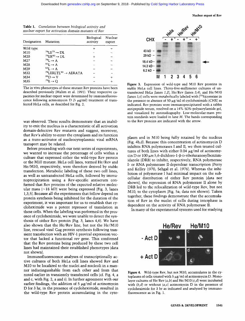
Before proceeding wi th our next series of experiments, we wanted to increase the percentage of cells wi th in a culture that expressed either the wild-type Rev protein or the M10 mutant . HeLa cell lines, termed He/Rev and He/M10, respectively, were therefore derived by stable transfection. Metabolic labeling of these two cell lines, as well as untransfected HeLa cells, followed by immu- noprecipitation using a Rev-specific ant iserum con- firmed that Rev proteins of the expected relative molec- ular mass ( -18 kD) were being expressed (Fig. 3, lanes 1,3,5). Because all the experiments that follow depend on protein synthesis being inhibi ted for the duration of the experiment, it was important for us to establish that cy- cloheximide was a potent repressor of translation in these cells. When the labeling was performed in the pres- ence of cycloheximide, we were unable to detect the syn- thesis of either Rev protein {Fig. 3, lanes 4,6). We have also shown that the He/Rev line, but not the He/M10 line, rescued viral Gag protein synthesis following tran- sient transfection wi th an HIV- 1 proviral expression vec- tor that lacked a functional rev gene. This confirmed that the Rev proteins being produced by these two cell lines had main ta ined their established phenotypes {data not shown).
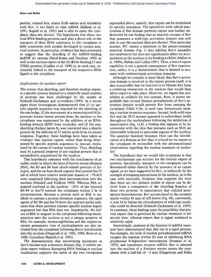
Immunofluorescence analyses of transcriptionally ac- tive cultures of both HeLa cell lines showed Rev and M10 to be localized to the nuclei and nucleoli in a man- ner indist inguishable from each other and from that noted earlier in t ransient ly transfected cells (cf. Fig. 4, a and c, wi th Fig. 2, a and i). In further agreement with our earlier findings, the addition of 5 Izg/ml of act inomycin D for 3 hr, in the presence of cycloheximide, resulted in the wild-type Rev protein accumulat ing in the cyto-
Figure 3. Expression of wild-type and M10 Rev proteins in stable HeLa cell lines. Thirty-five-millimeter cultures of un- transfected HeLa (lanes 1,2), He/Rev (lanes 3,4}, and He/M10 (lanes 5,61 cells were metabolically labeled with [3SS]cysteine in the presence or absence of 50 ~g/ml of cycloheximide (CHX) as indicated. Rev proteins were immunoprecipitated with a rabbit antipeptide serum, resolved on a 14% SDS-polyacrylamide gel, and visualized by autoradiography. Low-molecular-mass pro- tein standards were loaded in lane M. The bands corresponding to the Rev proteins are indicated with the arrow.
plasm and in M10 being fully retained by the nucleus (Fig. 4b, d). Because this concentration of ac t inomycin D inhibi ts RNA polymerases I and II, we then treated cul- tures of both lines wi th either 0.04 p~g/ml of actinomy- cin D or 100 p.M 5,6-dichloro-1-[3-D-ribofuranosylbenzim- idazole (DRB) to inhibit, respectively, RNA polymerase I- or RNA polymerase II-dependent transcription (Perry and Kelley 1970; Sehgal et al. 1976). Whereas the inhi- bition of polymerase I had m i n i m a l impact on the sub- cellular distribution of either Rev protein (data not shown), the repression of RNA polymerase II alone by DRB led to the relocalization of wild-type Rev, but not M10, to the cytoplasm (Fig. 5a; data not shown). Taken together, these findings demonstrate that the accumula- tion of Rev in the nuclei of cells during interphase is dependent on the activity of RNA polymerase II.
In many of the experimental systems used for studying
Figure 4. Wild-type Rev, but not M10, accumulates in the cy- toplasms of cells treated with 5 ~g/ml of actinomycin D. Mono- layer cultures of He/Rev (a,b} and He/M10 (c,d)were incubated with (b,d) or without (a,c) actinomycin D in the presence of cycloheximide for 3 hr as indicated and analyzed by immuno- fluorescence as in Fig. 1.
GENES & DEVELOPMENT 1541
Cold Spring Harbor Laboratory Press on September 9, 2018 - Published by genesdev.cshlp.orgDownloaded from

Meyer and Malim
+ DRB remove DRB
37oc C
~, (I,
4oc
Figure 5. Temperature dependence and reversibility of Rev's accumulation in the cytoplasm. He/Rev cultures were main- tained in cycloheximide and incubated at 37~ (a) or 4~ (b) with I00 p.M DRB for 3 hr. Cultures treated at 37~ were washed free of DRB and maintained at 37~ (c) or 4~ (d) for an addi- tional 2 hr. Samples were analyzed by immunofluorescence as in Fig. 1.
nucleocytoplasmic trafficking, the transport of macro- molecules in either direction across the nuclear mem- brane has been shown to be energy dependent (Dingwall et al. 1982; Richardson et al. 1988; Breeuwer and Gold- farb 1990; Mandell and Feldherr 1990; Dargemont and Kfihn 1992; Pifiol-Roma and Dreyfuss 1992). To deter- mine the energy requirements for Rev's egress out of the nucleus, cultures of He/Rev were treated with DRB at 37~ or 4~ (Fig. 5a, b). That Rev only accumulated in the cytoplasms of cells incubated at 37~ is indicative of Rev's export being mediated by a process that requires energy. By subsequently withdrawing the DRB from He/ Rev cultures incubated at 37~ and then maintaining them in DRB-free medium at 37~ or 4~ it was further established that the migration of Rev to the cytoplasm is, by definition, fully reversible (Fig. 5c) and that its reimport back into the nucleus requires energy utiliza- tion (Fig. 5d). In summary, these observations not only indicate that the wild-type Rev protein shuttles but also that active processes are involved in both the export and import segments of the shuttling cycle.
Although the data presented to this point strongly sug- gest that Rev constitutively exits and then reenters the nucleus, we nevertheless wanted to confirm this by uti-
lizing an assay system in which transcription was not inhibited. To this end, we adopted an approach, namely the formation of interspecies heterokaryons, that has been used successfully by a number of laboratories to confirm that certain nuclear proteins shuttle to and from the cytoplasm (Borer et al. 1989; Guiochon-Mantel et al. 1991; Pifiol-Roma and Dreyfuss 1992; Madan and De- Franco 1993; Schmidt-Zachmann et al. 1993). The HeLa cell lines expressing Rev or M10 (the "donor" cells) were therefore fused to mouse cells not expressing Rev (the "acceptor" cells) with polyethylene glycol and main- tained in cycloheximide-containing medium in the ab- sence of transcription inhibitors for an additional 45 min. Following fixation, the heterokaryons were sub- jected to double-label immunofluorescence using anti- bodies specific for Rev and the nonshuttling human hnRNP C1 and C2 proteins (Pifiol-Roma and Dreyfuss 1992). Because HeLa cells are of human origin, the donor nuclei within the heterokaryons were identified as those that contained the human C proteins, whereas the ac- ceptor nuclei were identified as those that did not (Fig. 6, cf. a with c and d with f). In contrast to the predicted restriction of the C proteins to the human cell-derived nuclei, the wild-type Rev protein was readily detected in both the acceptor and the donor nuclei of heterokaryons formed with the He/Rev cells (Fig. 6b). Because protein synthesis had been inhibited for the duration of this ex- periment, the import of Rev into the mouse cell-derived nuclei must have been preceded both by its export out of the human nuclei and by its subsequent passage through the cytoplasm. Of particular significance, however, was the observation that the M10 mutant protein was, like the human C proteins, confined to the donor nuclei of He/M10-derived heterokaryons (Fig. 6el. Because MIO is efficiently localized to the nuclei and nucleoli of express- ing cells (see Fig. 4c), its inability to accumulate in the mouse cell-derived nuclei of these heterokaryons must have been a consequence of its inability to escape from the donor nuclei. From these results we have concluded that the wild-type Rev t r a n s - a c t i v a t o r does shuttle be- tween the nuclear and cytoplasmic compartments of transcriptionally active cells but that mutant Rev pro- teins carrying nonfunctional activation domains do not.
anti-hnRNP C anti-Rev phase
Figure 6. Nucleocytoplasmic shuttling of HIV-1 Rev proteins in human-mouse interspecies heterokaryons. Mouse Ltk- cells were fused to He/Rev (a-c) or He/ MIO (d-fl cells using polyethylene glycol and main- tained at 37~ for an additional 45 min in cyclohexim- ide-containing medium. Double-label immunofluores- cence was then performed on fixed and permeabilized samples using monoclonal antibodies specific for the human hnRNP C proteins (a,d) and Rev (b,e). The po- sitions of the human (hu) and mouse (rno) nuclei are indicated with arrows. The corresponding phase-con- trast images of the heterokaryons are also shown (c,f).
He/Rev
He/MIO
i
3
~hu
0
4 - m o
hu_~ w #
e .4- Illi
m o .
1542 GENES & DEVELOPMENT
Cold Spring Harbor Laboratory Press on September 9, 2018 - Published by genesdev.cshlp.orgDownloaded from

Nuclear export of Rev
Discussion
In this paper we have used two different assay systems to show that the HIV-1 Rev trans-activator is not, as thought previously, confined solely to the nucleus. Al- though the indirect immunofluorescent analysis of pro- liferating cells shows that all detectable Rev localizes to the nucleus at any given time (Figs. 2 and 4; Cullen et al. 1988; Felber et al. 1989), the data presented here reveal that much of this protein is, to be accurate, in a state of constant flux between the nucleus and the cytoplasm. However, because the relative molecular mass of Rev is
18 kD and the diffusion limit of nuclear pores is esti- mated to be - 6 0 kD (for review, see Nigg et al. 1991; Davis 1992; Forbes 1992), it was particularly important for us to establish that Rev's export to the cytoplasm is a specific event and not merely the manifestation of pas- sive diffusion away from an area of high protein concen- tration in the nucleus.
Accordingly, we have provided two lines of evidence which, when taken together, are consistent with Rev's nucleocytoplasmic transport occurring by a process that requires a specific protein sequence and is dependent on energy utilization. First, in both transcription inhibition and heterokaryon experiments we demonstrate that Rev mutants with nonfunctional activation domains, which like Rev have relative molecular masses of - 18 kD, are unable to migrate out of the nucleus (Figs. 2, 4 and 6; Table 1). These observations establish the specificity of Rev's export to the cytoplasm and identify the leucine- rich activation domain of Rev as being essential for this process. Second, we show that the rapid accumulation of Rev in the cytoplasms of cells treated with inhibitors of transcription is suppressed when the cells are incubated at 4~ instead of 37~ (Fig. 5), a result consistent with export being an active process. Had the movement of Rev out of the nucleus been driven merely by diffusion down a concentration gradient, then reducing the tem- perature would not have been expected to have such a profound inhibitory effect. The finding that the Rev pro- tein of HIV-1 constitutively shuttles between the nu- cleus and the cytoplasm has implications both for the mechanism of Rev-mediated trans-activation and for un- derstanding how proteins are exported out of the nu- Glens.
Mechanism of Rev trans-activation
The HIV-1 Rev trans-activator is absolutely required for expression of unspliced -9-kb and singly spliced -4-kb HIV-1 transcripts in the cytoplasm. Because these tran- scripts represent both the genomic RNA found in viral particles {9-kb transcripts only) as well as the templates for translation of the viral structural proteins, for exam- ple, Gag and Env, their cytoplasmic expression is essen- tial for viral replication (Feinberg et al. 1986; Sodroski et al. 1986). Although it is generally accepted that Rev acts by inducing the translocation of intron-containing viral transcripts to the cytoplasm, the precise mechanism of trans-activation still remains to be elucidated. In partic-
ular, experiments in support of Rev operating either as an inhibitor of spliceosome assembly (Kjems et al. 1991; Kjems and Sharp 1993) and, hence, of viral mRNA splic- ing, or as a direct activator of mRNA export have been described (Emerman et al. 1989; Felber et al. 1989; Ham- marskj61d et al. 1989; Malim et al. 1989b}. Importantly, a recent study that used HIV-1-infected human T cells as the experimental system demonstrated that the Rev-de- pendent transport of 9- and 4-kb viral mRNAs to the cytoplasm occurs in the absence of any discernible effect on the level of expression of the fully spliced -2-kb tran- scripts (Malim and Cullen 1993). Because a splicing-in- hibition model would predict that expression of the 2-kb mRNAs would be reduced by Rev, this finding strongly supports the alternative viewpoint that Rev's primary function is to activate the nucleocytoplasmic transport of intron-containing viral mRNAs.
The demonstration that mutations in Rev's activation domain (e.g., M10) that abolish trans-activation also pre- vent Rev from entering the cytoplasm is therefore sug- gestive of the following model for the Rev-mediated in- duction of viral mRNA transport. After transcription, the full-length 9-kb transcript is spliced inefficiently to yield a heterogeneous population of viral transcripts in the nucleus that includes the 9-kb mRNA as well as 4- and 2-kb mRNAs. When Rev is present in sufficient abundance, it binds to the 9- and 4-kb mRNAs, via the RRE, to form ribonucleoprotein complexes that contain both Rev and viral RNA. Because Rev's leucine-rich ac- tivation domain is thought to mediate Rev-host cell pro- tein interactions (Malim et al. 1991), we hypothesize that such an interaction then takes place and the Rev- mRNA complexes are programmed for export to the cy- toplasm. As illustrated by mutants such as M10, failure to participate in a protein-protein interaction of this type results in Rev as well as the unspliced viral tran- scripts being confined to the nucleus. Once in the cyto- plasm, and perhaps after subserving a function there, we imagine that Rev completes the shuttling cycle by dis- engaging from the RNA and returning to the nucleus by a process that requires both its arginine-rich NLS (Malim et al. 1989a; Perkins et al. 1989) and RNA polymerase II-dependent transcription. Interestingly, in vitro-bind- ing experiments have shown that Rev can interact with the nucleolar shuttling protein B23/No38 (Fankhauser et al. 1991). Whether the shuttling of this protein has any influence on the activity and shuttling of Rev remains an open and intriguing question.
It has been predicted previously that proteins shuttling across the nuclear envelope are likely to be involved in the transport of proteins and RNAs between the nucleus and the cytoplasm (for review, see Goldfarb 1991; Laskey and Dingwall 1993). The discovery that Rev, a known activator of RNA transport, is also a shuttle protein ap- pears to bear out this prediction. Interestingly, human T-cell leukemia virus type I (HTLV-I), a human retrovi- rus that is very distinct from HIV-1, also encodes a nu- clear trans-activator that is essential for the expression of its unspliced mRNAs in the cytoplasm (Hidaka et al. 1988; Siomi et al. 1988; Hanly et al. 1989). Although this
GENES & DEVELOPMENT 1543
Cold Spring Harbor Laboratory Press on September 9, 2018 - Published by genesdev.cshlp.orgDownloaded from
Meyer and Malim
protein, termed Rex, shares little amino acid similarity with Rev, it too binds to viral mRNA (Ballaun et al. 1991; Bogerd et al. 1991) and is able to enter the cyto- plasm (data not shown). The hypothesis that these two viral RNA-binding proteins each play a direct role in the transport of their RNA substrates to the cytoplasm is fully consistent with models developed in certain non- viral systems. In particular, evidence has been presented to suggest that the shuttling of the mRNA-binding hnRNP A1 protein (Pifiol-Roma and Dreyfuss 1992) as well as the nuclear export of the 5S rRNA-binding L5 and THIIA proteins (Guddat et al. 1990) is, in each case, in- timately linked to the transport of the respective RNA ligand to the cytoplasm.
Implications for nuclear export
The notion that shuttling, and therefore nuclear export, is a specific process limited to a relatively small number of proteins has been challenged by the results of Schmidt-Zachmann and co-workers {1993}. In a recent paper these investigators demonstrated that (11 no spe- cific peptide sequence was required for the shuttling of the nucleolin, {2) the movement of a microinjected NLS- pyruvate kinase fusion protein from the nucleus to the cytoplasm was suppressed by the addition of an RNA- binding domain (RNP) from nucleolin, and (3) the non- shuttling chicken lamin B 2 was converted into a shuttle protein by the deletion of 27 amino acids from its amino terminus. Together, these findings have been taken as evidence that nuclear export, rather than being deter- mined by specific peptide sequences is, instead, modu- lated by the extent of nuclear retention. Thus, shuttling may be a general property of any nuclear protein that is incompletely sequestered by the nucleus.
This hypothesis contrasts with the conclusions of an earlier study in which the fates of bovine serum albumin {BSAI, the B3 and B4 heat shock-related proteins of Xe- nopus, and the rat heat shock cognate (hsc) protein hscT0 (all of which have relative molecular masses of -70-kD) were examined following their microinjection into the nucleus (Mandell and Feldherr 1990). Whereas BSA re- mained confined to the nucleus, -50% of the injected B3/B4 or hsc70 entered the cytoplasm within 2 hr of microinjection. Because a protein such as BSA is un- likely to contain a nuclear retention sequence, the rapid egress of B3/B4 and hsc70 from the injected nuclei indi- cates that these proteins contain specific nuclear export sequences but that BSA does not. Importantly, the fail- ure of BSA to migrate to the cytoplasm following micro- injection into the nucleus is not a unique property of BSA, for example, immunoglobulins and the trypsin-re- sistant pentameric core of nucleoplasmin are also ex- cluded from the cytoplasm following direct introduction into the nucleus (Dingwall et al. 1982, 1988; Borer et al. 1989; Guiochon-Mantel et al. 1991).
The demonstration that inactivating mutations in Rev's leucine-rich activation domain {Fig. 1) inhibit nu- clear export without disrupting RNA binding or nuclear localization supports the latter of the two viewpoints
expounded above, namely, that export can be modulated by specific sequences. The specificity with which inac- tivation of this domain prevents export was further un- derscored by our finding that an inactive mutant of Rev that possesses a wild-type activation domain was still able to exit the nucleus {data not shown). This particular mutant, M7, carries a mutation in the amino-terminal essential domain {Fig. 1) that inhibits Rev's assembly into multimers but does not significantly affect its accu- mulation in the nucleus or its binding to RNA {Malim et al. 1989a; Malim and Cullen 19911. Thus, a loss of export capability is not a general consequence of Rev inactiva- tion; rather, it is a characteristic that is limited to mu- tants with nonfunctional activation domains.
Although we consider it most likely that Rev's activa- tion domain is involved in the export process itself, it is also conceivable that its function is to liberate Rev from a confining interaction in the nucleus that would then allow export to take place. However, we regard this pos- sibility as unlikely for two reasons. First, it seems im- probable that several distinct perturbations of Rev's ac- tivation domain would prevent Rev from entering the cytoplasm (Table 1) by, in each case, fortuitously creat- ing a nuclear retention sequence. Second, both wild-type Rev and the M10 mutant appeared to redistribute freely throughout the nucleoplasm following the inhibition of transcription {Fig. le, k), a finding that appears to be in- consistent with the idea that mutants such as M10 are irreversibly tethered to particular regions of the nucleus. The question therefore becomes, How can the identifi- cation of a domain in Rev that is critical for its export to the cytoplasm be reconciled with the aforementioned observations regarding the nuclear transport of nucleo- lin?
The hypothesis that we would like to propose is that two mechanisms can account for the nuclear export of proteins. Specifically, transport to the cytoplasm can be determined either directly by the presence of an export signal, as we have suggested for Rev, or indirectly by the strength of retaining interactions in the nucleus, as is the case with nucleolin. Evidence that supports the view that there are two distinct modes of export can be de- rived from a comparison of the shuttling kinetics of these two proteins. In experiments that utilized inter- species heterokaryons, Rev accumulated in the acceptor nuclei within 45 min of cell fusion {Fig. 6b); conversely, it was 24 hr before the relocalization of wild-type nude- olin could be detected (Schmidt-Zachmann et al. 1993). In summary, these findings raise the possibility that pro- tein export that is governed by nuclear retention is rel- atively slow, whereas export that is signal mediated is relatively rapid.
Interestingly, analyses of the kinetics of mRNA trans- port have demonstrated that this too is a rapid process. For example, the bulk of nuclear polyadenylated mRNA escapes the nucleus within 20 min of inhibiting RNA polymerase II-dependent transcription {Penman et al. 1970), and transferrin receptor mRNA that is injected into the nucleus of a Xenopus oocyte enters the cyto- plasm with a half-life of - 2 min (Dargemont and Kfihn
1544 GENES & DEVELOPMENT
Cold Spring Harbor Laboratory Press on September 9, 2018 - Published by genesdev.cshlp.orgDownloaded from

Nuclear export of Rev
1992). G iven tha t Rev is required for the nuclear export of HIV-1 mRNA, it is perhaps no t surpris ing tha t it shut- tles to and f rom the cy top lasm wi th k ine t ics of the more rapid variety.
Mater ia l s and m e t h o d s
Expression vectors
pcREV constitutively expresses the wild-type HIV-1 Rev pro- tein (Malim et al. 1988). Missense derivatives of this vector that carry mutations in Rev's leucine-rich activation domain encode proteins that are either nonfunctional {M10, M27, M28, M29, M32) or have retained wild-type activity (M20, M34, M35) (Malim et al. 1989a, 1991) (see Fig. 1; Table 1). pgTAT expresses an intron-containing tat transcript whose expression in the cy- toplasm is dependent on Rev coexpression (Malim et al. 1988). For the construction of stable cell lines (see below), the pcREV and pM10 vectors were modified to further express the mouse dihydrofolate reductase gene, dhfr. This was accomplished by inserting a 1.8-kb PvuII-BamHI dhfr-gene-containing restric- tion fragment, derived from pSV2-dhfr (Subramani et al. 1981), into the unique StuI sites of pcREV and pM10.
Cell culture, transfections, and cell line construction
The human and mouse cell lines HeLa and Ltk- were main- tained in Iscove's modified Dulbecco's medium supplemented with 10% fetal bovine serum and transfected {HeLa cells only) using calcium phosphate (Cullen 1987). To obtain HeLa cell lines that stably express the wild-type Rev protein or the M10 mutant, termed He/Rev and He/M10, 5 lxg of the appropriate dhfr-containing plasmid was linearized with PvuI and cotrans- fected with 0.25 ~g of linearized pSV2-neo (Southern and Berg 1982) using calcium phosphate. Following ~2 weeks of selec- tion in 0.8 mg/ml of G418 (GIBCO BRL, Gaithersburg, MD), the pools of resistant cells were replated in selective medium con- taining 10 -r M amethopterin (Sigma Chemical Co., St. Louis, MO). As these cultures became resistant, the level of amethop- terin in the medium was gradually increased until a final con- centration of 10 - 6 M was attained. At this point, clonal popu- lations were derived by limiting dilution and those expressing higher levels of the Rev proteins, as judged by immunofluores- cence (see below), were used for further analysis. During the course of the experiments, cultures were supplemented vari- ously with 50 ~xg/ml of cycloheximide (Calbiochem Corp., La Jolla, CA) to inhibit protein synthesis and 0.04 ~g/ml of acti- nomycin D IBoehringer Mannheim Corp., Indianapolis, IN), 100 txM DRB {Calbiochem), or 5 txg/ml of actinomycin D to inhibit transcription directed by RNA polymerase I, RNA polymerase II, or both, respectively (Perry and Kelley 1970; Sehgal et al. 19761.
Indirect immunofluorescence and antibodies
Cells plated onto Lab-Tek chamber slides (Nunc Inc., Naper- ville, IL) were used for the determination of subcellular local- ization. In experiments requiring the transient transfection of HeLa cells, analyses were performed -40 hr after the addition of DNA to the monolayers. Cells were fixed using paraformalde- hyde and permeabilized with Triton-X100 as described (Ruben et al. 1989). For all experiments, except those involving heter- okaryons (see below), Rev was detected with an anti-Rev mono- clonal antibody (Repligen Corp., Cambridge, MA) at 1.25 Ixg/ ml, followed by a goat antimouse antibody conjugated to Texas red (FisherBiotech, Pittsburgh, PA). Samples were then visual-
ized by epifluorescence using a Nikon Microphot-FXA micro- scope at a magnification of 400 x.
Imm unoprecipita tion
Rev proteins from HeLa cells starved for 1 hr and metabolically labeled for 2 hr with [35S]cysteine (Amersham Corp., Arlington, IL) were immunoprecipitated with a 1:150 dilution of a rabbit antipeptide serum, resolved on 14% SDS--polyacrylamide gels, and visualized by autoradiography (Cullen et al. 1988; Malim et al. 1988}. When the labeling was performed in the presence of cycloheximide, the cycloheximide was also included in the cul- ture medium for the last 30 min of the starvation step.
Interspecies heterokaryons
To form heterokaryons, mouse Ltk- cells were first grown to subconfluence on glass coverslips. Suspensions of He/Rev or He/M10 in complete medium were then added, and the cells allowed to settle for 3.5 hr. Then, the medium was exchanged for medium supplemented with 100 ~g/ml of cycloheximide and the cells incubated for a further 30 min; this was done to ensure that all of the Rev protein was, at the time of heter- okaryon formation, localized to the nucleus. Cell fusion was performed by adding 50% polyethylene glycol 4000 (GIBCO BRL) for 105 sec at 37~ the cultures were then washed twice with prewarmed PBS and incubated for an additional 45 rain at 37~ in complete medium containing cycloheximide. Fixed and permeabilized samples were subjected to double-label immuno- fluorescence using monoclonal antibodies specific for the hu- man hnRNP C1 and C2 proteins (termed 41:4; see Pifiol-Roma and Dreyfuss 1991) and Rev (Repligen); these primary antibod- ies were detected, respectively, with fluorescein isothiocyanate conjugated anti-71 and Texas red-conjugated anti-72b subclass- specific antimouse antibodies raised in goats (FisherBiotech).
A c k n o w l e d g m e n t s
We thank Serafin Pifiol-Roma and Gideon Dreyfuss for the 4F4 antibody and for all their help and support, Kris Kelley for her expert assistance with the microscopy, Jim Alwine, Bob Doms, and Rebecca Oakey for their comments on the manuscript and Laurie Zimmerman for secretarial assistance. This work was supported by the Howard Hughes Medical Institute.
The publication costs of this article were defrayed in part by payment of page charges. This article must therefore be hereby marked "advertisement" in accordance with 18 USC section 1734 solely to indicate this fact.
Reierences
Adam, S. and L. Gerace. 1991. Cytosolic proteins that specifi- cally bind nuclear location signals are receptors for nuclear import. Cell 66: 837-847.
Ballaun, C., G.K. Farrington, M. Dobrovnik, J. Rusche, J. Hauber, and E. B6hnlein. 1991. Functional analysis of hu- man T-cell leukemia virus type I Rex-response element: Di- rect RNA binding of Rex protein correlates with in vivo activity. I. Virol. 65: 4408--4413.
Bogerd, H.P., G.L. Huckaby, Y.F. Ahmed, S.M. Hanly, and W.C. Greene. 1991. The type I human T-cell leukemia virus (HTLV-I) Rex trans-activator binds directly to the HTLV-I Rex and the type 1 human immunodeficiency virus Rev RNA response elements. Proc. Natl. Acad. Sci. 88: 5704- 5708.
B6hnlein, E., J. Berger, and J. Hauber. 1991. Functional mapping
GENES & DEVELOPMENT 1545
Cold Spring Harbor Laboratory Press on September 9, 2018 - Published by genesdev.cshlp.orgDownloaded from

Meyer and Malim
of the human immunodeficiency virus type 1 Rev RNA binding domain: New insights into the domain structure of Rev and Rex. J. Virol. 65: 7051-7055.
Borer, R.A., C.F. Lehner, H.M. Eppenberger, and E.A. Nigg. 1989. Major nucleolar proteins shuttle between nucleus and cytoplasm. Cell 56: 379-390.
Breeuwer, M. and D.S. Goldfarb. 1990. Facilitated nuclear trans- port of histone H1 and other small nucleophilic proteins. Cell 60: 999-1008.
Chang, D.D. and P.A. Sharp. 1989. Regulation by HIV Rev de- pends upon recognition of splice sites. Cell 59: 789-795.
Cullen, B.R. 1987. Use of eukaryotic expression technology in the functional analysis of cloned genes. Methods Enzymol. 152: 423-426.
~ . 1991. Human immunodeficiency virus as a prototypic complex retrovirus. J. Virol. 65: 1053-1056.
Cullen, B.R., J. Hauber, K. Campbell, J.G. Sodroski, W.A. Ha- seltine, and C.A. Rosen. 1988. Subcellular localization of the human immunodeficiency virus trans-acting art gene prod- uct. J. Virol. 62: 2498-2501.
Daly, T.J., K.S. Cook, G.S. Gray, T.E. Maione, and J.R. Rusche. 1989. Specific binding of HIV-1 recombinant Rev protein to the Rev-responsive element in vitro. Nature 342: 816-819.
Daly, T.J., P. Rennert, J.K. Barry, M. Dundas, J.R. Rusche, R.C. Doten, M. Auer, and G.K. Farrington. 1993. Perturbation of the carboxy terminus of HIV-1 Rev affects multimerization on the Rev responsive element. Biochemistry 32: 8945- 8954.
Dargemont, C. and L.C. Kfihn. 1992. Export of mRNA from microinjected nuclei of Xenopus laevis oocytes. J. Ceil Biol. 118: 1-9.
Davis, LT 1992. Control of nucleocytoplasmic transport. Curl Opin. Cell Biol. 4: 424--429.
Dingwall, C. and R.A. Laskey. 1992. The nuclear membrane. Science 258: 942-947.
1991. Nuclear targeting sequences--a consensus? Trends Biochem. Sci. 16: 478-481.
Dingwall, C., S.V. Shamick, and R.A. Laskey. 1982. A polypep- tide domain that specifies migration of nucleoplasmin into the nucleus. Cell 30: 449-458.
Dingwall, C., J. Robbins, S.M. Dilworth, B. Roberts, and W.D. Richardson. 1988. The nucleoplasmin nuclear location se- quence is larger and more complex than that of SV-40 large T antigen. J. Cell Biol. 107: 841-849.
Dworetzky, S.I. and C.M. Feldherr. 1988. Translocation of RNA-coated gold particles through the nuclear pores of oocytes. ]. Cell Biol. 106: 575-584.
Emerman, M., R. Vazeux, and K. Peden. 1989. The rev gene product of the human immunodeficiency virus affects enve- lope-specific RNA localization. Cell 57:1155-t 165.
Fankhauser, C., E. Izaurralde, Y. Adachi, P. Wingfield, and U.K. Laemmli. 1991. Specific complex of human immunodefi- ciency virus type 1 Rev and nucleolar B23 proteins: Disso- ciation by the Rev response element. Mol. Cell. Biol. 11: 2567-2575.
Feinberg, M.B., R.F. Jarrett, A. Aldovini, R.C. Gallo, and F. Wong-Staal. 1986. HTLV-III expression and production in- volve complex regulation at the levels of splicing and trans- lation of viral RNA. Cell 46: 807-817.
Felber, B.K., C.M. Drysdale, and G.N. Pavlakis. 1990. Feedback regulation of human immunodeficiency virus type 1 expres- sion by the Rev protein. J. Virol. 64: 3734-3741.
Felber, B.K., M. Hadzopoulou-Cladaras, C. Cladaras, T. Copel- and, and G.N. Pavlakis. 1989. Rev protein of human immu- nodeficiency virus type 1 affects the stability and transport of the viral mRNA. Proc. Natl. Acad. Sci. 86: 1495-1499.
Feldherr, C.M., E. Kallenbach, and N. Schultz. 1984. Movement of a karyophilic protein through the nuclear pores of oocytes. J. Cell Biol. 99: 2216--2222.
Forbes, D.J. 1992. Structure and function of the nuclear pore complex. Annu. Rev. Cell Biol. 8: 495-527.
Garcia-Bustos, J., J. Heitman, and M.N. Hall. 1991. Nuclear pro- tein localization. Biochim. Biophys. Acta 1071: 83-101.
Gerace, L. 1992. Molecular trafficking across the nuclear pore complex. Curr. Opin. Cell Biol. 4: 637-645.
Goldfarb, D.S. 1991. Shuttling proteins go both ways. Curl Biol. 1: 212-214.
Goldfarb, D. and N. Michaud. 1991. Pathways for the nuclear transport of proteins and RNAs. Trends Cell Biol. 1: 20--24.
Guddat, U., A.H. Bakken, and T. Pieler. 1990. Protein-mediated nuclear export of RNA: 5S rRNA containing small RNPs in Xenopus oocytes. Cell 60: 619-628.
Guiochon-Mantel, A., P. Lescop, S. Christin-Maitre, H. Loos- felt, M. Perrot-Applanat, and W. Milgrom. 1991. Nucleocy- toplasmic shuttling of the progesterone receptor. EMBO J. 10: 3851-3859.
Hadzopoulou-Cladaras, M., B.K. Felber, C. Cladaras, A. Atha- nassopoulos, A. Tse, and G.N. Pavlakis. 1989. The rev (trs/ art) protein of human immunodeficiency virus type 1 affects viral mRNA and protein expression via a cis-acting sequence in the env region. J. Virol. 63: 1265-1274.
Hammarskj61d, M.-L., J. Heimer, B. Hammarskj61d, I. Sangwan, L. Albert, and D. Rekosh. 1989. Regulation of human immu- nodeficiency virus env expression by the rev gene product. J. Virol. 63: 1959-1966.
Hanly, S.M., L.T. Rimsky, M.H. Malim, J.H. Kim, J. Hauber, M. Duc Dodon, S.-Y. Le, J.V. Maizel, B.R. Cullen, and W.C. Greene. 1989. Comparative analysis of the HTLV-I Rex and HIV-1 Rev trans-regulatory proteins and their RNA response elements. Genes & Dev. 3: 1534-1544.
Hidaka, M., J. Inoue, M. Yoshida, and M. Seiki. 1988. Post- transcriptional regulator (rex) of HTLV-1 initiates expression of viral structural proteins but suppresses expression of reg- ulatory proteins. EMBO J. 7: 519-523.
Hinshaw, J.E., B.O. Carragher, and R.A. Milligan. 1992. Archi- tecture and design of the nuclear pore complex. Cell 69:1133-1141.
Hope, T.J., X. Huang, D. McDonald, and T.G. Parslow. 1990a. Steroid-receptor fusion of the human immunodeficiency vi- res type 1 Rev trans-activator: Mapping cryptic functions of the arginine-rich motif. Proc. Natl. Acad. Sci. 87: 7787- 7791.
Hope, T.J., D. McDonald, X. Huang, J. Low, and T.G. Parslow. 1990b. Mutational analysis of the human immunodeficiency virus type 1 Rev trans-activator: Essential residues near the amino terminus. J. Virol. 64: 5360-5366.
Izaurralde, E. and I.W. Mattaj. 1992. Transport of RNA between nucleus and cytoplasm. Semin. Cell Biol. 3: 279-288.
Kambach, C. and I.W. Mattaj. 1992. Intracellular distribution of the U1A protein depends on active transport and nuclear binding to U1 snRNA. J. Cell Biol. 118: 11-21.
Kim, S., R. Bym, J. Groopman, and D. Baltimore. 1989. Tempo- ral aspects of DNA and RNA synthesis during human im- munodeficiency virus infection: Evidence for differential gene expression. J. Virol. 63: 3708-3713.
Kjems, J. and P.A. Sharp. 1993. The basic domain of Rev from human immunodeficiency virus type 1 specifically blocks the entry of U4/U6. U5 small nuclear ribonucleoprotein in spliceosome assembly. J. Virol. 67: 4769-4776.
Kjems, J., A.D. Frankel, and P.A. Sharp. 1991. Specific regula- tion of mRNA splicing in vitro by a peptide from HIV-1 Rev. Cell 67: 169-178.
1546 G E N E S & DEVELOPMENT
Cold Spring Harbor Laboratory Press on September 9, 2018 - Published by genesdev.cshlp.orgDownloaded from

Nuclear export of Rev
Knight, D.M., F.A. Flomerfelt, and J. Ghrayeb. 1987. Expression of the art/trs protein of HIV and study of its role in viral envelope synthesis. Science 236: 837-840.
Laskey, R.A. and C. Dingwall. 1993. Nuclear shuttling: The default pathway for nuclear proteins? Cell 74: 585-586.
Madan, A.P. and D.B. DeFranco. 1993. Bidirectional transport of glucocorticoid receptors across the nuclear envelope. Proc. Natl. Acad. Sci. 90: 3588-3592.
Malim, M.H. and B.R. Cullen. 1991. HIV-1 structural gene ex- pression requires the binding of multiple Rev monomers to the viral RRE: Implications for HIV-1 latency. Cell 65: 241- 248.
1993. Rev and the fate of pre-mRNA in the nucleus: Implications for the regulation of RNA processing in eukary- otes. Mol. Cell. Biol. 13: 6180--6189.
Malim, M.H., J. Hauber, R. Fenrick, and B.R. Cullen. 1988. Im- munodeficiency virus rev trans-activator modulates the ex- pression of the viral regulatory genes. Nature 335: 181-183.
Malim, M.H., S. B6hnlein, J. Hauber, and B.R. Cullen. 1989a. Functional dissection of the HIV-1 Rev trans-activator--der- ivation of a trans-dominant repressor of Rev function. Cell 58: 205-214.
Malim, M.H., J. Hauber, S.-Y. Le, J.V. Maizel, and B.R. Cullen. 1989b. The HIV-1 rev trans-activator acts through a struc- tured target sequence to activate nuclear export of unspliced viral mRNA. Nature 338: 254-257.
Malim, M.H., D.F. McCarn, L.S. Tiley, and B.R. Cullen. 1991. Mutational definition of the human immunodeficiency vi- rus type 1 Rev activation domain. J. Virol. 65: 4248-4254.
Mandell, R.B. and C.M. Feldherr. 1990. Identification of two HSP70-related Xenopus oocyte proteins that are capable of recycling across the nuclear envelope. J. Cell Biol. 111: 1775-1783.
Meier, U.T. and G. Blobel. 1992. Nopp 140 shuttles on tracks between nucleolus and cytoplasm. Cell 70: 127-138.
Mermer, B., B.K. Felber, M. Campbell, and G.N. Pavlakis. 1990. Identification of trans-dominant HIV-1 Rev protein mutants by direct transfer of bacterially produced proteins into hu- man cells. Nucleic Acids Res. 18: 2037-2044.
Muesing, M.A., D.H. Smith, C.D. Cabradilla, C.V. Benton, L.A. Lasky, and D.J. Capon. 1985. Nucleic acid structure and ex- pression of the human AIDS/lymphadenopathy retrovirus. Nature 313: 450-458.
Nigg, E.A., P.A. Baeuerle, and R. Lfihrmann. 1991. Nuclear im- port-export: In search of signals and mechanisms. Cell 66: 15-22.
Olsen, H.S., A.W. Cochrane, P.J. Dillon, C.M. Nalin, and C.A. Rosen. 1990. Interaction of the human immunodeficiency virus type 1 Rev protein with a structured region in env mRNA is dependent on multimer formation mediated through a basic stretch of amino acids. Genes & Dev. 4: 1357-1364.
Pavlakis, G.N. and B.K. Felber. 1990. Regulation of expression of human immunodeficiency virus. The N e w Biol. 2:20-31.
Penman, S., M. Rosbash, and M. Penman. I970. Messenger and heterogeneous nuclear RNA in HeLa cells: Differential in- hibition by cordycepin. Proc. Natl. Acad. Sci. 67: 1878- 1885.
Perkins, A., A.W. Cochrane, S.M. Ruben, and C.A. Rosen. 1989. Structural and functional characterization of the human im- munodeficiency virus rev protein. J. Acquired I m m u n e De- tic. Syndr. 2: 256-263.
Perry, R.P. and D.E. Kelley. 1970. Inhibition of RNA synthesis by actinomycin D: Characteristic dose-response of different RNA species. J. Cell. Physiol. 76: 127-140.
Pifiol-Roma, S. and G. Dreyfuss. 1991. Transcription-dependent
and transcription-independent nuclear transport of hnRNP proteins. Science 253:312-314.
~ . 1992. Shuttling of pre-mRNA binding proteins between nucleus and cytoplasm. Nature 355: 730-732.
Richardson, W.D., A.D. Mills, S.M. Dilworth, R.A. Laskey, and C. Dingwall. 1988. Nuclear protein migration involves two steps: Rapid binding at the nuclear envelope followed by slower translocation through nuclear pores. Cell 52: 655- 664.
Robert-Guroff, M., M. Popovic, S. Gartner, P. Markham, R.C. Gallo, and M.S. Reitz. 1990. Structure and expression of tat-, rev-, and her-specific transcripts of human irnmunodeficien- cy virus type 1 in infected lymphocytes and macrophages. J. Virol. 64: 3391-3398.
Rosen, C.A., E. Terwilliger, A. Dayton, J.G. Sodroski, and W.A. Haseltine. 1988. Intragenic cis-acting art gene-responsive se- quences of the human immunodeficiency virus. Proc. Natl. Acad. Sci. 85: 2071-2075.
Ruben, S., A. Perkins, R. Purcell, K. Joung, R. Sia, R. Burghoff, W.A. Haseltine, and C.A. Rosen. 1989. Structural and func- tional characterization of human immunodeficiency virus tat protein. J. Virol. 63: 1-8.
Schmidt-Zachmann, M.S., C. Dargemont, L.C. K~ihn, and E.A. Nigg. 1993. Nuclear export of proteins: The role of nuclear retention. Cell 74: 493-504.
Schwartz, S., B.K. Felber, D.M. Benko, E.-M. Feny6, and G.N. Pavlakis. 1990. Cloning and functional analysis of multiply spliced mRNA species of human immunodeficiency virus type 1. J. Virol. 64: 2519-2529.
Sehgal, P.B., J.E. Darnell, and I. Tamm. 1976. The inhibition by DRB (5,6-dichloro-l-B-D-ribofuranosylbenzimidazole) of hnRNA and mRNA production in HeLa cells. Cell 9: 473- 480.
Siomi, H., H. Shida, S.H. Nam, T. Nosaka, M. Maki, and M. Hatanaka. 1988. Sequence requirements for nucleolar local- ization of human T cell leukemia virus type I pX protein, which regulates viral RNA processing. Cell 55: 197-209.
Sodroski, J., W.C. Goh, C.A. Rosen, A. Dayton, E. Terwilliger, and W.A. Haseltine. 1986. A second post-transcriptional trans-activator gene required for HTLV-III replication. Na- ture 321: 412-417.
Southern, P.J. and P. Berg. 1982. Transformation of mammalian cells to antibiotic resistance with a bacterial gene under con- trol of the SV40 early region promoter. J. Mol. Appl. Genet. 1: 327-341.
Subramani, S., R. Mulligan, and P. Berg. 1981. Expression of the mouse dihydrofolate reductase complementary deoxyribo- nucleic acid in simian virus 40 vectors. Mol. Cell. Biol. 1: 854-864.
Tiley, L.S., S.J. Madore, M.H. Malim, and B.R. Cullen. 1992. The VP16 transcription activation domain is functional when targeted to a promoter-proximal RNA sequence. Genes & Dev. 6: 2077-2087.
Venkatesh, L.K. and G. Chinnadurai. 1990. Mutants in a con- served region near the carboxy-terminus of HIV-1 identify functionally important residues and exhibit a dominant neg- ative phenotype. Virology 178: 327-330.
Warner, J.R. 1989. Synthesis of ribosomes in Saccharomyces cerevisiae. Microbiol. Rev. 53: 256-271.
Zapp, M.L. and M.R. Green. 1989. Sequence-specific RNA bind- ing by the HIV-1 Rev protein. Nature 342: 714-716.
Zapp, M.L., T.J. Hope, T.G. Parslow, and M.R. Green. 1991. Oligomerization and RNA binding domains of the type 1 human immunodeficiency virus Rev protein: A dual func- tion for an arginine-rich binding motif. Proc. Natl. Acad. Sci. 88: 7734-7738.
GENES & DEVELOPMENT 1547
Cold Spring Harbor Laboratory Press on September 9, 2018 - Published by genesdev.cshlp.orgDownloaded from

10.1101/gad.8.13.1538Access the most recent version at doi: 8:1994, Genes Dev.
B E Meyer and M H Malim cytoplasm.The HIV-1 Rev trans-activator shuttles between the nucleus and the
References
http://genesdev.cshlp.org/content/8/13/1538.full.html#ref-list-1
This article cites 81 articles, 37 of which can be accessed free at:
License
ServiceEmail Alerting
click here.right corner of the article or
Receive free email alerts when new articles cite this article - sign up in the box at the top
Copyright © Cold Spring Harbor Laboratory Press
Cold Spring Harbor Laboratory Press on September 9, 2018 - Published by genesdev.cshlp.orgDownloaded from