the immune system chapter 40. primary immune response this is how immunity to disease is acquired...
TRANSCRIPT

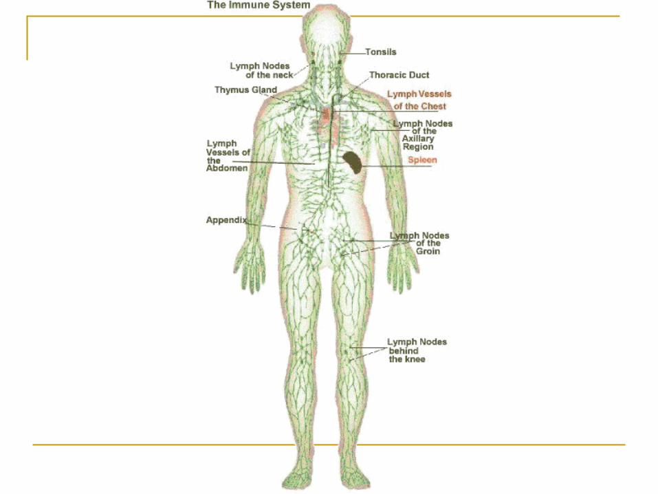
The Immune System
Chapter 40


Primary Immune Response
This is how immunity to disease is acquired Requires time Two components
Recognition Requires exposure to invading agent
Response Involves a searching effort (clonal selection)

Secondary Immune Response
The response of the immune system to the second or subsequent occasion on which it encounters a specific antigen.

Lymphocytes
Primary participants in the immune response
Types of lymphocytes: B-cells and T-cells
Both originate in red bone marrow (stem cells)

Differentiation Occurs in the Primary
Lymphoid Tissues (thymus and spleen)
Lymphocytes develop special recognition sites on their membranes
B-cells Differentiation occurs in
red marrow T-cells
Differentiation occurs in thymus

Migration
After differentiation, B-cells and T-cells migrate to Secondary Lymphoid Tissues Lymph nodes Spleen Adenoids Tonsils Peyer’s Patches: Intestinal
lining MALT

Lymph Nodes
B-cells, T-cells, and macrophages are “stored” here
Lymph nodes receive fluid from tissues and blood May contain bacteria,
viruses, etc. Brings lymphocytes in
contact with these invaders, allows for immune response
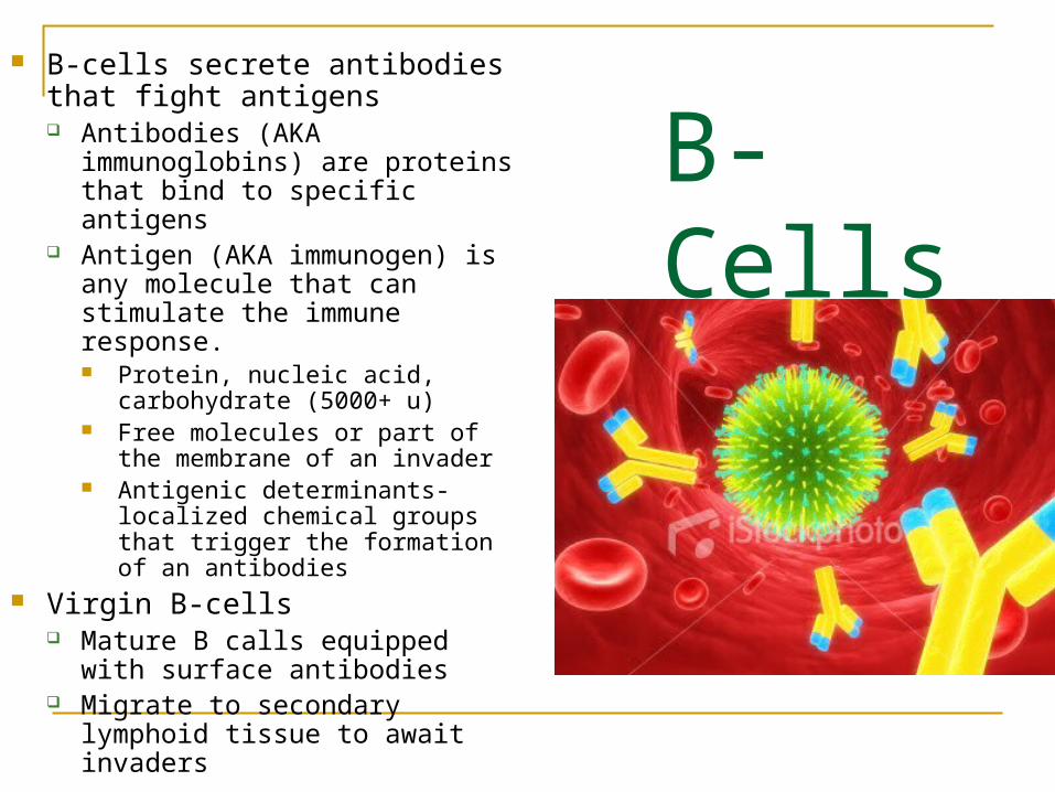
B-Cells
B-cells secrete antibodies that fight antigens Antibodies (AKA immunoglobins)
are proteins that bind to specific antigens
Antigen (AKA immunogen) is any molecule that can stimulate the immune response. Protein, nucleic acid,
carbohydrate (5000+ u) Free molecules or part of the
membrane of an invader Antigenic determinants-localized
chemical groups that trigger the formation of an antibodies
Virgin B-cells Mature B calls equipped with
surface antibodies Migrate to secondary lymphoid
tissue to await invaders

Structure of Antibody 4 polypeptide chains
2 heavy chains 2 light chains Chains held together by disulfide
linkages Y-shaped configuration
Variable regions 2 branches of the Y
Antigen binding Sites Contained in variable region Bind antibody to antigenic
determinant Millions of varieties
Constant Region Same in all antibodies in a given
class Fc Regions
Contained in constant region Make phagocytosis more
effective

Antibody Classes-Fig 40.11
IgG IgA IgM IgD IgE
Differ in structure, location, target, and action
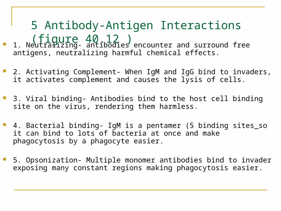
5 Antibody-Antigen Interactions (figure 40.12 )
1. Neutralizing- antibodies encounter and surround free antigens, neutralizing harmful chemical effects.
2. Activating Complement- When IgM and IgG bind to invaders, it activates complement and causes the lysis of cells.
3. Viral binding- Antibodies bind to the host cell binding site on the virus, rendering them harmless.
4. Bacterial binding- IgM is a pentamer (5 binding sites_so it can bind to lots of bacteria at once and make phagocytosis by a phagocyte easier.
5. Opsonization- Multiple monomer antibodies bind to invader exposing many constant regions making phagocytosis easier.
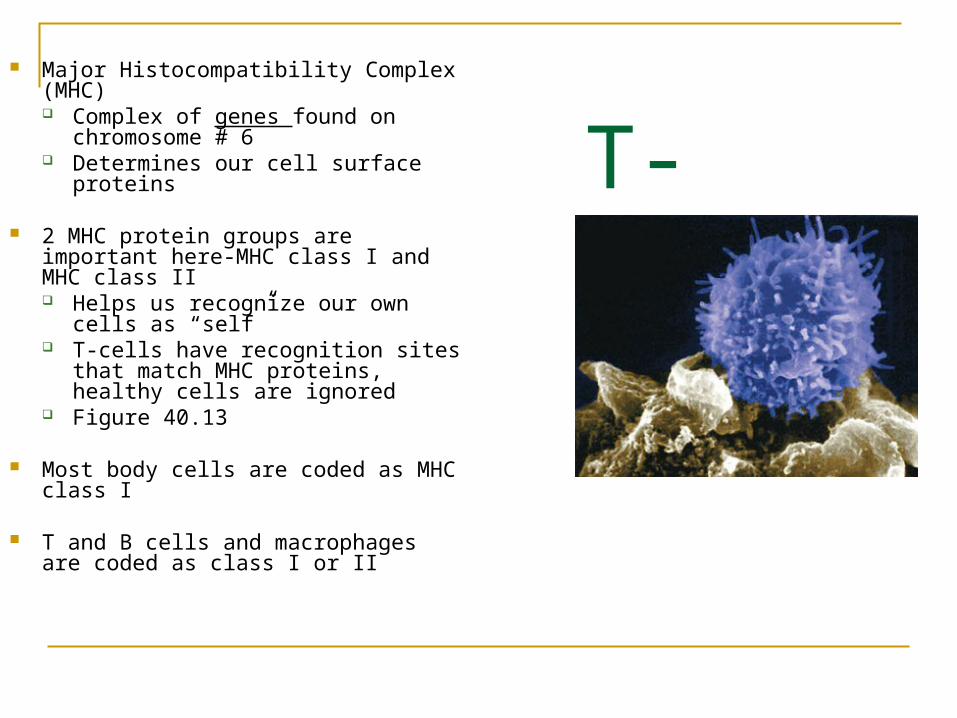
T-Cells Major Histocompatibility Complex
(MHC) Complex of genes found on
chromosome # 6 Determines our cell surface
proteins
2 MHC protein groups are important here-MHC class I and MHC class II Helps us recognize our own cells as
“self” T-cells have recognition sites that
match MHC proteins, healthy cells are ignored
Figure 40.13
Most body cells are coded as MHC class I
T and B cells and macrophages are coded as class I or II

Identification of Infected Cells by T-Cells Infected cells incorporate antigenic material into MHC protein T-cells have dual recognition sites
One recognizes MHC site (class I or II) One recognizes antigens Lots of variability in antigen site/T cells
Almost any antigen can be recognized Figure 40.14
Cytotoxic T-cells (Tc, T8) Recognize class I MHC proteins Seek out and attack diseased body cells
Helper T-cells (T4, Th) Recognize class II MHC proteins Recognize T-cells, B-cells, and macrophages and interact with them Coordinate Immune response
Virgin T-cells Mature T-cells equipped with dual recognition sites Migrate to secondary lymphoid tissue to await invaders

Primary Immune Response Complex set of events Occurs with first encounter w/new invader
(antigen) Takes time First Step—Clonal Selection
Immune cells with the correct antigen receptor must be located
Those cells must multiply Second Step--Response
Humoral or Cell-Mediated
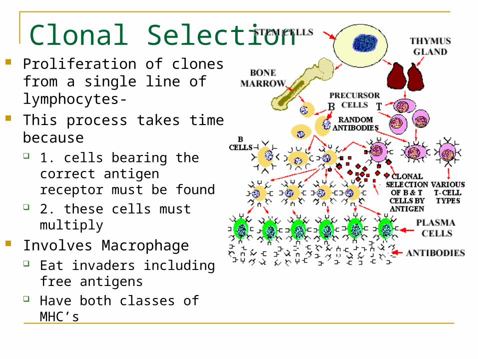
Clonal Selection Proliferation of clones from a
single line of lymphocytes- This process takes time
because 1. cells bearing the correct
antigen receptor must be found 2. these cells must multiply
Involves Macrophage Eat invaders including free
antigens Have both classes of MHC’s

Role of Macrophages in Clonal Selection
Serve as an intermediary trigger (T-cells cannot be activated by antigen)
Have both Class I and Class II proteins on their membranes

The Steps of Clonal Selection (see figure 40.17)1. Macrophage eats invader.
2. Material from invader conserved to make sites to bind with a T-cell.3. Conserved materials from extra-cellular pathogens (bacteria) incorporated into the macro’s Class 2 proteins 4. Conserved materials from intra-cellular pathogens (viruses) are incorporated into the macro’s Class 1 proteins 5. Surface of macro becomes the reciprocal of a T-cell dual recognition site6. The macrophage is now known as an antigen-presenting cell
7. The antigen presenting cell bumpsinto virgin T-cells till it finds one whose dual receptors form a match.
8. Because antigen presenting cells have both Class I and Class II MHC proteins, they can match (bind) with both Tc and Th cells.

Spreading The Alarm-Aroused T-Cells (figure 40.18)
T cell and macrophage (antigen presenting cell) bind causing macrophage to release a chemical messenger called interleukin 1.
This makes the attached T-cell undergo many cell divisions
When Th cells get aroused, they also release interleukin 2 that causes Tc cells to divide even more!
All of these new T-cells can recognize the antigen that initiates the process.
A small number of these new T-cells are set aside as memory cells

Tc cells –Cell Mediated Response Role is to “frisk” every cell till it finds
a matching antigen. Once a diseased cell is found the Tc
cell releases perforin (this chemical makes holes in the infected cell causing it to lyse.
Some Tc cells release lymphotoxin (this chemical activates enzymes of viruses to fragment their own DNA, thus preventing the virus from multiplying
Some release gamma-interferon (this chemical stimulates phagocytes to clear up cellular debris)
Can also fight cancer cells as long as the cancer has not metastasized.

Th cells—Humoral Response (figure 40.20) Two roles
1. increase cell division 2. activate B-cells
B-cells are aroused when the surface of a virgin B-cell binds to a matching free antigen.
B-cell takes in the free antigen then makes class 2 MHC proteins
Then the B-cell matches and binds to a Th cell.
The Th cell secretes interleukin 2 This chemical causes B-cells to multiply
(form clones) Some clones set aside as memory cells Once B-cell activated are called
plasma cells These cells are short lived (4-5
days) but secrete up to 2000 antibody molecules per second! See earlier discussion of antibodies
to remember the types of interactions they have with antigens.

B-cells Some B-cells don’t need to be
activated by Th cells The B-cell randomly
encounters the antigen that matches its MHC site
Antigen is taken in and triggers B-cell to divide (no chemicals)
Vast array of clones are made These type of B cells are very
limited

Suppressor T cells or Ts
Monitors the immune system and keeps
it from running out of control Brings the Primary Immune response to a halt
after danger has passed. These cells may inhibit remaining appropriate
virgin T and B cells. Since their lives are short (T and B cells) Ts cells
only need to suppress for a short time.

Memory cells and the 2o
response Some of the activated T and
B cells remain as memory cells
If another invasion of the same invaders occurs these cells recognize them immediately and start their attack.
We may not even notice that this is happening or the symptoms are very mild

