the impact of new methods of investigation and treatment on the ... · obviously demanding...
TRANSCRIPT

The impact of newmethods ofinvestigation andtreatment on theunderstanding ofthe pathology ofscleral inflammation
P Watson1,2,3,6 and A Romano4,5,6
Abstract
Recent advances in the understanding of the
initiation and perpetuation of the immune
response strongly suggest that all forms of
noninfective immunologically induced
scleral inflammation have a common origin.
Analysis of the progress of patients with
scleritis corroborates the current clinical
classification that, together with studies of
the immunohistology fluoresceine/ICG
angiography, 3D proteoglycan, and keratan
sulphate electron microscopy of scleritis,
strongly suggests that from the initiation of
the inflammatory process, necrotizing scleritis
and diffuse and nodular scleritis not only
pursue a different course but also have a
different pathogenesis; nonnecrotizing
scleritis being the consequence of an auto
immune response, whereas necrotizing
scleritis being the complication of an already
present (if not always manifest), systemic
immune-mediated systemic disease and its
associated vasculitis. The increasing imaging
capacity of anterior segment ocular coherence
tomography (OCT) and en face OCTenables the
changes occurring in the sclera during the
course of the disease to be observed for the first
time. These observations suggest that the
inflammatory changes involve the potential
suprachoroidal space between choroid and
sclera, an observation supported by the pre-
sence of subscleral granulomas on histo-
pathology. New imaging techniques have also
been able to explain the changes seen in the
cornea as a complication of scleritis. These
findings have implications for investigation
and the treatment of these conditions.
Eye (2014) 28, 915–930; doi:10.1038/eye.2014.110;
published online 30 May 2014
Introduction
Although scleral inflammation is uncommon,
accounting for only one new patient in every
thousand seen in the general hospital or clinical
practice,1 it needs to be accurately diagnosed to
ensure that those who have potentially life-
threatening disease are treated urgently and
effectively and the others are not given potentially
dangerous or inappropriate medication.
The presentation, investigation, and
treatment of most severe forms of scleral
inflammation have been extremely well covered
in recently revised texts by Watson et al,2 Sainz
de la Maza et al,3 the recent review articles by
Wieringa et al,4 by Watson and Young,5 and the
very extensive and thorough review of
Wakefield et al,6 and hence this information will
not be reiterated here.
The classification of the clinical mani-
festations of scleral inflammation that was
proposed in 1968 (see Watson et al7) (Figure 1)
has been used since then to differentiate
between the various forms of presentations of
inflammation of the sclera and thus act as a
guide to the appropriate treatment. However,
there are several questions that still need to
be answered.
The general questions are:
K Have the newer methods of investigation
and potential treatments available for the
management of scleritis altered the original
classification in any way?
K Are necrotizing scleritis and nonnecrotizing
diffuse and nodular scleritis manifestations
of the same pathological process or different
conditions?
1Department ofOphthalmology,Addenbrooke’s UniversityHospital, Cambridge, UK
2Moorfields Eye Hospital,London, UK
3University of Leiden,Leiden, The Netherlands
4Department ofOphthalmology, PaulistaSchool of Medicine, FederalUniversity of Sao Paulo,Sao Paulo, Brazil
5University of Miami, MillerSchool of Medicine, Miami,FL, USA
6Neovista Eye Center,Americana, Brazil
Correspondence:P Watson, 11 Perry Court,Clerk Maxwell Road,Cambridge CB3 0RS, UKTel: +44 (0)1223 353789;E-mail: [email protected] Jules FrancoisInternational Gold MedalResearch Oration was givenby Peter Watson atOphthalmologia Belgica,Brussels, on 8 November2013.
Received: 20 January 2014Accepted: 12 April 2014Published online:30 May 2014
RE
VIE
W
Eye (2014) 28, 915–930& 2014 Macmillan Publishers Limited All rights reserved 0950-222X/14
www.nature.com/eye

More specifically:
K Why does the inflammation occur in sclera, a tissue
that is structural in nature and has no direct blood
supply?
K What is it that initiates the immune response that
leads to scleral inflammation?
With regard to the corneal complications:
K How is it that the cornea becomes involved in scleral
disease when the structure and cellular constitution
are so different?
K Why should the cornea react in different ways in
patients with apparently similar scleral disease?
All of these questions can now be answered thanks to
new methods of imaging of the scleral and cornea in vivo
and revelations from researches in other immunologically
induced conditions.
Are necrotizing scleritis and nonnecrotizing diffuse and
nodular scleritis manifestations of the same
pathological process or different conditions?
Although there are different clinical presentations and
different types of vascular involvement in necrotizing
disease, such as the differences found in the systemic
vasculitides and those seen in rheumatoid arthritis, it
could well be that diffuse and nodular disease and
necrotizing disease have a similar underlying pathology.
However, observations of the clinical course of the
inflammatory process and the histological investigations
of Riono et al8 (confirmed by the examination of the
pathological specimens in the Wilmer Institute
Baltimore) (see ref. 2 pp 154–160) indicate that there are
differences at every stage in the progress of the disorders
up to the final stages of the disease between those who
present with nonnecrotizing scleritis and those who
develop necrotizing disease. The new imaging methods
of a speckle noise reduction algorithm and increased
scan length modified RTVue anterior segment ocular
coherence tomography (OCT; Optovue, Fremont, CA,
USA) and en face OCT together with anterior segment
fluorescein and indocyanin green (ICG) angiography and
the results of the recent methods of treatment pose a
hypothesis that necrotizing and nonnecrotizing scleritis
have different aetiologies with differing courses and
prognosis; something that considerably influences not
only our understanding of the underlying mechanisms
but also the management of the various manifestations of
scleral inflammation.
Clinical differences
Although there are bound to be overlaps and
misdiagnoses in the assessment of all the varieties of
scleritis particularly at the first presentation, it is almost
always possible to distinguish between infective scleritis,
noninfective necrotizing scleritis, and the less severe
diffuse and nodular anterior sclerits of immune-
mediated origin when the patient is first seen. Posterior
scleritis is invariably nonnecrotizing unless the
destructive process has extended from the anterior
segment. Acute scleral inflammation in the posterior
segment induces an exudative retinal detachment but no
necrosis of the sclera. Structural changes can occur that
are not due to inflammation as with the severe scleral
thinning encountered during surgery for retinal
detachment because of myopic or post-wound healing
changes in the sclera. In the vaso-occlusive form of
necrotizing scleritis, there is often a progressive
obliteration of the vascular networks leading to gradual
removal of scleral tissue without intense inflammation.
Anterior and posterior uveitis is not an accompaniment
of scleral disease except in its very severest form.
Presentation
Severe pain that wakes the patient at night, radiating to
the face and jaw and gradually improving during the
day, is the hallmark of all forms of scleritis except
scleromalacia perforans (scleromalacia perforans is now
a very rare condition seen in the late vasculsitic stages of
rheumatoid arthritis in which the sclera disintegrates
leaving bare choroid exposed). Although the onset of
necrotizing scleritis is acute, the pain severe and
accompanied by the rapid onset of severe inflammation,
Figure 1 The current classification of noninfective scleralinflammation modified from that proposed in 1968 (see ref. 2pp 154–160). Apart from the additional differentiation betweenthe nonnecrotizing and the necrotizing form of nodular scleritis,this classification remains correct. Posterior scleritis is non-necrotizing even when the choroid is also affected. SINS isscleritis following trauma often surgically induced.
Jules Francois lectureP Watson and A Romano
916
Eye
obviously demanding immediate care and attention, the
presentation of noninfective diffuse and nodular scleritis
may take days to develop with increasingly severe or
intermittent pain such that referral is often delayed for a
considerable period.
Demographics are of little help in distinguishing one
group from another at the onset of the disease except that
necrotizing scleritis is much more often bilateral than non
necrotizing disease. Overall, scleritis affects all races and
creeds equally with a female predominance and can occur
in anyone from 9 to 96 years of age. There is no genetic
association or HLA association peculiar to any form of
scleritis that does not accompany another systemic disease.
Course
Left untreated, the course of necrotizing scleritis is rapid,
inexorable, and destructive, whereas nonnecrotizing
disease is slowly progressive and even self-limiting, only in
the rare cases leaving visible changes in the affected sclera.
If the scleritis recurs, the type of scleritis that has been
diagnosed at its onset remains the same even if it recurs
at the initial site of the disease or elsewhere (Table 1).
A change from nonnecrotizing scleritis to necrotizing
scleritis was seen in only 13 of the 104 patients. Of the
10 patients who changed from nodular to necrotizing
scleritis, 9 were later found to have an overt underlying
systemic disease.
Associated disease
A cause or underlying disease is rarely found in those
who have nonnecrotizing noninfectious scleritis. When
found, it is usually associated with a sero-negative
condition such as Reiter’s or Behcet’s disease.
In contrast, necrotizing scleritis is associated with
systemic diseases, in particular the vasculitides or
autoimmune conditions associated with vasculitis.
Scleritis is most commonly seen in rheumatoid arthritis,
granulomatosis with polyangiitis (Wegener’s
granulomatosis), polyarteritis nodosa, and relapsing
polychondritis. Occasionally, it is the direct result of
viral, parasitic, or bacterial infection, particularly leprosy
in regions where this disease is endemic. Surgically
induced necrotizing scleritis (SINS) can occur after any
form of trauma. Where it follows surgery, it is noticed
that the post-operative inflammation is slow to settle or
even gets more intense. In SINS the scleral inflammatory
response is almost always necrotizing, severe, and very
resistant to treatment. Sometimes the scleritis occurs, not
at the site of the recent procedure, but at the place of
previous trauma such as a childhood strabismus
operation.
Investigations
The different types of nodular scleritis have, until now,
been difficult to diagnose because some are due to a
localized but intense form of benign scleral inflammation,
some present as an early manifestation of necrotizing
scleritis, and others are infective. The advent of speckle
noise reduction algorithm and increased scan length
Fourier domain anterior segment OCT and en face anterior
segment OCT developed by one of us (Andre Romano)
together with anterior segment ICG angiography has led to
a step change in diagnosis. Given the right tools the
diagnosis can be made as soon as the patient presents.
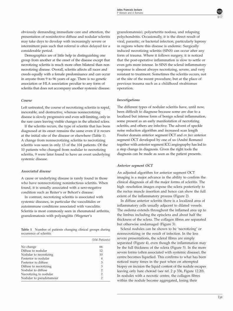
Anterior segment OCT
An adjusted algorithm for anterior segment OCT
imaging is a major advance in the ability to confirm the
clinical diagnosis of all the major forms of scleritis. The
high -resolution images expose the sclera posteriorly to
the rectus muscle insertion and hence can show the full
extent of the inflammatory process (Figure 2).
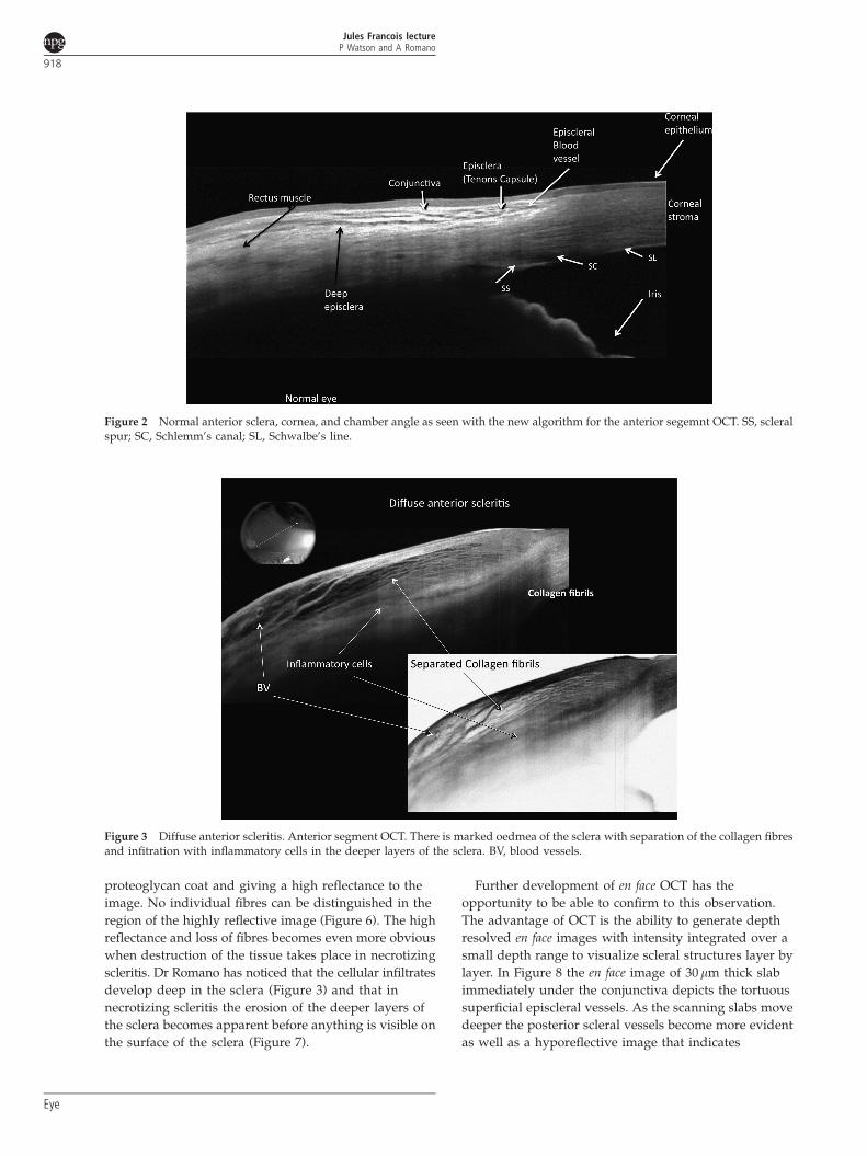
In diffuse anterior scleritis there is a localized area of
inflammatory cells usually adjacent to dilated vessels.
The oedema extends throughout the inflamed area up to
the limbus including the episclera and about half the
thickness of the sclera. The collagen fibres are separated
but otherwise undamaged (Figure 3).
Scleral nodules can be shown to be ‘necrotizing’ or
nonnecrotizing or the result of infection. In the less
severe presentations, the scleral fibres are simply
separated (Figure 4), even though the inflammation may
be the full thickness of the sclera (Figure 5). In the more
severe forms (often associated with systemic disease), the
centre becomes liquefied. This confirms to what has been
noticed many times in the past when on attempted
biopsy on incision the liquid content of the nodule escapes
leaving only bare choroid (see ref. 2 p 336, Figure 12.20).
In nodules with a necrotic centre, the collagen fibres
within the nodule become aggregated, losing their
Table 1 Number of patients changing clinical groups duringrecurrence of scleritis
(104 Patients)
No change 66Diffuse to nodular 12Nodular to necrotizing 10Posterior to nodular 4Posterior to diffuse 3Diffuse to necrotizing 3Nodular to diffuse 2Necrotizing to nodular 2Nodular to pseudotumour 2
Jules Francois lectureP Watson and A Romano
917
Eye
proteoglycan coat and giving a high reflectance to the
image. No individual fibres can be distinguished in the
region of the highly reflective image (Figure 6). The high
reflectance and loss of fibres becomes even more obvious
when destruction of the tissue takes place in necrotizing
scleritis. Dr Romano has noticed that the cellular infiltrates
develop deep in the sclera (Figure 3) and that in
necrotizing scleritis the erosion of the deeper layers of
the sclera becomes apparent before anything is visible on
the surface of the sclera (Figure 7).
Further development of en face OCT has the
opportunity to be able to confirm to this observation.
The advantage of OCT is the ability to generate depth
resolved en face images with intensity integrated over a
small depth range to visualize scleral structures layer by
layer. In Figure 8 the en face image of 30mm thick slab
immediately under the conjunctiva depicts the tortuous
superficial episcleral vessels. As the scanning slabs move
deeper the posterior scleral vessels become more evident
as well as a hyporeflective image that indicates
Figure 2 Normal anterior sclera, cornea, and chamber angle as seen with the new algorithm for the anterior segemnt OCT. SS, scleralspur; SC, Schlemm’s canal; SL, Schwalbe’s line.
Figure 3 Diffuse anterior scleritis. Anterior segment OCT. There is marked oedmea of the sclera with separation of the collagen fibresand infitration with inflammatory cells in the deeper layers of the sclera. BV, blood vessels.
Jules Francois lectureP Watson and A Romano
918
Eye

destruction of the posterior surface of the sclera. This
suggests that there is an intense inflammatory response
involving the sclera and the vessels deep to the sclera at
the site of the superficial visible destructive changes.
This is the first time that it has been possible, although
imperfectly at present, to observe changes deep to the
sclera during the course of the disease. The findings
suggest that the changes seen at the surface in scleral
inflammation are accompanied by, or even preceded by,
similar changes in the potential suprachoroidal space
between choroid and sclera (the space accessed in the
cyclodialysis operation). This can explain the subscleral
granulomas seen universally in the histopathology of eye
enucleated from patients with both anterior and
posterior scleritis (see ref. 2 p 155, Figures 7.2, 7.3, 7.5).
An extension of this technique using phase variance
OCT has the potential to discriminate between different
classes and activation of inflammatory cells that carry
different charges. This may depend on being able to
combine the imaging with tagged markers for each cell
subclass as well as utilizing energy sourcing such as the
Warburg effect9 for activation. If this is successful, then
specific rather than undirected treatments can be given.
Histology
The sclera is composed largely of type 1 collagen and
some elastic tissue. The collagen fibrils are surrounded
by proteoglycans that encircle the fibril, run axially
along its length, and join one fibril to the next (Figure 9,
lower panel). The predominant glycosaminoglycan is
chodroitin sulphate/dermatan sulphate. Its structure is
maintained by fibrocytes (sclerocytes) that are in contact
with each other via cellular processes. The only other
resident cell within the sclera is the resident tissue
macrophage. There is no direct blood supply to the
sclera; the nutrients, which includes the immuno-
globulins, arrive by diffusion from the vessels of the
choroid, ciliary body and the superficial anterior ciliary
arteries (Figure 10).
Figure 4 Nonnecrotizing nodular scleritis. Anterior segmentOCT. The nodule consists of extracellular fluid. The collagenfibres are separated but remain distinct. There is no necrosis oftissue.
Figure 5 Histology of a nonnecrotizing scleral nodule. Theinflammation affects the whole thickness of the sclera.
Figure 6 Necrotizing nodular scleritis. Anterior segment OCT. There is hyperreflectivity of the episcleral and deep tissues. Thehyporeflectivity of the scleral nodule shows that the deep layers of the nodule are liquefied and interpersed with blood vessels.
Jules Francois lectureP Watson and A Romano
919
Eye

Microscopic and electron microscopic studies of biopsy
specimens from patients with simple and nodular
episcleritis have been totally noncontributory in eluci-
dating the aetiology of these conditions. The inflamed
area is packed with lymphocytes and a few other
inflammatory cells, but there are no mast cells, plasma
cells, or eosinophils.
Because biopsy of the sclera is undesirable, most of the
information regarding the histopathological appearances
of scleral disease comes from enucleated specimens of
necrotizing scleritis. These confirm that even in the most
severe disease the anterior segment is primarily involved
and that purely posterior segment disease is very
unusual. The findings of Riono et al8 (confirmed by the
study of the specimens in the pathology laboratory of the
Wilmer Institute in Baltimore) (see ref. 2 pp 155–161) in
eyes that were enucleated because of the secondary
complications of glaucoma, retinal detachment, choroidal
involvement, uveitis, and vascular occlusion had a
histological appearance of nonzonal diffuse scleral
inflammation and had no association with systemic
disease. However, those with zonal necrotizing
granulomatous scleral inflammation, with the
histopathological characteristics of ‘fibrinoid necrosis’
Figure 7 Necrotizing scleritis. Apart from the obvious loss oftissue the collagen fibres are aggregated together, indicating aloss of the proteoglycan coat. There is not only erosion of thesuperficial tissues but also under surface of the sclera.
Figure 8 En face OCT in necrotizing scleritis. The depth, structure, and extent of the lesion can be determined. En face imageintegrated over the full-scan range of the sclera. Horizontal B-scan image through the centre of the scleral lesion in a patient withnecrotizing scleritis (i) with overlay contour lines indicating the positions of partial intensity en face images. En face image of the 30mmthick slab immediately underneath the conjunctival epithelium (a–h). A hyporeflective image indicates destruction of the posteriorsurface of the sclera (f–h).
Jules Francois lectureP Watson and A Romano
920
Eye

and vasculitis, showed various forms of massive
lymphocytic infiltration of the tissues and destructive
changes in areas of cell death (Figure 11), and 12 of the 14
cases examined were found to have come from patients
with an immune-mediated disease.8
Immunohistological changes
The immunohistological investigation of pathological
specimens by Usui et al10 show that the immunohistology
of nonnecrotizing and necrotizing scleritis also differed
(Figure 11). The cytokine profile of those with diffuse and
nodular scleritis consisted of 43% CD68 macrophages,
23% CD3 cells, 5% CD8, 1% DRC, but only 7% CD20 B
cells, highly suggestive of an autoimmune response. This
was in sharp contrast to specimens from those with
necrotizing scleritis where the profile was 43% CD20 B
cells, 35% CD68 macrophages, 17% CD3, 8% CD8, and
4% DRC, indicating the presence of a systemic vasculitis.
It has since been shown that those with an expanded
CD8 (þ ) T-cell memory population have a poor
prognosis.11 The relatively large number of macrophages
is also to be expected in this condition because of the
presence of necrotic tissue.
What is it that initiates and perpetuates the immune response
that leads to scleral inflammation? All the available
evidence suggests that with the exception of those cases of
infection, some unusual disorders of collagen such as the
Ehlers Danlos syndrome and in episcleral disorders where
there is localized vascular spasm, scleral and episcleral
inflammation is immune mediated (see ref. 2 p 155,
Figures 7.2, 7.3, 7.5).6 What has not been clear is what is
triggering the inflammatory response, what is the immune
response driven against, and what causes the inflamma-
tion to persist. In addition, it has not been certain why,
once the immune response has been triggered, the
manifestations of the disease are so different in each
individual. This is important because if these questions
can be answered, therapy can be targeted at a particular
response or even a subset of inflammatory cells.
In the nineteenth century, and presumably before this,
the commonest cause of scleritis was tuberculosis. The
inflammation was not the result of a primary invasion of
the tissue by the tubercle bacillus but an immune
response to it. As this disease was brought under control,
the commonest cause then became rheumatoid arthritis.
Now that rheumatoid arthritis can be treated early and
more effectively, scleritis associated with this disease
has become unusual and hence the commonest cause
of necrotizing scleritis is now from the various
manifestations of the systemic vasculitides. In countries
where immune deficiency is common, syphilis and
tuberculosis and the accompanying scleral complications
are prevalent again. In places where leprosy is common,
lepromatous leprosy should always be considered in the
differential diagnosis as scleritis can be the first clinical
manifestation of this disease.
How is it that the immune response is triggered by a variety of
microorganisms resulting in a condition that appears to be
identical clinically? The portal of entry of the infective
organism or particulate matter is through the lung and,
probably as in some bacteria, some virus infections and
acanthamoeba, the conjunctiva. Occasionally, in Borellia
Figure 9 Scleral collagen and its associated proteoglycans.Scleral collagen fibrils exhibit a–e cross-banding in longitudinalsections (top panel). Cuprolinic blue staining visualizesproteglycans as filaments is associated with the d bands. Thesefilaments lie along the fibril, encircle it, and connect the collagenfibrils to each other (lower panel).18
Figure 10 The distribution of immume proteins in cornea andsclera.19
Jules Francois lectureP Watson and A Romano
921
Eye

infection or following trauma or surgery, the portal of
entry is via the skin. Proteinases from these organisms
are recognized by the microbial pattern recognition
receptor (innate immune receptor), Toll-like receptor 4
(TL4) expressed on macrophages, and dendritic cells.
The Toll receptor family, collectively referred to as
pathogen-associated molecular patterns (PAMPs),
associate with interleukin-1 to form a receptor
superfamily. The immune system responds not only to
these evolutionary conserved danger signals but also to
damaged cells from the tissues themselves and from the
immune system itself (danger associated molecular
patterns (DAMPs).12,13 Many bacterial molecules and
DAMPS can activate TLR4 which may trigger a common
signalling pathway resulting in similar histological
changes. The foreign proteinases cleave the clotting
protein fibrinogen.14 The fibrinogen cleavage product
then acts as a TLR4 ligand on epithelial cells,
antigen-presenting cells, and macrophages, leading to
inflammation. This discovery of the importance of
fibrinogen cleavage in the allergic inflammatory process
is the missing link that has been elusive for so long.
In the experiments of Millien et al,14 they had to use
ovalbumen challenge as they were investigating the
fungal proteinases in asthma that does not occur
naturally in rats and mice. It is of interest that the only
animal model of scleritis to be produced was by
ovalbumen challenge.15 In this instance, the predominant
features were the presences of T cells, macrophages and
the activation of the fibrocytes by immune complexes.
Why does the inflammation occur in sclera, a tissue that is
structural in nature and has no direct blood
supply? Episcleritis has all the characteristics of a
localized hypersensitivity response affecting only the
superficial vascular plexuses with outpouring of fluid
and cells in the affected area. It is less clear why the
sclera, which derives its nutrition from branches of the
anterior ciliary artery and the terminal vessels of the long
posterior ciliary artery, should become involved in the
inflammatory process.
Characteristically, inflammation of the sclera and its
overlying episclera first appears in the upper inner or
outer quadrants of the eye. The reason why scleritis starts
in these regions between the extraocular muscles is that
the superficial blood supply is unusual in that there is an
artery to artery anastamosis between the terminal
branches of the anterior ciliary arteries and between the
terminal and perforating branches of the long posterior
ciliary arteries. Although this arrangement ensures an
adequate arterial blood supply to the anterior segment of
the eye at all times, it has the consequence, at least in the
superficial vasculature, that instead of a rapid flow of
blood from the arterial to the venous circulation through
a capillary network, the flow is sluggish, even oscillating.
Thus, if immune-competent cells leave the arteriolar
circulation in these areas, there is no immediate return to
the venules, giving time for immune reactions to be
triggered, enhanced, and perpetuated rather than being
dealt with by the innate immune response as in tissues
with a rapid circulation. Furthermore, if there is a
Figure 11 (Left above) Zonal necrotizing granulomatous inflammation in rheumatoid arthritis showing an acellular necrotic centrewith an infiltration of inflammatory cells and some giant cells. (Left below) Staining for CD20 B cells in the same patient. (Right above)Scleritis in a patient with no evidence of systemic disease. There is diffuse infiltration of the sclera with inflammatory cells. (Rightbelow) Staining for CD8 macrophages in the top right patient. The predominant cell is the macrophage (ref. 2 p 177, Figure 7.39).
Jules Francois lectureP Watson and A Romano
922
Eye

vasculitis of the terminal choroidal vessels, as can be seen
in end-stage histological specimens (see ref. 2 p 155
Figures 7.2, 7.3, 7.5), the inflammation can extend
through the sclera with the perforating vessels to the
surface, where isolated vasculitic lesions are known to
occur Figure 12. Hitherto it has not been suspected that
the inflammatory process could start deep to the sclera
rather than on the scleral surface and its overlying
membranes (Figures 3 and 6–8).
How is it that diffuse and nodular scleritis have such a
different course to necrotizing scleritis if their origin is the
same? The immunohistochemical evidence suggests
that, unlike necrotizing scleritis, diffuse and
nonnecrotizing nodular disease are the result of an
autoimmune process starting from within the sclera
itself; its components responding to, or initiating, not
only DAMP, activated circulating antigen-presenting
cells, and macrophages, but also under certain
circumstances resident macrophages. Resident tissue
macrophages are present at birth and remain throughout
life. Under certain circumstances, as in the induction of
diffuse and nonnecrotizing nodular scleritis, they may
assume an M1 inflammatory phenotype. The
involvement of these resident macrophages could also
explain why in SINS, when an area has been damaged
remote from the current insult, as in a previous
strabismus operation this becomes the site of the scleral
inflammation.
The immunohistochemistry suggests that the sequence
of events is different in necrotizing scleritis, a
complication of an already present (if not always
manifest) systemic immune-mediated systemic disease
and its associated vasculitis. Scleritis can be the
presenting feature of some of these (Figure 13), with its
presence in or under the sclera being because of the
sluggish circulations resulting from the unusual end
artery and interarterial connections or part of a systemic
multiorgan vasculitis.
Where the cytokine release is sufficient to cause
activation of the scleral stromal fibrocytes, then
destructive catabolic changes can take place. First,
the protective activity of tissue inhibitor of
metalloproteinases (TIMPs) (see ref. 2 p 158, Figures
7.10,7.11, 7.12 and pp 171-172, Figures 7.27, 7.28, 7.29) is
reduced, allowing proteoglycan to be removed from the
surface of the collagen fibril that then becomes
unravelled and separated from its neighbour Figure 14.
The exposure of the collagen, which has never before
been exposed to the immune system, can either result in
loss of tolerance or stimulate an inflammatory response,
leading to antibody production, immune complex
deposition, and the induction and perpetuation of the
autoimmune response.
Figure 12 Granulomatous scleritis in granulomatosis with polyangiitis (Wegener’s granulomatosis). (a) The characteristicpresentation where the inflammation includes the limbus and adjacent cornea (in rheumatoid arthritis the limbal area is spared).(b) Red Free and ICG after 2 weeks of intensive treatment. The eye appears completely quiet on the red free but ICG reveals an intensestaining at an area of vasculitis. Treatment should not be discontinued until these areas have healed.
Jules Francois lectureP Watson and A Romano
923
Eye

Why should the cornea react in different ways in patients with
apparently similar scleral disease? Anterior segment
fluorescein/ICG angiography has helped to resolve the
puzzle as to why the cornea reacts in different ways in
different patients with apparently similar disease.
The corneal changes that are seen in association with
scleral inflammation are of four types: Contact lens cornea,
scleralization of the cornea, peripheral corneal destruction/
guttering, and central corneal dissolution Figure 15.
Contact lens cornea
In contact lens cornea, the peripheral cornea becomes
thinner and thinner without any obvious infiltration,
coming to resemble a contact lens sitting on the cornea.
Fluorescein/ICG angiography shows the adjacent limbal
capillary network to be extensively attenuated, with a
few new vessels passing into the peripheral superficial
cornea. Nutrients for the maintenance of corneal collagen
come from the limbus and by diffusion from the anterior
chamber (Figure 10). The central cornea is maintained by
the elution of nutrients through the central cornea, but
the periphery is deprived of these because of the grossly
reduced blood supply and, as a consequence, gradually
diminishes in thickness (Figure 16).
Scleralization of the cornea
After prolonged or repeated attacks of scleral
inflammation at the same site, the cornea begins to
become opaque, the so-called scleralization of the cornea.
In the early and acute phases of scleral inflammation,
corneal infiltrates can be observed as an acute stromal
keratitis, sometimes with immune rings around them,
clearly the result of an antigen antibody response at that
site. It is probably fair to assume that the same process is
Figure 13 Necrotizing scleritis associated with granulomatosiswith polyangiitis (Wegener’s granulomatosis). A 57-year-oldwoman who presented with a red eye that was painful for 3months. (a) The corneal destructive changes involve the wholelimbus typical of that seen in a systemic vasculitis even thoughthe ANCA was negative at this time. (b) After 17 months. Advicethat she should be treated systemically was not accepted eventhough she had become ANCA positive by April 2001.
Figure 14 Normal collagen/proteoglycan structure on humansclera. In necrotising scleritis the proteoglycan separates fromthe collagen leading to unravelling of the collagen and itsexposure to cytokine activity. (a) Proteoglycan fibrils connectingcollagen to collagen. (b) Encircling proteoglycan fibril. (c)Proteoglycan fibril along the collagen (ref. 2 pp 158-159,Figures 7.10, 7.11, 7.12, 7.13).
Jules Francois lectureP Watson and A Romano
924
Eye

happening in the sclera at the same time. The opacification
of the cornea is occasionally because of the persistence
and extension of the precipitation of these immune
complexes following an acute sclerokeratitis, but more
often, as is shown by fluorescein/ICG angiography, the
opacification is the result of destruction of the normal
limbal arcade by the inflammation and the stem cells
contained within them. If this happens, the conjunctiva
and its vessels will overgrow the cornea, allowing
opacification of the tissue beneath.
Peripheral corneal destruction
In the presence of scleral inflammation there can also be
an infiltration and eventual loss of corneal tissue. In
idiopathic and rheumatoid-associated sclerokeratitis, this
peripheral change usually begins 2 mm inside the limbus
with an inflammatory infiltrate that, if the inflammatory
process is allowed to continue, results in a breakdown of
the cornea stroma. In these individuals, even though
there may be an associated necrotizing scleritis, the sclera
immediately adjacent to the limbus appears to be entirely
normal except for some abnormal vessels.
The site of destruction in both the sclera and cornea in
peripheral corneal/guttering, 2 mm either side of the
limbus, is the site of the highest concentration of immune
proteins (Figure 10). The limbus, which is fed and
drained by two different circulations, is spared because
of the high concentration of blood vessels there.
In rheumatoid arthritis, the characteristic changes seen in
this situation are those of a venular occlusive scleritis
affecting the vessels of the episcleral plexus. Fluorescein/ICG
angiography shows that the flow of blood within the
capillary and arteriolar circulations of the networks is slowed;
the vessels themselves becoming partially or completely
occluded (see ref. 2 pp 158–159, Figures 7.14, 7.16, 7.17).
In contrast, the vascular changes associated with the
corneoscleral pathology in a systemic, potentially fatal,
vasculitis are quite different. These limbal changes may
be the presenting feature of the systemic disorder, and
hence it is important to recognize granulomatous scleritis
as soon as it presents. In the presence of a systemic
vasculitis, the destructive changes involve the cornea,
limbus, and adjacent sclera equally; there is no area of
apparently normal sclera adjacent to the limbus. All the
tissues are involved together in the destructive process
(Figures 12 and 13). This is the result of the vasculitis that
affects blood vessels of all sizes. The limbal blood supply
revealed when all the other tissues have been digested
away is found to be enormous and is derived from all the
those vessels feeding the anterior segment. Therefore, it
is to be expected that the tissue of the limbus will be
affected in an inflammatory vascular disease.
Fluorescein/ICG angiography reveals grossly
abnormal vascular networks in the feeding vessels to the
site of inflammation, including aneurysms and most
importantly inflamed vessel walls Figure 12. Treatment
must not be stopped before these inflamed areas have
stopped leaking.
Figure 15 The corneal changes associated with scleral inflammation.
Jules Francois lectureP Watson and A Romano
925
Eye

Central corneal dissolution
Very occasionally in patients with rheumatoid arthritis,
the whole of the central corneal tissue will swell without
obvious cellular infiltration. The cause of this rare but
severe acute immune response is not known but is likely
because of the presence of a 54 kDa antigen found in the
cornea in this condition. Whatever the cause, if it is not
treated in time the stroma will dissolve, leaving only the
epithelium and Descemet’s membrane intact.
How is it that the cornea becomes involved in scleral disease
when the structure and constitution are so different? The
macromolecular composition of the collagens and
proteoglycans, its structural organization, and its fibrocytes
of cornea and sclera are all different. These differences,
which have been extensively investigated by Watson and
Young,5 reveal that the differences lie in the arrangement
of both collagen and its proteoglycan coat.
With one or two notable exceptions unless the
inflammatory response is very intense, the cornea is not
usually involved in scleral disease. The corneal collagen
network is regular in size and much more tightly bound
together and will only break apart if there is a major
influx of inflammatory cells as the result of intense
adjacent inflammation (Figure 17). In addition, if the
proposition is accepted that each specialized tissue is
capable of recognizing and responding to its own danger
signals, then corneal changes would not be expected.12
On the other hand, when the adaptive immune
response has been triggered and the sclera becomes
involved the proteoglycan bonds between the collagen,
fibrils break apart (see ref. 2 p 158, Figures 7.10, 7.11, 7.12).
In nonnecrotizing scleritis they may be restored by
remodelling if treatment is successful, but in necrotizing
scleritis the proteoglycans surrounding the collagen fibrils
may be permanently lost, exposing the fibrils to prolonged
enzymic attack, causing them to unravel. This may reveal
collagen neoepitopes never previously exposed to the
immune system that subsequently act as an antigen
inducing a further inflammatory response.
How does the perceived difference between nonnecrotizing disease
and its more serious counterpart affect treatment? Before
1950, the only treatment that could be offered to anyone
with scleral inflammation was palliative. It consisted of
atropine eye drops, aspirin, or morphine derivatives for
the pain, and bathing of the eye with either warm or cold
compresses that gave some form of counter irritant and
temporary comfort. As the common cause at that time
was tuberculosis, the equally ineffective treatments for
this condition were often added on top. If patients
recovered, it was because of their underlying good
constitution and the superb nursing of the patients of
that time rather than the intervention of the physician.
The introduction of locally applied steroid eye drops in
the 1950s not only led to a dramatic improvement in the
comfort of the patients but also to regression of the
signs and symptoms. The use of nonsteroidal anti-
inflammatory agents (NSAIDs) other than aspirin and
the use of high-dose and pulsed steroid was the major
advance of the 1960s. Because these regimes suppressed
the inflammation, the progression of the disease was
stopped and, if the sclera had been damaged, allowed
healing to take place. The other important revelation of
this treatment was that it showed that scleral
inflammation was the result of an immunological
process and rarely the result of direct infection that
had been assumed in the past. The next three decades
have been spent elucidating the cells that induced the
destructive process and their effect on the connective
tissue and other components of the sclera and its
coats.
Steroids, effective as they may be, have the effect of a
blunderbuss; undirected, widely effective but not
directed at a particular target. Oral steroids affect many
parts of the inflammatory pathway but do not target any
specific aspect of it and, in addition, have multiple
undesirable side effects. However, subconjunctival
triamcinalone has been shown to be effective in reducing
these to a minimum in nonnecrotizing scleritis. As it has
Figure 16 Contact lens cornea. Fluoresceine/ICG angiogramsshowing closure of the episleral capillary network associatedwith long-standing scleral inflammation. Fluorescein showsclosure (arrow) with secondary overgrowth of the conjunctivalvessels over the limbus to the edge of cornea of normalthickness. ICG images confirm the gross reduction of episcleralcapillaries and leakage from the remaining ones (betweenarrows).
Jules Francois lectureP Watson and A Romano
926
Eye

been shown that there is no overlap in the manifestations
of nonnecrotizing scleritis and the conditions that lead to
tissue destruction (Table 2), it follows that although
remission can be induced in diffuse and nodular scleritis
with NSAIDs, subconjunctival triamcinalole, or a short
course of steroids, necrotizing disease needs to be treated
from the outset with systemic immunosuppression and
disease-modifying agents such as methotrexate. It has
become recognized that TNF-a is involved in most of the
important parts of the inflammatory pathway and that
monoclonal antibodies to this substance are effective in
reducing the number and amount of inflammatory cells
of all classes, thus inhibiting the intense destructive
process and allowing healing to take place. The compounds
in common use are infliximab, adalimumab, and
rituximab. It has been found that rituximab, known to be
most effective against B cells (associated with vasculitis),
is much more effective in the treatment of scleral disease
than infliximab.
The management of scleral disease in the future
It is sometimes questioned why pioneers in
ophthalmology such as Jules Francois, von Graefe,
MacKenzie, Gonin, and my old mentor Sir Stewart Duke
Elder should be commemorated. It is because these were
the people who above all brought together all the
available knowledge of the eye and its diseases in their
era and hence allowed great leaps forward in the
diagnosis and treatment of the diseases that they brought
to everyone’s attention.
Jules Francois’ pioneering work and classic text on
genetically determined diseases of the eye has led to the
identification of those conditions that are possibly
amenable to the first successful stem cell implant
treatments in medicine.
These individuals marked the end of the era when
description of disease was all that could be done. The
succeeding decades have taken this knowledge and have
found ways, with the help of technology and pharmacy,
of dealing with most of the problems, reducing
dramatically the level of blindness worldwide.
Within the past 3 or 4 years, we have entered a new
revolutionary period that, I believe, will change the way
not only ophthalmology but the whole of medicine is
taught and practiced. This is the smart phone revolution.
Expert help in the diagnosis and treatment in unusual
conditions such as scleritis will be available to all in
however remote a community. Attachments to a smart
phone can measure visual acuity, do accurate visual
fields, measure intraocular pressure, refract, take
accurate high-quality images of corneal external diseases,
and excellent fundus photographs. Furthermore,
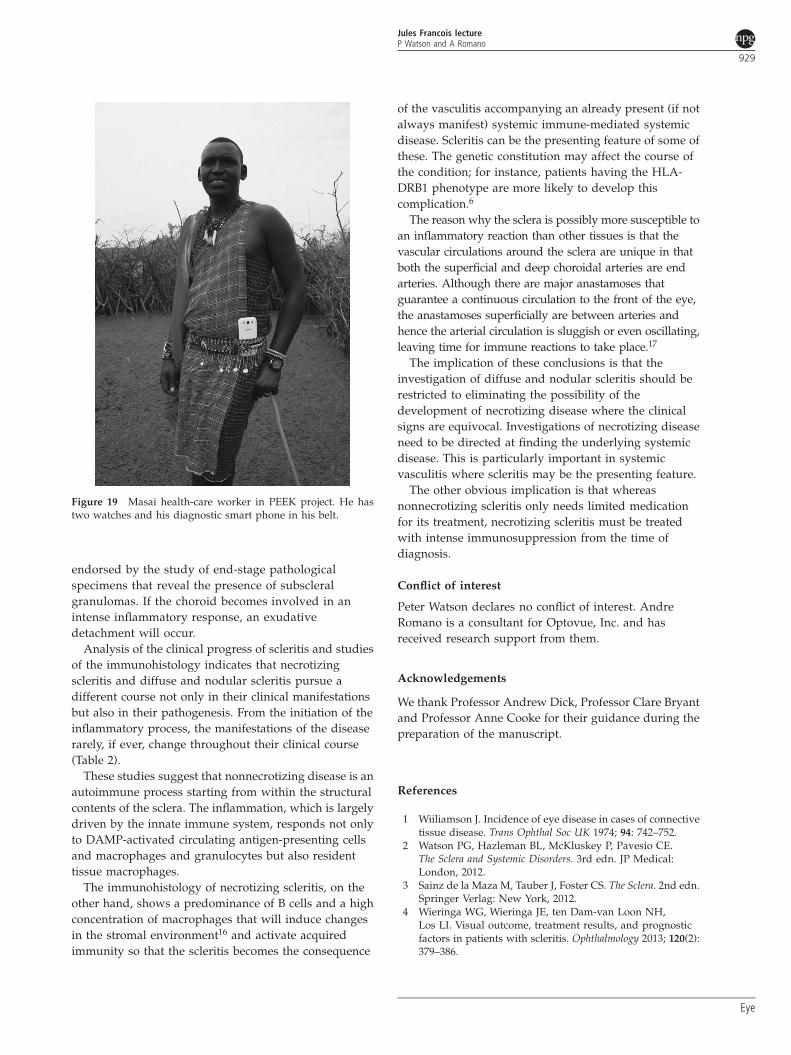
the PEEK project in Kenya has shown that these
investigations can be undertaken by health-care workers
who have had only a few hours of training (Figures 18
and 19).
As smart phones are now ubiquitous organized, or
even self, assessment is becoming widespread and not
CentreInner
peripheryMid
periphery
Outer periphery/
limbus ScleraP
oste
rior
stro
ma
Mid
str
oma
Ant
erio
r st
rom
a
120
90
60
30
0
0-3m
m
3-6m
m
6-9m
m
9-12
mm
12-1
5mm
corneal position
Ave
rage
fibr
il di
amte
r (n
m)
Anterior stroma
Mid stroma
Posterior stroma
Collagen fibril diameters from corneal centre to anterior sclera in bovine eye
Figure 17 Cross-sectional and longitudinal TEM micrographs of collagen fibrils across the bovine cornea and sclera. The collagenfibril diameter is uniform from the cornea centre until the limbus where it varies to increase significantly into the sclera. This is so inthe outer mid and deep stroma. Scale bar¼ 100 nm (cross-sectional) and 200 nm (longitudinal). The mean fibril diameter across thebovine cornea and sclera is shown in the graph. 0–3 nm¼ centre; 3–6 nm¼ inner perphery; 6–9¼mid periphery, 9–12¼ outerperiphery; and 12–15¼ sclera (Ho et al, submitted).
Jules Francois lectureP Watson and A Romano
927
Eye

confined to Africa, India, and Asia. It means that
ophthalmologists everywhere will have to become either
surgeons, specialists in some particular condition, or
expert advisors on where and how a patient should be
treated. Everything else will be done by ancillary staff.
Ophthalmologists may well find themselves treating
conditions remotely, perhaps without ever seeing the
patient. Training of budding medical students and
ophthalmologists will have to reflect these changes.
Times change and the loss of personal contact with the
patient is going to be a real problem. Technology is king
at the moment but, for me, it is good to have practised in
an era where one saw everyone face to face and could
ensure they left you with a smile on their face however
grim the diagnosis.
Conclusions
Analysis of the presentation and the course of
noninfective scleritis together with the additional
evidence derived from deep anterior segment OCT/
en face OCT, fluorescein/ICG imaging, histology, and the
immunohistochemical investigations confirms that the
original classification of scleral disease has stood the test
of time and is still correct.
The new algorithms for anterior segment OCT imaging
indicate that there are two forms of nodular scleritis: one
in which the collagen fibres are simply separated as in
the nonnecrotizing forms of scleritis and the other where
the contents liquefy in a similar manner to the
destructive changes of necrotizing scleritis.
In addition, the increased ability to observe the
deeper layers of the sclera with this technique
suggests that scleritis is associated with changes
within the potential suprachoroidal space, deep to the
sclera as well as superficial to it. This observation is
Table 2 The differences between necrotizing and nonnecrotizing scleritis, its investigations, its pathology, and the course of thedisorder from its presentation
Necrotizing scleritis Diffuse, nodular scleritis
Presentation Severe pain, intenseIntense inflammation
Gradual onset, moderate painDiffuse or nodular presentation
Course Unremitting Recurrent or self-healingIdentity No change throughout No change throughoutTrigger Trauma, infection Rarely determined, stressSystemic disease Common, RA, vasculitis UncommonArea of scleritis Anterior Anterior and posteriorCornea Often involved Rarely involvedAngiography Disturbed vascular pattern from onset, vasculitis Vascular pattern only altered in chronic or
recurrent diseaseOCT Destruction and aggregation of collagen fibrils superficial and
deep to scleraSeparation of collagen fibrils oedema, withcellular infiltrates
Histology Granulomatous, vasculitis fibrinoid necrosis adjacent to vessels.Central necrosis histiocytes, lymphocytes close to necrosis,plasma cells lymphocytes surrounding zonal necrosis
Diffuse inflammation of episcleral and scleraltissue lymphocytic infiltrates,nongranulomatous
Immunopathology 43% CD20 B cells,35% CD68 macrophage,and 8% CD8, indicating the presence of a systemic vasculitis
43% CD68 macrophage but only 7% CD20 Bcells, highly suggestive of an autoimmuneresponse
Complications Glaucoma uveitis frequent Rare except posterior scleritisTreatment Active immunosuppression required. Retuximab most effective Moderate inflammation NSAID or steroid even
spontaneous remission
Figure 18 The ‘stitched’ composite fundus photgraph takenwith an attachment to a smart phone of an exudative retinaldetachment and retinal haemorrhages taken by a health-careworker in the PEEK project in Kenya ready to be transmitted to adiagnostic centre.
Jules Francois lectureP Watson and A Romano
928
Eye
endorsed by the study of end-stage pathological
specimens that reveal the presence of subscleral
granulomas. If the choroid becomes involved in an
intense inflammatory response, an exudative
detachment will occur.
Analysis of the clinical progress of scleritis and studies
of the immunohistology indicates that necrotizing
scleritis and diffuse and nodular scleritis pursue a
different course not only in their clinical manifestations
but also in their pathogenesis. From the initiation of the
inflammatory process, the manifestations of the disease
rarely, if ever, change throughout their clinical course
(Table 2).
These studies suggest that nonnecrotizing disease is an
autoimmune process starting from within the structural
contents of the sclera. The inflammation, which is largely
driven by the innate immune system, responds not only
to DAMP-activated circulating antigen-presenting cells
and macrophages and granulocytes but also resident
tissue macrophages.
The immunohistology of necrotizing scleritis, on the
other hand, shows a predominance of B cells and a high
concentration of macrophages that will induce changes
in the stromal environment16 and activate acquired
immunity so that the scleritis becomes the consequence
of the vasculitis accompanying an already present (if not
always manifest) systemic immune-mediated systemic
disease. Scleritis can be the presenting feature of some of
these. The genetic constitution may affect the course of
the condition; for instance, patients having the HLA-
DRB1 phenotype are more likely to develop this
complication.6
The reason why the sclera is possibly more susceptible to
an inflammatory reaction than other tissues is that the
vascular circulations around the sclera are unique in that
both the superficial and deep choroidal arteries are end
arteries. Although there are major anastamoses that
guarantee a continuous circulation to the front of the eye,
the anastamoses superficially are between arteries and
hence the arterial circulation is sluggish or even oscillating,
leaving time for immune reactions to take place.17
The implication of these conclusions is that the
investigation of diffuse and nodular scleritis should be
restricted to eliminating the possibility of the
development of necrotizing disease where the clinical
signs are equivocal. Investigations of necrotizing disease
need to be directed at finding the underlying systemic
disease. This is particularly important in systemic
vasculitis where scleritis may be the presenting feature.
The other obvious implication is that whereas
nonnecrotizing scleritis only needs limited medication
for its treatment, necrotizing scleritis must be treated
with intense immunosuppression from the time of
diagnosis.
Conflict of interest
Peter Watson declares no conflict of interest. Andre
Romano is a consultant for Optovue, Inc. and has
received research support from them.
Acknowledgements
We thank Professor Andrew Dick, Professor Clare Bryant
and Professor Anne Cooke for their guidance during the
preparation of the manuscript.
References
1 Wiiliamson J. Incidence of eye disease in cases of connectivetissue disease. Trans Ophthal Soc UK 1974; 94: 742–752.
2 Watson PG, Hazleman BL, McKluskey P, Pavesio CE.The Sclera and Systemic Disorders. 3rd edn. JP Medical:London, 2012.
3 Sainz de la Maza M, Tauber J, Foster CS. The Sclera. 2nd edn.Springer Verlag: New York, 2012.
4 Wieringa WG, Wieringa JE, ten Dam-van Loon NH,Los LI. Visual outcome, treatment results, and prognosticfactors in patients with scleritis. Ophthalmology 2013; 120(2):379–386.
Figure 19 Masai health-care worker in PEEK project. He hastwo watches and his diagnostic smart phone in his belt.
Jules Francois lectureP Watson and A Romano
929
Eye

5 Watson PG, Young RD. Scleral structure, organisation anddisease. Exp Eye Res 2004; 78(3): 609–623.
6 Wakefield D, Di Girolamo N, Thurau S, Wildner G,McCluskey P. Scleritis: immunopathogenesis and molecularbasis for therapy. Prog Retin Eye Res 2013; 35: 44–62.
7 Watson PG, Hayreh SS, Awdry PN. Episcleritis and scleritis.Brit J Ophthalmol 1968; 52(3): 278–279.
8 Riono WP, Hidayat AA, Rao NA. Scleritis: aclinicopathologic study of 55 cases. Ophthalmology 1999;106(7): 1328–1333.
9 Paisson-McDermott EM, O’Neill LA. The Warburg effectthen and now: from cancer to inflammatory diseases.Bioessays 2013; 35(11): 965–973.
10 Usui Y, Parikh J, Goto H, Rao NA. Immunopathology ofnecrotising scleritis. Br J Ophthalmol 2008; 92(3): 417–419.
11 McKinney EF, Lyons PA, Carr EJ, Hollis JL, Jayne DR,Willcocks LC et al. A CD8þ T cell transcription signaturepredicts prognosis in autoimmune disease 2010; Nat Med16(5): 586–591.
12 Matzinger P. Friendly and danger signals:is the tissue incontrol? Nat Immunol 2007; 8: 11–13.
13 Matzinger P, Kamala T. Tissue based class control: the otherside of tolerance. Nat Rev Immunol 2011; 11: 221–230.
14 Millien VO, Lu W, Shaw J, Yuan X, Mak G, Roberts L et al.Cleavage of fibrinogen by proteinases elicits allergicresponses through Toll-like receptor 4. Science 2013;341(6147): 792–796.
15 Hembry RM, Playfair J, Watson PG, Dingle JT.Experimental model for scleritis. Arch Ophthalmol 1979;97: 1337–1340.
16 Riley GP, Harrrall RL, Watson PG, Cawston TE,Hazleman BL. Collagenase (MMP1) and TIMP-1 indestructive corneal disease associated with rheumatoidarthritis. Eye 1995; 9(6): 703–718.
17 Meyer PR. Patterns of blood flow in the episcleral vesselsstudied by low dose fluorescein angiography. Eye 1988; 2:533–536.
18 Young RD. The ultrastructural organization ofproteoglycans and collagen in human and rabbit scleralmatrix. J Cell Sci 1985; 74: 95–104.
19 Maurice D, Watson PG. The distribution and movement ofserum albumin in the cornea. Exp Eye Res 1965; 4: 355–336.
Jules Francois lectureP Watson and A Romano
930
Eye