the incidence and risk factors of severe lacerations
TRANSCRIPT

1
The incidence and risk factors of severe lacerations during forceps delivery in a
single teaching hospital where simulation training is held annually
Yasuko Sano, Chihiro Hirai, Shintaro Makino, Xianglan Li, Jun Takeda, Atsuo Itakura, Satoru Takeda
Department of Obstetrics and Gynecology, Juntendo University Faculty of Medicine, Tokyo, Japan
Corresponding author: Atsuo Itakura
Address: Juntendo University Faculty of Medicine, 2-1-1 Hongo, Bunkyo-ku, Tokyo, 113-8421, Japan
Telephone: +81-3-5802-1100
FAX: +81-3-5689-7460
Email address: [email protected]
A short running title: Severe lacerations of forceps delivery

2
Abstract
Aim: To evaluate the incidence of severe lacerations during forceps delivery and the risk factors
associated with such delivery in a hospital where simulation training is held annually, we retrospectively
reviewed the medical records of forceps delivery cases.
Methods: Data of 857 women who underwent forceps delivery at term with singleton cephalic
presentation from 2010 to 2015 were obtained. The relationship between clinical characteristics and birth
canal trauma was analyzed. Birth canal trauma included third- and fourth-degree perineal lacerations.
Univariable and multivariable models of logistic regression were employed to estimate the raw odds ratio
(OR) and then adjusted for cofactors with 95% confidence intervals (CI). Statistical significance was
defined as P<0.05.
Results: The incidence of severe lacerations was 10.1%. Birth weight, fetal head station, the rate of
malrotation, and the number of extractions was higher among women with severe lacerations (P<0.01),
whereas the use of obstetrical anesthesia was lower among women with such lacerations (P<0.01).
Neither the indication for forceps delivery nor qualifications of the operator showed any influence on the
incidence of severe lacerations.
Conclusions: The incidence of severe lacerations was relatively low. Risk factors for severe lacerations
with forceps delivery were identified as birth weight, fetal head station, malrotation, and the number of
extractions. Obstetric anesthesia may be protective against severe lacerations.
Keywords: Forceps delivery, Obstetrical anesthesia, Risk factors, Severe lacerations, Simulation training

3
Introduction
The use of forceps is an effective method to accomplish vaginal delivery when complications arise
during the second stage of labor.1 It is more likely to achieve vaginal birth in comparison with vacuum
extractors and is beneficial for parturients because it avoids cesarean delivery and its associated
morbidities.2 However, there has been a trend towards a greater occurrence of severe lacerations (third- or
fourth-degree lacerations) with forceps deliveries than with the vacuum technique.3 Severe lacerations are
associated with substantial maternal morbidity involving severe perineal pain, anal incontinence, and
fistula formation.4 The number of forceps deliveries has been decreasing worldwide because of the fear of
maternal and fetal complications.5 In addition, current training circumstances is associated with a decline
in forceps deliveries. The employment of forceps makes the operator more cautious about acquisition and
the accurate evaluation of the fetal head’s rotation, providing a safer option of operative vaginal delivery
(OVD).2 Most trainees reported that there were attending physicians who were available to teach them the
technique of forceps delivery, yet their experience with forceps deliveries had declined along with the
declining rate of such deliveries in the Unites States compared with that a decade ago,6 and rate of forceps
deliveries appeared to have deteriorated significantly since that time.7 The median number of forceps
deliveries in the 4-year U.S. training programs totals five for the most recent year; those circumstances
are inadequate to provide the technical skills required to perform forceps deliveries safely.7 Similarly, the
rates of cesarean and vacuum deliveries are 20% and 6%, respectively, while the rate of forceps delivery
is only 1% among all deliveries, except for planned cesarean deliveries, including those in high-level
Japanese medical facilities.8 Much discussion has centered on how to train the resident trainee regarding
forceps delivery. Some forms of “quality control” or practices have been studied, and some of them have
been shown to be useful.9–10
However, using such systems of training may be difficult in community hospitals. Therefore, the
prevention of severe lacerations should be emphasized during the training. Gossett et al. reported that
simulation-based forceps delivery training is associated with a significant reduction (22%) in severe
perineal lacerations.10 Risk factors of severe laceration have been investigated over the years including
vaginal delivery with or without OVD.11–15 However, PubMed search for articles with the key words
"simulation training" and "obstetric forceps" limited to the English language identified only 13 reports.
There are no reports on complications of and risk factors for forceps delivery from institutions where

4
residents are trained using a simulation model. Therefore, this study had two aims: first, to report the rates
of severe lacerations in a single teaching hospital where simulation training is held annually; second, to
clarify the risk factors for severe lacerations in forceps delivery.
Methods
Study design
This was a retrospective cohort study including forceps deliveries in Juntendo University Hospital
between January 2010 and December 2015. The study was approved by the Research Ethics Committee
of the Juntendo University. For this type of study, formal consent is not required. Clinical and obstetric
data from medical records were reviewed. Maternal backgrounds, neonatal characteristics, and
characteristics of operation were selected for analysis. Maternal data included age, body mass index
(BMI) at delivery, weight gain during pregnancy, parity, the use of obstetric anesthesia, induction, and
augmentation. We used epidural anesthesia or combined spinal and epidural anesthesia as obstetric
anesthesia. Neonatal characteristics included birth weight and gestational age. Operation characteristics
included indication for forceps delivery (non-reassuring fetal status, prolonged 2nd stage of labor,
shortening of the 2nd stage of labor), fetal head station (+2, 3 vs. +4, 5), malrotation, number of
extractions, and qualifications of the physician (trainee or specialist). Malrotation included occiput
posterior and occiput transverse positions (with rotation greater than 45°). The specialists had licenses of
obstetrics and gynecology that were qualified by the Japan Society of Obstetrics and Gynecology, and had
clinical experience of at least 3.5 years after they specialized in obstetrics and gynecology, which requires
them to have experience of no less than 100 deliveries.
Lacerations were defined as follows: third-degree lacerations (laceration extends to the anal sphincter)
and fourth-degree lacerations (laceration reaches the rectal mucosa). Severe lacerations included both
third- and fourth-degree lacerations. Episiotomy was performed in all forceps delivery cases to reduce
spontaneous severe lacerations in our hospital; therefore, episiotomy was not included in the analysis.
Clinical variables analyzed were compared between two groups: patients with severe lacerations and
controls. The control group included patients without laceration (only episiotomy) and patients with
laceration except for third- and fourth-degree lacerations. The data were analyzed using SAS 9.4 software
(SAS institute JAPAN). Univariate and multivariate models of logistic regression were employed to

5
estimate the raw odds ratio (OR) and were then adjusted for cofactors with 95% confidence intervals (CI).
Multivariate analysis included all predictive variables. The set that could best explain the occurrence of
severe lacerations was obtained using the backward method (Wald test: P value of 0.10). Statistical
significance was defined as P<0.05.
Delivery Procedure
Naegele forceps are mainly used in our institution, and they are the most commonly used forceps in
Japan. These forceps have fenestrated blades that make it possible to grasp the fetal head; they also have
adequate curves for pelvis and fetal head adjustments.16 They were made as non-rotational forceps. In
Japan, we have modified the Naegele forceps, making them lighter in weight (417 g), shorter in length
(35 cm), and with thinner blades, adapted for Japanese women; we have named this modified version the
UTokyo Naegele forceps.17 UTokyo Naegele forceps is mainly used during study period, and the image is
shown in (Fig. 1). If they are liken to the type of forceps those used worldwide, they are similar to Elliot
forceps for its fenestrated blades and adequate curves.
As prerequisites and precautions for OVD, the guidelines for obstetrical practice in Japan was
followed.18 When forceps are needed, doctors confirm that the fetal head has descended (fetal head
station) and the presence or absence of malrotation. This confirmation is done only by internal digital
examination or in combination with abdominal ultrasonography when internal examination is unclear.
The station is determined where the site of the largest fetal head circumference is passing. This is done as
follows: the plane of the pelvic inlet becomes station >+2 cm, the plane of the greatest pelvic dimensions
becomes station +2, +3 cm, the plane of the obstetric outlet becomes station +3, +4 cm, and the plane of
the anatomical outlet becomes station +5 cm. This original station is named the trapezoidal station
(T-station) in Japan.19 The definition is based on the trapezoidal plane, which consists of both the ischial
spines and the lower edge of the pubic symphysis along the pelvic axis, measuring the shortest distance
from the trapezoidal plane (base level: 0) to the presenting part. The T-station is diagnosed by measuring
the palpable distance between the symphysis and the anterior space of the sacrum. While the station is
defined according to levels of the leading portion of the fetal head horizontally at or below the levels of
maternal ischial spines (in cm) in DeLee’s station, the T-station is revised taking into account the fetal
head descent along the pelvic axis, which enables us to determine the fetal head descent objectively.
Based on the American College of Obstetricians and Gynecologists (ACOG) classification of 1988,20 a

6
station +2, 3 cm indicates the need for low forceps, whereas a station +4, 5 cm indicates the need for
outlet forceps. We perform forceps delivery only when the fetal head has reached no less than station +2
cm. An annual workshop of simulation training is held at our institution where the authors regularly share
and evaluate the use of forceps. Senior residents or operators who haven’t had enough experience of
forceps are intended to participate. Lecture was consists of two parts. First, expert specialist give the
lecture with slides in classroom. The content includes prerequisites for forceps application and how to
evaluate the fetal presentation, position, and asynclitism, extent of fetal head molding. After the lecture on
slides, participants actually perform forceps delivery using a female pelvis model. First, instructor tell
trainee the situation of the case and consequently trainee judge the prerequisites were met in that case
(they have to explain how they judged with reasons). Second, how to attach and extract forceps is
performed using female pelvis model. Participants fill the self-check sheet for performance of simulated
forceps delivery at the end of the program.
Results
A total of 5,588 deliveries were recorded during the period under review. The cesarean delivery was
performed in 40% without attempted of OVD (n=2,237). Among 3,351 attempted vaginal deliveries, 73%
cases (n=2,455) were delivered without OVD. In the remainder of the cases, vacuum extractions were
performed in 18 cases and a total 878 cases were attempted by forceps. Among those deliveries, 16 cases
were excluded because of preterm (gestational week <37) deliveries, and all resulted in successful forceps
deliveries. A total of 862 cases at term were attempted by forceps, and failed forceps delivery occurred in
5 cases (0.58%). Emergent cesarean sections were performed in all of these cases. The rate of success of
forceps delivery at term was 99.4%. A final sample of 857 patients in whom successful forceps delivery
was performed at term was included in the study (Fig. 2). This number accounted for 15% of all
deliveries and 26% of vaginal deliveries in this cohort. Among these, 87 (10.1%) patients had severe
lacerations.
The average maternal age of women successfully delivered by forceps at term was 35 years. Obstetric
anesthesia was performed in 56% of patients. More than 60% of forceps deliveries were low forceps
deliveries (station +4, +5) and performed by a trainee. Malrotations were recognized in 6.8% of the cases.
Other clinical and obstetric characteristics are summarized in Table 1.
7
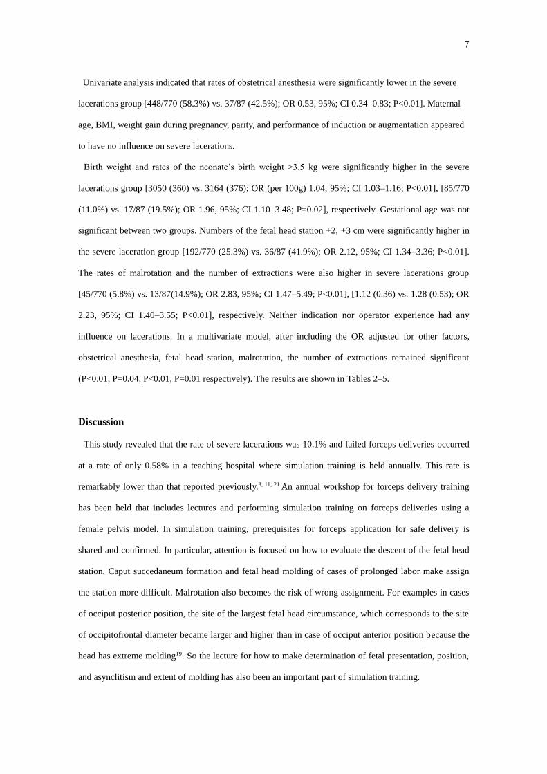
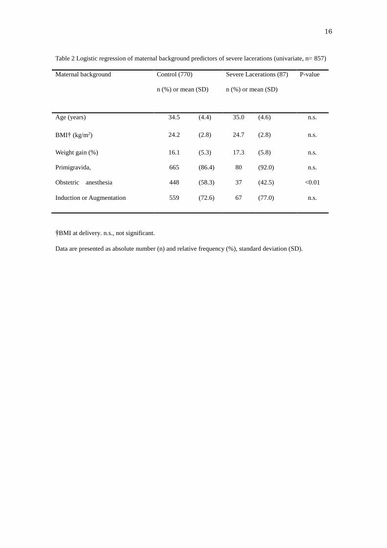
Univariate analysis indicated that rates of obstetrical anesthesia were significantly lower in the severe
lacerations group [448/770 (58.3%) vs. 37/87 (42.5%); OR 0.53, 95%; CI 0.34–0.83; P<0.01]. Maternal
age, BMI, weight gain during pregnancy, parity, and performance of induction or augmentation appeared
to have no influence on severe lacerations.
Birth weight and rates of the neonate’s birth weight >3.5 kg were significantly higher in the severe
lacerations group [3050 (360) vs. 3164 (376); OR (per 100g) 1.04, 95%; CI 1.03–1.16; P<0.01], [85/770
(11.0%) vs. 17/87 (19.5%); OR 1.96, 95%; CI 1.10–3.48; P=0.02], respectively. Gestational age was not
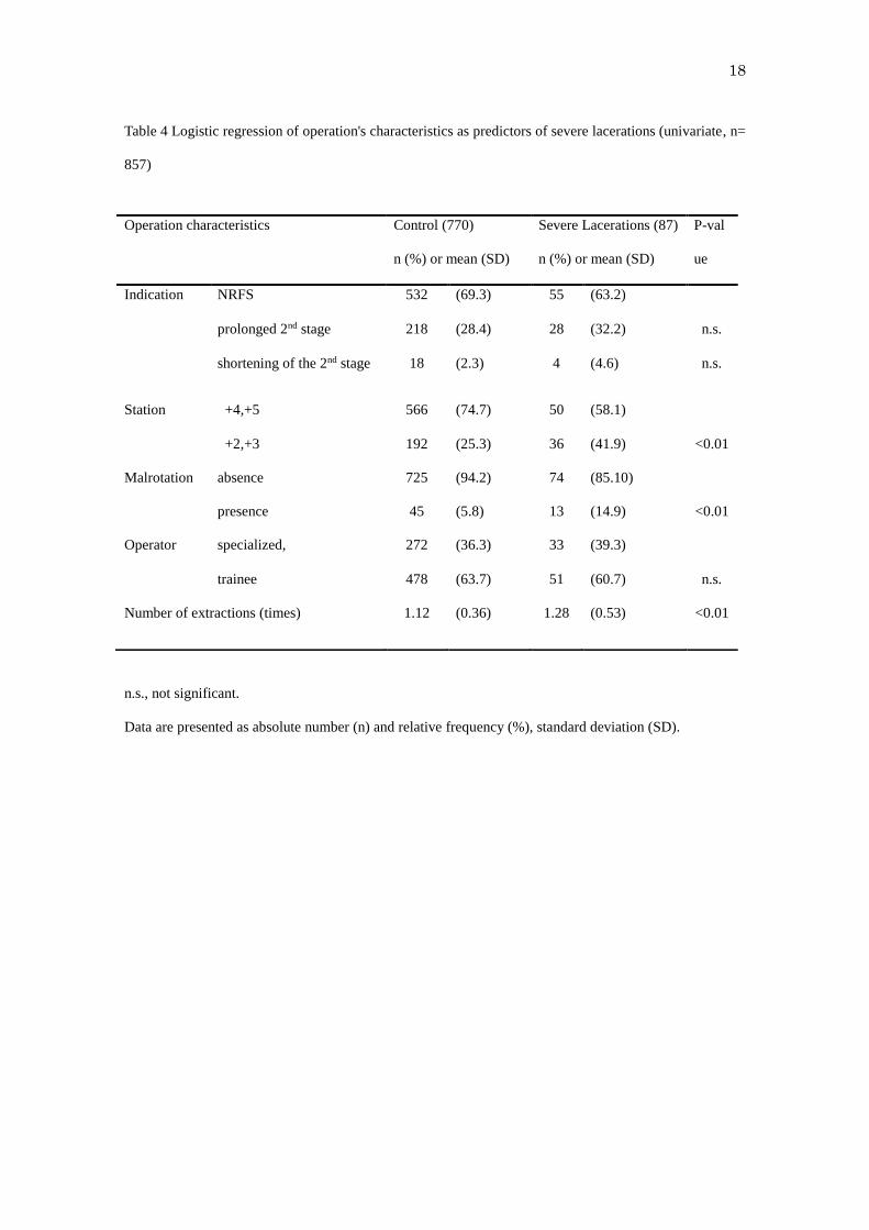
significant between two groups. Numbers of the fetal head station +2, +3 cm were significantly higher in
the severe laceration group [192/770 (25.3%) vs. 36/87 (41.9%); OR 2.12, 95%; CI 1.34–3.36; P<0.01].
The rates of malrotation and the number of extractions were also higher in severe lacerations group
[45/770 (5.8%) vs. 13/87(14.9%); OR 2.83, 95%; CI 1.47–5.49; P<0.01], [1.12 (0.36) vs. 1.28 (0.53); OR
2.23, 95%; CI 1.40–3.55; P<0.01], respectively. Neither indication nor operator experience had any
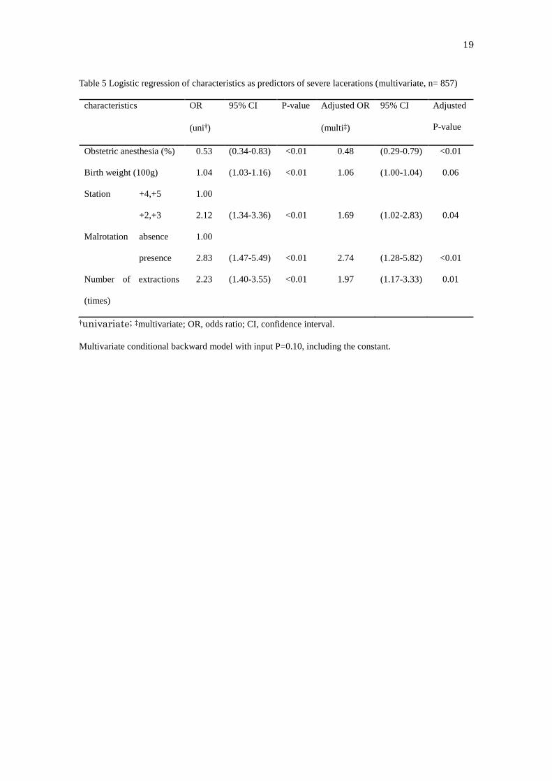
influence on lacerations. In a multivariate model, after including the OR adjusted for other factors,
obstetrical anesthesia, fetal head station, malrotation, the number of extractions remained significant
(P<0.01, P=0.04, P<0.01, P=0.01 respectively). The results are shown in Tables 2–5.
Discussion
This study revealed that the rate of severe lacerations was 10.1% and failed forceps deliveries occurred
at a rate of only 0.58% in a teaching hospital where simulation training is held annually. This rate is
remarkably lower than that reported previously.3, 11, 21 An annual workshop for forceps delivery training
has been held that includes lectures and performing simulation training on forceps deliveries using a
female pelvis model. In simulation training, prerequisites for forceps application for safe delivery is
shared and confirmed. In particular, attention is focused on how to evaluate the descent of the fetal head
station. Caput succedaneum formation and fetal head molding of cases of prolonged labor make assign
the station more difficult. Malrotation also becomes the risk of wrong assignment. For examples in cases
of occiput posterior position, the site of the largest fetal head circumstance, which corresponds to the site
of occipitofrontal diameter became larger and higher than in case of occiput anterior position because the
head has extreme molding19. So the lecture for how to make determination of fetal presentation, position,
and asynclitism and extent of molding has also been an important part of simulation training.

8
Dupuis et al. reported that performing forceps blade placement on a birth simulator allowed obstetricians
to improve their skills.9 Gossett et al. revealed a 22% reduction in severe perineal laceration in delivery
among women managed by residents who had completed forceps simulation training when compared
with women who were managed by residents who did not complete forceps stimulation training.10 Since
the number of forceps have fallen down as the rising rates of cesarean sections, how to teach the skill to
operators in training and perform forceps safely has been serious problems. And the situation is not
peculiar to forceps delivery. Cesarean section is a common operation in obstetrics and gynecology and it
is one of the first operations performed by trainees. However, maternal complications at cesarean section
increase when the primary surgeon is a trainee, Madsen et al. emphasized structured and validated
education and assessment methods should be used.22 Furthermore, managing patient safety and training at
once is common concern not only for the fields of obstetrics and gynecology. Quality of care and patient
safety have become a significant focus of medical practice, Rodriguez et al. has proposed different
training paradigm which integrate knowledge, skill based competencies and a culture of patient safety in
both simulated and real environments.23 As for forceps delivery, effectiveness of simulation training has
been already reported, so the training paradigm which integrate skills performing forceps obtained by
simulation and knowledge not only about prerequisites but also about the risk for misunderstanding the
fetal head station derived from various situations would be important for trainee. Thus simulation training
may be useful to avoid maternal complications, both for trainees who do not have enough experience and
for specialists to review their own methods.
In this study, risk factors for severe lacerations of forceps delivery were identified as birth weight, fetal
head station, malrotation, and the number of extractions. In contrast, we observed that obstetrical
anesthesia might be protective against severe laceration. To the best of our knowledge, this is the first
report to provide data indicating that obstetric anesthesia may be protective against severe laceration.
Risk factors for severe laceration including vaginal delivery with or without OVD have been
investigated in recent years. Birth weight is identified as one of the main factors12–14, and our analysis
supported this theory. Within OVD, previous reports revealed fetal head station and malrotation were
strong risk factors and that decreasing these factors would contribute to fewer severe lacerations.11, 15
Similarly, we found that the fetal head station and the rate of malrotation were significantly higher in the
severe lacerations group, even on multivariate analysis. Malrotations include occiput posterior as well as

9
occiput transverse positions in this study. Both forms of malrotation result in the need for a more forceful
extraction and an associated increase in severe lacerations. Higher stations also require more forceful
extraction than lower stations. In addition, factors of extraction vectors relate to lacerations. Low forceps
require extraction close to perpendicular to the ground before horizontal extraction of the fetal head under
the pubis. Extraction close to perpendicular to the ground can result in more severe lacerations because
the anal sphincter is located under the vagina; thus, higher stations would be considered as posing a risk
for lacerations. Authors also speculate the speed of extraction have possibility to influence the lacerations.
Compared to normal smooth delivery, fetal head descends more rapidly in forceps delivery. It’s one of the
reason to have higher rates of severe lacerations than normal delivery. Among forceps delivery, if operator
extract forceps rapidly more than necessary, it results in more pressure for birth canal thus leads to severe
lacerations. We are usually be careful to extract slowly as if we match the pace which fetal head come
down, but we rely on the senses based on experiences and difficult to explain objectively. And also
evaluation of the speed extraction is really difficult and because this study was retrospective one, we
could not take into account for analysis.
When all vaginal deliveries were analyzed, obstetrical anesthesia seemed to be associated with an
increase in severe lacerations because of increased risk of instrument use.24 Among forceps deliveries, we
found that the rates of obstetrical anesthesia were significantly lower in the severe lacerations group. We
speculate that this is because obstetrical anesthesia relaxes pelvic musculature, which may contribute to
fewer lacerations. Administration of analgesics is also known to be associated with a lower risk of failed
forceps delivery.25 This also suggests that anesthesia may have an indirect effect on pelvic musculature,
making it more relaxed. To identify the role and effect of obstetrical anesthesia on severe lacerations, a
larger number of cases including all vaginal deliveries are needed.
Few studies have analyzed the correlation between the number of extractions and morbidities or other
factors. Ramphul et al. reported that the rates of pulling more than three times were higher in suboptimal
replacement of OVD than in optimal OVD26, indicating that increased extraction derives from suboptimal
replacement. If forceps delivery was not completed in the first extraction, there could be inadequate
pushing, or partially misled station or direction of extraction, and the following extraction would result in
added friction on the vagina, resulting in severe lacerations.
The current study has several limitations. First, as simulation training was held during the entire study

10
period, we could not compare the rates of complications before and after the training began. Second,
current obstetrical practice in this hospital may be associated with lower rates of severe laceration. An
obstetrical specialist highly trained for forceps delivery always stays in the ward. If the trainee is not
experienced enough to handle the forceps delivery, an obstetrician with more experience can replace the
trainee at any time. Similarly, qualifications of the operator did not show any influence in our study, but
there was a tendency of specialists preferring to deliver suspected cases having a higher degree of
difficulty. Thus, we cannot make any conclusions within observed associations because this was
retrospective study under the specific circumstances. Third, as patients who underwent only forceps
delivery were included, we cannot compare our data with data obtained from spontaneous deliveries,
making it more difficult to evaluate the effect of obstetrical anesthesia on lacerations.
Malrotation can be a risk factor not only for severe lacerations but also for failed forceps delivery.
Malrotational forceps deliveries can be managed safely by using Kielland forceps.27–28 Employment of
Kielland forceps may be crucial for safe delivery in these circumstances. Additionally, fetal morbidity and
associated factors need to be clarified in the future, along with maternal morbidity. Therefore, research on
frequency, importance, and associated factors for fetal morbidity would enable the more frequent use
forceps delivery more often and in a safer manner. The results of these proposed studies may encourage
the use of forceps and thus, evaluation of forceps would be reviewed.
In conclusion, in a setting where simulation forceps delivery training is held annually, the rate of severe
lacerations was 10.1% and failed forceps deliveries occurred at a rate of only 0.57%. In this obstetrics
practice, risk factors for severe lacerations in forceps deliveries were revealed as birth weight, fetal head
station, malrotation, and the number of extractions. Obstetrical anesthesia may be protective against
severe lacerations. The accuracy of internal examination, recognition of risk factors, and skill in
extraction techniques obtained through simulation training may contribute to reduce severe lacerations.
Acknowledgements
Authors are very grateful to Masataka Sano (Chiba Institute of Technology) for assistance with data
analysis.
Disclosure

11
None of the authors have any conflicts of interest associated with this study.
References
1. The American College of Obstetricians and Gynecologists. ACOG Practice Bulletin: Operative
vaginal delivery. Obstet gynecol 2015; 126(5): e56–65.
2. O'mahony F, Hofmeyr GJ, Menon V. Choice of instruments for assisted vaginal delivery. Cochrane
Database Syst Rev 2010. DOI: 10.1002/14651858.CD005455.pub2.
3. Caughey AB, Sandberg PL, Zlatnik MG, Thiet MP, Parer JT, Laros RK. Forceps compared with
vacuum: rates of neonatal and maternal morbidity. Obstet Gynecol 2005; 106(5): 908–912
4. Homsi R, Daikoku NH, Littlejohn J, Wheeless CR. Episiotomy: risks of dehiscence and rectovaginal
fistula. Obstet Gynecol Surv 1994; 49(12): 803–808.
5. Patel RR, Murphy DJ. Forceps delivery in modern obstetric practice. BMJ 2004; 328: 1302–1305.
6. Powell J, Gilo N, Foote M, Gil K, Lavin JP. Vacuum and forceps training in residency: experience
and self-reported competency. J Perinatol 2007; 27: 343–346.
7. Dildy GA, Belfort MA, Clark SL. Obstetric Forceps: A Species on the Brink of Extinction. Obstet
Gynecol 2016; 128(3): 436–439.
8. Unno N, Masuzaki H, Kanayama N, Kubo T, Fujimori K, Matsuda Y. Annual report of perinatology
committee. Acta obstetrica et gynaecologica Japonica 2013; 65(6): 1377–1419. (Jpn).
9. Dupuis O, Decullier E, Clerc J, et al. (2011) Does forceps training on a birth simulator allow
obstetricians to improve forceps blade placement? Eur J Obstet Gynecol Reprod Biol 2011; 159(2):
305–309.
10. Gossett DR, Gilchrist-scott D, Wayne DB, Gerber SE. Simulation Training for Forceps-Assisted
Vaginal Delivery and Rates of Maternal Perineal Trauma. Obstet Gynecol 2016; 128(3): 429–435.
11. Miller ES, Barber EL, Mcdonald KD, Gossett DR. Association between obstetrician forceps volume
and maternal and neonatal outcomes. Obstet Gynecol 2014; 123(2): 248–254.
12. Vale de castro monteiro M, Pereira GM, Aguiar RA, Azevedo RL, Correia-junior MD, Reis ZS. Risk
factors for severe obstetric perineal lacerations. Int Urogynecol J Pelvic Floor Dysfunct 2016; 27(1):
61–67.
13. Meister MR, Cahill AG, Conner SN, Woolfolk CL, Lowder JL. Predicting obstetric anal sphincter

12
injuries in a modern obstetric population. Am J Obstet Gynecol 2016; 215(3): 310.e1–7.
14. Pergialiotis V, Vlachos D, Protopapas A, Pappa K, Vlachos G. Risk factors for severe perineal
lacerations during childbirth. Int J Gynaecol Obstet 2014; 125(1): 6–14.
15. Hirsch E, Elue R, Wagner A, et al. Severe perineal laceration during operative vaginal delivery: the
impact of occiput posterior position. J Perinatol 2014; 34(12): 898–900.
16. Cunningham F, Leveno KJ, Bloom SL, et al. Operative vaginal delivery. In Williams Obstetrics, 24th
edition. New York: McGraw-Hill Education, 2014; 574–586.
17. Matsumoto N, Takenaka T, Ikeda N, Yazaki S, Sato Y. Naegele Forceps Delivery and Association
between Morbidity and the Number of Forceps Traction Applications: A Retrospective Study. J
Pregnancy 2015. DOI:10.1155/2015/483195
18. Minakami H, Hiramatsu Y, Koresawa M, et al. Guidelines for obstetrical practice in Japan: Japan
Society of Obstetrics and Gynecology (JSOG) and Japan Association of Obstetricians and
Gynecologists (JAOG) 2011 edition. J Obstet Gynecol Res 2011; 37(9): 1174–1197.
19. TakedaS, TakedaJ, KoshiishiT, et al. Fetal station based on the trapezoidal plane and assessment of
head descent during instrumental delivery. Hypertension Research in Pregnancy
DOI:10.14390/jsshp.2.65
20. Hagadorn-freathy AS, Yeomans ER, Hankins GD. Validation of the 1988 ACOG forceps
classification system. Obstet Gynecol 1991; 77(3): 356–360.
21. Langeron A, Mercier G, Chauleur C, et al. Failed forceps extraction: risk factors and maternal and
neonatal morbidity. J Gynecol Obstet Biol Reprod (Paris) 2012; 41(4): 333–338.
22. Madsen K, Grønbeck L, Rifbjerg larsen C, et al. Educational strategies in performing cesarean
section. Acta Obstet Gynecol Scand 2013; 92(3): 256-263.
23. Rodriguez-paz JM, Kennedy M, Salas E, et al. Beyond "see one, do one, teach one": toward a
different training paradigm. Qual Saf Health Care 2009; 18(1): 63-68.
24. Carroll TG, Engelken M, Mosier MC, Nazir N. Epidural analgesia and severe perineal laceration in a
community-based obstetric practice. J Am Board Fam Pract 2003; 16(1): 1–6.
25. Ben-haroush A, Melamed N, Kaplan B, Yogev Y. Predictors of failed operative vaginal delivery: a
single-center experience. Am J Obstet Gynecol 2007; 197(3): 308.e1–5.
26. Ramphul M, Kennelly MM, Burke G, Murphy DJ. Risk factors and morbidity associated with

13
suboptimal instrument placement at instrumental delivery: observational study nested within the
Instrumental Delivery & Ultrasound randomised controlled trial ISRCTN 72230496. BJOG 2015;
122(4): 558–563.
27. Tempest N, Hart A, Walkinshaw S, Hapangama DK. A re-evaluation of the role of rotational forceps:
retrospective comparison of maternal and perinatal outcomes following different methods of birth for
malposition in the second stage of labour. BJOG 2013; 120(10): 1277–1284.
28. Bahl R, Van de venne M, Macleod M, Strachan B, Murphy DJ. Maternal and neonatal morbidity in
relation to the instrument used for mid-cavity rotational operative vaginal delivery: a prospective
cohort study. BJOG 2013; 120(12): 1526–1532.

14
Figure legends
Fig. 1 Image of UTokyo Naegele forceps is shown. This forceps have fenestrated blades and adequate
curves for pelvis and fetal head adjustments which was modified lighter weight and shorter length with
thinner blades from original Naegele forceps adapted for Japanese women.
Fig. 2 Flowchart showing selection criteria of patients who delivered in the hospital during study period
OVD; operated vaginal delivery

15
Table1 Overall clinical and obstetrics characteristics of cases successfully delivered by forceps at term
Clinical and obstetrics characteristics n (%) or mean (SD)
Maternal age (years) 34.6 (4.45)
BMI at delivery (kg/㎡) 24.3 (1.12 )
Weight gain (%) 16.2 (5.32)
Primigravida 745 (86.9)
Obstetrics anesthesia 485 (56.6)
Induction or augmentation 621 (72.5)
Birthweight (g) 3061.6 (363.2)
Gestational age (weeks) 39.7 (1.12)
Indication of NRFS 587 (68.4)
Indication of prolonged 2nd stage 246 (28.7)
Indication of shortening of the 2nd stage 22 (2.5)
Malrotation 58 (6.8)
Operator specialized 305 (35.6)
Operator trainee 529 (61.8)
Data are showed in terms of absolute number (n) and relative frequency (%), standard deviation (SD).
BMI, body mass index; NRFS, nonreassuring fetal status.
16
Table 2 Logistic regression of maternal background predictors of severe lacerations (univariate, n= 857)
Maternal background Control (770)
n (%) or mean (SD)
Severe Lacerations (87)
n (%) or mean (SD)
P-value
Age (years) 34.5 (4.4) 35.0 (4.6) n.s.
BMI† (kg/m2) 24.2 (2.8) 24.7 (2.8) n.s.
Weight gain (%) 16.1 (5.3) 17.3 (5.8) n.s.
Primigravida, 665 (86.4) 80 (92.0) n.s.
Obstetric anesthesia 448 (58.3) 37 (42.5) <0.01
Induction or Augmentation 559 (72.6) 67 (77.0) n.s.
†BMI at delivery. n.s., not significant.
Data are presented as absolute number (n) and relative frequency (%), standard deviation (SD).

17
Table 3 Logistic regression of obstetrical characteristics as predictors of severe lacerations (univariate n=
857)
n.s., not significant.
Data are presented as absolute number (n) and relative frequency (%), standard deviation (SD).
Obstetrical characteristics Control (770)
n (%) or mean (SD)
Severe Lacerations (87)
n (%) or mean (SD)
P-value
Birth weight (g) 3050 (360) 3164 (376) <0.01
Birth weight
≥3.5 (kg) 85 (11) 17 (19.5) 0.02
<3.5 (kg) 685 (89) 70 (80.5)
Gestational age, weeks (SD) 39.7 (1.1) 40 (1.1) n.s.
18
Table 4 Logistic regression of operation's characteristics as predictors of severe lacerations (univariate, n=
857)
n.s., not significant.
Data are presented as absolute number (n) and relative frequency (%), standard deviation (SD).
Operation characteristics Control (770)
n (%) or mean (SD)
Severe Lacerations (87)
n (%) or mean (SD)
P-val
ue
Indication
NRFS 532 (69.3) 55 (63.2)
prolonged 2nd stage 218 (28.4) 28 (32.2) n.s.
shortening of the 2nd stage 18 (2.3) 4 (4.6) n.s.
Station
+4,+5 566 (74.7) 50 (58.1)
+2,+3 192 (25.3) 36 (41.9) <0.01
Malrotation
absence 725 (94.2) 74 (85.10)
presence 45 (5.8) 13 (14.9) <0.01
Operator
specialized, 272 (36.3) 33 (39.3)
trainee 478 (63.7) 51 (60.7) n.s.
Number of extractions (times) 1.12 (0.36) 1.28 (0.53) <0.01
19
Table 5 Logistic regression of characteristics as predictors of severe lacerations (multivariate, n= 857)
characteristics OR
(uni†)
95% CI P-value Adjusted OR
(multi‡)
95% CI Adjusted
P-value
Obstetric anesthesia (%) 0.53 (0.34-0.83) <0.01 0.48 (0.29-0.79) <0.01
Birth weight (100g) 1.04 (1.03-1.16) <0.01 1.06 (1.00-1.04) 0.06
Station +4,+5 1.00
+2,+3 2.12 (1.34-3.36) <0.01 1.69 (1.02-2.83) 0.04
Malrotation
absence 1.00
presence 2.83 (1.47-5.49) <0.01 2.74 (1.28-5.82) <0.01
Number of extractions
(times)
2.23 (1.40-3.55) <0.01 1.97 (1.17-3.33) 0.01
†univariate; ‡multivariate; OR, odds ratio; CI, confidence interval.
Multivariate conditional backward model with input P=0.10, including the constant.

20
Fig. 1

21
Fig. 2
Total deliveries
(n= 5,588)
Attempted vaginal deliveries
(n= 3,351)
Attempted forceps deliveries
(n= 878)
Final sample
(n= 857)
Preterm forceps
<37 weeks gestation
(n= 16)
Severe lacerations
(n= 87)
Control
(n= 770)
Cesarean sections
(n= 2,237)
Failed forceps →cesarean sections
(n= 5)
Vacuum extractions
(n= 18)
Attempted forceps deliveries at term
(n= 862)
Without OVD
(n= 2,455)