the influence of growth hormone&insulin-like growth factor
TRANSCRIPT
ORIGINAL ARTICLE
The influence of growth hormone/insulin-like growth factordeficiency on prostatic dysplasia in pbARR2-Cre, PTEN knockoutmiceK Takahara1,2,6, N Ibuki1,6, M Ghaffari1,3, H Tearle1, CJ Ong1, H Azuma2, ME Gleave1,4, M Pollak5 and ME Cox1,4
BACKGROUND: Elevated insulin-like growth factor-I (IGF-I) serum levels and phosphatase and tensin homolog (PTEN) lossare prostate cancer (PCa) risk factors that enhance androgen-responsive and castration-resistant PCa xenografts growth.METHODS: The impact of suppressed growth hormone (GH)/IGF-I levels on neoplastic initiation of PTEN-deficient prostateepithelia was assessed histologically and by epithelial-to-mesenchymal marker expression in Ghrhr D60G homozygous (lit/lit)and heterozygous (lit/þ ) pbARR2-Cre, PTEN(fl/fl) (PTEN� /� ) mice. How suppressed GH/IGF-I levels impacted growth ofPTEN� /� mouse-derived prostate cells (MPPK) was examined by growth and survival signaling of cells cultured in lit/þ orlit/lit serum.RESULTS: Body weight, prostate weight and serum GH and IGF-I levels were reduced in lit/lit relative to lit/þ PTEN� /�littermates. While the anterior lobes of lit/þ PTEN� /� prostates consistently presented swollen, indicative of ductal blockage, thedegree of prostatic dysplasia in 15- and 20-week-old lit/lit and lit/þ PTEN� /� mice was indistinguishable as measured bynormalized prostatic weight, tissue histology, or probasin, PSP94, E-cadherin, N-cadherin and vimentin expression. However, growthand AKT activation of MPPK cells was decreased when cultured in lit/lit serum as compared with lit/þ serum and restored inlit/lit serum supplemented with IGF-I and, to a lesser extent, GH.CONCLUSIONS: These results suggest that initiation of prostate carcinogenesis by loss of PTEN is not influenced by germlinevariation of genes encoding signaling molecules in the GH/IGF-I axis, but suggests that these factors may affect the progression ofdysplastic phenotype and supports previous studies, indicating that the GH/IGF milieu does impact the growth of PTEN-deficientdysplastic prostatic cells once transformed.
Prostate Cancer and Prostatic Disease advance online publication, 21 May 2013; doi:10.1038/pcan.2013.14
Keywords: prostatic intraepithelial neoplasia; transgenic mouse; endocrine hormone; phosphatase tensin homolog; growthhormone receptor hormone
INTRODUCTIONThe growth hormone and insulin-like growth factor-I (GH/IGF-I)axis is an important regulator of growth, survival and metastaticpotential in a variety of malignancies and is strongly implicated inprostate cancer (PCa) etiology and risk.1–4 Perturbations in intrinsicexpression of IGF axis components by tumor cells are implicatedin susceptibility and progression of PCa.5–12 IGF-I receptor (IGF-1R)expression is elevated in metastatic PCa13,14 and castration-resistant disease progression is associated with increasedexpression of IGF-I and IGF-1R.15 Furthermore, maintaining IGF-Iresponsiveness facilitates PCa survival and growth, and is achievedthrough androgen-modulated IGF-1R) expression.14,16,17
IGF-I is a potent mitogen and antiapoptotic factor predo-minantly produced by the liver in response to GH signaling.18
Ligand activation of IGF-1R results in the activation of multipleintracellular signaling pathways, including RAS/extracellular signal-regulated kinases (ERKs) 1 and 2 and phosphatidylinositol-3 kinase(PI3K)/AKT that control the various IGF-mediated biological
effects.19 Activating mutations of PI3K have also been linkedto PCa progression,20 indicating the importance of deregulatedPI3K signaling in PCa. The phosphatidylinositol phosphatasetumor suppressor, PTEN, is an important negative regulator ofPI3K signaling. Homozygous loss of PTEN is a recurrent event inadvanced PCa,21 and among those patients who are not PTEN null,many exhibit loss of one PTEN allele.22 We have previouslyreported that the GH/IGF-I axis is important for androgen-respon-sive growth, castration-resistant progression and growth ofandrogen receptor (AR)-negative, PTEN-deficient PCa xenograftsin the lit/lit GH-releasing hormone-R loss-of-function murinehost.23 In addition, hemizygous PTEN loss, alone and in combina-tion with the presence of TMPRSS2-ERG gene rearrangements, arereported in early stages of disease and to predict an increased riskof biochemical progression.24,25 This prediction is supported bythe demonstration that a murine model for prostate-conditionalPTEN loss is sufficient to induce development26 and castration-resistant progression27 of PCa. These results indicate that PTEN
1The Vancouver Prostate Centre, Vancouver General Hospital, Vancouver, British Columbia, Canada; 2Department of Urology, Osaka Medical College, Osaka, Japan; 3Departmentof Medicine, University of British Columbia, Vancouver, British Columbia, Canada; 4Department of Urologic Sciences, University of British Columbia, Vancouver, British Columbia,Canada and 5Department of Medicine and Oncology, McGill University, Montreal, Quebec, Canada. Correspondence: Dr ME Cox, The Vancouver Prostate Centre, VancouverGeneral Hospital, 2660 Oak Street, Vancouver, British Columbia, Canada V6H 3Z6.E-mail: [email protected] authors contributed equally to this work.Received 17 January 2013; revised 12 March 2013; accepted 30 March 2013
Prostate Cancer and Prostatic Disease (2013), 1–9& 2013 Macmillan Publishers Limited All rights reserved 1365-7852/13
www.nature.com/pcan
loss can hypersensitize cells to PI3K activation by factors such asGH and IGF-I.
To test the hypothesis that dysplastic initiation of PTEN-deficient prostate epithelial cells may be due to hypersensitivityto GH/IGF-mediated PI3K signaling, we assessed development ofprostatic dysplasia in Ghrhr D60G (lit)� pbARR2-Cre, PTEN(fl/fl)mice (PTEN� /� ). Our in vivo and in vitro results suggest thatprostatic dysplasia induced by PTEN loss is not substantiallyaffected by Ghrhr deficiency, but subsequent growth of dysplasticepithelia is impaired in hosts carrying germline deficiencies for GHand IGF-I expression, and may affect the progression of dysplasticphenotype in PCa.
MATERIALS AND METHODSGhrhr(lit/lit) and (lit/þ ) pbARR2-Cre/PTEN(fl/fl) mousecharacterizationPTEN(fl/fl) C57BL/6J mice were obtained from T. Mak (Toronto, Canada)28
pbARR2-Cre created in a C57BL/6�DBA2 hybrid mouse strain wasobtained from P. Roy-Berman (Los Angeles, CA)29 and Ghrhr D60G (lit/lit)C57BL/6J mice were from Jackson Laboratory (Bar Harbor, ME).30 pbARR2-Cre, PTEN(fl/fl) mice (PTEN� /� ) generated, as described previously,31
were crossed with lit/lit mice to produce lit/lit and lit/þ PTEN� /� mice.This transgenic strain was genotyped to confirm the status of the Ghrhrallele mutation as described previously,23 as well as confirming Crerecombinase and PTEN(fl/fl) status from tail clip DNA extracted usingDirectPCR Lysis Reagent (Viagen Biotech, Los Angeles, CA, USA) andmaintained by backcrossing. The extent of prostatic dysplasia was assessedin lit/lit and lit/þ PTEN� /� mice at 15 and 20 weeks of age (10 mice percohort). Prostate weights were determined after dissection and lancing ofthe lit/þ anterior lobes to remove retained fluid. Prostate pathology wasscored from formalin-fixed, paraffin-embedded, hematoxylin- and eosin-stained specimens by a blinded pathologist (L Fazli). Probasin, PSP94,E-cadherin, N-cadherin and vimentin transcript levels were measured fromtotal RNA prepared from flash-frozen total prostates by quantitative real-time polymerase chain reaction normalized to murine b-actin transcript foreach specimen. Male mice were harvested in accordance with theguidelines of the Canadian Council on Animal Care and with appropriateinstitutional certification.
Cell linesMPPK cells were established from prostate tissue from a 40-week-oldpbARR2-Cre, PTEN(fl/fl) male mice as described31 and maintained inDulbecco’s modified Eagle’s medium supplemented with 10% fetal bovineserum. LNCaP cells (courtesy of Dr LWK Chung, City of Hope Hospital,Los Angeles, CA, USA) were maintained in RPMI1640þ 5% fetal bovineserum and DU145 cells purchased from American Type Culture Collection(Rockville, MD, USA) were maintained in Dulbecco’s modified Eagle’smediumþ 5% fetal bovine serum. Cells were cultured at 37 1C in ahumidified 5% CO2 atmosphere with media supplies purchased from LifeTechnologies (Burlington, Ontario, Canada).
In vitro cell proliferation assayMPPK cells were cultured in lit/lit and lit/þ serum and in lit/lit serumsupplemented with recombinant murine GH (R&D Systems, Minneapolis,MN, USA) or IGF-I (Calbiochem, San Diego, CA, USA) at the indicatedconcentrations. Proliferation was measured by pulse-labeling cells for2 h with 10 mM bromodeoxyuridine (BrdU) after 46 h in culture. DenaturedDNA in fixed cells was stained with a peroxidase-coupled anti-BrdU-antibody and peroxidase substrate (tetramethylbenzidine) for 30 min andBrdU incorporation was measured by densitometry (OD 370 nm). Eachassay was performed in triplicate.
Immunoblot analysisWhole-cell protein lysates of LNCaP, DU145 and MPPK cells cultured inserum-free RPMI-1640 for 2 days±1 nM methyltrienolone, a syntheticandrogen (R1881; Perkin-Elmer, Waltham MA, USA), were prepared in RIPAbuffer (50 mM Tris-HCl, pH 7.2, 1% NP-40, 0.1% deoxycholate, 0.1% sodiumdodecyl sulfate, 100 mM NaCl, 1� Roche complete protease inhibitorcocktail), subjected to sodium dodecyl sulfate-polyacrylamide gel electro-phoresis and transferred to nitrocellulose filters for immunoblotting with
primary antibody (Ab): anti-AR and anti-vimentin rabbit polyclonal Ab(Santa Cruz Biotechnology, Santa Cruz, CA, USA), anti-PTEN rabbitpolyclonal Ab (Cell Signaling Technology, Danvers, MA, USA), anti-E-cadherin and anti-N-cadherin mouse monoclonal Ab (BD Biosciences,San Jose, CA, USA) and anti-b-actin mouse monoclonal Ab (MilliporeCorporation, Billerica, MA, USA). Immunoreactive proteins were detectedby enhanced chemiluminescence western blotting analysis system(Amersham Life Science, Arlington Heights, IL, USA), using horseradishperoxide-conjugated anti-rabbit or anti-mouse IgG Ab (Santa Cruz).
Whole-cell lysates of MPPK cells cultured in media supplemented with1% lit/lit or 1% lit/þ mouse serum for 2 days, or cultured 24 h in serum-free media, washed and then stimulated with media containing 1% lit/litserum containing the indicated concentrations of recombinant murine GHor IGF-I for 20 min were immunoblotted with anti-vinculin mousemonoclonal Ab (Sigma Chemical, St Louis, MO, USA), anti-phospho-AKT(S473) rabbit polyclonal Ab, anti-AKT rabbit polyclonal Ab, anti-phospho-ERK1/2 (T185/Y187 and T202/Y204, respectively) rabbit polyclonal Ab, anti-ERK1/2 rabbit polyclonal Ab (all from Cell Signaling Technology) anddetected by enhanced chemiluminescent as above.
Statistical analysisData were analyzed by analysis of variance and Mann–Whitney post hoctest, except for paired tests that were analyzed by Student’s t-test. Levels ofstatistical significance were set at Po0.05.
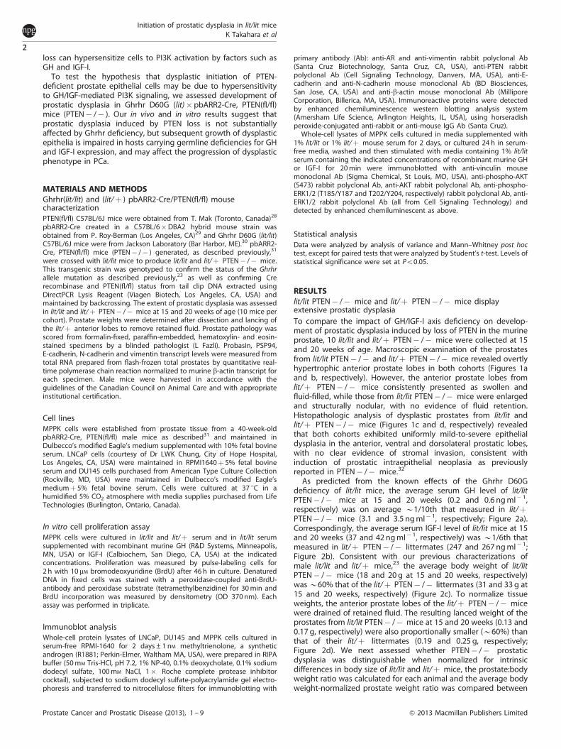
RESULTSlit/lit PTEN� /� mice and lit/þ PTEN� /� mice displayextensive prostatic dysplasiaTo compare the impact of GH/IGF-I axis deficiency on develop-ment of prostatic dysplasia induced by loss of PTEN in the murineprostate, 10 lit/lit and lit/þ PTEN� /� mice were collected at 15and 20 weeks of age. Macroscopic examination of the prostatesfrom lit/lit PTEN� /� and lit/þ PTEN� /� mice revealed overtlyhypertrophic anterior prostate lobes in both cohorts (Figures 1aand b, respectively). However, the anterior prostate lobes fromlit/þ PTEN� /� mice consistently presented as swollen andfluid-filled, while those from lit/lit PTEN� /� mice were enlargedand structurally nodular, with no evidence of fluid retention.Histopathologic analysis of dysplastic prostates from lit/lit andlit/þ PTEN� /� mice (Figures 1c and d, respectively) revealedthat both cohorts exhibited uniformly mild-to-severe epithelialdysplasia in the anterior, ventral and dorsolateral prostatic lobes,with no clear evidence of stromal invasion, consistent withinduction of prostatic intraepithelial neoplasia as previouslyreported in PTEN� /� mice.32
As predicted from the known effects of the Ghrhr D60Gdeficiency of lit/lit mice, the average serum GH level of lit/litPTEN� /� mice at 15 and 20 weeks (0.2 and 0.6 ng ml� 1,respectively) was on average B1/10th that measured in lit/þPTEN� /� mice (3.1 and 3.5 ng ml� 1, respectively; Figure 2a).Correspondingly, the average serum IGF-I level of lit/lit mice at 15and 20 weeks (37 and 42 ng ml� 1, respectively) was B1/6th thatmeasured in lit/þ PTEN� /� littermates (247 and 267 ng ml� 1;Figure 2b). Consistent with our previous characterizations ofmale lit/lit and lit/þ mice,23 the average body weight of lit/litPTEN� /� mice (18 and 20 g at 15 and 20 weeks, respectively)was B60% that of the lit/þ PTEN� /� littermates (31 and 33 g at15 and 20 weeks, respectively) (Figure 2c). To normalize tissueweights, the anterior prostate lobes of the lit/þ PTEN� /� micewere drained of retained fluid. The resulting lanced weight of theprostates from lit/lit PTEN� /� mice at 15 and 20 weeks (0.13 and0.17 g, respectively) were also proportionally smaller (B60%) thanthat of their lit/þ littermates (0.19 and 0.25 g, respectively;Figure 2d). We next assessed whether PTEN� /� prostaticdysplasia was distinguishable when normalized for intrinsicdifferences in body size of lit/lit and lit/þ mice, the prostate:bodyweight ratio was calculated for each animal and the average bodyweight-normalized prostate weight ratio was compared between
Initiation of prostatic dysplasia in lit/lit miceK Takahara et al
2
Prostate Cancer and Prostatic Disease (2013), 1 – 9 & 2013 Macmillan Publishers Limited
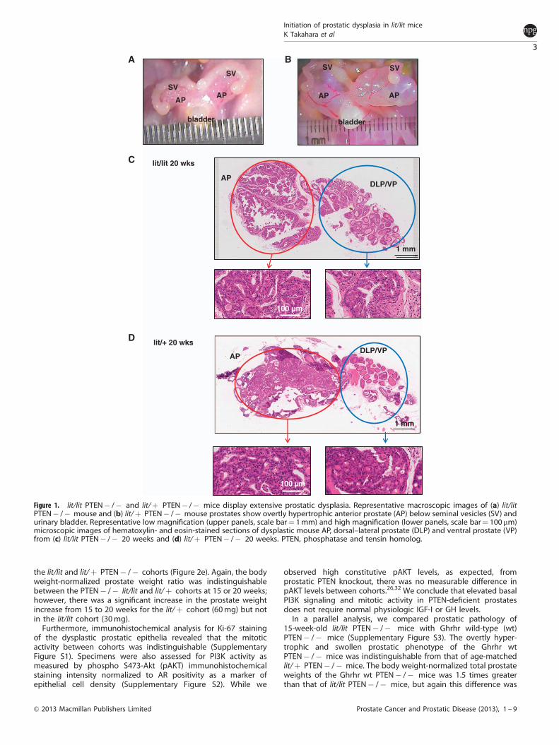
the lit/lit and lit/þ PTEN� /� cohorts (Figure 2e). Again, the bodyweight-normalized prostate weight ratio was indistinguishablebetween the PTEN� /� lit/lit and lit/þ cohorts at 15 or 20 weeks;however, there was a significant increase in the prostate weightincrease from 15 to 20 weeks for the lit/þ cohort (60 mg) but notin the lit/lit cohort (30 mg).
Furthermore, immunohistochemical analysis for Ki-67 stainingof the dysplastic prostatic epithelia revealed that the mitoticactivity between cohorts was indistinguishable (SupplementaryFigure S1). Specimens were also assessed for PI3K activity asmeasured by phospho S473-Akt (pAKT) immunohistochemicalstaining intensity normalized to AR positivity as a marker ofepithelial cell density (Supplementary Figure S2). While we
observed high constitutive pAKT levels, as expected, fromprostatic PTEN knockout, there was no measurable difference inpAKT levels between cohorts.26,32 We conclude that elevated basalPI3K signaling and mitotic activity in PTEN-deficient prostatesdoes not require normal physiologic IGF-I or GH levels.
In a parallel analysis, we compared prostatic pathology of15-week-old lit/lit PTEN� /� mice with Ghrhr wild-type (wt)PTEN� /� mice (Supplementary Figure S3). The overtly hyper-trophic and swollen prostatic phenotype of the Ghrhr wtPTEN� /� mice was indistinguishable from that of age-matchedlit/þ PTEN� /� mice. The body weight-normalized total prostateweights of the Ghrhr wt PTEN� /� mice was 1.5 times greaterthan that of lit/lit PTEN� /� mice, but again this difference was
A
bladder
APAP
SV
SV
B
C
APDLP/VP
lit/lit 20 wks
1 mm
100 µm
APDLP/VP
1 mm
lit/+ 20 wks
100 µm
D
SVSV
AP AP
bladder
Figure 1. lit/lit PTEN� /� and lit/þ PTEN� /� mice display extensive prostatic dysplasia. Representative macroscopic images of (a) lit/litPTEN� /� mouse and (b) lit/þ PTEN� /� mouse prostates show overtly hypertrophic anterior prostate (AP) below seminal vesicles (SV) andurinary bladder. Representative low magnification (upper panels, scale bar¼ 1mm) and high magnification (lower panels, scale bar¼ 100 mm)microscopic images of hematoxylin- and eosin-stained sections of dysplastic mouse AP, dorsal–lateral prostate (DLP) and ventral prostate (VP)from (c) lit/lit PTEN� /� 20 weeks and (d) lit/þ PTEN� /� 20 weeks. PTEN, phosphatase and tensin homolog.
Initiation of prostatic dysplasia in lit/lit miceK Takahara et al
3
& 2013 Macmillan Publishers Limited Prostate Cancer and Prostatic Disease (2013), 1 – 9
not seen when the Ghrhr wt PTEN� /� prostates were lanced todrain retained fluid. These results indicate that the lack of adifference between the lit/lit and lit/þ prostate pathology was notdue to a suppressed effect in response to Ghrhr D60Gheterozygosity.
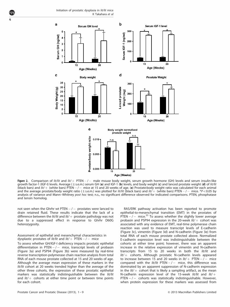
Assessment of epithelial and mesenchymal characteristics indysplastic prostates of lit/lit and lit/þ PTEN� /� miceTo assess whether GH/IGF-I deficiency impacts prostatic epithelialdifferentiation in PTEN� /� mice, transcript levels of probasin(Figure 3a) and PSP94 (Figure 3b) were measured by real-timereverse transcription-polymerase chain reaction analysis from totalRNA of each mouse prostate collected at 15 and 20 weeks of age.Although the average mean expression of these markers in thelit/lit cohort at 20 weeks trended higher than the average of theother three cohorts, the expression of these prostatic epithelialmarkers was statistically indistinguishable between the lit/litand lit/þ cohorts at either time point or between time pointsfor each cohort.
RAS/ERK pathway activation has been reported to promoteepithelial-to-mesenchymal transition (EMT) in the prostates ofPTEN� /� mice.33 To assess whether the slightly lower averageprobasin and PSP94 expression in the 20-week lit/þ cohort wasassociated with any evidence of EMT, real-time polymerase chainreaction was used to measure transcript levels of E-cadherin(Figure 3c), vimentin (Figure 3d) and N-cadherin (Figure 3e) fromtotal RNA of each mouse prostate collected above. NormalizedE-cadherin expression level was indistinguishable between thecohorts at either time point; however, there was an apparentincrease in the relative expression of vimentin and N-cadherintranscripts from 15 to 20 weeks in both the lit/lit andlit/þ cohorts. Although prostatic N-cadherin levels appearedto increase between 15 and 20 weeks in lit/þ PTEN� /� micecompared with the lit/lit PTEN� /� mice, this difference wasdominated by an apparent suppression of N-cadherin expressionin the lit/þ cohort that is likely a sampling artifact, as the meanN-cadherin expression level of the 15-week lit/lit and lit/þPTEN� /� cohorts was statistically indistinguishable. However,when protein expression for these markers was assessed from
Figure 2. Comparison of lit/lit and lit/þ PTEN� /� male mouse body weight, serum growth hormone (GH) levels and serum insulin-likegrowth factor-1 (IGF-I) levels. Average (±s.e.m.) serum GH (a) and IGF-1 (b) levels, and body weight (c) and lanced prostate weight (d) of lit/lit(black bars) and lit/þ (white bars) PTEN� /� mice at 15 and 20 weeks of age. (e) Prostate:body weight ratio was calculated for each animaland the average prostate/body weight ratio (±s.e.m.) was plotted for lit/lit (black bars) and lit/þ (white bars) PTEN� /� mice. *Po0.05 byanalysis of variance and Mann–Whitney post hoc test; n.s., no significant difference observed for indicated comparisons. PTEN, phosphataseand tensin homolog.
Initiation of prostatic dysplasia in lit/lit miceK Takahara et al
4
Prostate Cancer and Prostatic Disease (2013), 1 – 9 & 2013 Macmillan Publishers Limited

prostatic lysates by immunoblotting normalized to b-actin levels,these trends for increased vimentin and N-cadherin expressionin the 20-week cohorts were not confirmed (SupplementaryFigure S4).
We conclude that although the consistently observed swellingof the anterior prostate lobes in the lit/þ PTEN� /� mouse,indicative of blocked terminal prostatic ducts in these animals,suggests that dysplastic progression is delayed in the lit/lit hosts,morphologic and molecular marker expression changes indicatethat genomic alterations that result in suppressed GH/IGF-I axissignaling do not affect initiation of prostatic dysplasia because ofPTEN deletion.
Characterization of EMT marker expression in MPPK cellsWe next assessed whether the previously established PTEN� /�mouse prostate cell line, MPPK, harbored any of the EMTcharacteristics implicated above by characterizing them forexpression of prostatic epithelial and mesenchymal markersrelative to that observed in well-characterized, androgen-respon-sive human prostate cancer cell line, LNCaP, and androgen-independent, AR-negative human prostate cancer cell line, DU145(Figure 4a). MPPK cells expressed AR and this expression was
enhanced in the presence of R1881. Androgen responsiveness wasconfirmed by firefly luciferase reporter assay in MPPK cellstransfected with pbARR2-Luc and stimulated 24 h with R1881(Figure 4b). MPPK cells were also confirmed to be deficient forPTEN expression by comparison to expression in the PTEN wtDU145 cells and the PTEN-null LNCaP cells. MPPK cells exhibitedrobust E-cadherin expression, comparable to that observedin LNCaP cells. While MPPK cells expressed vimentin at lowlevels, comparable to that of LNCaP cells, relative to the robustexpression in DU145 cells, they did express high levels ofN-cadherin not observed in LNCaP and DU145 cells. Thisobservation is consistent with the suggestion that PTEN� /�prostatic epithelial cells acquire intermediary mesenchymalcharacteristics while still in an intraepithelial neoplastic state andindicate that the MPPK cell line accurately reflects the phenotypeof the dysplastic prostatic epithelia of PTEN� /� cells.
Growth of MPPK cells is suppressed in media supplemented withlit/lit serumTo determine if growth characteristics of MPPK cells were sensitiveto changes in GH/IGF-I levels, relative proliferation of MPPK cellscultured in basal mediaþ 1% lit/lit serum or 1% lit/þ serum was
Figure 3. Expression of prostatic differentiation markers and evidence of EMT in PTEN� /� lit/lit and lit/þ mice. Real-time reverse transcription-polymerase chain reaction was used to analyze probasin (a) and PSP94 (b) E-cadherin (c), vimentin (d) and N-cadherin (e) mRNA from totalprostate RNA collected at 15 and 20 weeks of age. Scatter plots represent relative mRNA level for each mouse prostate sample. Circles¼ lit/litanimals, squares¼ lit/þ animals, black¼ 15 week old animals and white¼ 20 week old animals. Bar designates mean relative expression ofrespective mRNAs for each PTEN� /� cohort. EMT, epithelial-to-mesenchymal transmition; PTEN, phosphatase and tensin homolog.
Initiation of prostatic dysplasia in lit/lit miceK Takahara et al
5
& 2013 Macmillan Publishers Limited Prostate Cancer and Prostatic Disease (2013), 1 – 9
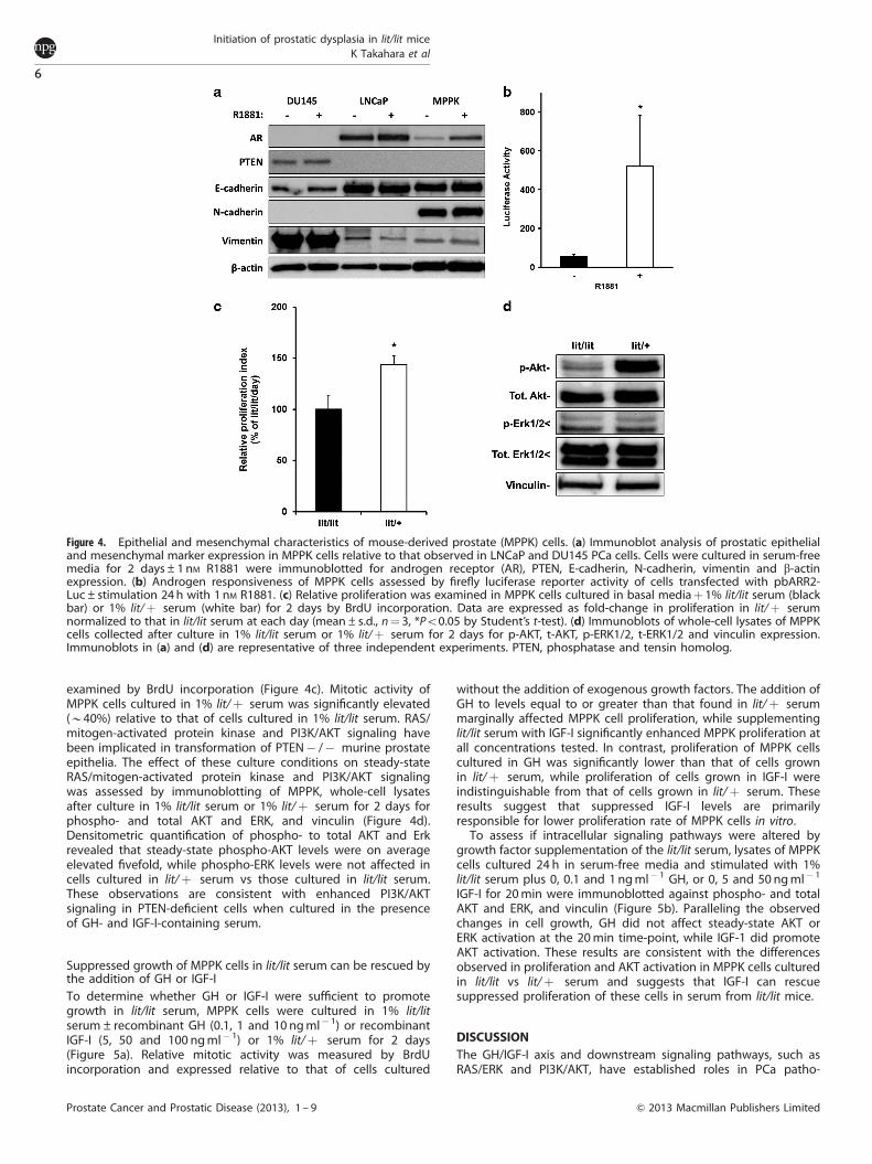
examined by BrdU incorporation (Figure 4c). Mitotic activity ofMPPK cells cultured in 1% lit/þ serum was significantly elevated(B40%) relative to that of cells cultured in 1% lit/lit serum. RAS/mitogen-activated protein kinase and PI3K/AKT signaling havebeen implicated in transformation of PTEN� /� murine prostateepithelia. The effect of these culture conditions on steady-stateRAS/mitogen-activated protein kinase and PI3K/AKT signalingwas assessed by immunoblotting of MPPK, whole-cell lysatesafter culture in 1% lit/lit serum or 1% lit/þ serum for 2 days forphospho- and total AKT and ERK, and vinculin (Figure 4d).Densitometric quantification of phospho- to total AKT and Erkrevealed that steady-state phospho-AKT levels were on averageelevated fivefold, while phospho-ERK levels were not affected incells cultured in lit/þ serum vs those cultured in lit/lit serum.These observations are consistent with enhanced PI3K/AKTsignaling in PTEN-deficient cells when cultured in the presenceof GH- and IGF-I-containing serum.
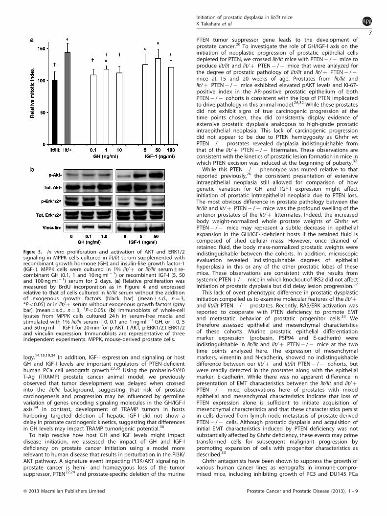
Suppressed growth of MPPK cells in lit/lit serum can be rescued bythe addition of GH or IGF-ITo determine whether GH or IGF-I were sufficient to promotegrowth in lit/lit serum, MPPK cells were cultured in 1% lit/litserum±recombinant GH (0.1, 1 and 10 ng ml� 1) or recombinantIGF-I (5, 50 and 100 ng ml� 1) or 1% lit/þ serum for 2 days(Figure 5a). Relative mitotic activity was measured by BrdUincorporation and expressed relative to that of cells cultured
without the addition of exogenous growth factors. The addition ofGH to levels equal to or greater than that found in lit/þ serummarginally affected MPPK cell proliferation, while supplementinglit/lit serum with IGF-I significantly enhanced MPPK proliferation atall concentrations tested. In contrast, proliferation of MPPK cellscultured in GH was significantly lower than that of cells grownin lit/þ serum, while proliferation of cells grown in IGF-I wereindistinguishable from that of cells grown in lit/þ serum. Theseresults suggest that suppressed IGF-I levels are primarilyresponsible for lower proliferation rate of MPPK cells in vitro.
To assess if intracellular signaling pathways were altered bygrowth factor supplementation of the lit/lit serum, lysates of MPPKcells cultured 24 h in serum-free media and stimulated with 1%lit/lit serum plus 0, 0.1 and 1 ng ml� 1 GH, or 0, 5 and 50 ng ml� 1
IGF-I for 20 min were immunoblotted against phospho- and totalAKT and ERK, and vinculin (Figure 5b). Paralleling the observedchanges in cell growth, GH did not affect steady-state AKT orERK activation at the 20 min time-point, while IGF-1 did promoteAKT activation. These results are consistent with the differencesobserved in proliferation and AKT activation in MPPK cells culturedin lit/lit vs lit/þ serum and suggests that IGF-I can rescuesuppressed proliferation of these cells in serum from lit/lit mice.
DISCUSSIONThe GH/IGF-I axis and downstream signaling pathways, such asRAS/ERK and PI3K/AKT, have established roles in PCa patho-
Figure 4. Epithelial and mesenchymal characteristics of mouse-derived prostate (MPPK) cells. (a) Immunoblot analysis of prostatic epithelialand mesenchymal marker expression in MPPK cells relative to that observed in LNCaP and DU145 PCa cells. Cells were cultured in serum-freemedia for 2 days±1nM R1881 were immunoblotted for androgen receptor (AR), PTEN, E-cadherin, N-cadherin, vimentin and b-actinexpression. (b) Androgen responsiveness of MPPK cells assessed by firefly luciferase reporter activity of cells transfected with pbARR2-Luc±stimulation 24 h with 1 nM R1881. (c) Relative proliferation was examined in MPPK cells cultured in basal mediaþ 1% lit/lit serum (blackbar) or 1% lit/þ serum (white bar) for 2 days by BrdU incorporation. Data are expressed as fold-change in proliferation in lit/þ serumnormalized to that in lit/lit serum at each day (mean±s.d., n¼ 3, *Po0.05 by Student’s t-test). (d) Immunoblots of whole-cell lysates of MPPKcells collected after culture in 1% lit/lit serum or 1% lit/þ serum for 2 days for p-AKT, t-AKT, p-ERK1/2, t-ERK1/2 and vinculin expression.Immunoblots in (a) and (d) are representative of three independent experiments. PTEN, phosphatase and tensin homolog.
Initiation of prostatic dysplasia in lit/lit miceK Takahara et al
6
Prostate Cancer and Prostatic Disease (2013), 1 – 9 & 2013 Macmillan Publishers Limited
logy.14,15,19,34 In addition, IGF-I expression and signaling or hostGH and IGF-I levels are important regulators of PTEN-deficienthuman PCa cell xenograft growth.23,35 Using the probasin-SV40T-Ag (TRAMP) prostate cancer animal model, we previouslyobserved that tumor development was delayed when crossedinto the lit/lit background, suggesting that risk of prostatecarcinogenesis and progression may be influenced by germlinevariation of genes encoding signaling molecules in the GH/IGF-Iaxis.34 In contrast, development of TRAMP tumors in hostsharboring targeted deletion of hepatic IGF-I did not show adelay in prostate carcinogenic kinetics, suggesting that differencesin GH levels may impact TRAMP tumorigenic potential.36
To help resolve how host GH and IGF levels might impactdisease initiation, we assessed the impact of GH and IGF-Ideficiency on prostate cancer initiation using a model morerelevant to human disease that results in perturbation in the PI3K/AKT pathway. A signature event impacting PI3K/AKT signaling inprostate cancer is hemi- and homozygous loss of the tumorsuppressor, PTEN22,24 and prostate-specific deletion of the murine
PTEN tumor suppressor gene leads to the development ofprostate cancer.26 To investigate the role of GH/IGF-I axis on theinitiation of neoplastic progression of prostatic epithelial cellsdepleted for PTEN, we crossed lit/lit mice with PTEN� /� mice toproduce lit/lit and lit/þ PTEN� /� mice that were analyzed forthe degree of prostatic pathology of lit/lit and lit/þ PTEN� /�mice at 15 and 20 weeks of age. Prostates from lit/lit andlit/þ PTEN� /� mice exhibited elevated pAKT levels and Ki-67-positive index in the AR-positive prostatic epithelium of bothPTEN� /� cohorts is consistent with the loss of PTEN implicatedto drive pathology in this animal model.26,32 While these prostatesdid not exhibit signs of true carcinogenic progression at thetime points chosen, they did consistently display evidence ofextensive prostatic dysplasia analogous to high-grade prostaticintraepithelial neoplasia. This lack of carcinogenic progressiondid not appear to be due to PTEN hemizygosity as Ghrhr wtPTEN� /� prostates revealed dysplasia indistinguishable fromthat of the lit/þ PTEN� /� littermates. These observations areconsistent with the kinetics of prostatic lesion formation in mice inwhich PTEN excision was induced at the beginning of puberty.32
While this PTEN� /� phenotype was muted relative to thatreported previously,26 the consistent presentation of extensiveintraepithelial neoplasia still allowed for comparison of howgenetic variation for GH and IGF-I expression might affectinitiation of prostatic intraepithelial neoplasia due to PTEN loss.The most obvious difference in prostate pathology between thelit/lit and lit/þ PTEN� /� mice was the profound swelling of theanterior prostates of the lit/þ littermates. Indeed, the increasedbody weight-normalized whole prostate weights of Ghrhr wtPTEN� /� mice may represent a subtle decrease in epithelialexpansion in the GH/IGF-I-deficient hosts if the retained fluid iscomposed of shed cellular mass. However, once drained ofretained fluid, the body mass-normalized prostatic weights wereindistinguishable between the cohorts. In addition, microscopicevaluation revealed indistinguishable degrees of epithelialhyperplasia in this or any of the other prostatic lobes of thesemice. These observations are consistent with the results fromsystemic PTENþ /� mice in which knockout of IRS2 did not affectinitiation of prostatic dysplasia but did delay lesion progression.37
This lack of overt phenotypic difference in prostatic dysplasticinitiation compelled us to examine molecular features of the lit/þand lit/lit PTEN� /� prostates. Recently, RAS/ERK activation wasreported to cooperate with PTEN deficiency to promote EMTand metastatic behavior of prostatic progenitor cells.33 Wetherefore assessed epithelial and mesenchymal characteristicsof these cohorts. Murine prostatic epithelial differentiationmarker expression (probasin, PSP94 and E-cadherin) wereindistinguishable in lit/lit and lit/þ PTEN� /� mice at the twotime points analyzed here. The expression of mesenchymalmarkers, vimentin and N-cadherin, showed no indistinguishabledifference between our lit/þ and lit/lit PTEN� /� cohorts, butwere readily detected in the prostates along with the epithelialmarker, E-cadherin. While there was no apparent difference inpresentation of EMT characteristics between the lit/lit and lit/þPTEN� /� mice, observations here of prostates with mixedepithelial and mesenchymal characteristics indicate that loss ofPTEN expression alone is sufficient to initiate acquisition ofmesenchymal characteristics and that these characteristics persistin cells derived from lymph node metastasis of prostate-derivedPTEN� /� cells. Although prostatic dysplasia and acquisition ofinitial EMT characteristics induced by PTEN deficiency was notsubstantially affected by Ghrhr deficiency, these events may primetransformed cells for subsequent malignant progression bypromoting expansion of cells with progenitor characteristics asdescribed.33
Ghrhr antagonists have been shown to suppress the growth ofvarious human cancer lines as xenografts in immune-compro-mised mice, including inhibiting growth of PC3 and DU145 PCa
Figure 5. In vitro proliferation and activation of AKT and ERK1/2signaling in MPPK cells cultured in lit/lit serum supplemented withrecombinant growth hormone (GH) and insulin-like growth factor-1(IGF-I). MPPK cells were cultured in 1% lit/þ or lit/lit serum±re-combinant GH (0.1, 1 and 10 ngml� 1) or recombinant IGF-I (5, 50and 100 ngml� 1) serum for 2 days. (a) Relative proliferation wasmeasured by BrdU incorporation as in Figure 4 and expressedrelative to that of cells cultured in lit/lit serum without the additionof exogenous growth factors (black bar) (mean±s.d., n¼ 3,*Po0.05) or in lit/þ serum without exogenous growth factors (graybar) (mean±s.d., n¼ 3, wPo0.05). (b) Immunoblots of whole-celllysates from MPPK cells cultured 24 h in serum-free media andstimulated with 1% lit/lit serumþ 0, 0.1 and 1 ngml� 1 GH, orþ 0, 5and 50 ngml� 1 IGF-I for 20min for p-AKT, t-AKT, p-ERK1/2,t-ERK1/2and vinculin expression. Immunoblots are representative of threeindependent experiments. MPPK, mouse-derived prostate cells.
Initiation of prostatic dysplasia in lit/lit miceK Takahara et al
7
& 2013 Macmillan Publishers Limited Prostate Cancer and Prostatic Disease (2013), 1 – 9
xenografts.38,39 We have previously demonstrated that decreasedcirculating GH and IGF-I were significant contributors tosuppressed growth of both androgen-responsive and castration-resistant PTEN-null PCa cells,23 and that antisense oligonucleotidesuppression of IGF-1R expression suppressed growth of theseandrogen-responsive and castration-resistant PTEN-null PCacells.35 These studies supported the contention that decreasedIGF-I availability and signaling from IGF-1R to AKT was the primarymissing growth stimulatory pathway for such cells in the lit/lithost. Consistent with these previous results, in this study, growthof MPPK cells was suppressed in media supplemented with lit/litserum as compared with lit/þ serum, and that restoration ofsuppressed growth of MPPK cells in lit/lit serum by the addition ofIGF-I and, to a lesser extent, GH was correlated with increasedsteady-state activation of AKT. In contrast to the results fromTRAMP crosses with lit/lit or hepatic IGF-I knockout mice thatimplicated a role for GH in driving TRAMP tumorigenesis, thesein vitro experiments and our previous studies implicate circulatingIGF-I as the predominant contributor to proliferation of PCa, andthat this effect is mediated primarily through PI3K/AKT activation.
CONCLUSIONOur in vivo and in vitro experiments suggest that loss of PTEN mayinitiate prostatic epithelial dysplasia and acquisition of mesen-chymal characteristics and that IGF-I may be an important factor insustaining proliferation of these dysplastic cells. We thereforesuggest that in early stages of PCa development, suppression ofGH/IGF-I axis may delay invasive PCa progression. Such treatmentsmight be best augmented by therapies targeting factors thatregulate EMT, a process mechanistically linked with stem cellsignatures in PCa cells40 and increased resistance to apoptosis,diminished senescence, escape from immune surveillance andeventual resistance to therapy.41 With the reports linking GH/IGF-Iaxis signaling to EMT and acquisition of stem-like properties, theseresults support continued effort to disrupt IGF axis signaling tocontrol PCa progression.
CONFLICT OF INTERESTThe authors declare no conflict of interest.
ACKNOWLEDGEMENTSWe thank Virginia Yago, Darrell Trendall, Estelle Li and Mary Bowden for theirexcellent technical assistance. This work was supported by Terry Fox Program inProstate Cancer Progression grant from the National Cancer Institute of Canada(no. 017007) and a CIHR Doctoral Fellowship award (to MG).
REFERENCES1 Chan JM, Stampfer MJ, Giovannucci E, Gann PH, Ma J, Wilkinson P et al.
Plasma insulin-like growth factor-I and prostate cancer risk: a prospective study.Science 1998; 279: 563–566.
2 LeRoith D, Roberts Jr CT. The insulin-like growth factor system and cancer.Cancer Lett 2003; 195: 127–137.
3 Papatsoris AG, Karamouzis MV, Papavassiliou AG. Novel insights into theimplication of the IGF-1 network in prostate cancer. Trends Mol Med 2005; 11:52–55.
4 Ryan CJ, Haqq CM, Simko J, Nonaka DF, Chan JM, Weinberg V et al. Expressionof insulin-like growth factor-1 receptor in local and metastatic prostate cancer.Urol Oncol 2007; 25: 134–140.
5 Pollak M. Insulin-like growth factor physiology and cancer risk. Eur J Cancer 2000;36: 1224–1228.
6 DiGiovanni J, Kiguchi K, Frijhoff A, Wilker E, Bol DK, Beltran L et al. Deregulatedexpression of insulin-like growth factor 1 in prostate epithelium leads to neoplasiain transgenic mice. Proc Natl Acad Sci USA 2000; 97: 3455–3460.
7 Chan JM, Stampfer MJ, Ma J, Gann P, Gaziano JM, Pollak M et al. Insulin-likegrowth factor-I (IGF-I) and IGF binding protein-3 as predictors of advanced-stageprostate cancer. J Natl Cancer Inst 2002; 94: 1099–1106.
8 Grimberg A. Mechanisms by which IGF-I may promote cancer. Cancer Biol Ther2003; 2: 630–635.
9 Hellawell GO, Turner GD, Davies DR, Poulsom R, Brewster SF, Macaulay VM.Expression of the type 1 insulin-like growth factor receptor is up-regulated inprimary prostate cancer and commonly persists in metastatic disease. Cancer Res2002; 62: 2942–2950.
10 Scorilas A, Plebani M, Mazza S, Basso D, Soosaipillai AR, Katsaros N et al.Serum human glandular kallikrein (hK2) and insulin-like growth factor 1 (IGF-1)improve the discrimination between prostate cancer and benignprostatic hyperplasia in combination with total and %free PSA. Prostate 2003; 54:220–229.
11 Oliver SE, Barrass B, Gunnell DJ, Donovan JL, Peters TJ, Persad RA et al.Serum insulin-like growth factor-I is positively associated with serum prostate-specific antigen in middle-aged men without evidence of prostate cancer.Cancer Epidemiol Biomarkers Prev 2004; 13: 163–165.
12 Frasca F, Pandini G, Sciacca L, Pezzino V, Squatrito S, Belfiore A et al. The role ofinsulin receptors and IGF-I receptors in cancer and other diseases. Arch PhysiolBiochem 2008; 114: 23–37.
13 Hellawell GO, Ferguson DJ, Brewster SF, Macaulay VM. Chemosensitization ofhuman prostate cancer using antisense agents targeting the type 1 insulin-likegrowth factor receptor. BJU Int 2003; 91: 271–277.
14 Krueckl SL, Sikes RA, Edlund NM, Bell RH, Hurtado-Coll A, Fazli L et al. Increasedinsulin-like growth factor I receptor expression and signaling are componentsof androgen-independent progression in a lineage-derived prostate cancerprogression model. Cancer Res 2004; 64: 8620–8629.
15 Nickerson T, Chang F, Lorimer D, Smeekens SP, Sawyers CL, Pollak M. In vivoprogression of LAPC-9 and LNCaP prostate cancer models to androgen inde-pendence is associated with increased expression of insulin-like growth factor I(IGF-I) and IGF-I receptor (IGF-IR). Cancer Res 2001; 61: 6276–6280.
16 Huynh H, Seyam RM, Brock GB. Reduction of ventral prostate weight by finas-teride is associated with suppression of insulin-like growth factor I (IGF-I) and IGF-Ireceptor genes and with an increase in IGF binding protein 3. Cancer Res 1998; 58:215–218.
17 Pandini G, Mineo R, Frasca F, Roberts Jr CT, Marcelli M, Vigneri R et al. Androgensup-regulate the insulin-like growth factor-I receptor in prostate cancer cells.Cancer Res 2005; 65: 1849–1857.
18 Ohlsson C, Mohan S, Sjogren K, Tivesten A, Isgaard J, Isaksson O et al. The role ofliver-derived insulin-like growth factor-I. Endocr Rev 2009; 30: 494–535.
19 Butler AA, Yakar S, Gewolb IH, Karas M, Okubo Y, LeRoith D. Insulin-like growthfactor-I receptor signal transduction: at the interface between physiology and cellbiology. Comp Biochem Physiol B 1998; 121: 19–26.
20 Thomas GV, Horvath S, Smith BL, Crosby K, Lebel LA, Schrage M et al. Antibody-based profiling of the phosphoinositide 3-kinase pathway in clinical prostatecancer. Clin Cancer Res 2004; 10: 8351–8356.
21 Muller M, Rink K, Krause H, Miller K. PTEN/MMAC1 mutations in prostate cancer.Prostate Cancer Prostatic Dis 2000; 3(Suppl 1): S32.
22 Trotman LC, Niki M, Dotan ZA, Koutcher JA, Di Cristofano A, Xiao A et al. Pten dosedictates cancer progression in the prostate. PLoS Biol 2003; 1: 27.
23 Takahara K, Tearle H, Ghaffari M, Gleave ME, Pollak M, Cox ME. Human prostatecancer xenografts in lit/lit mice exhibit reduced growth and androgen-indepen-dent progression. Prostate 2011; 71: 525–537.
24 Whang YE, Wu X, Suzuki H, Reiter RE, Tran C, Vessella RL et al. Inactivation of thetumor suppressor PTEN/MMAC1 in advanced human prostate cancer through lossof expression. Proc Natl Acad Sci USA 1998; 95: 5246–5250.
25 Yoshimoto M, Joshua AM, Cunha IW, Coudry RA, Fonseca FP, Ludkovski O et al.Absence of TMPRSS2:ERG fusions and PTEN losses in prostate cancer is associatedwith a favorable outcome. Mod Pathol 2008; 21: 1451–1460.
26 Wang S, Gao J, Lei Q, Rozengurt N, Pritchard C, Jiao J et al. Prostate-specificdeletion of the murine Pten tumor suppressor gene leads to metastatic prostatecancer. Cancer Cell 2003; 4: 209–221.
27 Mulholland DJ, Tran LM, Li Y, Cai H, Morim A, Wang S et al. Cell autonomous roleof PTEN in regulating castration-resistant prostate cancer growth. Cancer Cell2011; 19: 792–804.
28 Suzuki A, Yamaguchi MT, Ohteki T, Sasaki T, Kaisho T, Kimura Y et al. T cell-specificloss of Pten leads to defects in central and peripheral tolerance. Immunity 2001;14: 523–534.
29 Wu X, Wu J, Huang J, Powell WC, Zhang J, Matusik RJ et al. Generation of aprostate epithelial cell-specific Cre transgenic mouse model for tissue-specificgene ablation. Mech Dev 2001; 101: 61–69.
30 Godfrey P, Rahal JO, Beamer WG, Copeland NG, Jenkins NA, Mayo KE. GHRHreceptor of little mice contains a missense mutation in the extracellular domainthat disrupts receptor function. Nat Genet 1993; 4: 227–232.
31 Moussavi M, Fazli L, Tearle H, Guo Y, Cox M, Bell J et al. Oncolysis of prostatecancers induced by vesicular stomatitis virus in PTEN knockout mice. Cancer Res2010; 70: 1367–1376.
Initiation of prostatic dysplasia in lit/lit miceK Takahara et al
8
Prostate Cancer and Prostatic Disease (2013), 1 – 9 & 2013 Macmillan Publishers Limited
32 Luchman HA, Benediktsson H, Villemaire ML, Peterson AC, Jirik FR. The pace ofprostatic intraepithelial neoplasia development is determined by the timing ofPten tumor suppressor gene excision. PLoS One 2008; 3: 15.
33 Mulholland DJ, Kobayashi N, Ruscetti M, Zhi A, Tran LM, Huang J et al. Pten lossand RAS/MAPK activation cooperate to promote EMT and metastasis initiatedfrom prostate cancer stem/progenitor cells. Cancer Res 2012; 72: 1878–1889.
34 Majeed N, Blouin MJ, Kaplan-Lefko PJ, Barry-Shaw J, Greenberg NM, Gaudreau Pet al. A germ line mutation that delays prostate cancer progression and prolongssurvival in a murine prostate cancer model. Oncogene 2005; 24: 4736–4740.
35 Furukawa J, Wraight CJ, Freier SM, Peralta E, Atley LM, Monia BP et al. Antisenseoligonucleotide targeting of insulin-like growth factor-1 receptor (IGF-1R) inprostate cancer. Prostate 2010; 70: 206–218.
36 Anzo M, Cobb LJ, Hwang DL, Mehta H, Said JW, Yakar S et al. Targeted deletion ofhepatic Igf1 in TRAMP mice leads to dramatic alterations in the circulating insulin-like growth factor axis but does not reduce tumor progression. Cancer Res 2008;68: 3342–3349.
37 Szabolcs M, Keniry M, Simpson L, Reid LJ, Koujak S, Schiff SC et al. Irs2 inactivationsuppresses tumor progression in Ptenþ /� mice. Am J Pathol 2009; 174:276–286.
38 Schally AV, Varga JL. Antagonists of growth hormone-releasing hormone inoncology. Comb Chem High Throughput Screen 2006; 9: 163–170.
39 Stangelberger A, Schally AV, Varga JL, Hammann BD, Groot K, Halmos G et al.Antagonists of growth hormone releasing hormone (GHRH) and ofbombesin/gastrin releasing peptide (BN/GRP) suppress the expressionof VEGF, bFGF, and receptors of the EGF/HER family in PC-3 andDU-145 human androgen-independent prostate cancers. Prostate 2005; 64:303–315.
40 Kong D, Banerjee S, Ahmad A, Li Y, Wang Z, Sethi S et al. Epithelial tomesenchymal transition is mechanistically linked with stem cell signatures inprostate cancer cells. PLoS One 2010; 5: e12445.
41 van der Pluijm G. Epithelial plasticity, cancer stem cells and bone metastasisformation. Bone 2011; 48: 37–43.
Supplementary Information accompanies the paper on the Prostate Cancer and Prostatic Diseases website (http://www.nature.com/pcan)
Initiation of prostatic dysplasia in lit/lit miceK Takahara et al
9
& 2013 Macmillan Publishers Limited Prostate Cancer and Prostatic Disease (2013), 1 – 9