the internationalisation of the new zealand journal of medical
TRANSCRIPT
Established
in 1946
Volume 66 Number 2 August 2012ISSN 1171-0195
New Zealand Journal ofMedicalLaboratoryScience
Official Publication of the New Zealand Institute of Medical Laboratory Science Incorporated
2

Conquer your space limitations
STA Satellite®
Con
cept
ion
L2R
- ©
200
5 D
iagn
ostic
a St
ago
- A
ll ri
ghts
res
erve
d -
07/2
011
Diagnostica Stago Pty. Ltd. 651 Doncaster Road Doncaster, Victoria, 3108 Australia Phone (AUS): 1800 4 STAGO Phone (NZ): 0508 4 STAGO Fax: +61 3 9855 8999 [email protected] www.stago.com.au
NZ J Med Lab Science 2012
33
Editor Rob Siebers, PGCertPH FNZIC FNZIMLS CBiol FSB, University of Otago, Wellington
Deputy Editor Terry Taylor, BSc DipMLS MNZIMLS, Southern Community Laboratories, Dunedin
Editorial BoardChris Kendrick, GradDipSc MSc MNZIMLS, Massey University, Palmerston NorthMichael Legge, PhD MSB FIBMS FNZIMLS FFSc(RCPA), University of Otago, DunedinHolly Perry, MApplSci(Hons) DipMLT MNZIMLS, AUT, AucklandCat Ronayne, BMLSc DipGrad MNZIMLS, University of Otago, DunedinJohn Stirling, BSc(Hons) MLett AFRCPA MAIMS FRMS, South Australia Pathology, AdelaideAnn Thornton, CertMS FNZIMLS, University of Otago, WellingtonTony Woods, BA BSc(Hons) PhD MAIMS FFSc(RCPA), University of South Australia, Adelaide
Statistical EditorNevil Pierse, BSc MSc PhD, University of Otago, Wellington
About the Journal The Journal publishes original articles, technical communications, scientifi c letters, review articles and case studies on all subjects pertaining to medical laboratory science. The Journal was established in 1946 and is currently published three times per year (April, August and November). Circulation is to all New Zealand Institute of Medical Laboratory Science (NZIMLS) members, and to universities in New Zealand and overseas. Current circulation is about 2,200 copies per issue. The Journal is the offi cial publication of the NZIMLS who owns the copyright and no parts of this publication may be reproduced in any form without the written permission of the NZIMLS. Opinions expressed in the Journal are not necessarily those of the Editors or the NZIMLS.
Article submissionSubmit articles electronically to the Editor ([email protected]). Full instructions to authors are on the NZIMLS website (www.nzimls.org.nz) together with the author form in which each author’s contribution to the study must be stated. If accepted, copyright of the article is transferred to the NZIMLS. Authors are responsible for the scientifi c content and views.
Abstracting The Journal is open access (www.nzimls.org.nz/nzimls-journal.html) and is abstracted by EMBASE, CINAHL, EBSCO, Biosis Citation Index, SCOPUS, ProQuest and Thomson Gale. The Journal’s Editors are members of the World Association of Medical Editors (www.wame.org).
Subscription and advertising Enquiries regarding subscription or address changes, and advertising should be addressed to the NZIMLS Executive Offi ce, PO Box 505, Rangiora 7440, New Zealand. Ph: +64 3 313 4761; email: [email protected].
PrintingISSN 1171-0195. Printed by Keyprint Printing Ltd., Auckland on FSC Certifi ed Mixed Source pulp from well-managed and legally harvested forests.
N u m b e r 2Volume 66August 2012ISSN 1171-0195
N e w Z e a l a n d J o u r n a l o f
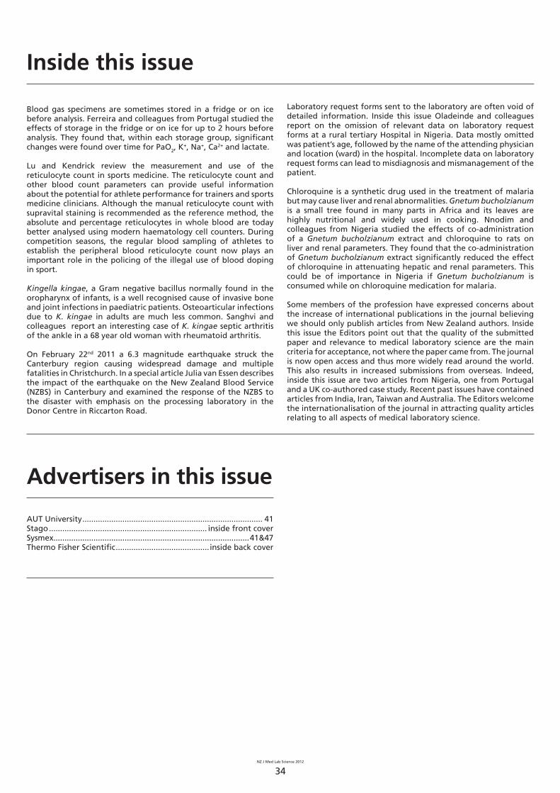
EditorialThe internationalisation of the New Zealand Journal of Medical Laboratory ScienceTerry Taylor and Rob Siebers .......................................................... 35
Review articleReticulocyte counts in sports medicineJun Lu and Christopher J Kendrick ..................................................36-38
Short communicationEvaluation of laboratory request forms for incomplete data at a rural tertiary hospital in NigeriaBankole Henry Oladeinde, Richard Omoregie, Eguagie Osareniro Osakue and Adekunde Abdufattai Onifade ...............................39-41
Original articleStability of blood gases when refrigeratedJoão Pedro Ferreira, Sara Vieira Silva, Patrícia Rodrigues, Miguel Araújo Abreu, José Miguel Maia, Daniela Carvalho and Luísa Carvalho ............................................................................................42-45
Scientifi c letterAttenuation of chloroquine-induced hepatotoxicity and renal damage by Gnetum bucholzianum leaf extractJohnkennedy Nnodim, Augustine Ihim and Hellen Ifeoma Udujih ...................................................................................46-47
Case studySeptic arthritis due to Kingella kingae in an adult patientAjay Sanghvi, Michael Addidle and Kate Grimwade ....................48-49
Special articleThe Christchurch earthquake and its effect on the New Zealand Blood Service processing laboratoryJulia van Essen ...................................................................................50-51
Book reviewFever: How Malaria Has Ruled Mankind for 500,000 Years by Sonia ShahReviewed by Leanne Mayhew ........................................................ 52
Regular featuresAdvertisers in this issue ................................................................... 34Barry Edwards/Rod Kennedy Scholarship ...................................... 54Editorial Board ................................................................................ 33Fellowship of the NZIMLS ............................................................... 53In this issue ...................................................................................... 34Instructions to authors .................................................................... 33Journal questionnaire ..................................................................... 55NZIMLS journal case study prize ..................................................... 55Olympus photo competition .......................................................... 53Pacifi c Way column ....................................................................62-64Reviewers for 2011/2012 ................................................................. 54Special Interest Groups ..............................................................56-61Up and coming NZIMLS events ....................................................... 47
NZ J Med Lab Science 2012
34
Inside this issue
Blood gas specimens are sometimes stored in a fridge or on ice before analysis. Ferreira and colleagues from Portugal studied the effects of storage in the fridge or on ice for up to 2 hours before analysis. They found that, within each storage group, signifi cant changes were found over time for PaO2, K
+, Na+, Ca2+ and lactate.
Lu and Kendrick review the measurement and use of the reticulocyte count in sports medicine. The reticulocyte count and other blood count parameters can provide useful information about the potential for athlete performance for trainers and sports medicine clinicians. Although the manual reticulocyte count with supravital staining is recommended as the reference method, the absolute and percentage reticulocytes in whole blood are today better analysed using modern haematology cell counters. During competition seasons, the regular blood sampling of athletes to establish the peripheral blood reticulocyte count now plays an important role in the policing of the illegal use of blood doping in sport.
Kingella kingae, a Gram negative bacillus normally found in the oropharynx of infants, is a well recognised cause of invasive bone and joint infections in paediatric patients. Osteoarticular infections due to K. kingae in adults are much less common. Sanghvi and colleagues report an interesting case of K. kingae septic arthritis of the ankle in a 68 year old woman with rheumatoid arthritis.
On February 22nd 2011 a 6.3 magnitude earthquake struck the Canterbury region causing widespread damage and multiple fatalities in Christchurch. In a special article Julia van Essen describes the impact of the earthquake on the New Zealand Blood Service (NZBS) in Canterbury and examined the response of the NZBS to the disaster with emphasis on the processing laboratory in the Donor Centre in Riccarton Road.
Advertisers in this issue
AUT University ................................................................................. 41Stago ....................................................................... inside front coverSysmex. .......................................................................................41&47Thermo Fisher Scientifi c ..........................................inside back cover
Laboratory request forms sent to the laboratory are often void of detailed information. Inside this issue Oladeinde and colleagues report on the omission of relevant data on laboratory request forms at a rural tertiary Hospital in Nigeria. Data mostly omitted was patient’s age, followed by the name of the attending physician and location (ward) in the hospital. Incomplete data on laboratory request forms can lead to misdiagnosis and mismanagement of the patient.
Chloroquine is a synthetic drug used in the treatment of malaria but may cause liver and renal abnormalities. Gnetum bucholzianum is a small tree found in many parts in Africa and its leaves are highly nutritional and widely used in cooking. Nnodim and colleagues from Nigeria studied the effects of co-administration of a Gnetum bucholzianum extract and chloroquine to rats on liver and renal parameters. They found that the co-administration of Gnetum bucholzianum extract signifi cantly reduced the effect of chloroquine in attenuating hepatic and renal parameters. This could be of importance in Nigeria if Gnetum bucholzianum is consumed while on chloroquine medication for malaria.
Some members of the profession have expressed concerns about the increase of international publications in the journal believing we should only publish articles from New Zealand authors. Inside this issue the Editors point out that the quality of the submitted paper and relevance to medical laboratory science are the main criteria for acceptance, not where the paper came from. The journal is now open access and thus more widely read around the world. This also results in increased submissions from overseas. Indeed, inside this issue are two articles from Nigeria, one from Portugal and a UK co-authored case study. Recent past issues have contained articles from India, Iran, Taiwan and Australia. The Editors welcome the internationalisation of the journal in attracting quality articles relating to all aspects of medical laboratory science.
NZ J Med Lab Science 2012
35
Recently there have been a number of concerns raised from some members of the New Zealand Institute of Medical Laboratory Science (NZIMLS) about the amount of overseas articles published within the NZIMLS Journal believing that we should only be publishing articles from New Zealand authors’ seeing it is a New Zealand journal.
Since the journal has become open access around the world and abstracting by international data bases this has dramatically increased the potential reader numbers. This is evidenced by an increasing number of published articles from the journal that are cited in the international biomedical literature (1). There has also been an increase in the number of overseas submissions to the journal. In this August issue is an original article from Portugal and two short communications from Nigeria. In the past we have also published articles from Taiwan, Nigeria, Iran, India, the UK and Australia. Later this year there will be further articles from the USA and Australia. With this in mind this has also seen an opportunity for overseas researchers to look at publishing in our journal. While this may not be an ideal scenario in some of our member’s eyes, it has also given an opportunity to many others to publish articles. One thing that should always be remembered is that we have a panel of experienced and very knowledgeable peer reviewers to rely on to scrutinize articles put forward for publication. What you read in the journal is often signifi cantly different from what was fi rst presented to the reviewer.
This does not answer the question of why we don’t have more publications published from our members. The answer is, we simply do not get many articles submitted from our New Zealand members. If we had to rely on this the journal would be lucky to have more than a couple of articles each issue. There are many different types of scientifi c writing that can be submitted for publication. This includes original articles, reviews, opinion pieces, short technical communications and scientifi c letters.
Basically if people want to see more local content then keep encouraging our peers to publish. We all have a tremendous amount of data, case studies and expertise available within the laboratories that we work in. Many excellent presentations are given at the NZIMLS Special Interest Group meetings, the Annual Scientifi c Meeting and User Group Meetings. Yet, very few end up in the journal despite active encouragement from the Editors and Editorial Board Members. Just remember, what may seem mundane and not worth a lot to one person may in turn be just the information another person was looking for. The NZIMLS Journal provides a target audience for sharing of this knowledge.
The internationalisation of the New Zealand Journal of Medical Laboratory ScienceTerry Taylor and Rob Siebers
In conclusion, the NZIMLS Journal is now becoming more international. The basic aim of the journal has been, and will continue to be to publish quality articles relating to all aspects of medical laboratory science. Whether they come from New Zealand or overseas is immaterial. Of course, being the Institute’s journal we would like to see the NZIMLS members contribute more. Benefi ts for NZIMLS members is accrual of 20 CPD points per published article (whether as primary or co-author) and being eligible for the journal prizes. Remember, when you give your presentation at a scientifi c meeting you are reaching only about 50 to 150 people. In the print version you are reaching a readership of about 2,200. Open access worldwide of the journal your target audience is unlimited. You owe it to yourself professionally, to your colleagues and to the world to share your fi ndings and knowledge to the advancement of medical laboratory science. We look forward to your submissions. On a fi nal note, when members tell us that we should only publish articles from New Zealand as it is our (New Zealand) journal, we just ask them a simple question. What have you contributed to the journal?
References
1. Siebers R. Which journals are citing articles from the New Zealand Journal of Medical Laboratory Science. N Z J Med Lab Sci 2012; 66: 14-17.
Author information
*Terry Taylor, BSc DipMLS MNZIMLS, Medical Laboratory Scientist1
and Deputy Editor2
Rob Siebers, PGCertPH FNZIC, FNZIMLS CBiol FSB, Associate Professor3 and Editor2
1Southern Community Laboratories, Dunedin2New Zealand Institute of Medical Laboratory Science, Rangiora3School of Medicine and Health Sciences, University of Otago, Wellington
* Email: [email protected]
N Z J Med Lab Sci 2012; 66: 35

NZ J Med Lab Science 2012
36
Abstract Reticulocytes are juvenile red blood cells (RBCs) containing remnant ribonucleic acid (RNA). Their percentage in the peripheral blood (PB) is a useful indication of erythropoiesis in the bone marrow. In the context of sport, the reticulocyte count and other complete blood count (CBC) parameters can provide useful information about the potential for athlete performance for trainers and sports medicine clinicians. In order to obtain reliable measurements, pre-analytical variables have to be controlled, including the timing of blood collection and the standardisation of phlebotomy procedures. Although the manual reticulocyte count with supravital staining is recommended as the reference method, the absolute and percentage reticulocytes in whole blood are today better analysed using modern haematology cell counters. The comparability of the results from the manual and automated methods remains in doubt mostly due to the lack of suitable calibration and control bloods for both methods.
Mild haemolysis and increased oxygen demand are associated with exercise, especially during endurance training. The production of erythropoietin is enhanced in this setting inducing erythropoiesis resulting in an elevated PB reticulocyte count. Athlete training at high altitudes produces a physiological PB hypoxic effect in an attempt to maximise oxygen carrying capacity and performance. Studies into the reticulocyte response in athletes show that minimal intensity and duration of training will initiate the erythropoietic response to increase PB reticulocyte numbers. Efforts to raise athlete performance by increasing the number of circulating RBCs have led to the banning of autologous and/or homologous blood transfusions and the use of recombinant human erythropoietin (rHuEpo). The illegal use of these methods to boost performance is a major focus for some athletes and a major challenge for anti-doping agencies trying to keep sport clean from the use of banned performance enhancers. As part of the screening for the use of performance boosting activities, the OFF score is used for the detection of recent usage of rHuEpo and is based on the PB reticulocyte percentage. More research and studies are still necessary to standardise reticulocyte measurement, particularly for its application in blood doping in sports.
Key words: reticulocyte, recombinant human erythropoietin, blood doping, training, sports
N Z J Med Lab Sci 2012; 66: 36-38
IntroductionImmature red cells with remnants of RNA are termed reticulocytes and they are present in both the peripheral blood and the bone marrow (1). As they mature to become functionally normal erythrocytes, the haemoglobin content of the reticulocyte increases and the size of the cell decreases (1). The concentration of reticulocytes in the PB is estimated from a PB specimen with the reticulocyte count a test frequently performed in the haematology laboratory. Most reticulocyte testing in the lab is performed on patient samples, providing useful information about the physiologic BM response to disease and response to medication and other treatments. For some sports trainers and sports medicine clinicians, it is becoming more popular to monitor athlete performance against the effective rate of RBC output from the bone marrow (2). Moreover, during competition seasons, the regular blood sampling of athletes to establish the peripheral blood reticulocyte count now plays an important role in the policing of the illegal use of blood doping in sport (2, 3).
Reticulocyte counts in sports medicine Jun Lu and Christopher J Kendrick
Pre-analytical variablesThe control of pre-analytical and analytical variables is crucial for gaining accurate reticulocyte measurements if the reticulocyte count is to be used to detect those athletes using rHuEpo to boost performance. To eliminate variations in the reticulocyte count a number of pre-analytical variables must be standardised. Studies have shown that prolonged tourniquet times during blood collection can produce false haemoglobin (Hb) and haematocrit (Hct) results due to altered cellular fl uid distribution. While there is no real evidence that this has an infl uence on the reticulocyte count it is recommended by doping agencies that a standardised venepuncture procedure for sample collections be followed at all times (2). As with other biological substances, the reticulocyte count shows diurnal variation peaking at around 1:00am with a corresponding rise in the erythropoietin (EPO) concentration (2,5). Blood collections for reticulocyte counts used to monitor human athletic performance need to be performed at a similar time interval to minimise intra-personal variability. Adherence to a standardised collection time for blood sampling has become an issue for agencies monitoring athletes following intercontinental fl ights (2).
Generally, reticulocyte numbers in whole blood remain stable for up to 24 hours after collection, if samples are refrigerated. Results can become falsely low if analysis is further delayed as reticulocytes mature in the blood sample prior to testing (1,2). It is therefore important to ensure the optimal transportation and storage conditions for specimens being tested for reticulocyte numbers in order to show genuine stimulation of the bone marrow in athletes using rHuEpo (2).
Analytical variationThe reference range of the reticulocyte count for the general population is 0.5-2.5%, which is similar to the range expected for athletes (2,4). It is suggested that a reticulocyte count of less than 0.4% or greater than 2.6% in athletes may be considered abnormal (2). Differences among ethnicities have been reported for some of the reticulocyte related parameters, such as increased values for the reticulocyte haematocrit (RetHt) in some African athletes (2).
The reference staining method for the manual reticulocyte count uses a dye such as new methylene blue and supravital staining (2). Standard microscopic examination of blood fi lms classifi es reticulocytes as a red cell containing at least two blue dots or strands of fi lamentous reticulum (1,2). The subjective, time-consuming and imprecise nature of the manual method has given way to use of analysers in today’s laboratories (2,4). Cell analysers use either supravital staining or a fl uorophore technique to measure emitted fl uorescence from stained reticulocytes (2,3). Besides the percentage and absolute number values for the reticulocyte count, the volume (MCVr), immature reticulocyte fraction (IRF) and haemoglobin content of reticulocytes (CHr) can also be provided by some analysers. These can be useful parameters to establish recent reticulocyte stimulation of reticulocytes in response to rHuEpo usage contrasting with results seen in normal physiologic RBC production. (2,4). Research found that the MCVr and CHr values are elevated in athletes either using or having used rHuEpo within the previous three weeks (2).
A number of studies have been done to evaluate the consistency of reticulocyte results measured by various automated haematology analysers. Satisfactory agreement of measurement have been established for some analysers (2,3). The major problem with testing is the consistency of the reticulocyte count across all
NZ J Med Lab Science 2012
37
automated platforms (2,3). The lack of a reliable calibrator means the result from one analyser may not be comparable with the results obtained from the same blood on another analyser. Even comparisons of results obtained from the same series of instruments in different laboratories may produce variable results (2). Ashenden et al (2004) introduced a concept of analyser-specifi c bias using a samplator protocol. This integrated the mean values of a large sample measured at sea level using two analysers and they calculated the bias between the two analysers of interest allowing for the fi nal reticulocyte count to be compensated (3). It is therefore important to factor in analyser variables when using reticulocyte analysis for athletes during training and for anti-doping purposes (2). The variability of analyser estimations of the reticulocyte count in this setting has led to the suggestion that it might be more relevant to compare an athlete’s reticulocytes against his/her baseline results. These could be documented in the form of a so-called haematological passport, rather than using a generalised reference range for all athletes (2).
Effects of trainingMechanical damage and oxidative stress on circulating red cells are listed as the main causes of RBC destruction during active training, particularly in endurance running (6). The level of haemolysis can be raised in plasma and can be measured with a consequential loss of plasma haptoglobin (2,4,6,7). Increased red cell turnover combined with an expanded plasma volume in athletes can lead to reduced oxygen tension in the kidneys. These events enhance the expression of EPO by hypoxia-inducible factor 1 (HIF-1), with a rise in the reticulocyte count and the immature reticulocyte fraction (IRF) (5-8). Additionally, depleted iron stores that can occur due to excessive loss or insuffi cient intake over time, can lead to or contribute to existing anaemia, especially in female athletes (2,6,7).
Studies have shown that the percentage of PB reticulocytes can be decreased in athletes and the IRF may show a slightly increase or remain stable during a competitive season. There was no signifi cant correlation between the reticulocyte values across various sports disciplines (2,4). For many athletes the variations observed are still within the physiologically normal reference range (2,4).
The theory of improved performance during training at high altitude or the so-called living high – training low protocol is to trigger the hypoxic response in the body. The supposed improved athletic performance associated with this relies upon an increased RBC mass and adjusted oxygen perfusion to the tissues at altitude. Following a return to a lower altitude the increased RBC mass improves oxygen intake at normal atmospheric pressure leading to better performance among athletes (2,5,8). Julian et al researched the effects of simulated hypoxia on the red cell population. In their experiments the research subjects inhaled intermittently hypoxic and normally pressurised air (5). They found no signifi cant correlation between changes in the haematological values and individual athlete performance (5). In other studies the lack of a control population meant it was not clear whether observed performance improvements in athletes were induced by hypoxia or simply by concentrated training during the studies (2,5). Today it is thought that it takes a minimum altitude of 2100-2500m with a certain intensity and duration of training to initiate a measurable reticulocyte response (2,5,8).
Blood dopingBlood doping among athletes is undertaken to illegally and exogenously increase Hb and Hct values to elevate the oxygen carrying capacity of the blood to increase the oxygen supply to muscle tissue and improved physical performance in sport (7). The earliest form of doping involved the transfusion of autologous and/or homologous blood prior to sports events (2,7). These activities exposed athletes to transfusion reactions and transfusion-related infections following unsupervised blood transfusions (7). The autologous transfusion process fi rstly involves the removal of a large volume of blood inducing an increase of effective erythropoiesis and an elevated reticulocyte count (2). Following the re-infusion of
the athletes’ autologous blood prior to competition, EPO levels are suppressed and low reticulocyte counts and low levels of soluble transferrin receptors are found in the blood (2,5). An increase of Hb by more than 7.5% in an athlete over a short time interval could be indicative of a recent transfusion and is usually followed up by the anti-doping agencies (2).
Recombinant human erythropoietin (rHuEpo)Erythropoietin is a hormone that regulates bone marrow erythropoiesis. The release of synthetic rHuEpo within the last 10 years has led to its misuse in sport. Recombinant HuEpo has similar properties to natural EPO and has wide applications in medicine for the treatment of patients with renal failure and anaemia caused by chronic disease (1,9). Exogenous or pharmaceutical EPO is being used illegally by some athletes to improve performance in competitive sport in endurance events (2,3,7). A number of health risks are associated with the use of rHuEpo in sport depending upon the dose of the drug. With high dosage there is a marked increase of blood viscosity and polycythaemia including thrombocytosis. Increased blood viscosity can lead to heart failure and increased platelet numbers coupled with increased blood viscosity can expose athletes to a higher risk of the life-threatening venous thrombosis (7). A normal dose (50U/kg) of rHuEpo causes a considerable rise in the reticulocyte count within a few days which can be elevated for a week (2). The response to the doses eventually elevates the Hb and Hct which can stay elevated for weeks (1,2). After a treatment of high dose rHuEpo (200U/kg), a lower than normal baseline reticulocyte value can sometimes be the response (2,3). Over time it has been discovered that the administration of a much lower dose of rHuEpo over a longer period of time masks the reticulocyte response. The use of the lower dose reduced the peaks in the reticulocyte count yet still allowed for an eventual increase in the Hb and/or Hct (2).
Anti-doping agencies have developed a number of approaches to the detection of exogenous EPO. Urine sampling for EPO testing of athletes was introduced in 2000 after a method was developed in France (2,7). Differentiating endogenous EPO from rHuEpo administered as a performance enhancing drug has proven diffi cult and there have been lingering doubts about the ability of this method to differentiate the two types of EPO. Another approach to EPO detection examines the Hb and Hct values of athletes (2). Those with a Hb >170g/L and/or a Hct of >0.50L/L are today considered to be involved in EPO doping (10). The ON-OFF scoring system was developed to assist in the process of monitoring rHuEpo usage in athletes. A score greater than the ON-model threshold, coupled with a concomitant elevated reticulocyte percentage value, suggests current rHuEpo usage. A score that is higher than the OFF-model threshold with a depressed reticulocyte value also is suggestive of recent rHuEpo doping (2,10,11). The initially proposed OFF-model was based on the RetHt value that can only be obtained from the Bayer haematology analysers (2). The “OFF-score” system or “stimulation index” was then reviewed to include the Hb value and the reticulocyte count allowing for universal application of the model by various haematology analysers. The OFF-score is calculated by taking the Hb (g/L) value minus x60 the √ retic % (2,3,11). Scores of over 133 are considered to be evident of doping with the normal range value between 85-95 (2,3). In these studies the reference analyser used for the reticulocyte counts was the ADVIA 120 (2,3). The use of differing models of haematology analysers continues to cause interpretation diffi culties for using the reticulocyte count as an indication of exogenous rHuEpo usage (2,3).
The OFF-score continues to be used as a screening test for recent use of rHuEpo in athletes. Individual responses to rHuEpo can be quite variable, which may help to explain an occasional disagreement between fi ndings from the OFF-score and other supplementary laboratory results such as urine rHuEpo (2,8). Several studies suggest the detection of gene markers related to erythropoiesis at molecular levels could be more sensitive for future anti-doping testing (2,8). More recently, an abnormal blood profi le score based on the statistical analysis of biomarkers that are indirectly related

NZ J Med Lab Science 2012
38
to erythropoiesis has been proposed to detect blood manipulations of both rHuEpo and transfusion in athletes (2).
ConclusionsThe reticulocyte count has traditionally been used in medicine as a marker of bone marrow erythropoiesis. In the last decade the reticulocyte count has been applied for use in the detection of illegal blood doping practise among the world’s athletes. By limiting possible pre-analytical and analytical variables, the use of the reticulocyte count in anti-doping testing has become more useful. The comparability of the reticulocyte count derived from differing analysers and applied to sports testing remains in some doubt because of the lack of a standardised reticulocyte calibrator. It has been suggested that this problem could be countered with the application of analyser-specifi c bias for the reticulocyte calculation offering a more standardised result. Both haemolysis and hypoxia that form with athlete training induces erythropoiesis and improved physical performance at sea level. Illegal blood transfusions and the use of rHuEpo are traceable among athletes and the application of the OFF-score system based on the reticulocyte count is an important tool used by anti-doping agencies in the investigation of performance enhancing drug use. In the fi eld of sports medicine, further research into more accurate methods of identifying athletes undergoing manipulation of the PB red cell mass to improve performance is required.
AcknowledgmentsThis article was part of the academic requirement for 202.471 Advanced Haematology paper offered by Massey University.
Author informationJun (Rebecca) Lu, GCertScTech BMLSc MNZIMLS, Medical Laboratory Scientist1
Christopher J Kendrick, GradDipSci MSc MNZIMLS, Senior Lecturer in Haematology and Transfusion Science2
1Automation Department, Southern Community Laboratories, Southland Hospital, Invercargill2Massey University, Palmerston North.
Author contributionsJun (Rebecca) Lu: topic research and substantive drafting of the main manuscript. Christopher Kendrick: substantive rewriting of parts of the article for critical content. The authors declare no confl icts of interest.
Author for correspondenceJun (Rebecca) Lu, Automation Department, Southern Community Laboratories, Southland Hospital, PO Box 828, Invercargill. Email: [email protected]
References
1. Hoffbrand AV, Moss PAH, Pettit JE (Editors). Essential Haematology. 5th ed. Blackwell Publishing, UK; 2006.
2. Banfi G. Reticulocytes in sports medicine. Sports Med 2008; 38: 187-211.
3. Ashenden MJ, Sharpe K, Damsgaard R, Jarvis L. Standardization of reticulocyte values in an antidoping context. Am J Clin Pathol 2004; 121: 816-825.
4. Banfi G, Del Fabbro M. Behaviour of reticulocyte counts and immature reticulocyte fraction during a competitive season in elite athletes of four different sports. Int J Lab Hematol 2007; 29: 127-131.
5. Julian CG, Gore CJ, Wilber RL, Daniels JT, Fredericson M, Stray-Gundersen J, et al. Intermittent normobaric hypoxia does not alter performance or erythropoietic markers in highly trained distance runners. J Appl Physiol 2004; 96: 1800-1807.
6. Telford RD, Sly GJ, Hahn AG, Cunningham RB, Bryant C, Smith JA. Footstrike is the major cause of hemolysis during running. J Appl Physiol 2003; 94: 38-42.
7. Shaskey DJ, Green GA. Sports haematology. Sports Med 2000; 29: 27-38.
8. González AJ, Hérnandez D, De Vera A, Barrios Y, Salido E, Torres A, et al. ACE gene polymorphism and erythropoietin in endurance athletes at moderate altitude. Med Sci Sports Exerc 2006; 38: 688-693.
9. Ramos AS, Schmidt CA, Andrade SS, Fronza M, Rafferty B, Dalmora SL. Biological evaluation of recombinant human erythropoietin in pharmaceutical products. Braz J Med Biol Res 2003; 36: 1561-1569.
10. Sharpe K, Hopkins W, Emslie KR, Howe C, Trout GJ, Kazlauskas R, et al. Development of reference ranges in elite athletes for markers of altered erythropoiesis. Haematologica 2002; 87: 1248-1257.
11. Ashenden MJ, Gore CJ, Parisotto R, Sharpe K, Hopkins W, Hahn AG. Effect of altitude on second-generation blood tests to detect erythropoietin abuse by athletes. Haematologica 2003; 88: 1053-1062.

NZ J Med Lab Science 2012
39
AbstractObjective: To determine type and frequency of omission of relevant data on laboratory request forms at a rural tertiary Hospital in Nigeria. Methods: A total of 2,362 laboratory request forms sent to the Pathology Department of Igbinedion University Teaching Hospital Okada within a 10 month period were scrutinized for specifi c parameters. The forms were evaluated to determine what section was incorrect and/or incomplete and the frequency of such errors. Results: Data mostly omitted was patient’s age, observed in 48.3% of request forms reviewed. Complete documentation was only observed in respect to patients name and signature of attending physician. The name of the attending physician, however, was missing in 19.8% of forms audited. Information regarding patient’s gender and location (ward) in the hospital was absent in 1.1%, and 20.1% cases respectively. 151(6.4%) of audited forms were void of working diagnosis, while type of clinical sample was not documented in 2.7% of laboratory request forms evaluated.Conclusions: Data mostly omitted in laboratory forms audited was patient’s age, (48.3%) followed by location of patient (20.1%). Incomplete data on laboratory request forms can lead to misdiagnosis and mismanagement of patients. Renewed emphasis on relevance of completeness of data on laboratory request test forms is strongly advocated.
Key words: laboratory request forms, incomplete data, tertiary hospital, Nigeria
N Z J Med Lab Sci 2012; 66: 39-41 IntroductionIn the face of currently emerging and re-emerging diseases observed in medicine today, the need for improved health care services and assessment of quality indicators of same cannot be overemphasized. The pivotal role of laboratory medicine in effective management of diseases is not questionable, as reports shows that laboratory services play a role in as much as 60-70% of decisions related to hospital admission, prescribed medication and discharges (1). This dependence of patients’ management on laboratory data underlines the need for regular assessment of quality indicators that may have a profound effect on accuracy, reliability and usefulness of test results.
Following the development of high quality analytical techniques, and observed increased emphasis on analytical portion of testing process, analytical mistakes now account for a minimal percentage of error in clinical laboratory testing processes (2,3). Data shows that laboratory errors primarily occur in the pre- analytical phase, severely affecting quality of patient management (2,4). The pre–analytical phase refers to procedures performed neither in the clinical laboratory nor under the control of laboratory
Evaluation of laboratory request forms for incomplete data at a rural tertiary hospital in NigeriaBankole Henry Oladeinde, Richard Omoregie, Eguagie Osareniro Osakue and Adekunle Abdufattai Onifade
personnel (1), e.g. specimen identifi cation, phlebotomy, sample handling, transportation and completion of laboratory request forms. Several studies have shown that most laboratory request forms sent to the laboratory are void of detailed information (5,6), which is key to proper processing of samples. This trend has led to misidentifi cation of clinical samples, diffi culty in choice of antibiotics to use on clinical isolates, and interpretation of test results among many others. Valuable work time is often lost seeking for essential patient information by laboratory personnel leading to lack of productivity. This no doubt negatively impacts on general management of the patient. Pre-analytical error rates are reduced by automation (7). While this may be readily available in most developed countries, many clinical laboratories in resource poor settings still carry out most pre-analytical procedures manually giving room for high error rates. Laboratory errors made during the pre-analytical phase can have profound effect on clinical care (7). Against this background, and the paucity of reports on rates of pre-analytical errors in Nigerian hospitals, this study focused on determining the type and frequency of omission of relevant data in laboratory request forms at a rural tertiary hospital in Nigeria.
Materials and methodsThis study was conducted at the Igbinedion University Teaching Hospital Okada, Okada, Nigeria. Igbinedion University Teaching Hospital is the only tertiary health care provider in Okada Edo State. Nigeria. Okada a rural community in Edo State, Nigeria.
A total of 2362 laboratory request forms sent to the Pathology Department of Igbinedion University Teaching Hospital Okada within a 10 month period were scrutinized for completeness of patient, clinician and sample information. Patients and clinicians confi dentiality were maintained. The study was approved by the Ethical Committee of the Igbinedion University, Teaching Hospital, Okada. Nigeria. ResultsThe results are summarized in Table 1. Of all parameters examined only the patient’s name and signature of attending clinician was observed on all the laboratory forms audited. Information regarding patient’s age, gender and location (ward) was missing in 48.3%, 1.1% and 20.1% respectively of all forms evaluated. There was no provision on the laboratory form for a telephone number of the attending clinician. The name of attending clinician was not documented in 19.8% of laboratory request forms.
With respect to information on the clinical sample, the type of sample was not documented in 2.7% of forms audited. Sample collection date and type of test required were not supplied in 5.6% and 1.5% respectively of all forms evaluated. A total of 151 (6.4%) of forms did not carry information on the working diagnosis.
NZ J Med Lab Science 2012
40
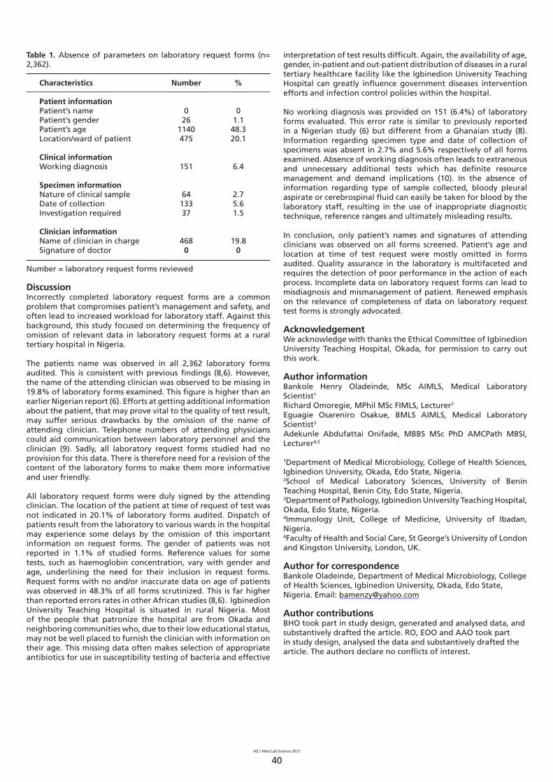
Table 1. Absence of parameters on laboratory request forms (n= 2,362).
Characteristics Number %
Patient informationPatient’s name 0 0Patient’s gender 26 1.1Patient’s age 1140 48.3Location/ward of patient 475 20.1
Clinical informationWorking diagnosis 151 6.4
Specimen informationNature of clinical sample 64 2.7Date of collection 133 5.6Investigation required 37 1.5
Clinician informationName of clinician in charge 468 19.8Signature of doctor 0 0
Number = laboratory request forms reviewed
DiscussionIncorrectly completed laboratory request forms are a common problem that compromises patient’s management and safety, and often lead to increased workload for laboratory staff. Against this background, this study focused on determining the frequency of omission of relevant data in laboratory request forms at a rural tertiary hospital in Nigeria.
The patients name was observed in all 2,362 laboratory forms audited. This is consistent with previous fi ndings (8,6). However, the name of the attending clinician was observed to be missing in 19.8% of laboratory forms examined. This fi gure is higher than an earlier Nigerian report (6). Efforts at getting additional information about the patient, that may prove vital to the quality of test result, may suffer serious drawbacks by the omission of the name of attending clinician. Telephone numbers of attending physicians could aid communication between laboratory personnel and the clinician (9). Sadly, all laboratory request forms studied had no provision for this data. There is therefore need for a revision of the content of the laboratory forms to make them more informative and user friendly.
All laboratory request forms were duly signed by the attending clinician. The location of the patient at time of request of test was not indicated in 20.1% of laboratory forms audited. Dispatch of patients result from the laboratory to various wards in the hospital may experience some delays by the omission of this important information on request forms. The gender of patients was not reported in 1.1% of studied forms. Reference values for some tests, such as haemoglobin concentration, vary with gender and age, underlining the need for their inclusion in request forms. Request forms with no and/or inaccurate data on age of patients was observed in 48.3% of all forms scrutinized. This is far higher than reported errors rates in other African studies (8,6). Igbinedion University Teaching Hospital is situated in rural Nigeria. Most of the people that patronize the hospital are from Okada and neighboring communities who, due to their low educational status, may not be well placed to furnish the clinician with information on their age. This missing data often makes selection of appropriate antibiotics for use in susceptibility testing of bacteria and effective
interpretation of test results diffi cult. Again, the availability of age, gender, in-patient and out-patient distribution of diseases in a rural tertiary healthcare facility like the Igbinedion University Teaching Hospital can greatly infl uence government diseases intervention efforts and infection control policies within the hospital.
No working diagnosis was provided on 151 (6.4%) of laboratory forms evaluated. This error rate is similar to previously reported in a Nigerian study (6) but different from a Ghanaian study (8). Information regarding specimen type and date of collection of specimens was absent in 2.7% and 5.6% respectively of all forms examined. Absence of working diagnosis often leads to extraneous and unnecessary additional tests which has defi nite resource management and demand implications (10). In the absence of information regarding type of sample collected, bloody pleural aspirate or cerebrospinal fl uid can easily be taken for blood by the laboratory staff, resulting in the use of inappropriate diagnostic technique, reference ranges and ultimately misleading results.
In conclusion, only patient’s names and signatures of attending clinicians was observed on all forms screened. Patient’s age and location at time of test request were mostly omitted in forms audited. Quality assurance in the laboratory is multifaceted and requires the detection of poor performance in the action of each process. Incomplete data on laboratory request forms can lead to misdiagnosis and mismanagement of patient. Renewed emphasis on the relevance of completeness of data on laboratory request test forms is strongly advocated.
AcknowledgementWe acknowledge with thanks the Ethical Committee of Igbinedion University Teaching Hospital, Okada, for permission to carry out this work.
Author informationBankole Henry Oladeinde, MSc AIMLS, Medical Laboratory Scientist1
Richard Omoregie, MPhil MSc FIMLS, Lecturer2
Eguagie Osareniro Osakue, BMLS AIMLS, Medical Laboratory Scientist3
Adekunle Abdufattai Onifade, MBBS MSc PhD AMCPath MBSI, Lecturer4,5
1Department of Medical Microbiology, College of Health Sciences, Igbinedion University, Okada, Edo State, Nigeria.2School of Medical Laboratory Sciences, University of Benin Teaching Hospital, Benin City, Edo State, Nigeria.3Department of Pathology, Igbinedion University Teaching Hospital, Okada, Edo State, Nigeria.4Immunology Unit, College of Medicine, University of Ibadan, Nigeria. 4Faculty of Health and Social Care, St George’s University of London and Kingston University, London, UK.
Author for correspondenceBankole Oladeinde, Department of Medical Microbiology, College of Health Sciences, Igbinedion University, Okada, Edo State, Nigeria. Email: [email protected]
Author contributionsBHO took part in study design, generated and analysed data, and substantively drafted the article. RO, EOO and AAO took part in study design, analysed the data and substantively drafted the article. The authors declare no confl icts of interest.
NZ J Med Lab Science 2012
41
References
1. Plebani M. Errors in clinical laboratories or errors in laboratory medicine? Clin Chem Lab Med 2006; 44: 750-759.
2. Bonini P, Plebani M, Ceriotti F, Rubboli F. Errors in laboratory medicine. Clin Chem 2002; 48: 691-698.
3. Plebani M, Carraro P. Mistakes in a stat laboratory: types and frequency. Clin Chem 1997; 43: 1348-1351.
4. Lippi G, Guidi GC, Mattiuzzi C, Plebani M. Pre-analytical variability: the dark side of the moon in laboratory testing. Clin Chem Lab Med 2006; 44: 358-365.
5. Nutt L, Zemlin AE, Erasmus RT. Incomplete laboratory request forms: the extent and impact on critical results at a tertiary hospital in South Africa. Ann Clin Biochem 2008; 45: 463-466.
6. Adegoke OA, Idowu A A, Jeje O A. Incomplete laboratory request forms as a contributory factor to pre-analytical errors in a Nigerian teaching hospital. Afr J Biochem Res 2011; 14: 82-85.
7. Vanker N, van Wyk J, Zemlin AE, Erasmus RT. A Six Sigma approach to the rate and clinical effect of registration errors in a laboratory. J Clin Pathol 2010; 63: 434-437.
8. Olayemi E, Asiamah-Brini R. Evaluation of request forms submitted to the haematology laboratory in a Ghanaian tertiary hospital. Pan Afr Med J 2011; 8: 33.
9. Sharif MA, Mushtaq S, Mamoon N, Jamal S, Luqman M. Clinician’s responsibility in pre-analytical quality assurance of histopathology. Pak J Med Sci 2007; 23: 720-723.
10. Burton JL, Stephenson TJ. Are clinicians failing to supply adequate information when requesting a histopathological investigation? J Clin Pathol 2001; 54: 806-808.
SYSMEX CONFERENCE 2012:
Medical Lab Scientists, Lab Managers, IT Specialists
Interested in innovation in Health IT?Wednesday 10th - Friday 12th October 2012, Te Papa, Wellington
A unique opportunity for networking and education
- attend the Sysmex Conference 2012 to extend your
knowledge and learn from industry leaders.
The conference programme features presentations
on innovations that are advancing the provision of
healthcare, highlighting achievements in laboratory IT
such as electronic ordering, mobilisation, structured
pathology reporting and regionalisation.
Sysmex Conference 2012 is CPD point qualified.
Register Nowsysmex.co.nz/
conference2012
FURTHER YOUR CAREER IN MEDICAL LABORATORY SCIENCE.
SCHOOL OF APPLIED SCIENCE
to find out more:
0800 AUT UNIautsciences.ac.nz
The University for the changing world
Medical laboratory scientists are facing new challenges as technologies evolve and the nature of diagnostic services change. Top level evaluative and management skills are in demand along with specialised skills in research and development. AUT postgraduate qualifications in medical laboratory science are designed to ensure you are prepared for the challenges ahead.
AUT offers you the options of a Postgraduate Certificate, Diploma or full research Masters, with two pathways; a specialist scientist pathway to enhance your scientific knowledge or our management pathway.
To find out more, get in touch today.
NZ J Med Lab Science 2012
42
AbstractBackground: Blood gas analysis is a widely used procedure. In clinical practice, the physicians may not always have a blood gas analyzer in their proximity. Not infrequently, blood gas samples are stored in a fridge or on ice and read retrospectively. Continued anaerobic and aerobic metabolism in the blood may alter blood gases in the interval between drawing arterial blood and its analysis, which may cause a fall in the PaO2 and pH and a rise in the PaCO2.Methods: Two sets of arterial blood samples were obtained from hospitalized patients. After the initial analysis, one sample from each patient was put in raw ice within a specimen bag (0 to +1
oC) and the other in the fridge (+4 to +8 oC). These samples were submitted to serial analysis at 30 minutes, 1 hour and 2 hours after the initial analysis.Results: Two hundred arterial blood gas results from 25 patients were analysed. The mean values of PaO2, PaCO2, HCO3-, Na+, K+, Ca2+ and lactate at 0 minutes, 30 minutes, 1 hour and 2 hours were not signifi cantly different between the two alternatives of storage. However, within each group, signifi cant changes were found over time for PaO2, K
+, Na+, Ca2+ and lactate.Conclusions: When using plastic syringes, arterial blood gas analysis should be processed shortly after collecting the sample. Despite the fact that low temperatures can slow down the metabolism, neither the ice nor the fridge preserved all the sample parameters.
Keywords: blood gas analysis, refrigeration, ice, plastic syringes
N Z J Med Lab Sci 2012; 66: 42-45
IntroductionBlood gas analysis is a widely used procedure; however, in clinical practice, the physicians do not always have a blood gas analyzer in their proximity and, not infrequently, the samples are stored in a fridge or on ice and read retrospectively.
Continued anaerobic and aerobic metabolism in the blood after collection may alter blood gas composition in the interval between drawing arterial blood and its analysis, such as a fall in the PaO2 and pH and a rise in the PaCO2. These changes are temperature and time dependent (1-3). In addition, oxygen will diffuse into the sample, particularly in plastic syringes (2). However, previous research has indicated that if blood gas analysis is done within 30 minutes, there seems to be no reason to keep it on ice (2), but we found no previous studies comparing longer periods of preservation on ice versus in the fridge.
Based on the limited existing information, we designed this study to answer the following questions:
How long does it take for blood gas composition to change? • Which parameters change and when does that change • become statistically signifi cant? Are there any signifi cant differences between keeping the • samples in ice or in the fridge?
Stability of blood gases when refrigerated João Pedro Ferreira, Sara Vieira Silva, Patrícia Rodrigues, Miguel Araújo Abreu, José Miguel Maia, Daniela Carvalho and Luísa Carvalho
MethodsArterial blood samples were obtained randomly from hospitalized patients as part of their normal clinical management. All patients had an arterial line placed. One milliliter (mL) of blood was collected in 1 mL heparinized (2 IU/mL) plastic syringes (RAPID Lyte® 1 mL L/S Syringe, Siemens).
For each patient, two arterial blood samples were collected at the same time using the arterial line. Samples were mixed and care was taken to ensure no air entered the syringes. If air bubbles were found, these were carefully removed from the syringe after each analysis to avoid equilibration with room air. Samples were analysed immediately. After the initial analysis, one sample was placed on ice (0 to +1 oC) and the other one was placed in the fridge (+4 to +8 oC). The samples were capped using fi lter caps that came with the syringes. These samples were submitted to serial testing at 30 minutes, 1 hour and 2 hours after the initial analysis. The pH, PaO2, PaCO2, HCO3
-, Na+, K+, Ca2+ and lactate were measured.
All the determinations were performed using a RAPIDLab® 1200 Blood Gas Analyzer (Siemens Healthcare Diagnostics Inc., Deerfi eld, IL, USA) according to the manufacturer’s instructions. All samples were analysed under a standard temperature (4).
The number of samples needed (n=25) to obtain potentially signifi cant results was determined using Piface software (3). Statistical analysis was performed using Software IBM SPSS Statistics 19® (IBM Corporation, Somers, NY, USA).
Mean values of all parameters at 0 min, 30 min, 1 hr and 2 hr were compared between groups by an independent two-tailed t test within each group, and the magnitude of change was examined by comparing the differences in the measurements for each value at 0, 30 min, 1 hr, and 2 hr using a paired two-tailed t test. A p value of ≤0.05 was defi ned as signifi cant.
Ethics approval was obtained from the local ethics committee. Informed consent was obtained from the patients.
ResultsTwo hundred arterial blood determinations from 25 patients were entered in the study - 100 measurements for each of the two experimental conditions. The mean values of PaO2, PaCO2, HCO3
-, Na+, K+, Ca2+ and lactate at 0 min, 30 min, 1 hr and 2 hr were not signifi cantly different between the two groups (Table 1).

NZ J Med Lab Science 2012
43
Table 1. Blood gas values at 0, 30 min, 1 hr and 2 hr. Comparisons between groups.
Blood gas parameter Time Ice Fridge p
pH 0 7.39 ±0.09 7.40 ±0.09 NS
30 min 7.40 ±0.09 7.41 ±0.09 NS
1 hr 7.40 ±0.09 7.40 ±0.09 NS
2 hr 7.39 ±0.10 7.40 ±0.10 NS
PaO2
mm hg
0 71.96 ±14.0 72.25 ±12.90 NS
30 min 77.09 ±16.22 78.75 ±17.07 NS
1 hr 84.62 ±21.29 84.84 ±21.55 NS
2 hr 92.49 ±25.16 95.39 ±27.15 NS
PaCO2
mm hg
0 42.25 ±25.54 43.92 ±25.26 NS
30 min 44.66 ±24.86 43.07 ±24.05 NS
1 hr 44.56 ±25.36 43.84 ±24.99 NS
2 hr 44.44 ±25.31 42.96 ±24.64 NS
HCO3-
mmol/L
0 25.99 ±8.05 25.10 ±7.47 NS
30 min 25.84 ±8.13 24.87 ±7.10 NS
1 hr 25.92 ±8.02 24.99 ±7.62 NS
2 hr 25.54 ±7.99 24.32 ±7.04 NS
K+
mmol/L
0 4.17 ±0.91 4.03 ±1.00 NS
30 min 4.28 ±0.94 3.98 ±0.99 NS
1 hr 4.41 ±0.93 4.04 ±1.01 NS
2 hr 4.53 ±0.94 4.37 ±1.19 NS
Na+
mmol/L
0 138.08 ±5.96 138.58 ±6.00 NS
30 min 138.90 ±5.58 139.42 ±6.49 NS
1 hr 139.95 ±6.24 140.34 ±6.75 NS
2 hr 139.02 ±5.24 141.84 ±6.34 NS
Ca2+
mmol/L
0 1.13 ±0.09 1.11 ±0.09 NS
30 min 1.10 ±0.10 1.06 ±0.11 NS
1 hr 1.07 ±0.10 1.06 ±0.12 NS
2 hr 1.01 ±0.10 0.99 ±0.18 NS
Lactate mmol/L
0 1.31 ±0.60 1.32 ±0.59 NS
30 min 1.47 ±0.61 1.47 ±0.64 NS
1 hr 1.59 ±0.60 1.64 ±0.62 NS
2 hr 1.83 ±0.52 1.93 ±0.67 NS
NS=not signifi cant
However, within each group, signifi cant changes were found over time in the blood gases and electrolytes. The PaO2, K+, Na+ and lactate increased; PaCO2 and Ca2+ decreased (Table 2). The increase in the PaO2 was signifi cant in both groups, compared with baseline values. These changes were noticed at 30 min in both groups, increasing even further afterwards. The decrease in PaCO2 was statistically signifi cant at 30 min and 2 hr in the fridge, compared to baseline. No signifi cant differences were found when these samples were kept on ice. The pH remained stable in both groups; however the lactate levels increased signifi cantly in both groups after 30 min. The decrease in HCO3
- was statistically signifi cant only after 2 hr storage in the fridge.

NZ J Med Lab Science 2012
44
Table 2. Changes in blood gas values from 0 min to 30 min, 1 hr and 2 hr. Comparisons within groups.
Blood gas parameter
Values in comparison Ice p Fridge p
pH 0 and 30 min 0.01 ±0.03 NS 0.02 ±0.04 NS
0 min and 1hr 0.01 ±0.03 NS 0.01 ±0.03 NS
0 min and 2 hr 0.01 ±0.03 NS 0.01 ±0.04 NSPaO2
mm hg0 and 30 min 5.02 ±4.31 <0.001 6.28 ±7.10 <0.001
0 min and 1hr 12.39 ±13.21 <0.001 12.34 ±11.19 <0.001
0 min and 2hr 20.53 ±15.71 <0.001 23.27 ±17.01 <0.001PaCO2
mm hg0 and 30 min -0.58 ±2.16 NS -0.84 ±2.09 0.05
0 min and 1hr -0.68 ±2.23 NS -0.07 ±3.86 NS
0 min and 2hr -0.80 ±2.65 NS -1.04 ±2.27 =0.03HCO3
-
mmol/L0 and 30 min -0.14 ±0.93 NS -0.26 ±0.89 NS
0 min and 1hr -0.07 ±1.12 NS -0.09 ±1.78 NS
0 min and 2hr -0.45 ±1.21 NS -0.80 ±0.74 <0.001K+
mmol/L0 and 30 min 0.11 ±0.26 =0.05 -0.07 ±0.18 NS
0 min and 1hr 0.24 ±0.28 <0.001 0.01 ±0.19 NS
0 min and 2hr 0.36 ±0.34 <0.001 0.32 ±0.77 0.05Na+
mmol/L0 and 30 min 0.84 ±1.00 <0.001 0.83 ±1.90 0.04
0 min and 1hr 1.86 ±1.97 <0.001 1.71 ±2.58 0.003
0 min and 2hr 0.94 ±5.41 NS 3.28 ±2.90 <0.001Ca2+
mmol/L0 and 30min -0.03 ±0.06 NS -0.05 ±0.05 <0.001
0 min and 1hr -0.06 ±0.05 <0.001 -0.06 ±0.05 <0.001
0 min and 2hr -0.12 ±0.07 <0.001 -0.15 ±0.12 <0.001Lactatemmol/L
0 and 30min 0.15 ±0.13 <0.001 0.15 ±0.12 <0.001
0 min and 1hr 0.27 ±0.18 <0.001 0.31 ±0.17 <0.001
0 min and 2hr 0.46 ±0.22 <0.001 0.55 ±0.29 <0.001
NS=not signifi cant
The K+ started to increase signifi cantly after 30 min on ice, but when kept in the fridge signifi cant changes were observed only after 2 hr. Na+ increased in samples stored on ice at 30 min and 1hr, whereas in the fridge Na+ increased signifi cantly throughout the three time periods. The decrease in ionized Ca2+ was sustained in both groups, but this change appeared to begin later in the samples kept on ice.
DiscussionRefrigerating the blood may delay changes in arterial blood gas composition (5,6). A robust and well conducted study by Liss and Payne analysed the changes in PaO2, PaCO2 and pH at 0, 15 and 30 min (7). They found a statistically signifi cant increase in the PaO2 at 15 and 30 min in both groups, and a statistically signifi cant decrease in the PaCO2 at 15 min in both groups. There was also a statistically signifi cant decrease in the pH at 15 min in both groups. There were no differences between the samples stored at room temperature or in ice. In our study the changes in the blood gas composition over time were not in the direction as previously found (5, 8-9). The authors of those studies attributed their fi nding to the use of plastic syringes. Plastic syringes can act as semipermeable membranes and allow diffusion of gases (10). This was corroborated by another study (12).
According to Fletcher et al, the PaO2 of oxygenated water stored in glass syringes remains stable for 1 hr if kept on ice or at room temperature, but not in plastic syringes (8). Another study comparing plastic versus glass syringes regarding blood gas tensions
in samples with high oxygen partial pressures concluded that glass syringes are superior to plastic syringes in preserving samples with a high PaO2, and prompt and adequate cooling of such samples is essential for accurate blood gas analysis (12).
Our fi ndings are similar to those described by Liss and Payne (7). However, instead of comparing ice with room temperature, we compared ice with refrigeration using baseline measurements as standard. We also extended the time length, and analyzed changes in the ions and lactate.
Comparisons between groups (ice vs fridge) did not show statistically signifi cant differences. The most signifi cant changes in both groups were a rise in lactate levels and PaO2 and a decrease in Ca2+ over 2 hr. As in another study, no changes in pH, pCO2 and HCO3
- were found in samples stored on ice (13).
The changes in PaO2 are consistent and signifi cant in both groups, i.e. independently of storing blood gas samples on ice or in the fridge, the PaO2 rose signifi cantly at 30 min, 1 hr and at 2 hr. As other authors have already observed, when plastic syringes are placed on ice there is an infl ow of oxygen into the samples, because iced water exhibits a very high oxygen concentration (13). Besides, as the temperature decreases, there is a shift in the oxygen-hemoglobin dissociation curve towards the left and an increase in the solubility of oxygen in the plasma, resulting in an increase of the measured PaO2. A limitation is that we cannot exclude the possibility that sample manipulation and homogenization may have contributed to air entry into the sample.
NZ J Med Lab Science 2012
45
The decrease in PaCO2 could be explained using the same principle of gas equilibration between the syringe and the environment. However, the decrease in PaCO2 and HCO3- was signifi cant only after 2 hr in the fridge, explaining why there were no major changes in pH.
The increase in K+ and decrease in ionized Ca2+ can be explained by cell lysis that occurs over time, releasing K+ and PO4
-. PO4- binds
to Ca2+, decreasing ionized Ca2+. In addition K+ leaks out of red blood cells at temperatures around 4oC due to the inability of the membrane Na+K+-ATPase pump to work correctly and this could also be an explanation for the K+ results.
The increase in lactate was also expected because of anaerobic metabolism, particularly by red blood cells that utilize anaerobic pathways preferentially in their metabolism (11). Unexpectedly, a decrease in both the HCO3
- and pH was not observed and this interesting fi nding may be explained by the release of intracellular HCO3
- as cell lysis occurs.
ConclusionsWhen using plastic syringes, the arterial blood gas analysis should be processed shortly after the sample is collected. Despite the fact that low temperatures can slow down metabolism, neither the ice nor the fridge preserved all the sample characteristics. If however, the analysis of the blood gases needs to be postponed, the clinician should be aware that storage in ice or in the fridge are not very different (even though the former is possibly slightly better) and that the levels of PaO2 and lactate are probably overestimated.
AcknowledgementsThe authors acknowledge Dr. Álvaro Ferreira for his support and help in recruiting the participants for the study.
Author informationJoão Pedro Ferreira, MD, Internal Medicine Resident1
Sara Vieira Silva, MD, Internal Medicine Resident1
Patrícia Rodrigues, MD, Cardiology Resident1,2
Miguel Araújo Abreu, MD, Infectious Diseases Resident1,3
José Miguel Maia, MD, Internal Medicine Resident1
Daniela Carvalho, MD, Internal Medicine Resident1
Luísa Carvalho, MD, Internal Medicine Hospital Assistant1
1Internal Medicine Department, 2Cardiology Department and 3Infectious Diseases Department, Centro Hospitalar do Porto, Porto, Portugal.
Author contributionsJPF conceived the study, contributed to the analytic work, data acquisition and data analysis, and substantially drafted the main article. SVS contributed to the analytic work, data acquisition and data analysis, and added critical content to the article. PR contributed to data acquisition and added critical content to the article. MAA conceived the study and added critical content to the article. JMM, DC and LC added critical content to the article. The authors declare no confl icts of interest.
Author for correspondenceDr João Pedro Ferreira, Internal Medicine Department, Centro Hospitalar do Porto, Largo Prof. Abel Salazar, 4099-001 Porto, Portugal. Email: [email protected]
References
1. Foster JM, Terry ML. Studies on the energy metabolism of human leukocytes. 1. Oxidative phosphorylation by human leukocyte mitochondria. Blood 1967; 30: 168-175.
2. Shapiro BA. Clinical application of blood gases. Year Book Medical Publishers, Chicago. 1973: 100-101.
3. Milkulcik P. Análises Rapid – gases no sangue e muito mais (1st ed.). Siemens Healthcare Diagnostics Inc., 2008.
4. Lenth RV. (2006-9). Java Applets for Power and Sample Size [Computer software]. Retrieved from http://www.stat.uiowa.edu/~rlenth/Power.
5. Lenfant C, Aucutt C. Oxygen uptake and change in carbon dioxide tension in human blood stored at 37 C. J Appl Physiol 1965; 20: 503-508.
6. Fell WL Sr. Sampling and measurement of blood gases. In: Lane EE, Walker JF Jr, Eds. Clinical Arterial Blood Gas Analysis. CV Mosby, St. Louis; 1987: 199-229.
7. Liss HP, Payne CP Jr. Stability of blood gases in ice and at room temperature. Chest 1993; 103: 1120-1122.
8. Fletcher G, Barber JL. Effect of sampling technique on the determination of PaO2 during oxygen breathing. J Appl Physiol 1966; 21: 463-468.
9. Andersen OS. Sampling and storing of blood for determination of acid-base status. Scand J Clin Lab Invest 1961; 13: 196-204.
10. Mahoney JJ, Harvey JA, Wong RJ, Van Kessel AL. Changes in oxygen measurements when whole blood is stored in iced plastic or glass syringes. Clin Chem 1991; 37: 1244-1248.
11. Hess CE, Nichols AB, Hunt WB, Suratt PM. Pseudohypoxemia secondary to leukemia and thrombocytosis. N EngI J Med 1979; 301: 361-363.
12. Pretto JJ, Rochford PD. Effects of sample storage time, temperature and syringe type on blood gas tensions in samples with high oxygen partial pressures. Thorax 1994; 49: 610–612.
13. Beaulieu M, Lapointe Y, Vinet B. Stability of PO2, PCO2 and pH in fresh blood samples stored in a plastic syringe with low heparin in relation to various blood-gas and hematological parameters. Clin Biochem 1999; 32: 101-107.

NZ J Med Lab Science 2012
46
Gnetum bucholzianum belongs to the gnetaceae family usually with climbing jointed stems (1). It is a small tree with tiered branches and divaricate branchlets having broad glossy dark green leaves. It is found in Amucha Njaba LGA, Imo State Nigeria as well as other parts of the South Eastern Nigeria. In Igbo land it is called “Ukazi” or “Ukasi” while Efi k call it afang. Nutritionally G. bucholzianum is very rich in proteins and minerals. The leaves contain high nutritional values as it contains eight amino acids in signifi cant quantities (2). Dishes based on G. bucholzianum leaves are prominent on the menu list in some restaurants and hotels in Owerri, Nigeria.
Chloroquine is a member of an important series of chemically related anti-malarial agents, the quinolone derivatives. It is a synthetic drug used in the treatment of malaria. Being a 4-aminoquinoline, it is a rapidly acting blood schizontocide with some gametocytocidal activity (2). In this study, the effect of G. bucholzianum was evaluated to provide information on its attenuation effect on chloroquine-induced hepatoxicity and renal damage in Wistar rats.
Chloroquine (Emzor) was purchased from a standard pharmacy shop in Owerri, Imo State, Nigeria. The tablets were dissolved in distilled water according to the required concentrations required for administration to Wistar rats on the basis of their body weight.
G. bucholzianum was obtained from the Ekeonunwa market in Owerri Nigeria. The botanical identifi cation and authentication was confi rmed by Dr. C. Okere (Head of Department of Plant Science and Biotechnology, Imo State University ,Owerri). The plant material was sun dried for seven days. The dried leaves of the G. bucholzianum were milled to achieve a coarse powder used for extraction. The powder was macerated in a 400g percolator with
Attenuation of chloroquine-induced hepatoxicity and renal damage by Gnetum bucholzianum leaf extract Johnkennedy Nnodim, Augustine Ihim and Hellen Ifeoma Udujih
200ml of distilled water. The mixture was allowed to stand for 48 hours after which it was fi ltered. The fi ltrate was then placed in an oven to evaporate and the solid residue referred to as extract. The appropriate concentrations of the extract were made in distilled water for the experiments. Hence, the following concentrations: 200mg and 400mg were prepared.
Wistar albino rats, weighing between 160 and 240g and aged 8-12 weeks, were used in the study. These animals were obtained from the Animal House of College of Medicine and Health Sciences, Imo State University, Owerri Nigeria. They were kept under standard laboratory conditions, fed with commercial growers mash (Tops Feeds Ltd, Sapele, Nigeria). Water and feed were provided ad libitum. The animals were left for two weeks to acclimatize. The experimental protocol was approved by the local ethical committee for animal experimentation. The animals were handled in accordance with institutional guidelines for the care and use of animals for experimental purposes.
The animals were randomly assigned to four experimental groups (n = 6 in each group). The fi rst group of animals, which served as the control group, was given distilled water. Groups II, III and 1V were given chloroquine (970mg/kg body weight) and G. bucholzianum extract (200mg/body weight); chloroquine and G. bucholzianum extract (400mg/body weight) respectively for 14 days. In all groups the drug was administered through the oral route using a feeding tube attached to a 5ml syringe. All animals were allowed free access to food and water throughout the experiment.
Twenty four hours after the last doses were administered, the animals were sacrifi ced and blood collected for biochemical analysis.
Table 1. Hepatic and renal function parameters in rats given chloroquine with 200mg/kg and 400mg/kg body weight of extract of G. bucholzianum.
Group
Treatment AST IU/L ALT IU/L ALP IU/L Bilirubin mg/dl Ureamg/dl
Creatininemg/dl
1Control 14.3 ±2.3 12.1 ±2.8 62.7 ±8.4 0.5 ±0.2 24.7 ±3.5 0.7 ±0.11
2Chloroquine 29.5 ± 3.6* 23.3 ±2.7* 89.4 ±9.1* 1.6 ±0.99* 57.3 ±5.9* 2.2 ±0.09*
3Chloroquine
+200 Gb exract20.0 ±1.8* 17.5 ±2.9* 69.6 ±9.4* 1.0 ±0.08* 36.4 ±4.7* 1.7 ±0.13*
4Chloroquine
+400 Gb extract17.3 ±3.1* 15.9 ±3.5* 66.3 ±7.8* 0.8 ±0.19* 31.7 ±5.2* 1.3 ±0.15*
Gb: G. bucholzianum *Signifi cantly different from control (P<0.05)
NZ J Med Lab Science 2012
47
In this study, chloroquine administration in a dose of 970 mg body weight of Wistar rats elevated serum hepatic and renal parameters. The damage to the liver and kidney resulted in signifi cantly increased bilirubin, AST, ALT, ALP, urea and creatinine levels..The liver and renal damage may be due to generation of free radicals by chloroquine overdose which is also partly responsible for its anti-malaria effects (4). This harmful effect could be caused by free radicals produced during peroxide formation. The level of hydroxylchoroquine treatment may be responsible for the hepatic and renal impairment (5). However, the simultaneous administration of Gnetum bucholzianum signifi cantly reduced the effect of chloroquine by attenuating hepatic and renal parameters. This could be of importance in Nigeria if Gnetum bucholzianum is consumed while on chloroquine medication for malaria.
Author informationJohnkennedy Nnodim*, BMLS MSc AMLSCN, Lecturer1
Augustine Ihim, BMLS MSc AMLSCN, Medical Laboratory Scientist2
Hellen Ifeoma Udujih, BMLS MSc AMLSCN, Lecturer1
1Department of Medical Laboratory Science, Faculty of Health Science, Imo State University, Imo State, Nigeria2Department of Medical Laboratory Science, Nnamdi Azikiwe University, Nnewi Campus, Anambra State, Nigeria
*To whom correspondence should be addressed. Email: [email protected]
References
Nkwatoh AF, Labode P, Iyassa SM, Nkwatoh FW, Ndumbe NL, 1. Ewane ME. Harvesting and marketing of Gnetum species (Engl) in Cameroon and Nigeria. J Ecol Nat Environ 2010; 2: 187-193.Lowe J. Gnetum in west Africa. 2. Nigeria Field 1984; 49 (1-4): 99-104.Sowunmi A, Fehintola FA, Adedeji AA, Falade AG, Falade 3. CO, Akinyinka OO, Oduola AMJ. Comparative effi cacy of chloroquine plus chlorpheniramine alone and in a sequential combination with sulfadoxine-pyrimethamine for the treatment of acute, uncomplicated, falciparum malaria in children. Ann Trop Med Parasitol 2000; 94: 209- 217.Ansari NM, Houlihan L, Hussain B, Pieroni A. Antioxidant 4. activity of fi ve vegetables traditionally consumed by south-Asian migrants in Bradford, Yorkshire, UK. Phytother Res 2005; 19: 907 – 911. Nnodim JK, 5. Adamma E, Austin A, Chukwunyere NNE. Alterations in biochemical parameters of wistar rats administrated with sulfudoxine and pyrimethamine (FansidarR). Al Almeen J Med Sci 2010; 3: 317-321.
N Z J Med Lab Sci 2012; 66: 46-47
Up and Coming NZIMLS Events
Event Location Date
2012
NZIMLS Conference Wellington Convention Centre
27-31 August 2012
NZIMLS Annual General Meeting
Wellington Convention Centre
29 August 2012
PreAnalytical SIG Seminar
Waipuna Hotel & Conference Centre, Auckland
6 October 2012
Histology SIG Seminar
Coachman Hotel, Palmerston North
13 October 2012
Mortuary SIG Seminar
Palmerston North Hospital
3 November 2012
2013
Haematology SIG Seminar
Napier War Memorial
2 March 2013
NZIMLS Conference Claudelands Events Centre, Hamilton
August 2013
NZIMLS Annual Scientific Meeting
Delphic LIS Workshop: Focussing on the Future.28th August 2012, 3.30 – 5pm, Wellington Town Hall
The Delphic LIS is the backbone for the medical laboratory – streamlining all processes with accuracy, efficiency and reliability.
Sysmex is holding a LIS workshop as part of the scientific programme at the NZIMLS Annual Scientific Meeting.
This workshop is open to everyone who has a keen interest in the laboratory information system, to share and discuss ideas that will lead the LIS forward with your labs.
For more information Contact Colin McKenzie
NZ J Med Lab Science 2012
48
AbstractKingella kingae, a Gram negative bacillus normally found in the oropharynx of infants, is a well recognised cause of invasive bone and joint infections in paediatric patients. Osteoarticular infections due to K. kingae in adults are much less common. We report an interesting case of K. kingae septic arthritis of the ankle in a 68 year old woman with rheumatoid arthritis, followed by a brief literature review.
Key words: Kingella kingae, septic arthritis, ankle
N Z J Med Lab Sci 2012; 66: 48-49
IntroductionKingella kingae, a short Gram negative bacillus, is best known for being a constituent of the HACEK group of organisms; i.e. Aggregatibacter (formerly the aphrophilus group of Haemophilus and Actinobacillus), Cardiobacterium, Eikenella, and Kingella spp (1). It is part of the normal pharyngeal fl ora in children, and is well recognised as a cause of invasive bone and joint infections in this age group (2). However infections due to K. kingae in adult patients are much less common and the patients usually have a degree of immunocompromise. We report here an unusual case of ankle septic arthritis due to K. kingae in an adult patient.
Case ReportA 68 year old woman, with a 40 year history of rheumatoid arthritis, was admitted with a 2 day history of acute on chronic worsening of pain and swelling in her right ankle. Having undergone left total hip and bilateral knee joint replacements, she was awaiting a right ankle replacement. At presentation she was taking azathioprine, prednisone and celecoxib for her rheumatoid arthritis.
On examination she was febrile with a warm, swollen, erythematous right ankle, with pain on movement and tenderness over the medial and lateral malleoli. Blood tests showed a normal leucocyte count but raised infl ammatory markers (CRP: 161mg/L, ESR: 72 mm/hr). X-rays confi rmed arthritic changes in the ankle and foot.
Aspiration of the ankle recovered 1.5ml of turbid fl uid. A formal white cell count was not performed. A Gram stain demonstrated large numbers of white cells, which were predominantly neutrophils, but no organisms were visualised. The sample was of insuffi cient volume for biochemistry analysis. The aspirate was inoculated on to blood and chocolate agar (CO2 incubation at 37°C) and Fastidious Anaerobe Agar (anaerobic incubation at 37°C). There was insuffi cient aspirate to inoculate a blood culture bottle. After 48 hours incubation, small colonies were visualised on both the chocolate and blood agar plates. Gram stain of these colonies revealed a short, plump Gram negative bacillus. The colonies were slightly haemolytic with a strong positive oxidase reaction. Catalase testing was negative. Biochemical profi le testing (Rapid NH, Remel) confi rmed the identifi cation of K. kingae (code 1120, 99.9% probability). The isolate was susceptible to penicillin with an MIC of 0.16mg/l. Peripheral blood cultures yielded no growth after 5 days incubation.
Empirical treatment with fl ucloxacillin (2g 6-hourly) and benzyl penicillin (1.2g 6-hourly) was rationalised to benzyl penicillin alone 1.2g 4-hourly. The patient improved rapidly on the antimicrobial
Septic arthritis due to Kingella kingae in an adult patient Ajay Sanghvi, Michael Addidle and Kate Grimwade
therapy alone and washout of the joint was not undertaken. She was discharged to the community where she received a benzyl penicillin infusion of 8g over 24 hours via PICC line for 6 weeks. She continued to make an uneventful recovery and has subsequently gone on to have an arthrodesis of her ankle, which was preferred to replacement following the infection.
DiscussionKingella kingae was originally placed under the Moraxella genus and named Moraxella kingii after Elizabeth O King of the US Centers for Disease Control(CDC) who isolated the bacterium in 1960 (3). It was later transferred to its own genus and renamed Kingella kingae in 1976 (4). K. kingae are facultative anaerobic Gram negative rods which lie together in small clusters and decolourise unevenly on Gram stain. Small colonies are seen after 48 hours and usually have a small zone of beta haemolysis on blood agar. They are oxidase positive.
K. kingae has been shown to be a commensal of the oropharyngeal tract in early childhood. Previously considered a rare cause of human infection, it is now recognised as an important cause of invasive infection in paediatric patients, predominantly under the age of 2 years (2). Infections have most commonly been reported in the bones and joints of children. K. kingae constitutes one of the HACEK group of organisms which are collectively responsible for 3-5% of cases of bacterial endocarditis (2). They are also known to cause lower-respiratory tract infections, meningitis, ocular infections and stomatitis. According to the literature, almost 90% of patients with invasive Kingella kingae infections are under the age of 5 years, with 60% of episodes occurring in the under 2 year age group (5). Based on studies on children in day-care centres it is thought Kingella kingae is transmitted from child to child via saliva particles (6). In contrast to the healthy children acquiring Kingella kingae infections, adults frequently have pre-disposing factors such as rheumatoid arthritis, Felty’s syndrome, liver cirrhosis, systemic lupus erythematosus, renal disease, sickle cell anaemia, malignancies and HIV(2,7-9). The fact that relatively few invasive K. kingae infections occur in immunocompetent adults indicates that protection from colonisation and infection requires an acquired immune response (2).
Few previous cases of adult joint sepsis caused by K. kingae have been reported in the literature. Of these few, most involve the knee and one case involved a septic elbow (7, 10-13). As far as we are aware, this is the fi rst reported case of adult septic arthritis caused by K. kingae, occurring in an ankle joint.
The subtle clinical manifestations of K. kingae have been noted previously with patients often presenting only with mild malaise and no or minimal fevers (2,7). K. kingae infections predominantly affect the large weight bearing joints such as hips, knees and ankles (2). Patients with osteo-articular infections due to K. kingae often have blood leucocyte counts, c-reactive proteins and erythrocyte sedimentation rates that are within normal limits or only mildly elevated. The bacterial count in the synovial fl uid is often low. As with other HACEK organisms K. kingae is a fastidious bacterium. Recovery of K kingae from body fl uids and pus can thus be problematic because these types of specimens seem to be inhibitory to the bacteria. Inoculating synovial fl uid into blood culture medium has been shown to increase the likelihood of isolating the organism (2,10).

NZ J Med Lab Science 2012
49
The technology available for rapid identifi cation of bacteria from clinical samples is now rapidly evolving. Targeted PCR is currently the most sensitive method for detecting K. kingae directly from sterile site aspirates (14). However, this is not practical when looking for many potential pathogens in a clinical sample. 16S rRNA gene sequencing offers a more broad based approach, with the ability to look for many different potential pathogens at once, but is less sensitive at detecting K. kingae than targeted PCR (15). The recent introduction of MALDI-TOF (Matrix Associated Laser Desorption and Ionisation-Time of Flight) technology into many of the bigger diagnostic laboratories may expedite identifi cation once K. kingae colonies are growing on the plate (16). However there has been no research done as yet on the ability of MALDI-TOF technology to detect HACEK organisms directly from patient samples. Other technology which may be of value here is PCR-Electrospray Ionisation/Mass Spectrometry, which combines both PCR and mass spectrometry technology to allow rapid and sensitive identifi cation of a wide spectrum of pathogens directly from patient samples (17). Micro-array may also offer a sensitive and practical approach to the molecular diagnosis of K. kingae infections in the future; although at the time of writing there is little available commercially using microarray technology for bacterial identifi cation.
There are no specifi c guidelines or optimal therapy for K. kingae. Patients are treated empirically until cultures reveal the isolate and its susceptibility pattern. The organism is usually susceptible to beta-lactams however beta-lactamase producing K. kingae isolates have been described (2). Intravenous penicillin is a standard treatment for those isolates which have tested susceptible (18).
ConclusionThis case demonstrates that K. kingae can occasionally cause septic arthritis in adults, particularly those with underlying immunocompromise. Inoculation of synovial fl uid into blood culture media improves chances of isolating the organism and should be recommended as routine procedure to improve the recovery rate of these types of fastidious organisms. Newly emerging identifi cation technologies may in the future assist with the more rapid and sensitive diagnosis of this organism.
Author informationAjay Sanghvi, BSc MBChB1
Michael Addidle, MBChB MRCP DTM&H FRCPath2 Kate Grimwade, MBChB MRCP PhD DTM&H3
1John Radcliffe Hospital, Oxford, UK2Pathlab Bay of Plenty, Tauranga, New Zealand3Tauranga Hospital, Tauranga, New Zealand
Author contributionsAS: substantive drafting of the main article. MA: revision of article and literature review. KG: clinical care of patient and revision of article. The authors declare no confl icts of interest.
Author for correspondenceMichael Addidle, Pathlab Bay of Plenty, PO Box 130, Tauranga 3140. Email: [email protected]
References
1. J Versalovic (Editor). Manual of Clinical Microbiology, 10th Ed; American Society of Microbiology Press, USA. Vol 1: pg 1192.
2. Yagupsky P. Kingella kingae: from medical rarity to an emerging paediatric pathogen. Lancet Infect Dis 2004; 4: 358-367.
3. Henriksen SD, Bøvre K. Moraxella kingii sp. Nov., a haemolytic saccharolytic species of the genus Moraxella. J Gen Microbiol 1968; 58: 377-385.
4. Henriksen SD, Bøvre K. Transfer of Moraxella kingii Henriksen and Bφvre to the genus Kingella gen. nov. in the family Neiseriaceae. Int J Syst Bacteriol 1976; 26: 447-50.
5. Yagupsky P, Dagan R, Howard CB, Einhorn M, Kassis I, Simu A. Clinical features and epidemiology of invasive Kingella kingae infections in southern Israel. Pediatrics 1993; 92: 800-804.
6. Slonim A, Walker ES, Mishori E, Porat N, Dagan R, Yagupsky P. Person-to-person transmission of Kingella kingae among day care center attendees. J Infect Dis 1998; 178: 1843-1846.
7. Lewis DA, Settas L. Kingella kingae causing septic arthritis in Felty’s syndrome. Postgrad Med J 1983; 59: 525-526.
8. Wolak T, Abu-Shakra M, Flusser D, Liel-Cohen N, Buskila D, Sukenik S. Kingella endocarditis and meningitis in a patient with SLE and associated antiphospholipid syndrome. Lupus 2000; 9: 393-396.
9. Urs S, D’Silva BS, Jeena CP, Sona CP, Beena K, Shetty KR. Kingella kingae septicemia in association with HIV disease. Trop Doct 1994; 24: 127.
10. Elyès B, Mehdi G, Haj Slama Kamel B, Hela Z, Smida Imen B. Kingella kingae septic arthritis with endocarditis in an adult. Joint Bone Spine 2006; 73: 472-473.
11. Estève V, Porcheret H, Clerc D, Dorfmann H, Le Pennec MP. Septic arthritis due to Kingella kingae in an adult. Joint Bone Spine 2001; 68: 85-86.
12. Vincent J, Podewell C, Franklin GW, Korn JH. Septic arthritis due to Kingella (Moraxella) kingae: case report and review of literature. J Rheumatol 1981; 8: 501-503.
13. Salminen I, Von Essen R, Koota K, Nissinen A. A pitfall in purulent arthritis brought out in Kingella kingae infection of the knee. Ann Rheum Dis 1984; 43: 656-657.
14. Moumile K, Merckx J, Glorion C, Berche P, Ferroni A. Osteoarticular infections caused by Kingella kingae in children: contribution of polymerase chain reaction to the microbiologic diagnosis. Paediatr Infect Dis J 2003; 22: 837-839.
15. Cherkaoui A, Ceroni D, Emonet S, Lefevre Y, Schrenzel J. Molecular diagnosis of Kingella kingae osteoarticular infections by specifi c real-time PCR assay. J Med Microbiol 2009; 58: 65-68.
16. Couturier MR, Mehinovic E, Croft AC, Fisher MA. Identifcation of HACEK clinical isolates by matrix-assisted laser desorption-time of fl ight mass spectrometry. J Clin Microbiol 2011; 49: 1104-1106.
17. Kaleta EJ, Clark AE, Cherkaoui A, Wysocki VH, Ingram EL, Schrenzel J, et al. Comparative analysis of PCR–electrospray ionization/mass spectrometry (MS) and MALDI-TOF/MS for the identifi cation of bacteria and yeast from positive blood culture bottles. Clin Chem 2011; 57: 1057-1067.
18. Sordillo EM, Rendel M, Sood R, Belinfanti J, Murray O, Brook D. Septicaemia due to beta-lactamase-positive Kingella kingae. Clin Infect Dis 1993; 17: 818-819.
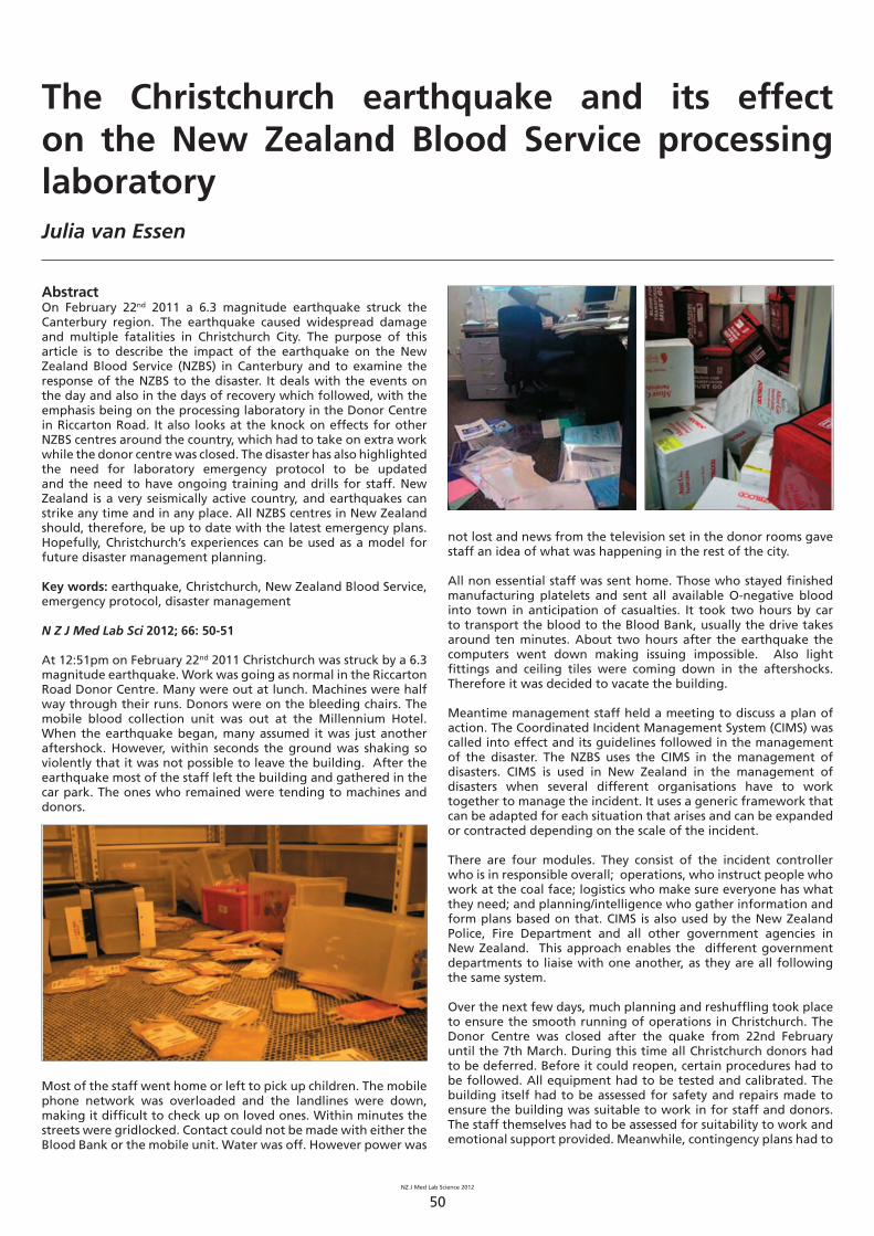
NZ J Med Lab Science 2012
50
AbstractOn February 22nd 2011 a 6.3 magnitude earthquake struck the Canterbury region. The earthquake caused widespread damage and multiple fatalities in Christchurch City. The purpose of this article is to describe the impact of the earthquake on the New Zealand Blood Service (NZBS) in Canterbury and to examine the response of the NZBS to the disaster. It deals with the events on the day and also in the days of recovery which followed, with the emphasis being on the processing laboratory in the Donor Centre in Riccarton Road. It also looks at the knock on effects for other NZBS centres around the country, which had to take on extra work while the donor centre was closed. The disaster has also highlighted the need for laboratory emergency protocol to be updated and the need to have ongoing training and drills for staff. New Zealand is a very seismically active country, and earthquakes can strike any time and in any place. All NZBS centres in New Zealand should, therefore, be up to date with the latest emergency plans. Hopefully, Christchurch’s experiences can be used as a model for future disaster management planning.
Key words: earthquake, Christchurch, New Zealand Blood Service, emergency protocol, disaster management
N Z J Med Lab Sci 2012; 66: 50-51
At 12:51pm on February 22nd 2011 Christchurch was struck by a 6.3 magnitude earthquake. Work was going as normal in the Riccarton Road Donor Centre. Many were out at lunch. Machines were half way through their runs. Donors were on the bleeding chairs. The mobile blood collection unit was out at the Millennium Hotel. When the earthquake began, many assumed it was just another aftershock. However, within seconds the ground was shaking so violently that it was not possible to leave the building. After the earthquake most of the staff left the building and gathered in the car park. The ones who remained were tending to machines and donors.
Most of the staff went home or left to pick up children. The mobile phone network was overloaded and the landlines were down, making it diffi cult to check up on loved ones. Within minutes the streets were gridlocked. Contact could not be made with either the Blood Bank or the mobile unit. Water was off. However power was
The Christchurch earthquake and its effect on the New Zealand Blood Service processing laboratoryJulia van Essen
not lost and news from the television set in the donor rooms gave staff an idea of what was happening in the rest of the city.
All non essential staff was sent home. Those who stayed fi nished manufacturing platelets and sent all available O-negative blood into town in anticipation of casualties. It took two hours by car to transport the blood to the Blood Bank, usually the drive takes around ten minutes. About two hours after the earthquake the computers went down making issuing impossible. Also light fi ttings and ceiling tiles were coming down in the aftershocks. Therefore it was decided to vacate the building.
Meantime management staff held a meeting to discuss a plan of action. The Coordinated Incident Management System (CIMS) was called into effect and its guidelines followed in the management of the disaster. The NZBS uses the CIMS in the management of disasters. CIMS is used in New Zealand in the management of disasters when several different organisations have to work together to manage the incident. It uses a generic framework that can be adapted for each situation that arises and can be expanded or contracted depending on the scale of the incident.
There are four modules. They consist of the incident controller who is in responsible overall; operations, who instruct people who work at the coal face; logistics who make sure everyone has what they need; and planning/intelligence who gather information and form plans based on that. CIMS is also used by the New Zealand Police, Fire Department and all other government agencies in New Zealand. This approach enables the different government departments to liaise with one another, as they are all following the same system.
Over the next few days, much planning and reshuffl ing took place to ensure the smooth running of operations in Christchurch. The Donor Centre was closed after the quake from 22nd February until the 7th March. During this time all Christchurch donors had to be deferred. Before it could reopen, certain procedures had to be followed. All equipment had to be tested and calibrated. The building itself had to be assessed for safety and repairs made to ensure the building was suitable to work in for staff and donors. The staff themselves had to be assessed for suitability to work and emotional support provided. Meanwhile, contingency plans had to
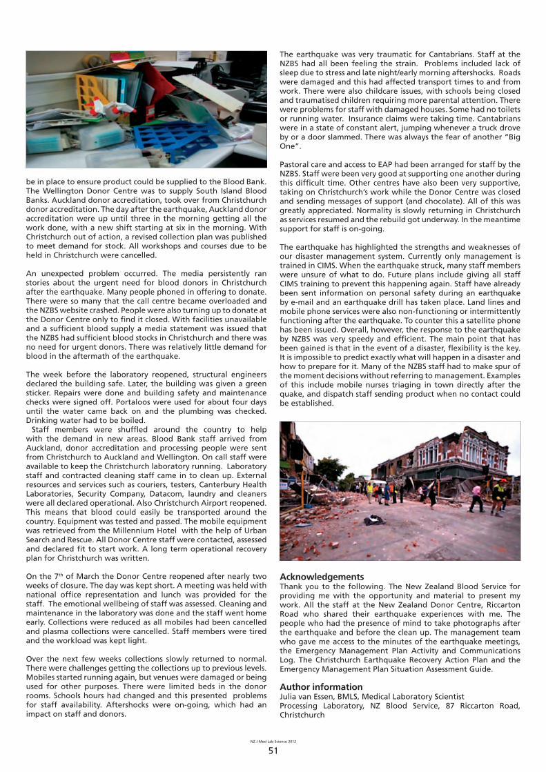
NZ J Med Lab Science 2012
51
be in place to ensure product could be supplied to the Blood Bank. The Wellington Donor Centre was to supply South Island Blood Banks. Auckland donor accreditation, took over from Christchurch donor accreditation. The day after the earthquake, Auckland donor accreditation were up until three in the morning getting all the work done, with a new shift starting at six in the morning. With Christchurch out of action, a revised collection plan was published to meet demand for stock. All workshops and courses due to be held in Christchurch were cancelled.
An unexpected problem occurred. The media persistently ran stories about the urgent need for blood donors in Christchurch after the earthquake. Many people phoned in offering to donate. There were so many that the call centre became overloaded and the NZBS website crashed. People were also turning up to donate at the Donor Centre only to fi nd it closed. With facilities unavailable and a suffi cient blood supply a media statement was issued that the NZBS had suffi cient blood stocks in Christchurch and there was no need for urgent donors. There was relatively little demand for blood in the aftermath of the earthquake.
The week before the laboratory reopened, structural engineers declared the building safe. Later, the building was given a green sticker. Repairs were done and building safety and maintenance checks were signed off. Portaloos were used for about four days until the water came back on and the plumbing was checked. Drinking water had to be boiled. Staff members were shuffl ed around the country to help with the demand in new areas. Blood Bank staff arrived from Auckland, donor accreditation and processing people were sent from Christchurch to Auckland and Wellington. On call staff were available to keep the Christchurch laboratory running. Laboratory staff and contracted cleaning staff came in to clean up. External resources and services such as couriers, testers, Canterbury Health Laboratories, Security Company, Datacom, laundry and cleaners were all declared operational. Also Christchurch Airport reopened. This means that blood could easily be transported around the country. Equipment was tested and passed. The mobile equipment was retrieved from the Millennium Hotel with the help of Urban Search and Rescue. All Donor Centre staff were contacted, assessed and declared fi t to start work. A long term operational recovery plan for Christchurch was written.
On the 7th of March the Donor Centre reopened after nearly two weeks of closure. The day was kept short. A meeting was held with national offi ce representation and lunch was provided for the staff. The emotional wellbeing of staff was assessed. Cleaning and maintenance in the laboratory was done and the staff went home early. Collections were reduced as all mobiles had been cancelled and plasma collections were cancelled. Staff members were tired and the workload was kept light.
Over the next few weeks collections slowly returned to normal. There were challenges getting the collections up to previous levels. Mobiles started running again, but venues were damaged or being used for other purposes. There were limited beds in the donor rooms. Schools hours had changed and this presented problems for staff availability. Aftershocks were on-going, which had an impact on staff and donors.
The earthquake was very traumatic for Cantabrians. Staff at the NZBS had all been feeling the strain. Problems included lack of sleep due to stress and late night/early morning aftershocks. Roads were damaged and this had affected transport times to and from work. There were also childcare issues, with schools being closed and traumatised children requiring more parental attention. There were problems for staff with damaged houses. Some had no toilets or running water. Insurance claims were taking time. Cantabrians were in a state of constant alert, jumping whenever a truck drove by or a door slammed. There was always the fear of another “Big One”.
Pastoral care and access to EAP had been arranged for staff by the NZBS. Staff were been very good at supporting one another during this diffi cult time. Other centres have also been very supportive, taking on Christchurch’s work while the Donor Centre was closed and sending messages of support (and chocolate). All of this was greatly appreciated. Normality is slowly returning in Christchurch as services resumed and the rebuild got underway. In the meantime support for staff is on-going.
The earthquake has highlighted the strengths and weaknesses of our disaster management system. Currently only management is trained in CIMS. When the earthquake struck, many staff members were unsure of what to do. Future plans include giving all staff CIMS training to prevent this happening again. Staff have already been sent information on personal safety during an earthquake by e-mail and an earthquake drill has taken place. Land lines and mobile phone services were also non-functioning or intermittently functioning after the earthquake. To counter this a satellite phone has been issued. Overall, however, the response to the earthquake by NZBS was very speedy and effi cient. The main point that has been gained is that in the event of a disaster, fl exibility is the key. It is impossible to predict exactly what will happen in a disaster and how to prepare for it. Many of the NZBS staff had to make spur of the moment decisions without referring to management. Examples of this include mobile nurses triaging in town directly after the quake, and dispatch staff sending product when no contact could be established.
AcknowledgementsThank you to the following. The New Zealand Blood Service for providing me with the opportunity and material to present my work. All the staff at the New Zealand Donor Centre, Riccarton Road who shared their earthquake experiences with me. The people who had the presence of mind to take photographs after the earthquake and before the clean up. The management team who gave me access to the minutes of the earthquake meetings, the Emergency Management Plan Activity and Communications Log. The Christchurch Earthquake Recovery Action Plan and the Emergency Management Plan Situation Assessment Guide.
Author informationJulia van Essen, BMLS, Medical Laboratory ScientistProcessing Laboratory, NZ Blood Service, 87 Riccarton Road, Christchurch
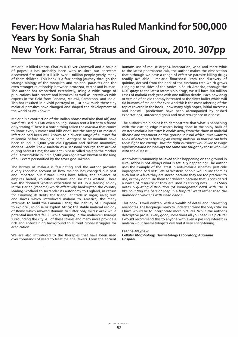
NZ J Med Lab Science 2012
52
Malaria. It killed Dante, Charles II, Oliver Cromwell and a couple of popes. It has probably been with us since our ancestors discovered fi re and it still kills over 1 million people yearly, many of them children. This book is a fascinating journey through the strange biology of the mosquito and malarial parasites and the even stranger relationship between protozoa, vector and human. The author has researched extensively, using a wide range of publications both recent and historical as well as interviews with experts in the fi eld from Panama, Malawi, Cameroon, and India. This has resulted in a vivid portrayal of just how much these tiny malarial parasites have changed and shaped the development of the world as we know it.
Malaria is a contraction of the Italian phrase mal’aire (bad air) and was fi rst used in 1740 when an Englishman sent a letter to a friend drily stating “There is a horrid thing called the mal’aria that comes to Rome every summer and kills one”. But the ravages of malarial infection had been well known to a diverse range of cultures for millennia before having a name. Antigens to plasmodium have been found in 5,000 year old Egyptian and Nubian mummies; ancient Greeks knew malaria as a seasonal scourge that arrived during harvest time; the ancient Chinese called malaria the mother of all fevers while in India 3,500 years ago it was known as the King of all Fevers personifi ed by the fever god Takman.
The history of malaria is fascinating and the author provides a very readable account of how malaria has changed our past and impacted our future. Cities have fallen, the advance of empires halted, countless nations and societies wasted. There was the doomed Scottish expedition to set up a trading colony in the Darien (Panama) which effectively bankrupted the country leading Scotland to surrender its autonomy to England, in return for assuming its debts; the triangular trade in sugar, silver, rum and slaves which introduced malaria to America; the many attempts to build the Panama Canal; the inability of Europeans to explore , colonise or exploit Africa; the stable malarial ecology of Rome which allowed Romans to suffer only mild P.vivax while potential invaders fell ill while camping in the malarious swamps surrounding the city. All of these stories and many more provide a rich and entertaining background to current global struggles for eradication.
We are also introduced to the therapies that have been used over thousands of years to treat malarial fevers. From the ancient
Fever: How Malaria has Ruled Mankind for 500,000 Years by Sonia ShahNew York: Farrar, Straus and Giroux, 2010. 307pp
Romans use of mouse organs, incantation, wine and more wine to the latest pharmaceuticals, the author makes the observation that although we have a range of effective parasite-killing drugs readily available – malaria fl ourishes! From the discovery of quinine, derived from the bark of the cinchona tree which grows clinging to the sides of the Andes in South America, through the DDT sprays to the latest artemisinin drugs, we still have 300 million cases of malaria each year with one million deaths. Each new drug or version of an old therapy is treated as the silver bullet which will rid humans of malaria for ever. And this is the most sobering of the topics covered in the book – how many high hopes, initial successes and boastful predictions have been accompanied by dashed expectations, unreached goals and new resurgence of disease.
The author’s main point is to demonstrate that what is happening with the cutting edge research being done in the shiny halls of western malaria institutes is worlds away from the chaos of malarial disease and treatment on the ground in rural Africa. “We want to think of Africans as battling an enemy, malaria, so that we can help them fi ght the enemy…but the fi ght outsiders would like to wage against malaria isn’t always the same one fought by those who live with the disease”. And what is commonly believed to be happening on the ground in rural Africa is not always what is actually happening! The author uses the example of the latest in anti-malaria schemes, pesticide impregnated bed nets. We as Western people would use them as such but in Africa they are stored because they are too precious to use, or they don’t use them for children because that is considered a waste of resource or they are used as fi shing nets…….as Shah notes “Equating distribution (of impregnated nets) with use is like counting the bars of soap in a hospital ward rather than the number of clinicians with clean hands”.
This book is well written, with a wealth of detail and interesting anecdotes. The language is easy to understand and the only criticism I have would be to incorporate more pictures. While the author’s descriptive prose is very good, sometimes all you need is a picture! I would recommend this to anyone with even a passing interest in malaria – but haematologists will fi nd it very enlightening.
Leanne MayhewCellular Morphology, Haematology Laboratory, Auckland Hospital
NZ J Med Lab Science 2012
53
Fellowship of the NZIMLS
Are you a medical laboratory scientist?
Do you have a post graduate qualifi cation?
If yes to the two above you may be exempt from sitting Part 1 (examination) of Fellowship of the NZIMLS and go straight to Part 2 consisting of a dissertation of 3000 - 5000 words.
The dissertation may take the form of a review, development of a hypothesis or any other presentation that meets with the approval of the Fellowship Committee.Qualifi cations recognised by the NZIMLS for the purpose of exemption to sit the Part 1 examinations are:
An academic postgraduate qualifi cation, normally at least a • postgraduate diploma, in medical laboratory science or closely related subject. The course of study must meet the minimum requirement of one year’s full-time studyFellowship of the Australian Institute of Medical Scientists • (FAIMS)Fellowship of the Institute of Biomedical Science (FIBMS)• Fellowship of the Australasian Association of Clinical Biochemists • (FAACB)Fellowship of the Institute of Biology, London (FSB)•
For full Fellowship regulations and the application process visit the NZIMLS web site: www.nzimls.org.nz or contact the Fellowship Committee Chair: Associate Professor Rob Siebers at [email protected]
Fellowship CommitteeRob Siebers, FNZIMLSAnn Thornton, FNZIMLSJillian Broadbent, FNZIMLS
The Olympus Journal Imaging Competition
The NZIMLS Journal invites applications for the annual Olympus photo competition giving NZIMLS members the chance to win an Olympus digital camera and have their photo published in the journal.
The general theme is “Medical Laboratory Science”, so whether it is related to haematology / histology, laboratory personnel, instruments, humour, or other, there’s plenty of scope for
keen photographers to showcase their talents.
Olympus, a leading manufacturer of professional opto-digital products, has generously donated a digital camera as the prize for the best photo.
Entries should be submitted as an email attachment to Rob Siebers, Editor of the NZIMLS Journal, at [email protected]. A title for the photo, together with the entrant’s name, place of work and email address, should accompany the attachment. Submissions can be in colour or black and white.
Entries close on 5pm on Friday 14th September 2012, with the winning photo appearing in the November 2012 issue of the Journal. Previously submitted entries will not be considered.
Judging will be carried out by the Editor, the Deputy-Editor and an Olympus representative. Their decision will be fi nal and no correspondence will be entered into. Entrants must be current fi nancial members of the NZIMLS to be eligible.
The NZIMLS Journal invites applications forthe annual Olympus photo competition givingNZIMLS members the chance to win an Olympusdigital camera and have their photo published inthe journal.
The general theme is “Medical LaboratoryScience”, so whether it is related to haematology/ histology, laboratory personnel, instruments,
NZ J Med Lab Science 2012
54
New Zealand Institute of Medical Laboratory Science
The Barrie Edwards & Rod Kennedy Scholarships
The Barrie Edwards & Rod Kennedy scholarship is one of the most signifi cant awards offered by the NZIMLS. The scholarship provides the winner with support to attend an international or national scientifi c meeting up to a maximum value of $7,500 for each.
Applications for this prestigious scholarship isinvited from Fellows, Members and Associate Members of the NZIMLS. Applicants must be a current fi nancial member of the NZIMLS and have been a fi nancial member for at least two concurrent years prior to application. To be eligible applicants must make an oral presentation or present a poster as 1st author at their nominated scientifi c meeting.
All applications will be considered by a panel consisting of the President and Vice-President of the NZIMLS and the Editor of the New Zealand Journal of Medical Laboratory Science (who are ineligible to apply for the scholarships). The applications will be judged on your professional and academic abilities together with your participation in the profession. The panel’s decision is fi nal and no correspondence will be entered into.
Application is by letter. Please address all correspondence to:
NZIMLS Executive Offi cerPO Box 505Rangiora 7440
There is one scholarship awarded in each calendar year. Closing date is December 20th in any given year.
In your application letter please provide the following details:
• Full name, position, work address, email address and contact phone number
• The length of time you have been a fi nancial member of the NZIMLS
• The conference you wish to attend - please provide dates • A budget comprising airfares, conference registration and
accommodation costs • The abstract of your intended oral or poster presentation
and whether it has been accepted for presentation (proof required)
• Your intentions to publish your results
• State briefl y your history of participation in the profession over the last 5 years
• State the reasons why you wish to attend your nominated scientifi c meeting
Successful applicants will be required to provide a full written report on return which will be published in the Journal. If not intended to publish elsewhere, successful applicants will be required to submit their study results for consideration by the New Zealand Journal of Medical Laboratory Science.
We thank the listed individuals who reviewed articles submitted to the Journal from September 2011 to August 2012, some more than once. All submitted articles undergo peer review in order that the Journal maintains its high standard. Additionally, thoughtful comments and suggestions made by reviewers help authors in ensuring that their articles, if accepted, are put in front of the reader in the best possible light. The Editors cannot be experts in all disciplines of medical laboratory science and thus rely on quality peer review by others.
Reviewers for September 2011 to August 2012Tony Barnett, NelsonMary Bilkey, AucklandShona Brougham, AucklandGlen Devanie, AucklandMike Legge, DunedinCat Ronayne, DunedinRob Siebers, WellingtonAnn Thornton, WellingtonPhilip Wakem, Wellington
We also thank the Journal’s Statistical Editor, Nevil Pierse, for his thoughtful review of statistics in submitted articles and Members of the Editorial Board for their advice and guidance.
Rob Siebers, FNZIMLS, EditorTerry Taylor, MNZIMLS, Deputy-Editor
Journal reviewers 2011/2012
NZ J Med Lab Science 2012
55
Journal questionnaire
Below are 10 questions based on articles in the August 2012 Journal issue. Read the articles fully and carefully, most questions require more than one answer.
Answers are to be submitted through the NZIMLS web site. Make sure you supply your correct email address and membership number. It is recommended that you write your answers in a word document and then cut and paste your answers on the web site.
The site has been developed for use with Microsoft’s Internet Explorer web browser. If you are having problems submitting your questionnaire and you are using the Firefox web browser, try resubmitting from a computer or system using Microsoft’s Internet Explorer.
You are reminded that to claim valid CPD points for successfully completing the journal questionnaire you must submit an individual entry. It must not be part of a consultative or group process. In addition, members who have successfully completed the journal questionnaire can only claim 5 CPD points. You cannot then claim additional CPD points for reading the articles from which the questions were derived.
The site will remain open until Friday 12th October 2012. You must get a minimum of 8 questions right to obtain 5 CPD points.
The CPD Co-ordinator Jillian Broadbent now marks the journal questionnaire, not the Editor. Please direct any queries to her at [email protected].
August 2012 journal questions
1. How are reticulocytes classifi ed in standard microscopic examination of blood fi lms?
2. What are the main causes of RBC destruction during active training?
3. What are the effects of a high dose of recombinant human erythropoietin?
4. What may alter blood gas composition in the interval between drawing arterial blood and its analysis?
5. What were the most signifi cant changes when storing blood samples on ice or in the fridge?
6. Apart from bacterial endocarditis, what other conditions can Kingella kingae cause?
7. Which predisposing conditions do adults acquiring Kingella kingae infections frequently have?
8. Before the Christchurch donor centre could re-open after the earthquake, what procedures had to be followed?
9. What were the two most commonly omitted data on laboratory request forms and what can this lead to?
10. Which laboratory parameters increased signifi cantly after administration of chloroquine?
Questions and answers for the April 2012 journal questionnaire
1. What are KPC-carbapenemases capable of hydrolysing? Carbapenems, penicillins, cephalosporins and aztreonam2. How many Gram-negative isolates were reported as resistant
to carbapenem and what were these organisms? 16 isolates. 10 were E. coli and 6 were Klebsiella
pneumoniae.3. Give an example of overlap in the frequency of auto-
antibodies within any one autoimmune-based connective tissue disorder.
While 70% of patients with Sjogren’s syndrome will be reactive for anti-SSA, so too will 50% of patients with SLE.
4. When is an anti-nuclear antibody (ANA) test performed? When an autoimmune-based connective tissue disorder (CTD)
is suspected where compatible symptoms are present, or to exclude a CTD where clinical features are unclear.
5. Which antibodies are considered as diagnostic markers for SLE, PSS and autoimmune myositis?
Sm and Ribo-P for SLE, Scl-70 for PSS and Jo-1 for autoimmune myositis.
6. What should methodologies for anti-ENA testing ideally demonstrate?
Consistently high diagnostic specifi city and clinically relevant levels of sensitivity across the range of markers.
7. What is a valid measure of assay performance in the routine diagnostic laboratory setting?
The assessment over time in external quality assurance profi ciency (EQA) programmes.
8. What is the prime purpose of references in published articles?
To acknowledge the relevance of published works of other authors and for the authors to present and discuss the relevance of their research in a concise manner.
9. What is the most cited article ever and what did it describe? The 1951 methods paper of Lowry describing the
measurement of protein with the Folin phenol reagent.10. Describe self-plagiarism. The reuse of signifi cant, identical, or nearly identical portions
of one’s own work without acknowledging that one is doing so or without citing the original work.
NZIMLS Journal Prize
Council of the NZIMLS has approved an annual Journal prize ($300) for the best case study published in the Journal during the calendar year.
Case studies bring together laboratory results with the patient’s medical condition and are very educational. Many such studies are presented at the Annual Scientifi c Meeting, SIG meetings, and the North and South Island Seminars, yet are rarely submitted to the Journal for wider dissemination to the profession. Consider submitting your case study presentation to the Journal. If accepted, you are in consideration for the NZIMLS Journal Prize and will also earn
you CPD points. Please contact the Editor or any Editorial Board Member for advice and help. Contact details are on the NZIMLS web site (www.nzimls.org.nz) as are instructions to authors. Refer to the article “How to write a laboratory-based case study for the journal” published in the April 2010 issue of the journal, pages 22-23, for guidance.
No formal application is necessary but you must be a fi nancial member of the NZIMLS during the calendar year to be eligible. All case studies accepted and published during the calendar year (April, August and November issues) will be considered. The Editor, Deputy Editor and the President of the NZIMLS will judge all eligible articles in December each calendar year. Their decision will be fi nal and no correspondence will be entered into.

NZ J Med Lab Science 2012
56
Haematology Special Interest Group Meeting 2012
On Saturday 25 February, 40 delegates met at the Tahuna Function Centre in sunny Nelson for the NZIMLS Haematology Special Interest Group Meeting. It was the fi rst Haem SIG to be held in the South Island for a number of years, and Nelson really turned on the weather for us.
The morning session was kicked off by Dr Luke Merriman from Nelson Malborough DHB with a thought provoking presentation about why thrombophilia testing has fallen from favour. Margaret Anderson then introduced us to the diverse world of veterinary haematology. Just before lunch Alex Beavis discussed the impact of dabigatran on her laboratory, which lead to much discussion.
A broad range of haematology subjects were presented in the afternoon sessions, including massive transfusion protocols, point of care testing, websites for continuing education, case studies - even religion! There were some interesting perspectives on haematology in far fl ung places, and the experiences of a new graduate thrown in the deep end. The afternoon was rounded off with a fun quiz arranged by the Nelson haematology laboratory staff. I’m sure that some of us were wishing that we had an SOP handy!
With 15 great and varied presentations to choose from, judges were spoilt for choice. Many of the presentations are available to view on the institute website. The NZIMLS best fi rst time speaker prize was awarded to Tania Feary for her weird and wonderful case studies, “When Haematology gets Munted”. The NZIMLS best presentation prize went to Cat Ronayne for “A Lupy Case”.
Thanks to Shona Brougham and her organising team, presenters, delegates and sponsors (Roche Diagnostics NZ, Diagnostica Stago and the NZIMLS) for such an informative and enjoyable meeting. Over a lively dinner at the conference venue, it was decided that Hawkes Bay will host the 2013 meeting. I’m not sure if they remember that, but now it is in writing and I recommend that anyone with an interest in haematology should attend.
Cat Ronayne
North Island Seminar 2012
The North Island Seminar was held on Saturday 12th May in Hamilton at the very new Claudelands Event Centre. The facilities for holding the seminar were excellent, with plenty of options available for rooms of varying sizes and a very pleasant outdoor area for enjoying the fresh air in the autumn sunshine. There were two huge screens, one on each side of the presenter, which meant everyone was able to see. The morning and afternoon teas and lunch were superb and I am sure no-one went hungry. There were 150 registrations for the seminar with most originating from the top half of the North Island.
We had 18 excellent presentations, covering most disciplines, which meant there was something for everyone. Two invited speakers, Dr Dell Hood who is a Medical Offi cer of Health and Dr Stephen Du Toit, Biochemistry specialist, gave very interesting presentations on arsenic in the environment with particular reference to a situation in a Thames suburb as a result of gold mining in the hills above the town.
Best fi rst time speaker was Louisa Simon from Pathlab Waikato, with a talk entitled “Citrate sample quality and stability in plasma-
based coagulation testing”. Her prize, which was donated by BIO-RAD, was presented by Judith Lawrence.
The Best overall presentation was awarded to Mark de Hora from LabPLUS, with a talk entitled “Biochemical genetics”. His prize was presented by John Muir from Abbott.
The day fi nished with a dinner held at a restaurant in town. We had a very nice meal and a catch-up with old friends and colleagues. Thank you to all presenters, delegates and sponsors for your support of this informative, interesting and sometimes very funny event. Sponsors were Abbott Diagnostics, BIO-RAD, Beckman Coulter, Roche, and the NZIMLS
Jan Bird
Biochemistry Special Interest Group meeting 2012
This year the BSIG seminar was held at the Quality Hotel Plymouth International, New Plymouth on 9 June. We had 61 delegates attend, with 28 guests for a very lovely dinner in the Orangery Conservatory. The scientifi c program was full and varied, with 13 speakers. The keynote speaker was Dr Neil Pollock, an anaesthetist from Palmerston North Hospital, who gave a very interesting presentation about malignant hyperthermia.
The Biorad best fi rst time presenter prize was awarded to Virginia Nairn from Medlab Wanganui with a presentation entitled “What the……? High viscosity samples and the effects on a nightshift worker”. The Abbott best overall presentation award went to Chris Sies from Canterbury Health Laboratories, Christchurch for his presentation entitled “Are you really getting what you want?” The judges for the awards at the BSIG have a diffi cult task as the proffered presentations are always of a very high standard. A very big thank you to everyone who offered to present at this meeting.
Thank you to all the sponsors: Beckman Coulter, Abbott Diagnostics, Roche, Biorad and LabPLUS and most importantly, the delegates who gave up a precious weekend to attend.
Sandy Woods
Molecular Diagnostics Special Interest Group meeting 2012
New Zealand (IMLS) has got talent! For me, it was a day of Firsts; fi rst time attendance at a Special Interest Group, and fi rst time convener at a SIG. A daunting prospect, I can tell you! Added to that, it was the fi rst NZIMLS SIG in Molecular Diagnostics. I decided that this could only be a good thing for me, for if it turned out to be a disaster, there would be nothing to compare it with! As it turned out, I needn’t have worried. From the feedback I have received so far, it was a great success. Why was that the case? Two reasons really: the NZIMLS “Support Unit” (Ross Hewett & Fran van Til) and the NZIMLS membership. I just helped put all the right people together. Ross was the true driving force in making this SIG happen, and I am really glad he gave me the opportunity to be convener, considering that I had no previous experience in that
NZ J Med Lab Science 2012
57
role. It is at this point that I would also like to thank Fran van Til for all her help in the preparation leading up to the day, and Chris Pickett for her presence and help on the day itself.
The challenge was to get microbiologists, virologists, geneticists and other related scientists, all in the same room to share their knowledge, experience and expertise. I now know that we do indeed have a huge amount in common at the molecular level, especially with regard to technology.
Hopefully, by the time this report goes to press, all 12 of the presentations will be available for viewing on the NZIMLS website. This will serve as a source of reference for those of us who were present, and will give a glimpse of what was missed to those colleagues that were not able to attend.
Our fi rst guest speaker, Peter Stone, Professor of Fetal Medicine at Auckland Hospital, set the tone for the day by opening up the possibility that the future of molecular testing in New Zealand could involve a partnership between the laboratory and clinicians. This was followed by presentations from virology, genetics and microbiology, each with their own slant on molecular diagnostics in a routine testing context.
Some scientists expressed surprise at the decision to only have four presentations in the morning, followed by an early lunch that lasted over an hour and a half. However, judging from the level of discussion that was generated by the small groups of scientists that formed over lunch, it seemed to be quite a positive feature of the day.
Lunch over, and the middle session was chaired by Alice George, Technical Head of Diagnostic Genetics at Auckland hospital, and also one of the main contributors to making the day a success (Special thanks to Alice and Don Love for all their behind the scenes work as “The Committee”). Talks in this session included: HLA antigen genotyping, molecular oncology, molecular techniques replacing traditional cytogenetic karyotyping, and a molecular haematology presentation from Tarn Donald, which went on to win the prize for best presentation. We then had a twenty-fi ve minute break for afternoon tea, before the fi nal session.
“The Don Love Show” is the only adequate way I can describe the second guest speaker slot, which opened the fi nal session of presentations. If you have not experienced such a performance/presentation/lecture for yourself, it is diffi cult to explain how you are able to learn and laugh at the same time! Order was fi nally restored with a further three fi ne presentations on subjects ranging from MGMT gene testing, PCR testing for the detection of Bordetella pertussis, and Neisseria testing on the COBAS 4800. This last talk of the day, from Amanda Miller, became the other prizewinning presentation.
It gave me great pleasure to be able to hand out these two $300 prizes (provided by the NZIMLS), as a reward for such hard work which went far beyond the presenter’s job descriptions.
If there were one negative point to say about the day, it would have to be the fact that so few of us were able to stay on for the offi cial dinner. A combination of circumstances: the extra cost of the dinner, the fact that it was a Saturday and an All Blacks match that evening in Auckland, meant that few people were able to stay after the offi cial end of the SIG. All feedback will be passed on to the NZIMLS, to look into ways to improve future events. Full marks to the Waipuna Hotel & Conference Centre for providing such a lovely venue and great food. Again, this is based on feedback from others, rather than just a personal view.
As you can probably tell from this report, the Molecular Diagnostics SIG has no real template to work from, it evolves as it moves forward. The important thing is that now it has started; it continues. I am very aware that molecular genetics is only one piece of the puzzle
that makes up molecular diagnostics. I rely on, and look forward to, your ideas and feedback on how to steer this SIG forwards.
Thanks to all of you who gave up their Saturday to attend this very special and interesting group, but the biggest thank you would have to go to the twelve fantastic presenters, who really set the bar high for all future SIGs! Until next time.
Roberto Mazzaschi
Microbiology Special Interest Group meeting – 2012
The 2012 Microbiology Special Interest Group (MSIG) was hosted by Labtests in May this year at the Ponsonby Cruising Club, Auckland. This venue was selected primarily to showcase our beautiful Auckland Harbour, as it has panoramic views of the Waitemata. In typical fashion, however, our Auckland weather refused to cooperate and served up the only day with heavy fog we’ve had all year! Fortunately by late afternoon it had lifted and our guests were able to enjoy the outlook.
The meeting had a full programme of speakers with topics ranging from rheumatic fever to toxoplasmosis. There were some last minute schedule changes due to some of our attendees and speakers from the South Island being unable to land in Auckland due to the fog – luckily, they all made it by the end of the day.
The session which generated the most interest was a multi-laboratory review of MALDI-TOF MS (matrix assisted laser desorption/ ionization-time of fl ight mass spectrometry) technology, an exciting new automated method of organism identifi cation. Representatives from Labtests, Middlemore Hospital and Canterbury Heath Laboratories discussed their experience with using MALDI-TOF and subsequent changes to workfl ows.
There were many comments from delegates on the extremely high standard of presentations and variety of topics. Prizes were awarded at the close of the day to Ivy Richards (best fi rst time speaker, sponsored by IMMUNZ) and Liselle Bisessor (best overall speaker, sponsored by Biorad).
After the conclusion of our scientifi c programme delegates enjoyed drinks and canapés at the Cruising Club before heading to the nearby Headquarters Restaurant for dinner. Feedback on the seminar has all been excellent. We would like to thank all the delegates for joining us – these meetings provide such a wonderful opportunity to network with other laboratories. We were very pleased with the attendance – we had over 30 different laboratories represented and over 150 delegates.
We would also like to acknowledge our presenters and thank them for such an enjoyable and informative programme. A big thank you also to our sponsors – Labtests, NZIMLS, Biorad, IMMUNZ, Ngaio, Thermofi sher, Roche and Biomerieux. We very much appreciate your ongoing support.
Sarah Hughes
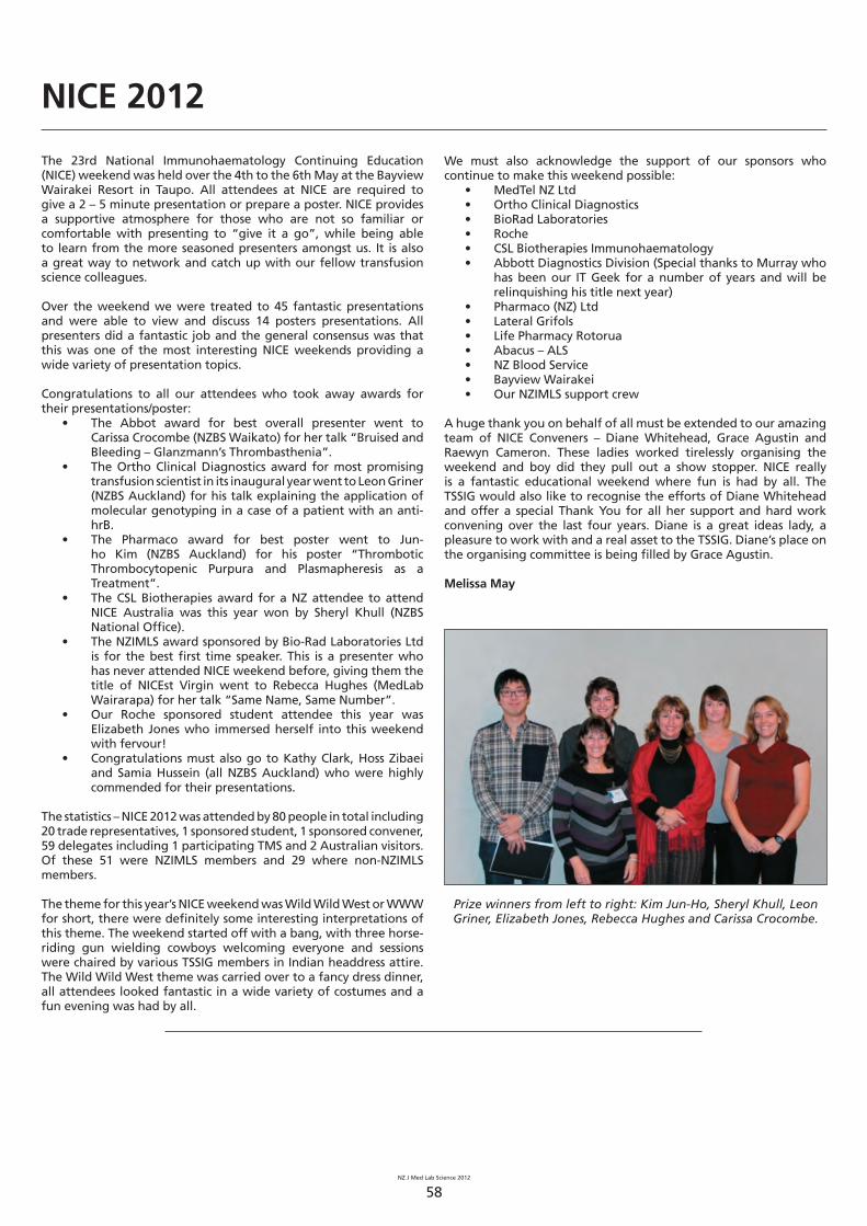
NZ J Med Lab Science 2012
58
NICE 2012
The 23rd National Immunohaematology Continuing Education (NICE) weekend was held over the 4th to the 6th May at the Bayview Wairakei Resort in Taupo. All attendees at NICE are required to give a 2 – 5 minute presentation or prepare a poster. NICE provides a supportive atmosphere for those who are not so familiar or comfortable with presenting to “give it a go”, while being able to learn from the more seasoned presenters amongst us. It is also a great way to network and catch up with our fellow transfusion science colleagues.
Over the weekend we were treated to 45 fantastic presentations and were able to view and discuss 14 posters presentations. All presenters did a fantastic job and the general consensus was that this was one of the most interesting NICE weekends providing a wide variety of presentation topics.
Congratulations to all our attendees who took away awards for their presentations/poster:
• The Abbot award for best overall presenter went to Carissa Crocombe (NZBS Waikato) for her talk “Bruised and Bleeding – Glanzmann’s Thrombasthenia”.
• The Ortho Clinical Diagnostics award for most promising transfusion scientist in its inaugural year went to Leon Griner (NZBS Auckland) for his talk explaining the application of molecular genotyping in a case of a patient with an anti-hrB.
• The Pharmaco award for best poster went to Jun-ho Kim (NZBS Auckland) for his poster “Thrombotic Thrombocytopenic Purpura and Plasmapheresis as a Treatment”.
• The CSL Biotherapies award for a NZ attendee to attend NICE Australia was this year won by Sheryl Khull (NZBS National Offi ce).
• The NZIMLS award sponsored by Bio-Rad Laboratories Ltd is for the best fi rst time speaker. This is a presenter who has never attended NICE weekend before, giving them the title of NICEst Virgin went to Rebecca Hughes (MedLab Wairarapa) for her talk “Same Name, Same Number”.
• Our Roche sponsored student attendee this year was Elizabeth Jones who immersed herself into this weekend with fervour!
• Congratulations must also go to Kathy Clark, Hoss Zibaei and Samia Hussein (all NZBS Auckland) who were highly commended for their presentations.
The statistics – NICE 2012 was attended by 80 people in total including 20 trade representatives, 1 sponsored student, 1 sponsored convener, 59 delegates including 1 participating TMS and 2 Australian visitors. Of these 51 were NZIMLS members and 29 where non-NZIMLS members.
The theme for this year’s NICE weekend was Wild Wild West or WWW for short, there were defi nitely some interesting interpretations of this theme. The weekend started off with a bang, with three horse-riding gun wielding cowboys welcoming everyone and sessions were chaired by various TSSIG members in Indian headdress attire. The Wild Wild West theme was carried over to a fancy dress dinner, all attendees looked fantastic in a wide variety of costumes and a fun evening was had by all.
We must also acknowledge the support of our sponsors who continue to make this weekend possible:
• MedTel NZ Ltd• Ortho Clinical Diagnostics• BioRad Laboratories • Roche• CSL Biotherapies Immunohaematology• Abbott Diagnostics Division (Special thanks to Murray who
has been our IT Geek for a number of years and will be relinquishing his title next year)
• Pharmaco (NZ) Ltd• Lateral Grifols• Life Pharmacy Rotorua• Abacus – ALS• NZ Blood Service• Bayview Wairakei• Our NZIMLS support crew
A huge thank you on behalf of all must be extended to our amazing team of NICE Conveners – Diane Whitehead, Grace Agustin and Raewyn Cameron. These ladies worked tirelessly organising the weekend and boy did they pull out a show stopper. NICE really is a fantastic educational weekend where fun is had by all. The TSSIG would also like to recognise the efforts of Diane Whitehead and offer a special Thank You for all her support and hard work convening over the last four years. Diane is a great ideas lady, a pleasure to work with and a real asset to the TSSIG. Diane’s place on the organising committee is being fi lled by Grace Agustin.
Melissa May
Prize winners from left to right: Kim Jun-Ho, Sheryl Khull, Leon Griner, Elizabeth Jones, Rebecca Hughes and Carissa Crocombe.
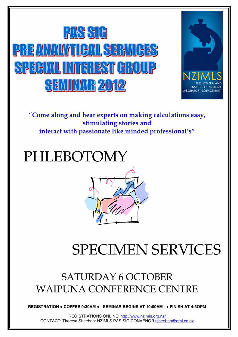
“Come along and hear experts on making calculations easy,
stimulating stories and
interact with passionate like minded professional’s”
PHLEBOTOMY
SPECIMEN SERVICES
SATURDAY 6 OCTOBER
WAIPUNA CONFERENCE CENTRE
REGISTRATION COFFEE 9:30AM SEMINAR BEGINS AT 10:00AM FINISH AT 4:3OPM
REGISTRATIONS ONLINE: http://www.nzimls.org.nz/CONTACT: Theresa Sheehan: NZIMLS PAS SIG CONVENOR [email protected]
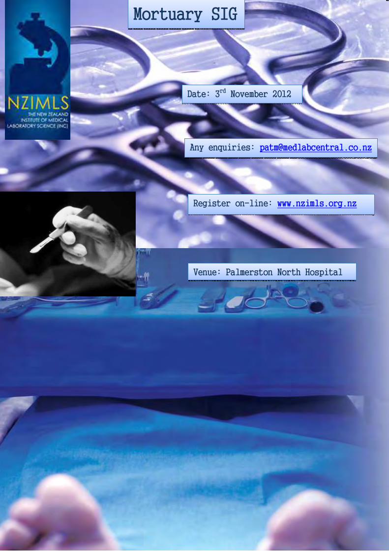
NZ J Med Lab Science 2012
60
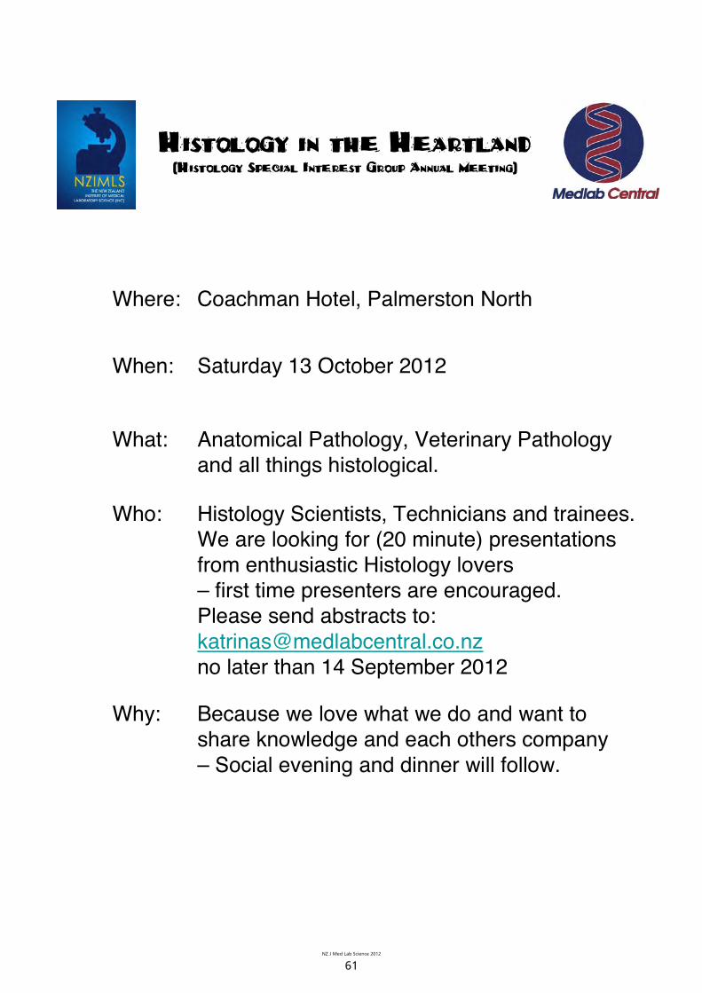
NZ J Med Lab Science 2012
61
Histology in the Heartland(Histology Special Interest Group Annual Meeting)
Where: Coachman Hotel, Palmerston North
When: Saturday 13 October 2012
What: Anatomical Pathology, Veterinary Pathology and all things histological.
Who: Histology Scientists, Technicians and trainees.We are looking for (20 minute) presentations from enthusiastic Histology lovers – first time presenters are encouraged.Please send abstracts to: [email protected] later than 14 September 2012
Why: Because we love what we do and want to share knowledge and each others company – Social evening and dinner will follow.

NZ J Med Lab Science 2012
62
Greetings from the PPTC
The PPTC wishes to extend to you the very best wishes and is once again looking forward to continue working with you all on the many projects scheduled for the remainder of this year 2012.
New PPTC staff appointments
The PPTC Board of Management has the greatest of pleasure in welcoming both Navin Karan and Russell Cole to the permanent staff of the PPTC.
Russell Cole , DipMLT MNZIMLS
Up until August 2011, Russell fi lled the position of charge microbiology and laboratory co-ordinator at Whakatane Hospital and during that time had oversight of QC and IANZ documentation for the laboratory. As a registered medical laboratory scientist and microbiologist, Russell has had over 25yrs experience in the medical laboratory diagnostic sciences and has excelled in the particular areas of laboratory management, human resources and laboratory accreditation.
The World Health Organization (WHO) has recently developed an Asia-Pacifi c Strategy for Strengthening Health Laboratory Services (2010-2015) which was endorsed by Ministers of Health for the WHO Western Pacifi c Region in October 2009. The strategy takes a health systems strengthening approach to its objective of assisting countries to provide comprehensive laboratory services, aimed at improving health outcomes. This has created some momentum for an increased focus on laboratory services in the Pacifi c.
The PPTC’s work on laboratory quality management is increasingly guided by the WHO Asia-Pacifi c Strategy for Strengthening Health Laboratory Services 2010-2015, and many countries in the Pacifi c are preparing for or have begun signifi cant health sector reform. The PPTC is to play a progressive role in the development and implementation of LQMS in selected Pacifi c Island countries and Russell appointed by the PPTC as its laboratory quality management co-ordinator will be responsible for the supervision of this programme.
Navin Karan, BMLSc (Otago) MNZIMLS
Born and raised in Fiji, Navin graduated from the University of Otago, and qualifi ed as a New Zealand registered medical laboratory scientist in 2008 specialising both in clinical microbiology and diagnostic molecular pathology. Since then he has gained a wealth of experience working in medical diagnostic laboratories throughout New Zealand and is now the programme co-ordinator for all teaching and training programmes provided by the PPTC to students throughout the Pacifi c.
The PPTC is a Collaborating Centre of the World Health Organization, Western Pacifi c Region and its mission is to provide training in appropriate medical laboratory technology, external quality assurance programmes and development assistance for the clinical laboratory and blood transfusion services of developing countries with particular emphasis on those of the Pacifi c Island Region.
The teaching, training and development aid programmes offered by the PPTC are governed by one principle: ‘They must be appropriate, affordable and sustainable for the health care setting in which they will be used’.
The emphasis is on appropriate and practical short-term training and teaching, both in country and in New Zealand that will ensure immediate benefi t for the trainees in their work setting. In 2005 the Centre commenced a distance learning Diploma programme in conjunction with WHO and now provides courses in the majority of the medical laboratory science disciplines in addition to the training courses run at the PPTC. Navin is responsible for the consolidation of all our teaching and training programmes taking us to the next step in our development cycle.
Courses held at the PPTC from the beginning of 2012 to midyear
Haematology and blood fi lm examinationIn March of this year, Phil Wakem our PPTC Manager and haematology technical consultant provided a haematology and blood fi lm examination course to Pacifi c students nominated to attend, and once again this proved to be a great success.
This course provided trainees with guidelines for the objective microscopic evaluation of white cells, red cells and platelets in both health and disease. Trainees were introduced to the workings of the microscope in terms of correct operation, correct use of objectives, and essential maintenance. They learnt the principles of Romanowsky staining, the preparation of stains and buffers, causes of inconsistent staining quality and the correct staining techniques used in the identifi cation of malarial parasites. Students were introduced to the blood fi lm in terms of sample quality, the effects of anticoagulants, the correct technique in blood fi lm making, morphological artefacts, buffy coat preparations, and the correct storage of blood fi lms. Students also learnt extensively the correlation of blood fi lm fi ndings with results obtained from manual and/ or automated methods for red cell, white cell and platelet parameters. Morphological terminology with reference to origin and correct application was also discussed. The lineage of all blood cells was followed through systematically from the common stem cell through all stages of development. A comprehensive account of both normal haematology and pathological haematology was given over the 4 week teaching programme. The course was designed to give trainees confi dence in the preparation, staining and examination of blood fi lms, be able to differentiate the white cell count into both normal and abnormal populations and fi nally recognise and comment on with confi dence, abnormal fi lm fi ndings in an extensive range of common blood cell disorders.

NZ J Med Lab Science 2012
63
The eight students who attended this course were as follows: Sione Isoa and Timote Fakasi’ieiki from Tonga, Elizabeth Asamole from Papua New Guinea, Robert Daniel and Jeffery Vutilolo from Vanuatu, Lenburg Ligohr from Pohnpei, Bernard Paul from Kiribati, and Ishael Ken from the Marshall Islands.
It was the greatest pleasure to have Lavea’i Ioane, Ministry of Foreign Affairs and Trade, present to the students their certifi cates at the fi nal graduation. Her speech to the students was very meaningful and this is recorded as follows:
Kia Ora, Talofa and Pacifi c Greetings to you all. I would like to take this opportunity to say a few words. First and foremost, I would like to say thank you to Phil and the team, for inviting me along today, it’s a privilege and an honor for me to be here today, to partake in this wonderful celebration. Thank you.
PPTC is recognized internationally as a ‘core of excellence’ in terms of their specialist training, laboratory quality assurance and laboratory strengthening to the National Health laboratories of the Pacific nations. The Ministry of Foreign Affairs and Trade NZ, have provided fi nancial support to PPTCs various activities since 1981, in particular, their:
Regional external assurance programmeo Training programmeo Laboratory quality management systems o programme
We have received positive feedback from Pacifi c countries on how much they value the work PPTCs doing around strengthening their National Health laboratories, this is testimonial to the signifi cant contribution PPTC’s making in the Pacific, and it illustrates how much the island nation appreciates the hard work your team here are doing.
For the graduates, our congratulations go out to you, for the hard work you’ve put into the course, hence we’re here today. We applaud you for empowering yourselves, leaving your loved ones, your home land to take on this opportunity to up skill yourselves in foreign land, for the betterment of the services you deliver to your people.
I wish you all the best as you start the next leg of your journey in your respective roles back home, with the new skills and knowledge you are armed with. Lastly I would like to leave you with a Maori proverb signifi cant for this occasion; “Naku te rourou, nau te rourou ka ora te iwi”: With your basket and my basket the people will live. Thank you.Lavea’i Ioane.
Haematology 2012 PPTC staff and students
Laboratory quality management courseReported by Russell ColeTeaching and advising Pacifi c Island laboratories in the intricacies of quality management is both a privilege and extreme challenge rolled into one. Like many New Zealand laboratories in the 90’s, lab staff throughout the Pacifi c are in anguish over the cash strapped resources, short staffed departments and the apparent insurmountable task ISO15189 presents to them. The WHO is well aware of the laboratory status of the majority of Pacifi c Island nations and along with the enormous contributions of our own John Elliot, has produced a minimum standards guideline for lab quality standards and their implementation. For those of us reared on an IANZ diet where accreditation meant funding and survival for the laboratory, these WHO guidelines seem rather basic and watered down. Rightfully so too, because they are pitched at a level Pacifi c Island labs can build foundational blocks of management on and over the next few years expand their QM structure, step by step, to encompass the 12 essential facets of 15189 within the next 5 years. A task more achievable in Pacifi c cultures and their adorable “Island time” concepts.
Throughout May, the PPTC ran an intensive quality management course covering the practical implementation of quality manuals, documentation control and structure, standard operating procedures, reporting and result processing. A big emphasis was put on hands on experience in creating policies and documents that were relevant and workable documents that would be adopted easily into Pacifi c Island labs and utilized on a daily basis. An extensive tour of Wellington, Kenepuru, Hutt Valley Hospital laboratories and the NZ Blood service in Wellington were excellent in providing valuable experience and exposure to the daily working operations of accredited laboratories. To see these laboratories actively functioning and monitoring their performances under the confi nes of ISO15189 was very impressionable to our students, fi lling them with immeasurable enthusiasm and motivation to make it happen in their home laboratories. A very big thank you to Filipo Faiga, Clare Murphy, Russell Cooke, Stuart Clarke and Kevin Fomiatti for their time and input in hosting these visits.
Many aspects of laboratory management throughout New Zealand are heavily associated with LIS systems, automated procedures and internet reporting. These aspects are seen by Pacifi c Island laboratories as luxuries rather than essentials, a wish list of commodities to spend the limited health dollar on rather than mandatory requirements of an LQM system. Creating business cases, justifying additional expenditure and examining the risk factors involved in implementing ISO standards occupied a big portion of this course including the complications of electronic procurement systems (i-proc) and electronic documentation control (Q pulse). Above all, establishing systems and written processes for staff and operational processes was our priority, along with a good deal of fun and enjoyment thrown in.
Students attending the course were Nanise Malupo (CWM Fiji), Berenadeta Lutua (Lautoka,Fiji) and Wesley Puri (PNG). A big thank you to Associate Professor Rob Siebers for presenting the students with their certifi cates at the fi nal graduation. The PPTC would also like to thank the senior medical laboratory scientists from Wellington Hospital for their time, wisdom and valuable contribution in making this course hugely rewarding and successful, including Martijn Groen from the IT Department.
Distance learning programme
Diploma in Medical Laboratory ScienceIn March of this year, the 2012 microbiology module was released to students registered for the PPTC Diploma of Medical Laboratory Science and this is now to be followed by transfusion science at the end of June which is the fi nal module of the Diploma. Once again it is a requirement of the PPTC that all students register for each individual module and therefore it is important to complete registration with both the PPTC and the WHO POLHN offi ce in Suva well before modules are to be released.

NZ J Med Lab Science 2012
64
The PPTC external quality assessment programme
It is with greatest pleasure that we welcome to the PPTC, Dan Gyles, Nicky Beamish and Marita Smit as specialist co-ordinators for the PPTC EQA programme which covers over 40 diagnostic laboratories throughout the Pacifi c and South East Asia regions.
Dan is our specialist/consultant for blood banking and transfusion services, Nicky is our specialist/consultant for microbiology and Marita is our specialist/consultant for serology. Phil continues as specialist/consultant for haematology and Clare as our specialist/consultant for biochemistry. Christine co-ordinates and administers the EQA programme.
Our EQA team:
Christine Phil Clare
Dan Nicky Marita
Country visits
The PPTC has been extremely busy since the beginning of this year visiting at least twice, the countries - Samoa, Vanuatu, Kiribati, and Tonga laying the foundations for the implementation of laboratory quality management in each of the national laboratories. Navin and Russell have both travelled extensively in the last few months throughout the Pacifi c and must be congratulated for the valuable contribution that has been made towards evaluation and introductory implementation of quality management in each of these nominated countries.
The PPTC would like to thank Filipo Faiga, section head biochemistry, Wellington Hospital and Dan Gyles, team leader Wellington blood bank, New Zealand Blood Service for carrying out PPTC consultancies in Samoa. The PPTC is very fortunate to have accessibility to such high levels of experience and technical expertise provided by senior New Zealand medical laboratory scientists towards the development and strengthening of Pacifi c laboratories.
Filipo also visited Kiribati, at the end of June to conduct biochemistry training in the newly established Marine Training Laboratory located in Tarawa. Phil travelled to Vanuatu in May to conduct a two week haematology and blood fi lm morphology training course in Port Vila and in Espiritu Santo for all staff and especially for those currently registered in the PPTC Diploma programme. This was a great success and the students very much appreciated the time given to each of them by Phil throughout the training period. Russell travelled to Vanuatu at the end of June to conduct an LQMS workshop for over 20 students in Santo, and then represented the PPTC at the 5th LabNet Technical Working Group meeting held in Noumea, New Caledonia from 26th - 27th June.
Laboratory quality management course May 2012.PPTC staff and students.
Phil pictured with students, Santo, Vanuatu
PPTC courses for 2012
Microbiology: 6th August - 31st August 2012This course will provide trainees with an update on developments in microbiological procedures. The theoretical and practical aspects of current methods used in the isolation, identifi cation and antimicrobial susceptibility testing of microorganisms will be covered along with discussions on emerging and re-emerging bacterial organisms likely to cause infectious diseases.
Serological and other rapid methods for the identifi cation of bacterial and viral diseases including Hepatitis A, B, and C, HIV and other STIs, will be discussed as will the role of the microbiology laboratory in the surveillance of nosocomial infections and identifi cation of infections of public health importance.
Blood bank technology: November 2012This course will include units of study covering the theoretical and practical aspects of the following topics; routine blood grouping, blood group antigens, cross match techniques, antibody detection, transfusion reactions, haemolytic disease of the newborn, screening blood for infectious agents, blood donor selection, organisation of a blood bank, and the appropriate use of blood components in transfusion medicine.
The Thermo Scientific Herasafe KS Class II Biological Safety Cabinet offers maximum safety, comfort and convenience.
DC motors mean a reduction in operating costs up to 75% over traditional biological safety cabinets with AC motors
Maximised safety through our innovative Smartflow design with digital airflow verification
Increased comfort and encourages safe working habits with the sloped front, bright workspace and low noise level
2 year warranty
Maximum safety comfort and convenience
For further information call: 0800 933 966 or visit us at: www.thermofisher.co.nz
Thermo Scientific’s Multiskan FC Microplate Photometer is a filter-based microplate photometer for a wide variety of research and routine applications. The Multiskan FC, developed using 30 years of experience in microplate photometry, offers proven perfomance and reliable results.
Wide wavelength range of 340 to 850nm
Fast reading of both 96- and 384-well plates
USB port for easy data transfer
Shaking and incubation up to 50°C for temperature critical assays
Proven performance and reliability
For further information call: 0800 933 966 or visit us at: www.thermofisher.co.nz
Thermo Scientific’s Multiskan FC Microplate Photomeeteter iis a filterr--for a wide variety of research and routinene aappppllications. TThe MMulultitiskskyears of experience in microplp ate e phphototomometetry, ofoffefersrs p provenn perfo
WiW dede wwave
Fast reaeadin
USB portrt f
Shaking anntemperatur
For fuurtr her innfofooro visit uus s at:
