the metal binding properties of the zinc site of yeast copper
TRANSCRIPT

JBIC (2000) 5 : 189±203 � SBIC 2000
ORIGINAL ARTICLE
Thomas J. Lyons ´ Aram Nersissian ´ Huijuan HuangHyeyeong Yeom ´ Clinton R. NishidaJanet A. Graden ´ Edith Butler GrallaJoan Selverstone Valentine
The metal binding properties of the zinc site of yeast copper-zincsuperoxide dismutase: implications for amyotrophic lateral sclerosis
Received: 17 September 1999 / Accepted: 8 December 1999
Abstract We have investigated factors that influencethe properties of the zinc binding site in yeast copper-zinc superoxide dismutase (CuZnSOD). The proper-ties of yeast CuZnSOD are essentially invariant frompH 5 to pH 9. However, below this pH range there isa change in the nature of the zinc binding site whichcan be interpreted as either (1) a change in metalbinding affinity from strong to weak, (2) the expulsionof the metal bound at this site, or (3) a transitionfrom a normal distorted tetrahedral ligand orientationto a more symmetric arrangement of ligands. Thischange is strongly reminiscent of a similar pH-inducedtransition seen for the bovine protein and, based onthe data presented herein, is proposed to be a prop-erty that is conserved among CuZnSODs. The transi-tion demonstrated for the yeast protein is not onlysensitive to the pH of the buffering solution but alsoto the occupancy and redox status of the adjacentcopper binding site. Furthermore, we have investi-
gated the effect of single site mutations on the pH-and redox-sensitivity of Co2+ binding at the zinc site.Each of the mutants H46R, H48Q, H63A, H63E,H80C, G85R, and D83H is capable of binding Co2+ toa zinc site with a distorted tetrahedral geometry sim-ilar to that of wild-type. However, they do so only ifCu+ is bound at the copper site or if the pH in raisedto near physiological levels, indicating that the changeat the zinc binding site seen in the wild-type is con-served in the mutants, albeit with an altered pKa. Themutants H71C and D83A did not bind Co2+ in a wild-type-like fashion under any of the conditions tested.This study reveals that the zinc binding site is exqui-sitely sensitive to changes in the protein environment.Since three of the mutant yeast proteins investigatedhere contain mutations analogous to those that causeALS (amyotrophic lateral sclerosis) in humans, thisfinding implicates improper metal binding as a mech-anism by which CuZnSOD mutants exert their toxicgain of function.
Key words Copper ´ Zinc ´ Superoxide dismutase ´Amyotrophic lateral sclerosis ´ Yeast
Introduction
Copper-zinc superoxide dismutase (CuZnSOD) is a32 kD homodimeric metalloenzyme that catalyzes thedisproportionation of superoxide anion (O2
± ) to givehydrogen peroxide (H2O2) and dioxygen (O2) [1].Each subunit of the dimer contains one catalyticcopper ion and one zinc ion [2, 3]. In the oxidizedform of the enzyme, Cu2+ is bound to the protein viafour histidine residues (His46, His48, His63, andHis120, using the yeast CuZnSOD numbering system)arranged in a distorted square planar geometry. Aweakly bound water molecule assumes an axial posi-tion, making the copper pentacoordinate. The Zn2+
ion is coordinated in a distorted tetrahedral arrange-ment by three histidine residues (His63, His71, and
T.J. LyonsNutritional Sciences Program, Department of MolecularBiology, 217 Gwynn Hall, University of Missouri-Columbia,Columbia, MO 65211, USA
A. Nersissian ´ H. Yeom ² ´ E.B. Gralla ´ J.S. Valentine ())Department of Chemistry and Biochemistry,University of California, Los Angeles, 607 Circle Drive South,Box 951569, Los Angeles, CA 90095-1569, USATel.: +1-310-8259835Fax: +1-310-2067197e-mail: [email protected]
H. HuangDepartment of Computer Science, University of California,Los Angeles, 405 Hilgard, Los Angeles, CA 90095-1569, USA
C.R. NishidaSchool of Pharmacy, University of California, San Fransisco,513 Parnassus Avenue, Box 0446, Room S-1134, San Fransisco,CA 94143 USA
J.A. GradenKelly Scientific Resources, 1710 Walton Road, Suite 301,Bluebell, PA 19420, USA

190
His80) and one aspartate residue (Asp83). The doublydeprotonated His63 side-chain couples the Cu2+ andZn2+ ions via a bridging imidazolate ligand [4, 5]. Insolution, reduction of Cu2+ to Cu+ is accompanied bythe rupture of this bridge [6]. While a recent crystalstructure of reduced yeast CuZnSOD confirms thisrearrangement of the active site upon reduction, show-ing Cu+ in a trigonal planar geometry with the copperion detached from His63 [7], another crystal structureof reduced bovine CuZnSOD shows an intact bridge[8]. The two metal binding sites are also bridged by ahydrogen bond network that connects His46 in thecopper site with His71 in the zinc site via Asp124 [9](see Fig. 1).
Both the spectroscopic properties and the enzy-matic activity of bovine CuZnSOD are essentiallyinvariant between pH 5 and 9, leading to the conclu-sion that the enzyme retains its structure over a widepH range [2]. However, if the pH of the bufferingsolution falls below pH 4, a reversible and kineticallyslow conformational change was shown to occur at thezinc binding site of the bovine protein that results inthe possible expulsion of the metal ion bound at thatsite [10]. It was also shown that the pKa of this transi-tion did not change significantly with the nature of themetal bound at the zinc site; viz., the transitionoccurred at roughly the same pH when either Zn2+,Co2+, or Cu2+ were bound there. This observation sug-gested that simple competition for the metal ligandsby protons was not the only cause of the phenomenon.Rather, a pH-dependent conformational change of theprotein was implicated [11].
CuZnSOD isolated from bovine blood has been thesubject of extensive investigation since the discoveryof its catalytic function in 1969 [12], while consid-erably less information is available about the enzymefrom other species. Using bovine CuZnSOD as a flag-ship enzyme, detailed structural information about theCuZnSOD family has been provided over the years bya plethora of analytical techniques. In this paper wedemonstrate that this low pH transition is not only aproperty of the bovine enzyme but that it occurs inthe yeast and human enzymes as well. In addition, wereport the investigation of the structural parametersgoverning the properties of the zinc binding site usingthe yeast wild-type protein and several site-directedmutants. The ability of the yeast protein to form anative-like zinc binding site is not only sensitive to thepH of the solution but it is also sensitive to the statusof the adjacent copper binding site and to a diverseset of mutations in the protein matrix.
Little is known about why CuZnSOD evolved tocontain zinc. The prevailing theory is that it plays astructural role. However, there is ample evidence thatzinc can also modulate the reactivity of the enzyme(see Discussion). This idea becomes important in lightof the discovery in 1993 that point mutations in thehuman CuZnSOD gene result in an unknown gain offunction that causes approximately 2% of cases ofamyotrophic lateral sclerosis (ALS). ALS is a neu-rodegenerative disorder that strikes motor neurons,causing paralysis and ultimately death. Three of themutants investigated in this paper are analogous tomutations in the human enzyme that cause ALS [13,
Fig. 1 Active site diagram ofyeast CuZnSOD. Mutationsthat are the subject of thisinvestigation are labeled withbold print (underlined and ital-icized mutations are merelydiscussed and are not the sub-ject of this investigation)
191
14, 15], raising the possibility of a relationshipbetween the ability of this protein to form a nativezinc site and the presumed disease-causing gain offunction.
Materials and methods
Lyophilized yeast CuZnSOD was purchased from Peptech (Den-mark; batch no. 8997368, product no. 3007; mfg. date 10/17/89).Recombinant yeast CuZnSOD mutants were expressed in andisolated from BL21 Escherichia coli using the pET-3d expressionplasmid. The mutants H63A and H63E (the bridging histidine atposition 63 changed to alanine and glutamic acid, respectively),G85R (glycine at position 85 changed to arginine), and D83Aand D83H (aspartic acid at position 83 changed to alanine andhistidine, respectively) were prepared by oligonucleotide-di-rected mutagenesis and purified by published procedures [16, 17,18]. H46R and H48Q were prepared by overlap extension PCRmutagenesis and purified by published procedures [17, 18].H46C and H80C were prepared and purified by published pro-cedures [19]; purity was confirmed by gel electrophoresis.Human wild-type CuZnSOD was purified via published pro-cedures [20]. DEAE cellulose was purchased from Whatmanand G-75 superfine Sephadex was purchased from Sigma.
We have found previously that the metal binding propertiescharacteristic of apo bovine CuZnSOD can be reproduced foronly one subunit of the yeast apoprotein dimer. One subunit iscompetent to bind metals in a bovine-like fashion and the othersubunit (the ªphantomº subunit) is not competent to do so anddoes not participate in metal titrations until the competent sub-unit has been filled and even then the metal binding propertiesof the phantom subunit are abnormal [20]. For this study weused a molar extinction coefficient at 280 nm of 3000 M ± 1 cm ± 1
per dimer to determine the concentration of apoprotein. Only1 equiv per dimer of each metal is added so that the competentsubunit is filled and the ªphantomº subunit remains empty. Fora more detailed description of the rationale for this assumption,see [20].
Apoproteins were obtained by dialysis against 100 mM ace-tate buffer, pH 3.8, in the presence of 10 mM EDTA (four timesagainst 1 L each time). EDTA was then removed by dialysisagainst 100 mM acetate buffer, pH 3.8, in the presence of100 mM NaCl (three times against 1 L each time) [2]. Metalcontent was determined by atomic absorption spectroscopy toconfirm that the samples were indeed apo, containing less than10% of residual metal ions. Experiments were carried out in100 mM acetate buffer at pH 5.5 unless otherwise noted. Metal-substituted proteins were obtained by directly infusing solutionsof the apoprotein with aliquots of either 10 mM or 100 mMstock solutions of the appropriate metal sulfate salt (Ag+ wasadded as AgNO3). Titration experiments required approximately200 mL of 0.3 mM apoprotein solution (see figure legends forexact concentrations used in each experiment).
pH adjustments were made by addition of 100 mM or 1 Macid or base (NaOH or HCl) and pH was measured using anOrion Research Microprocessor Ionalyzer/901 (Boston, Mass.)with an Ingold microelectrode (Wilmington, Mass.).
UV-visible spectra from 900 to 240 nm were determinedusing a Cary 3 UV-visible spectrometer (Varian, Sunnyvale, Cal-if.). Samples were centrifuged at 14,000 rpm for 1 min beforespectra were taken to remove trace turbidity. Reduced proteinswere obtained by addition of a single small crystal of soliddithionite directly to the protein solutions.
pKa values were determined by plotting the change inabsorption at a certain wavelength as a function of pH. Best fitsigmoidal plots of these data and first derivative graphs of thebest fit curves were obtained to estimate the apparent pKavalues for these transitions.
Results
Addition of Co2+ and Cu2+ to apo wild-type yeastCuZnSOD
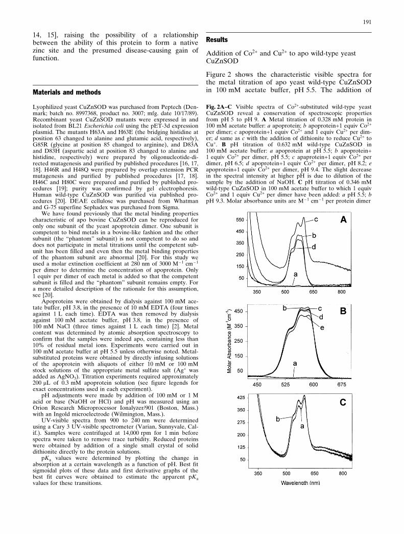
Figure 2 shows the characteristic visible spectra forthe metal titration of apo yeast wild-type CuZnSODin 100 mM acetate buffer, pH 5.5. The addition of
Fig. 2A±C Visible spectra of Co2+-substituted wild-type yeastCuZnSOD reveal a conservation of spectroscopic propertiesfrom pH 5 to pH 9. A Metal titration of 0.328 mM protein in100 mM acetate buffer: a apoprotein; b apoprotein+1 equiv Co2+
per dimer; c apoprotein+1 equiv Co2+ and 1 equiv Cu2+ per dim-er; d same as c with the addition of dithionite to reduce Cu2+ toCu+. B pH titration of 0.632 mM wild-type CuZnSOD in100 mM acetate buffer: a apoprotein at pH 5.5; b apoprotein+1 equiv Co2+ per dimer, pH 5.5; c apaprotein+1 equiv Co2+ perdimer, pH 6.5; d apoprotein+1 equiv Co2+ per dimer, pH 8.2; eapoprotein+1 equiv Co2+ per dimer, pH 9.4. The slight decreasein the spectral intensity at higher pH is due to dilution of thesample by the addition of NaOH. C pH titration of 0.346 mMwild-type CuZnSOD in 100 mM acetate buffer to which 1 equivCo2+ and 1 equiv Cu2+ per dimer have been added: a pH 5.5; bpH 9.3. Molar absorbance units are M ± 1 cm ± 1 per protein dimer

192
1 equiv of Co2+ per dimer resulted in the appearanceof three relatively intense overlapping bands at525 nm, 565 nm, and 585 nm due to Co2+ binding tothe zinc site (Fig. 2Ab). These bands were essentiallyinvariant from pH 5.50 to pH 9.4, as can be seen inFig. 2Ba±e.
The further addition of Cu2+ to the above Co2+-sub-stituted species at pH 5.5 resulted in the superpositionof the Co2+ bands on top of a 670 nm Cu2+ band, aswell as a slight red shift of the 585 nm Co2+ band to595 nm (see Fig. 2Ac). This red shift has been attrib-uted to the formation of the imidazolate bridge in thecase of the bovine enzyme [21]. The spectra of thecopper and cobalt-substituted derivative were found tobe relatively invariant from pH 5.5 to pH 9 (shown inFig. 2Ca,b). Reduction of Cu2+ to Cu+ at pH 5.5resulted in the disappearance of the 670 nm Cu2+
band and Co2+ bands that are essentially identical tothe Co2+ bands seen in the absence of copper (seeFig. 2Ad and compare with Fig. 2Ab).
pH titration of as-isolated and Co2+-substitutedderivative of wild-type protein
Wild-type CuZnSOD isolated from yeast contains afull complement of two copper ions and two zinc ions.When the pH of the buffering solution was droppedfrom 5.5 to 3.0, there was a concomitant change in thevisible spectrum at 450 nm. A small shoulder at thiswavelength, attributed to a copper imidazolate chargetransfer, disappeared at low pH. The loss of this bandas a function of pH is graphed in Fig. 3 (closedsquares) and the pKa of the spectral change was calcu-lated to be 3.8, a value that is identical to the low pHtransition found in the bovine enzyme.
When a solution containing apo wild-typeCuZnSOD to which 1 equiv Co2+ per dimer wastitrated down in pH, the high-intensity Co2+ bandsalso disappeared (see Fig. 3, open squares), much likethe 450 nm charge transfer band mentioned above.Interestingly, the pKa of the loss of these bands was4.7, not 3.8, suggesting either a marked differencebetween the as-isolated and half-reconstituted proteinsor a greatly altered affinity of the yeast CuZnSODzinc binding site for Co2+ relative to that of bovineCuZnSOD. Note that in the case of the bovine proteinthe pKa for the low pH transition is the same for theas-isolated protein as it is for the protein reconstitutedwith either Zn2+, Co2+, or Cu2+ [11]. Also note thatthe pH titration of human wild-type CuZnSOD towhich 2 equiv Co2+ per dimer have been added (seeFig. 3, inverted triangles) revealed that the pKa of thechange at the zinc site in the human enzyme isroughly the same as it is for the bovine enzyme(pKa = 4.0).
Kinetics of the loss of zinc site integrity at low pH forwild-type CuZnSOD
Co2+ bound selectively to the zinc site when only1 equiv Co2+ per dimer was added to the wild-typeapoprotein at pH 5.5, as judged by the appearance ofthe characteristic d-d band at 585 nm. Figure 4Ashows that upon a decrease in pH from 5.5 to 4.5there was an immediate reduction in the intensity ofthe Co2+ bands, reaching equilibrium within the timeit took to measure the pH and record a spectrum (lessthan 5 min). However, if Cu2+ was present in the adja-cent copper site, the change in Co2+ binding upon thesame drop in pH was slower and equilibrium was notreached until 105 min later (see Fig. 4B).
Effect of the copper site on Co2+ binding for wild-typeCuZnSOD
In order to investigate further the role the copper siteplays in influencing the nature of the zinc site, weobtained visible spectra at both pH 5.5 and pH 4.5 ofa sample of wild-type apoprotein to which 1 equivCo2+ per dimer has been added (see Fig. 5a). Asexpected from the data in Fig. 4, significantly lessabsorptivity due to Co2+ was seen at the lower pH(Fig. 5b). This pH 4.5 sample was then divided intotwo aliquots. To the first aliquot, 1 equiv Cu2+ perdimer was added. This addition resulted in the appear-
Fig. 3 pH titrations of wild-type yeast CuZnSOD and mutantsdemonstrate pH-dependent metal binding to the zinc site. Thedata represented by closed squares (J) are the pH titration ofthe yeast wild-type protein as isolated from yeast (0.568 mM)containing 2 equiv Cu2+ and 2 equiv Zn2+ per dimer. The lowpH transition in this case was monitored by the change inabsorbance of the 450 nm copper-imidazolate charge transferband. The other data are for samples to which 1 equiv Co2+ wasadded per dimer (2 equiv Co2+ per dimer in the case of apohuman CuZnSOD). The graph monitors the absorbance of theCo2+ chromophore as a function of pH and normalizes thevalues to the maximum value obtained during the titration.Other symbols: m 0.273 mM yeast wild-type, monitored at585 nm; p 0.521 mM H46C, at 585 nm; P 0.320 mM H46R, at585 nm; g 0.119 mM H48Q, at 585 nm; Z 0.437 mM H80C, at625 nm; h 0.427 mM human wild-type, at 585 nm

193
ance of a Cu2+ band at 670 nm and the furtherdecrease of the Co2+ bands (Fig. 5c). The subsequentreduction of Cu2+ to Cu+ by dithionite resulted in thereturn of the high-intensity Co2+ bands to a levelgreater than that found in the absence of copper(Fig. 5d). To the second aliquot, 1 equiv Ag+ perdimer was added (Ag+ binds to the copper site in asimilar fashion as Cu+ [2]), resulting in an even greaterreturn of the Co2+ band intensities than was seen forthe Cu+ bound protein (Fig. 5e). The occupancies ofthe copper site can be ordered with respect to thebeneficial effects each has on the ability of the wild-type zinc site to bind Co2+ normally: Ag+ bound at thecopper site > Cu+ bound > empty copper site > Cu2+
bound.
Co2+-substituted derivatives of several metal ligandmutants of CuZnSOD also undergo a pH-inducedchange in the Co2+-binding affinity at the zinc site
When 1 equiv Co2+ per dimer was added to metal-freepreparations of the site-directed mutants H46C,H46R, H48Q, and H80C, all were found to bind Co2+
in a relatively normal fashion to their zinc bindingsites. However, this binding was pH dependent; thepH profiles of Co2+ binding for each of the aforemen-tioned proteins are shown in Fig. 3 (H46C, open cir-cles; H46R, closed circles; H48Q, open triangles;H80C, open diamonds). Following the absorbance at585 nm (625 nm for H80C), we monitored the changein Co2+ binding as a function of pH. The apparentpKa of the loss of Co2+ binding for the wild-type pro-tein is 4.7. The apparent pKa values of this transitionfor the mutant proteins are as follows: H46C, 4.7;H46R, 6.0; H48Q, 5.6; and H80C, 5.6.
H48Q converts the zinc site from a strong binding siteto a weak binding site
Titration of apo H48Q with Co2+ at pH 5.5 resulted inCo2+ bands that appeared normal in shape and posi-tion but were drastically reduced in intensity. Further-more, a careful titration with Co2+ demonstrated thatthe Co2+ spectrum did not saturate until greater than4 equiv of Co2+ per dimer had been added to the solu-tion, revealing weak zinc site binding behavior(Fig. 6). The inset graph in Fig. 6 is a plot of theintensity of Co2+ bands as a function of equivalentsCo2+ added to the apoprotein. While Fig. 6 dem-onstrates that the zinc site in H48Q is apparently nor-mal, albeit with a lowered metal affinity, Fig. 3 (opentriangles) demonstrates that this weak binding site isconverted to a strong binding site at higher pH values.
Fig. 4A,B Kinetics of the disappearance of Co2+ peaks inresponse to a drop in pH. A 0.273 mM apo yeast wild-type in100 mM acetate buffer to which 1 equiv Co2+ per dimer hasbeen added. The spectrum was recorded at pH 5.5 (a) and thepH was dropped from 5.5 to 4.5 quickly and a new spectrum (b)was recorded as soon as possible (5 min). B Same experiment asA except 1 equiv Co2+ and 1 equiv Cu2+ have been added andthe decrease in Co2+ absorptivity is monitored over time: apH 5.5 before pH was decreased; b pH 4.5, 15 min after; cpH 4.5, 60 min after; d pH 4.5, 105 min after (low pH spectrafor both A and B did not change even after 2 days, indicatingequilibrium had been reached). Molar absorbance units areM ± 1 cm ± 1 per protein dimer
Fig. 5 The occupancy of the copper site strongly affects theextent of conversion of the zinc site at low pH. Metal titrationof 0.175 mM apo wild-type CuZnSOD in 100 mM acetate buff-er: a apoprotein+1 equiv Co2+ per dimer, pH 5.5; b apoprotein+1 equiv Co2+ per dimer, pH 4.5; c apoprotein+1 equiv Co2+ and1 equiv Cu2+ per dimer, pH 4.5; d apoprotein+1 equiv Co2+ and1 equiv Cu+ per dimer, pH 4.5 (Cu2+ reduced to Cu+ withdithionite); e apoprotein+1 equiv Co2+ and 1 equiv Ag+ per dim-er, pH 4.5. Molar absorbance units are M ± 1 cm ± 1 per proteindimer

194
Co2+ binding affinity at pH 5.5 for the zinc site of themutants G85R, D83H, and H63E can be improved ifCu+ is bound at the copper site; this is not true for themutants D83A and H63A
While some of the metal binding behavior of themutants G85R (analogous to the human ALS mutanthG85R), H63E, and H63A have been previouslyreported [16, 17, 18], those studies incorrectly deter-mined protein concentration by a factor of two andtherefore those results must be reinterpreted [20].Here we reinterpret and expand upon those studies.
As was reported previously, addition of 1 equiv ofCo2+ to apo G85R at pH 5.5 in acetate buffer resultedin the appearance of a non-wild-type-like Co2+ bandof moderate intensity centered at roughly 550 nm(Fig. 7Aa). The fine structure and lowered intensity ofthe Co2+ band suggest that the Co2+ may be penta-coordinate [22] and therefore that it is probably notbound at the zinc site, but rather at the copper site.Further addition of Cu2+ to this species resulted in the
Fig. 6 The mutation H48Q converts the zinc binding site from astrong binding site to a weak one at pH 5.5. Co2+ titration of0.217 mM apo yeast H48Q in 100 mM acetate buffer pH 5.5: ais a spectrum of the apo protein and b ± r are spectra of sequen-tial additions of 0.25 equiv Co2+ per dimer up to 4.25 equiv Co2+
per dimer. Inset is a plot of the intensity of the Co2+ absorptivityat 585 nm as a function of equiv of Co2+ added as well as a bestfit sigmoidal curve of the data. Molar absorbance units areM ± 1 cm ± 1 per protein dimer
Fig. 7A ± E The Co2+ bindingaffinity at pH 5.5 of the zincsite is strongly influenced bythe occupancy of the coppersite in several mutants. Metaltitrations in 100 mM acetatebuffer at pH 5.5 for A0.300 mM apo yeast G85R; B0.312 mM apo yeast D83H; C0.862 mM apo yeast H63E; D0.600 mM apo yeast D83A;and E 0.202 mM apo yeastH63A. For all figures: a apop-rotein+1 equiv Co2+ per dimer;b apoprotein+1 equiv Co2+
and 1 equiv Cu2+ per dimer; capoprotein+1 equiv Co2+ and1 equiv Cu+ per dimer (Cu2+
reduced to Cu+ with dithion-ite). Molar absorbance unitsare M ± 1 cm ± 1 per proteindimer
195
complete disappearance of this Co2+ band and theappearance of a 670 nm Cu2+ peak typical of Cu2+ atthe copper site (Fig. 7Ab). These data suggest thatCo2+ and Cu2+ are competing for the same bindingsite at pH 5.5, i.e., that Co2+ binds to the copper siteand is then displaced by the Cu2+. Reduction of Cu2+
resulted in the rebinding of Co2+, this time to the zincsite, as judged by the appearance of a normal wild-type-like Co2+ spectrum (Fig. 7Ac).
The titration of apo D83H (zinc ligand Asp83mutated to histidine) proceeded very similarly to thetitration of apo G85R. The addition of 1 equiv Co2+
per dimer to the apoprotein yielded a broad visibleband identical to the one seen for the similarly met-allated G85R (Fig. 7Ba). The addition of 1 equiv Cu2+
to this derivative caused the disappearance of theCo2+ band and the appearance of a 670 nm Cu2+ band(Fig. 7Bb) and reduction of Cu2+ then caused the par-tial return of wild-type-like Co2+ bands (Fig. 7Bc).The fact that the return of wild-type-like Co bands inD83H is not as complete as for G85R may reflect themore drastic nature of the D83H mutation (D83 beinga zinc binding ligand).
Despite some differences in the shape and positionof the relevant bands, the titration of apo H63E (thehistidine that acts as a bridging ligand between copperand zinc, H63, replaced with a glutamate) also pro-ceeds very similarly to that of apo G85R. Addition of1 equiv Co2+ per dimer yielded Co2+ bands of rel-atively low intensity and of poor structural definition(Fig. 7Ca). This again would suggest Co2+ bound atthe copper site. The addition of Cu2+ to this derivativeresulted in the loss of Co2+ bands and the appearanceof a 670 nm Cu2+ band, reflecting competition by thetwo metal ions for the same site (Fig. 7Cb). Thereduction of Cu2+ to Cu+ resulted in the reappearanceof high-intensity, albeit not wild-type-like, Co2+ bands.The Co2+ bands in this last derivative were of lowerintensity than those observed for wild-type and werered-shifted (Fig. 7Cc). Such bands are, however, indic-ative of tetrahedral binding and the fact that they arenot similar to those of wild-type may reflect the differ-ent liganding properties of the glutamate side chainthat has replaced His63 [22].
Both mutants D83A (zinc ligand Asp83 mutated toalanine) and H63A (bridging histidine, H63, replacedwith alanine) present examples of mutants that arenot capable of binding Co2+ to the zinc site at pH 5.5even in the presence of Cu+ bound to the copper site.If 1 equiv Co2+ per dimer is added to the apo deriva-tive of either mutant, an amorphous band of lowintensity appears centered around 550 nm (Fig. 7Daand Ea). As before, these bands strongly suggest thatthe Co2+ has bound to the copper site. The addition ofCu2+ displaces the Co2+ and results in the appearanceof a 670 nm Cu2+ band (Fig. 7Db and Eb). However,the reduction of Cu does not result in the formationof a zinc site with a tetrahedral geometry as was thecase with G85R, D83H, and H63E. Instead, the Co2+
remains unbound or weakly bound to a low-affinityzinc site (Fig. 7Dc and Ec). It is apparent that at thispH a liganding amino acid is necessary at both posi-tion 63 and position 83 for a strong binding zinc siteto form.
The Co2+ binding affinity of the zinc sites of G85R,D83H, H63E, and H63A can be improved if the pH isincreased; D83A was not observed to form awild-type-like zinc site at any pH
As stated above, if 1 equiv Cu2+ and 1 equiv Co2+ perdimer is added to each of the apo derivatives ofG85R, D83H, H63E, and H63A, the resulting visiblespectrum shows only a 670 nm Cu2+ band (Fig. 8Aa,Ba, Ca, and Da, respectively). However, if the pH ofthese derivatives is increased, wild-type-like Co2+
bands emerge in the spectra of all four mutants whilethe copper band remains and is relatively unper-turbed. The inset graphs in Fig. 8 show the increase inabsorbance at 595 nm due to Co2+ as a function of pHfor each mutant, and from these graphs we canapproximate the pKa values for the emergence of theCo2+ bands. While it was not possible to obtain anexact pKa of these transitions for all the mutantsowing to a blue shift of the Cu2+ band at pH valuesabove 7, we can state with some certainty that theirpKa values have all been shifted to much higher values(the values are as follows: G85R, pKa >> 5; D83H,pKa " 5.8; H63E, pKa " 7.3; and H63A pKa " 6.2). NoCo2+ bands emerged when the pH was increased forthe D83A mutant (Fig. 8Ea±e), suggesting that evenat high pH a liganding amino acid is critical at posi-tion 83 in the sequence.
Metal binding properties of the secondary bridgemutants H71C and H46C
We also investigated the role that the secondarybridge (His46-Asp124-His71; see Fig. 1) plays in for-mation of the zinc site. Mutation of the zinc ligandHis71 to cysteine resulted in a mutant that was notcapable of binding Co2+ normally. Addition of 1 equivof Co2+ per dimer to the apoprotein resulted in theappearance of Co2+ bands of relatively low intensity(Fig. 9Ab), indicative of penta- or hexacoordination[22]. These bands did not significantly change uponincreasing the pH of the buffering solution from 5.5 to7 (Fig. 9Ac). The small increase in Co2+ intensity seenat pH 7 was also seen in the UV region (see inset ofFig. 9A) and cannot be eliminated by centrifugation,facts which indicate that this increase is most likelycaused by increased light scattering due to proteinaggregation at the higher pH and not because there isa change in the affinity of the zinc site for Co2+.
At first glance, mutation of the copper ligand His46to cysteine had seemingly negligible effects on the

196
zinc binding site (Fig. 9B). Upon lowering the pH ofthe 1 equiv Co2+ per dimer derivative (Fig. 9Ba) ofthis mutant from 5.5 to 4.5 there was, as expected, adecrease in absorbance at 585 nm due to Co2+ with anapparent pKa of 4.7 (Fig. 9Bb; also see Fig. 3, opencircles). However, if 1 equiv Cu2+ per dimer was thenadded to this low pH species, there was little noticea-ble change in the Co2+ bands (Fig. 9Bc) (although theCo2+ bands were mostly obscured by an intenseabsorption which is assigned to a Cu2+-thiolate chargetransfer band; see inset Fig. 9B). In addition, reduc-tion of Cu2+ to Cu+ in this low pH species did notresult in the return of high-intensity Co2+ bands(Fig. 9Bd), a result that differs from results of similarexperiments for the wild-type protein (compare Figs. 5and 7B).
Discussion
Cobalt as a probe of zinc binding sites
The significance of these experiments cannot be eval-uated without first providing an explanation of thebenefits of using cobalt to probe the zinc binding site.Zn2+ is a d10 metal ion and as such has completelyfilled d-orbitals and is therefore spectroscopicallysilent. While there are methods to monitor the bindingof zinc to metalloproteins [23], it is often easier toreplace the zinc with a more spectroscopically ver-satile metal ion and hope that the protein retains itsproperties.
Co2+ is an excellent mimic of Zn2+, having a similarionic radius and geometric preferences [22]. Co2+ isbeneficial in that its visible spectroscopy is extremelysensitive to the nature of the ligands and their geo-metrical arrangement around Co2+. For instance, Co2+
in an octahedral ligand field has minimal absorbancein the visible region (e of approximately 5 M ± 1 cm ± 1)
Fig. 8A ± E The Co2+ bindingaffinity of the zinc site canalso be increased at higherpH. pH titrations in 100 mMacetate buffer of variousmutants to which 1 equiv Co2+
and 1 equiv Cu2+ have beenadded. A 0.230 mM G85R ata pH 5.65; b pH 5.85; cpH 5.84; d pH 5.97; e pH 6.18;f pH 6.62. B 0.686 mM yeastD83H at a pH 5.50; b pH 6.22;c pH 6.76; d pH 7.56. C0.548 mM yeast H63E at apH 5.51; b pH 5.57; c pH 5.63;d pH 5.71; e pH 5.80; fpH 5.90; g pH 6.12; h pH 6.35;i pH 6.72; j pH 7.22; kpH 8.19. D 0.294 mM apoyeast H63A at a pH 5.58; bpH 5.92; c pH 6.04; d pH 6.11;e pH 6.24; f pH 6.37; g pH6.67; h pH 7.00; i pH 8.28. E0.600 mM apo yeast D83A ata pH 5.50; b pH 6.00; cpH 7.00; d pH 7.65; e pH 8.49.Insets: plot of the intensity ofCo2+ absorptivity at 595 nm asa function of pH as well as abest fit sigmoidal curve of thedata. Molar absorbance unitsare M ± 1 cm ± 1 per proteindimer

197
while Co2+ in a tetrahedral geometry yields bands thatare approximately 100-fold more intense [22]. Con-sequently, the intensities of bands in the visible spec-trum are superb gauges of the structure of the zincsite. Co2+ bands are also sensitive to the properties ofthe ligands that are bound to it. For example, Co2+
bound to the zinc site of copper-free CuZnSOD bindstwo types of ligands, three imidazole nitrogens andone carboxylate oxygen, and this ligand set results in avery unique visible spectrum. However, when copperbinds, the imidazolate bridge forms and changes oneof the imidazole ligands to an imidazolate. This elec-tronic change causes a shift of one of the Co2+ bandsto a higher wavelength (red shift) [21]. Thus the finestructure of Co2+ bands can be used as a fingerprint ofa certain ligand set [22].
Co2+ can substitute for Zn2+ in wild-type bovineCuZnSOD without appreciably affecting the dis-mutase activity or the crystal structure [2, 24] and has
been repeatedly used as a reporter of the structuraland metal binding properties of CuZnSOD. This studymakes use of the descriptive spectroscopy of Co2+ toinvestigate the factors that govern the proper for-mation of the zinc site in yeast CuZnSOD. The infor-mation gleaned from these studies can be comparedwith similar studies on the bovine enzyme in order tointerpret its significance.
pH-dependent conformational changes in bovineCuZnSOD
Several studies have shown that there is a reversibleconformational change at low pH in bovineCuZnSOD. This transition is accompanied by smallchanges in the properties of the bound Cu2+ ion.These changes were found to include the loss of thestrong shoulder at 450 nm found in the visible spec-trum and an increase in the A|| value in the EPR spec-trum [10]. Similar changes were seen at high pH uponremoval of the Zn2+ ion from the protein, suggestingthat this conformational change is a result of a changeat the zinc site [2].
A later study characterized this transition using theCo2+-substituted enzyme (Cu2Co2SOD). In this case,Co2+ replaced Zn2+ in the zinc site, and a decrease inpH from 5.5 to 3 resulted in a decrease in Co2+ bandintensity in the visible spectrum without significantchanges in the copper chromophore. The change inCo2+ spectroscopy supported the hypothesis that aconformational change was occurring at the zinc site,resulting in an alteration of the tetrahedral geometryof this site. This idea was further supported by EPRstudies which showed that, while Cu2Co2SOD is EPR-silent at high pH owing to the coupling of the Cu2+
and Co2+ spins across the imidazolate bridge, loweringthe pH to 3 resulted in the appearance of a Cu2+ EPRsignal that was, again, very similar to the signalobtained for the Zn2+-free enzyme at pH 7 [25, 26].Therefore, the data suggested that imidazolate bridgebreakage and gross geometrical change occurred atthe zinc site upon a decrease in pH, at least in thebovine enzyme.
Another study demonstrated that the metal boundat the zinc site was most likely expelled from the pro-tein at low pH, or at least the metal became weaklybound and could be easily removed from the proteinby dialysis [11]. This study also demonstrated that theapparent pKa of this pH-dependent binding was thesame regardless of which metal was bound at the zincsite; that is to say, the pKa did not change whetherZn2+, Co2+, or Cu2+ was bound. The implication ofthis finding is that the phenomenon was probably nota simple competition by protons for the metal bindingligands; rather it was more likely the result of a con-formational change in the protein structure that modu-lates the affinity or integrity of the zinc site. Regard-less, all three of the aforementioned studies showed
Fig. 9A,B Metal binding properties of the secondary bridgemutants H71C and H46C. A pH titration of 0.246 mM apo yeastH71C in 100 mM acetate buffer: a apoprotein, pH 5.5; b apopro-tein+1 equiv Co2+ per dimer, pH 5.5; c apoprotein+1 equiv Co2+
per dimer, pH 7.0. Inset in A shows the UV region of the samespectra. B Visible spectra of metal-substituted derivatives at var-ious pH values for apo yeast H46C in 100 mM acetate buffer: aapoprotein+1 equiv Co2+ per dimer, pH 5.5; b apoprotein+1 equiv Co2+ per dimer, pH 4.5; c apoprotein+1 equiv Co2+ and1 equiv Cu2+ per dimer, pH 4.5; d apoprotein+1 equiv Co2+ and1 equiv Cu+ per dimer, pH 4.5 (Cu2+ reduced to Cu+ withdithionite). Inset in B expands the range for trace c, showing thestrong copper-thiolate charge transfer band that dominates thespectrum. Molar absorbance units are M ± 1 cm ± 1 per proteindimer
198
that this low pH transition has an apparent pKa ofapproximately 3.8.
Low pH conformational change at the zinc site ofyeast CuZnSOD
We have investigated the possibility that this low pHtransition is a general property of CuZnSODs usingthe enzymes from humans and Saccharomyces cerevis-iae. Indeed, Fig. 3 shows that for native yeastCuZnSOD (which contains two Cu2+ and two Zn2+
per dimer) there is a pH-dependent conformationalchange that results in uncoupling of the copper andzinc sites at low pH. The pKa of this transition isapproximately 3.8, indicating a remarkable degree ofconservation from the yeast to bovine proteins. Inaddition, the human protein reconstituted with 2 equivCo2+ per dimer also retains this pH-dependent changeat the zinc site with approximately the same pKa (4.0).
Using Co2+ as a probe of the integrity of the zincsite, we studied the ability of the wild-type yeastenzyme and several site-directed mutants to form thedistorted tetrahedral zinc site at various pH valuesand under different redox conditions. Figure 2 showsthat the zinc site of the yeast enzyme is capable ofbinding at least one Co2+ at pH 5.5 (the second sub-unit of yeast CuZnSOD has been shown previously tobe incapable of binding Co2+ under any of the con-ditions tested [20]). The intensity of the Co2+ bands(e= 400 M ± 1 cm ± 1) confirms that the ligands in thiscase are oriented about the Co2+ in a distorted tet-rahedral geometry [22]. The shape and intensity ofthese bands are invariant from pH 5.5 to 9.4, a resultthat is reminiscent of studies on the bovine enzyme[2].
Figure 3 demonstrates that, for the wild-type pro-tein below pH 5.5, there is loss of Co2+ absorptivitywith an apparent pKa of 4.7. Identical experiments forthe site-directed mutants H46C (pKa = 4.7), H46R(pKa = 6.0), H48Q (pKa = 5.6), and H80C (pKa = 5.6)are also plotted in Fig. 3, demonstrating that while thezinc site geometry and the low pH transition are con-served in yeast wild-type and some site-directedmutants, the pKa of the transition is markedly sensi-tive to changes in the protein structure. It should benoted that the pKa of this transition for reconstitutedwild-type yeast (half the subunits filled with Co2+) issignificantly higher than the pKa for the as-isolatedyeast wild-type, reconstituted human, and reconsti-tuted and as-isolated bovine proteins. The source ofthis discrepancy is most likely to be the fact that theapo yeast wild-type CuZnSOD can only be reconsti-tuted with copper and cobalt in one subunit [20].Thus, unlike the as-isolated yeast protein, which iscomposed of two fully metallated and equivalent sub-units, the half-reconstituted protein may have differentstructural features that, while retaining pH depend-ence, alter the pKa of the transition.
Effect of copper on the integrity of the zinc site atlow pH
Since these experiments were done in the absence ofmetal bound at the copper site, it was necessary inaddition to investigate the effect the copper site hason the zinc site. Figure 2 shows that binding of Cu2+
to the copper site at pH 5.5 causes a slight decrease inintensity of the Co2+ bands as well as a small red shiftthat is most likely due to the anionic nature of theimidazolate ligand [21]. However, reduction of theCu2+ to Cu+ in this last species results in loss of theCu2+ band and a reversion of the Co2+ bands to anappearance identical to that seen in the absence ofany metal bound at the copper site. Thus, the reducedwild-type and copper-free wild-type proteins havenearly identical zinc sites at pH 5.5. Based on thesedata, copper has seemingly small effects on the struc-ture of the zinc site at high pH values.
However, copper does affect the low pH transition,as demonstrated by the experiment shown in Fig. 4.This transition occurs if Cu2+ is bound at the coppersite, as can be seen by the decrease in absorptivitydue to Co2+ at pH 4.5. However, unlike the copper-free protein, which equilibrates rapidly at low pH, theCu2+-replete protein takes nearly 2 h to reach equilib-rium at pH 4.5. Thus, the presence of metal bound atthe copper site affects the kinetics of reorganization ofthe zinc site at low pH.
The nature of the metal bound at the copper siteclearly affects the extent of reorganization at low pH.The Co2+-substituted protein (copper site empty) has anabsorptivity of approximately 400 M ± 1 cm ± 1 at 585 nmat pH 5.5. If the pH is decreased to 4.5, this absorptivitydecreases by roughly 75% (shown in Fig. 5). (Since thefine structure of these peaks does not change signifi-cantly, this is interpreted to mean that the site has beenconverted from a strong binding site to a weak bindingone, although we cannot rule out definitively the possi-bility that all of the Co2+ remains bound in an alterna-tive configuration at low pH.)
If Cu2+ is added to the Co2+-substituted protein atpH 4.5, there is a further decrease in the Co2+ absorp-tivity, indicating that Cu2+ causes an even higher per-centage of zinc sites to change their configuration. Inother words, an empty copper site seems to be moreconducive to proper zinc site formation at this pHthan is a copper site filled with Cu2+. If the Cu2+ inthis species is reduced to Cu+, an increase in Co2+
absorptivity is seen. In fact, the Co2+ absorptivity inthe Cu+ species is larger than that seen in the absenceof metal bound at the copper site, indicating that Cu+
is yet more conducive to zinc site formation at this pHthan is an empty copper site. The binding of Ag+ atpH 4.5 elicits a further increase in Co2+ absorptivity,suggesting that Ag+ is even better still at promotingnative zinc site formation than is Cu+.
Since reduction of wild-type bovine CuZnSOD hasbeen shown to result in conformational changes in the

199
protein [27] and may also result in increased proteinstability [28], it is not surprising that Cu+ and Ag+
binding have such a profound and beneficial effect onthe zinc site. The magnitude of their effects may resultfrom their different Lewis acidities, which would mod-ify the dynamics of any hydrogen bond networksinfluenced by the copper site.
The redox status of the copper site is influential athigher pH in certain site-directed mutants
The major effects that copper has on the metal bind-ing properties of a wild-type zinc site do not manifestthemselves at physiological pH. However, if the pro-tein architecture is perturbed by point mutations, thisinfluence can be readily seen at pH values above 5.Of the mutants G85R, D83H, and H63E, none iscapable of binding Co2+ in a native-like fashion if thecopper site is empty. In fact all the evidence supportsthe conclusion that Co2+ does not bind to the zinc siteat all but to the copper site (which is expected to havea more symmetrical arrangement of ligands and there-fore Co2+ absorption bands of lower intensity and lessstructural definition [22]). Although it is possible that,at this pH, the Co2+ is bound to a weak binding zincsite and as such has lowered intensity, the assignmentof Co2+ bound to the copper site is supported by thefine structure and also by the fact that addition ofcopper at pH 5.5 results in the complete disappear-ance of Co2+ bands [strongly suggesting the presenceof hexaaquocobalt(II) and hence the expulsion ofCo2+ from the protein]. If Cu2+ is then reduced to Cu+
, high-intensity wild-type-like Co2+ bands return in thespectrum, suggesting that Co2+ can now bind to anative-like zinc site (see Eq. 1):
Apo ÿ!�Co2�Co2�E ÿ!�Cu2�
Cu2�E� Co2�aq ÿ!�eÿ
Cu�Co2�
where E � empty zinc site�1�
The mutants D83A and H63A behave similarly tothe others up to a point. Adding Co2+ to these apop-roteins also results in what is probably Co2+ bound atthe copper site. The addition of Cu2+ results in theexpulsion of Co2+ from the copper site. However,these mutants are distinctly different in that the reduc-tion of Cu2+ to Cu+ does not yield a native-like zincsite. In fact there is no evidence whatsoever that atpH 5.5 the zinc sites of these mutants are capable ofbinding metals. Equation 2 summarizes the proposedmetal binding scheme of these two mutants at pH 5.5:
Apo ÿ!�Co2�Co2�E ÿ!�Cu2�
Cu2�E� Co2�aq ÿ!�eÿ
Cu�E� Co2�aq
where E � empty zinc site �2�
Increasing the pH to near-physiological levelssubstantially enhances the affinity of the zinc site forCo2+ in most site-directed mutants
As we have shown, the ability of the mutants G85R,D83H, D83A, H63E, and H63A to bind Co2+ to anative-like zinc site is impaired at pH 5.5 if either thecopper site remains empty or Cu2+ is bound to thatsite. For all the mutants except D83A, this can bechanged if the pH is increased to physiological levels.The 1 equiv Co2+, 1 equiv Cu2+ derivatives of theseproteins show no appreciable Co2+ binding at pH 5.5,but if the pH is increased then Co2+ bands begin toappear superimposed over the Cu2+ band. The pKa ofthe return of these bands varies from mutant tomutant and is much higher than wild-type. Thesebands appear normal in shape and intensity and sug-gest that at higher pH the zinc site is converted backto a high-affinity site. The metal binding scheme of allof these mutants is summarized in Eq. 3:
Apo ÿ!�Co2��Cu2�Cu2�E� Co2�
aq ÿ! Cu2�Co2�
where E � empty zinc site�3�
The above data suggest that the copper site com-municates with the zinc site and helps govern itsproper formation. One obvious way in which this canoccur is via the imidazolate bridge. The ability ofHis63 to coordinate at the zinc site may be a factor indetermining the integrity of the site. This may helpexplain why Cu+ (and by corollary Ag+, which is amimic of Cu+) is more conducive to formation of anative zinc site at low pH, as the imidazolate bridgedetaches from the copper site when Cu+ is bound [6,7], perhaps facilitating the formation of a bond withthe metal ion in the zinc site.
The site-directed mutants H63E and H63A, inwhich the bridging histidine is mutated to a glutamicacid and an alanine, respectively, seem to support thisidea. Both mutants have profound effects of the pKaof the zinc site transition. Neither mutant apoproteinis capable of binding Co2+ normally at pH 5.5 if thecopper site is vacant or has Cu2+ bound to it. Whilereduction of the 1 equiv Cu2+, 1 equiv Co2+ derivativeof H63E causes intense and highly red-shifted Co2+
bands to appear, the same does not occur in the samederivative of H63A. One possible explanation forthese observations is that, in the case of H63E, reduc-tion liberates Glu63 from binding to the copper siteand allows it to bind to the ion in the zinc site throughits carboxyl group. Coordination of this unorthodoxcarboxyl group to Co2+ might then be the source ofthe red-shifted bands in the visible spectrum [19].Reduction would not have such an effect on theH63A mutant because at this pH a liganding aminoacid is needed at position 63, and alanine cannot func-tion in this capacity.
200
At higher pH values, both of these mutants (H63A,H63E) behave more like each other. In both cases, ifthe pH is increased, high-intensity Co2+ bands appearsuperimposed on the Cu2+ band at 670 nm. The pKavalues of Co2+ binding for H63E and H63A areroughly 7.3 and 6.2, respectively. Thus, at higher pH,it appears that a more native-like zinc site can beformed for both of these mutants and that a ligandingamino acid at position 63 is not necessary for zinc siteformation. As a brief aside, it should be mentionedthat it has been reported that mutation of His63 tocysteine in the human enzyme was found to yield aprotein that bound a substoichiometric amount ofCo2+ in a tetrahedral manner at pH 5.5, althoughhigher pH values were not investigated [29].
Influence of the secondary bridge on the zinc site
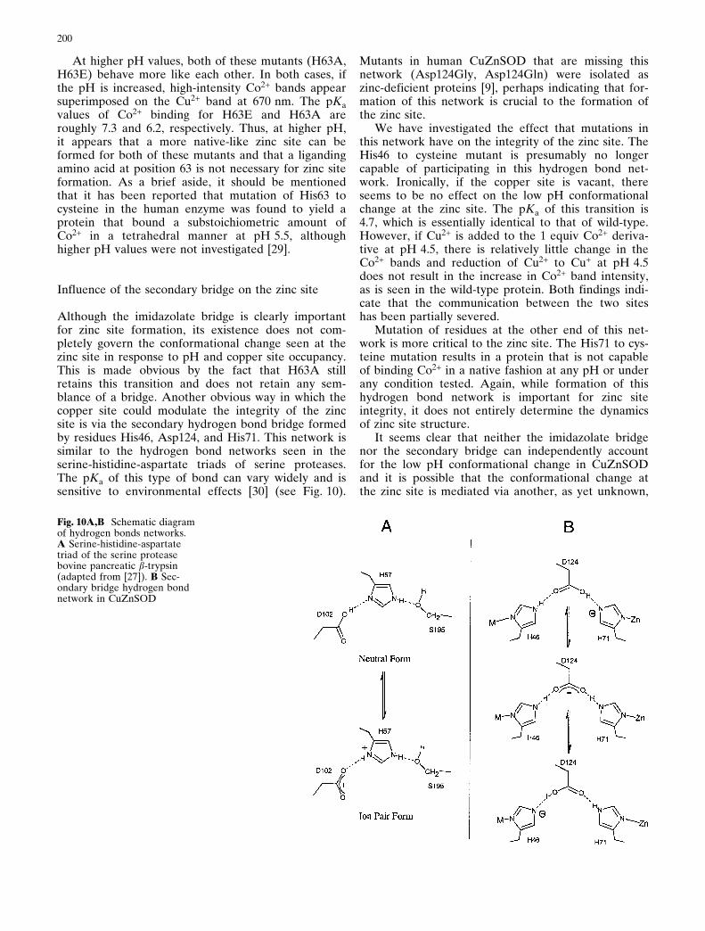
Although the imidazolate bridge is clearly importantfor zinc site formation, its existence does not com-pletely govern the conformational change seen at thezinc site in response to pH and copper site occupancy.This is made obvious by the fact that H63A stillretains this transition and does not retain any sem-blance of a bridge. Another obvious way in which thecopper site could modulate the integrity of the zincsite is via the secondary hydrogen bond bridge formedby residues His46, Asp124, and His71. This network issimilar to the hydrogen bond networks seen in theserine-histidine-aspartate triads of serine proteases.The pKa of this type of bond can vary widely and issensitive to environmental effects [30] (see Fig. 10).
Mutants in human CuZnSOD that are missing thisnetwork (Asp124Gly, Asp124Gln) were isolated aszinc-deficient proteins [9], perhaps indicating that for-mation of this network is crucial to the formation ofthe zinc site.
We have investigated the effect that mutations inthis network have on the integrity of the zinc site. TheHis46 to cysteine mutant is presumably no longercapable of participating in this hydrogen bond net-work. Ironically, if the copper site is vacant, thereseems to be no effect on the low pH conformationalchange at the zinc site. The pKa of this transition is4.7, which is essentially identical to that of wild-type.However, if Cu2+ is added to the 1 equiv Co2+ deriva-tive at pH 4.5, there is relatively little change in theCo2+ bands and reduction of Cu2+ to Cu+ at pH 4.5does not result in the increase in Co2+ band intensity,as is seen in the wild-type protein. Both findings indi-cate that the communication between the two siteshas been partially severed.
Mutation of residues at the other end of this net-work is more critical to the zinc site. The His71 to cys-teine mutation results in a protein that is not capableof binding Co2+ in a native fashion at any pH or underany condition tested. Again, while formation of thishydrogen bond network is important for zinc siteintegrity, it does not entirely determine the dynamicsof zinc site structure.
It seems clear that neither the imidazolate bridgenor the secondary bridge can independently accountfor the low pH conformational change in CuZnSODand it is possible that the conformational change atthe zinc site is mediated via another, as yet unknown,
Fig. 10A,B Schematic diagramof hydrogen bonds networks.A Serine-histidine-aspartatetriad of the serine proteasebovine pancreatic b-trypsin(adapted from [27]). B Sec-ondary bridge hydrogen bondnetwork in CuZnSOD

201
mechanism. What is evident is that the pH and redoxdependence of this interconversion is sensitive to theenvironment of the protein matrix.
The role of the zinc site in the structure and functionof CuZnSOD
To assess appropriately the importance of these find-ings, it is first necessary to analyze the role that thezinc site plays in CuZnSOD. CuZnSOD was first iso-lated in 1939 and determined to be a copper proteinof unknown function [32]. It was not until 1970, a yearafter the SOD function was discovered, that zinc wasproven to be a cofactor of the enzyme [33]. Zinc canbe replaced with Co2+, Hg2+, Cd2+, Cu2+, Ni2+, orVO2+ with only modest effects on the catalytic func-tion of the enzyme [2, 34]. Even the zinc-free enzymeis fully active at physiological pH. However, while theactivity of the holo enzyme is relatively constant overa wide pH range, the activity of the zinc free enzymeis markedly pH dependent and falls off rapidly abovepH 7 [35].
Zinc can be thought of as playing a structural rolein CuZnSOD, a role that is supported by data whichdemonstrated that zinc binding to the bovine enzymeenhances stability towards denaturation by SDS [36,37] and guanidinium chloride [38], and towards degra-dation by proteolysis [39]. In addition, zinc has beendemonstrated to be a major contributing factor to theunusual thermal stability in both the bovine [40] andhuman [41] proteins. Various techniques have alsoshown that the binding of zinc alone to the apoproteinis sufficient to induce a native fold on the protein [36,40, 41, 42]. Recent results from our laboratory indicatethat the binding of one zinc per dimer is sufficient toinduce a native fold for the entire protein (H. Zhu, J.S. Valentine, unpublished results).
Zinc also has important effects on the structure andreactivity of the copper site. It is thought that zincbinding pre-forms the copper site [23] and enhancesthe affinity of the copper site for metal ions [43].Along with the aforementioned effects on catalyticactivity, zinc deficiency also results in several otherchanges in copper site reactivity. For instance, zinc-de-ficient yeast CuZnSOD is more rapidly reduced byascorbate than is the holoprotein [17] and zinc-defi-cient human CuZnSOD reacts more readily with per-oxynitrite to nitrate tyrosine residues [31]. Zinc-defi-cient bovine CuZnSOD, as well as derivatives inwhich the zinc has been replaced by Cu2+ and Co2+,are more readily inactivated by peroxide than is theholoprotein [34, 44].
At low pH, where the copper and zinc sites areuncoupled, the copper ion can no longer be reducedby ferrocyanide as is possible at higher pH [45]. Inaddition, there are gross changes in the electrochem-istry of the copper site of bovine CuZnSOD at low
pH, making the site more accessible to the electrode[46] as well as changing the redox potential [47].
This study adds to our understanding of the factorsthat govern the formation of the zinc site in yeastCuZnSOD, and yet our understanding is still far fromcomplete. Based on these studies, a histidine at posi-tion 71 seems to be absolutely critical to zinc site for-mation. Liganding amino acids at positions 63 and 83also appear to be very important. While disruption ofthe secondary bridge on one end can be tolerated(H46C, H46R, and G85R), disconnection of this net-work from the other end is calamitous for the zinc site(H71C, D124G, D124N). Table 1 summarizes theresults discussed in this paper.
Conclusions
Understanding the structural parameters of the zincsite is important for the study of mutations that causeALS [14], as several mutants have been found to havea lowered affinity for zinc [31] and others have beenshown to display abnormal zinc site spectroscopy [17].These changes in zinc site properties may be relatedto the changes that occur in the wild-type at low pH.
Table 1. Summary of zinc site status for individual proteinsa
Protein Zinc site status pKa oftransi-tion
Human wild-type reconstitutedwith Co2+
Td at pH 5.5 4.0
Bovine wild-type as-isolatedand reconstituted with Zn2+,Co2+, or Cu2+
Td at pH 4 ± 9 3.8
Yeast wild-type as-isolated Td at pH 4 ± 9 3.8Yeast wild-type reconstitutedwith Co2+
Td at pH 5 ± 9 4.7c
Yeast H46Cb Like yeast wild-typeat high pH
4.66
Yeast H46Rb Td at high pH 6.05c
Yeast H48Qb Td at high pH 5.58Yeast H63Ab Td at high pH 6.2Yeast H63Eb Td at high pH and
at pH 5.5 if Cu2+ isreduced to Cu+
7.3
Yeast H71Cb Not Td under anycondition tested
NA
Yeast H80Cb Td at high pH 5.60c
Yeast D83Ab Not Td under anycondition tested
NA
Yeast D83Hb Td at high pH andat pH 5.5 if Cu2+ isreduced to Cu+
5.8
Yeast G85Rb Td at high pH andat pH 5.5 if Cu2+ isreduced to Cu+
> 5
a Td = zinc site with ligands arranged in a distorted tetrahedralgeometryb Using the visible spectroscopy of Co2+ to monitor the geome-try of the zinc sitec Value determined with no metal ion bound at the copper site
202
The zinc site seems to be able to exist in two states:one state binds Co2+ to a strong binding site with tet-rahedral geometry and another forms a weak bindingzinc site. The conversion from one to the other is reg-ulated by pH and by the metal ion bound at thecopper site. It is also apparent that mutations all overthe protein impact this transition, generally raising thepKa to values closer to physiologically relevant ones.This finding may help explain a recent discovery bythis group that yeast CuZnSOD that has been demet-allated is composed of two inequivalent subunits, onewhich is competent to bind metals normally andanother that is not [20]. Apo yeast CuZnSOD maycontain one subunit that is ªfrozenº in the wrong con-figuration. The ramifications of these findings are farreaching, especially concerning the relationshipbetween CuZnSOD and ALS.
In this paper we discuss three yeast mutants thatare analogous to mutations that cause ALS. All threeALS mutations investigated appear to have a pH-in-duced transition similar to that of the wild-type, butwith a pKa that has been shifted up to near physiologi-cal pH. This suggests that it is a real possibility that,at physiological pH, these proteins might be expectedto contain a substantial portion of zinc sites that havebeen converted to the weak Co2+ binding configura-tion, thus making this phenomenon potentially rel-evant for the etiology of ALS.
It is possible that the ALS-causing mutations resultin a protein that does not bind zinc as tightly as thewild-type and is therefore less stable and more proneto aggregation and denaturation. It is also possiblethat the less-stable zinc site allows for undesirablereactivity to occur at an ªunprotectedº copper site.An even more interesting possibility is that the ALSmutants have modified zinc sites that allow other met-als besides zinc to bind at this site and participate innovel chemical reactions. The zinc site in the wild-typebovine CuZnSOD does indeed have a naturally highaffinity for copper [48]. This high affinity apparentlyincreases if the pH is raised from 7 and there is nometal bound at the zinc site [48, 49]. It is possible thatthe ALS mutants possess zinc sites which, whileretaining a high affinity for zinc, have increased affini-ties for other metals.
Since it is now generally accepted that mutations inhuman CuZnSOD cause ALS via a gain of function[50], it is imperative that we uncover the intricatemechanisms by which this enzyme achieves its remark-able stability. It may be through understanding howthis protein folds correctly that we stumble upon thetrait or traits that contribute to the toxicity of itsmutants. However, we must stress that, while thereare many similarities between the yeast and humanenzymes, there are distinct differences in their bio-physical properties [20, 51]. While it is possible thatthese differences may simply reflect a greater propen-sity of the yeast enzyme to undergo certain changesthat are characteristic of all CuZnSODs, the fact that
they exist must temper any conclusions we draw. It isalso quite possible that the yeast protein is an aberra-tion, a fact that diminishes its importance for thoseresearching ALS.
Acknowledgements This research was funded by the NationalInstitute of General Medical Sciences GM28222 (J.S.V.). Wewould also like to thank Dr. Daryl Eggers for editorial sugges-tions. This work is dedicated to the memory of Dr. HyeyeongYeom.
References
1. Cabelli DE, Riley D, Rodriguez JA, Valentine JS, Zhu H(2000) In: Meunier B (ed) Biomimetic oxidations. ImperialCollege Press, London (in press)
2. Valentine JS, Pantoliano MW (1981) In: Spiro TG (ed)Copper proteins. Wiley, New York, pp 292±358
3. Bertini I, Mangar S, Viezzoli MS (1998) Adv Inorg Chem45 : 127±250
4. Djinovic K, Gatti G, Coda A, Antolini L, Pelosi G, DesideriA, Falconi M, Marmocchi F, Rotilio G, Bolognesi M (1992)J Mol Biol 225 : 791±809
5. Tainer JA, Getzoff ED, Beem KM, Richardson JS, Richard-son DC (1982) J Mol Biol 160 : 181±217
6. Bertini I, Luchinat C, Monnanni R (1985) J Am Chem Soc107 : 2178±2179
7. Ogihara NL, Parge HE, Hart PJ, Weiss MS, Goto JJ, CraneBR, Tsang J, Slater K, Roe JA, Valentine JS, Eisenberg D,Tainer JA (1996) Biochemistry 35 : 2316±2321
8. Banci L, Bertini I, Bruni B, Carloni P, Luchinat C, MagnaniS, Orioli PL, Piccioli M, Rypniewski WS, Wilson KS (1994)Biochem Biophys Res Commun 202 : 1088±1095
9. Banci L, Bertini I, Hallewell RA, Tung JW, Viezzoli MS(1991) Eur J Biochem 196 : 123±128
10. Fee JA, Phillips WD (1975) Biochim Biophys Acta412 : 26±38
11. Pantoliano MW, Valentine JS, Mammone RJ, Scholler DM(1982) J Am Chem Soc 104 : 1717±1723
12. McCord JM, Fridovich I (1969) J Biol Chem 244 : 6049±605513. Siddique T, Nijhawan D, Hentati A (1997) J Neural Transm
Suppl 49 : 219±23314. Deng H-X, Hentati A, Tainer JA, Iqbal Z, Cayabyab A,
Hung W-Y, Getzoff ED, Hu P, Herzfeld B, Roos RP,Warner C, Deng G, Soriano E, Smith C, Parge HE, AhmedA, Roses AD, Hallewell RA, Pericak-Vance MA, SiddiqueT (1993) Science 261 : 1047±1051
15. Lyons TJ, Gralla EB, Valentine JS (1999) In : Sigel A, SigelH (eds) Metal ions in biological systems. Dekker, NewYork, pp 125±177
16. Graden JA, Ellerby LM, Cabelli D, Gralla EB, Valentine JS(1994) J Am Chem Soc 116 : 9743±9744
17. Lyons TJ, Liu H, Goto JJ, Nersissian A, Roe JA, GradenJA, CafØ C, Ellerby LM, Bredesen DE, Gralla EB, Valen-tine JS (1996) Proc Natl Acad Sci USA 93 : 12240±12244
18. Nishida CR, Gralla EB, Valentine JS (1994) Proc Natl AcadSci USA 91 : 9906±9910
19. Lu Y, Gralla EB, Roe JA, Valentine JS (1992) J Am ChemSoc 114 : 3560±3562
20. Lyons TJ, Nersissian A, Goto JJ, Zhu H, Gralla EB, Valen-tine JS (1998) JBIC 3 : 650±662
21. Cupane A, Leone M, Militello V, Stroppolo ME, PolticelliF, Desideri A (1995) Biochemistry 34 : 16313±16319
22. Maret W, Vallee BL (1993) Methods Enzymol 226 : 52±7123. Cass AEG, Hill HAO, Bannister JV, Bannister WH (1979)
Biochem J 177 : 477±48624. Djinovic K, Coda A, Antolini L, Pelosi G, Desideri A, Fal-
coni M, Rotilio G, Bolognesi M (1992) J Mol Biol226 : 227±238

203
25. Pantoliano MW, McDonnell PJ, Valentine JS (1979) J AmChem Soc 101 : 6454±6456
26. Calabrese L, Cocco D, Morpurgo L, Mondovi B, Rotilio G(1975) FEBS Lett 59 : 29±31
27. Dong A, Huang P, Caughey WS (1995) Arch Biochem Bio-phys 320 : 59±64
28. Hart PJ, Liu H, Pellegrini M, Nersissian AM, Gralla EB,Valentine JS, Eisenberg DE (1998) Protein Sci 7 : 545±555
29. Banci L, Bertini I, Borsari M, Viezzoli MS, Hallewell RA(1995) Eur J Biochem 232 : 220±225
30. Umeyama H, Nakagawa S (1982) J Theor Biol 99 : 759±77531. Crow JP, Sampson JB, Zhuang Y, Thompson JA, Beckman
JS (1997) J Neurochem 69 : 1936±194432. Mann T, Keilin D (1939) Proc R Soc London Ser B 126 : 30333. Carrico RJ, Deutsch HF (1970) J Biol Chem 245 : 723±72734. Kajihara J, Enomoto M, Katoh K, Mitsuta K, Kohno M
(1990) Agric Biol Chem 54 : 495±49935. Pantoliano MW, Valentine JS, Burger AR, Lippard SJ
(1982) J Inorg Biochem 17 : 325±34136. Marmocchi F, Caulini G, Venardi G, Cocco D, Calabrese L,
Rotilio G (1975) Physiol Chem Phys 7 : 465±47137. Caulini G, Marmocchi F, Venardi G, Rotilio G (1974) Boll
Soc Ital Biol Sper 50 : 1091±109438. Mach H, Dong Z, Middaugh CR, Lewis RV (1991) Arch
Biochem Biophys 287 : 41±47
39. Rotilio G, Calabrese L, Bossa F, Barra D, Agro AF, Mon-dovi B (1972) Biochemistry 11 : 2182±2186
40. Roe JA, Butler A, Scholler DM, Valentine JS, Marky L,Breslauer KJ (1988) Biochemistry 27 : 950±958
41. Biliaderis CG, Weselake RJ, Petkau A, Friesen AD (1987)Biochem J 248 : 981±984
42. Sun WY, Fang JL, Cheng M, Xia PY, Tang WX (1997) Bio-polymers 42 : 297±303
43. Roe JA, Peoples R, Scholler DM, Valentine JS (1990) J AmChem Soc 102 : 1538±1545
44. Goto JJ, Gralla EB, Valentine JS, Cabelli DE (1998) J BiolChem 273 : 30104±30109
45. Morpurgo L, Mavelli I, Calabrese L, Agro AF, Rotilio G(1976) Biochem Biophys Res Commun 70 : 607±614
46. Iyer RN, Schmidt WE (1992) Bioelectrochem Bioenerg27 : 393±404
47. Verhagen MFJM, Meussen ETM, Hagen WR (1995) Bio-chim Biophys Acta 1244 : 99±103
48. Hirose J, Yamada M, Hayakawa C, Nagao H, Noji M,Kidani Y (1984) Biochem Int 8 : 401±408
49. Valentine JS, Pantoliano MW, McDonnell PJ, Burger AR,Lippard SJ (1979) Proc Natl Acad Sci USA 76 : 4245±4249
50. Brown RH Jr (1995) Curr Opin Neurobiol 5 : 841±84651. Lepock JR, Arnold LD, Torrie BH, Andrews B, Kruuv J
(1985) Arch Biochem Biophys 241 : 243±251