zinc, zinc transporters and type 2 diabetes · binding proteins (andreini et al., 2006)....
TRANSCRIPT

Zinc, Zinc Transporters and Type 2 Diabetes Stephen A Myers
Collaborative Research Network School of Health Sciences
Federation University Australia, Australia
Alex Nield Collaborative Research Network
School of Health Sciences Federation University Australia, Australia

1 Introduction
Insulin resistance is an important characteristic of Type 2 Diabetes (T2D) and is commonly associated with obesity, hypertension and cardiovascular disease (Carsten, 2000; Hulver and Lynis, 2004). Insulin resistance reduces insulin-stimulated glucose disposal due to multiple post-receptor intracellular defects in insulin signaling with subsequent reductions in glucose transport, glucose oxidation and incorporation of glucose into glycogen (Abdul-Ghani and DeFronzo, 2010; Peppa et al., 2010). The intracellular post-receptor regulatory effects of insulin include the regulation of the cellular glucose transport system, adap-tive changes in gene expression and subsequent biosynthesis and action of the enzymes involved in the preservation of metabolism, and the modulation of genes that contribute to increased pro-mitotic, prolif-erative and anti-apoptotic activity of cells (Taton et al., 2010). Accordingly, the reduced activity of insu-lin action in any, or all of these post-receptor regulatory actions is insulin resistance. Given that insulin resistance usually precedes the development of T2D and is a major component of the progressive nature of this disease (Pagel-Langenickel et al., 2010), understanding the pathophysiology of insulin resistance will enable the development of therapeutic strategies to prevent or manage disease progression. Although many theories have been forthcoming, the primary mechanism of insulin resistance remains largely elu-sive.
In this context, research on T2D has revealed an exciting role for zinc signaling in this disease. Ionic zinc has insulin “mimetic” activity where it is involved in insulin receptor signal transduction and insulin storage, secretion and distribution (Fukada et al., 2011). In peripheral tissue such as fat and mus-cle, zinc ions facilitate insulin-induced glucose transport and glycaemic control through the regulation of essential pathways involved in glucose homeostasis (Jansen et al., 2009). However there is insufficient information on how the concentration of free ionic zinc in cells is controlled. The proteins that transport zinc presumably facilitate cell signaling processes that contribute to glycaemic control in peripheral tissue through the modulation of ionic zinc concentrations in the cytosol. For example, aberrant subcellular par-titioning or signaling of zinc could contribute to altered insulin responsiveness (Mocchegiani et al., 2008) and therefore facilitate insulin resistance. However, how zinc transporter proteins effectively facilitate zinc flux and contribute to cellular metabolism is not clear.
Accordingly, this review will discuss the zinc transporter gene family and what is currently known regarding their various roles in cellular signaling in disease processes with a particular focus on T2D. It is envisaged that understanding the potential relevance of dysfunctional ionic zinc partitioning in diseases such as T2D will created opportunities for translating basic research into clinically important applica-tions. Therefore, knowing the specific molecular targets of ionic zinc in cellular signaling in the context of insulin resistance and T2D will provide opportunities to develop novel therapeutic approaches to pre-vent disease progression or treat this disease.
2 Type 2 Diabetes Mellitus
Type 2 Diabetes (T2D) is a progressive and chronic metabolic disease that is rapidly increasing in preva-lence worldwide. In 2030, it is predicted that adults diagnosed with this disorder will rise from the current count of 246 million to 552 million (Pal and McCarthy, 2013). Patients with T2D are predisposed to a number of microvascular and macrovascular complications and therefore have a significant increase in mortality and morbidity (Shin et al., 2012). Consequently, T2D is an important global public health prob-

lem due to the considerable cost of appropriate disease control and management of chronic complications (Shin et al., 2012). For example, diabetes cost the global economy nearly US $50 billion in 2010 and that figure is expected to increase to US $745 billion in 2030 (Bloom et al., 2011).
Lifestyle factors that lead to obesity, in particular poor nutrition and reduced physical activity, con-tribute to the development of T2D. In this context, for the prevention of factors leading to T2D, it is vital that blood glucose levels are maintained within a normal range (i.e. fasting blood glucose of less than 100 mg/dL and a 2 hour postprandial blood glucose of less than 140 mg/dL are usually considered normal) (Nyenwe et al., 2011). Medical nutrition therapy and exercise programs are the basis for managing T2D, however the majority of patients with this disease also require pharmacotherapy to manage this disorder successfully (Kourtoglou, 2011).
T2D results when the endocrine pancreas fails to meet increasing metabolic demands and compen-sate for peripheral tissue insulin resistance (Osto et al., 2013) and usually develops in patients with pan-creatic β-cell dysfunction in the presence of insulin resistance in peripheral tissue such as liver, fat and muscle (Lin and Sun, 2010). Insulin resistance is described as a reduced and/or delayed response to insu-lin in peripheral tissue and is generally associated with dysfunctional insulin signaling rather than the production of insulin (Lin and Sun, 2010). Even in the absence of T2D, insulin resistance is often associ-ated with hypertension, obesity, polycystic ovarian syndrome, dyslipidemia and atherosclerosis (Saltiel and Pessin, 2002). In T2D, the majority of patients will eventually require the administration of insulin, either alone or in combination with other antidiabetic agents to control their diabetes (Kourtoglou, 2011).
Recently there has been a growing interest in the role of zinc signaling in T2D based on the fact that reduced levels of serum zinc have been observed in patients with diabetes (Basaki et al., 2012; Jansen et al., 2009; Jansen et al., 2012; Zhao et al., 2011) and zinc supplementation in several animal models of T2D and human patients showed improved glycaemic control (Adachi et al., 2006; Jansen et al., 2009; Jayawardena et al., 2012; Liu et al., 2011a; Pathak et al., 2011; Wang et al., 2012). The clinical relevance of zinc in T2D is further emphasized by recent developments in our understanding of dysregu-lation of zinc partitioning in this disease (Mocchegiani et al., 2008). T2D is characterized by defects in both insulin secretion and insulin sensitivity, of which zinc is particularly important based on its role in insulin receptor signal transduction (Haase et al., 2005a, 2005b), and insulin storage and secretion (Figlewicz et al., 1984; Vardatsikos et al., 2013).
3 Zinc
Zinc is one of the most important trace elements in nature where it is indispensable for the growth and development of microorganisms, plants and animals (Chasapis et al., 2011). In the human body there is approximately 2-4 g of total zinc making it the most abundant trace metal in tissue next to iron (approxi-mately 4 g which is localized primarily in blood). Zinc is found in relatively high abundance with the highest concentration of zinc is found in the prostate (84-211 µg/g), while the pancreas contains 140 µg/g; muscle has approximately 51 µg/g of zinc while in plasma approximately 14-16 µM of zinc can be meas-ured, which contributes to the mobile zinc pool that is required for cellular distribution (Jansen et al., 2009). At the cellular level, approximately 30-40% of total cellular zinc is found in the nucleus, approxi-mately 50% in the cytosol and its organelles, and the remainder in the plasma membrane (Vallee et al., 1993). Zinc found in these compartments is essentially bound to macromolecules including zinc pro-teins/enzymes, lipids, and DNA/RNA (Chasapis et al., 2011; Maret, 2011a, 2011b). In fact, research of

the human genome has established that approximately 10% of the proteome consists of potential zinc-binding proteins (Andreini et al., 2006). Accordingly, the compartmentalization, availability, transport and re-distribution of zinc must be tightly controlled in order to maintain cellular zinc homeostasis. Thus, the mechanisms dedicated to controlling these processes is principally achieved by the metallothioneins [for a comprehensive review see (Maret, 2011a), and references therein], and a family of transmembrane zinc transporter proteins (Kambe, 2011; Myers et al., 2012).
Zinc ions are widely used as a cofactor for numerous proteins, and in addition to their structural and catalytic role, zinc facilitates information transfer and cellular control (Maret, 2011a, 2011b). In fact, zinc is critical for the function of over 300 enzymes including members of all classes. These include the oxidoreductases, transferases, hydrolases, lyases, isomerases and ligases, and are examples of all the six enzymes classes established by the International Union of Biochemistry (Vallee and Falchuk, 1993). Zinc is also involved in a number of cellular processes including extracellular signal recognition; second messenger activity; protein phosphorylation and dephosphorylation, and the regulation of transcription factors (Beyersmann and Haase, 2001).
Zinc has insulin-mimetic and anti-diabetic effects in cells and animal models of type 1 and T2D. The molecular mechanisms responsible for the insulin-mimetic effects of zinc include the activation of several key signaling molecules of the insulin signaling pathway such as the extracellular signal-regulated kinase 1/2 (ERK1/2) and phosphatidylinositol 3-kinase (PI3-K)/protein kinase B/Akt (PKB/Akt) path-ways (Vardatsikos et al., 2013). Moreover, disturbances of zinc homeostasis are associated with several disease states including diabetes, liver cirrhosis (Bode et al., 1988), cancer (Dufner-Beattie et al., 2004) and impaired function of the immune system (Haase et al., 2006). However, the molecular mechanisms responsible for alterations in cellular zinc status that lead to dysfunctional signaling and disease are un-known.
3.1 Zinc and health: brief historical perspectives
The importance of zinc in biological systems was first described in 1869 where it was shown to be essen-tial for the growth of Aspergillus niger, the common bread mold (Raulin, 1869). Subsequently, zinc was found to be vital for the growth of plants (Sommer et al., 1926) and rats (Todd et al., 1934). In 1955 it was identified that parakeratosis in swine was due a deficiency in zinc (Tucker and Salmon, 1955) and the addition of zinc to food rations with established cases of this disease produced immediate and rapid weight gain and essentially eliminated the skin lesions (Lewis et al., 1956). Moreover, chickens with a severe deficiency in zinc were slow growing, had abnormal respiration, poor calcification of bone, and evidence of parakeratosis (O'Dell et al., 1958). Although by 1960 several experiments reported the essen-tiality of zinc in several animal models; due to its abundance, zinc deficiency and subsequent clinical manifestations in humans was considered unlikely (Prasad, 2012).
The idea that zinc was essential for humans was first discovered in 1958 where symptoms of se-vere anemia, growth retardation, hypogonadism, heptosplenomegaly, skin abnormalities and mental leth-argy and geophagia (clay eating) in men from Iran were attributed to zinc deficiency (Prasad et al., 1961). Subsequently, many other studies have identified the importance of dietary zinc in humans (Alves et al., 2012; Atasoy and Ulusoy, 2012; Bribiescas, 2003; Coble et al., 1971; Hambidge et al., 1972; Makonnen et al., 2003; Ronaghy et al., 1969; Sandstead et al., 1967), and the recognition that zinc deficiency is po-tentially a widespread problem in both developing and developed countries (Ackland et al., 2006). Alt-hough zinc deficiency has compound effects on human health, perturbations in cellular ionic zinc status

can lead to several chronic diseases including cancer, cardiovascular disease, Alzheimer’s disease and diabetes (Bosomworth et al., 2013; Devirgiliis et al., 2007, Mocchegiani et al., 2008).
4 Zinc Transporters
Zinc transporters belong to a family of transmembrane proteins that control zinc transport across cellular membranes and contribute to the uptake, distribution and compartmentalization of this metal ion (Myers et al., 2012) (Figure 1). The zinc transporters1 belong to two major gene families: the ZnT proteins (so-lute-linked carrier 30, SLC30) and the ZIP (Zrt/Irt-like, solute-linked carrier 39, SLC39) (Kambe, 2011; Liuzzi et al., 2004). Among these, in mammals there are ten members of the zinc efflux (SLC30/ZnT; Znt1-10) transporters proteins that transport zinc out of the cell or into subcellular compartments in the presence of high cytoplasmic zinc, and fourteen members of the zinc influx (SLC39/ZIP; ZIP1-14) pro-teins that transport zinc into the cell or out of subcellular compartments when cytosolic zinc is low or depleted (Figure 1) (Gaither and Eide, 2001).
Figure 1: Cellular localization of the ZIP and ZnT zinc transporters. Green arrows depict the ZnTs while red arrows show the ZIP family. The position of the arrows indicates the direction of zinc flux. Note: Golgi (Golgi apparatus) and ER (endoplasmic reticulum). Figure was produced using Servier Medical Art, http://www.servier.com/.
1 The alias terminology ZIP and ZnT (SLC39 and SLC30, respectively) will be used throughout for consistency.

5.1 The SLC39/ZIP family
The first member of the ZIP family of transporters to be identified was in Saccharomyces cerevisiae based on a similarity to that of Irt1p, an Fe(II) transporter from Arabidopsis thaliana (Eide et al., 1996; Zhao et al., 1996). The ZRT1 gene was found to encode a zinc transporter protein with a high affinity for zinc and moreover, this transporter was induced at the transcriptional level in zinc-depleted conditions (Zhao et al., 1996). Furthermore, a mutation in this gene eliminated the high-affinity uptake of zinc and inhibited growth on zinc-limiting media suggesting that ZRT1 is necessary for growth in zinc-limiting conditions. Since the discovery of ZRT1, fourteen gene members have been identified in mammals des-ignated SLC39A1-SLC39A14, (Table 1) encoding for the proteins ZIP1-14, respectively (Jeong and Eide, 2013). Most ZIP transporters have a predicted 8 transmembrane domain (TMD) with the N- and C-termini localized extra-cytoplasmically (Figure 1). A long loop region that often contains a histidine-rich region is localized between TMD III and TMD IV (Jeong and Eide, 2013) where it is thought to be in-volved in binding metal ions. Indeed, studies in PC-3 prostate cancer cells identified that mutation of one or both histidine residues in the loop region (H158 and H160) of ZIP1 resulted in a decrease in zinc accu-mulation in PC-3 cells (Milon et al., 2006). More recently, a loop region located between TM II and III of the IRT1 transporter from Arabidopsis thaliana was shown to have unique zinc-binding features where imidazoles from two histidines (His-96 and His-116), a cysteine thiolate (Cys-109) and one of a glutamic acid carboxyl group was responsible for the formation of the zinc-IRT1 complex, and thus the transport of zinc ions (Potocki et al., 2013). These data are supported by earlier studies in the zinc-binding protein ZnuA from Escherichia coli where bound zinc was coordinated by three histidine residues (His-78, His-161 and His-225) and one glutamate residue (Glu-77) (Li et al., 2007).
The ZIP transporters are expressed in a range of tissue types and their proteins are localized to dis-tinct subcellular compartments. In addition to regulation by intracellular and extracellular zinc concentra-tions, many of these transporters are also regulated by hormones and cytokines (Table 1) (Lichten and Cousins, 2009). Such diverse regulation, tissue expression patterns and subcellular localization of these transporters provides some insight into their many physiological roles. Moreover, the ZIP transporters are implicated in a number of pathophysiological processes including prostate, breast and pancreatic cancer, carotid artery disease, acrodermatitis enteropathica, schizophrenia, Ehlers-Danlos syndrome, and asthma (Table 1).
5.2 The SLC30/ZnT family
The mammalian SLC30 zinc transporters belong to a large family of cation diffusion facilitators (CDF/ZnT) and include zinc transporter members with similar topology from bacteria, fungi, nematodes, insects, plants and mammals (Huang and Tepaamorndech, 2013). Members of this family are predicted to have six transmembrane (TM) domains and a histidine-rich loop between TMD IV and TMD V with the N- and C-termini on the cytoplasmic side of the membrane (Palmiter and Huang, 2004) (Figure 1). At present, there is little information available on the structure of the ZnTs, however, one CDF bacterial homologs, Yiip from Escherichia coli has been characterized functionally and has 25-30% sequence similarity to their mammalian counterparts (Lu and Fu, 2007). The Yiip protein is a homodimer consist-ing of two 32.9-kDa integral membrane proteins composed of six TMD (Wei et al., 2004) and a tetrahe-dral zinc-binding site that is essential for transport (Lu and Fu, 2007).

Gene/protein Tissue expression and cellular localization
Regulation of ZIP transporters Disease/pathology association
Slc39a1/ZIP1 Ubiquitously expressed, plasma membrane
↑ Prolactin and testosterone (LNCaP and PC3 prostate cancer cells)b
Prostate cancer
Slc39a2/ZIP2 Blood, prostate, plasma mem-brane
Unknown Carotid artery disease
Slc39a3/ZIP3 Mammary gland, prostate, plasma membrane, intracellular compartments
↑ Prolactin (mammary gland)c Pancreatic cancerl
Slc39a4/ZIP4 Small intestine, stomach, colon, kidney, brain, plasma mem-brane, apical membranes
↓ Zinc deficiency (embryonic visceral yolk sack isolated from pregnant mice)d
Pancreatic cancer, acrodermatitis enteropathica (AE)
Slc39a5/ZIP5 Pancreas, kidney, liver, spleen, colon, stomach, plasma mem-brane, basolateral membranes
Unknown Unknown
Slc39a6/ZIP6 Ubiquitously expressed, plasma membrane
↑ Estrogen, ↓ Tamoxifen and fulves-trant (MCF-7 breast cancer cells)e, ↑
glucose (pancreatic mouse islets)f
Breast cancer; cervical cancerm
Slc39a7/ZIP7 Ubiquitously expressed, Golgi apparatus, endoplasmic reticu-lum
↑ Estrogen (MCF-7 breast cancer cells)e, ↑ glucose (pancreatic mouse islets)f
Breast cancer
Slc39a8/ZIP8 Ubiquitously expressed, vesicles ↑ TNF-α (monocytesg, human lung epithelial cells, hLECs)h
Inflammation
Slc39a9/ZIP9 Ubiquitously expressed, trans-Golgi network
Unknown Unknown
Slc39a10/ZIP10 Ubiquitously expressed, plasma membrane
↑ Thyroid hormone (rat intestines and kidney)i
Breast cancer
Slc39a11/ZIP11 Mammary gland, testesa, stom-acha, small and large intestinea, Golgi apparatus
↑ Zinc (spleen and liver in gavage-fed mice)a
Unknown
Slc39a12/ZIP12 Retina, brain, testis, lung Unknown Schizophrenia, neuronal differen-tiationn
Slc39a13/ZIP13 Ubiquitously ex-pressed, Golgi apparatus
Unknown Ehlers-Danlos syndrome
Slc39a14/ZIP14 Ubiquitously ex-pressed, plasma membrane
↑ Estrogen, ↓ tamoxifen and fulves-trant (MCF-7 breast cancer cells)e; ↑
IL-6 (mouse liver parenchymal cellsj, adipose tissue and muscle)k
Asthma, in-flammmationk
Table Source: modified from Myers et al. 2012 and references herein. Regulation of zinc transporters: ↑ Upregulation; ↓ Down regulation, a Yu et al., 2013; b Costello et al., 1999; c Kelleher et al., 2011; d Dufner-Beattie et al., 2004; e Taylor et al., 2007; f Bellomo et al., 2011; g Begum et al., 2002; h Besecker et al., 2008; i Pawan et al., 2007; j Liuzzi et al., 2005; k Beker et al., 2012; l Costello et al., 2012; m Zhao et al., 2007; n Chowanadisai et al., 2013.
Table 1: SlC39/ZIP transporters: tissue expression and cellular localization, regulation and dis-ease/pathology association.

The first mammalian ZnT transporter (ZnT1) was isolated from a rat kidney cDNA library where it was shown to facilitate zinc resistance to zinc toxicity in the zinc-sensitive baby hamster kidney (BHK) cell line (Palmiter and Findley, 1995). Since the discovery of ZnT1, ten members of the mammalian SLC30 family have been identified and designated SLC30A1-SLC30A10 and encoding for the proteins ZnT1-10, respectively (Table 2) (Huang and Tepaamorndech, 2013). Similar to the ZIP family members, the ZnTs are expressed in a wide-range of tissues; have specific subcellular localizations and are impli-cated in a number of pathophysiological disease states (Table 2). Equally, they are also regulated by in-tracellular and extracellular zinc status, hormones and cytokines and other molecules (Table 2).
6 Zinc, Zinc Transporters and Cell Signaling
Zinc transporters typically act as zinc sensors and respond to cellular zinc availability to maintain intra-cellular zinc homeostasis. Cellular homeostasis of zinc is complex and there are a number of significant and comprehensive reviews on these processes (Jeong and Eide, 2013; Kambe, 2011; Liuzzi and Cousins, 2004; Myers et al., 2012, and references therein). Therefore, this section aims to briefly present a number of important processes by which zinc and zinc transporters maintain cellular homeostasis. A particular focus will be made to highlight the role of these transporters in cellular signaling. Zinc mimics the action of hormones, growth factors and cytokines and given the large number of zinc transporters that are dedi-cated to controlling zinc homeostasis (Table 1 and Table 2); it is not surprising that this ion is quickly taking precedence as a leading signaling molecule analogous to calcium. In this context, the processes of cellular zinc signaling have been designated into two main mechanisms of action; these are 1) early zinc signaling (EZS), and 2) late zinc signaling (LZS).
6.1 Early and late zinc signaling
EZS is a transcriptional-independent mechanism that involves a rapid intracellular change in levels of free zinc ions that occurs in minutes due to an extracellular stimulus (Fukada et al., 2011). This mecha-nism was first reported in bone marrow-derived mast cells (BMMCs) where treatment of these cells with the high affinity IgE receptor (FcεRI) resulted in a rapid increase (within minutes) in intracellular free zinc from the perinuclear region that includes the endoplasmic reticulum (ER) (Yamasaki et al., 2007). This zinc ‘wave’ as defined by these authors was also observed under conditions in which either the ex-tracellular zinc influx or the exocytosis of zinc-rich granules, was blocked. LZS is also triggered by an extracellular signal but involves transcriptional-dependent changes in expression of proteins implicated in zinc homeostasis such as storage proteins or transporters (Yamasaki et al., 2007). In LZS the intracellular zinc concentrations are usually altered over several hours following an external stimulus (Fukada et al., 2011). Since zinc has an important role in maintaining cellular function, and dysregulation or altered par-titioning of intracellular zinc causes disease in humans and animal models (Foster and Samman, 2010; Fukada et al., 2011; Haase and Maret, 2005a; Jansen et al., 2012; Pfaffl and Windisch, 2003; Prasad, 1991), understanding the proteins that transport zinc into and out of cells and subcellular organelles (the ZnTs and ZIPs) will be important in identifying novel therapeutic opportunities.

Gene/protein Tissue expression and cellular localization
Regulation of ZnT transporters Disease/pathology association
Slc30a1/ZnT1 Ubiquitously expressed, plasma membrane
↑ Zinc (intestine, liver and kidneye, pan-creatic cancer cellsf, HeLa cells)g, ↑ nitric oxide (rat cerebral cortex)h, ↓ Cadmium (mouse decidua, yolk sac, and embryo)i
Alzheimer's dis-ease, heart disease, pancreatic cancer
Slc30a2/ZnT2 Pancreas, kidney, testis, epithe-lial cells, small intestine, pros-tate, vesicles, lysosomes
↑ Zinc (intestine, liver and kidney)e,↑Nitric oxide (rat cerebral cortex)h, ↑ Prolactin (human mammary epithelial cells)j; ↑ neu-rotrophic factors (retinal pigment epitheli-um cell lines)k
Low zinc milk concentrations
Slc30a3/ZnT3 Brain, testis, vascular smooth muscle cellsa; adipose tissueb, synaptic vesicles
↓angiotensin (vascular smooth muscle)a, ↑
High glucose (INS-1E cells)b; ↓ Chronic metabolic acidosis (rat duodenal epithelial cells)j; ↑ Synthetic androgen R1881 (AIDL prostate cancer cells)l
Alzheimer's dis-ease
Slc30a4/ZnT4 Mammary gland, brain, small intestine, placenta, blood, epi-thelial cells, intracellular com-partments
↑ Nitric oxide (rat cerebral cortex)h Lethal Zn-deficient milk production, Alz-heimer's disease, asthma
Slc30a5/ZnT5 Ubiquitously expressed, secre-tory vesicles, Golgi apparatus
↑ Low glucose (INS-1E cells)b, ↑ TPEN (HeLa cells)g, ↓ High dietary zinc (human ileal mucosa and Caco-2 cells)m
Osteopenia
Slc30a6/ZnT6 Small intestine, liver, brain, adipose tissue, secretory vesi-cles, Golgi apparatus
Unknown Alzheimer's dis-ease
Slc30a7/ZnT7 Retina, small intestine, liver, blood, epithelial cells, spleen, secretory vesicles, Golgi appa-ratus
↑ TPEN (HeLa cells)g Prostate cancer
Slc30a8/ZnT8 Pancreatic β-cells, retinac, se-cretory vesicles
↑ Exendin-4 (pancreas in db/db mice)n, ↓ High glucose (INS-1E cells)b
Type 1 and 2 dia-betes mellitus, retinopathy
Slc30a9/ZnT9 Ubiquitously expressed, cyto-plasm, nucleus
Unknown
Slc30a10/ZnT10 Liver, brain, small intestined, Golgi apparatus
↓Angiotensin (vascular smooth muscle)a Parkinson’s dis-ease, dystonia, liver disease, Alz-heimer's disease
Table Source: modified from Myers et al. 2012 and references herein. Regulation of zinc transporters: ↑ Upregula-tion; ↓ Down regulation, a Patrushev et al., 2012; b Smidt and Rungby., 2012; c Deniro and Al-Mohanna., 2012; d
Bosomworth et al., 2012; e Liuzzi et al., 2001; f Jayaraman et al., 2011; gDevergnas et al., 2004; hAguilar-Alonso et al., 2008; i Fernandez et al., 2007; j Wongdee et al., 2009; k Leung et al., 2008; l Iguchi et al., 2004; m Cragg et al., 2005 n Liu et al., 2011b.
Table 2: SlC30/ZnT transporters: tissue expression, cellular localization and regulation, and dis-ease association.

6.2 Zinc and cellular signaling
The role of zinc and its function as a signaling mediator is well established and the literature is replete with many important findings (see, Fukada et al., 2011, and references therein). Early studies suggesting that zinc ions could function as a signaling molecule were identified in rat adipocytes where it was shown that zinc stimulated lipogenesis that was independent, and additive to that of insulin (Coulston and Dandona, 1980). Similarly, rat adipocytes treated with zinc for 30 minutes stimulated cAMP phospo-diesterase and the translocation of the glucose transporter to the plasma membrane that was not depend-ent on insulin receptor stimulated kinase activity (Ezaki, 1989). Zinc was also shown to induce EGF re-ceptor phosphorylation and subsequent EGF signaling cascade in human airway bronchial cells (Wu et al., 1999).
Since these early studies implicating zinc as a cellular signaling molecule, there is increasing evi-dence that this ion is implicated in extracellular signal recognition (Yamasaki et al., 2007), second mes-senger metabolism (Zhao et al., 2011), protein kinase activity (Tang and Shay, 2001), protein phosphory-lation (Pandey et al., 2010; Yoshikawa et al., 2004) and the modulation of transcription factors (Rutherford and Bird, 2004). These studies highlight zinc’s dynamic role as a cellular second messenger in the control of cellular systems that are associated with insulin signaling and glucose homeostasis (Hwang et al., 2011; Mocchegiani et al., 2008; Yamasaki et al., 2007).
In this context, the mechanisms of zinc and its insulin-mimetic activity have been delineated in glucose (Ilouz et al., 2002; May and Contoreggi, 1982; Moniz et al., 2011; Simon and Taylor, 2001; Tang and Shay, 2001; Wijesekara et al., 2009; Yoshikawa et al., 2004) and lipid (Coulston and Dandona, 1980; Yoshikawa et al., 2004) metabolism. For example, zinc mediates control of glucose homeostasis through the inhibition of protein tyrosine phosphatases (PTPs) which are an important class of enzymes involved in the removal of phosphate groups (Haase and Maret, 2003, 2005a; Wilson et al., 2012; Yamasaki et al., 2007). Thus, in the context of the insulin receptor (IR), the inhibition of protein tyrosine phosphatases by zinc facilitates an increase in the net phosphorylation of the IR and activates its signaling cascade (Haase and Maret, 2003, 2005a). For example, the elevated expression and activity of PTP1B (a negative regulator of insulin and leptin signaling pathways) in the liver of hyperglycemic insulin receptor substrate 2 (IRS2-/-) mice facilitated the association of the IR with PTP1B and impaired IR/IRS1-mediated insulin signaling (González-Rodríguez et al., 2010). Similarly, zinc inhibition of PTP1B aug-ments tyrosine phosphorylation of the IGF-1 receptor and the insulin receptor substrate 1 in C6 rat glioma cells (Haase and Maret, 2003) and mice lacking PTP1B are lean and have increased insulin sensitivity (Xue et al., 2007).
Thus, the zinc-mediated effect on cellular homeostasis is numerous and includes the stimulation of glucose uptake and lipogenesis in adipocytes (Tang and Shay, 2001), tyrosine phosphorylation of the in-sulin/IGF-1 receptor and insulin receptor substrate-1 (Haase and Maret., 2003, 2005a; Pandey et al., 2010), activation of epidermal growth factor receptor (Pandey et al., 2010; Taylor et al., 2008), inhibition of protein tyrosine phosphatase (PTP) (Haase and Maret, 2005a; Mocchegiani et al., 2008) and subse-quent activation of mitogen-activated protein kinases (MAPKs) including extracellular-signal-regulated kinases 1 and 2 (ERK1/2), c-Jun N-terminal kinase (JNK) and p38 (Hogstrand et al., 2009) and an in-crease in glycogen synthesis through the inhibition of glycogen synthase kinase-3 (Ilouz et al., 2002).
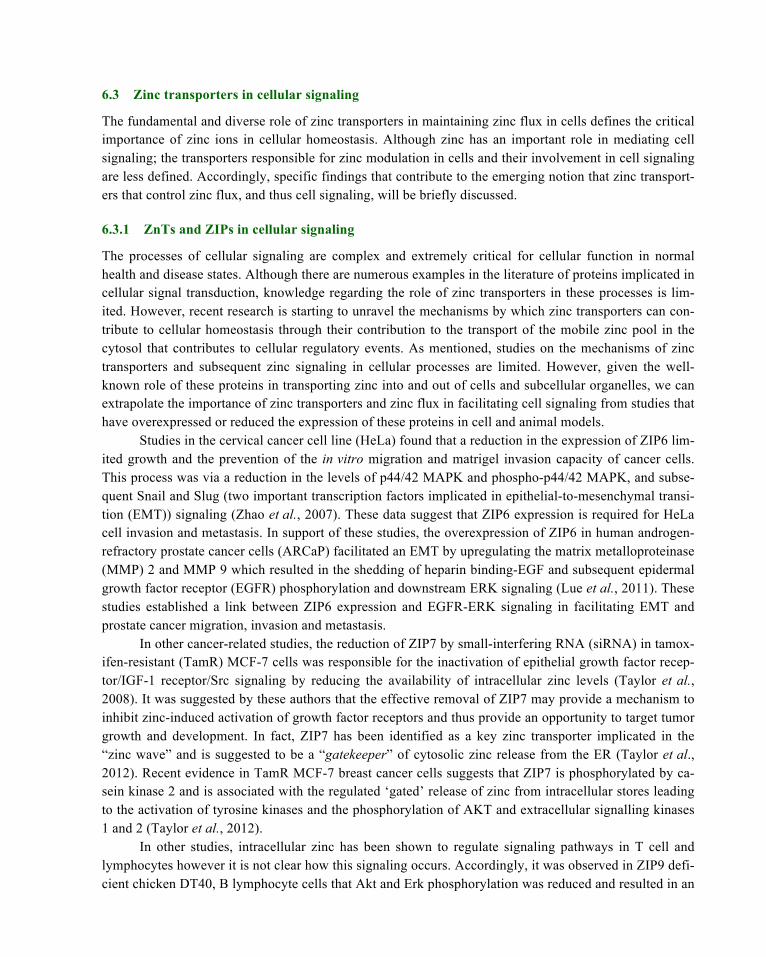
6.3 Zinc transporters in cellular signaling
The fundamental and diverse role of zinc transporters in maintaining zinc flux in cells defines the critical importance of zinc ions in cellular homeostasis. Although zinc has an important role in mediating cell signaling; the transporters responsible for zinc modulation in cells and their involvement in cell signaling are less defined. Accordingly, specific findings that contribute to the emerging notion that zinc transport-ers that control zinc flux, and thus cell signaling, will be briefly discussed.
6.3.1 ZnTs and ZIPs in cellular signaling
The processes of cellular signaling are complex and extremely critical for cellular function in normal health and disease states. Although there are numerous examples in the literature of proteins implicated in cellular signal transduction, knowledge regarding the role of zinc transporters in these processes is lim-ited. However, recent research is starting to unravel the mechanisms by which zinc transporters can con-tribute to cellular homeostasis through their contribution to the transport of the mobile zinc pool in the cytosol that contributes to cellular regulatory events. As mentioned, studies on the mechanisms of zinc transporters and subsequent zinc signaling in cellular processes are limited. However, given the well-known role of these proteins in transporting zinc into and out of cells and subcellular organelles, we can extrapolate the importance of zinc transporters and zinc flux in facilitating cell signaling from studies that have overexpressed or reduced the expression of these proteins in cell and animal models.
Studies in the cervical cancer cell line (HeLa) found that a reduction in the expression of ZIP6 lim-ited growth and the prevention of the in vitro migration and matrigel invasion capacity of cancer cells. This process was via a reduction in the levels of p44/42 MAPK and phospho-p44/42 MAPK, and subse-quent Snail and Slug (two important transcription factors implicated in epithelial-to-mesenchymal transi-tion (EMT)) signaling (Zhao et al., 2007). These data suggest that ZIP6 expression is required for HeLa cell invasion and metastasis. In support of these studies, the overexpression of ZIP6 in human androgen-refractory prostate cancer cells (ARCaP) facilitated an EMT by upregulating the matrix metalloproteinase (MMP) 2 and MMP 9 which resulted in the shedding of heparin binding-EGF and subsequent epidermal growth factor receptor (EGFR) phosphorylation and downstream ERK signaling (Lue et al., 2011). These studies established a link between ZIP6 expression and EGFR-ERK signaling in facilitating EMT and prostate cancer migration, invasion and metastasis.
In other cancer-related studies, the reduction of ZIP7 by small-interfering RNA (siRNA) in tamox-ifen-resistant (TamR) MCF-7 cells was responsible for the inactivation of epithelial growth factor recep-tor/IGF-1 receptor/Src signaling by reducing the availability of intracellular zinc levels (Taylor et al., 2008). It was suggested by these authors that the effective removal of ZIP7 may provide a mechanism to inhibit zinc-induced activation of growth factor receptors and thus provide an opportunity to target tumor growth and development. In fact, ZIP7 has been identified as a key zinc transporter implicated in the “zinc wave” and is suggested to be a “gatekeeper” of cytosolic zinc release from the ER (Taylor et al., 2012). Recent evidence in TamR MCF-7 breast cancer cells suggests that ZIP7 is phosphorylated by ca-sein kinase 2 and is associated with the regulated ‘gated’ release of zinc from intracellular stores leading to the activation of tyrosine kinases and the phosphorylation of AKT and extracellular signalling kinases 1 and 2 (Taylor et al., 2012).
In other studies, intracellular zinc has been shown to regulate signaling pathways in T cell and lymphocytes however it is not clear how this signaling occurs. Accordingly, it was observed in ZIP9 defi-cient chicken DT40, B lymphocyte cells that Akt and Erk phosphorylation was reduced and resulted in an

increase in PTPase activity (Taniguchi et al., 2013). Furthermore, overexpression of human ZIP9 in the chicken ZIP9 knockout DT40 cells restored Akt and Erk phosphorylation while PTPase activity was de-creased in response to zinc treatment. These data suggest that ZIP9 increases cytosolic zinc and activation of cell signaling by inhibition of PTPases in these cells (Taniguchi et al., 2013).
In metabolic studies, glucose stimulation of pancreatic insuinoma RIN5mf cells that had an over-expression of ZnT7, resulted in an increase in insulin secretion (Huang et al., 2010). This was also asso-ciated with an increase in insulin biosynthesis and a subsequent increase in cellular insulin content and suggested that ZnT7 plays an important role in insulin expression. Similarly, ZnT7 null mice were more susceptible to diet-induced glucose intolerance and insulin resistance and this was associated with a re-duction in the expression of the insulin receptor (IR), IR substrate 1, IR substrate 2 and Akt mRNA in primary skeletal myotubes (Huang et al., 2012). In other studies on ZnT7, the overexpression of this transporter in mouse osteoblasts MC3T3-E1 cells facilitated cytoprotection from H2O2 oxidative stress-induced apoptosis via the activation PI3K/Akt and MAPK/ERK pathways (Liang et al., 2013).
ZnT3, ZnT5 and ZnT8 gene expression is differentially regulated by glucose in INS-1E cells and a ZnT3 ‘knock down’ decreased insulin gene expression and secretion and resulted in hyperglycemia in streptozotocin-treated ZnT3 null mice (Smidt et al., 2009). Similarly, elevated glucose concentrations increased free cytosolic zinc in mouse pancreatic islets and were associated with an increase in the mRNA expression of ZIP6-8 (Bellomo et al., 2011). These authors suggested that glucose induces cyto-solic zinc leading to the processing and storage of insulin and associated increase in the ZIP importers.
Fasting gluconeogenesis is impaired in the livers of ZIP14 knockout mice which is attributable to dysregulation of G-protein coupled receptor (GPCR) signaling (Hojyo et al., 2011). The reduction in sig-naling is due to reduced levels of basal cAMP as a consequence of increased phosphodiesterase (PDE) activity and suggests that ZIP14 plays a role in facilitating GPCR-mediated cAMP-CREB activity by re-ducing basal PDE in the liver (Hojyo et al., 2011). Moreover, ZIP14 null mice have greater body fat, hy-poglycemia and increase levels of insulin that was concomitant with increased liver glucose and increase insulin receptor, PI3K and Akt phosphorylation (Beker et al., 2012).
Recently we have identified that the reduction in ZIP7 by siRNA in mouse skeletal muscle C2C12 cells is associated with the downregulation of several genes implicated in glucose metabolism including the IR, IR-substrate 1, IR-substrate 2, Glut4 and glycogen branching enzyme (Gbe) (Myers et al. 2013). This was concomitant with a reduction in glycogen synthesis and pAKT and suggests that ZIP7 controls glycogen synthesis in these cells via pAKT and Glut4 signaling.
Given that the zinc transporters are involved in the cellular regulation of signal transduction path-ways in a number of biological systems, and specifically, their ability to modulate pathways involved in glucose and lipid metabolism, suggests that these transporters will have significance in disease states as-sociated with insulin resistance and type 2 diabetes. Although several studies in cell and animal models have provided some insight into the mechanisms of zinc transporters in the context of metabolic process-es associated with insulin signaling and insulin resistance, their role in type 2 diabetes remains vague.
7 Zinc, Zinc Transporters and Type 2 Diabetes
Type 2 diabetes (T2D) is a complex disorder characterized by insulin resistance and impaired glucose homeostasis. Recently, intensive research has aimed to elucidate the role that dysfunctional zinc signaling plays in this disease. Moreover, given that the global incidence of T2D is escalating at an alarming rate

(Zimmet, 2002), there is considerable interest in understanding the molecular mechanisms of zinc transport and zinc action on cellular pathways associated with glucose metabolism. Although dietary zinc supplements given to animal models of diabetes and human diabetic patients has shown some benefits in improving glycaemic control (Hwang et al., 2011; Jansen et al., 2009; Jayawardena et al., 2012; Simon and Taylor, 2001; Song et al., 2001), there are also contradictory outcomes in human studies on zinc sup-plementation to treat diabetes (see, Miao et al., 2013, for a comprehensive review and references therein), and is beyond the scope of this review. While these studies on zinc supplementation as an adjunct therapy to treat diabetes are ongoing, the transporters implicated in controlling zinc flux in cells and their role in diabetes is limited.
7.1 ZnT8 and diabetes
Zinc has an integral role in the processing, storage, secretion and action of insulin in response to changes in elevated glucose concentrations (Mocchegiani et al., 2008; Wijesekara et al., 2009). Zinc is required for the storage of insulin in the secretory granules of the pancreas as an inactive Zn2+-insulin hexamer. When released into blood serum, a change in pH drives dissociation of the hexamer into a monomer which is the physiologically active form of insulin (Xu et al., 2011). To date, the most well-studied zinc transporter in diabetes is ZnT8. This transporter is almost exclusively expressed in the β-cells of the pan-creas where it plays a crucial role in transporting zinc into insulin secretory vesicles and therefore is criti-cal for the synthesis, storage and action of insulin (for comprehensive reviews, see Chimienti et al., 2005; Kawasaki., 2012; Mocchegiani et al., 2008 and references therein).
It has been shown in pancreatic INS-1 cells that overexpression of ZnT8 facilitated glucose-stimulated insulin secretion (Chimienti et al., 2006) while reduced expression of this transporter in these cells resulted in a reduction in insulin content and secretion in response to a hyperglycaemic stimulus (Fu et al., 2009). Moreover, mice with a target-specific ZnT8 knockout in pancreatic β-cells show compro-mised glucose tolerance (Wijesekara et al., 2010) while global ZnT8 null mice showed abnormalities in diet-dependent glucose tolerance, insulin secretion (Nicolson et al., 2009; Pound et al., 2009) and body weight (Hardy et al., 2012; Nicolson et al., 2009). An association of ZnT8 with diabetes risk in humans emerged when a non-synonymous single nucleotide polymorphism (SNP) (rs13266634 C>T) which changes an arginine (R) to tryptophan (W) at amino acid position 325 was identified as a susceptibility locus of T2D in genome-wide association studies in European patients (Kommoju and Reddy, 2011; Saxena et al., 2007; Sladek et al., 2007; Xu et al., 2012). The rs13266634 SNP disrupts a protein kinase A and protein kinase C recognition motif that alters the function of this transporter (Kawasaki, 2012). Accordingly, defects in ZnT8 protein structure will affect the accumulation of zinc ions in the secretory granule where insulin is matured and stored as hexamers bound to zinc ions (Nicolson et al., 2009). In this context, the risk allele of ZnT8 was found to be associated with impaired conversion of proinsulin to insulin in human patients (Kirchhoff et al., 2008).
It is well-established that type 1 diabetes (T1D) is associated with autoimmune-mechanisms (type 1A) and both genetic and environmental factors affect the disease onset and progression (Kawasaki, 2012). T1D is characterized by the selective destruction of the β-cells of the pancreas resulting in signifi-cant insulin deficiency and subsequent hyperglycemia. Circulating autoantibodies targeting pancreatic β-cell proteins are currently the most reliable biomarkers in the early stage of T1D and therefore provides utility for therapeutic intervention. To date, the four major humoral autoantigens in T1D are [pro]-insulin, glutamic acid decarboxylase (GAD65), insulinoma-associated antigen 2 (IA-2) and ZnT8 (Wenzlau and Hutton, 2013).

The discovery of ZnT8 as the fourth humoral autoantigen involved microarray gene expression profiling of human and rodent pancreas and islet cells that were subsequently screened with new-onset T1D and prediabetic sera (Wenzlau et al., 2007). These studies identified that ZnT8 is a diabetes-associated autoantigen where autoantibodies were found in 60-80% of new onset cases of T1D (Wenzlau et al., 2007). Recent genome-wide association studies revealed that the above mentioned SNP rs13266634 is a key determinant of humoral autoreactivity to ZnT8 (Wenzlau et al., 2008; Kawasaki et al., 2008). Moreover, autoantibodies that recognize ZnT8R, ZnT8W, or both at the polymorphic site are common in newly diagnosed T1D patients (Skarstrand et al., 2013). However, it is not clear on the im-portance of ZnT8 epitope-specific autoantibodies and β-cell destruction in T1D. Nevertheless, because ZnT8 plays a critical role in insulin biology, and has recently emerged as a genetic marker for diabetes, it represents an attractive candidate for therapy for the treatment of diabetes and other disorders related to pancreatic pathology.
7.2 Other zinc transporters and type 2 diabetes
A recent study investigated the effect of zinc and α-linolenic acid (ALA) supplementation on circulating concentrations of inflammatory markers and zinc transporter gene expression in peripheral blood mono-nuclear cells (PBMCs) of forty-three postmenopausal women with T2D (Foster et al., 2013). These stud-ies identified that zinc and ALA supplementation had no effect on inflammatory markers, however, circu-lating base levels of the inflammatory cytokine IL-6 could predict the expression levels of mRNA for ZnT5, ZnT7, ZIP1, ZIP7 and ZIP10 in such that each unit increase in IL-6 produced an 8%-15% increase in zinc transporter gene expression (Foster et al., 2013). These authors suggest that IL-6 increases the expression of this group of zinc transporters to facilitate the redistribution of zinc into PBMC organelles as part of a coordinated inflammatory response.
Apart from research in animal models and cell culture systems (see 6.2 Zinc and cellular signaling and 6.3 Zinc transporters in cellular signaling) there is little additional information available on the role of other members of the zinc transporter family in human type 2 diabetes.
8 Zinc, Zinc transporters and therapeutic utility
The relevance of intracellular zinc signaling for normal physiology and for the pathogenesis of several disease states including T2D has recently been highlighted. Although zinc supplementation in zinc-deficient T2D patients might be appropriate treatment in genetically susceptible individuals, the potential mechanisms that lead to dysfunctional intracellular zinc signaling and altered zinc subcellular distribution, rather than zinc deficiency will require the development innovative drugs that target zinc transporters for example. Moreover, several issues regarding zinc supplementation for T2D patients requires intensive investigation as this type for treatment may not have the maximal benefit required to improve glycaemic control. Although zinc in general is less toxic, excessive zinc intake may have undesirable effects such as the elevation of HbA1c and high blood pressure (Miao et al., 2013).
While zinc has insulin-mimetic actions and is implicated in cellular signaling events, its applica-tion in the clinical setting in lowering blood glucose in patients with T2D is complicated by the fact that absorption rates are low and high doses and long-term supplementation are required. In this context, sev-eral zinc complexes have been developed to overcome these complexities (see Miao et al., 2013 for a comprehensive review). Zinc complexes with nicotinamide, maltol, amino acids, picolinic acid, pico-

linamide, and their derivatives have high insulin-mimetic activities in vitro and in vivo studies, however it was found that these complexes were only effective in lowering blood glucose in T2D subjects by giving daily injections (Yoshikawa et al., 2003). For example, daily intraperitoneal injections of a zinc complex with maltol (Zn(ma)2) normalized blood glucose levels in KKAy mice with T2D (Yoshikawa et al., 2001).
In the context of a clinical application for zinc complexes, it is desirable to identify therapies that can be administered orally. Accordingly, many studies have aimed to synthesis novel zinc complexes that have insulin-mimetic activities that can be administered orally. For example, the daily oral administration of the zinc complex Zn(II)-thioallixin-N-methyl (Zn(tanm)2) over four weeks significantly improved hy-perglycemia, glucose intolerance, insulin resistance, hyperleptinemia, obesity and hypertension in KKAy mice (Adachi et al., 2006). Similarly, the development and synthesis of a novel of zinc complex [Di(1-oxy-2-pyridinethiolato)Zn] displayed superior insulin-mimetic and anti-diabetic activity compared to ZnCl2 or the clinically administered pioglitazone in KKAy diabetic rats (Yoshikawa et al., 2011). Moreo-ver, this complex was shown to have high gastrointestinal absorption when compared to ZnCl2 alone. Although zinc complexes have been shown to have beneficial effects in animal models of T2D, their utility and efficacy in human diabetes is not known. Accordingly, opportunities for translating zinc-complex therapies in animal models of diabetes into clinically relevant applications will require intensive investigation into the properties of these zinc complexes, their absorption rates, tissue distribution, toxici-ty, insulin-mimetic activity and efficacy in ameliorating blood glucose levels in human T2D.
Given our current understanding on the role of zinc as a critical modulator of cellular signaling and homeostasis, and the fact that the storage, compartmentalization and cellular distribution of zinc is tightly regulated by zinc transporters, it is reasonable that dysregulation of zinc flux will play an important role in disease processes. Moreover, the processes of zinc-related cellular dysfunction are predominately due to alternations in the proteins that control zinc flux and homeostasis rather than an issue of zinc deficien-cy or accumulation. Accordingly, drugs that target zinc transporters may provide utility to correct or im-prove dysfunctional zinc homeostasis and could therefore be more efficient than zinc supplementation. Although zinc is implicated in cellular signaling events, several questions remain to be resolved: For ex-ample, what extracellular stimulus regulates the expression of the zinc transporters? Are the zinc trans-porters differentially regulated in a tissue or cell-specific manner? How does each specific zinc trans-porter regulate cellular signaling pathways? Moreover, genetic studies such as the targeted, tissue-specific disruption of these transporters in animal models will assist in determining the function of these proteins.
9 Summary
• Zinc is an essential trace element that is indispensable for the growth and development of organ-isms.
• The ZIP and ZnT zinc transporter family are involved in regulating the intracellular availability of zinc in cells to maintain homeostasis.
• Zinc is an insulin-mimetic involved in cellular signaling events. • Dysfunction zinc signaling is implicated in a number of disease states including liver cirrhosis,
cancer, impaired immune function and diabetes. • A single nucleotide polymorphism in the zinc transporter, ZnT8 is associated with greater risk
for the development of T2D.

• Zinc supplementation in T2D patients with zinc deficiency has shown to be beneficial in some cases but not all.
• Novel zinc complexes for lowering blood glucose in animal models of T2D are promising but will require extensive clinical trials to determine their efficacy in humans.
• Drugs that target altered zinc transporter activity or dysfunctional zinc signaling may provide utility to understand aberrant zinc homeostasis in diseases such as T2D.
10 Conclusion
Zinc is an essential trace element that is implicated in a numerous normal and patho-physiological cellu-lar functions. The emerging role of zinc as an insulin mimetic and the ubiquitous nature of this ion in maintaining cellular function suggests that abnormal cellular partitioning and levels of zinc will have bio-logical and clinical effects. Although our current understandings on the role of zinc transporters in type 2 diabetes is limited, it is clear from studies on ZnT8 that this family of transporters has utility for the de-velopment of novel diabetic therapies. While ZnT8 plays a significant role in insulin biology and there-fore represents an attractive target for diabetes therapy, the other members of the zinc transporter family in diabetes are less defined. However, we can speculate from the information presented in this review that the other transporters are involved in processes that facilitate insulin signaling and glycaemic control and therefore could offer exciting new targets that are amendable to therapeutic intervention in the treatment of diseases associated with insulin resistance and type 2 diabetes.
References
Abdul-Ghani, M.A., & DeFronzo, R. A. (2010). Pathogenesis of Insulin Resistance in Skeletal Muscle. Journal of Biomedicine and Biotechnology, doi:10.115/2010/476279.
Ackland, M. L., & Michalczyk, A. (2006). Zinc deficiency and its inherited disorders-a review. Genes & Nutrition, 1, 41-50.
Adachi, Y., Yoshida, J., Kodera, Y., Kiss, T., Jakusch, T., Enyedy, E. A., Yoshikawa, Y., & Sakurai, H. (2006). Oral administration of a zinc complex improves type 2 diabetes and metabolic syndromes. Biochemical and Biophysical Research Communications, 351, 165-170.
Aguilar-Alonso, P., Martinez-Fong, D., Pazos-Salazar, N. G., Brambila, E., Gonzalez-Barrios, J. A., Mejorada, A., Flores, G., Millan-Perezpena, L., Rubio, H., & Leon-Chavez, B. A. (2008). The increase in zinc levels and upregulation of zinc transporters are mediated by nitric oxide in the cerebral cortex after transient ischemia in the rat. Brain Research, 1200, 89-98.
Alves, C. X., Vale, S. H., Dantas, M. M., Maia, A. A., Franca, M. C., Marchini, J. S., Leite, L. D., & Brandao-Neto, J. (2012). Positive effects of zinc supplementation on growth, GH, IGF1, and IGFBP3 in eutrophic children. Journal of Pediatric Endocrinology and Metabolism, 25, 881-887.
Andreini, C., L. Banci, I. Bertini and A. Rosato (2006). Counting the zinc-proteins encoded in the human genome. Journal of Proteome Research, 5, 196-201.
Atasoy, H. B., & Ulusoy, Z. I. (2012). The relationship between zinc deficiency and children's oral health. Pediatric Dentistry, 34, 383-386.
Basaki, M., Saeb, M., Nazifi, S., & Shamsaei, H. A. (2012). Zinc, Copper, Iron, and Chromium Concentrations in Young Patients with Type 2 Diabetes Mellitus. Biological Trace Element Research, 148, 161-164.

Begum, N. A., Kobayashi, M., Moriwaki, Y., Matsumoto, M., Toyoshima, K., & Seya, T. (2002). Mycobacterium bovis BCG cell wall and lipopolysaccharide induce a novel gene, BIGM103, encoding a 7-TM protein: identification of a new protein family having Zn-transporter and Zn-metalloprotease signatures. Genomics, 80, 630-645.
Beker Aydemir, T., Chang, S. M., Guthrie, G. J., Maki, A. B., Ryu, M. S., Karabiyik, A., & Cousins, R. J. (2012). Zinc transporter ZIP14 functions in hepatic zinc, iron and glucose homeostasis during the innate immune response (endotoxemia). PLoS One, 7, e48679.
Bellomo, E. A., Meur, G., & Rutter, G. A. (2011). Glucose Regulates Free Cytosolic Zn2+ Concentration, Slc39 (ZiP), and Metallothionein Gene Expression in Primary Pancreatic Islet β-Cells. Journal of Biological Chemistry, 286, 25778-25789.
Besecker, B., Bao, S., Bohacova, B., Papp, A., Sadee, W., & Knoell, D. L. (2008). The human zinc transporter SLC39A8 (Zip8) is critical in zinc-mediated cytoprotection in lung epithelia. American Journal of Physiology - Lung Cellular and Molecular Physiology, 294, L1127-L1136.
Beyersmann, D., & Haase, H. (2001). Functions of zinc in signaling, proliferation and differentiation of mammalian cells. BioMetals, 14:331-341.
Bloom, D. E., Cafiero, E. T., Jane-Llopis, E., Abrahams-Gessel, S., Bloom, L. R., Fathima, S., Feigl, A. B., Gaziano, T., Mowafi, M., Pandya, A., Prettner, K., Rosenberg, L., Seligman, B., Stein, A. Z., & Weinstein, C. (2011). The Global Economic Burden of Noncommunicable Diseases. Geneva: World Economic Forum, Geneva Switzerland. www.weforum.org/EconomicsOfNCD
Bode, J. C., Hanisch, P., Henning, H., Koenig, W., Richter, F. W., & Bode, C. (1988). Hepatic zinc content in patients with various stages of alcoholic liver disease and in patients with chronic active and chronic persistent hepatitis. Hepatology, 8:1605-1609.
Bosomworth, H. J., Adlard, P. A., Ford, D., & Valentine, R. A. (2013). Altered Expression of ZnT10 in Alzheimer's Disease Brain. PLoS One, 8, e65475.
Bosomworth, H. J., Thornton, J. K., Coneyworth, L. J., Ford, D., & Valentine, R. A. (2012). Efflux function, tissue-specific expression and intracellular trafficking of the Zn transporter ZnT10 indicate roles in adult Zn homeostasis. Metallomics, 4, 771-779.
Bribiescas, R. G. (2003). Effects of oral zinc supplementation on serum leptin levels in Ache males of eastern Paraguay. American Journal of Human Biology, 15, 681-687.
Carsten, S-P. (2000). Signalling aspects of insulin resistance in skeletal muscle: mechanisms induced by lipid oversupply. Cellular Signalling, 12, 583-594.
Chasapis, C., Loutsidou, A., Spiliopoulou, C., & Stefanidou, M. (2011). Zinc and human health: an update. Archives of Toxicology, 86, 1-14.
Chimienti, F., Devergnas, S., Pattou, F., Schuit, F., Garcia-Cuenca, R., Vandewalle, B., Kerr-Conte, J., Van L., Leentje, G., Didier, F. A., & Seve, M. (2006). In vivo expression and functional characterization of the zinc transporter ZnT8 in glucose-induced insulin secretion. Journal of Cell Science, 119, 4199-4206.
Chimienti, F., Favier, A., & Seve, M. (2005). ZnT-8, A Pancreatic Beta-Cell-Specific Zinc Transporter. BioMetals, 18, 313-317.
Chowanadisai, W., Graham, D. M., Keen, C. L., Rucker, R. B., & Messerli, M. A. (2013). Neurulation and neurite extension require the zinc transporter ZIP12 (slc39a12). Proceedings of the National Academy of Sciences, 110, 9903-9908.
Coble, Y. D., Jr., Bardin, C. W., Ross, G. T., & Darby, W. T. (1971). Studies of endocrine function in boys with retarded growth, delayed sexual maturation and zinc deficiency. The Journal of Clinical Endocrinology & Metabolism, 32, 361-367.
Costello, L. C., Zou, J., Desouki, M. M., & Franklin, R. B. (2012). Evidence for changes in RREB-1, ZIP3, and Zinc in the early development of pancreatic adenocarcinoma. Journal of Gastrointestinal Cancer, 43, 570-578.

Costello, L. C., Liu, Y., Zou, J., & Franklin, R. B. (1999). Evidence for a Zinc Uptake Transporter in Human Prostate Cancer Cells Which Is Regulated by Prolactin and Testosterone. Journal of Biological Chemistry, 274, 17499-17504.
Coulston, L., & Dandona, P. (1980). Insulin-like Effect of Zinc on Adipocytes. Diabetes, 29, 665-667.
Cragg, R. A., Philips, S. R., Piper, J. M., Varma, J. S., Campbell, F. C., Mathers, J. C & Ford, D. (2005). Homeostatic regulation of zinc transporters in the human small intestine by dietary zinc supplementation. Gut, 54, 469-478.
Deniro, M., & Al-Mohanna, F. A. (2012). Zinc transporter 8 (ZnT8) expression is reduced by ischemic insults: a potential therapeutic target to prevent ischemic retinopathy. PLoS One, 7, e50360.
Devergnas, S., Chimienti, F., Naud, N., Pennequin, A., Coquerel, Y., Chantegrel, J., Favier, A., & Seve, M. (2004). Differential regulation of zinc efflux transporters ZnT-1, ZnT-5 and ZnT-7 gene expression by zinc levels: a real-time RT–PCR study. Biochemical Pharmacology, 68, 699-709.
Devirgiliis, C., P. D. Zalewski, G. Perozzi and C. Murgia (2007). Zinc fluxes and zinc transporter genes in chronic diseases. Mutation Research, 622, 84-93.
Dufner-Beattie, J., Kuo, Y. M., Gitschier, J., & Andrews, G. K. (2004). The Adaptive Response to Dietary Zinc in Mice Involves the Differential Cellular Localization and Zinc Regulation of the Zinc Transporters ZIP4 and ZIP5. Journal of Biological Chemistry, 279, 49082-49090.
Eide, D., Broderius, M., Fett, J., & Guerinot, M. L. (1996). A novel iron-regulated metal transporter from plants identified by functional expression in yeast. Proceedings of the National Academy of Sciences, 93, 5624-5628.
Ezaki, O. (1989). IIb group metal ions (Zn2+, Cd2+, Hg2+) stimulate glucose transport activity by post-insulin receptor kinase mechanism in rat adipocytes. Journal of Biological Chemistry, 264, 16118-16122.
Fernandez, E. L., Dencker, L, & Tallkvist, J. (2007). Expression of ZnT-1 (Slc30a1) and MT-1 (Mt1) in the conceptus of cadmium treated mice. Reproductive Toxicology, 24, 353-358.
Figlewicz, D. P., Forhan, S. E., Hodgson, A. T., & Grodsky, G. M. (1984). 65Zinc and endogenous zinc content and distribution in islets in relationship to insulin content. Endocrinology, 115, 877-881.
Foster, M., Petocz, P., & Samman, S. (2013). Inflammation markers predict zinc transporter gene expression in women with type 2 diabetes mellitus. The Journal of Nutritional Biochemistry, 24, 1655-1661.
Foster, M., & Samman, S. (2010). Zinc and redox signaling: perturbations associated with cardiovascular disease and diabetes mellitus. Antioxidants & redox signaling, 13, 1549-1573.
Fu, Y., Tian, W., Pratt, E. B., Dirling, L. B., Shyng, S. L., Meshul, C. K., & Cohen, D. M. (2009). Down-Regulation of ZnT8 Expression in INS-1 Rat Pancreatic Beta Cells Reduces Insulin Content and Glucose-Inducible Insulin Secretion. PLoS One, 4, e5679.
Fukada, T., Yamasaki, S., Nishida, K., Murakami, M., & Hirano, T. (2011). Zinc homeostasis and signaling in health and diseases. Journal of Biological Inorganic Chemistry, 16, 1123-1134.
Gaither, L. A. & Eide, D (2001). Eukaryotic zinc transporters and their regulation. BioMetals, 14, 251-270.
González-Rodríguez, Á., Gutierrez, J. A. M., Sanz-González, S., Ros, M., Burks, D. J., & Valverde, Á. M. (2010). Inhibition of PTP1B Restores IRS1-Mediated Hepatic Insulin Signaling in IRS2-Deficient Mice. Diabetes, 59, 588-599.
Haase, H., & Maret, W. (2003). Intracellular zinc fluctuations modulate protein tyrosine phosphatase activity in insulin/insulin-like growth factor-1 signaling. Experimental Cell Research, 291, 289-298.
Haase, H., & Maret, W. (2005a). Fluctuations of cellular, available zinc modulate insulin signaling via inhibition of protein tyrosine phosphatases. Journal of Trace Elements in Medicine and Biology, 19, 37-42.
Haase, H., & Maret, W. (2005b). Protein Tyrosine Phosphatases as Targets of the Combined Insulinomimetic Effects of Zinc and Oxidants. BioMetals, 18, 333-338.
Haase, H., Mocchegiani, E., & Rink, L. (2006). Correlation between zinc status and immune function in the elderly. Biogerontology, 7:412-428.
Hambidge, K. M., Hambidge, C., Jacobs, M., & Baum, J. D. (1972). Low levels of zinc in hair, anorexia, poor growth, and hypogeusia in children. Pediatric Research, 6, 868-874.
Hardy, A. B., Wijesekara, N., Genkin, I., Prentice, K. J., Bhattacharjee, A., Kong, D., Chimienti, F., & Wheeler, M. B. (2012). Effects of high-fat diet feeding on Znt8-null mice: differences between beta-cell and global knockout of Znt8. American Journal of Physiology: Endocrinology and Metabolism, 302, E1084-1096.
Hogstrand, C., Kille, P., Nicholson, R. I., & Taylor, K. M. (2009). Zinc transporters and cancer: a potential role for ZIP7 as a hub for tyrosine kinase activation. Trends in Molecular Medicine, 15, 101-111.
Hojyo, S., Fukada, T., Shimoda, S., Ohashi, W., Bin, B-H., Koseki, H., & Hirano, T. (2011). The Zinc Transporter SLC39A14/ZIP14 Controls G-Protein Coupled Receptor-Mediated Signaling Required for Systemic Growth. PLoS One, 6, e18059.
Huang, L., Kirschke, C. P., Lay, Y. E., Levy, L. B., Lamirande, D. E., & Zhang, P. H. (2012). Znt7-null mice are more susceptible to diet-induced glucose intolerance and insulin resistance. Journal of Biological Chemistry. 287:33883-33896.
Huang, L., & Tepaamorndech, S. (2013). The SLC30 family of zinc transporters – A review of current understanding of their biological and pathophysiological roles. Molecular Aspects of Medicine, 34, 548-560.
Huang, L., Yan, M., & Kirschke, C. P. (2010). Over-expression of ZnT7 increases insulin synthesis and secretion in pancreatic β-cells by promoting insulin gene transcription. Experimental Cell Research, 316, 2630-2643.
Hulver, M. W., & Lynis D, G. (2004). The molecular mechanism linking muscle fat accumulation to insulin resistance. Proceedings of the Nutrition Society, 63, 375-380.
Hwang, I., Yoon, T., Kim, C., Cho, B., Lee, S., & Song, M. K. (2011). Different roles of zinc plus arachidonic acid on insulin sensitivity between high fructose- and high fat-fed rats. Life Sciences, 88, 278-284.
Iguchi, K., Otsuka, T., Usui, S., Ishii, K., Onishi, T., Sugimura, Y., & Hirano, K. (2004). Zinc and metallothionein levels and expression of zinc transporters in androgen-independent subline of LNCaP cells. Journal of Andrology, 25, 154-161.
Ilouz, R., Kaidanovich, O., Gurwitz, D., & Eldar-Finkelman, H. (2002). Inhibition of glycogen synthase kinase-3β by bivalent zinc ions: insight into the insulin-mimetic action of zinc. Biochemical and Biophysical Research Communications, 295, 102-106.
Jansen, J., Karges, W., & Rink, L. (2009). Zinc and diabetes — clinical links and molecular mechanisms. Journal of Nutritional Biochemistry, 20, 399-417.
Jansen, J., Rosenkranz, E., Overbeck, S., Warmuth, S., Mocchegiani, E., Giacconi, R., Weiskirchen, R., Karges, W., & Rink, L. (2012). Disturbed zinc homeostasis in diabetic patients by in vitro and in vivo analysis of insulinomimetic activity of zinc. The Journal of Nutritional Biochemistry, 23, 1458-1466.
Jayaraman, A. K., & Jayaraman, S. (2011). Increased level of exogenous zinc induces cytotoxicity and up-regulates the expression of the ZnT-1 zinc transporter gene in pancreatic cancer cells. The Journal of Nutritional Biochemistry, 22, 79-88.
Jayawardena, R., Ranasinghe, P., Galappatthy, P., Malkanthi, R. L. D. K., Constantine, G. R., & Katulanda, P. (2012). Effects of zinc supplementation on diabetes mellitus: a systematic review and meta-analysis. Diabetology & Metabolic Syndrome, 4, 13.
Jeong, J., & Eide, D. J. (2013). The SLC39 family of zinc transporters. Molecular Aspects of Medicine, 34, 612-619.
Kambe, T. (2011). An Overview of a Wide Range of Functions of ZnT and Zip Zinc Transporters in the Secretory Pathway. Bioscience Biotechnology and Biochemistry, 75, 1036-1043.
Kawasaki, E. (2012). ZnT8 and type 1 diabetes [Review]. Endocrine Journal, advpub, 1203050683-1203050683.
Kelleher, S. L., McCormick, N. H., Velasquez, V., & Lopez, V. (2011). Zinc in Specialized Secretory Tissues: Roles in the Pancreas, Prostate, and Mammary Gland. Advances in Nutrition: An International Review Journal, 2, 101-111.
Kirchhoff, K., Machicao, F., Haupt, A., Schafer, S. A., Tschritter, O., Staiger, H., Stefan, N., Haring, H-U., & Fritsche, A. (2008). Polymorphisms in the TCF7L2, CDKAL1 and SLC30A8 genes are associated with impaired proinsulin conversion. Diabetologia, 51:597-601.
Kommoju, U. J., & Reddy, B. M. (2011). Genetic etiology of type 2 diabetes mellitus: a review. International Journal of Diabetes in Developing Countries, 31, 51-64.
Kourtoglou, G. I. (2011). Insulin therapy and exercise. Diabetes Research and Clinical Practice, 93, Supplement 1, S73-S77.
Leung, K. W., Liu, M., Xu, X., Seiler, M. J., Barnstable, C. J., & Tombran-Tink, J. (2008). Expression of ZnT and ZIP zinc transporters in the human RPE and their regulation by neurotrophic factors. Investigative Ophthalmology and Visual Science, 49, 1221-1231.
Lewis, P. K. Jr., Hoekstra, W. G., Grummer, R. H., & Philips, P. H. (1956). The Effect of Certain Nutritional Factors including Calcium, Phosphorus and Zinc on Parakeratosis in Swine. Journal of Animal Science, 15, 741-751.
Li, H, & Jogl, G. (2007). Crystal Structure of the Zinc-binding Transport Protein ZnuA from Escherichia coli Reveals an Unexpected Variation in Metal Coordination. Journal of Molecular Biology, 368, 1358-1366.
Liang, D., Xiang, L., Yang, M., Zhang, X., Guo, B., Chen, Y., Yang, L., & Cao, J. (2013). ZnT7 can protect MC3T3-E1 cells from oxidative stress-induced apoptosis via PI3K/Akt and MAPK/ERK signaling pathways. Cellular Signalling, 25, 1126-1135.
Lichten, L. A., & Cousins, R. J. (2009). Mammalian Zinc Transporters: Nutritional and Physiologic Regulation. Annual Review of Nutrition, 29, 153-176.
Lin, Y., & Sun, Z. (2010). Current views on type 2 diabetes. Journal of Endocrinology, 204, 1-11.
Liu, B. Y., Jiang, Y. Z., Lu, Z. Y., Li, S. F., Lu, D. B., & Chen, B. (2011b). Down-regulation of zinc transporter 8 in the pancreas of db/db mice is rescued by Exendin-4 administration. Molecular Medicine Reports, 4, 47-52.
Liu, J., Bao, W., Jiang, M., Zhang, Y., Zhang, X., & Liu, L. (2011a). Chromium, Selenium, and Zinc Multimineral Enriched Yeast Supplementation Ameliorates Diabetes Symptom in Streptozocin-Induced Mice. Biological Trace Element Research, 1-10.
Liuzzi, J. P., Blanchard, R. K., & Cousins, R. J. (2001). Differential Regulation of Zinc Transporter 1, 2, and 4 mRNA Expression by Dietary Zinc in Rats. The Journal of Nutrition, 131, 46-52.
Liuzzi, J. P., & Cousins, R. J. (2004). Mammalian Zinc Transporters. Annual Review of Nutrition, 24, 151-172.
Liuzzi, J. P., Lichten, L. A., Rivera, S., Blanchard, R. K., Aydemir, T. B., Knutson, M. D., Ganz, T., & Cousins, R. J. (2005). Interleukin-6 regulates the zinc transporter Zip14 in liver and contributes to the hypozincemia of the acute-phase response. Proceedings of the National Academy of Sciences, 102, 6843-6848.
Lu, M., & Fu, D. (2007). Structure of the Zinc Transporter YiiP. Science, 317, 1746-1748.
Lue, H. W., Yang, X., Wang, R., Qian, W., Xu, R. Z. H., Lyles, R., Osunkoya, A. O., Zhou, B. P., Vessella, R. L., Zayzafoon, M., Liu, Z-R., Zhau, H. E., & Chung, L. W. K. (2011). LIV-1 Promotes Prostate Cancer Epithelial-to-Mesenchymal Transition and Metastasis through HB-EGF Shedding and EGFR-Mediated ERK Signaling. PLoS One, 6, e27720.
Makonnen, B., Venter, A., & Joubert, G. (2003). A randomized controlled study of the impact of dietary zinc supplementation in the management of children with protein-energy malnutrition in Lesotho. II: Special investigations. Journal of Tropical Pediatrics, 49, 353-360.
Maret, W. (2011a). Metals on the move: zinc ions in cellular regulation and in the coordination dynamics of zinc proteins. BioMetals, 24, 411-418.
Maret, W. (2011b). New perspectives of zinc coordination environments in proteins. Journal of Inorganic Biochemistry, 111, 110-116.
May, J. M., & Contoreggi, C. S. (1982). The mechanism of the insulin-like effects of ionic zinc. Journal of Biological Chemistry, 257, 4362-4368.

Miao, X., Sun, W., Fu, Y., Miao, L., & Cai, Lu. (2013). Zinc homeostasis in the metabolic syndrome and diabetes. Frontiers of Medicine, 7, 31-52.
Milon, B., Wu, Q., Zou, J., Costello, L. C., & Renty, F. B. (2006). Histidine residues in the region between transmembrane domains III and IV of hZip1 are required for zinc transport across the plasma membrane in PC-3 cells. Biochimica et Biophysica Acta (BBA) - Biomembranes, 1758, 1696-1701.
Mocchegiani, E., Giacconi, R., & Malavolta, M. (2008). Zinc signalling and subcellular distribution: emerging targets in type 2 diabetes. Trends in Molecular Medicine, 14, 419-428.
Moniz, T., Amorim, M. J., Ferreira, R., Nunes, A., Silva, A., Queirós, C., Leite, A., Gameiro, P., Sarmento, B., Remião, F., Yoshikawa, Y., Sakurai, H., & Rangel, M. (2011). Investigation of the insulin-like properties of zinc(II) complexes of 3-hydroxy-4-pyridinones: Identification of a compound with glucose lowering effect in STZ-induced type I diabetic animals. Journal of Inorganic Biochemistry, 105, 1675-1682.
Myers, S. A., Nield, A., Chew, G. S., & Myers, M. A. (2013) The Zinc Transporter, Slc39a7 (Zip7) Is Implicated in Glycaemic Control in Skeletal Muscle Cells. PLoS ONE, 8:e79316.
Myers, S. A., Nield, A., & Myers, M. (2012). Zinc Transporters, Mecahnisms of Action and Therapeutic Utility: Implications for Type 2 Diabetes Mellitus. Journal of Nutrition and Metabolism, doi:10.1155/2012/173712).
Nicolson, T. J., Bellomo, E. A., Wijesekara, N., Loder, M. K., Baldwin, J. M., Gyulkhandanyan, A. V., Koshkin, V., Tarasov, A. I., Carzaniga, R., Kronenberger, K., Taneja, T. K., da Silva Xavier, G., Libert, S., Froguel, P., Scharfmann, R., Stetsyuk, V., Ravassard, P., Parker, H., Gribble, F. M., Reimann, F., Sladek, R., Hughes, S. J., Johnson, P. R.V., Masseboeuf, M., Burcelin, R., Baldwin, S. A., Liu, M., Lara-Lemus, R., Arvan, P., Schuit, F. C., Wheeler, M. B., Chimienti, F., & Rutter, G. A. (2009). Insulin Storage and Glucose Homeostasis in Mice Null for the Granule Zinc Transporter ZnT8 and Studies of the Type 2 Diabetes–Associated Variants. Diabetes, 58, 2070-2083.
Nyenwe, E. A., Jerkins, T. W., Umpierrez, G. E., & Kitabchi, A. E. (2011). Management of type 2 diabetes: evolving strategies for the treatment of patients with type 2 diabetes. Metabolism, 60, 1-23.
O'Dell, B. L., Newberne, P. M., & Savage, J. E. (1958). Significance of dietary zinc for the growing chicken. The Journal of Nutrition, 65, 503-518.
Osto, M., Zini, E., Reusch, C. E., & Lutz, T. A. (2013). Diabetes from humans to cats. General and Comparative Endocrinology, 182, 48-53.
Pagel-Langenickel, I., Bao, J., Pang, L., & Sack, M. N. (2010). The Role of Mitochondria in the Pathophysiology of Skeletal Muscle Insulin Resistance. Endocrine Reviews, 31, 25-51.
Pal, A., & McCarthy, M. I. (2013). The genetics of type 2 diabetes and its clinical relevance. Clinical Genetics, 83, 297-306.
Palmiter, R. D, & Findley, S. D. (1995). Cloning and functional characterization of a mammalian zinc transporter that confers resistance to zinc. EMBO J., 14, 639-649.
Palmiter, R. D, & Huang, L. (2004). Efflux and compartmentalization of zinc by members of the SLC30 family of solute carriers. Pflugers Archiv., 447, 744-751.
Pawan, K., Sharma, N., Kumar, S., Radha, K. R., Prasad, R. (2007). Upregulation of Slc39a10 gene expression in response to thyroid hormones in intestine and kidney. Biochimica et Biophysica Acta (1769), 117–123.
Pandey, N., Vardatsikos, G., Mehdi, M., & Srivastava, A. (2010). Cell-type-specific roles of IGF-1R and EGFR in mediating Zn2+-induced ERK1/2 and PKB phosphorylation. Journal of Biological Inorganic Chemistry, 15, 399-407.
Pathak, A., Sharma, V., Kumar, S., & Dhawan, D. (2011). Supplementation of zinc mitigates the altered uptake and turnover of 65Zn in liver and whole body of diabetic rats. BioMetals, 24, 1027-1034.
Patrushev, N., Seidel-Rogol, B., & Salazar, G. (2012). Angiotensin II requires zinc and downregulation of the zinc transporters ZnT3 and ZnT10 to induce senescence of vascular smooth muscle cells. PLoS One, 7, e33211.
Peppa, M., Koliaki, C., Nikolopoulos, P., & Raptis, S. A. (2010). Skeletal Muscle Insulin Resistance in Endocrine Disease. Journal of Biomedicine and Biotechnology, 10.1155/2010/527850.

Pfaffl, M. W., & Windisch, W. (2003). Influence of zinc deficiency on the mRNA expression of zinc transporters in adult rats. Journal of Trace Elements in Medicine and Biology, 17, 97-106.
Potocki, S., Valensin, D., Camponeschi, F., & Kozlowski, H. (2013). The extracellular loop of IRT1 ZIP protein - the chosen one for zinc? Journal of Inorganic Biochemistry. doi: 10.1016/j.jinorgbio.2013.05.003
Pound, L. D., Sarkar, S. A., Benninger, R. K. P., Wang, Y. D., Suwanichkul, A., Shadoan, M. K., Printz, R. L., Oeser, J. K., Lee, C. E., Piston, D. W., McGuinness, O. P., Hutton, J. C., Powell, D. R., & O'Brien, R. M. (2009). Deletion of the mouse Slc30a8 gene encoding zinc transporter-8 results in impaired insulin secretion. Biochemical Journal, 421, 371-376.
Prasad, A. (1991). Discovery of human zinc deficiency and studies in an experimental human model. The American Journal of Clinical Nutrition, 53, 403-412.
Prasad, A. S., Halsted, J. A., & Nadimi, M. (1961). Syndrome of iron deficiency anemia, hepatosplenomegaly, hypogonadism, dwarfism and geophagia. American Journal of Medicine, 31, 532-546.
Prasad, A. S. (2012). Discovery of human zinc deficiency: 50 years later. Journal of Trace Elements in Medicine and Biology, 26, 66-69.
Raulin, J. (1869). Etudes clinique sur la vegetation. Annales des Scienceas Naturelle: Botanique, 11, 93-299.
Rink, L. (2011). Zinc in Human Health. Amsterdam, Netherlands: IOS Press BV, pp.577.
Ronaghy, H., Fox, M. R., Garnsm, Israel, H., Harp, A., Moe, P. G., & Halsted, J. A. (1969). Controlled zinc supplementation for malnourished school boys: a pilot experiment. American Journal of Clinical Nutrition, 22, 1279-1289.
Rutherford, J. C., & Bird, A. J. (2004). Metal-Responsive Transcription Factors That Regulate Iron, Zinc, and Copper Homeostasis in Eukaryotic Cells. Eukaryotic Cell, 3, 1-13.
Rutter, G. A. (2010). Think zinc: New roles for zinc in the control of insulin secretion. Islets, 2, 49-50.
Saltiel, A. R., & Pessin, J. E. (2002). Insulin signaling pathways in time and space. Trends in Cell Biology, 12, 65-71.
Sandstead, H. H., Prasad, A. S., Schulert, A. R., Farid, Z., Miale, A., Jr., Bassilly, S., & Darby, W. J. (1967). Human zinc deficiency, endocrine manifestations and response to treatment. American Journal of Clinical Nutrition, 20, 422-442.
Saxena, R., Voight, B. F., Lyssenko, V., Burtt, N. P., de Bakker, P. I. W., Chen, H., Roix, J. J., Kathiresan, S., Hirschhorn, J. N., Daly, M. J., Hughes, T. E., Groop, L., Altshuler, D., Almgren, P., Florez, J. C., Meyer, J., Ardlie, K., Bengtsson B. K., Isomaa, B., Lettre, G., Lindblad, U., Lyon, H. N., Melander, O., Newton-Cheh, C., Nilsson, P., Orho-Melander, M., Råstam, L., Speliotes, E. K., Taskinen, M-R., Tuomi, T., Guiducci, C., Berglund, A., Carlson, J., Gianniny, L., Hackett, R., Hall, L., Holmkvist, J., Laurila, E., Sjögren, M., Sterner, M., Surti, A., Svensson, M., Svensson, M., Tewhey, R., Blumenstiel, B., Parkin, M., DeFelice, M., Barry, R., Brodeur, W., Camarata, J., Chia, N., Fava, M., Gibbons, J., Handsaker, B., Healy, C., Nguyen, K., Gates, C., Sougnez, C., Gage, D., Nizzari, M., Gabriel, S. B., Chirn, G-W., Ma, Q., Parikh, H., Richardson, D., Ricke, D., & Purcell, S. (2007). Genome-Wide Association Analysis Identifies Loci for Type 2 Diabetes and Triglyceride Levels. Science, 316, 1331-1336.
Skarstrand, H., Lernmark, A & Vaziri-Sani, F. (2013). Antigenicity and Epitope Specificity of ZnT8 Autoantibodies in Type 1 Diabetes. Scandinavian Journal of Immunology, 77:21-29.
Shin, J. A., Lee, J. H., Kim, H. S., Choi, Y. H., Cho, J. H., & Yoon, K. H. (2012). Prevention of diabetes: a strategic approach for individual patients. Diabetes/Metabolism Research and Reviews, 28, 79-84.
Simon, S. F., & Taylor, C. G. (2001). Dietary Zinc Supplementation Attenuates Hyperglycemia in db/db Mice. Experimental Biology and Medicine, 226, 43-51.
Sladek, R., Rocheleau, G., Rung, J., Dina, C., Shen, L., Serre, D., Boutin, P., Vincent, D., Belisle, A., Hadjadj, S., Balkau, B., Heude, B., Charpentier, G., Hudson, T. J., Montpetit, A., Pshezhetsky, Al. V., Prentki, M., Posner, B. I., Balding, D. J., Meyre, D., Polychronakos, C., & Froguel, P. (2007). A genome-wide association study identifies novel risk loci for type 2 diabetes. Nature, 445, 881-885.
Smidt, K., & Rungby, J. (2012). ZnT3: a zinc transporter active in several organs. Biometals, 25,1-8.

Smidt, K., Jessen, N., Petersen, A. B., Larsen, A., Magnusson, N., Jeppesen, J. B., Stoltenberg, M., Culvenor, J. G., Tsatsanis, A., Brock, B., Schmitz, O., Wogensen, L., Bush, A. I., & Rungby, J. (2009). SLC30A3 Responds to Glucose- and Zinc Variations in beta-Cells and Is Critical for Insulin Production and In Vivo Glucose-Metabolism During beta-Cell Stress. PLoS One, 4, e568410.
Sommer, A. L., & Lipman, C. B. (1926). Evidence on the indispensable nature of zinc and boron for higher green plants. Plant Physiology, 1, 231-249.
Song, M. K., Rosenthal, M. J., Hong, S., Harris, D. M., Hwang, I., Yip, I., Golub, M. S., Ament, M. E., & Go, V. L. W. (2001). Synergistic antidiabetic activities of zinc, cyclo (His-Pro), and arachidonic acid. Metabolism, 50, 53-59.
Tang, X. H., & Shay, N. F. (2001). Zinc Has an Insulin-Like Effect on Glucose Transport Mediated by Phosphoinositol-3-Kinase and Akt in 3T3-L1 Fibroblasts and Adipocytes. The Journal of Nutrition, 131, 1414-1420.
Taniguchi, M., Fukunaka, A., Hagihara, M., Watanabe, K., Kamino, S., Kambe, T., Enomoto, S., & Hiromura, M. (2013). Essential role of the zinc transporter ZIP9/SLC39A9 in regulating the activations of Akt and Erk in B-cell receptor signaling pathway in DT40 cells. PLoS One, 8, e58022.
Taton, J., Piatkiewicz, P., & Czech, A. (2010). Molecular physiology of cellular glucose transport - a potential area for clinical studies in diabetes mellitus. Endokrynologia Polska, 61, 303-310.
Taylor, K. M., Morgan, H. E., Smart, K., Zahari, N. M., Pumford, S., Ellis, I. O., Robertson, J. F., & Nicholson, R. I. (2007). The emerging role of the LIV-1 subfamily of zinc transporters in breast cancer. Molecular Medicine, 13, 396-406.
Taylor, K. M., Hiscox, S., Nicholson, R. I., Hogstrand, C., & Kille, P. (2012). Protein Kinase CK2 Triggers Cytosolic Zinc Signaling Pathways by Phosphorylation of Zinc Channel ZIP7. Science Signaling, 5, ra11.
Taylor, K. M., Vichova, P., Jordan, N., Hiscox, S., Hendley, R., & Nicholson, R. I. (2008). ZIP7-Mediated Intracellular Zinc Transport Contributes to Aberrant Growth Factor Signaling in Antihormone-Resistant Breast Cancer Cells. Endocrinology, 149, 4912-4920.
Todd, W. R., Elvehjem, C. A., & Hart, E. B. (1934). Zinc in the nutrition of the rat. The American Journal of Physiology, 107, 146-156.
Tucker, H. F, & Salmon, W. D. (1955). Parakertosis or zinc deficiency disease in pig. Proceedings of the Society for Experimental Biology and Medicine, 88, 613-616.
Vallee, B. L., & Falchuk, K. H. (1993). The biochemical basis of zinc physiology. Physiological Reviews, 73, 79-118.
Vardatsikos, G., Pandey, N. R., & Srivastava, A. K. (2013). Insulino-mimetic and anti-diabetic effects of zinc. Journal of Inorganic Biochemistry, 120, 8-17.
Wang, X., Li, H., Fan, Z., & Liu, Y. (2012). Effect of zinc supplementation on type 2 diabetes parameters and liver metallothionein expressions in Wistar rats. Journal of Physiology and Biochemistry, 68, 1-10.
Wei, Y., Li, H., & Fu, D. (2004). Oligomeric State of the Escherichia coli Metal Transporter YiiP. Journal of Biological Chemistry, 279, 39251-39259.
Wenzlau, J. M & Hutton, J. C. (2013). Novel Diabetes Autoantibodies and Prediction of Type 1 Diabetes. Current Diabetes Reports, 13, 608-615.
Wenzlau, J. M., Juhl, K., Yu, L., Moua, O., Sarkar, S. A., Gottlieb, P., Rewers, M., Eisenbarth, G. S., Jensen, J., Davidson, H. W., & Hutton, J. C. (2007). The cation efflux transporter ZnT8 (Slc30A8) is a major autoantigen in human type 1 diabetes. Proceedings of the National Academy of Sciences, 104, 17040-17045.
Wenzlau, J. M., Liu, Y., Yu, L., Moua, O., Fowler, K. T., Rangasamy, S., Walters, J., Eisenbarth, G. S., Davidson, H. W., & Hutton, J. C. (2008). A common non-synonymous single nucleotide polymorphism in the SLC30A8 gene determines ZnT3 autoantibody specificity in type 1 diabetes. Diabetes, 57:2693-2697.
Wijesekara, N., Chimienti, F., & Wheeler, M. B. (2009). Zinc, a regulator of islet function and glucose homeostasis. Diabetes, Obesity and Metabolism, 11, 202-214.

Wijesekara, N., Dai, F. F., Hardy, A. B., Giglou, P. R., Bhattacharjee, A., Koshkin, V., Chimienti, F., Gaisano, H. Y., Rutter, G. A., & Wheeler, M. B. (2010). Beta cell-specific Znt8 deletion in mice causes marked defects in insulin processing, crystallisation and secretion. Diabetologia, 53, 1656-1668.
Wilson, M., Hogstrand, C., & Maret, W. (2012). Picomolar Concentrations of Free Zinc(II) Ions Regulate Receptor Protein-tyrosine Phosphatase β Activity. Journal of Biological Chemistry, 287, 9322-9326.
Wongdee, K., Teerapornpuntakit, J., Riengrojpitak, S., Krishnamra, N., & Charoenphandhu, N. (2009). Gene expression profile of duodenal epithelial cells in response to chronic metabolic acidosis. Molecular Cell Biochemistry, 321, 173-188.
Wu, W., Graves, L. M., Jaspers, I., Devlin, R. B., Reed, W., & Samet, J. M. (1999). Activation of the EGF receptor signaling pathway in human airway epithelial cells exposed to metals. American Journal of Physiology, 277(5 Pt 1), L924-931.
Xu, J., Wang, J., & Chen, B. (2012). SLC30A8 (ZnT8) variations and type 2 diabetes in the Chinese Han population. Genetics and Molecular Research, 11, 1592-1598.
Xu, Y., Yan, Y., Seeman, D., Sun, L., & Dubin, P. L. (2011). Multimerization and Aggregation of Native-State Insulin: Effect of Zinc. Langmuir. doi: 10.1021/la202902a
Xue, B., Kim, Y. B., Lee, A., Toschi, E., Bonner-Weir, S., Kahn, C. R., Neel, B. G., & Kahn, B. B. (2007). Protein-tyrosine Phosphatase 1B Deficiency Reduces Insulin Resistance and the Diabetic Phenotype in Mice with Polygenic Insulin Resistance. Journal of Biological Chemistry, 282, 23829-23840.
Yamasaki, S., Sakata-Sogawa, K., Hasegawa, A., Suzuki, T., Kabu, K., Sato, E., Kurosaki, T., Yamashita, S., Tokunaga, M., Nishida, K., & Hirano, T. (2007). Zinc is a novel intracellular second messenger. The Journal of Cell Biology, 177, 637-645.
Yoshikawa, Y., Murayama, A., Adachi, Y., Sakurai, H., & Yasui, H. (2011). Challenge of studies on the development of new Zn complexes (Zn(opt)2) to treat diabetes mellitus. Metallomics, 3:686-692.
Yoshikawa, Y., Ueda, E., Miyake, H., Sakurai, H., & Kojima, Y. (2001). Insulinomimetic bis(maltolato)zinc(II) Complex: Blood Glucose Normalizing Effect in KK-Ay Mice with Type 2 Diabetes Mellitus. Biochemical and Biophysical Research Communications, 281, 1190-1193.
Yoshikawa, Y., Ueda, E., Sakurai, H., & Kojima, Y. (2003). Anti-diabetes Effect of Zn(II)/Carnitine Complex by Oral Administration. Chemical and Pharmaceutical Bulletin, 51:230-231. 281:1190-1193.
Yoshikawa, Y., Ueda, E., Kojima, Y., & Sakurai, H. (2004). The action mechanism of zinc(II) complexes with insulinomimetic activity in rat adipocytes. Life Sciences, 75, 741-751.
Yu, Y., Wu, A., Zhang, Z., Yan, G., Zhang, F., Zhang, L., Shen, X., Hu, R., Zhang, Y., Zhang, K., & Wang, F. (2013). Characterization of the GufA subfamily member SLC39A11/Zip11 as a zinc transporter. Journal of Nutritional Biochemistry. doi: 10.1016/j.jnutbio.2013.02.010
Zhao, H., & Eide, D. (1996). The yeast ZRT1 gene encodes the zinc transporter protein of a high-affinity uptake system induced by zinc limitation. Proceedings of the National Academy of Sciences, 93, 2454-2458.
Zhao, L., Chen, W., Taylor, K. M., Cai, B., & Li, X. (2007). LIV-1 suppression inhibits HeLa cell invasion by targeting ERK1/2-Snail/Slug pathway. Biochemical and Biophysical Research Communications, 363, 82-88.
Zhao, Y., Tan, Y., Dai, J., Wang, B., Li, B., Guo, L., Cui, J., Wang, G., Li, W., & Cai, L. (2011). Zinc deficiency exacerbates diabetic down-regulation of Akt expression and function in the testis: essential roles of PTEN, PTP1B and TRB3. The Journal of Nutritional Biochemistry, 23, 1018-1026.
Zimmet, P. (2002). Review: Epidemiology of diabetes — its history in the last 50 years. The British Journal of Diabetes & Vascular Disease, 2, 435-439.