the neglected significance of "antioxidative stress"
DESCRIPTION
DNA Repair, oxidation, B. Poljsak and I. MilisavHindawi Publishing Corporation Oxidative Medicine and Cellular Longevity Volume 2012, Article ID 480895, 12 pages doi:10.1155/2012/480895TRANSCRIPT
Hindawi Publishing CorporationOxidative Medicine and Cellular LongevityVolume 2012, Article ID 480895, 12 pagesdoi:10.1155/2012/480895
Review Article
The Neglected Significance of “Antioxidative Stress”
B. Poljsak1 and I. Milisav1, 2
1 Laboratory of Oxidative Stress Research, Faculty of Health Sciences, University of Ljubljana, Zdravstvena pot 5,SI-1000 Ljubljana, Slovenia
2 Institute of Pathophysiology, Faculty of Medicine, University of Ljubljana, Zaloska 4, SI-1000 Ljubljana, Slovenia
Correspondence should be addressed to I. Milisav, [email protected]
Received 18 January 2012; Accepted 17 February 2012
Academic Editor: Felipe Dal-Pizzol
Copyright © 2012 B. Poljsak and I. Milisav. This is an open access article distributed under the Creative Commons AttributionLicense, which permits unrestricted use, distribution, and reproduction in any medium, provided the original work is properlycited.
Oxidative stress arises when there is a marked imbalance between the production and removal of reactive oxygen species (ROS)in favor of the prooxidant balance, leading to potential oxidative damage. ROSs were considered traditionally to be only a toxicbyproduct of aerobic metabolism. However, recently, it has become apparent that ROS might control many different physiologicalprocesses such as induction of stress response, pathogen defense, and systemic signaling. Thus, the imbalance of the increasedantioxidant potential, the so-called antioxidative stress, should be as dangerous as well. Here, we synthesize increasing evidenceon “antioxidative stress-induced” beneficial versus harmful roles on health, disease, and aging processes. Oxidative stress is notnecessarily an un-wanted situation, since its consequences may be beneficial for many physiological reactions in cells. On theother hand, there are potentially harmful effects of “antioxidative stress,” especially in the cases of overconsumption of syntheticantioxidants. Antioxidants can neutralize ROS and decrease oxidative stress; however, this is not always beneficial in regard todisease formation or progression (of, e.g., cancer) or for delaying aging.
1. Introduction
The process of aging or senescence is complex; it may derivefrom a variety of different mechanisms and is caused by avariety of different factors. In recent years, oxidative stresshas been implicated in a wide variety of degenerative pro-cesses, diseases, and syndromes, including the mutagenesis,cell transformation, and cancer; atherosclerosis/arterioscle-rosis, heart attacks, strokes, and ischemia/reperfusion injury;chronic inflammatory diseases, such as rheumatoid arthritis,lupus erythematosus, and psoriatic arthritis; acute inflam-matory problems, such as wound healing; photooxidativestresses to the eye, such as cataract; central nervous systemdisorders, such as certain forms of familial amyotrophic lat-eral sclerosis, certain glutathione peroxidase-linked adoles-cent seizures, Parkinson’s disease and Alzheimer’s dementia;a wide variety of age-related disorders, perhaps even includ-ing factors underlying the aging process itself [1]. There aremany theories trying to explain the aging process, each froma different angle. The most recent studies support the ideathat oxidative stress is a significant marker of senescence;
this was established in different species [2]. Harman firstproposed the free radical theory of aging in the 1950s andextended this idea to implicate mitochondrial production ofreactive oxygen species in the 1970s [3]. According to thefree radical theory of aging [4–7], enhanced and unopposedmetabolism-driven oxidative stress plays a major role indiverse chronic age-related disorders. The free-radical theoryof aging states that organisms age because their cells accumu-late free radical damage over time. Halliwell and Gutteridgelater suggested to rename this free radical theory of agingas the “oxidative damage theory of aging” [8], since agingand diseases are caused not only by free radicals but alsoby other reactive oxygen and nitrogen species. Theory linksoxygen consumption, metabolism, ATP, and ROS formationand holds that increases in ROS accompany aging andlead to functional alterations, pathological conditions, andeven death [9]. Furthermore, impairment of mitochondrialactivity is assumed to be one of the main causes of theaging process [10–12]. Mitochondria are the main site ofintracellular oxygen consumption and the main source ofROS formation [10, 13, 14]. Mitochondrial ROSs originate
2 Oxidative Medicine and Cellular Longevity
from the electron transport chain and the nitric oxide syn-thase reactions. Nonmitochondrial sources of ROS includeenvironmental pollutants, pollutants in food, radiation,or they are the by-products of other metabolic processeswithin organisms. Majority of free radicals are generatedinside the cell rather than coming from the environment[10, 15, 16]. The mitochondrial damage theory has beenrecently reviewed by Wilken [17]. Age-related functionaldeficits have been observed in some, but not all, studies ofaging mitochondria, adding support to the mitochondrialdamage theory. The age-related increases in the levels of bothoxidative damage and mutational load of mtDNA predictedby the mitochondrial theory of aging have been confirmed inmultiple species and organ systems [18]. However, whetherthis damage affects mitochondrial function or significantlymodulates, the physiology of aging has remained controver-sial [19, 20]. On the other hand, the “vicious cycle” theory,which states that free radical damage to mitochondrial DNAleads to mitochondria that produce more superoxide, hasbeen questioned by some scientists since the most damagedmitochondria are degraded by autophagy (mitophagy),whereas some defective mitochondria (which produce lessATP as well as less superoxide) remain to reproduce them-selves [21]. Several lines of direct and indirect evidencegenerated over the past two decades have demonstrated apositive relationship between the increased oxidative stressin vivo and biological aging. In reality, the oxidative damagepotential is greater than antioxidant defense, and thus thereis a constant free radicals formation in low amounts, whichescapes the cell defenses. Estimates of how much oxygenis turned into free radicals vary; however, typically citedvalues are around 1.5–5% of the total consumed oxygen[7, 22]. These estimates have been questioned by Hansfordet al. [23] and Staniek and Nohl [24], who suggested thatH2O2 production rates were lower than 1% of consumed O2.Yet, even if we accept a conservative value of 0.15%, it stillrepresents a substantial amount of free radicals formation[25]. Thus, high levels of reactive oxygen species (ROS)compared to antioxidant defenses are considered to play amajor role in diverse chronic age-related diseases and aging.Reduction of oxidative stress was reported to be associatedwith prolongation of life expectancy, and ROS-loweringinterventions were proposed as antiaging strategies [26–40].
Since a biological antioxidant has been defined as anysubstance that, when present at low concentrations com-pared to those of an oxidizable substrate, significantly delaysor prevents oxidation of that substrate [8], the questions arisewhether reduction of oxidative stress in cell environmentwith antioxidant treatment would be beneficial; how it wouldinfluence the health outcome and what adverse effects couldthis trigger. At this point, it should be stressed that theantioxidants cannot distinguish among the radicals that playa beneficial physiological role and those that cause oxidativedamage to biomolecules.
Oxidative stress was first defined by Sies as “a disturbancein the pro-oxidant/antioxidant balance in favor of the former,leading to potential damage.” The term “antioxidative stress”was used for the first time by Dundar and Aslan [41] for thenegative effects of antioxidants. Here, we present and discuss
the evidence on how “antioxidative” and oxidative stresses orantioxidative imbalance can be damaging for the organismon the examples of cancerogenesis and aging process.
2. Antioxidants and Human Trials
The results of many clinical trials in which individualsreceived one or more synthetic antioxidants failed to obtainbeneficial results. Recent evidence suggests that antioxidantsupplements (although highly recommended by the pharma-ceutical industry and taken by many individuals) do not offersufficient protection against oxidative stress, oxidative dam-age or increase the lifespan. Some recent studies showed thatantioxidant therapy has no effect and can even increase mor-tality [42–53]. Schulz and coworkers showed that nutritiveantioxidants completely abolish the extension of lifespan byinhibiting an adoptive reaction to ROS called mitohormesis[54]. It was demonstrated by in vivo and in vitro studies thatvitamin C, vitamin E, SOD, glutathione and beta carotenehave a potential to cause also “antioxidative stress” in ad-dition to prooxidative stress under certain conditions [55–59]. There are evidently homeostatic mechanisms in cellsthat govern the amount of allowable antioxidant activity.This indicates that other substances in fruits and vegetables,or a complex mix of substances (e.g., inhibitors of P450from grapefruit, garlic; inhibitors of cell proliferation, e.g.,resveratrol, green tea polyphenols, curcumin; antagonists ofestrogen, e.g., some flavonoids; inhibitors of metastases, likesome flavonoids; inhibitors of angiogenesis, like genistein,epigallocatechin gallate), contribute to better cardiovascularhealth and decreased cancer incidence observed in individu-als who consume more of fruit and vegetable [60, 61].
Dosing the cells with exogenous antioxidants mightdecrease the rate of synthesis or uptake of endogenousantioxidants, so that the total “cell antioxidant potential”remains unaltered. Cutler described “the oxidative stresscompensation model” which explains why dietary supple-ments of antioxidants have minimum effect on longevity[62, 63]. Most humans are able to maintain their set pointof oxidative stress and so no matter how much additionalantioxidant supplement they consumed in their diet furtherdecrease in oxidative stress does not occur. Antioxidant sup-plements thus do not appear to significantly decrease oxida-tive stress or/and increase life expectancy in humans [64].Besides, to exert beneficial physiological effects, antioxidantsshould be well absorbed in the body and reach the site ofROS formation in cells, where they should be present and inappropriate amounts for a sufficient time.
3. ROS as Signaling Molecules, Regulators ofTranscription Factor Activities, and OtherDeterminants of Gene Expression
Human cells also generate some hydrogen peroxide andother ROS molecules deliberately to use them as chemicalsignals to regulate everything from glucose metabolism tocellular growth and proliferation [65]. The most commonlysynthesized ROSs are superoxide radical and NO, which
Oxidative Medicine and Cellular Longevity 3
are produced by NADPH oxidases and NO synthases,respectively, in different places of the organism [66]. Theseenzymes are highly active in most of the reproductive tissues,indicating that some ROSs are required for reproduction.
ROSs induce various biological processes that include atransient elevation of intracellular Ca2+ concentration, phos-phorylation of specific proteins, activation of specific tran-scription factors, modulation of eicosanoid metabolism, andstimulation of cell growth [67]. Nitric oxide was identifiedas a signaling molecule as early as 1987 [68] and is nowa well-known regulator of some transcription factor activ-ities and other determinants of gene expression. Hydrogenperoxide and superoxide have similar intracellular effects[69]. ROS can affect directly conformation and/or activitiesof all sulfhydryl-containing molecules, such as proteinsor glutathione (GSH), by oxidation of their thiol moiety.Among many other enzymes and membrane receptors, thistype of redox regulation affects many proteins importantin signal transduction and carcinogenesis, such as proteinkinase C, Ca2+-ATPase, collagenase, and tyrosine kinase [70].For several transcription factors, ROSs are physiologicalmediators of transcription control [71]. The well-knownexamples of redox-sensitive transcription factors are nuclearfactor-κB (NF-κB) and activator Protein-1 (AP-1). Thus,increased oxidative stress is not beneficial for the cell;however, an increase in cellular antioxidants might impactredox regulation and signal transduction. The completeelimination of free radicals would thus disrupt, rather thanextend, the normal functioning of the body.
In addition to ROS, regulators of transcription factoractivities and other determinants of gene expression also actas signaling molecules. Even the products oxidized by ROS(e.g., lipids, proteins, sugars, and nucleic acids) may act in thesimilar way. For example, lipid peroxidation products maymodulate signal transduction pathways and mediate bio-logical processes through receptors or receptor-independentpathways. At the same time, it has been shown that thelipid peroxidation products induce adaptive response andincrease tolerance against forthcoming oxidative stress byupregulating defense capacity [72].
4. “Antioxidative Stress” and Cancer
Increasing cellular viability by antioxidants prior or afterthe toxic compound-induced toxicity (e.g., Cr(VI), UV-radiation, ionizing radiation) might not always be beneficial.Carcinogen-induced growth arrest and apoptosis are at themolecular decision point between carcinogen toxicity andcarcinogen carcinogenesis [73]. When normally growingcells are in contact with carcinogens, they may respondby undergoing growth arrest, apoptosis, or necrosis. Apopulation of genetically modified cells may also emerge,which exhibits either intrinsic or induced resistance toapoptosis [73]. Control of intracellular concentration of ROSis critical for the survival of cancer cells. It was demonstratedrecently in human lung cancer cells that acute increases inintracellular concentrations of ROS caused the inhibitionof glycolysis through glycolytic enzyme pyruvate kinase M2
thus diverted the glucose flux into the pentose phosphatepathway [74]. This generated sufficient reducing potential(antioxidant response) for detoxification of ROS. Thispromoted the metabolic changes required for proliferationand enabled the cancer cells to withstand oxidative stress.Therefore, precancer cells may lead to neoplasia as a result oftheir altered growth/death ratio, due to disrupted cell cyclecontrol, genomic instability, or altered metabolism. This,however, raises the question whether decreasing carcinogentoxicity by antioxidants might actually increase the incidenceof cancer by allowing the inappropriate survival of alteredcells. This hypothesis was confirmed by human interventionstudies in which smoking male volunteers were exposedfor 5–8 years to daily supplements of vitamin E and betacarotene. The overall mortality of male smokers increasedin those taking supplements of beta carotene and was mostprobably due to its effect on cell proliferation [8, 75].Supplementation by alpha-tocopherol or beta-carotene didnot prevent lung cancer in older men who smoke [75].Beta-carotene supplementation at pharmacologic levels maymodestly increase lung cancer incidence in cigarette smokers,and this effect may be associated with heavier smokingand higher alcohol intake. Randomized human trials havedemonstrated the antineoplastic and neoplastic effects ofantioxidants, with neoplastic effects in patients from higherrisk groups due to smoking and alcohol consumption,or patients undergoing chemo- or radiation therapy. Theinfluence of “antioxidative” stress and cancer was confirmedalso by the study of Schafer and coworkers [76], who revealedan unanticipated mechanism for survival of epithelial cellsdetached from the extracellular matrix. Normally such cellslose the ability to take up glucose, which results in ATPdeficiency. Such cells do not survive normally in these condi-tions but can be rescued through two different pathways. Theexpression of cancer-promoting oncogene ErbB2 in detachedendothelial cells restores the cell’s ability to take up glucoseand reduces the levels of ROS through the antioxidant-generating pentose phosphate pathway. Alternatively, theATP deficiency could be rescued by antioxidant treatment,which stimulates fatty acid oxidation otherwise inhibitedby detachment-induced ROS. The latter cell rescue occurswithout the rescue of glucose uptake. Therefore, the additionof antioxidants promotes the survival of preinitiated tumorcells even in unnatural matrix environments by alteringmetabolic regulation, which results in enhanced malignancy[76].
Antioxidants may thus have dichotomous activities withrespect to tumorigenesis, namely, suppressing tumorigenesisby preventing oxidative damage to DNA [77, 78] andpromoting tumorigenesis by allowing survival of cells thatare metabolically impaired (e.g., in altered matrix environ-ments).
Also, iron metabolism, iron balance, and iron stores inrelations with antioxidants (Fenton’s chemistry) are impor-tant in ROS-induced damage (see Section 4.1). Given that thecells require iron, restricting its supply could also limit thegrowth of cells, including tumor cells [79–90]. Conversely,the iron carrier neutrophil gelatinase-associated lipocalin(NGAL, Siderocalin) is overexpressed in tumours [91–93].

4 Oxidative Medicine and Cellular Longevity
The role of iron-promoting oncogenesis is documented well[94–110].
4.1. Is Iron Overload the Reason Why Human Trials with Syn-thetic Antioxidative Supplements Failed to Obtain BeneficialResults? Potential strategies to decrease oxidative stress mayinvolve not only overall reduction of oxidative stress butalso the use of iron and other metal chelators hamperingFenton-type chemistry [111]. Harmful reactions, in whichpartially reduced oxygen species such as superoxide (O2
•−)and hydrogen peroxide (H2O2) may be formed, are, in part,mediated by redox-active metals, the most physiologicallyrelevant of which are iron and copper. These transition metalions can play an important role in ROS production in thecell. Reduced forms of redox active metal ions participatein the Fenton reaction where the hydroxyl radical (HO•) isgenerated from hydrogen peroxide.
Fenton Reaction
Metal(n+1) + H2O2 −→ Metal(n+1)+ + HO• + HO− (1)
For example,
Fe2+ + H2O2 −→ Fe3+ + OH• + OH− (2)
Furthermore, the Haber-Weiss reaction, which involvesthe oxidized forms of redox active metal ions and superoxideanion, generates the reduced form of metal ion, which canbe coupled to Fenton chemistry to generate hydroxyl radical(see also below). Superoxide and H2O2 themselves are notexcessively reactive; thus, they are not especially damaging atphysiological concentrations. However, their reactions withpoorly liganded iron species can lead to the production ofhydroxyl radical, which is extremely damaging, and a majorcause of chronic inflammation [112].
Haber-Weiss Reaction
Metal(n+1)+ + O2•− −→ Metal(n+1) + O2 (3)
For example,
Fe3+ + O2•−/AA −→ Fe2+ + O2/AA• (4)
AA ascorbateAA• ascorbate•.
In the net reaction molecule of hydrogen peroxide isconverted into hydroxyl radical and water in the presence ofiron as a catalyst. Because the iron concentration in biologicalsystems is often very low, the very important factor forFenton chemistry activity in biological systems is the pres-ence of functional metal redox-cycling mechanism [113].Superoxide anion radical (O2
•−) or certain antioxidants (e.g.,ascorbate) play the role of such reducing agent in biologicalsystems. Antioxidants reduce Fe(III) to Fe(II) which reentersthe Fenton reaction. Thus, although ascorbate (or someother antioxidant) is “reducing” and an “antioxidant” agent,
its reaction with O2, especially when catalyzed by Fe(II),produces superoxide and thence HO• radicals that haveprooxidant properties. It is clear that any increase in thelevels of superoxide anion, hydrogen peroxide or the redoxactive metal ions are likely to lead to the formation of highlevels of hydroxyl radical by the chemical mechanisms listedabove.
Some compounds contribute to antioxidant defenseby chelating transition metals and preventing them fromcatalyzing the production of free radicals in the cell. Metal-chelating antioxidants such as transferrin, albumin, andceruloplasmin avoid radical production by inhibiting theFenton reaction catalyzed by copper and iron. Particularlyimportant is the ability to sequester iron, which is the func-tion of iron-binding proteins such as transferrin and ferritin[114]. The cellular pools of low-molecular-weight iron arenot characterized well [115, 116]. If these are in contact withascorbate (or other antioxidants), prooxidant effects mayoccur. As already mentioned, ascorbic acid reduces Fe(III)to Fe(II) which reduces oxygen to hydroxyl radical [117].According to Herbert [118], every advertisement and labelfor iron supplements and/or vitamin C should warn theconsumers: “Do not take this product until your blood ironstatus has been determined.” Six percent of Americans havethe negative iron balance, and such multivitamin/mineralproduct may help them. Twelve percent of Americans havepositive iron balance, and such product may hurt them.If the inappropriate iron balance is a confounding factorin trials with antioxidants, then the ranges of negativeeffects from these studies should overlap with those of ironbalance. Indeed, there was approximately 5–15% rise whendifferent antioxidants were assessed for increased mortality.For example, when the different antioxidants were assessedseparately in the study of Bjelakovic et al. [49], there wasa significantly increased mortality by vitamin A (RR 1.16,95% CI 1.10 to 1.24), beta-carotene (RR 1.07, 95% CI 1.02 to1.11), and vitamin E (RR 1.04, 95% CI 1.01 to 1.07); however,there was no significant detrimental effect of vitamin C (RR1.06, 95% CI 0.94 to 1.20). The effects of a combination ofbeta carotene and of retinol (vitamin A) on the incidenceof lung cancer were compared by Omenn et al. [42]. Therelative risk of death from any cause was 1.17 (95 percentconfidence interval, 1.03 to 1.33) in the treated group: 1.46(95 percent confidence interval, 1.07 to 2.00) of death fromlung cancer and 1.26 (95 percent confidence interval, 0.99to 1.61) of death from cardiovascular diseases. As there isno direct evidence to connect the inappropriate iron balancewith antioxidants treatment, the only conclusion could bethat iron status should be considered in further clinical trialswith antioxidants.
Dietary antioxidants can therefore act in complex andsynergistic ways depending on iron status and promote theproduction of OH• radicals in the presence of inappro-priately or inadequately liganded Fe(II) [119–122]. In thisregard, the use of elemental iron plus ascorbate in foodsupplements [123] does not seem appropriate. The problemof iron overload increases with aging since iron stores tendto increase with age [124–128], partly due to dietary reasons[129–131]. Several commonly used dietary vitamin and

Oxidative Medicine and Cellular Longevity 5
mineral supplements may be associated with increased totalmortality risk in elderly women; this association is strongestwhen supplemented iron [47]. On the other hand, anemiawas reported in older population [132, 133]. Killilea et al.[134] reported additionally that the iron content of cellsincreases also as the cells age normally. Some excessive ironcan be removed from the body by regular blood transfusions,like in the case of haemochromatosis. Additionally, certainpolyphenols inhibit the absorption of iron. Flavonoids actas antioxidants, through, both, free radical scavenging andmetal chelation [135]. Many other iron chelators have beenreviewed elsewhere (e.g., [85, 86, 112]). The reduction ofredox stress thus requires appropriate amounts of bothantioxidants and effective iron chelators.
Additionally, calorie restriction (CR) is thought to be theonly experimental manipulation that considerably increasesboth mean and maximum lifespan in a phylogeneticallywide variety of animals [136]. CR provokes a mild stressresponse, causing enhanced cell defences and metabolicchanges, coordinated by the endocrine system. CR mayimprove iron status, since age-related iron accumulation wasfound to be markedly suppressed by dietary restriction (DR)in all tissues of 344 male Fischer rats [137]. This improvediron status could in part promote longevity [138]. Whilewe have comparatively little influence on the production ofsuperoxide and peroxide in mitochondria, we can try andimprove the speciation of iron ions by pharmacological ordietary means [112].
5. ROS and Adaptive Responses
The key to the future success of regulating oxidative stressinduced damage should be the suppression of oxidativedamage without disruption of the well-integrated antioxi-dant defense network. Approach to neutralize free radicalswith antioxidants should be changed into prevention offree radical formation in the first place. Evidence that suchapproaches are possible was summarized in the recent articleby Poljsak [2].
The term “hormesis” describes beneficial actions result-ing from the response of an organism to a low-intensitystressor. The basic concept behind the idea of hormesis isto provoke the intrinsic capability of a body rather than tosupply exogenous natural or synthetic antioxidants to try tocompensate for age-related decline of physiological activitiesin the overall maintenance mechanisms of life [139].
Hormetic pathways activated by phytochemicals mayinvolve kinases and transcription factors that induce theexpression of genes that encode antioxidant enzymes, proteinchaperones, phase-2 enzymes, neurotrophic factors, andother cytoprotective proteins. Specific examples of suchpathways include the sirtuin/FOXO pathways, the NF-κBpathway, and the Nrf-2/ARE pathway [140]. Examplesof moderate (usually intermittent) stress inducers includeischemic preconditioning, exercise, and dietary energyrestriction [141]. The work of Lee and Yu [142], Koizumiet al. [143], and Chen and Lowry [144] strongly suggestthat food restriction (energetic stress) enhances the overall
antioxidant capacity to maintain the optimal status of intra-cellular environments through the concerted interactions ofcellular components to regulate ROS and membrane stabilityagainst peroxidative stress [137]. However, recent studiesdo not provide compelling evidence that the CR-associatedlongevity is mediated by antioxidants [145]. Attention is thusdirected to other targets of nutrient signaling that result inthe increased production of cytoprotective and restorativeproteins in cells.
Finkel and Holbrook [146] stated that the best strategyto enhance endogenous antioxidant levels may actually beoxidative stress itself, based on the classical physiologicalconcept of hormesis. Studies have reported the antiaging andlife-prolonging effects of a wide variety of the so-called stres-sors, such as prooxidants, aldehydes, H2O2 [147], CR [148–151], shear stress [152, 153], irradiation [154], radiationstress [155], UV-radiation [156], physical exercise [157], heatshock, hyperbaric conditions [158], and hypergravity. ROSproduction is increased by several environmental factors ofstress, such as exposition to high levels of light, drought,heavy metals, salt concentrations, temperature extremes, airpollution, UV radiation, herbicides, and pathogen attacks.Whether ROS will act as damaging, protective, or signalingfactors depends on the delicate equilibrium between ROSproduction and scavenging at the proper site and time[159]. Heat and cold can also work as hormesis agents[160–164]. Thermal stress induces a heat-shock responseinvolving increased expression of heat-shock proteins (HSP).Overexpression of certain HSPs in the mitochondria cansignificantly extend the longevity of normal-lived animals[165, 166]. The so-called adaptive response processes mayexplain how ROS formation could culminate in promotionof health and life span. For example, moderate physicalactivity enhanced mitochondrial activity and subsequentlyincreased ROSs formation that ultimately induces an adap-tive response, which culminates in metabolic health andextended longevity. On the other hand, health-promotingeffects were demonstrated to be reduced, if the subjectsexposed to physical activity were treated also with antioxi-dant supplements [167, 168].
According to Ristow and Schmeisser [145], endogenouslyproduced ROS presumably not only induce ROSs defenseenzymes, but also increase activities of phase II responseenzymes that protect from damage beyond ROS. Increasedoxidative stress over extended period of time is harmfulas well as is the extended period of antioxidative stress.Increased ROSs induce endogenous defence mechanismsculminating in increased stress resistance. Interestingly, lowdoses of ROS with intermittent duration seem to exert thehormesis effects, whereas higher doses of ROS are unques-tionably detrimental [145]. The question arises weather LeChatelier’s principle can be used also in situation of antiox-idative stress to predict the effect of a change in conditionson a chemical equilibrium. The principle states
“If a system in equilibrium is subjected to a stressthe equilibrium will shift in the direction whichtends to relieve that stress.”
6 Oxidative Medicine and Cellular Longevity
6. The Importance of Determination of theOxidative/Antioxidative Status In Vivo
To establish the mechanism of toxicity as ROS mediated,there are direct and indirect methods. Direct methods relateto ROS measurement such as superoxide, H2O2, OH•. Thesespecies are very reactive, and their quantitation can bedifficult. The only technique that can detect free radicalsdirectly is the spectroscopic technique of electron spinresonance (ESR), sometimes called electron paramagneticresonance (EPR). The indirect methods are used in orderto overcome these problems. Indirect methods usuallymeasure changes in endogenous antioxidant defense systemsor measure the products of damage by ROS among thecellular components [169]. Therefore, changes in endoge-nous antioxidant defense systems and damages on cellularcomponents, caused by ROS, are measured by indirectmethods. The principle behind fingerprinting methods is tomeasure products of damage by ROS, that is, to measurenot the species themselves but the damage that they cause.Measuring the damage caused by ROS instead of directmeasuring of ROS seems logical, since it is the damagecaused by ROS that is important rather than the total amountof ROS generated. A good marker of oxidative damagemust be increased in the presence of oxidative stress, andit must remain unchanged in its absence [170]. The markermust measure a product that is endogenously present, notproduced during the isolation procedure. Multiple methodsof measurement are available today, each has their ownbenefits and limitations. Ascertaining the true importance ofROS has been hard, as these evanescent species are difficultto measure in vivo. It is generally accepted that two or moreassays should be used whenever possible to enhance theirvalidity, since different parameters are measured by differentassays. There are inherent limitations in all methods, andno method can be said to measure accurately the amountsof ROS by itself [171]. For example, in order to determineoxidative stress, both, the ROS potential as well as theantioxidative defense potential should be measured. Besides,it is not possible for a reactive free radical produced inan extravascular space of living tissue, with a lifetime ofmicroseconds, to diffuse into the blood to be detected atthe distant site. Numerous in vitro methods are describedfor the antioxidation potential determination that are easyto perform and largely used in screening. However, theresults of such tests are relevant only partially for humansas certain active compounds (e.g., those with large molecularmasses) are poorly absorbed from the gastrointestinal tractand/or may undergo metabolic degradation. Therefore, newexperimental models are required to provide informationif protective effects take place in humans under realisticconditions.
There are many challenges regarding the methodologyfor oxidative state detection on cellular level, for example,which method(s) to use, which antioxidant should be usedfor a standard, on the most appropriate positive control(e.g., an oxidising agent or catalysts of Fenton reaction),and so forth. There are no specific recommendations onmethods which evaluate best the oxidative stress in biological
systems although there are several test systems with differentend-points [169]. Additionally, there are no reference valuesof what is the optimal antioxidative potential in urine,blood, or even intracellularly. It is not known how muchantioxidants and in which combinations they are neededfor a beneficial antioxidant effect in vivo. Besides, typicaloxidative stress status of an individual is not establishedyet because it is difficult to measure [172]. Determinationof antioxidative potential per se is thus not enough, sinceit is difficult to establish how the individual antioxidantswork: by preventing the formation of ROS, by scavenging freeradicals, by inducing the signaling pathways, or by repairingthe oxidative damage. The improvement in methodology willlikely overcome at least some of these drawbacks.
7. Antioxidant Supplements and theConsequences of Oxidative Imbalance
Mitochondria are the main site of intracellular oxygen con-sumption and the main source of ROS formation [10–14].Cellular and tissue defenses against ROS include the enzymessuperoxide dismutase (Mn-SOD, Cu/Zn-SOD, extracellular(EC)-SOD), catalase, glutathione peroxidase, peroxiredox-ins, and the nonenzymatic antioxidants, like glutathione(GSH), thioredoxin, ascorbate, α-tocopherol, and uric acid[173]. In reality, the oxidative damage potential is graterthan antioxidant defence, and thus there is a constantsmall amount of toxic free radical formation, which escapesthe cellular defenses. These may lead to a conclusion thatendogenous levels of antioxidants are not high enough tofight the stress insults and that the addition of exogenousantioxidants could enhance the cellular antioxidant capacity.
On the other hand, low levels of ROS function as sig-nalling molecules to induce adaptive responses. Over-consumption of exogenous antioxidants could thus lead tothe state of “antioxidative” stress, where antioxidants mightattenuate or block the adaptive stress responses. Addition-ally, reactive oxygen species in moderate concentrationsare essential mediators of defence against microorganismsand unwanted cells. Thus, if administration of antioxidantsupplements decreases the levels of free radicals, this mayinterfere with essential defence mechanisms for removal ofdamaged cells, including those that are precancerous andcancerous [174]. Therefore, antioxidant supplements mayactually cause some harm [44, 175–177]. Our diets typicallycontain safe levels of vitamins; however, supplementing themwith high levels of antioxidant supplements can potentiallyupset an important physiological balance [44, 175–177].A systematic review and meta-analysis done by Bjelakovicet al. [176] concluded that the long-term treatment withbeta carotene, vitamin A, and vitamin E may increasemortality. It is not known whether the increased mortalityis due to iron overload, adaptive stress response prevention,repression of immune system, or any other mechanism. Pre-sented findings provide indirect evidences for the hypothesisthat “anti-oxidative” stress influences ROS production andsubsequently inappropriate induction of ROS defenses. Onthe other hand, also excessive oxidative stress for an extended
Oxidative Medicine and Cellular Longevity 7
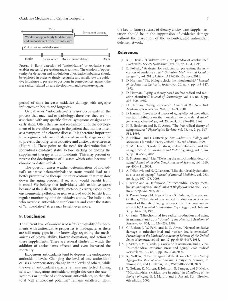
Disease manifestation
CurePermanent damage
Oxidative/ antioxidative stress
Window of opportunity for detection
and modulation of oxidative imbalance
Disease onsetHealth Death
Figure 1: Early detection of “antioxidative” or oxidative stressenables successful prevention and treatment. The window of oppor-tunity for detection and modulation of oxidative imbalance shouldbe explored in order to timely recognise and ameliorate the oxida-tive imbalance to prevent or postpone its consequences, namely, thefree radical-related disease development and premature aging.
period of time increases oxidative damage with negativeinfluences on health and longevity.
Oxidative or “antioxidative” stresses occur early in theprocess that may lead to pathology; therefore, they are notassociated with any specific clinical symptoms or signs at anearly stage. Often they are not recognized until the develop-ment of irreversible damage to the patient that manifest itselfas a symptom of a chronic disease. It is therefore importantto recognise oxidative imbalance at an early stage in orderto prevent the long-term oxidative and antioxidative stresses(Figure 1). These point to the need for determination ofindividual’s oxidative status before starting or ending thesupplement therapy with antioxidants. This may prevent orreverse the development of diseases which arise because ofchronic oxidative imbalance.
The question arises weather determination of individ-ual’s oxidative balance/imbalance status would lead to abetter preventive or therapeutic interventions that may slowdown the aging process. If so, who would benefit fromit most? We believe that individuals with oxidative stressbecause of their diets, lifestyle, metabolic errors, exposure toenvironmental pollution or iron overload would benefit fromregular monitoring of their oxidative status. The individualswho overdose antioxidant supplements and enter the statusof “antioxidative” stress may benefit even more.
8. Conclusion
The current level of awareness of safety and quality of supple-ments with antioxidative properties is inadequate, as thereare still many gaps in our knowledge regarding the mech-anisms of bioavailability, biotransformation, and action ofthese supplements. There are several studies in which theaddition of antioxidants affected and even increased themortality.
Exogenous antioxidants tend to depress the endogenousantioxidant levels. Changing the level of one antioxidantcauses a compensatory change in the levels of others, whilethe overall antioxidant capacity remains unaffected. Dosingcells with exogenous antioxidants might decrease the rate ofsynthesis or uptake of endogenous antioxidants, so that thetotal “cell antioxidant potential” remains unaltered. Thus,
the key to future success of dietary antioxidant supplemen-tation should be in the suppression of oxidative damagewithout the disruption of the well-integrated antioxidantdefense network.
References
[1] K. J. Davies, “Oxidative stress: the paradox of aerobic life,”Biochemical Society Symposium, vol. 61, pp. 1–31, 1995.
[2] B. Poljsak, “Strategies for reducing or preventing the gen-eration of oxidative stress,” Oxidative Medicine and CellularLongevity, vol. 2011, Article ID 194586, 15 pages, 2011.
[3] D. Harman, “The biologic clock: the mitochondria?” Journalof the American Geriatrics Society, vol. 20, no. 4, pp. 145–147,1972.
[4] D. Harman, “Aging: a theory based on free radical and radi-ation chemistry,” Journal of Gerontology, vol. 11, no. 3, pp.298–300, 1956.
[5] D. Harman, “Aging: overview,” Annals of the New YorkAcademy of Sciences, vol. 928, pp. 1–21, 2001.
[6] D. Harman, “Free radical theory of aging: effect of free radicalreaction inhibitors on the mortality rate of male laf mice,”Journals of Gerontology, vol. 23, no. 4, pp. 476–482, 1968.
[7] K. B. Beckman and B. N. Ames, “The free radical theory ofaging matures,” Physiological Reviews, vol. 78, no. 2, pp. 547–581, 1998.
[8] B. Halliwell and J. Gutteridge, Free Radicals in Biology andMedicine, Clarendon Press, Oxford, UK, 3rd edition, 1999.
[9] T. M. Hagen, “Oxidative stress, redox imbalance, and theaging process,” Antioxidants and Redox Signaling, vol. 5, no.5, pp. 503–506, 2003.
[10] B. N. Ames and J. Liu, “Delaying the mitochondrial decay ofaging,” Annals of the New York Academy of Sciences, vol. 1019,pp. 406–411, 2004.
[11] A. Trifunovic and N. G. Larsson, “Mitochondrial dysfunctionas a cause of ageing,” Journal of Internal Medicine, vol. 263,no. 2, pp. 167–178, 2008.
[12] I. Bratic and A. Trifunovic, “Mitochondrial energy meta-bolism and ageing,” Biochimica et Biophysica Acta, vol. 1797,no. 6-7, pp. 961–967, 2010.
[13] R. Perez-Campo, M. Lopez-Torres, S. Cadenas, C. Rojas, andG. Barja, “The rate of free radical production as a deter-minant of the rate of aging: evidence from the comparativeapproach,” Journal of Comparative Physiology B, vol. 168, no.3, pp. 149–158, 1998.
[14] G. Barja, “Mitochondrial free radical production and agingin mammals and birds,” Annals of the New York Academy ofSciences, vol. 854, pp. 224–238, 1998.
[15] C. Richter, J. W. Park, and B. N. Ames, “Normal oxidativedamage to mitochondrial and nuclear dna is extensive,”Proceedings of the National Academy of Sciences of the UnitedStates of America, vol. 85, no. 17, pp. 6465–6467, 1988.
[16] J. Sastre, F. V. Pallardo, J. Garcıa de la Asuncion, and J. Vina,“Mitochondria, oxidative stress and aging,” Free RadicalResearch, vol. 32, no. 3, pp. 189–198, 2000.
[17] R. Wilken, “Healthy aging: skeletal muscla,” in HealthyAging—The Role of Nutrition and Lifestyle, S. Stanner, R.Thompson, and J. Buttriss, Eds., Wiley-Blackwell, 2009.
[18] T. Golden, K. Morten, F. Johnson, E. Samper, and S. Melov,“Mitochondria: a critical role in aging,” in Handbook of theBiology of Aging, E. J. Masoro and S. Austad, Eds., Elsevier,6th edition, 2006.
8 Oxidative Medicine and Cellular Longevity
[19] H. T. Jacobs, “The mitochondrial theory of aging: dead oralive?” Aging Cell, vol. 2, no. 1, pp. 11–17, 2003.
[20] J. W. Pak, A. Herbst, E. Bua, N. Gokey, D. McKenzie, and J.M. Aiken, “Rebuttal to jacobs: the mitochondrial theory ofaging: alive and well,” Aging Cell, vol. 2, no. 1, pp. 9–10, 2003.
[21] A. D. N. J. de Grey, “Reactive oxygen species production inthe mitochondrial matrix: implications for the mechanismof mitochondrial mutation accumulation,” RejuvenationResearch, vol. 8, no. 1, pp. 13–17, 2005.
[22] L. Casteilla, M. Rigoulet, and L. Penicaud, “Mitochondrialros metabolism: modulation by uncoupling proteins,” IubmbLife, vol. 52, no. 3–5, pp. 181–188, 2002.
[23] R. G. Hansford, B. A. Hogue, and V. Mildaziene, “Depen-dence of H2O2 formation by rat heart mitochondria onsubstrate availability and donor age,” Journal of Bioenergeticsand Biomembranes, vol. 29, no. 1, pp. 89–95, 1997.
[24] K. Staniek and H. Nohl, “H2O2 detection from intactmitochondria as a measure for one-electron reduction ofdioxygen requires a non-invasive assay system,” BiochimicaEt Biophysica Acta, vol. 1413, no. 2, pp. 70–80, 1999.
[25] J. R. Speakman, C. Selman, J. S. McLaren, and E. J. Harper,“Living fast, dying when? the link between aging andenergetics,” Journal of Nutrition, vol. 132, supplement 6, pp.1583S–1597S, 2002.
[26] L. A. Harrington and C. B. Harley, “Effect of vitamin eon lifespan and reproduction in Caenorhabditis elegans,”Mechanisms of Ageing and Development, vol. 43, no. 1, pp.71–78, 1988.
[27] J. P. Phillips, S. D. Campbell, D. Michaud, M. Charbonneau,and A. J. Hilliker, “Null mutation of copper/zinc superoxidedismutase in Drosophila confers hypersensitivity to paraquatand reduced longevity,” Proceedings of the National Academyof Sciences of the United States of America, vol. 86, no. 8, pp.2761–2765, 1989.
[28] W. C. Orr and R. S. Sohal, “Extension of life-span byoverexpression of superoxide dismutase and catalase inDrosophila melanogaster,” Science, vol. 263, no. 5150, pp.1128–1130, 1994.
[29] T. L. Parkes, A. J. Elia, D. Dickinson, A. J. Hilliker, J. P.Phillips, and G. L. Boulianne, “Extension of Drosophila lifes-pan by overexpression of human SOD1 in motorneurons,”Nature Genetics, vol. 19, no. 2, pp. 171–174, 1998.
[30] S. Melov, J. Ravenscroft, S. Malik et al., “Extension of life-span with superoxide dismutase/catalase mimetics,” Science,vol. 289, no. 5484, pp. 1567–1569, 2000.
[31] J. Moskovitz, S. Bar-Noy, W. M. Williams, J. Requena, B. S.Berlett, and E. R. Stadtman, “Methionine sulfoxide reductase(MsrA) is a regulator of antioxidant defense and lifespan inmammals,” Proceedings of the National Academy of Sciences ofthe United States of America, vol. 98, no. 23, pp. 12920–12925,2001.
[32] V. V. Bakaev and M. B. Lyudmila, “Effect of ascorbicacid on longevity in the nematode Caenorhabditis elegans,”Biogerontology, vol. 3, supplement 1, pp. 12–16, 2002.
[33] H. Ruan, X. D. Tang, M. L. Chen et al., “High-qualitylife extension by the enzyme peptide methionine sulfoxidereductase,” Proceedings of the National Academy of Sciences ofthe United States of America, vol. 99, no. 5, pp. 2748–2753,2002.
[34] N. Ishii, N. Senoo-Matsuda, K. Miyake et al., “Coenzyme Q10can prolong C. elegans lifespan by lowering oxidative stress,”Mechanisms of Ageing and Development, vol. 125, no. 1, pp.41–46, 2004.
[35] T. T. Huang, M. Naeemuddin, S. Elchuri et al., “Genetic mod-ifiers of the phenotype of mice deficient in mitochondrialsuperoxide dismutase,” Human Molecular Genetics, vol. 15,no. 7, pp. 1187–1194, 2006.
[36] S. Zou, J. Sinclair, M. A. Wilson et al., “Comparativeapproaches to facilitate the discovery of prolongevity inter-ventions: effects of tocopherols on lifespan of three inverte-brate species,” Mechanisms of Ageing and Development, vol.128, no. 2, pp. 222–226, 2007.
[37] J. Kim, M. Takahashi, T. Shimizu et al., “Effects of a potentantioxidant, platinum nanoparticle, on the lifespan ofCaenorhabditis elegans,” Mechanisms of Ageing and Develop-ment, vol. 129, no. 6, pp. 322–331, 2008.
[38] K. L. Quick, S. S. Ali, R. Arch, C. Xiong, D. Wozniak, and L. L.Dugan, “A carboxyfullerene sod mimetic improves cognitionand extends the lifespan of mice,” Neurobiology of Aging, vol.29, no. 1, pp. 117–128, 2008.
[39] D. F. Dai, L. F. Santana, M. Vermulst et al., “Overexpression ofcatalase targeted to mitochondria attenuates murine cardiacaging,” Circulation, vol. 119, no. 21, pp. 2789–2797, 2009.
[40] A. Shibamura, T. Ikeda, and Y. Nishikawa, “A method for oraladministration of hydrophilic substances to Caenorhabditiselegans: effects of oral supplementation with antioxidantson the nematode lifespan,” Mechanisms of Ageing andDevelopment, vol. 130, no. 9, pp. 652–655, 2009.
[41] Y. Dundar and R. Aslan, “Antioxidative stress,” EasternJournal of Medicine, vol. 5, no. 2, pp. 45–47, 2000.
[42] G. S. Omenn, G. E. Goodman, M. D. Thornquist et al.,“Effects of a combination of β carotene and vitamin A onlung cancer and cardiovascular disease,” The New EnglandJournal of Medicine, vol. 334, no. 18, pp. 1150–1155, 1996.
[43] G. Bjelakovic, D. Nikolova, R. G. Simonetti, and C. Gluud,“Antioxidant supplements for prevention of gastrointestinalcancers: a systematic review and meta-analysis,” The Lancet,vol. 364, no. 9441, pp. 1219–1228, 2004.
[44] E. R. Miller, R. Pastor-Barriuso, D. Dalal, R. A. Riemersma, L.J. Appel, and E. Guallar, “Meta-analysis: high-dosage vitaminE supplementation may increase all-cause mortality,” Annalsof Internal Medicine, vol. 142, no. 1, pp. 37–46, 2005.
[45] R. Collins, J. Armitage, S. Parish, P. Sleight, and R. Peto,“MRC/BHF heart protection study of antioxidant vitaminsupplementation in 20, 536 high-risk individuals: a ran-domised placebo-controlled trial,” The Lancet, vol. 360, no.9326, pp. 23–33, 2002.
[46] Age-Related Eye Disease Study Research Group, “A random-ized, placebo-controlled, clinical trial of high-dose supple-mentation with vitamins C and E and β carotine for age-related cataract and vision loss: AREDS report no. 9,” Archivesof Ophthalmology, vol. 119, no. 10, pp. 1439–1452, 2001.
[47] J. Mursu, K. Robien, L. J. Harnack, K. Park, and D. R. JacobsJr., “Dietary supplements and mortality rate in older women:the Iowa women’s health study,” Archives of Internal Medicine,vol. 171, no. 18, pp. 1625–1633, 2011.
[48] E. A. Klein, I. M. Thompson Jr., C. M. Tangen et al., “VitaminE and the risk of prostate cancer: the selenium and vitaminE cancer prevention trial (SELECT),” Journal of the AmericanMedical Association, vol. 306, no. 14, pp. 1549–1556, 2011.
[49] G. Bjelakovic, D. Nikolova, L. L. Gluud, R. G. Simonetti,and C. Gluud, “Antioxidant supplements for prevention ofmortality in healthy participants and patients with variousdiseases,” Cochrane Database of Systematic Reviews, no. 2,Article ID CD007176, 2008.
[50] S. Hercberg, K. Ezzedine, C. Guinot et al., “Antioxidantsupplementation increases the risk of skin cancers in women
Oxidative Medicine and Cellular Longevity 9
but not in men,” Journal of Nutrition, vol. 137, no. 9, pp.2098–2105, 2007.
[51] A. Bardia, I. M. Tleyjeh, J. R. Cerhan et al., “Efficacyof antioxidant supplementation in reducing primary can-cer incidence and mortality: systematic review and meta-analysis,” Mayo Clinic Proceedings, vol. 83, no. 1, pp. 23–34,2008.
[52] B. D. Lawenda, K. M. Kelly, E. J. Ladas, S. M. Sagar, A.Vickers, and J. B. Blumberg, “Should supplemental antiox-idant administration be avoided during chemotherapy andradiation therapy?” Journal of the National Cancer Institute,vol. 100, no. 11, pp. 773–783, 2008.
[53] S. K. Myung, Y. Kim, W. Ju, H. J. Choi, and W. K. Bae,“Effects of antioxidant supplements on cancer prevention:meta-analysis of randomized controlled trials,” Annals ofOncology, vol. 21, no. 1, pp. 166–179, 2010.
[54] T. J. Schulz, K. Zarse, A. Voigt, N. Urban, M. Birringer,and M. Ristow, “Glucose restriction extends Caenorhabditiselegans life span by inducing mitochondrial respiration andincreasing oxidative stress,” Cell Metabolism, vol. 6, no. 4, pp.280–293, 2007.
[55] J. G. Ionescu and B. Poljsak, “Metal ions mediated pro-oxidative reactions with vitamin C: possible implications fortreatment of different malignancies,” International Journal ofCancer Prevention, vol. 3, no. 3, pp. 149–174, 2010.
[56] B. Poljsak, I. Milisav, T. Lampe, and I. Ostan, “Reproductivebenefit of oxidative damage: an oxidative stress “malevo-lence”?” Oxidative Medicine and Cellular Longevity, vol. 2011,Article ID 760978, 9 pages, 2011.
[57] M. J. Fettman, K. D. Valerius, G. K. Ogilvie et al., “Effects ofdietary cysteine on blood sulfur amino acid, glutathione, andmalondialdehyde concentrations in cats,” American Journalof Veterinary Research, vol. 60, no. 3, pp. 328–333, 1999.
[58] A. Bast, G. R. M. M. Haenen, and C. J. A. Doelman,“Oxidants and antioxidants: state of the art,” AmericanJournal of Medicine, vol. 91, no. 3C, pp. 2S–13S, 1991.
[59] B. Halliwell, M. A. Murcia, S. Chirico, and O. I. Aruoma,“Free radicals and antioxidants in food and in vivo: what theydo and how they work,” Critical Reviews in Food Science andNutrition, vol. 35, no. 1-2, pp. 7–20, 1995.
[60] A. Cherubini, G. B. Vigna, G. Zuliani, C. Ruggiero, U.Senin, and R. Fellin, “Role of antioxidants in atherosclerosis:epidemiological and clinical update,” Current PharmaceuticalDesign, vol. 11, no. 16, pp. 2017–2032, 2005.
[61] S. B. Lotito and B. Frei, “Consumption of flavonoid-richfoods and increased plasma antioxidant capacity in humans:cause, consequence, or epiphenomenon?” Free Radical Biol-ogy and Medicine, vol. 41, no. 12, pp. 1727–1746, 2006.
[62] R. G. Cutler and M. P. Mattson, “Measuring oxidative stressand interpreting its relevance in humans,” in Oxidative Stressand Aging, R. G. Cutler and H. Rodriguez, Eds., WorldScientific, Hackensack, NJ, USA, 2003.
[63] R. G. Cutler, “Genetic stability, dysdifferentiation, andlongevity determinant genes,” in Critical Reviews of OxidativeStress and Damage, R. G. Cutler and H. Rodriguez, Eds.,World Scientific, Hackensack, NJ, USA, 2003.
[64] L. J. Green, The Dermatologist’s Guide to Looking Younger,Crossing Press, Freedom, Calif, USA, 1999.
[65] S. G. Rhee, “Redox signaling: hydrogen peroxide as intracel-lular messenger,” Experimental and Molecular Medicine, vol.31, no. 2, pp. 53–59, 1999.
[66] K. Bedard and K. H. Krause, “The NOX family of ROS-generating NADPH oxidases: physiology and pathophysiol-ogy,” Physiological Reviews, vol. 87, no. 1, pp. 245–313, 2007.
[67] N. Kaul and H. J. Forman, “Reactive oxygen species in phys-iology and toxicology: from lipid peroxidation to transcrip-tional activation,” in Toxicology of the Human Environment:The Critical Role of Free Radicals, Cr. Rhodes, Ed., pp. 310–335, Taylor and Francis, New York, NY, USA, 2000.
[68] R. M. J. Palmer, A. G. Ferrige, and S. Moncada, “Nitric oxiderelease accounts for the biological activity of endothelium-derived relaxing factor,” Nature, vol. 327, no. 6122, pp. 524–526, 1987.
[69] H. Kamata and H. Hirata, “Redox regulation of cellularsignalling,” Cellular Signalling, vol. 11, no. 1, pp. 1–14, 1999.
[70] T. P. Dalton, H. G. Shertzer, and A. Puga, “Regulationof gene expression by reactive oxygen,” Annual Review ofPharmacology and Toxicology, vol. 39, pp. 67–101, 1999.
[71] J. Nordberg and E. S. J. Arner, “Reactive oxygen species,antioxidants, and the mammalian thioredoxin system,” FreeRadical Biology and Medicine, vol. 31, no. 11, pp. 1287–1312,2001.
[72] Z. H. Chen and E. Niki, “Two faces of lipid peroxidationproducts: the “Yin and Yang” principles of oxidative stress,”Journal of Experimental and Integrative Medicine, vol. 1, no.4, pp. 215–219, 2011.
[73] D. L. Carlisle, D. E. Pritchard, J. Singh et al., “Apoptosisand p53 induction in human lung fibroblasts exposedto chromium (VI): effect of ascorbate and tocopherol,”Toxicological Sciences, vol. 55, no. 1, pp. 60–68, 2000.
[74] D. Anastasiou, G. Poulogiannis, J. M. Asara et al., “Inhibitionof pyruvate kinase M2 by reactive oxygen species contributesto cellular antioxidant responses,” Science, vol. 334, no. 6060,pp. 1278–1283, 2011.
[75] D. Albanes, O. P. Heinonen, P. R. Taylor et al., “α-Tocopheroland β-carotene supplements and lung cancer incidence in theα-tocopherol, β-carotene cancer prevention study: effects ofbase- line characteristics and study compliance,” Journal ofthe National Cancer Institute, vol. 88, no. 21, pp. 1560–1570,1996.
[76] Z. T. Schafer, A. R. Grassian, L. Song et al., “Antioxidant andoncogene rescue of metabolic defects caused by loss of matrixattachment,” Nature, vol. 461, no. 7260, pp. 109–113, 2009.
[77] P. Gao, H. Zhang, R. Dinavahi et al., “HIF-dependentantitumorigenic effect of antioxidants in vivo,” Cancer Cell,vol. 12, no. 3, pp. 230–238, 2007.
[78] B. A. Narayanan, “Chemopreventive agents alters global geneexpression pattern: predicting their mode of action andtargets,” Current Cancer Drug Targets, vol. 6, no. 8, pp. 711–727, 2006.
[79] N. Rakba, F. Aouad, C. Henry et al., “Iron mobilization andcellular protection by a new synthetic chelator O-Trensox,”Biochemical Pharmacology, vol. 55, no. 11, pp. 1797–1806,1998.
[80] N. T. V. Le and D. R. Richardson, “The role of iron in cellcycle progression and the proliferation of neoplastic cells,”Biochimica Et Biophysica Acta, vol. 1603, no. 1, pp. 31–46,2002.
[81] J. L. Buss, F. M. Torti, and S. V. Torti, “The role of ironchelation in cancer therapy,” Current Medicinal Chemistry,vol. 10, no. 12, pp. 1021–1034, 2003.
[82] D. B. Lovejoy and D. R. Richardson, “Iron chelators as anti-neoplastic agents: current developments and promise of thePIH class of chelators,” Current Medicinal Chemistry, vol. 10,no. 12, pp. 1035–1049, 2003.
[83] J. L. Buss, B. T. Greene, J. Turner, F. M. Torti, and S. V. Torti,“Iron chelators in cancer chemotherapy,” Current Topics inMedicinal Chemistry, vol. 4, no. 15, pp. 1623–1635, 2004.

10 Oxidative Medicine and Cellular Longevity
[84] J. I. Hanai, T. Mammoto, P. Seth et al., “Lipocalin 2 dimin-ishes invasiveness and metastasis of Ras-transformed cells,”The Journal of Biological Chemistry, vol. 280, no. 14, pp.13641–13647, 2005.
[85] D. S. Kalinowski and D. R. Richardson, “The evolution ofiron chelators for the treatment of iron overload disease andcancer,” Pharmacological Reviews, vol. 57, no. 4, pp. 547–583,2005.
[86] P. M. B. Pahl and L. D. Horwitz, “Cell permeable iron chela-tors as potential cancer chemotherapeutic agents,” CancerInvestigation, vol. 23, no. 8, pp. 683–691, 2005.
[87] G. Nie, G. Chen, A. D. Sheftel, K. Pantopoulos, and P.Ponka, “In vivo tumor growth is inhibited by cytosolic irondeprivation caused by the expression of mitochondrialferritin,” Blood, vol. 108, no. 7, pp. 2428–2434, 2006.
[88] G. Edgren, O. Nyren, and M. Melbye, “Cancer as a ferrotoxicdisease: are we getting hard stainless evidence?” Journal of theNational Cancer Institute, vol. 100, no. 14, pp. 976–977, 2008.
[89] X. Huang, “Does iron have a role in breast cancer?” TheLancet Oncology, vol. 9, no. 8, pp. 803–807, 2008.
[90] L. R. Zacharski, B. K. Chow, P. S. Howes et al., “Decreasedcancer risk after iron reduction in patients with peripheralarterial disease: results from a randomized trial,” Journal ofthe National Cancer Institute, vol. 100, no. 14, pp. 996–1002,2008.
[91] S. P. Stoesz, A. Friedl, J. D. Haag, M. J. Lindstrom, G. M.Clark, and M. N. Gould, “Heterogeneous expression of thelipocalin NGAL in primary breast cancers,” InternationalJournal of Cancer, vol. 79, no. 6, pp. 565–572, 1998.
[92] Z. Tong, X. Wu, D. Ovcharenko, J. Zhu, C. S. Chen, andJ. P. Kehrer, “Neutrophil gelatinase-associated lipocalin as asurvival factor,” Biochemical Journal, vol. 391, no. 2, pp. 441–448, 2005.
[93] R. Lim, N. Ahmed, N. Borregaard et al., “Neutrophilgelatinase-associated lipocalin (NGAL) an early-screeningbiomarker for ovarian cancer: NGAL is associated withepidermal growth factor-induced epithelio-mesenchymaltransition,” International Journal of Cancer, vol. 120, no. 11,pp. 2426–2434, 2007.
[94] R. L. Nelson, F. G. Davis, E. Sutter, L. H. Sobin, J. W.Kikendall, and P. Bowen, “Body iron stores and risk of colonicneoplasia,” Journal of the National Cancer Institute, vol. 86,no. 6, pp. 455–460, 1994.
[95] S. Okada, “Iron-induced tissue damage and cancer: the roleof reactive oxygen species-free radicals,” Pathology Interna-tional, vol. 46, no. 5, pp. 311–332, 1996.
[96] S. Toyokuni, “Iron-induced carcinogenesis: the role of redoxregulation,” Free Radical Biology and Medicine, vol. 20, no. 4,pp. 553–566, 1996.
[97] E. D. Weinberg, “The role of iron in cancer,” European Journalof Cancer Prevention, vol. 5, no. 1, pp. 19–36, 1996.
[98] E. D. Weinberg, “Iron loading and disease surveillance,”Emerging Infectious Diseases, vol. 5, no. 3, pp. 346–352, 1999.
[99] M. Nunez, V. Tapia, S. Toyokuni, and S. Okada, “Iron-induced oxidative damage in colon carcinoma (Caco-2)cells,” Free Radical Research, vol. 34, no. 1, pp. 57–68, 2001.
[100] M. Glei, G. O. Latunde-Dada, A. Klinder et al., “Iron-overload induces oxidative DNA damage in the human coloncarcinoma cell line HT29 clone 19A,” Mutation Research, vol.519, no. 1-2, pp. 151–161, 2002.
[101] S. Toyokuni, “Iron and carcinogenesis: from Fenton reactionto target genes,” Redox Report, vol. 7, no. 4, pp. 189–197,2002.
[102] Y. Deugnier, “Iron and liver cancer,” Alcohol, vol. 30, no. 2,pp. 145–150, 2003.
[103] J. E. Klaunig and L. M. Kamendulis, “The role of oxidativestress in carcinogenesis,” Annual Review of Pharmacology andToxicology, vol. 44, pp. 239–267, 2004.
[104] P. Storz, “Reactive oxygen species in tumor progression,”Frontiers in Bioscience, vol. 10, no. 2, pp. 1881–1896, 2005.
[105] S. K. Lee, J. J. Lee, H. J. Lee et al., “Iron chelator-inducedgrowth arrest and cytochrome c dependent apoptosis inimmortalized and malignant oral keratinocytes,” Journal ofOral Pathology and Medicine, vol. 35, no. 4, pp. 218–226,2006.
[106] M. Valko, C. J. Rhodes, J. Moncol, M. Izakovic, and M.Mazur, “Free radicals, metals and antioxidants in oxidativestress-induced cancer,” Chemico-Biological Interactions, vol.160, no. 1, pp. 1–40, 2006.
[107] J. Wu, J. Eckard, H. Chen, M. Costa, K. Frenkel, and X.Huang, “Altered iron homeostasis involvement in arsenite-mediated cell transformation,” Free Radical Biology andMedicine, vol. 40, no. 3, pp. 444–452, 2006.
[108] A. R. Kallianpur, S. A. Lee, Y. T. Gao et al., “Dietary animal-derived iron and fat intake and breast cancer risk in theShanghai Breast Cancer Study,” Breast Cancer Research andTreatment, vol. 107, no. 1, pp. 123–132, 2008.
[109] D. Pra, S. I. R. Franke, R. Giulian et al., “Genotoxicity andmutagenicity of iron and copper in mice,” Biometals, vol. 21,no. 3, pp. 289–297, 2008.
[110] W. S. Yang and B. R. Stockwell, “Synthetic lethal screeningidentifies compounds activating iron-dependent, nonapop-totic cell death in oncogenic-RAS-harboring cancer cells,”Chemistry and Biology, vol. 15, no. 3, pp. 234–245, 2008.
[111] A. Terman and U. T. Brunk, “Oxidative stress, accumulationof biological “garbage”, and aging,” Antioxidants and RedoxSignaling, vol. 8, no. 1-2, pp. 197–204, 2006.
[112] D. B. Kell, “Iron behaving badly: inappropriate iron chelationas a major contributor to the aetiology of vascular and otherprogressive inflammatory and degenerative diseases,” BMCMedical Genomics, vol. 2, article 2, 2009.
[113] L. Deguillaume, M. Leriche, and N. Chaumerliac, “Impact ofradical versus non-radical pathway in the Fenton chemistryon the iron redox cycle in clouds,” Chemosphere, vol. 60, no.5, pp. 718–724, 2005.
[114] J. A. Imlay, “Pathways of oxidative damage,” Annual Reviewof Microbiology, vol. 57, pp. 395–418, 2003.
[115] A. Jacobs, “Low molecular weight intracellular iron transportcompounds,” Blood, vol. 50, no. 3, pp. 433–439, 1977.
[116] A. Voogd, W. Sluiter, H. G. van Eijk, and J. F. Koster, “Lowmolecular weight iron and the oxygen paradox in isolatedrat hearts,” Journal of Clinical Investigation, vol. 90, no. 5, pp.2050–2055, 1992.
[117] B. Halliwell and J. M. C. Gutteridge, Free Radicals in Biologyand Medicine, Oxford University Press, Oxford, UK, 2005.
[118] V. Herbert, “The antioxidant supplement myth,” AmericanJournal of Clinical Nutrition, vol. 60, no. 2, pp. 157–158, 1994.
[119] D. M. Miller, G. R. Buettner, and S. D. Aust, “Transitionmetals as catalysts of “autoxidation” reactions,” Free RadicalBiology and Medicine, vol. 8, no. 1, pp. 95–108, 1990.
[120] B. Lachili, I. Hininger, H. Faure et al., “Increased lipidperoxidation in pregnant women after iron and vitamin Csupplementation,” Biological Trace Element Research, vol. 83,no. 2, pp. 103–110, 2001.
[121] D. W. Reif, “Ferritin as a source of iron for oxidative damage,”Free Radical Biology and Medicine, vol. 12, no. 5, pp. 417–427,1992.

Oxidative Medicine and Cellular Longevity 11
[122] I. FaBIaN and V. Csordas, “Metal ion catalyzed autoxidationreactions: kinetics and mechanisms,” Advances in InorganicChemistry, vol. 54, pp. 395–461, 2003.
[123] M. Hoppe, L. Hulthen, and L. Hallberg, “The relative bio-availability in humans of elemental iron powders for use infood fortification,” European Journal of Nutrition, vol. 45, no.1, pp. 37–44, 2006.
[124] L. R. Zacharski, D. L. Ornstein, S. Woloshin, and L. M.Schwartz, “Association of age, sex, and race with body ironstores in adults: analysis of of NHANES III data,” AmericanHeart Journal, vol. 140, no. 1, pp. 98–104, 2000.
[125] R. F. Ritchie, G. E. Palomaki, L. M. Neveux, O. Navolotskaia,T. B. Ledue, and W. Y. Craig, “Reference distributions forserum iron and transferrin saturation: a comparison of alarge cohort to the world’s literature,” Journal of ClinicalLaboratory Analysis, vol. 16, no. 5, pp. 246–252, 2002.
[126] R. F. Ritchie, G. E. Palomaki, L. M. Neveux, O. Navolotskaia,T. B. Ledue, and W. Y. Craig, “Reference distributions forserum iron and transferrin saturation: a practical, simple,and clinically relevant approach in a large cohort,” Journal ofClinical Laboratory Analysis, vol. 16, no. 5, pp. 237–245, 2002.
[127] J. M. Liu, S. E. Hankinson, M. J. Stampfer, N. Rifai, W. C.Willett, and J. Ma, “Body iron stores and their determinantsin healthy postmenopausal US women,” American Journal ofClinical Nutrition, vol. 78, no. 6, pp. 1160–1167, 2003.
[128] P. T. Doulias, C. Vlachou, C. Boudouri, P. Kanavaros, K. C.Siamopoulos, and D. Galaris, “Flow cytometric estimationof “labile iron pool” in human white blood cells reveals apositive association with ageing,” Free Radical Research, vol.42, no. 3, pp. 253–259, 2008.
[129] D. J. Fleming, K. L. Tucker, P. F. Jacques, G. E. Dallal, P. W.F. Wilson, and R. J. Wood, “Dietary factors associated withthe risk of high iron stores in the elderly Framingham HeartStudy cohort,” American Journal of Clinical Nutrition, vol. 76,no. 6, pp. 1375–1384, 2002.
[130] J. Beard, “Dietary iron intakes and elevated iron stores inthe elderly: is it time to abandon the set-point hypothesis ofregulation of iron absorption?” American Journal of ClinicalNutrition, vol. 76, no. 6, pp. 1189–1190, 2002.
[131] J. E. Cade, J. A. Moreton, B. O’Hara et al., “Diet and geneticfactors associated with iron status in middle-aged women,”American Journal of Clinical Nutrition, vol. 82, no. 4, pp. 813–820, 2005.
[132] J. M. Guralnik, R. S. Eisenstaedt, L. Ferrucci, H. G. Klein,and R. C. Woodman, “Prevalence of anemia in persons 65years and older in the united states: evidence for a high rate ofunexplained anemia,” Blood, vol. 104, no. 8, pp. 2263–2268,2004.
[133] E. A. Price, “Aging and erythropoiesis: current state ofknowledge,” Blood Cells, Molecules, and Diseases, vol. 41, no.2, pp. 158–165, 2008.
[134] D. W. Killilea, H. Atamna, C. Liao, and B. N. Ames, “Ironaccumulation during cellular senescence in human fibrob-lasts in vitro,” Antioxidants and Redox Signaling, vol. 5, no.5, pp. 507–516, 2003.
[135] V. Glauce Socorro de Barros, A. Luzia Kalyne, M. Leal, andJ. B. Fontenele, “Role of plant extracts and polyphenoliccompounds in oxidative stress-related diseases,” in Handbookof Free Radicals: Formation, Types and Effects, D. Kozyrev andV. Slutsky, Eds., Nova Science Publishers, Inc., New York, NY,USA, 2010.
[136] R. Gredilla and G. Barja, “Mitochondrial oxidative stress andcaloric restriction,” in Metabolism and Lifespan Determina-
tion, M. P. Mattson, Ed., vol. 14, pp. 105–122, Advances inCell Aging and Gerontology, 2003.
[137] C. I. Cook and B. P. Yu, “Iron accumulation in aging:modulation by dietary restriction,” Mechanisms of Ageing andDevelopment, vol. 102, no. 1, pp. 1–13, 1998.
[138] G. Reverter-Branchat, E. Cabiscol, J. Tamarit, and J. Ros,“Oxidative damage to specific proteins in replicative andchronological-aged Saccharomyces cerevisiae—common tar-gets and prevention by calorie restriction,” The Journal ofBiological Chemistry, vol. 279, no. 30, pp. 31983–31989, 2004.
[139] S. Goto, Z. Radak, and R. Takahasi, “Biological implicationsof protein oxidation,” in Critical Review of Oxidative Stressand Aging, R. Cutler and H. Rodriguez, Eds., World Scientific,Hackensack, NJ, USA, 2003.
[140] T. G. Son, S. Camandola, and M. P. Mattson, “Hormeticdietary phytochemicals,” Neuromolecular Medicine, vol. 10,no. 4, pp. 236–246, 2008.
[141] M. P. Mattson, “Hormesis defined,” Ageing Research Reviews,vol. 7, no. 1, pp. 1–7, 2008.
[142] D. W. Lee and B. P. Yu, “Food restriction as an effectivemodulator of free radical metabolism in rats,” KoreanBiochemical Journal, vol. 24, pp. 148–154, 1991.
[143] A. Koizumi, R. Weindruch, and R. L. Walford, “Influencesof dietary restriction and age on liver enzyme activities andlipid peroxidation in mice,” Journal of Nutrition, vol. 117, no.2, pp. 361–367, 1987.
[144] L. H. Chen and S. R. Lowry, “Cellular antioxidant defensesystem,” Progress in Clinical and Biological Research, vol. 287,pp. 247–256, 1989.
[145] M. Ristow and S. Schmeisser, “Extending life span by increas-ing oxidative stress,” Free Radical Biology and Medicine, vol.51, no. 2, pp. 327–336, 2011.
[146] T. Finkel and N. J. Holbrook, “Oxidants, oxidative stress andthe biology of ageing,” Nature, vol. 408, no. 6809, pp. 239–247, 2000.
[147] Z. H. Chen, Y. Yoshida, Y. Saito, and E. Niki, “Adaptationto hydrogen peroxide enhances PC12 cell tolerance againstoxidative damage,” Neuroscience Letters, vol. 383, no. 3, pp.256–259, 2005.
[148] E. J. Masoro, “Hormesis and the antiaging action of dietaryrestriction,” Experimental Gerontology, vol. 33, no. 1-2, pp.61–66, 1998.
[149] S. I. Rattan and D. Demirovic, “Hormesis as a mechanismfor the anti-aging effects of calorie restriction,” in CalorieRestriction, Aging and Longevity, A. V. Everitt, S. I. S. Rattan,D. G. Couteur, and R. D. Cabo, Eds., pp. 233–245, Springer,Dordrecht, The Netherlands, 2010.
[150] B. P. Yu and H. Y. Chung, “Stress resistance by caloricrestriction for longevity,” Annals of the New York Academy ofSciences, vol. 928, pp. 39–47, 2001.
[151] E. Masoro, “The role of hormesis in life extension by dietaryrestriction,” Interdisciplinary Topics in Gerontology, vol. 35,pp. 1–17, 2007.
[152] E. Warabi, Y. Wada, H. Kajiwara et al., “Effect on endothelialcell gene expression of shear stress, oxygen concentration,and low-density lipoprotein as studied by a novel flow cellculture system,” Free Radical Biology and Medicine, vol. 37,no. 5, pp. 682–694, 2004.
[153] E. Warabi, W. Takabe, T. Minami et al., “Shear stress stabilizesNF-E2-related factor 2 and induces antioxidant genes inendothelial cells: role of reactive oxygen/nitrogen species,”Free Radical Biology and Medicine, vol. 42, no. 2, pp. 260–269,2007.
12 Oxidative Medicine and Cellular Longevity
[154] M. Plews, S. L. R. Simon, D. R. Boreham et al., “A radiation-induced adaptive response prolongs the survival of prion-infected mice,” Free Radical Biology and Medicine, vol. 49, no.9, pp. 1417–1421, 2010.
[155] S. Yanase, P. S. Hartman, A. Ito, and N. Ishii, “Oxidativestress pretreatment increases the X-radiation resistance of thenematode Caenorhabditis elegans,” Mutation Research, vol.426, no. 1, pp. 31–39, 1999.
[156] B. Poljsak, Skin aging, Free Radicals and Antioxidants, NovaScience Publisher, New York, NY, USA, 2012.
[157] L. Packer, E. Cadenas, and K. J. A. Davies, “Free radicals andexercise: an introduction,” Free Radical Biology and Medicine,vol. 44, no. 2, pp. 123–125, 2008.
[158] A. Boveris and B. Chance, “The mitochondrial generationof hydrogen peroxide. general properties and effect ofhyperbaric oxygen,” Biochemical Journal, vol. 134, no. 3, pp.707–716, 1973.
[159] P. L. Gratao, A. Polle, P. J. Lea, and R. A. Azevedo,“Making the life of heavy metal-stressed plants a little easier,”Functional Plant Biology, vol. 32, no. 6, pp. 481–494, 2005.
[160] S. I. S. Rattan, R. Gonzalez-Dosal, E. R. Nielsen, D. C. Kraft,J. Weibel, and S. Kahns, “Slowing down aging from within:mechanistic aspects of anti-aging hormetic effects of mildheat stress on human cells,” Acta Biochimica Polonica, vol. 51,no. 2, pp. 481–492, 2004.
[161] S. I. S. Rattan, “Hormesis in aging,” Ageing Research Reviews,vol. 7, no. 1, pp. 63–78, 2008.
[162] B. Conti, M. Sanchez-Alavez, R. Winsky-Sommerer et al.,“Transgenic mice with a reduced core body temperature havean increased life span,” Science, vol. 314, no. 5800, pp. 825–828, 2006.
[163] R. Hosono, Y. Mitsui, and Y. Sato, “Life span of the wildand mutant nematode Caenorhabditis elegans: effects of sex,sterilization, and temperature,” Experimental Gerontology,vol. 17, no. 2, pp. 163–172, 1982.
[164] S. S. Ali, M. C. G. Marcondes, H. Bajova, L. L. Dugan, and B.Conti, “Metabolic depression and increased ROS productionby isolated mitochondria at moderately lower temperatures,”The Journal of Biological Chemistry, vol. 285, no. 42, pp.32522–32528, 2010.
[165] R. Kurapati, H. B. Passananti, M. R. Rose, and J. Tower,“Increased hsp22 RNA levels in Drosophila lines geneticallyselected for increased longevity,” Journals of GerontologySeries A, vol. 55, no. 11, pp. B552–B559, 2000.
[166] G. Morrow, S. Battistini, P. Zhang, and R. M. Tanguay,“Decreased lifespan in the absence of expression of the mito-chondrial small heat shock protein hsp22 in Drosophila,”Journal of Biological Chemistry, vol. 279, no. 42, pp. 43382–43385, 2004.
[167] M. Ristow, K. Zarse, A. Oberbach et al., “Antioxidantsprevent health-promoting effects of physical exercise inhumans,” Proceedings of the National Academy of Sciences ofthe United States of America, vol. 106, no. 21, pp. 8665–8670,2009.
[168] M. C. Gomez-Cabrera, E. Domenech, M. Romagnoli etal., “Oral administration of vitamin C decreases musclemitochondrial biogenesis and hampers training-inducedadaptations in endurance performance,” American Journal ofClinical Nutrition, vol. 87, no. 1, pp. 142–149, 2008.
[169] B. Poljsak and P. Jamik, “Methodology for oxidative statedetection in biological systems,” in Handbook of Free Radicals:Formation, Types and Effects, D. Kozyrev and V. Slutsky,Eds., Cell biology research progress series, Nova SciencePublishers, New York, NY, USA, 2010.
[170] S. Miwa, F. L. Muller, and K. B. Beckman, “The basics ofoxidative biochemistry,” in Oxidative Stress in Aging. FromModel Systems to Human Diseases, S. Miwa, F. L. Muller, andK. B. Beckman, Eds., Humana Press, Totowa, NJ, USA, 2008.
[171] B. Halliwell and J. M. C. Gutteridge, “The chemistryof oxygen radicals and other oxygen-derived species,” inFree Radicals in Biology and Medicine, pp. 20–64, OxfordUniversity Press, New York, NY, USA, 1985.
[172] S. Arguelles, A. Gomez, A. Machado, and A. Ayala, “A pre-liminary analysis of within-subject variation in humanserum oxidative stress parameters as a function of time,”Rejuvenation Research, vol. 10, no. 4, pp. 621–636, 2007.
[173] R. Radi, G. Peluffo, M. N. Alvarez, M. Naviliat, and A. Cayota,“Unraveling peroxynitrite formation in biological systems,”Free Radical Biology and Medicine, vol. 30, no. 5, pp. 463–488,2001.
[174] R. I. Salganik, “The benefits and hazards of antioxidants:controlling apoptosis and other protective mechanisms incancer patients and the human population,” Journal of theAmerican College of Nutrition, vol. 20, no. 5, pp. 464S–472S,2001.
[175] D. P. Vivekananthan, M. S. Penn, S. K. Sapp, A. Hsu, and E.J. Topol, “Use of antioxidant vitamins for the prevention ofcardiovascular disease: meta-analysis of randomised trials,”The Lancet, vol. 361, no. 9374, pp. 2017–2023, 2003.
[176] G. Bjelakovic, D. Nikolova, L. L. Gluud, R. G. Simonetti,and C. Gluud, “Mortality in randomized trials of antiox-idant supplements for primary and secondary prevention:systematic review and meta-analysis,” Journal of the AmericanMedical Association, vol. 297, no. 8, pp. 842–857, 2007.
[177] M. Caraballoso, M. Sacristan, C. Serra, and X. Bonfill, “Drugsfor preventing lung cancer in healthy people,” CochraneDatabase of Systematic Reviews, no. 2, Article ID CD002141,2003.