the neurofibromatosis 2 tumor suppressor merlin in
TRANSCRIPT

Helsinki Graduate School in Biotechnology and Molecular Biology
The Neurofibromatosis 2 tumor suppressor merlin
in cytoskeleton organization and cell cycle regulation
Taru Muranen
Program of Molecular Neurology Department of Pathology
Biomedicum Helsinki Faculty of Medicine
University of Helsinki Helsinki, Finland
Academic dissertation To be publicly discussed with the permission of the
Faculty of Medicine of the University of Helsinki,
in Biomedicum Helsinki, Haartmaninkatu 8, lecture hall 2,
Helsinki, November 16th, 2007, at 1 pm.
HELSINKI 2007

2
Thesis supervisors Professor Olli Carpén, M.D., Ph.D. Department of Pathology, University of Turku and Turku University Hospital Program of Molecular Neurology, Biomedicum Helsinki University of Helsinki Helsinki, Finland
Mikaela Grönholm, Ph.D. Program of Molecular Neurology, Biomedicum Helsinki University of Helsinki Helsinki, Finland
Thesis reviewers Marko Kallio, Ph.D VTT Medical Biotechnology and Turku Centre for Biotechnology University of Turku Turku, Finland
Professor Kari Majamaa, M.D., Ph.D. Department of Neurology University of Turku Turku, Finland
Thesis opponent Helen Morrison Institute of Age Research and Fritz-Lipmann-Institute Jena, Germany Forschungszentrum Karlsruhe, Institute of Toxicology and Genetics, Karlsruhe, Germany
ISBN 978-952-92-2844-7 (paperback) ISBN 978-952-10-4253-9 (PDF) Yliopistopaino Helsinki 2007

3
On ne voit bien qu’avec le coeur. L’essentiel est invisible pour les yeux.
Le Petit Prince, Antoine de Saint-Exupéry

4
CONTENTS
CONTENTS 4
ABBREVIATIONS 6
LIST OF ORIGINAL PUBLICATIONS 8
ABSTRACT 9
REVIEW OF THE LITERATURE 11
1. The Cell-division cycle 11
2. Genes and cancer 13
2.1 Oncogenes 13
2.2 Tumor suppressor genes 14
2.2.1 Gatekeepers, caretakers and landscapers 15
3. The Cytoskeleton 16
3.1 Actin cytoskeleton 16
3.2 Microtubules 17
3.3 Intermediate filaments 20
4. FERM-domain proteins 21
4.1 The FERM-domain 21
4.2 ERM-proteins and merlin 22
5. Neurofibromatosis 2 (NF2) 25
5.1 Merlin 26
5.2 Model organisms 27
5.3 Schwann cells 28
6. Functions and regulation of merlin 29
6.1 Interactions with the cytoskeleton and the plasma membrane 29
5
6.2 Phosphorylation and conformational regulation 31 6.2.1 Rho GTPases 32
6.3 Merlin regulation of signaling pathways 33
AIMS OF THE STUDY 35
MATERIALS AND METHODS 37
RESULTS AND DISCUSSION 42
1. Nucleo-cytoplasmic shuttling of merlin (I) 42
2. Interaction of merlin with a cell cycle-regulator HEI10 (II) 44
3. Merlin regulates the microtubule cytoskeleton of
primary Schwann cells (III) 46
4. Phosphorylation of merlin N-terminus by PKA (IV) 48
CONCLUDING REMARKS AND FUTURE PERSPECTIVES 51
ACKNOWLEDGEMENTS 54
REFERENCES 56

6
ABBREVIATIONS
aa amino acid
A Alanine
AKAP Protein kinase A-anchoring protein
cAMP Cyclic adenosine mono phosphate
C-ERMAD C-terminal ERM association domain
Cdk Cyclin-dependent kinase
CPI-17 17-kDa protein kinase C potentiated inhibitor
CRM1 Chromosome region maintenance 1
D Aspartic acid
EBP50 ERM-binding phosphoprotein 50
EGFR Endothelial growth factor receptor
ErbB2 V-erb-b2 erythroblastic leukemia viral oncogene homolog
2/Her-2/neu
ERK Extracellular-signal regulated kinase
ERM Ezrin-radixin-moesin
EVH1 Enabled/VASP-1
FAK Focal adhesion kinase
FERM-domain Band four-point-one ezrin-radixin-moesin homology-domain
FRAP Fluorescence recovery after photobleaching
G1 (phase) Gap 1 (phase)
G2 (phase) Gap 2 (phase)
G-actin Globular actin
GDP Guanosine diphosphate
GFP Green fluorescent protein
Grb2 Growth factor receptor-bound protein 2
GST Glutathione S-transferase
GTP Guanosine triphosphate
HEI10 Human Enhancer of Invasion 10
Hrs Hepatocyte growth factor receptor substrate
ICAM Intracellular adhesion molecules
JNK C-Jun N-terminal kinase
mAb Monoclonal antibody

7
M (phase) Mitosis (phase)
MAP Microtubule-associated proteins
Mdm2 Mouse double minute 2
MEF Mouse embryonic fibroblast
miRNA Micro ribonucleic acid
mRNA Messenger ribonucleic acid
MYPT-1-PP1δ myosin phosphatase
NF2 Neurofibromatosis 2
NGB NF2-associated GTP binding protein
NHE Sodium-hydrogen exchanger
NHERF1 NHE regulatory factor 1
N-WASP Neural Wiscott-Aldrich syndrome protein
pAb Polyclonal antibody
PAK P21-activated kinase
PDGFR Platelet derived growth factor receptor
PDZ PSD-95/Disc large/ZO-1
PI3K Phosphoinositol-3 kinase
PIKE-L PI3-kinase enhancer
PIP2 Phosphatidylinositol 4,5-bisphosphate
PKA Protein kinase A
PKCα Protein kinase Cα
PKCθ Protein kinase Cθ
PTB Phospho-tyrosine binding
pVHL Von Hippel Lindau protein
S Serine
S (phase) Synthesis (phase)
RalGDS Ral guanine nucleotide dissociation stimulator
Rho-GDI Rho-guanine dissociation inhibitor
8
LIST OF ORIGINAL PUBLICATIONS
This thesis is based on the following submitted manuscript and original publications,
which are referred to in the text by their Roman numerals.
I Taru Muranen*, Mikaela Grönholm*, G. Herma Renkema and Olli Carpén.
Cell cycle-dependent nucleo-cytoplasmic shuttling of the neurofibromatosis 2
tumor suppressor merlin. (2005). Oncogene 24:1150-1158.
II Mikaela Grönholm*, Taru Muranen*, Garabet G. Toby, Tamara Utermark,
C. Oliver Hanemann, Erica A. Golemis and Olli Carpén. A functional
association between merlin and HEI10, a cell cycle regulator. (2006).
Oncogene 25:4389-4398.
III Taru Muranen, Mikaela Grönholm, Aurelie Lampin, Dominique Lallemand,
Fang Zhao, Marco Giovannini and Olli Carpén. The tumor suppressor merlin
interacts with microtubules and modulates Schwann cell microtubule
cytoskeleton. (2007). Hum Mol Genet. 16:1742-1751.
IV Minja Laulajainen*, Taru Muranen*, Olli Carpén and Mikaela Grönholm.
Protein kinase A mediated phosphorylation of the NF2 tumor suppressor
protein merlin at serine 10 affects actin cytoskeleton. Submitted.
* equal contribution, the publications I and II have also been used in the thesis work
of Mikaela Grönholm.

9
ABSTRACT
Neurofibromatosis 2 (NF2) is a dominantly inherited disorder, which predisposes to
multiple tumours of the nervous system, typically schwannomas and meningiomas.
Biallelic inactivation of the NF2 gene occurs both in sporadic and NF2-related
schwannomas and in most meningiomas.
The NF2 gene product merlin (or schwannomin) is structurally related to the ERM
proteins, ezrin, radixin and moesin, which act as molecular linkers between the actin
cytoskeleton and the plasma membrane. Merlin partly colocalizes with the ERM
proteins in regions of dynamic cytoskeletal remodeling and forms heterodimers with
ezrin. Like ezrin, merlin undergoes conformational regulation, which is at least
partially mediated by phosphorylation. Merlin is a tumor suppressor that participates
in cell cycle regulation. Merlin’s phosphorylation status appears to be associated with
its tumour suppressor activity, i.e. non-phosphorylated merlin functions as a tumour
suppressor, whereas protein phosphorylation results in loss of functional activity. At
least two kinases, p21-activated kinase (PAK) and cyclic AMP (cAMP) dependent
protein kinase (PKA), have been shown to phosphorylate merlin.
This thesis study was initiated to investigate merlin’s role as a tumor suppressor and
growth inhibitor. These studies show, that like many other tumor suppressors, also
merlin is targeted to the nucleus at some stages of the cell cycle. Merlin’s nuclear
localization is regulated by cell cycle phase, contact inhibition and adhesion. In
addition, a potential nuclear binding partner for merlin was identified, Human
Enhancer of Invasion 10 (HEI10), a cyclin B interacting protein, which functions as
an ubiquitin ligase for cyclin B and controls its accumulation during cell cycle. HEI10
undergoes nucleo-cytoplasmic shuttling and colocalizes with merlin in the nucleus at
the G1 phase of the cell cycle and at the plasma membrane during other phases of the
cell cycle. Increased merlin expression in primary human schwannoma cell cultures,
results in changes in the subcellular localization of HEI10. Merlin also regulates the
amount of HEI10 by increasing its degradation.
Many tumor suppressors interact with microtubules and this thesis work shows that
also merlin colocalizes with microtubules in mitotic structures. Merlin binds
microtubules directly, and increases their polymerization in vitro and in vivo. In
addition, primary mouse Schwann cells lacking merlin displays disturbed microtubule

10
cytoskeleton. This is of interest since cytoskeletal defects occur also in schwannomas
that are isolated from patients.
Fourth part of this thesis work began from the notion that in addition to C-terminal
phosphorylation, PKA phosphorylates also an unidentified site from the merlin N-
terminus. Therefore the N-terminal phosphorylation was investigated further. Our
studies show that serine 10 is a target for PKA and modulation of this residue regulates
cytoskeletal organization, lamellipodia formation and cell migration.
Dephosphorylation of this residue inhibits lamellipodia formation and decreases the
amount of actin filaments.
In summary, this thesis work shows that merlin’s role is much more versatile than
previously thought. It has a yet unidentified role in the nucleus and it participates in
the regulation of both microtubules and the actin cytoskeleton. These studies have led
to a better understanding of this enigmatic tumor suppressor, which eventually will aid
in the design of specific drugs for the NF2 disease.

11
REVIEW OF THE LITERATURE
1. The Cell-division cycle
Cells originate from other cells. Cell division is essential for unicellular organisms
that use it to propagate and for multi-cellular organisms, which need cell division for
production of the gametes (meiosis) and for growth and development (mitosis). It is
the method by which a single fertilized egg develops into a mature organism, and by
which tissue homeostasis is maintained. The somatic cell cycle of all animals consists
of a series of events that leads to the duplication of all cellular material and to the
birth of two genetically identical daughter cells. Errors in these finely tuned series of
events can lead to genetic aberrations and cancer.
The cell cycle consists of four different phases (Fig. 1): Mitosis (M), Gap 1 (G1),
Synthesis (S) and Gap 2 (G2). During these four phases the cell must replicate its
genome, double its mass and duplicate the organelles in the cytoplasm. M phase is the
physical separation of the chromosomes and cytoplasm to two daughter cells. During
S phase the cell replicates its genetic material. G1 and G2 phases separate M and S
phase and prepare the cell for the next phase in its cycle. During G2 the cell doubles
its mass for the preceding cell cleavage at M phase. By this means daughter cells
maintain their size during every round of symmetric cell division. Cells that are not
dividing exit the cell cycle in G1, become quiescent and enter phase G0. The cell-
division cycle is controlled by various checkpoints and the cycle is considered to be
irreversible.
Three key classes of molecules regulate the progression of the cell-division cycle:
cyclins, cyclin-dependent kinases (Cdks) and phosphatases (Hartwell, 2002, Hunt,
2002, Nurse, 2002, Boutros et al, 2007). Cyclins form the regulatory subunits and the
Cdks the catalytic subunits of the heterodimer. Together a Cdk-cyclin heterodimer is
an active kinase, which phosphorylates its downstream targets, ultimately leading to
cell cycle progression. Phosphatases on the other hand function on the opposite
manner, by removing phosphate groups from their targets. Precise timing is also
important in the regulation of phosphatases during different cell cycle stages. They
have the power to remove inhibitory phosphates from Cdks (Cdc25 and Calcineurin)
(Boutros et al, 2007, Kahl & Means, 2004) thus promoting cell cycle progression or to
remove inhibitory phosphates from cell cycle controllers (such as pRb, protein

12
phosphatase 1) (Tamrakar et al, 2000) thus inhibiting cell cycle progression. Different
sets of Cdks, cyclins and phosphatases regulate different phases of the cell cycle. The
amount of cyclins varies in a recurring fashion during cell cycle. For example
degradation of an M-phase cyclin, cyclin B, is required for exit from mitosis. Yeast
cells require only one Cdk, which binds all classes of cyclins and drives all phases of
the cell cycle by changing cyclin partners. Vertebrate cells, however, contain 11
different Cdks, four key Cdk-cyclin complexes are considered to drive the cell cycle;
Cdk2-cyclin E complex that governs cell cycle progression from G1 to S phase and
Cdk1/2-cyclin A that ensures progression through S phase and entry to G2/M, Cdk1-
cyclin B that is needed for mitosis, and Cdk4/5-cyclin D that controls entry into and
progression of G1, and entry back into the cell cycle from G0 requires the Cdk4/6-
cyclin D complex.
Figure 1. Four phases of the mammalian cell division cycle. The cell cycle consists of four distinct phases (G1-S-G2-M), which are normally irreversible. Progression through these phases is controlled by Cdks, their cyclin partners and phosphatases. The resting phase is called G0. Cell’s genetic material is replicated in S phase, the cell doubles its mass in G2 phase and the division takes place in M phase. Cdc25 and Calcineurin remove inhibitory phosphate groups from Cdks (green arrows).
Cyclin partners do not simply activate Cdk’s; they also direct the kinase to its specific
targets. As a result, each Cdk-cyclin heterodimer phosphorylates a different set of
substrates, leading to different outcomes, during different phases of the cell cycle

13
(Bloom & Cross, 2007, Hartwell, 2002, Hunt, 2002, Murray, 2004, Nurse, 2002,
Wikman & Kettunen, 2006).
2. Genes and cancer
Cancer is uncontrollable growth of a cell population that originates from normal
tissue and is able to invade and/or metastasize. The path that leads to a healthy cell
becoming an invasive cancer cell has many steps that in most cases involve
consecutive accumulation of genetic changes. Mutations that cause cancer usually
appear in chromosomal areas, which encode for genes that regulate cell growth. These
mutations are usually caused by chemical carcinogens, irradiation or by viruses that
are able to insert their DNA into genome. Mutations may also occur spontaneously
and it is possible to inherit a mutated gene, which makes a person more susceptible to
cancer. In these cases cancer can be inherited. Cancer risk increases with age as our
cells harbor more genetic alterations.
Cancer cells have many unique characteristics (Fig. 2). They are able to evade
apoptosis, they have unlimited growth potential due to reactivation of telomerase and
they do not require growth factors from outside. In addition, cancer cells have faster
cell cycle rate and their ability to differentiate is altered. They show no contact
inhibition of growth and they are able to invade neighboring tissues and to
metastasize and to promote growth of blood vessels
(http://www.nature.com/nrc/poster/subpathways/index.html) (Kastan, 2007, Sherr &
McCormick, 2002, Stewart & Weinberg, 2006, Wikman & Kettunen, 2006).
2.1 Oncogenes A proto-oncogene is a normal gene that becomes an oncogene as a result of a
mutation or altered gene expression. Cell growth, cell division, survival and
differentiation are all processes driven by proto-oncogenes. Typical proto-oncogenes
are c-Myc and Ras; their mutations are detected in numerous human cancers. C-Myc
functions normally as a transcription factor that promotes growth and also inhibits
Cdk inhibitors. However, its functions are heavily dependent on mitogenic signaling.
Ras on the other hand operates at the membrane where it can initiate multiple
signaling cascades leading to cell proliferation. Gene duplications, chromosomal

14
translocations and mutations in proto-oncogenes can alter their functions, increasing
their activity (Hemann & Narita, 2007).
Figure 2. Alterations leading to cancer (modified from Hanahan and Weinberg, 2000) (Hanahan & Weinberg, 2000).
Recently, microRNAs (miRNAs), a class of non-protein-coding small RNAs, have
been indicated in the regulation of cell proliferation and apoptosis. They influence
gene expression by translational repression of their target genes, by mRNA cleavage
and mRNA decay. These miRNAs are involved in several human cancers, where they
can act as both oncogenes and tumor suppressors (Dalmay & Edwards, 2006, Zhang
et al, 2007).
2.2 Tumor suppressor genes The discovery of oncogenes proposed the presence of a distinct class of anti-
oncogenes. Over the last 17 years such genes, tumor suppressor genes, have been
identified (reviewed in Sherr, 2004). Tumor suppressor genes regulate a wide variety
of cellular functions. They are involved in cell cycle checkpoint responses, detection
and repair of DNA damage, protein ubiquitination and degradation, mitogenic
signaling, differentiation, migration, and tumor angiogenesis (Sherr, 2004). Tumor
suppressor genes provide protection against cancer, and the presence of only a single
allele of a tumor suppressor gene is in most cases sufficient for its protective function.
Thus, tumor suppressor genes require a ‘two-hit’ inactivation of both alleles for

15
cancer to develop (Knudson, 1971). In cancers, tumor suppressor genes harbor loss-
of-function mutations or are completely deleted. In hereditary cancer syndromes
patients carry one inactivated tumor suppressor gene in their germ line and an
additional somatic mutation, loss-of heterozygosity or an epigenetic mechanism is
required for the complete loss of gene function. The same tumor suppressor genes are
frequently mutated in sporadic cancers (Sherr, 2004) and therefore, studying these
tumor suppressors is beneficial for understanding cancer in general.
2.2.1 Gatekeepers, caretakers and landscapers Gatekeepers are tumor suppressor genes that directly regulate cell growth by
inhibiting it or by promoting cell death. The most classical examples are p53 and pRb,
the retinoblastoma gene product. pRb was the first tumor suppressor to be
characterized (Knudson, 1971). It physically interacts with transcription factors and
represses genes that regulate cell cycle progression, apoptosis and differentiation. It
prevents cells from entering S phase, serving a cell cycle checkpoint function.
Phosphorylation of the pRb-protein by mitogen-activated Cdk’s cancels the pRb-
mediated repression. This provides a link between Cdk’s, extracellular signals and
cell cycle checkpoint control; loss of pRb dissociates the cell cycle from pRb-
mediated extracellular signals (reviewed in Sherr 2004). p53 has a cell cycle
checkpoint function and it becomes activated by DNA damage. Activation of p53
causes it to arrest the cell cycle at G1. In response to various cellular stresses such as
DNA damage and osmotic shock p53 activation leads to a transcriptional response
that either inhibits cell proliferation or induces apoptosis (Kastan et al, 1991). Many
cancers have both Rb and p53 inactivated, and for instance human papilloma virus
can inactivate both tumor suppressors to promote growth (Scheffner et al, 1991,
Slebos et al, 1994).
Gatekeepers form one point of restriction for tumor formation whereas caretakers
that, for example, repair DNA or control oncogene expression, act as ‘caretakers’ of
the genome. Inactivation of a caretaker gene does not promote tumor formation but
rather increases mutation rate, thereby increasing the probability of tumorigenic
mutations (Kinzler & Vogelstein, 1997). Therefore, persons carrying germ line
mutations that affect caretaker tumor suppressor genes are often more prone to

16
tumors, as several mutations are required for the full development of cancer (Hanahan
& Weinberg, 2000).
Landscapers are cancer susceptibility genes that work through less direct mechanisms
than gatekeepers or caretakers. The discovery of the landscapers raised essential
questions about the relation between tumor cells and other cells that together
constitute a tumor mass. Landscaper defects are not yet well characterized. One
example of a landscaper effect is found in the juvenile polyposis syndrome. The
epithelial cells within and surrounding the polyp are initially devoid of neoplastic
features, but are nevertheless at increased risk of becoming malignant as a result of an
abnormal microenvironment. This effect can be thought of as a "landscaper" defect in
the epithelial cells (Kinzler & Vogelstein, 1998).
3. The Cytoskeleton
The cytoskeleton is essential for all eukaryotic cells. It is formed by three different,
but interconnected filament structures (Fig. 4): the actin cytoskeleton, microtubules
and intermediate filaments. The mechanical properties of these structures provide
shape and strength to the cell, justifying the use of the term cytoskeleton.
Cytoskeleton also enables the cell to move. In addition, it is required for cell division
and transport of material inside the cell. The term cytoskeleton is misleading,
however, in the sense that it suggests a static structure. Cytoskeletal polymers are in
fact highly dynamic, capable of polymerizing, depolymerizing, and moving within the
cytoplasm on a time scale of seconds to minutes. Many pathogenic conditions, such as
invasive cancer, immunity, and host cell infection by pathogens, are linked to defects
in the cytoskeleton (Fuchs, 1996).
3.1 Actin cytoskeleton The actin cytoskeleton forms the ‘muscles’ of a cell and helps to maintain cell shape.
Actin filaments, also known as microfilaments or F-actin, are polar, helical and
flexible fibers that are formed through polymerization of globular actin monomers (G-
actin). In muscle tissue actin filaments participate in muscle contraction; in non-
muscle cells actin filaments contribute to various cellular activities such as changes in

17
cell shape, spreading, motility, cytokinesis and polarity. Actin filaments are
concentrated just beneath the plasma membrane where they form various structures
that help cells to move. These protrusive structures of the plasma membrane are
called lamellipodia, filopodia and pseudopodia (Fig. 3). All of these structures contain
different, specialized actin networks, depending on the accessory proteins
participating in network formation (Pantaloni et al, 2001, Pollard et al, 2000).
Figure 3. A schematic presentation of various actin structures. Lamellipodia are a characteristic feature at the front, the leading edge, of motile cells. They pull the cell forward during cell migration. Lamellipodia are formed by a two-dimensional actin meshwork. Within the lamellipodia are protrusions of actin called filopodia. They are formed from actin filaments cross-linked into bundles by actin-binding proteins. Filopodia form focal adhesions with the substratum, linking them to the cell surface. Stress fibers form when a cell makes stable connections with a substrate via integrins (transmembrane adhesion molecules), and they provide mechanical support to cells.
3.2 Microtubules Microtubules are polymers of tubulin protein found in all dividing eukaryotic cells
and in most differentiated cell types (Table 1). During cell division, a large dynamic
array of microtubules, the mitotic spindle, functions to physically segregate the
chromosomes and to orient the plane of cell cleavage. In non-dividing cells,
microtubules organize the cytoplasm, position the nucleus and organelles, transport
vesicles and serve as the principal structural element of axons, dendrites, flagella and
cilia.

18
Figure 4. A schematic drawing of different cytoskeletal networks and proteins. Actin filaments are formed from actin proteins. They supply cell with structural support and enable it to move. Microtubules are hollow sylinders made from tubulin dimers (arrow). They participate e.g. in mitosis and in vesicle transport. Intermediate filaments provide structural support to cells and help to keep epithelial cells together. They are formed from filamentous proteins.
Microtubules are more rigid structures than actin filaments with an intrinsic resistance
to bending and compression. They are hollow cylindrical filaments formed of
globular α- and β-tubulin dimers. Microtubules are also polar structures, usually with
the minus end of the microtubule attached to the centrosomes. They are the
motorways of the cell. They allow intracellular trafficking as vesicles glide along the
microtubules with the help of motor proteins (such as kinesin and dynein) to their
targets. Microtubules are very dynamic in most structures, especially the mitotic
spindle where the non-kinetochore microtubule turnover is only 20 seconds and in

19
kinetochore microtubules 300 seconds (Cimini et al, 2006). These dynamics are
highly regulated by microtubule-associated proteins (MAPs), especially in neurons,
where some structures are stabilized and some structures need rapid reformation.
MAPs are proteins that bind microtubules and induce their
polymerization/depolymerization (catastrophins). Another proteins are needed for
microtubule nucleation (γ-tubulin) (reviewed in Desai and Mitchison 1997). To date,
many tumor suppressors have been shown to bind or regulate microtubules
(Chaudhuri et al, 1999, Dallol et al, 2004, Fisk et al, 2002, Gregory et al, 1993,
Hergovich et al, 2003, Jiang & Yeung, 2006, Moshnikova et al, 2006, Sankaran et al,
2005, Vos et al, 2004).
Tubulin proteins Microtubule associated proteins (MAPs)
α-tubulin (isotypes I-VI) MAP1A Tau
β-tubulin (isotypes I-VI) MAP1B Stathmin/OP18
γ-tubulin MAP2a BRCA1
Δ-tubulin MAP2b Survivin
ε-tubulin MAP2c pVHL
MAP4 Eg5
MAP7 MAP215/TOG
Table 1. Various tubulin and microtubule associated proteins. Δ- and ε-tubulin are also involved in cetrosome formation. MAP-proteins participate both in microtubule polymerization and depolymerization, and include some motor proteins as well.
Microtubules have become popular targets for cancer therapy. Microtubule-targeting
agents, such as paclitaxel, colchicine and vinca alkaloids are used in cancer treatment.
Their effectiveness is based on their ability to effect microtubule dynamics. This has
an impact on cancer cells, which are constantly dividing, and require a functional
mitotic spindle. These microtubule-disrupting agents hinder cancer cells from
dividing. However, cancer cells have developed multiple ways of evading these
microtubule-disrupting agents. They become resistant to these drugs for example by
over-expressing mdr1 gene that encodes for an efflux pump that keeps the drugs out
of the cells. Alternative strategies for cancer cells to avoid death by these drugs are
altered tubulin gene expression, altered interaction of the drugs with the microtubules
or inadequate induction of apoptotic signaling. New and better microtubule-disrupting

20
agents are sought, and the conventional drugs (colcisin and paclitaxel) have also
unwanted side effects such as neurotoxicity (due to high amount of microtubules in
neurons) (Dumontet & Sikic, 1999). Another emerging field of research focuses on
MAPs as possible cancer therapy targets (Bhat & Setaluri, 2007).
3.3 Intermediate filaments Intermediate filaments are ropelike protein fibers that tolerate stretching and bending.
They constitute the internal three-dimensional structure of the cell and the protective
cage around the nucleus, the nuclear envelope. They also keep epithelial cell sheets
together, help neuronal cells to extend axons and form tough appendages, such as hair
and nails. Intermediate filaments are made up of filamentous proteins. So far, 67
genes encoding intermediate filament proteins have been identified, which makes this
gene family one of the largest in the human genome (Table 2). Many members of the
intermediate filament protein family are expressed abundantly and differentially in
complex patterns during embryonic development and in the terminally differentiated
cell types, making intermediate filaments cytoskeletal ‘identity cards’. Intermediate
filaments also provide each cell type with unique cytoskeletal architecture. One of the
most well known families of intermediate filament proteins is the keratin-family,
which form our skin, nails and hair.
The individual proteins of intermediate filaments are elongated molecules with an
extended α-helical domain that forms a parallel coiled-coil with another molecule.
This dimer then associates with another dimer to form a tetramer. The assembled
intermediate filament lacks the polarity that is characteristic of microtubules and
microfilaments (Chang & Goldman, 2004, Helfand et al, 2004).
Subtype I and II Subtype III Subtype IV Subtype V Subtype VI
Epithelial keratins Desmin α-Internexin Lamins Nestin
Trichocytic (hair)
keratins
Glial fibrillary
acidic protein
Neurofilaments
(H/L/M)
Peripherin Synemin α/β
Vimentin Syncoilin
Table 2. Intermediate filament subtypes (http://www.interfil.org/index.php)

21
All three cytoskeletal systems are interconnected via proteins that are able to bind the
different cytoskeletal proteins. For instance, during cell division and cell migration,
microtubules and actin cytoskeleton need to perform their tasks in an orderly manner.
However, this crosstalk is not yet completely understood, and the mechanisms by
which these cross-linking proteins function to connect these two systems are still
somewhat unclear. Small GTPases, such as Rac, Rho and Cdc42 have been shown to
influence both networks, but they do this by modulating signaling networks rather
than binding actin or tubulin directly (Kodama et al, 2004).
4. FERM-domain proteins
Ezrin-radixin-moesin-proteins (ERM) form a family of closely related intracellular
proteins that crosslink actin filaments to the plasma membrane, and participate in
signal-transduction pathways. They regulate the structure and function of specific
domains of the cell cortex. They are widely expressed, membrane-associated proteins,
that are highly homologous and they also share a common feature, the band four-
point-one ezrin-radixin-moesin homology-domain (FERM-domain) that makes them
members of the band 4.1 protein superfamily. The Neurofibromatosis 2 tumor
suppressor merlin (also known as schwannomin) contains the FERM-domain and is
highly homologous to the ERM-proteins from its FERM-domain (reviewed in
Bretscher et al 2002).
4.1 The FERM-domain The FERM-domain is a widespread protein module of approximately 300 amino acids
that is involved in localizing proteins to the plasma membrane. The FERM-domain
was originally identified in the band 4.1 protein that was isolated from human
erythrocytes (Leto & Marchesi, 1984). The 30-kDa FERM domain of protein band 4.1
is a cysteine-rich, basic globular module (Chishti et al, 1998). Most FERM-domain
proteins function as conformationally regulated membrane:cytoskeleton cross-linkers
in actin-rich cell surface structures (reviewed in Bretscher et al 2002).
The structure of FERM-domain has been resolved by protein crystallography
(Pearson et al, 2000). This globular domain consists of three sub-domains (F1, F2 and
F3) that are arranged in a cloverleaf-like fashion. Even though there seems to be no

22
Figure 5. Domain structure of merlin in comparison to the other ERM-proteins. ERM-proteins consist of the amino-terminal FERM-domain (three sub-domains have been marked F1, F2 and F3), the central α-helical domain and the C-terminal domain (C-ERMAD, C-terminal ERM association domain. Percentages indicate the amino-acid sequence similarity.
evident sequence conservation, all three sub-domains are homologous to protein
domains that have been described before: the F1-domain resembles ubiquitin, F2
resembles Acyl-CoA binding protein and F3 shows structural homology to the
phospho-tyrosine binding (PTB), pleckstrin-homology and Enabled/VASP-1 (EVH1)
domains (Pearson et al, 2000). The FERM-domain binds peptide and lipid ligands in
signaling and cytoskeletal proteins. The FERM-domains of ERM-proteins and their
homologues from different species show remarkable sequence conservation. ERM-
proteins show 74-82% identity between their FERM-domains and also merlin
displays 60% identity to ezrin’s FERM-domain. This indicates that the structure has
been exceptionally well conserved (Bretscher et al, 2002).
4.2 ERM-proteins and merlin Ezrin, radixin and moesin, together with tumor suppressor merlin comprise one class
of the band 4.1 superfamily (Fig. 5) (Rouleau et al, 1993, Sato et al, 1992, Trofatter et
al, 1993). They link various membrane proteins to the actin cytoskeleton and

23
participate in a wide range of signaling pathways, such as the Ras-pathway. They also
regulate the structure and function of specific domains of the cell cortex. Ezrin was
the first family member that was identified as a component of actin rich membrane
structures, such as microvilli and membrane ruffles (Bretscher, 1983, Pakkanen et al,
1987). Radixin was isolated from hepatic adherens junctions (Tsukita et al, 1989), and
was shown to localize to microvilli (Amieva et al, 1994, Henry et al, 1995) and the
cleavage furrow (Sato et al, 1991) in several cell types. Moesin is also enriched in
actin rich membrane structures (Amieva & Furthmayr, 1995, Franck et al, 1993),
although it was originally discovered for its ability to bind heparin (Lankes &
Furthmayr, 1991).
ERM-proteins link F-actin to membrane proteins in a regulated fashion (Tsukita et al,
1994). They contain an F-actin binding site in their C-terminal part, however, this
binding site is masked in the dormant molecule (Nakamura et al, 1999, Pestonjamasp
et al, 1995, Turunen et al, 1994). Regulated attachment of membrane proteins to actin
is important in many cellular functions, such as determination of cell shape, polarity,
cell adhesion, motility and integration of signaling pathways with membrane
transport.
Merlin was found in 1993 when the Neurofibromatosis 2 (NF2) tumor suppressor
gene was identified (Rouleau et al, 1993, Trofatter et al, 1993). The subsequent
protein product joined the family of ERM-proteins as it showed high similarity
structurally and functionally to ERM-proteins, and hence, the protein was named
merlin for moesin-ezrin-radixin-like protein (Trofatter et al, 1993), or schwannomin
(Rouleau et al, 1993), describing its suppressor role in schwannoma formation. Like
the other ERM-proteins, also merlin links the plasma membrane to the cytoskeleton
and participates in conveying signals from the plasma membrane to the cell interior.
However, merlin functions as a tumor suppressor. Despite the apparent sequence
homology between merlin and ERM-proteins, merlin seems to perform a defined set
of functions that no other protein is able to compensate, whereas the ERM-proteins
seem to have overlapping functions (Takeuchi et al, 1994). Merlin knock-out in mice
is embryonically lethal (McClatchey et al, 1997) whereas ezrin knock-out mice are
viable although they have problems in epithelial organization in the developing
intestine (Saotome et al, 2004). Radixin knock-out mice are normal at birth but
develop mild liver injuries at the age of 8 weeks (Kikuchi et al, 2002), and moesin

24
knock-out mice appear to be normal, showing no change in ezrin or radixin
expression (Doi et al, 1999).
Although the FERM-domains in all four proteins are quite similar (60% homology
between human ERMs and merlin) (Pearson et al, 2000), there are regions in merlin
that are conserved between mammalian and Drosophila merlin but that are distinct
from the ERM-proteins. The FERM-domain structure shows that most divergent
residues between merlin and the ERM-proteins are found clustered at the surface of
the FERM-domain where these exposed residues might serve as sites for effector
binding or regulatory interactions (reviewed in Bretscher et al., 2002) (Bretscher et al,
2002). Another conserved feature between mammalian and Drosophila merlin is the
‘blue box’ domain, a perfectly conserved seven-amino-acid stretch of basic residues
(aa 177-183) (LaJeunesse et al, 1998) which has been shown to be essential for
merlin’s functions both in flies (LaJeunesse et al, 1998) and humans (Stokowski &
Cox, 2000).
ERM-proteins and merlin are capable of forming homo- and heterotypic associations
with the other ERM-proteins, thus forming dimers and oligomers (Berryman et al,
1995, Gary & Bretscher, 1993, Gronholm et al, 1999). The accessibility of merlin and
ERM-proteins is regulated by intramolecular association between their N- and the C-
terminal domains (Fig. 6). When the FERM-domain binds the C-terminal domain it
forms a ‘closed’ protein molecule that masks many of the binding sites and inhibits
interactions with some of the binding partners as well as the homo- and
heterodimerization (Grönholm et al, 1999). Activation is needed to ‘open’ the
molecule and for the separation of the two domains. This activation takes place in
response to various cellular signals, such as phosphorylation and binding of lipids.
Phosphorylation of ezrin, radixin and moesin on a conserved threonine (T558 in
moesin, T567 in ezrin and T564 in radixin) and binding of the lipid,
phosphatidylinositol 4,5-bisphosphate (PtdIns(4,5)P2, or PIP2), has been shown to
open and ‘activate’ the molecule (Gautreau et al, 2000, Hirao et al, 1996, Nakamura
et al, 1995, Nakamura et al, 1999). Several kinases phosphorylate the conserved
threonine: Rho kinase (Matsui et al, 1998, Tran Quang et al, 2000), protein kinase Cα
(PKCα) (Ng et al, 2001), protein kinase Cθ (PKCθ) (Pietromonaco et al, 1998,

25
Simons et al, 1998) and phosphatidylinositol-4-phosphate-5-kinase (Matsui et al,
1999).
ERM-proteins anchor F-actin to the plasma membrane by binding transmembrane
receptors, such as hyaluronan receptor CD44 (Tsukita et al, 1994) and intracellular
adhesion molecules (ICAM’s) (Heiska et al, 1998). ERM-proteins bind CD44 only in
the presence of PIP2 (Hirao et al, 1996) and further studies have revealed that PIP2 is
required to unmask the F-actin binding site in ERM-proteins in vitro (Nakamura et al,
1999).
Figure 6. Model of conformational regulation of merlin and ERM-proteins. Merlin’s FERM-domain (black) forms a clover-leaf like structure which is able to bind its C-terminus (grey) to form a closed conformation (right). Upon phosphorylation (P) molecule is activated and conformation becomes opened (left).
5. Neurofibromatosis 2 (NF2)
NF2 disease is an autosomal dominant, needing only one affected allele from either
parent to inherit the disease manifested as multiple tumors of the central nervous
system, in particular schwannomas and meningiomas. Biallelic inactivation of the
NF2 gene is also commonly found in sporadically occurring schwannomas and
meningiomas (Lomas et al, 2005, Ruttledge et al, 1994, Stemmer-Rachamimov et al,
1997b). In addition, inactivation of the NF2 gene has been reported to occur in
asbestos exposed mesotheliomas of the lung (Bianchi et al, 1995, Sekido et al, 1995),
sporadic thyroid carcinomas (Sheikh et al, 2004), hepatocellular carcinomas and in
perineurial cell tumors (Lasota et al, 2001, Pineau et al, 2003, Sheikh et al, 2004).
The incidence of the NF2 disease is low, 1 in 35.000-70.000, with high penetrance
(>95%) (Antinheimo et al, 2000, Evans et al, 1992). The most typical hallmark of the
disease is the formation of bilateral schwannomas (Schwann cell tumors) around the
vestibular branches of the eight cranial nerves. In addition, some patients develop
multiple schwannomas associated with other cranial nerves, meningiomas (tumors
P

26
derived from meningeal cells), astrocytomas (derived from astrocytes) and intraspinal
ependymomas (derived from ependymal cells) (reviewed in Baser et al. 2003). The
tumors in NF2 are usually slowly growing and benign. They respond poorly to
radiation and are difficult to operate due to their location and therefore, the morbidity
among NF2 patients is high. Vestibular schwannoma growth rates among young
patients are generally high and extremely variable even among members of the same
family with the same age (Mautner et al, 2002). This indicates that other factors than
simply the mutation in the NF2 gene affect the outcome of the disease. Genotype-
phenotype correlations have been examined in NF2. Patients with nonsense or frame-
shift mutations usually have a more severe disease than patients with missense
mutations, in-frame deletions or large deletions (Baser et al, 2002, Baser et al, 2005).
It has been shown that all schwannomas lack functional NF2 protein, for
meningiomas, the case is not as simple. All NF2 related meningiomas lack functional
NF2 gene, but in sporadic meningiomas only 50% lack functional NF2 gene,
suggesting that additional factors influence the formation of sporadic meningiomas. In
addition, NF2 status strongly depends on the meningiomas subtype, depending on the
subtype, 20-80% of meningiomas lack functional NF2 gene (Hanemann & Evans,
2006, Riemenschneider et al, 2006).
The NF2 gene is located on chromosome 22q12 (Rouleau et al, 1993, Trofatter et al,
1993) and encodes the merlin protein. It is composed of 17 exons, with two
alternatively spliced major isoforms, I and II (Hara et al, 1994). Isoform I lacks exon
16, whereas isoform II contains exon 16 but lacks exon 17, thus replacing 16 C-
terminal residues with 11 new residues. In addition, more alternatively spliced
isoforms have been reported (Hara et al, 1994, Schmucker et al, 1999), however,
these isoforms are not widely expressed and at least some of these isoforms are
degraded by the ubiquitin-proteasome pathway (Gautreau et al, 2002).
5.1 Merlin During mouse embryonic development, high amount of merlin is detected in extra-
embryonic tissues. Merlin is also expressed in nervous and skeletal systems and in the
heart of mice and human. In adult tissues, merlin is widely expressed but expression
levels are low. Merlin mRNA is found in heart, brain, spleen, lung, liver, skeletal
muscle and kidney. Significant amounts of the protein are expressed in Schwann

27
cells, meningeal cells, lens and nerve cells whereas smaller amounts of the protein are
expressed in lung, intestine, muscle, spleen, kidney and erythrocytes (Claudio et al,
1995, Gronholm et al, 2005, Hara et al, 1994, Huynh et al, 1996, Jindal et al, 2006,
Rouleau et al, 1993).
Studies of merlin localization in cells have been complicated by the low amount of
the endogenous protein. Therefore, many of the studies were done by expressing
exogenous protein, which may not reflect the true localization of the endogenous
protein. However, in cultured mammalian cells, both endogenous and over-expressed
merlin are localized to actin-rich structures at the membrane and to cell:cell contacts,
perhaps reflecting merlin’s role in mediating contact dependent inhibition of growth
(Gonzalez-Agosti et al, 1996, Lallemand et al, 2003, Sainio et al, 1997). Merlin is
also shown to localize diffusely to the cytoplasm and to punctuate structures. These
are thought to represent localization to intracellular vesicles or to lipid rafts
(McCartney & Fehon, 1996, Stickney et al, 2004). In the central nervous system
(CNS) merlin is expressed in coarse cytoplasmic granules in glia and neurons and in
synaptic junctions in cultured polarized neurons (Grönholm et al, 2005, Stemmer-
Rachamimov et al, 1997a).
5.2 Model organisms Merlin orthologs have been found in a wide variety of species (Drosophila
melanogaster, Anopheles gambiae, Apis mellifera, Caernohabditis elegans, Xenopus
laevis, Danio rerio, Oryzias latipes, Gallus gallus, Mus musculus, Homo sapiens)
(Golovnina et al, 2005), but not in yeast (Saccharomyces cerevisiae).
Mouse and human merlin show 98% sequence identity. Three different Nf2 mutant
mice have been engineered that have a homozygous germ-line mutations of the Nf2
gene. These mice are not viable, due to embryonic death at early stage of
development. Embryos fail to initiate gastrulation, and to form extra-embryonic
structures (Giovannini et al, 2000, McClatchey et al, 1997). Merlin function is also
required during D. melanogaster development and lack of merlin leads to
overproliferation of cells of D. melanogaster (Fehon et al, 1997, LaJeunesse et al,
1998). Heterozygous Nf2 mutant mice develop bone tumors that have lost the Nf2
wild type allele (Giovannini et al, 2000, McClatchey et al, 1997), but do not
spontaneously develop tumors that are classical for the NF2 disease in humans

28
(reviewed in McClatchey and Giovannini 2005) (McClatchey & Giovannini, 2005).
However, inactivation of both Nf2 alleles in only Schwann cells produces mice with
features found in NF2 patients; these mice develop Schwann cell hyperplasia,
Schwann cell tumors and cerebral calcifications, but not meningiomas (Giovannini et
al, 2000). However, Nf2 mice with targeted deletion of the Nf2 gene in
leptomeningeal cells display meningioma formation (Kalamarides et al, 2002).
5.3 Schwann cells Schwann cells express ERM proteins and merlin, but only the absence of merlin
results in NF2-related schwannomas. This implicates a special role for merlin in this
cell type. Schwann cells are a subtype of neuroglia, which provide insulation to
neurons by forming myelin sheaths around axons (Fig. 7). Schwann cells provide
myelin for the axons of the peripheral nervous system, whereas oligodendrocytes
myelinate axons of the central nervous system. Myelination increases the rate of
conduction of the action potential dramatically, and thus reduces the necessity to
increase the axon diameter. The myelin sheath is interrupted at regular intervals by
the nodes of Ranvier, where sodium channels of the axon are concentrated.
Schwannoma cells that are isolated from the NF2 patients show cytoskeletal changes
in their actin and intermediate filament cytoskeletons when compared to normal
Schwann cells (Bashour et al, 2002, Pelton et al, 1998, Rosenbaum et al, 1998,
Utermark et al, 2005).
Figure 7. Mouse primary Schwann cells. Stained for α-tubulin, 1000x magnification.

29
6. Functions and regulation of merlin
Already early experiments with merlin showed its potential as a tumor suppressor.
Over-expressed wild type merlin inhibits cell proliferation in NIH3T3 (Lutchman &
Rouleau, 1995) and schwannoma cells (Sherman et al, 1997), and can reverse the
Ras-induced malignant phenotype in NIH3T3 cells (Tikoo et al, 1994). Merlin also
negatively regulates cyclin D1 levels, a major contributor of cell cycle progression in
G1 (Xiao et al, 2005). In addition, merlin enhances the stability of p53, by degrading
a p53-inhibitor mouse double minute 2 (Mdm2) (Kim et al, 2004). Heterozygous Nf2
mice develop a wide range of tumors (Giovannini et al, 2000), and cultured cells
lacking merlin fail to undergo contact dependent growth arrest (Lallemand et al,
2003). Due to merlin’s localization to the areas of the plasma membrane:cytoskeleton
interface, it has been considered a unique type of tumor suppressor. Therefore, merlin
has been studied mainly in this cellular compartment.
6.1 Interactions with the cytoskeleton and the plasma membrane Merlin binds F-actin directly with its FERM-domain, but lacks the C-terminal F-actin
binding site found in other ERM-proteins (Brault et al, 2001, James et al, 2001, Xu &
Gutmann, 1998). Merlin can also bind actin indirectly via its binding partners β-
spectrin/fodrin (Scoles et al, 1998), paxillin (Fernandez-Valle et al, 2002), other
ERM-proteins (Grönholm et al, 1999, Meng et al, 2000) and neural Wiskott-Aldrich
syndrome protein (N-WASP) (Manchanda et al, 2005). Merlin’s interaction with N-
WASP has been shown to regulate actin dynamics in vitro, since this interaction
inhibits actin polymerization (Manchanda et al, 2005). In addition, merlin stabilizes
actin polymers in vitro by directly binding to actin filaments through a lateral
association (James et al, 2001). These two functions appear to be independent from
each other (Manchanda et al, 2005).
Merlin can anchor actin fibers to the plasma membrane by binding to transmembrane
receptors, such as CD44 (Morrison et al, 2001, Sainio et al, 1997), layilin (Bono et al,
2005), β1-integrin (Obremski et al, 1998), ErbB2 (Fernandez-Valle et al, 2002) and
paranodin (Denisenko-Nehrbass et al, 2003). Additionally it can bind membrane
proteins indirectly by binding PDZ-domain containing protein NHE regulatory factor
1/ERM-binding phosphoprotein 50 (NHERF1/EBP50) (Murthy et al, 1998).
NHERF1/EBP50 can link merlin to other membrane proteins, such as platelet-derived

30
growth factor receptors (PDGF) (Voltz et al, 2001). Through its interaction with
transmembrane receptors merlin is thought to convey signals from the extracellular
matrix inside the cell. For example its interaction with hyaluronic acid receptor CD44
is regulated by cell density and by the degree of merlin phosphorylation (Morrison et
al, 2001). It is thought that merlin together with CD44 forms a molecular switch that
conveys signals of growth arrest and proliferation (Morrison et al, 2001).
Recently, merlin has been linked to growth factor receptor recycling and endocytic
trafficking (reviewed in McClatchey and Giovannini 2005). The first clue in this
direction came from studies where merlin was shown to interact with hepatocyte
growth factor receptor substrate (Hrs), which is an important controller of lysosomal
trafficking of membrane receptors (Scoles et al, 2000), and regulates receptor tyrosine
kinase trafficking to the degradation pathway (Lloyd et al, 2002). Merlin also
interacts with other proteins with roles in growth factor receptor signaling (Table 3.).
These include growth factor receptor-bound protein 2 (Grb2), magicin/MED28
(Wiederhold et al, 2004) and NHERF-1/EBP50 (Murthy et al, 1998). Merlin also
inhibits platelet derived growth factor receptor (PDGFR) degradation (Fraenzer et al,
2003). It localizes to vesicular structures in both, mammalian cells and Drosophila
(Maitra et al, 2006, Scoles et al, 2000). Recently merlin was shown to regulate
endothelial growth factor receptor (EGFR) recycling and turnover in Drosophila
(Maitra et al, 2006) and in mouse embryonic fibroblasts (Curto et al, 2007), and to
have a role in ErbB2 receptor recycling in Schwann cells (Lallemand et al., personal
communication).
Merlin’s binding partners:
cytoskeletal: transmembrane: receptor recycling: signaling pathways: tubulin CD44 Hrs RIβ RalGDS ERM laylin Grb2 PAK magicin F-actin β1-integrin NHERF1/EBP50 PIKE-L TRBP N-WASP ErbB2 ErbB2 HEI10 syntenin βII-spectrin paranodin Rho-GDI NGB paxillin MYPT-1 MAP eIF3c
Table 3. Merlin’s known binding partners classified according to their function. (Transactivation-responsive RNA-binding protein (TRBP) (Lee et al, 2004b, Lee et al, 2006a), syntenin (Jannatipour et al, 2001), merlin-associating protein (MAP) (Lee et al, 2004a)), eukaryotic initiating factor subunit 3c (eIF3c) (Scoles et al, 2006).

31
6.2 Phosphorylation and conformational regulation Merlin undergoes analogous conformational regulation between its N- and C-terminus
as the ERM proteins (Grönholm et al, 1999, Gutmann et al, 1999, Gutmann et al,
2001, Huang et al, 1998, Meng et al, 2000, Neill & Crompton, 2001, Rong et al,
2004a), but the activation mechanism for merlin is still unclear. The threonine (T576)
involved in conformational activation of ERM-proteins is conserved in merlin, but
this residue has not been shown to play any role in its conformational activation.
However, another C-terminal residue, not found in the other ERM-proteins, serine
518 (S518), is phosphorylated in merlin and appears, at least to some extent, to
regulate merlin’s conformation. Phosphorylation of S518 inactivates the protein’s
tumor suppressor mechanism and promotes growth (Morrison et al, 2001, Shaw et al,
2001). Two kinases have been shown to phosphorylate merlin at S518, p21 activated
kinase (PAK) 1 and 2 (Kissil et al, 2002, Shaw et al, 2001), and cAMP activated PKA
(Alfthan et al, 2004). PAKs 1-3 are downstream effectors of Rho GTPases Rac and
Cdc42 (see below). PAKs mediate signals of cytoskeletal reorganization, cell motility
and transcriptional activation (reviewed in Kumar et al., 2006). Merlin is not only
phosphorylated by PAK, but is also able to modulate PAK, by binding and inhibiting
PAK activity, thus forming a negative feedback loop. Binding is enhanced by cell
confluency (Hirokawa et al, 2004, Kissil et al, 2003) perhaps indicating that it is the
dephosphorylated merlin that binds PAK.
PAK and PKA act independently from each other since PKA phosphorylates merlin
in cells where PAK activity is suppressed (Alfthan et al, 2004). cAMP regulates a
wide variety of cellular processes and it has been shown that cAMP driven activation
of PKA leads to proliferation in Schwann cells (Kim et al, 1997), and is required for
myelin formation (Howe & McCarthy, 2000). Merlin also binds the PKA regulatory
subunit RIβ and may work as a protein kinase A-anchoring protein (AKAP) in
neurons (Grönholm et al, 2003). An additional kinase indicated in merlin
phosphorylation is the Slik-kinase, found to affect merlin phosphorylation in
Drosophila. However, it is still unclear whether this phosphorylation is direct
(Hughes & Fehon, 2006).
A recent paper describes myosin phosphatase MYPT-1-PP1δ as the first phosphatase
that dephosphorylates merlin at S518 (Jin et al, 2006). Inhibiting MYPT-1-PP1δ with
a 17-kDa protein kinase C potentiated inhibitor (CPI-17), results in merlin
32
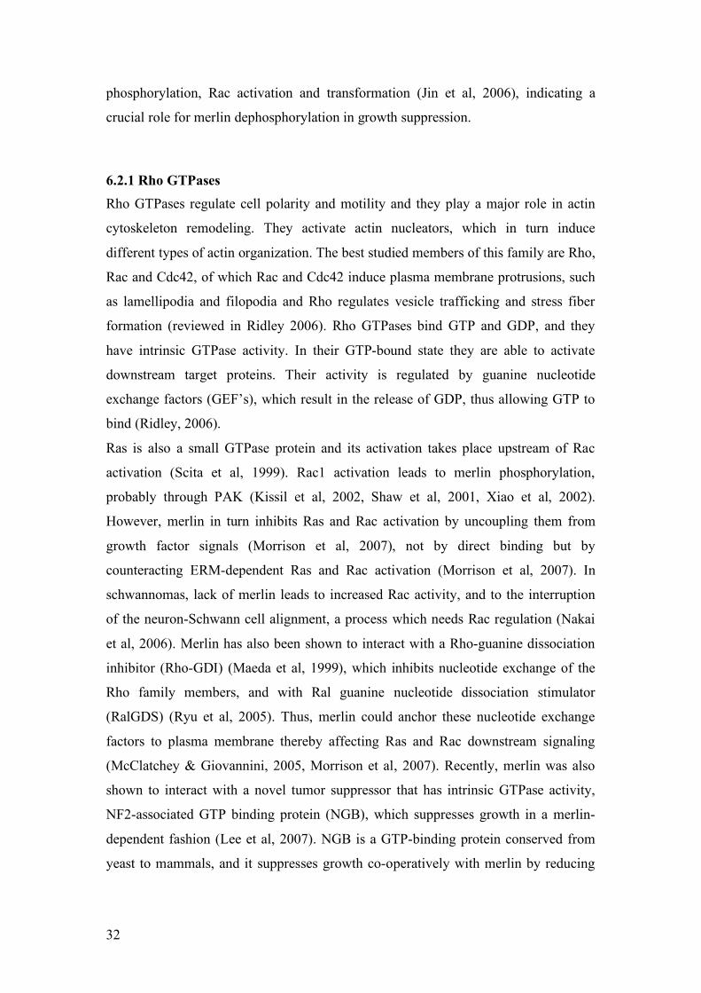
phosphorylation, Rac activation and transformation (Jin et al, 2006), indicating a
crucial role for merlin dephosphorylation in growth suppression.
6.2.1 Rho GTPases Rho GTPases regulate cell polarity and motility and they play a major role in actin
cytoskeleton remodeling. They activate actin nucleators, which in turn induce
different types of actin organization. The best studied members of this family are Rho,
Rac and Cdc42, of which Rac and Cdc42 induce plasma membrane protrusions, such
as lamellipodia and filopodia and Rho regulates vesicle trafficking and stress fiber
formation (reviewed in Ridley 2006). Rho GTPases bind GTP and GDP, and they
have intrinsic GTPase activity. In their GTP-bound state they are able to activate
downstream target proteins. Their activity is regulated by guanine nucleotide
exchange factors (GEF’s), which result in the release of GDP, thus allowing GTP to
bind (Ridley, 2006).
Ras is also a small GTPase protein and its activation takes place upstream of Rac
activation (Scita et al, 1999). Rac1 activation leads to merlin phosphorylation,
probably through PAK (Kissil et al, 2002, Shaw et al, 2001, Xiao et al, 2002).
However, merlin in turn inhibits Ras and Rac activation by uncoupling them from
growth factor signals (Morrison et al, 2007), not by direct binding but by
counteracting ERM-dependent Ras and Rac activation (Morrison et al, 2007). In
schwannomas, lack of merlin leads to increased Rac activity, and to the interruption
of the neuron-Schwann cell alignment, a process which needs Rac regulation (Nakai
et al, 2006). Merlin has also been shown to interact with a Rho-guanine dissociation
inhibitor (Rho-GDI) (Maeda et al, 1999), which inhibits nucleotide exchange of the
Rho family members, and with Ral guanine nucleotide dissociation stimulator
(RalGDS) (Ryu et al, 2005). Thus, merlin could anchor these nucleotide exchange
factors to plasma membrane thereby affecting Ras and Rac downstream signaling
(McClatchey & Giovannini, 2005, Morrison et al, 2007). Recently, merlin was also
shown to interact with a novel tumor suppressor that has intrinsic GTPase activity,
NF2-associated GTP binding protein (NGB), which suppresses growth in a merlin-
dependent fashion (Lee et al, 2007). NGB is a GTP-binding protein conserved from
yeast to mammals, and it suppresses growth co-operatively with merlin by reducing

33
Cyclin D1 levels and increases merlin turnover by reducing its ubiquitinylation (Lee
et al, 2007).
6.3 Merlin regulation of signaling pathways Merlin is implicated in the regulation of several proteins that affect cell-signaling
pathways (Fig. 8). Focal adhesion kinase (FAK) is up-regulated in many cancers and
promotes cell migration and invasion. Down-regulation of merlin in mouse embryonic
fibroblasts leads to increased spreading and invasion, whereas merlin expression leads
to increased FAK phosphorylation. This, in turn, disrupts its interaction with the
regulatory subunit of PI3K, p85 and Src (Poulikakos et al, 2006), an interaction
required for cell migration (Shen & Guan, 2001).
Recently, merlin, together with tumor suppressor protein Expanded, a related FERM-
domain protein, were found to negatively regulate the Hippo signaling pathway in D.
melanogaster (Hamaratoglu et al, 2006). The Hippo signaling pathway consists of a
kinase cascade that ultimately leads to the inhibition of a transcriptional co-activator
Yorkie, which normally controls cell growth, survival and proliferation (reviewed in
Edgar 2006). Merlin and Expanded activate Hippo/Salvador kinase in response to a
yet unidentified signal from a membrane receptor. The activated kinase complex then
phosphorylates its downstream target, the Warts/Mats kinase complex, which in turn
phosphorylates Yorkie. In its phosphorylated state Yorkie is inactive and inhibits
transcription (Edgar, 2006, Hamaratoglu et al, 2006). This signaling cascade forms a
negative feedback loop, as Yorkie also regulates transcription of merlin and Expanded
(Hamaratoglu et al, 2006). All components of this pathway are conserved in humans,
but so far it is only the lack of merlin that causes tumors.
Merlin has also been shown to interact with a novel cytoskeletal protein, magicin
(merlin and Grb2 interacting cytoskeletal protein) (Wiederhold et al, 2004), also
known as MED28 (Sato et al, 2004). Merlin forms a complex with magicin and Grb2,
that can regulate receptor tyrosine kinase and Ras signaling (Wiederhold et al, 2004).
Merlin also interacts with the PI3-kinase enhancer (PIKE-L), which is a brain specific
GTPase that binds PI3-kinase and increases its lipid kinase activity. By binding PIKE-
L merlin inhibits the PIKE-L:PI3-kinase interaction, thus inhibiting the PI3-kinase.

34
Interestingly, knocking down PIKE-L abolishes merlin’s tumor suppressor activity in
RT4 rat schwannoma cells (Rong et al, 2004b).
Lack of merlin leads to upregulation of both phosphorylated extracellular-signal
regulated kinase (ERK) (Chadee et al, 2006, Jung et al, 2005, Lim et al, 2006,
Rangwala et al, 2005), c-Jun N-terminal kinase (JNK) and PAK (Chadee et al, 2006,
Kaempchen et al, 2003). Upon phosphorylation ERK translocates into the nucleus
where it activates downstream targets, which ultimately leads to cell growth and
proliferation. Activation of the Rac1-PAK-JNK-pathway also leads to transcriptional
activation and cell proliferation.
Figure 8. Merlin signaling pathways. Extracellular growth-factors bind to growth-factorreceptors. This results in various downstream signaling cascades, that merlin is able toregulateor participate. These signaling cascades ultimately lead to the activation or repressionof transcription. (GPCR (G-protein coupled receptor) conveys signals that lead to activationof adenylyl cyclase and increase in cAMP. It also activates phospholipase C, Src and PI3K,which then leads to Raf, Ras and Akt activation (not shown)). These molecules represent onlya fraction of all the proteins that take part and regulate these processes.
Thus, merlin’s role in cell proliferation includes participation in several signaling
pathways that merlin is able to regulate, perhaps by affecting phosphorylation or
interactions of signaling cascade partners at the cell membrane. Despite all the
studies, the precise mechanism of action how merlin performs its specific functions in

35
a spatio-temporal manner and in different tissues remains unclear. Clarifying these
mechanisms would be of great importance to cancer biology as this knowledge may
translate into therapeutic opportunities in the treatment of NF2 patients.

36
AIMS OF THE STUDY
At the beginning of this study, merlin was considered as a unique type of tumor
suppressor, which performs its function at the membrane. However, preliminary
observations of merlin’s localization during different stages of the cell cycle led us to
challenge this view. Therefore I set out to study what additional functions merlin
could have, and what purpose would these novel functions serve. The specific aims of
this study were:
1) To study merlin’s localization during different stages of the cell cycle and to
determine the mechanisms and consequences of merlin’s nucleo-cytoplasmic
shuttling.
2) To identify interaction partners for merlin in the nucleus, and to study the
functional relevance of these interactions.
3) To study merlin’s association with the microtubule cytoskeleton, and to
examine merlin’s role in the regulation of the microtubule cytoskeleton in
primary mouse Schwann cells and other relevant cell types.
4) To further study the role of phosphorylation in the regulation of merlin, and
more specifically, to analyze the consequences of N-terminal phosphorylation
of merlin by PKA.

37
MATERIALS AND METHODS
The materials and methods used in this study are listed below, and are described in
detail in the original publications, which are referred to here by using their Roman
numerals.
Antibodies and probes used in this study:
NAME DESCRIPTION REFERENCE USED IN
A-19 (sc-331) Merlin pAb Santa Cruz Biotechnology I, II, III,
IV
C-18 (sc-332) Merlin pAb Santa Cruz Biotechnology
1398 NF2 Merlin pAb (den Bakker et al, 1995a) I
schwannomin Merlin pAb (Lutchman & Rouleau, 1995) I
HM2175 Merlin pS518 pAb (Kissil et al, 2002) I
1C4 Merlin mAb (Gonzalez-Agosti et al, 1996) II
KF10 Merlin mAb (den Bakker et al, 1995b) I, II
α-tubulin α-tubulin mAb Sigma-Aldrich III, IV
β-tubulin β-tubulin mAb Sigma-Aldrich III
N356 mAb α-tubulin Amesham Pharmacia
Biotech.
I, II
Acetyl-tubulin Acetylated tubulin
mAb
Sigma-Aldrich III
α-HEI10 HEI10 pAb (Toby et al, 2003) II
α-porin α-porin mAb Invitrogen-Molecular Probes I
ERK2 ERK2 pAb Santa Cruz Biotechnology
p75 p75 mAb (Huber & Chao, 1995) III
MAT-1 fl-309 Mat1 pAb Santa Cruz Biotechnology I
Hermes CD44 mAb (Jalkanen et al, 1987) I
α-Myc Myc mAb Covance Research Products I
α-HA HA mAb Roche I
GST GST pAb goat GE Healthcare

38
X63 mAb, negative control ATCC I
TO-PRO 3 Nucleic acid stain Invitrogen-Molecular Probes I, II
phalloidin Oregon green and
rhodamine conjugated
Invitrogen-Molecular Probes I, IV
Cell lines used in this study:
CELL LINE DESCRIPTION SOURCE USED IN
U2OS Human osteosarcoma ATCC I, II
U251MG Human glioma Japan cell line collection I, II, III
293HEK Human kidney epithelial ATCC I, II
293A Human kidney epithelial Stratagene III, IV
RT4 Rat schwannoma (Morrison et al, 2001) II
RT4-5-4 Rat schwannoma (Morrison et al, 2001) II
Schwann Human primary (Hanemann et al, 1998) II
Schwannoma Human primary (Rosenbaum et al, 1998) II
Schwann Mouse primary (Manent et al, 2003) III, IV
COS-7 Monkey kidney ATCC IV
Nf2 -/- MEFs Mouse embryonic
fibroblasts
(Giovannini et al, 2000) IV
Expression vectors used in this study:
PLASMID DESCRIPTION AND INSERTION SOURCE USED IN
pEF-BOS
T402E
PAK2
Constitutively active PAK2 with myc-
HA tag
(Alfthan et al,
2004)
I
pEBB-ME
PAK2 1-248
H82/85L
Dominant negative PAK2 with myc tag (Alfthan et al,
2004)
I
pcDNA3 Mammalian expression vector
containing various merlin (aa): 1-595,
Invitrogen I, II, IV

39
1-590, 1-547, 1-537, 1-569, 1-587, 1-
314, 492-595, 1-547/ins307 and HEI10
constructs
pYESTrp2 Yeast two hybrid prey vector with
merlin (aa) 1-547 construct with
transposon mutations
Invitrogen II
pGEX4T-1 Bacterial expression vector containing
several merlin (aa): 1-314, 314-595,
492-595, 492-595 S518A, 1-100, 492-
547, ezrin 1-585 and HEI10: 1-277,
46-277, constructs
Amersham
Biosciences
II, III, IV
JG4-5 Yeast two hybrid prey vector
containing various HEI10 constructs
(aa): 1-277, 1-143, 1-200, 40-250, 116-
277, 175-277
(Gyuris et al,
1993)
II, IV
EG202 Yeast two hybrid bait vector containing
various merlin constructs (aa): 1-595,
1-595 S518A, 1-595 S518D, 1-595
T576A, 1-595 T576D, 1-590, 1-339, 1-
546, 252-595, 252-595 A468P, 252-
595 L316W, 252-595 L316T, 339-595,
306-478, 492-595 and ezrin (aa): 1-
585, 1-309, 278-585
(Gyuris et al,
1993)
II, IV
pEGFP-Tub Mammalian expression vector
containing tubulin-GFP
Invitrogen III
pBaculoGold Baculovirus vestor containing merlin
isoform 1 and (aa) 1-547
BD
Biosciences
III
pShuttle Shuttle vector for adenovirus vector
pAdEasy containing merlin isoform 1,
merlin isoform 1 S10A/S10D
Stratagene III, IV
pAdEasy Adenovirus vector with merlin isoform
1, isoform 1 S10A/S10D
Stratagene III, IV
pENTR3C Gateway entry vector with merlin Invitrogen IV

40
isoform 1
pDEST-27 Gateway target vector containing
merlin isoform 1
Invitrogen IV
GFP-α-
actinin
Mammalian expression vector
containing GFP-α-actinin
(Hotulainen &
Lappalainen,
2006)
IV
Methods used in this study:
METHOD USED IN STUDY
Cytochalasin D treatment of cells I
Subcellular fractionation I
Flow and laser scanning cytometry I, II
Cell culture I, II, III, IV
Cell transfections I, II, III, IV
Production of recombinant DNA constructs I, II, III, IV
Cell cycle synchronization I, II, III
Immunofluorescence and laser scanning confocal microscopy I, II, III, IV
Western blot analysis and immunoblotting I, II, III, IV
Co-immunoprecipitation I, II, IV
Insertion of random mutations by transposon insertion system II
Primary culture of Schwann and schwannoma cells II, III
Production of recombinant protein in E.coli II, III, IV
Yeast two hybrid analysis II, IV
Production of recombinant protein by in vitro translation II, III
Site-specific mutagenesis of DNA constructs II, IV
Protein affinity precipitation II, IV
Tubulin purification from bovine brain III
Tubulin pull-down assay III
Baculovirus production III
Production of recombinant protein in insect cells III
Fluorescence recovery after photobleaching (FRAP) III

41
Deconvolution and image analysis III
In vitro and in vivo tubulin polymerization assay III
Live cell imaging III, IV
Adenovirus production III, IV
In vitro phosphorylation assay III, IV
Adenovirus infections III, IV
Latrunculin B treatment of cells IV
Production of recombinant protein in mammalian cells IV
Wound healing assay IV
Metabolic labeling of mammalian cells IV

42
RESULTS AND DISCUSSION
1. Nucleo-cytoplasmic shuttling of merlin (I)
In our earlier studies we had seen a subset of cells display nuclear localization of
endogenous merlin. The vast majority of tumor suppressors localize to the nucleus, at
least in some phases of the cell cycle (Fabbro & Henderson, 2003). Merlin, however,
was considered a cytoskeletal tumor suppressor. Previous studies had shown that
some rare isoforms of merlin localize to the nucleus (Kressel & Schmucker, 2002,
Schmucker et al, 1999), however, these studies were done using merlin mutants that
were later shown to be degraded by the ubiquitin-proteasome pathway (Gautreau et al,
2002). We decided to study the localization of merlin further and used two different
cell lines, U2OS osteosarcoma cells and U251 glioma cells, which contain detectable
amounts of endogenous merlin (article I, Fig. 1 and 2). Since only 15% of the cells
contained nuclear merlin we first studied whether we could increase the amount of
merlin in the nucleus by inhibiting chromosome region maintenance 1
(CRM1)/exportin-dependent nuclear export with Leptomycin B. This treatment
increased the amount of nuclear merlin significantly, indicating that merlin is
dependent on CRM1/exportin nuclear export (I, Fig. 3). As only a small subset of
cells displayed nuclear localization in normal growth conditions, we wanted to see
whether this is dependent on cell cycle stage or confluency. We tested this by
synchronizing the cell cycle or growing cell cultures to confluency and analyzing the
cells for nuclear merlin. According to our results merlin enters the nucleus after
mitosis, at early G1 and leaves the nucleus as the G1 phase proceeds (I, Fig 4 and 5).
Confluent cells were completely devoid of merlin. However, merlin still shuttled to
the nucleus in these cells since Leptomycin B treatment caused merlin to accumulate
into the nucleus. This indicates that the export rate of merlin exceeds that of the
import rate in confluent cells.
Mitotic cells have a round shape. After mitosis they gain their normal shape and
adhere more firmly to the substratum, which reminds of the situation of cell adhesion.
We therefore tested whether it is the adhesion that plays a role in the nuclear
localization of merlin during cell cycle, not necessarily the cell cycle stage per se.
Indeed we saw merlin localizing to the nucleus of adhering interphase cells, which
would suggest that after mitosis, adherence induces merlin’s nuclear localization (I,

43
Fig. 7). However, in contrast to ERK, this localization was not dependent on an intact
actin cytoskeleton (I, Fig. 8) since merlin localized to the nucleus of adhering cells
even if the actin cytoskeleton was disrupted by Cytochalasin D treatment. We also
studied whether merlin phosphorylation plays a role in nuclear shuttling. In nuclear
extracts, both phosphorylated forms of merlin could be found, and stimulation of
PKA or PAK did not inhibit nuclear localization, suggesting that nuclear localization
is not, at least directly, regulated by phosphorylation of serine 518 (I, Fig. 6).
Our finding that, like so many other tumor suppressor, also merlin localizes to the
nucleus is interesting since merlin is considered to be an essential part of the
cytoskeleton. Why would a cytoskeletal protein undergo nucleo-cytoplasmic
shuttling? Of interest are results where merlin-binding partners such as paxillin,
syntenin (Aplin & Juliano, 2001), PIKE-L (Rong et al, 2004b) and magicin/MED28
(Lee et al, 2006b, Sato et al, 2004) have been shown to undergo nucleo-cytoplasmic
shuttling or which have been implicated in transcriptional or translational regulation.
For instance paxillin and syntenin have been shown to play a role in transcription
(Aplin & Juliano, 2001), but it is unclear whether merlin regulates these functions.
One possibility for studying whether merlin regulates paxillins nuclear functions,
would be to inhibit the merlin-paxillin interaction. Paxillin has been shown to bind
polyA-binding protein 1 (PABP1) and to enhance PABP1 bound mRNA export from
the nucleus (Woods et al, 2005). Paxillin targets mRNA to focal adhesions, where
protein translation takes place. Localized, efficient protein translation also has an
important role in cell migration (Woods et al, 2005). If merlin plays a role in paxillin
dependent mRNA transport, then either inhibiting the merlin-paxillin interaction or
creating a stable merlin-paxillin interaction at the membrane, could have role in
paxillin dependent mRNA transport, focal adhesion formation and cell migration.
Recently, another binding partner of merlin, actin, was shown to localize to the
nucleus and control gene expression (Vartiainen et al, 2007).
The role of merlin in the nucleus and its nucleo-cytoplasmic shuttling is still unclear.
However, increasing evidence suggests that the nucleus is not merely a convenient
compartment where cytoskeletal proteins can be relocated when they are no longer
needed in their more conventional locations (e.g. plasma membrane), but they also
have other functions in the nucleus, perhaps independent from their cytoplasmic roles.
In the case of merlin this could be tested by transfecting cells with a form of merlin,
which contains a mutated cytoplasmic retention and nuclear export signals (Kressel &

44
Schmucker, 2002). This merlin would be trapped in the nucleus and its effects on cell
cycle and migration could be studied. Another possibility would be to mutate
merlin’s, yet unidentified, nuclear import signal.
2. Interaction of merlin with a cell cycle-regulator HEI10 (II)
In a yeast-two hybrid screen in search for merlin-interacting proteins, we identified a
novel binding partner for merlin, a cyclin B-binding protein and cell cycle regulator
HEI10 that has been shown to localize to the nucleus (Toby et al, 2003). Our aim was
to study whether HEI10 could be a nuclear binding partner for merlin and to study the
functional consequences of the merlin-HEI10 interaction. HEI10 had been previously
characterized as a protein capable of enhancing invasion in yeast. It is a cell cycle
regulator both in yeast and mammals, controlling the accumulation of cyclin B.
HEI10 is a 277 amino acid protein containing an N-terminal RING-finger motif, often
found in E3 ubiquitin ligases, a coiled-coil domain and a C-terminal domain which is
phosphorylated by cyclin B/Cdc2. HEI10 interacts with the UbcH7 E2 ubiquitin
conjugating enzyme and with cyclin B and regulates its degradation (Toby et al,
2003). A recent publication shows that HEI10 also negatively regulates cell migration
and metastasis, as HEI10 depleted cells migrate and invade more efficiently than wild
type cells. In addition, depletion of HEI10 in these cells results in upregulation of
p130Cas, paxillin, Cdk1 and cyclin B2, but not merlin (Singh et al, 2007).
We showed that merlin and HEI10 interact through their coiled-coil α-helical regions,
which are domains frequently involved in protein interactions (II, Fig. 1-3). In
addition, we analyzed merlin and HEI10 localization in U2OS osteosarcoma cells in
which both proteins are expressed endogenously. The two proteins localize
underneath the cell membrane during the entire cell cycle, and in a small subset of
cell, they also colocalize in the nucleus (II, Fig. 4 and 5). HEI10 is expressed mostly
in the nucleus (also during S-phase), whereas merlin and HEI10 colocalize in the
nucleus only after mitosis at G1 and during cell attachment. We also studied whether
merlin could influence HEI10 expression. In RT4-5-4 rat schwannoma cells and
human Schwann (expressing merlin) and schwannoma (no merlin expression) cells
that also express HEI10. Expression of merlin translocates HEI10 from the nucleus to
the cell membrane (II, Fig. 6). In addition, we tested whether merlin expression
influences HEI10 protein amount in 293HEK cells. When cells were transfected with

45
a mutant merlin lacking the C-terminus (merlin 1-547), the amount of HEI10 protein
was reduced by 59% (P<0.01), and with the wild type protein, HEI10 amount was
reduced by 23% (P<0.05). This degradation was seen after 72 hours of transfection
(II, Fig. 7), which might indicate that merlin influences HEI10 degradation only when
cells are beginning to reach contact inhibition of growth.
In this study we identified HEI10, a cell cycle and cell migration regulator, as a
merlin binding partner. This is the first potential binding partner for merlin in the
nucleus, and the two proteins also colocalize during mitosis at the centrosomes.
HEI10 regulates cell cycle by degrading cyclin B, whereas merlin regulates HEI10
levels. This provides a link for merlin to cell cycle regulation, worth further
investigation. It would be of interest to express a merlin construct that would
stimulate constitutive HEI10 degradation, to see whether this would have any impact
on cyclin B degradation or on cell cycle progression during mitosis. It is shown that
depletion of HEI10 leads to enhanced motility and migration and up-regulation of
several pro-motility genes (Singh et al, 2007). Does the depletion of the whole protein
mimic the situation where its amount is regulated by degradation? Perhaps merlin is
required for the regulation of HEI10, that it is then needed for efficient cyclin B
degradation at the end of mitosis. This could be tested by studying MEF’s lacking
merlin; do these cells have altered cell cycle or cyclin B levels with or without HEI10
expression?
Whether these two proteins act together at the membrane or in the nucleus to govern
cell cycle progression, is still unknown. Our unpublished data shows that merlin and
HEI10 could together influence cell cycle progression, by blocking the cell cycle
either in G1 or S phase. Constitutively open merlin and HEI10 block the cell cycle in
S phase, whereas wild type merlin together with HEI10 block it in G1 even more
strongly than merlin alone. Another interesting piece of data is the finding that HEI10
expression is different in tumors and in normal tissue. HEI10 is up-regulated in
melanomas (Smith et al, 2004) and a translocation involving HEI10 has been reported
in uterine leiomyoma (Mine et al, 2001). It would be of interest to study whether this
up-regulation in HEI10 coincides with the lack of merlin in tumors.

46
3. Merlin regulates the microtubule cytoskeleton of primary
Schwann cells (III)
Another common feature of tumor suppressors, in addition to their nuclear
localization, is their ability to localize to mitotic structures and to interact with
microtubules (Fisk et al, 2002, Hergovich et al, 2003). For example Von Hippel
Lindau tumor suppressor protein (pVHL) interacts with microtubules and stabilizes
them against depolymerization. In addition, patient mutations residing in the tubulin-
binding area of pVHL influence its tubulin binding ability, suggesting that at least
some of the patient phenotypes are caused by its inability to bind tubulin properly
(Hergovich et al, 2003, Lolkema et al, 2004). We had previously seen merlin localize
to mitotic structures (I, II) and this led us to study merlin in context with
microtubules. Merlin has previously been shown to bind microtubules in vitro
(Stokowski & Cox, 2000, Xu & Gutmann, 1998), however these studies have been
controversial and no additional in vivo evidence has been presented. Therefore, we
wanted to further investigate the merlin-tubulin interaction.
In this study we identified two tubulin-binding sites in merlin, one in the N-terminus
(aa 1-100) and other in the C-terminus (aa 492-547) of merlin (III, Fig. 2). We saw
the two proteins colocalize in the centrosomes and mitotic spindle during mitosis and
in the mid-body during cytokinesis in U251 glioma cells (III, Fig. 1). Merlin is able to
form a closed conformation by intramolecular association, which blocks many of its
binding sites (Grönholm et al, 1999). We therefore studied whether intramolecular
association or serine 518 phosphorylation, thought to open the molecule, could
regulate merlin-tubulin binding. Merlin’s intramolecular association, and also
phosphorylation of the S518, inhibited merlin-tubulin binding, implicating a more
complex mechanism for merlin activation than simply phosphorylation of S518 (III,
Fig. 3). This would suggest that merlin exists in a conformation where it is open but
unphosphorylated on S518.
Many microtubule-associated proteins regulate tubulin polymerization and, according
to this study, also merlin modulates microtubule polymerization in an in vitro assay
by enhancing the rate of polymerization (III, Fig. 4). It is of interest, that the full-
length protein was required for this effect, whereas C-terminally deleted mutant
merlin (aa 1-547) could not induce polymerization, even though it contains both

47
identified tubulin binding sites, which further shows the importance of the correct
regulation of merlin.
To gain further in vivo insight in the merlin-tubulin interaction we used Nf2 -/-
primary mouse Schwann cells (Giovannini et al, 2000). In these cells no clear
colocalization with adeno-infected wild type merlin and tubulin could be seen. Only
partial colocalization was seen under the cell membrane where both proteins resided
(III, Fig. 1). However, when these cells lost merlin, their microtubule cytoskeleton
became disorganized, and the cell morphology changed dramatically (Fig. 9); the cells
lost their spindle shape and obtained a more spread morphology (III, Fig. 5).
Figure 9. Mouse schwann cells expressing merlin (left) and lacking merlin (right). α-tubulin staining, 1000x magnification.
This change in the microtubule cytoskeleton is probably due to differences in
microtubule dynamics, which has been shown previously to be responsible for fluid
sheer stress dependent cell shape change (Malek & Izumo, 1996). The Nf2 -/-
Schwann cells contained more soluble tubulin in Triton-X 100 lysates. However, the
overall tubulin amount in -/- and merlin expressing cells was similar when analyzed
from 6M Urea lysates (III, Fig. 5). This might suggest that more tubulin is associated
with microtubules in merlin expressing cells. We then tested whether merlin enhances
microtubule polymerization also in vivo. Merlin expression increased the tubulin
polymerization rates in Schwann cells treated with Nocodazole, a drug that
depolymerizes microtubules, since they were able to re-polymerize their microtubules
faster than the -/- cells. Using FRAP-analysis we could also see that merlin expressing
Schwann cells had enhanced microtubule dynamics (III, Fig. 6).

48
According to our study, merlin-tubulin binding is under strict regulation. These
proteins colocalize only briefly and it still remains unclear if this interaction affects
merlin’s tumor suppressor function. One possibility is in the regulation of
endocytosis. Microtubules regulate endocytic pathways (Murray & Wolkoff, 2003,
Petiot et al, 2003) and it has been shown that also merlin plays a role in endocytic
processes by localizing to endocytic structures and regulating growth factor receptor
availability (Fraenzer et al, 2003, Lloyd et al, 2002, Maitra et al, 2006, Scoles et al,
2000, Curto et al, 2007). Endocytic processes regulate growth-factor availability and
recycling, and it has been shown that also microtubules play a role in these processes.
Fast and slow microtubules sort their vesicles into different compartments, fast ones
more often deliver their cargo to lysosomes for degradation than the slow ones. This
rapid movement and maturation is completely dependent on microtubules; if
microtubules are disrupted, the dynamic vesicles will non-selectively join all early
endosomes and are less efficiently degraded (Lakadamyali et al, 2006). We
hypothesize that merlin could function as a linker at the plasma membrane, helping
receptor-containing endocytosed vesicles to attach to microtubules. In cells lacking
merlin, this receptor recycling might be slower, and the growth factor receptors would
be cleared from the membrane less efficiently. Merlin could have a dual role at the
membrane of Schwann cells, partly by transferring early endocytic vesicles to
microtubules and partly by increasing the microtubule polymerization rate, thus
guiding them to lysosomes. Nf2 -/- and add-back Schwann cells have differences in
their growth factor receptor recycling rates (D. Lallemand, manuscript). If the merlin-
tubulin interplay is important for clearing up the receptors one would hypothesize that
Nocodazole treatment would increase the amount of receptors at the membrane of
both Schwann cell lines. However, if merlin alone is responsible for this receptor
recycling, the Nf2 -/- cells would still have more receptors at their membrane.
As we did not see disorganized microtubules in any other cell types lacking merlin,
this might be a way for merlin to specifically regulate Schwann cell proliferation.
4. Phosphorylation of merlin N-terminus by PKA (IV)
Our previous studies had identified a PKA phosphorylation site in the merlin FERM-
domain, in addition to the previously described C-terminal site (Alfthan et al, 2004).
It has also been shown that PKA activity increases the proliferation rate of Schwann

49
cell (Kim et al, 1997). For these reasons we wanted to study PKA mediated merlin
phosphorylation further.
In our study we identified serine 10 (S10) as the N-terminal phosphorylation site. In
addition to S10 and S518, no additional PKA phosphorylation sites were found (IV,
Fig. 1). We studied whether S10 phosphorylation affects S518 phosphorylation, but
could not see any effect when serine 10 was mutated either to alanine (A) or aspartic
acid (D) (IV, Fig. 2), mimicking the unphosphorylated and phosphorylated residue,
respectively. S518 phosphorylation has been shown to promote heterodimerization
with ezrin (Alfthan et al, 2004). However, merlin phosphorylated at S10 bound ezrin
similarly as unphosphorylated merlin (IV, Fig. 1).
The first 18 amino acids are unique to merlin. They are not included in the FERM
domain, and are indicated in the merlin binding of actin (Brault et al, 2001).
Therefore, we hypothesized that N-terminal phosphorylation could play a role in the
modulation of the merlin-actin interaction. Our first clue came from transfection
experiments where merlin S10 mutants induced more diverse morphologies than did
the wild type merlin in COS-7 and Nf2 -/- MEFs (IV, Fig. 3). Cells transfected with
merlin wild type had an increased amount of membrane extensions compared to
untransfected cells, whereas cells expressing merlin S10A displayed much elongated
extensions not seen with wild type merlin or S10D. S10D transfection resulted in a
different phenotype, producing short filopodia-like structures and rarely long
extensions seen with S10A. When the same experiment was performed with merlin
isoform II the phenotypes were even more pronounced (IV, Fig. 3), which is of
interest as it is shown to bind actin more strongly than the wild-type merlin (James et
al, 2001).
We next studied the F-actin structures in transfected Nf2 -/- MEFs (IV, Fig. 4). Cells
expressing wild type merlin had a dense, fine network of actin in lamellipodia areas.
The actin meshwork structure was seen in wild type, S10D and S518A transfected
cells; however, in S10A transfected cells the F-actin staining was reduced compared
to wild type or S10D cells and the meshwork was missing. Since it has been shown
that merlin can stabilize actin filaments (James et al, 2001) we then tested whether
merlin mutants could protect F-actin from Latrunculin B treatment (an agent that
inhibits actin polymerization). We noticed that cells expressing S10D were more
resistant to the depolymerizing effect of Latrunculin B than cells with wild type or
S10A merlin (IV, Fig. 5). We then studied whether S10 plays a role in cell motility.

50
When MEFs were transfected with various merlin constructs, cells expressing merlin
S10A were unable to produce lamellipodia and migrated slower (IV, Fig. 6).
One of merlin’s actin binding sites lies within the first 18 amino acids, however, when
we tested merlin S10A ability to directly bind actin it was not affected (unpublished
data). Therefore we hypothesize that it must be through a different mechanism than
direct actin binding, that S10 phosphorylation of merlin affects cytoskeletal
organization, lamellipodia formation and migration. This effect could be mediated
through merlin’s binding partners involved in the regulation of actin dynamics; the
most promising candidates are β-spectrin, paxillin, N-WASP and ERM-proteins, or
through the Rac-signaling pathway, but this requires further study. It will be of
interest to study how S10A mutant affects the interplay between merlin and its
binding partners.

51
CONCLUDING REMARKS
AND FUTURE PERSPECTIVES
This study began in 2002 with the observation on merlin’s surprising subcellular
localization in the nucleus and centrosomes. This direction of research seemed a bit
hazardous at the time as most research efforts were targeted to identifying kinases
phosphorylating merlin, mechanisms for its membrane:cytoskeleton interaction, and
how these interactions modulated merlin’s tumor suppressor activity. Since then, the
areas of merlin research have vastly expanded, now covering various aspects of
cellular functions that merlin is able to regulate, and identifying more and more
interaction partners for merlin. It is now acknowledged that merlin has a versatile role
in cells, perhaps depending on cell type or developmental stage of the organism.
The studies presented in this thesis have expanded merlin’s role and found alternative
functions that might explain why schwannomas are the typical hallmark of the NF2
disease. Our studies have shown that merlin actively shuttles in and out of the nucleus
(I), and more and more evidence is published, showing nuclear localization of merlin
binding partners (Aplin & Juliano, 2001, Rong et al, 2004b, Vartiainen et al, 2007).
Unfortunately our research efforts have still not answered the question of what the
functional consequence of merlin’s nucleo-cytoplasmic shuttling might be. We
hypothesize that merlin could regulate the localization of nuclear binding partners,
perhaps retaining them in or transporting them to the cytoplasm. In the case of HEI10
this could be studied by identifying and mutating the nuclear import signal in merlin,
to see whether this influences HEI10 localization and/or function. Alternatively,
merlin could, as a cytoskeletal scaffolding protein, participate in re-building the
nuclear envelope, after cell division, based on its nuclear localization at G1. To this
point we have no evidence that merlin itself would bind RNA or DNA directly, but
this needs further investigation. In our study of the HEI10-merlin interaction, we
discovered that these two proteins can localize to the nucleus simultaneously,
however, so far we have no data showing their interaction in the nucleus, since these
experiments have been technically difficult to perform. We could only show that
merlin can influence HEI10 protein levels and localization, retaining it at the
membrane. There are not many studies published on HEI10, but recently Singh et al.

52
(2007) showed that depletion of HEI10 enhances cell migration by the upregulation of
cyclin B/Cdk1. They hypothesize that merlin could relocate HEI10 in cells so that it
would not be able to enhance migration (Singh et al, 2007). Our merlin-HEI10 study
connects cyclin B to merlin. As cyclin B/Cdk1 regulates passage through M phase
and merlin is not shown to influence the progression of mitosis it is unlikely that
merlin would regulate this aspect of HEI10 function. However, our preliminary
results show that merlin has some role in mitosis or alternatively in centrosome
replication as 293HEK cells expressing constitutively open merlin display increased
amount of multipolar spindles. Whether this is through a direct mechanism or through
HEI10 is still unknown, as no role for HEI10 in causing spindle abnormalities has
been shown. It is perhaps more likely that merlin affects other HEI10 mediated
functions, such as migration or metastasis, linking the proteins to tumor formation.
The third study in this thesis expanded our early notions of merlin’s localization to
mitotic structures. This work showed for the first time, that merlin influences
microtubules, their structure and dynamics. The finding is important, since
microtubules are involved in various cellular processes, including cell growth and
division. What makes this finding even more intriguing is that we could only see
microtubule disorganization in primary Schwann cells lacking merlin, indicating that
merlin has an important role in regulating Schwann cell microtubule cytoskeleton.
This may partly explain the presence of such specific tumors, vestibular
schwannomas, in NF2 patients. However, the link between merlin, microtubule
organization and membrane traffic still needs to be clarified.
Finally, the fourth study in this thesis identified a novel phosphorylation site in
merlin. This phosphorylation seems to regulate merlin’s impact on the actin
cytoskeleton. The finding is interesting as the first 18 amino acids containing the
phosphorylated serine are not included in the FERM-domain. We believe that the
extreme N-terminal residues could form a flexible tail that participates in regulating
binding to interacting partners, depending on its phosphorylation status. Our
experiments suggest that this tail does not modulate actin cytoskeleton directly as we
could not see any difference in actin binding of the phosphorylated vs.
unphosphorylated N-terminus. However, merlin has several binding partners that are
able to modulate actin cytoskeleton, and it might be that it is through these binding
partners that merlin regulates lamellipodia formation. Another interesting aspect is
that merlin regulates PAK (Kissil et al, 2003), Rac and Ras activity (Morrison et al,

53
2007), which provides another link to cytoskeletal regulation. It will be interesting to
study, through which pathway the S10 phosphorylation performs its role.
Despite the progress in NF2 research, we are still lacking specific treatment for the
NF2 disease. This thesis work has widened the field and brought upon new avenues of
research that may eventually offer new therapeutic targets in the fight against NF2
disease. In the beginning of a study it is hard to evaluate the relevance of its outcome.
Yet, it is the basic research that lays the foundation for effective treatment and drug
design.

54
ACKNOWLEDEMENTS These studies were carried out at the University of Helsinki, Department of Pathology and Program of Molecular Neuroscience in Biomedicum Helsinki, during the years 2002-2007. I would like to express my gratitude to professors Veli-Pekka Lehto, Eero Saksela, Antti Vaheri, Olli Jänne and Tomi Mäkelä for excellent working facilities and inspiring research environment. I would also like to acknowledge the Helsinki Graduate School in Biotechnology and Molecular Biology (GSBM) for years of financial support, and for their excellent courses and Christmas parties. This theses was also financially supported by Emil Aaltonen’s Foundation, Ida Montin’s Foundation, Biomedicum Helsinki Foundation, Finnish Cancer Organization, Maire Taponen’s Foundation, Svenska Kulturfonden, Maud Kuistila’s Memorial Foundation, Oskar Öflund’s Foundation, Medicinska Understödsföreningen Liv och Hälsa, CIMO, the French Embassy in Helsinki, The Academy of Finland and the US Army Neurofibromatosis research grants DAMD17-00-0550 and W81XWH-05-1-0469. This thesis work was done in the laboratory of professor Olli Carpén. I am extremely grateful for Olli for taking me in as a summer student and leading me into the fascinating world of science. He provided an excellent research environment and offered his support when needed. He also supported our innovative (not-always-so-successful) new methods, experimental set-ups and research avenues. His guidance has always encouraged independent reasoning, letting us follow our own ideas, sometimes succeeding, sometimes failing. Making mistakes and learning from them makes an effective tutor. I am eternally indebted to my other thesis supervisor, doctor Mikaela Grönholm. Her help and collaboration have laid the foundations of this thesis. Her never-failing support and optimism, our sessions brainstorming for new ways to beat scientific obstacles and her never-ending energy (chocolate-based) have made an ever-lasting impression, which I can only admire in awe. Professor Kari Majamaa and docent Marko Kallio, are warmly acknowledged for their careful and thoughtful review of this thesis. I would especially like to thank them for all their excellent suggestions that improved this thesis. I also want to express my gratitude to my thesis committee follow-up group, professors Marikki Laiho and Ilkka Julkunen. Their comments and suggestions over the years have been very valuable; they have helped me in sticking to the timetable and also provided the final push that I needed to start writing this thesis. Today it is difficult to achieve scientific advance without deep collaborations. I extend my deepest appreciation to my collaborators abroad. Professor Oliver Hanemann and doctor Tamara Utermark are thanked for their help with the HEI10 work, which greatly improved the story. Professor Marco Giovannini is warmly acknowledged for providing us Nf2-/- cells and for letting me into his lab in Paris to learn the secrets of working with Schwann cells. I warmly thank all the personnel in Dr. Giovanninis lab for their help and encouragement. I am especially grateful to doctor Dominique Lallemand who has been a generous source of ideas and support, and whose help has been instrumental for the tubulin article. This work couldn’t have been done without the help of all the present and former members of the Ollilab. They have all been excellent in their support, providing ideas, solving problems and of course, great company in and out of the lab and on work trips around the world. In chronological order: Miku Grönholm, not only an excellent supervisor but also a great friend. I’ve never met anyone but Leena Heiska who can answer just about any question and who has used all the methods imaginable, millions of thanks for all those A4’s filled with methods that always worked (I intend to take them with me abroad). Tuula Halmesvaara always provided help if needed. Fang Zhao has been invaluable help first at tutoring me at the confocal

55
microscope and later with the help of baculoviruses. Juha Jääskeläien, not really a part of the Ollilab, but in any case responsible for getting me here today. If it hadn’t been for him, I probably would have never heard of Ollilab. I wish to thank Anu Taivainen, Paula Salmikangas, Maciej Lalowski and Luca Mologni who all worked at the Ollilab when I just started and helped me in various ways as I had never been to a lab before. During later years, when I already knew my way around, new members joined the lab, Veera Palokangas, Angela Flint, Jenni Paajanen and Hanne Ahola are all acknowledged for their help and great company, I would especially like to thank Suvi Natunen for all the help with the ever-so-scary columns and biochemistry stuff, and Kaija Alfthan for her great humor, impact on the merlin project, and establishing the concept of “karvatiede”. And finally the present members of the Ollilab, I would like to express my deepest gratitude for all the help, enjoyable coffee breaks, deep analysis of the peculiarities of human nature, and the great time we have had. I wish to thank Pernilla von Nandelstadh for her down-to-earth attitude, Heli Suila for all the help with strange problems, Monica Moza (our Master of the Mice), Pyttan Wahlström (our Turku station), Masha Melikova (3D matrix), Ville Järvi (the only guy left in the lab) and Mikko Rönty (for help with the LSC machine). I would like to give my heartfelt thanks to Helena Ahola who is absolutely amazing; she is the guru of the lab (with all the protocols, not just cloning anymore), she keeps everything rolling, and just makes life easy. Finally I would like to thank the rest of the merlin crew, Arna who just recently joined the lab, but seems to do the work of three people, she is an excellent addition to merlin crew. Minja Laulajainen, a co-author of the PKA article. Her enthusiasm, hard work, great company and sharp mind makes her an ideal collaborator, not to mention the excellent swimming pool sessions we have had when attending meetings across the Atlantic. A big thanks goes also to members of the Mäkelä lab, whenever I need weird You-Tube broadcasts, this is the place to go. Wartiovaara lab, thanks for all the help with always so strange problems that I seem to have, I would also like to thank Anu for her careful reviewing of the first article. Mika Hukkanen and Mikko Liljeström for all the possible help with the imaging stuff, which has been invaluable. I also want to thank the person who inspired me to study biology, if it hadn’t been for my teacher Raija Sarvilinna, I would have ended up as an archeologist. And finally we come to friends and family. Without your help this work could not have been done. My friends have kept me sane by taking me out of the lab and distracting my mind from work. I especially want to thank Heta, my childhood friend, who, despite the distance, remains still my friend, and Emilia whom I can always trust for company, whether it involves champagne, meatballs, movies or Lost&Found. Thank you for putting up with me. My deepest appreciation goes to my family who has been there for me always. My brothers who prevent my “upper lip from becoming too stiff” and are always ready to defend me. My parents, who have always encouraged my choices in life, without any pressure, letting me be me. And finally, to Thomas, my friend, my companion, my love, who shares my life and my passion not only to science, but also to yet finer things in life. You make every day worthwhile. Thank you,
Helsinki, November 2007.

56
REFERENCES
Alfthan K, Heiska L, Gronholm M, Renkema GH, & Carpen O (2004) Cyclic AMP-dependent protein kinase phosphorylates Merlin at serine 518 independently of P21-activated kinase and promotes Merlin-Ezrin heterodimerization. J Biol Chem
Amieva MR & Furthmayr H (1995) Subcellular localization of moesin in dynamic filopodia, retraction fibers, and other structures involved in substrate exploration, attachment, and cell-cell contacts. Exp Cell Res 219: 180-196
Amieva MR, Wilgenbus KK, & Furthmayr H (1994) Radixin is a component of hepatocyte microvilli in situ. Exp Cell Res 210: 140-144
Antinheimo J, Sankila R, Carpen O, Pukkala E, Sainio M, & Jaaskelainen J (2000) Population-based analysis of sporadic and type 2 neurofibromatosis-associated meningiomas and schwannomas. Neurology 54: 71-76
Aplin AE & Juliano RL (2001) Regulation of nucleocytoplasmic trafficking by cell adhesion receptors and the cytoskeleton. J Cell Biol 155: 187-191
Baser ME, Friedman JM, Aeschliman D, Joe H, Wallace AJ, Ramsden RT, & Evans DG (2002) Predictors of the risk of mortality in neurofibromatosis 2. Am J Hum Genet 71: 715-723
Baser ME, Kuramoto L, Woods R, Joe H, Friedman JM, Wallace AJ, Ramsden RT, Olschwang S, Bijlsma E, Kalamarides M, Papi L, Kato R, Carroll J, Lazaro C, Joncourt F, Parry DM, Rouleau GA, & Evans DG (2005) The location of constitutional neurofibromatosis 2 (NF2) splice site mutations is associated with the severity of NF2. J Med Genet 42: 540-546
Baser ME, R Evans DG, & Gutmann DH (2003) Neurofibromatosis 2. Curr Opin Neurol 16: 27-33
Bashour AM, Meng JJ, Ip W, MacCollin M, & Ratner N (2002) The neurofibromatosis type 2 gene product, merlin, reverses the F-actin cytoskeletal defects in primary human Schwannoma cells. Mol Cell Biol 22: 1150-1157
Berryman M, Gary R, & Bretscher A (1995) Ezrin oligomers are major cytoskeletal components of placental microvilli: a proposal for their involvement in cortical morphogenesis. J Cell Biol 131: 1231-1242
Bhat KR & Setaluri V (2007) Microtubule-Associated Proteins as Targets in Cancer Chemotherapy. Clin Cancer Res 13: 2849-2854
Bianchi AB, Mitsunaga SI, Cheng JQ, Klein WM, Jhanwar SC, Seizinger B, Kley N, Klein-Szanto AJ, & Testa JR (1995) High frequency of inactivating mutations in the neurofibromatosis type 2 gene (NF2) in primary malignant mesotheliomas. Proc Natl Acad Sci U S A 92: 10854-10858
Bloom J & Cross FR (2007) Multiple levels of cyclin specificity in cell-cycle control. Nat Rev Mol Cell Biol 8: 149-160
Bono P, Cordero E, Johnson K, Borowsky M, Ramesh V, Jacks T, & Hynes RO (2005) Layilin, a cell surface hyaluronan receptor, interacts with merlin and radixin. Exp Cell Res 308: 177-187

57
Boutros R, Lobjois V, & Ducommun B (2007) CDC25 phosphatases in cancer cells: key players? Good targets? Nat Rev Cancer 7: 495-507
Brault E, Gautreau A, Lamarine M, Callebaut I, Thomas G, & Goutebroze L (2001) Normal membrane localization and actin association of the NF2 tumor suppressor protein are dependent on folding of its N-terminal domain. J Cell Sci 114: 1901-1912
Bretscher A (1983) Purification of an 80,000-dalton protein that is a component of the isolated microvillus cytoskeleton, and its localization in nonmuscle cells. J Cell Biol 97: 425-432
Bretscher A, Edwards K, & Fehon RG (2002) ERM proteins and merlin: integrators at the cell cortex. Nat Rev Mol Cell Biol 3: 586-599
Chadee DN, Xu D, Hung G, Andalibi A, Lim DJ, Luo Z, Gutmann DH, & Kyriakis JM (2006) Mixed-lineage kinase 3 regulates B-Raf through maintenance of the B-Raf/Raf-1 complex and inhibition by the NF2 tumor suppressor protein. Proc Natl Acad Sci U S A 103: 4463-4468
Chang L & Goldman RD (2004) Intermediate filaments mediate cytoskeletal crosstalk. Nat Rev Mol Cell Biol 5: 601-613
Chaudhuri AR, Khan IA, Prasad V, Robinson AK, Luduena RF, & Barnes LD (1999) The tumor suppressor protein Fhit. A novel interaction with tubulin. J Biol Chem 274: 24378-24382
Chishti AH, Kim AC, Marfatia SM, Lutchman M, Hanspal M, Jindal H, Liu SC, Low PS, Rouleau GA, Mohandas N, Chasis JA, Conboy JG, Gascard P, Takakuwa Y, Huang SC, Benz EJ,Jr, Bretscher A, Fehon RG, Gusella JF, Ramesh V et al (1998) The FERM domain: a unique module involved in the linkage of cytoplasmic proteins to the membrane. Trends Biochem Sci 23: 281-282
Cimini D, Wan X, Hirel C, & Salmon ED (2006) Aurora Kinase Promotes Turnover of Kinetochore Microtubules to Reduce Chromosome Segregation Errors. 16: 1711-1718
Claudio JO, Lutchman M, & Rouleau GA (1995) Widespread but cell type-specific expression of the mouse neurofibromatosis type 2 gene. Neuroreport 6: 1942-1946
Curto M, Cole BK, Lallemand D, Liu CH, & McClatchey AI (2007) Contact-dependent inhibition of EGFR signaling by Nf2/Merlin. J Cell Biol 177: 893-903
Dallol A, Agathanggelou A, Fenton SL, Ahmed-Choudhury J, Hesson L, Vos MD, Clark GJ, Downward J, Maher ER, & Latif F (2004) RASSF1A interacts with microtubule-associated proteins and modulates microtubule dynamics. Cancer Res 64: 4112-4116
Dalmay T & Edwards DR (2006) MicroRNAs and the hallmarks of cancer. Oncogene 25: 6170-6175
den Bakker MA, Riegman PH, Hekman RA, Boersma W, Janssen PJ, van der Kwast TH, & Zwarthoff EC (1995a) The product of the NF2 tumour suppressor gene localizes near the plasma membrane and is highly expressed in muscle cells. Oncogene 10: 757-763
den Bakker MA, Tascilar M, Riegman PH, Hekman AC, Boersma W, Janssen PJ, de Jong TA, Hendriks W, van der Kwast TH, & Zwarthoff EC (1995b) Neurofibromatosis type 2 protein co-localizes with elements of the cytoskeleton. Am J Pathol 147: 1339-1349

58
Denisenko-Nehrbass N, Goutebroze L, Galvez T, Bonnon C, Stankoff B, Ezan P, Giovannini M, Faivre-Sarrailh C, & Girault JA (2003) Association of Caspr/paranodin with tumour suppressor schwannomin/merlin and beta1 integrin in the central nervous system. J Neurochem 84: 209-221
Desai A & Mitchison TJ (1997) Microtubule polymerization dynamics. Annu Rev Cell Dev Biol 13: 83-117
Doi Y, Itoh M, Yonemura S, Ishihara S, Takano H, Noda T, & Tsukita S (1999) Normal development of mice and unimpaired cell adhesion/cell motility/actin-based cytoskeleton without compensatory up-regulation of ezrin or radixin in moesin gene knockout. J Biol Chem 274: 2315-2321
Dumontet C & Sikic B (1999) Mechanisms of Action of and Resistance to Antitubulin Agents: Microtubule Dynamics, Drug Transport, and Cell Death. J Clin Oncol 17: 1061
Edgar BA (2006) From cell structure to transcription: Hippo forges a new path. Cell 124: 267-273
Evans DG, Huson SM, Donnai D, Neary W, Blair V, Teare D, Newton V, Strachan T, Ramsden R, & Harris R (1992) A genetic study of type 2 neurofibromatosis in the United Kingdom. I. Prevalence, mutation rate, fitness, and confirmation of maternal transmission effect on severity. J Med Genet 29: 841-846
Fabbro M & Henderson BR (2003) Regulation of tumor suppressors by nuclear-cytoplasmic shuttling. Exp Cell Res 282: 59-69
Fehon RG, Oren T, LaJeunesse DR, Melby TE, & McCartney BM (1997) Isolation of mutations in the Drosophila homologues of the human Neurofibromatosis 2 and yeast CDC42 genes using a simple and efficient reverse-genetic method. Genetics 146: 245-252
Fernandez-Valle C, Tang Y, Ricard J, Rodenas-Ruano A, Taylor A, Hackler E, Biggerstaff J, & Iacovelli J (2002) Paxillin binds schwannomin and regulates its density-dependent localization and effect on cell morphology. Nat Genet 31: 354-362
Fisk HA, Mattison CP, & Winey M (2002) Centrosomes and tumour suppressors. Curr Opin Cell Biol 14: 700-705
Fraenzer JT, Pan H, Minimo L,Jr, Smith GM, Knauer D, & Hung G (2003) Overexpression of the NF2 gene inhibits schwannoma cell proliferation through promoting PDGFR degradation. Int J Oncol 23: 1493-1500
Franck Z, Gary R, & Bretscher A (1993) Moesin, like ezrin, colocalizes with actin in the cortical cytoskeleton in cultured cells, but its expression is more variable. J Cell Sci 105 ( Pt 1): 219-231
Fuchs E (1996) The cytoskeleton and disease: genetic disorders of intermediate filaments. Annu Rev Genet 30: 197-231
Gary R & Bretscher A (1993) Heterotypic and homotypic associations between ezrin and moesin, two putative membrane-cytoskeletal linking proteins. Proc Natl Acad Sci U S A 90: 10846-10850

59
Gautreau A, Louvard D, & Arpin M (2000) Morphogenic effects of ezrin require a phosphorylation-induced transition from oligomers to monomers at the plasma membrane. J Cell Biol 150: 193-203
Gautreau A, Manent J, Fievet B, Louvard D, Giovannini M, & Arpin M (2002) Mutant products of the NF2 tumor suppressor gene are degraded by the ubiquitin-proteasome pathway. J Biol Chem 277: 31279-31282
Giovannini M, Robanus-Maandag E, van der Valk M, Niwa-Kawakita M, Abramowski V, Goutebroze L, Woodruff JM, Berns A, & Thomas G (2000) Conditional biallelic Nf2 mutation in the mouse promotes manifestations of human neurofibromatosis type 2. Genes Dev 14: 1617-1630
Golovnina K, Blinov A, Akhmametyeva EM, Omelyanchuk LV, & Chang LS (2005) Evolution and origin of merlin, the product of the Neurofibromatosis type 2 (NF2) tumor-suppressor gene. BMC Evol Biol 5: 69
Gonzalez-Agosti C, Xu L, Pinney D, Beauchamp R, Hobbs W, Gusella J, & Ramesh V (1996) The merlin tumor suppressor localizes preferentially in membrane ruffles. Oncogene 13: 1239-1247
Gregory PE, Gutmann DH, Mitchell A, Park S, Boguski M, Jacks T, Wood DL, Jove R, & Collins FS (1993) Neurofibromatosis type 1 gene product (neurofibromin) associates with microtubules. Somat Cell Mol Genet 19: 265-274
Gronholm M, Sainio M, Zhao F, Heiska L, Vaheri A, & Carpen O (1999) Homotypic and heterotypic interaction of the neurofibromatosis 2 tumor suppressor protein merlin and the ERM protein ezrin. J Cell Sci 112 ( Pt 6): 895-904
Gronholm M, Teesalu T, Tyynela J, Piltti K, Bohling T, Wartiovaara K, Vaheri A, & Carpen O (2005) Characterization of the NF2 protein merlin and the ERM protein ezrin in human, rat, and mouse central nervous system. Mol Cell Neurosci 28: 683-693
Gronholm M, Vossebein L, Carlson CR, Kuja-Panula J, Teesalu T, Alfthan K, Vaheri A, Rauvala H, Herberg FW, Tasken K, & Carpen O (2003) Merlin links to the cAMP neuronal signaling pathway by anchoring the RIbeta subunit of protein kinase A. J Biol Chem 278: 41167-41172
Gutmann DH, Haipek CA, Burke SP, Sun CX, Scoles DR, & Pulst SM (2001) The NF2 interactor, hepatocyte growth factor-regulated tyrosine kinase substrate (HRS), associates with merlin in the "open" conformation and suppresses cell growth and motility. Hum Mol Genet 10: 825-834
Gutmann DH, Haipek CA, & Hoang Lu K (1999) Neurofibromatosis 2 tumor suppressor protein, merlin, forms two functionally important intramolecular associations. J Neurosci Res 58: 706-716 Guyris JE, Golemis E, Chertkov H & Brent R (1993) Cdi 1, a human G1 and S phase protein phosphatase that associates with Cdk2. Cell, 75, 791-803.
Hamaratoglu F, Willecke M, Kango-Singh M, Nolo R, Hyun E, Tao C, Jafar-Nejad H, & Halder G (2006) The tumour-suppressor genes NF2/Merlin and Expanded act through Hippo signalling to regulate cell proliferation and apoptosis. Nat Cell Biol 8: 27-36
Hanahan D & Weinberg RA (2000) The hallmarks of cancer. Cell 100: 57-70

60
Hanemann CO & Evans DG (2006) News on the genetics, epidemiology, medical care and translational research of Schwannomas. J Neurol 253: 1533-1541
Hanemann CO, Rosenbaum C, Kupfer S, Wosch S, Stoegbauer F, & Muller HW (1998) Improved culture methods to expand Schwann cells with altered growth behaviour from CMT1A patients. Glia 23: 89-98
Hara T, Bianchi AB, Seizinger BR, & Kley N (1994) Molecular cloning and characterization of alternatively spliced transcripts of the mouse neurofibromatosis 2 gene. Cancer Res 54: 330-335
Hartwell LH (2002) Nobel Lecture. Yeast and cancer. Biosci Rep 22: 373-394
Heiska L, Alfthan K, Gronholm M, Vilja P, Vaheri A, & Carpen O (1998) Association of ezrin with intercellular adhesion molecule-1 and -2 (ICAM-1 and ICAM-2). Regulation by phosphatidylinositol 4, 5-bisphosphate. J Biol Chem 273: 21893-21900
Helfand BT, Chang L, & Goldman RD (2004) Intermediate filaments are dynamic and motile elements of cellular architecture. J Cell Sci 117: 133-141
Hemann MT & Narita M (2007) Oncogenes and senescence: breaking down in the fast lane. Genes Dev 21: 1-5
Henry MD, Gonzalez Agosti C, & Solomon F (1995) Molecular dissection of radixin: distinct and interdependent functions of the amino- and carboxy-terminal domains. J Cell Biol 129: 1007-1022
Hergovich A, Lisztwan J, Barry R, Ballschmieter P, & Krek W (2003) Regulation of microtubule stability by the von Hippel-Lindau tumour suppressor protein pVHL. Nat Cell Biol 5: 64-70
Hirao M, Sato N, Kondo T, Yonemura S, Monden M, Sasaki T, Takai Y, Tsukita S, & Tsukita S (1996) Regulation mechanism of ERM (ezrin/radixin/moesin) protein/plasma membrane association: possible involvement of phosphatidylinositol turnover and Rho-dependent signaling pathway. J Cell Biol 135: 37-51
Hirokawa Y, Tikoo A, Huynh J, Utermark T, Hanemann CO, Giovannini M, Xiao GH, Testa JR, Wood J, & Maruta H (2004) A clue to the therapy of neurofibromatosis type 2: NF2/merlin is a PAK1 inhibitor. Cancer J 10: 20-26
Hotulainen P & Lappalainen P (2006) Stress fibers are generated by two distinct actin assembly mechanisms in motile cells. J Cell Biol 173: 383-394
Howe DG & McCarthy KD (2000) Retroviral inhibition of cAMP-dependent protein kinase inhibits myelination but not Schwann cell mitosis stimulated by interaction with neurons. J Neurosci 20: 3513-3521
Huang L, Ichimaru E, Pestonjamasp K, Cui X, Nakamura H, Lo GY, Lin FI, Luna EJ, & Furthmayr H (1998) Merlin differs from moesin in binding to F-actin and in its intra- and intermolecular interactions. Biochem Biophys Res Commun 248: 548-553
Huber LJ & Chao MV (1995) Mesenchymal and neuronal cell expression of the p75 neurotrophin receptor gene occur by different mechanisms. Dev Biol 167: 227-238

61
Hughes SC & Fehon RG (2006) Phosphorylation and activity of the tumor suppressor Merlin and the ERM protein Moesin are coordinately regulated by the Slik kinase. J Cell Biol 175: 305-313
Hunt T (2002) Nobel Lecture. Protein synthesis, proteolysis, and cell cycle transitions. Biosci Rep 22: 465-486
Huynh DP, Tran TM, Nechiporuk T, & Pulst SM (1996) Expression of neurofibromatosis 2 transcript and gene product during mouse fetal development. Cell Growth Differ 7: 1551-1561
Jalkanen S, Bargatze RF, de los Toyos J, & Butcher EC (1987) Lymphocyte recognition of high endothelium: antibodies to distinct epitopes of an 85-95-kD glycoprotein antigen differentially inhibit lymphocyte binding to lymph node, mucosal, or synovial endothelial cells. J Cell Biol 105: 983-990
James MF, Manchanda N, Gonzalez-Agosti C, Hartwig JH, & Ramesh V (2001) The neurofibromatosis 2 protein product merlin selectively binds F-actin but not G-actin, and stabilizes the filaments through a lateral association. Biochem J 356: 377-386
Jannatipour M, Dion P, Khan S, Jindal H, Fan X, Laganiere J, Chishti AH, & Rouleau GA (2001) Schwannomin isoform-1 interacts with syntenin via PDZ domains. J Biol Chem 276: 33093-33100
Jiang X & Yeung RS (2006) Regulation of microtubule-dependent protein transport by the TSC2/mammalian target of rapamycin pathway. Cancer Res 66: 5258-5269
Jin H, Sperka T, Herrlich P, & Morrison H (2006) Tumorigenic transformation by CPI-17 through inhibition of a merlin phosphatase. Nature 442: 576-579
Jindal HK, Yoshinaga K, Seo PS, Lutchman M, Dion PA, Rouleau GA, Hanada T, & Chishti AH (2006) Purification of the NF2 tumor suppressor protein from human erythrocytes. Can J Neurol Sci 33: 394-402
Jung JR, Kim H, Jeun SS, Lee JY, Koh EJ, & Ji C (2005) The Phosphorylation status of merlin is important for regulating the Ras-ERK pathway. Mol Cells 20: 196-200
Kaempchen K, Mielke K, Utermark T, Langmesser S, & Hanemann CO (2003) Upregulation of the Rac1/JNK signaling pathway in primary human schwannoma cells. Hum Mol Genet 12: 1211-1221
Kahl CR & Means AR (2004) Calcineurin regulates cyclin D1 accumulation in growth-stimulated fibroblasts. Mol Biol Cell 15: 1833-1842
Kalamarides M, Niwa-Kawakita M, Leblois H, Abramowski V, Perricaudet M, Janin A, Thomas G, Gutmann DH, & Giovannini M (2002) Nf2 gene inactivation in arachnoidal cells is rate-limiting for meningioma development in the mouse. Genes Dev 16: 1060-1065
Kastan MB (2007) Wild-type p53: tumors can't stand it. Cell 128: 837-840
Kastan MB, Onyekwere O, Sidransky D, Vogelstein B, & Craig RW (1991) Participation of p53 protein in the cellular response to DNA damage. Cancer Res 51: 6304-6311
Kikuchi S, Hata M, Fukumoto K, Yamane Y, Matsui T, Tamura A, Yonemura S, Yamagishi H, Keppler D, Tsukita S, & Tsukita S (2002) Radixin deficiency causes conjugated

62
hyperbilirubinemia with loss of Mrp2 from bile canalicular membranes. Nat Genet 31: 320-325
Kim H, Kwak NJ, Lee JY, Choi BH, Lim Y, Ko YJ, Kim YH, Huh PW, Lee KH, Rha HK, & Wang YP (2004) Merlin neutralizes the inhibitory effect of Mdm2 on p53. J Biol Chem 279: 7812-7818
Kim HA, DeClue JE, & Ratner N (1997) cAMP-dependent protein kinase A is required for Schwann cell growth: interactions between the cAMP and neuregulin/tyrosine kinase pathways. J Neurosci Res 49: 236-247
Kinzler KW & Vogelstein B (1998) Landscaping the cancer terrain. Science 280: 1036-1037
Kinzler KW & Vogelstein B (1997) Cancer-susceptibility genes. Gatekeepers and caretakers. Nature 386: 761, 763
Kissil JL, Johnson KC, Eckman MS, & Jacks T (2002) Merlin phosphorylation by p21-activated kinase 2 and effects of phosphorylation on merlin localization. J Biol Chem 277: 10394-10399
Kissil JL, Wilker EW, Johnson KC, Eckman MS, Yaffe MB, & Jacks T (2003) Merlin, the product of the Nf2 tumor suppressor gene, is an inhibitor of the p21-activated kinase, Pak1. Mol Cell 12: 841-849
Knudson AG,Jr (1971) Mutation and cancer: statistical study of retinoblastoma. Proc Natl Acad Sci U S A 68: 820-823
Kodama A, Lechler T, & Fuchs E (2004) Coordinating cytoskeletal tracks to polarize cellular movements. J Cell Biol 167: 203-207
Kressel M & Schmucker B (2002) Nucleocytoplasmic transfer of the NF2 tumor suppressor protein merlin is regulated by exon 2 and a CRM1-dependent nuclear export signal in exon 15. Hum Mol Genet 11: 2269-2278
Kumar R, Gururaj AE, & Barnes CJ (2006) p21-activated kinases in cancer. Nat Rev Cancer 6: 459-471
LaJeunesse DR, McCartney BM, & Fehon RG (1998) Structural analysis of Drosophila merlin reveals functional domains important for growth control and subcellular localization. J Cell Biol 141: 1589-1599
Lakadamyali M, Rust MJ, & Zhuang X (2006) Ligands for clathrin-mediated endocytosis are differentially sorted into distinct populations of early endosomes. Cell 124: 997-1009
Lallemand D, Curto M, Saotome I, Giovannini M, & McClatchey AI (2003) NF2 deficiency promotes tumorigenesis and metastasis by destabilizing adherens junctions. Genes Dev 17: 1090-1100
Lankes WT & Furthmayr H (1991) Moesin: a member of the protein 4.1-talin-ezrin family of proteins. Proc Natl Acad Sci U S A 88: 8297-8301
Lasota J, Fetsch JF, Wozniak A, Wasag B, Sciot R, & Miettinen M (2001) The neurofibromatosis type 2 gene is mutated in perineurial cell tumors: a molecular genetic study of eight cases. Am J Pathol 158: 1223-1229

63
Lee H, Kim D, Dan HC, Wu EL, Gritsko TM, Cao C, Nicosia SV, Golemis EA, Liu W, Coppola D, Brem SS, Testa JR, & Cheng JQ (2007) Identification and characterization of putative tumor suppressor NGB, a GTP-binding protein that interacts with the neurofibromatosis 2 protein. Mol Cell Biol 27: 2103-2119
Lee IK, Kim KS, Kim H, Lee JY, Ryu CH, Chun HJ, Lee KU, Lim Y, Kim YH, Huh PW, Lee KH, Han SI, Jun TY, & Rha HK (2004a) MAP, a protein interacting with a tumor suppressor, merlin, through the run domain. Biochem Biophys Res Commun 325: 774-783
Lee JY, Kim H, Ryu CH, Kim JY, Choi BH, Lim Y, Huh PW, Kim YH, Lee KH, Jun TY, Rha HK, Kang JK, & Choi CR (2004b) Merlin, a tumor suppressor, interacts with transactivation-responsive RNA-binding protein and inhibits its oncogenic activity. J Biol Chem 279: 30265-30273
Lee JY, Moon HJ, Lee WK, Chun HJ, Han CW, Jeon YW, Lim Y, Kim YH, Yao TP, Lee KH, Jun TY, Rha HK, & Kang JK (2006a) Merlin facilitates ubiquitination and degradation of transactivation-responsive RNA-binding protein. Oncogene 25: 1143-1152
Lee MF, Beauchamp RL, Beyer KS, Gusella JF, & Ramesh V (2006b) Magicin associates with the Src-family kinases and is phosphorylated upon CD3 stimulation. Biochem Biophys Res Commun 348: 826-831
Leto TL & Marchesi VT (1984) A structural model of human erythrocyte protein 4.1. J Biol Chem 259: 4603-4608
Lim JY, Kim H, Jeun SS, Kang SG, & Lee KJ (2006) Merlin inhibits growth hormone-regulated Raf-ERKs pathways by binding to Grb2 protein. Biochem Biophys Res Commun 340: 1151-1157
Lloyd TE, Atkinson R, Wu MN, Zhou Y, Pennetta G, & Bellen HJ (2002) Hrs regulates endosome membrane invagination and tyrosine kinase receptor signaling in Drosophila. Cell 108: 261-269
Lolkema MP, Mehra N, Jorna AS, van Beest M, Giles RH, & Voest EE (2004) The von Hippel-Lindau tumor suppressor protein influences microtubule dynamics at the cell periphery. Exp Cell Res 301: 139-146
Lomas J, Bello MJ, Arjona D, Alonso ME, Martinez-Glez V, Lopez-Marin I, Aminoso C, de Campos JM, Isla A, Vaquero J, & Rey JA (2005) Genetic and epigenetic alteration of the NF2 gene in sporadic meningiomas. Genes Chromosomes Cancer 42: 314-319
Lutchman M & Rouleau GA (1995) The neurofibromatosis type 2 gene product, schwannomin, suppresses growth of NIH 3T3 cells. Cancer Res 55: 2270-2274
Maeda M, Matsui T, Imamura M, Tsukita S, & Tsukita S (1999) Expression level, subcellular distribution and rho-GDI binding affinity of merlin in comparison with Ezrin/Radixin/Moesin proteins. Oncogene 18: 4788-4797
Maitra S, Kulikauskas RM, Gavilan H, & Fehon RG (2006) The tumor suppressors Merlin and expanded function cooperatively to modulate receptor endocytosis and signaling. Curr Biol 16: 702-709
Malek A & Izumo S (1996) Mechanism of endothelial cell shape change and cytoskeletal remodeling in response to fluid shear stress. J Cell Sci 109: 713-726

64
Manchanda N, Lyubimova A, Ho HY, James MF, Gusella JF, Ramesh N, Snapper SB, & Ramesh V (2005) The NF2 tumor suppressor Merlin and the ERM proteins interact with N-WASP and regulate its actin polymerization function. J Biol Chem 280: 12517-12522
Manent J, Oguievetskaia K, Bayer J, Ratner N, & Giovannini M (2003) Magnetic cell sorting for enriching Schwann cells from adult mouse peripheral nerves. J Neurosci Methods 123: 167-173
Matsui T, Maeda M, Doi Y, Yonemura S, Amano M, Kaibuchi K, Tsukita S, & Tsukita S (1998) Rho-kinase phosphorylates COOH-terminal threonines of ezrin/radixin/moesin (ERM) proteins and regulates their head-to-tail association. J Cell Biol 140: 647-657
Matsui T, Yonemura S, Tsukita S, & Tsukita S (1999) Activation of ERM proteins in vivo by Rho involves phosphatidyl-inositol 4-phosphate 5-kinase and not ROCK kinases. Curr Biol 9: 1259-1262
Mautner VF, Baser ME, Thakkar SD, Feigen UM, Friedman JM, & Kluwe L (2002) Vestibular schwannoma growth in patients with neurofibromatosis Type 2: a longitudinal study. J Neurosurg 96: 223-228
McCartney BM & Fehon RG (1996) Distinct cellular and subcellular patterns of expression imply distinct functions for the Drosophila homologues of moesin and the neurofibromatosis 2 tumor suppressor, merlin. J Cell Biol 133: 843-852
McClatchey AI & Giovannini M (2005) Membrane organization and tumorigenesis--the NF2 tumor suppressor, Merlin. Genes Dev 19: 2265-2277
McClatchey AI, Saotome I, Ramesh V, Gusella JF, & Jacks T (1997) The Nf2 tumor suppressor gene product is essential for extraembryonic development immediately prior to gastrulation. Genes Dev 11: 1253-1265
Meng JJ, Lowrie DJ, Sun H, Dorsey E, Pelton PD, Bashour AM, Groden J, Ratner N, & Ip W (2000) Interaction between two isoforms of the NF2 tumor suppressor protein, merlin, and between merlin and ezrin, suggests modulation of ERM proteins by merlin. J Neurosci Res 62: 491-502
Mine N, Kurose K, Konishi H, Araki T, Nagai H, & Emi M (2001) Fusion of a sequence from HEI10 (14q11) to the HMGIC gene at 12q15 in a uterine leiomyoma. Jpn J Cancer Res 92: 135-139
Morrison H, Sherman LS, Legg J, Banine F, Isacke C, Haipek CA, Gutmann DH, Ponta H, & Herrlich P (2001) The NF2 tumor suppressor gene product, merlin, mediates contact inhibition of growth through interactions with CD44. Genes Dev 15: 968-980
Morrison H, Sperka T, Manent J, Giovannini M, Ponta H, & Herrlich P (2007) Merlin/neurofibromatosis type 2 suppresses growth by inhibiting the activation of Ras and Rac. Cancer Res 67: 520-527
Moshnikova A, Frye J, Shay JW, Minna JD, & Khokhlatchev AV (2006) The growth and tumor suppressor NORE1A is a cytoskeletal protein that suppresses growth by inhibition of the ERK pathway. J Biol Chem 281: 8143-8152
Murray AW (2004) Recycling the cell cycle: cyclins revisited. Cell 116: 221-234

65
Murray JW & Wolkoff AW (2003) Roles of the cytoskeleton and motor proteins in endocytic sorting. Adv Drug Deliv Rev 55: 1385-1403
Murthy A, Gonzalez-Agosti C, Cordero E, Pinney D, Candia C, Solomon F, Gusella J, & Ramesh V (1998) NHE-RF, a regulatory cofactor for Na(+)-H+ exchange, is a common interactor for merlin and ERM (MERM) proteins. J Biol Chem 273: 1273-1276
Nakai Y, Zheng Y, MacCollin M, & Ratner N (2006) Temporal control of Rac in Schwann cell-axon interaction is disrupted in NF2-mutant schwannoma cells. J Neurosci 26: 3390-3395
Nakamura F, Amieva MR, & Furthmayr H (1995) Phosphorylation of threonine 558 in the carboxyl-terminal actin-binding domain of moesin by thrombin activation of human platelets. J Biol Chem 270: 31377-31385
Nakamura F, Huang L, Pestonjamasp K, Luna EJ, & Furthmayr H (1999) Regulation of F-actin binding to platelet moesin in vitro by both phosphorylation of threonine 558 and polyphosphatidylinositides. Mol Biol Cell 10: 2669-2685
Neill GW & Crompton MR (2001) Binding of the merlin-I product of the neurofibromatosis type 2 tumour suppressor gene to a novel site in beta-fodrin is regulated by association between merlin domains. Biochem J 358: 727-735
Ng T, Parsons M, Hughes WE, Monypenny J, Zicha D, Gautreau A, Arpin M, Gschmeissner S, Verveer PJ, Bastiaens PI, & Parker PJ (2001) Ezrin is a downstream effector of trafficking PKC-integrin complexes involved in the control of cell motility. EMBO J 20: 2723-2741
Nurse P (2002) Cyclin dependent kinases and cell cycle control (nobel lecture). Chembiochem 3: 596-603
Obremski VJ, Hall AM, & Fernandez-Valle C (1998) Merlin, the neurofibromatosis type 2 gene product, and beta1 integrin associate in isolated and differentiating Schwann cells. J Neurobiol 37: 487-501
Pakkanen R, Hedman K, Turunen O, Wahlstrom T, & Vaheri A (1987) Microvillus-specific Mr 75,000 plasma membrane protein of human choriocarcinoma cells. J Histochem Cytochem 35: 809-816
Pantaloni D, Le Clainche C, & Carlier MF (2001) Mechanism of actin-based motility. Science 292: 1502-1506
Pearson MA, Reczek D, Bretscher A, & Karplus PA (2000) Structure of the ERM protein moesin reveals the FERM domain fold masked by an extended actin binding tail domain. Cell 101: 259-270
Pelton PD, Sherman LS, Rizvi TA, Marchionni MA, Wood P, Friedman RA, & Ratner N (1998) Ruffling membrane, stress fiber, cell spreading and proliferation abnormalities in human Schwannoma cells. Oncogene 17: 2195-2209
Pestonjamasp K, Amieva MR, Strassel CP, Nauseef WM, Furthmayr H, & Luna EJ (1995) Moesin, ezrin, and p205 are actin-binding proteins associated with neutrophil plasma membranes. Mol Biol Cell 6: 247-259
Petiot A, Faure J, Stenmark H, & Gruenberg J (2003) PI3P signaling regulates receptor sorting but not transport in the endosomal pathway. J Cell Biol 162: 971-979

66
Pietromonaco SF, Simons PC, Altman A, & Elias L (1998) Protein kinase C-theta phosphorylation of moesin in the actin-binding sequence. J Biol Chem 273: 7594-7603
Pineau P, Marchio A, Cordina E, Tiollais P, & Dejean A (2003) Homozygous deletions scanning in tumor cell lines detects previously unsuspected loci. Int J Cancer 106: 216-223
Pollard TD, Blanchoin L, & Mullins RD (2000) Molecular mechanisms controlling actin filament dynamics in nonmuscle cells. Annu Rev Biophys Biomol Struct 29: 545-576
Poulikakos PI, Xiao GH, Gallagher R, Jablonski S, Jhanwar SC, & Testa JR (2006) Re-expression of the tumor suppressor NF2/merlin inhibits invasiveness in mesothelioma cells and negatively regulates FAK. Oncogene
Rangwala R, Banine F, Borg JP, & Sherman LS (2005) Erbin regulates mitogen-activated protein (MAP) kinase activation and MAP kinase-dependent interactions between Merlin and adherens junction protein complexes in Schwann cells. J Biol Chem 280: 11790-11797
Ridley AJ (2006) Rho GTPases and actin dynamics in membrane protrusions and vesicle trafficking. Trends Cell Biol 16: 522-529
Riemenschneider MJ, Perry A, & Reifenberger G (2006) Histological classification and molecular genetics of meningiomas. Lancet Neurol 5: 1045-1054
Rong R, Surace EI, Haipek CA, Gutmann DH, & Ye K (2004a) Serine 518 phosphorylation modulates merlin intramolecular association and binding to critical effectors important for NF2 growth suppression. Oncogene 23: 8447-8454
Rong R, Tang X, Gutmann DH, & Ye K (2004b) Neurofibromatosis 2 (NF2) tumor suppressor merlin inhibits phosphatidylinositol 3-kinase through binding to PIKE-L. Proc Natl Acad Sci U S A 101: 18200-18205
Rosenbaum C, Kluwe L, Mautner VF, Friedrich RE, Muller HW, & Hanemann CO (1998) Isolation and characterization of Schwann cells from neurofibromatosis type 2 patients. Neurobiol Dis 5: 55-64
Rouleau GA, Merel P, Lutchman M, Sanson M, Zucman J, Marineau C, Hoang-Xuan K, Demczuk S, Desmaze C, & Plougastel B (1993) Alteration in a new gene encoding a putative membrane-organizing protein causes neuro-fibromatosis type 2. Nature 363: 515-521
Ruttledge MH, Sarrazin J, Rangaratnam S, Phelan CM, Twist E, Merel P, Delattre O, Thomas G, Nordenskjold M, & Collins VP (1994) Evidence for the complete inactivation of the NF2 gene in the majority of sporadic meningiomas. Nat Genet 6: 180-184
Ryu CH, Kim SW, Lee KH, Lee JY, Kim H, Lee WK, Choi BH, Lim Y, Kim YH, Lee KH, Hwang TK, Jun TY, & Rha HK (2005) The merlin tumor suppressor interacts with Ral guanine nucleotide dissociation stimulator and inhibits its activity. Oncogene 24: 5355-5364
Sainio M, Zhao F, Heiska L, Turunen O, den Bakker M, Zwarthoff E, Lutchman M, Rouleau GA, Jaaskelainen J, Vaheri A, & Carpen O (1997) Neurofibromatosis 2 tumor suppressor protein colocalizes with ezrin and CD44 and associates with actin-containing cytoskeleton. J Cell Sci 110 ( Pt 18): 2249-2260
Sankaran S, Starita LM, Groen AC, Ko MJ, & Parvin JD (2005) Centrosomal microtubule nucleation activity is inhibited by BRCA1-dependent ubiquitination. Mol Cell Biol 25: 8656-8668

67
Saotome I, Curto M, & McClatchey AI (2004) Ezrin is essential for epithelial organization and villus morphogenesis in the developing intestine. Dev Cell 6: 855-864
Sato N, Funayama N, Nagafuchi A, Yonemura S, Tsukita S, & Tsukita S (1992) A gene family consisting of ezrin, radixin and moesin. Its specific localization at actin filament/plasma membrane association sites. J Cell Sci 103 ( Pt 1): 131-143
Sato N, Yonemura S, Obinata T, Tsukita S, & Tsukita S (1991) Radixin, a barbed end-capping actin-modulating protein, is concentrated at the cleavage furrow during cytokinesis. J Cell Biol 113: 321-330
Sato S, Tomomori-Sato C, Parmely TJ, Florens L, Zybailov B, Swanson SK, Banks CA, Jin J, Cai Y, Washburn MP, Conaway JW, & Conaway RC (2004) A set of consensus mammalian mediator subunits identified by multidimensional protein identification technology. Mol Cell 14: 685-691
Scheffner M, Munger K, Byrne JC, & Howley PM (1991) The state of the p53 and retinoblastoma genes in human cervical carcinoma cell lines. Proc Natl Acad Sci U S A 88: 5523-5527
Schmucker B, Tang Y, & Kressel M (1999) Novel alternatively spliced isoforms of the neurofibromatosis type 2 tumor suppressor are targeted to the nucleus and cytoplasmic granules. Hum Mol Genet 8: 1561-1570
Scita G, Nordstrom J, Carbone R, Tenca P, Giardina G, Gutkind S, Bjarnegard M, Betsholtz C, & Di Fiore PP (1999) EPS8 and E3B1 transduce signals from Ras to Rac. Nature 401: 290-293
Scoles DR, Huynh DP, Chen MS, Burke SP, Gutmann DH, & Pulst SM (2000) The neurofibromatosis 2 tumor suppressor protein interacts with hepatocyte growth factor-regulated tyrosine kinase substrate. Hum Mol Genet 9: 1567-1574
Scoles DR, Huynh DP, Morcos PA, Coulsell ER, Robinson NG, Tamanoi F, & Pulst SM (1998) Neurofibromatosis 2 tumour suppressor schwannomin interacts with betaII-spectrin. Nat Genet 18: 354-359
Scoles DR, Yong WH, Qin Y, Wawrowsky K, & Pulst SM (2006) Schwannomin inhibits tumorigenesis through direct interaction with the eukaryotic initiation factor subunit c (eIF3c). Hum Mol Genet 15: 1059-1070
Sekido Y, Pass HI, Bader S, Mew DJ, Christman MF, Gazdar AF, & Minna JD (1995) Neurofibromatosis type 2 (NF2) gene is somatically mutated in mesothelioma but not in lung cancer. Cancer Res 55: 1227-1231
Shaw RJ, Paez JG, Curto M, Yaktine A, Pruitt WM, Saotome I, O'Bryan JP, Gupta V, Ratner N, Der CJ, Jacks T, & McClatchey AI (2001) The Nf2 tumor suppressor, merlin, functions in Rac-dependent signaling. Dev Cell 1: 63-72
Sheikh HA, Tometsko M, Niehouse L, Aldeeb D, Swalsky P, Finkelstein S, Barnes EL, & Hunt JL (2004) Molecular genotyping of medullary thyroid carcinoma can predict tumor recurrence. Am J Surg Pathol 28: 101-106
Shen TL & Guan JL (2001) Differential regulation of cell migration and cell cycle progression by FAK complexes with Src, PI3K, Grb7 and Grb2 in focal contacts. FEBS Lett 499: 176-181

68
Sherman L, Xu HM, Geist RT, Saporito-Irwin S, Howells N, Ponta H, Herrlich P, & Gutmann DH (1997) Interdomain binding mediates tumor growth suppression by the NF2 gene product. Oncogene 15: 2505-2509
Sherr CJ (2004) Principles of tumor suppression. Cell 116: 235-246
Sherr CJ & McCormick F (2002) The RB and p53 pathways in cancer. Cancer Cell 2: 103-112
Simons PC, Pietromonaco SF, Reczek D, Bretscher A, & Elias L (1998) C-terminal threonine phosphorylation activates ERM proteins to link the cell's cortical lipid bilayer to the cytoskeleton. Biochem Biophys Res Commun 253: 561-565
Singh MK, Nicolas E, Gherraby W, Dadke D, Lessin S, & Golemis EA (2007) HEI10 negatively regulates cell invasion by inhibiting cyclin B/Cdk1 and other promotility proteins. Oncogene
Slebos RJ, Lee MH, Plunkett BS, Kessis TD, Williams BO, Jacks T, Hedrick L, Kastan MB, & Cho KR (1994) p53-dependent G1 arrest involves pRB-related proteins and is disrupted by the human papillomavirus 16 E7 oncoprotein. Proc Natl Acad Sci U S A 91: 5320-5324
Smith AP, Weeraratna AT, Spears JR, Meltzer PS, & Becker D (2004) SAGE identification and fluorescence imaging analysis of genes and transcripts in melanomas and precursor lesions. Cancer Biol Ther 3: 104-109
Stemmer-Rachamimov AO, Gonzalez-Agosti C, Xu L, Burwick JA, Beauchamp R, Pinney D, Louis DN, & Ramesh V (1997a) Expression of NF2-encoded merlin and related ERM family proteins in the human central nervous system. J Neuropathol Exp Neurol 56: 735-742
Stemmer-Rachamimov AO, Xu L, Gonzalez-Agosti C, Burwick JA, Pinney D, Beauchamp R, Jacoby LB, Gusella JF, Ramesh V, & Louis DN (1997b) Universal absence of merlin, but not other ERM family members, in schwannomas. Am J Pathol 151: 1649-1654
Stewart SA & Weinberg RA (2006) Telomeres: cancer to human aging. Annu Rev Cell Dev Biol 22: 531-557
Stickney JT, Bacon WC, Rojas M, Ratner N, & Ip W (2004) Activation of the tumor suppressor merlin modulates its interaction with lipid rafts. Cancer Res 64: 2717-2724
Stokowski RP & Cox DR (2000) Functional analysis of the neurofibromatosis type 2 protein by means of disease-causing point mutations. Am J Hum Genet 66: 873-891
Takeuchi K, Sato N, Kasahara H, Funayama N, Nagafuchi A, Yonemura S, Tsukita S, & Tsukita S (1994) Perturbation of cell adhesion and microvilli formation by antisense oligonucleotides to ERM family members. J Cell Biol 125: 1371-1384
Tamrakar S, Rubin E, & Ludlow JW (2000) Role of pRB dephosphorylation in cell cycle regulation. Front Biosci 5: D121-37
Tikoo A, Varga M, Ramesh V, Gusella J, & Maruta H (1994) An anti-Ras function of neurofibromatosis type 2 gene product (NF2/Merlin). J Biol Chem 269: 23387-23390
Toby GG, Gherraby W, Coleman TR, & Golemis EA (2003) A novel RING finger protein, human enhancer of invasion 10, alters mitotic progression through regulation of cyclin B levels. Mol Cell Biol 23: 2109-2122

69
Tran Quang C, Gautreau A, Arpin M, & Treisman R (2000) Ezrin function is required for ROCK-mediated fibroblast transformation by the Net and Dbl oncogenes. EMBO J 19: 4565-4576
Trofatter JA, MacCollin MM, Rutter JL, Murrell JR, Duyao MP, Parry DM, Eldridge R, Kley N, Menon AG, & Pulaski K (1993) A novel moesin-, ezrin-, radixin-like gene is a candidate for the neurofibromatosis 2 tumor suppressor. Cell 75: 826
Tsukita S, Hieda Y, & Tsukita S (1989) A new 82-kD barbed end-capping protein (radixin) localized in the cell-to-cell adherens junction: purification and characterization. J Cell Biol 108: 2369-2382
Tsukita S, Oishi K, Sato N, Sagara J, Kawai A, & Tsukita S (1994) ERM family members as molecular linkers between the cell surface glycoprotein CD44 and actin-based cytoskeletons. J Cell Biol 126: 391-401
Turunen O, Wahlstrom T, & Vaheri A (1994) Ezrin has a COOH-terminal actin-binding site that is conserved in the ezrin protein family. J Cell Biol 126: 1445-1453
Utermark T, Schubert SJ, & Hanemann CO (2005) Rearrangements of the intermediate filament GFAP in primary human schwannoma cells. Neurobiol Dis 19: 1-9
Vartiainen MK, Guettler S, Larijani B, & Treisman R (2007) Nuclear Actin Regulates Dynamic Subcellular Localization and Activity of the SRF Cofactor MAL. Science 316: 1749-1752
Voltz JW, Weinman EJ, & Shenolikar S (2001) Expanding the role of NHERF, a PDZ-domain containing protein adapter, to growth regulation. Oncogene 20: 6309-6314
Vos MD, Martinez A, Elam C, Dallol A, Taylor BJ, Latif F, & Clark GJ (2004) A role for the RASSF1A tumor suppressor in the regulation of tubulin polymerization and genomic stability. Cancer Res 64: 4244-4250
Wiederhold T, Lee MF, James M, Neujahr R, Smith N, Murthy A, Hartwig J, Gusella JF, & Ramesh V (2004) Magicin, a novel cytoskeletal protein associates with the NF2 tumor suppressor merlin and Grb2. Oncogene 23: 8815-8825
Wikman H & Kettunen E (2006) Regulation of the G1/S phase of the cell cycle and alterations in the RB pathway in human lung cancer. Expert Rev Anticancer Ther 6: 515-530
Woods A, Kantidakis T, Sabe H, Critchley D, & Norman J (2005) Interaction of Paxillin with Poly(A)-Binding Protein 1 and Its Role in Focal Adhesion Turnover and Cell Migration. Mol Cell Biol 25: 3763-3773
Xiao GH, Beeser A, Chernoff J, & Testa JR (2002) p21-activated kinase links Rac/Cdc42 signaling to merlin. J Biol Chem 277: 883-886
Xiao GH, Gallagher R, Shetler J, Skele K, Altomare DA, Pestell RG, Jhanwar S, & Testa JR (2005) The NF2 tumor suppressor gene product, merlin, inhibits cell proliferation and cell cycle progression by repressing cyclin D1 expression. Mol Cell Biol 25: 2384-2394
Xu HM & Gutmann DH (1998) Merlin differentially associates with the microtubule and actin cytoskeleton. J Neurosci Res 51: 403-415

70
Zhang B, Pan X, Cobb GP, & Anderson TA (2007) microRNAs as oncogenes and tumor suppressors. Dev Biol 302: 1-12