the palm family - zeiss · slide or cell culture dish. the special advantage of the capmover lies...
TRANSCRIPT
M i c r o d i s s e c t i o n f r o m C a r l Z e i s s
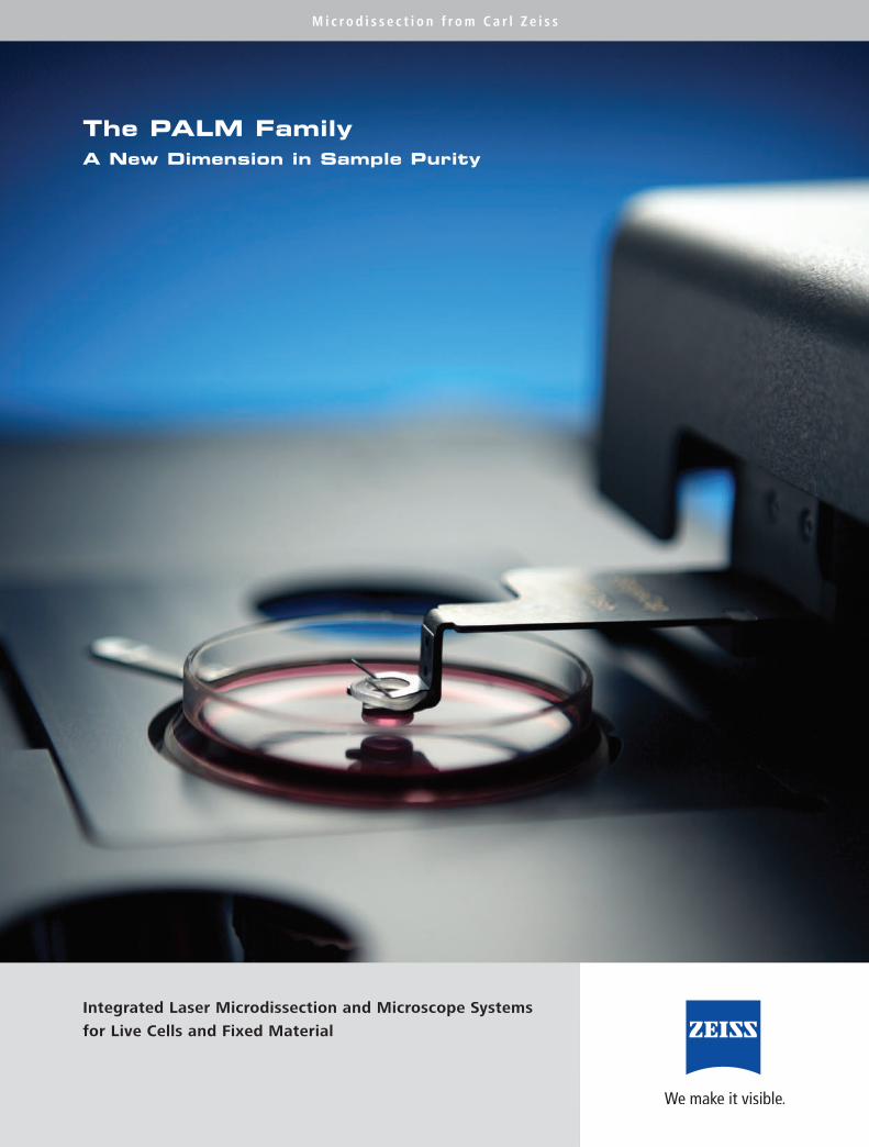
The PALM FamilyA New Dimension in Sample Purity
Integrated Laser Microdissection and Microscope Systems
for Live Cells and Fixed Material

At the Forefront of Science
The biomedical sciences with their rapid development rate is one of the most mean-
ingful and promising research fields. Increasingly efficient technologies are leading
to a deeper understanding of the complex mechanisms that underlie living systems
at the molecular, cellular and tissue level.
For more than 160 years Carl Zeiss has been at the forefront of research and devel-
opment in advanced optical technologies. Excellence in support, but above all, an
understanding of the individual needs of scientists means we can create the best
environment for successful research.


The isolation of single cells and cell groups, as well as biomolecules, is central to life sciences.
For biomedical analysis a method is required for the extraction of your samples, particularly with heterogeneous
tissue, that is free of contamination and guarantees preservation of the material.
Clearly the preparation and staining of samples as well as the actual analyses are critical processes.
However, in molecular biological analysis methods such as PCR, separation of the desired elements is of critical
importance and requires the maximum precision and purity. Sample purity is the deciding factor
for the success of your research.
Sample Purity :The Key Factor in Science
2
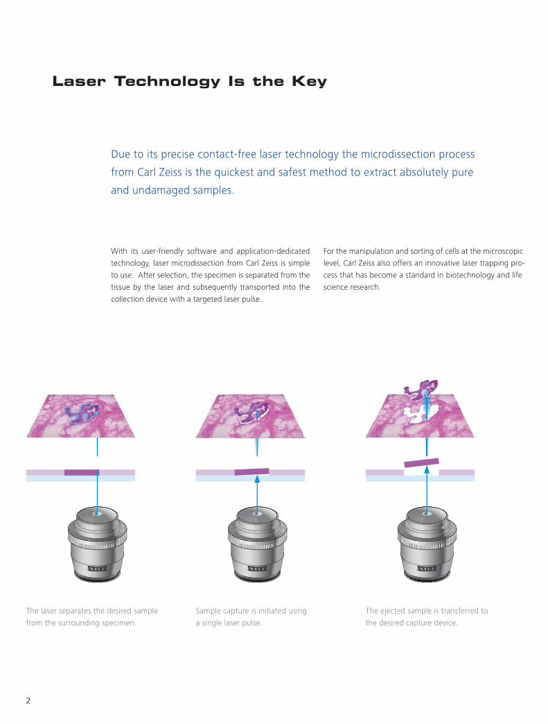
Laser Technology Is the Key
Due to its precise contact-free laser technology the microdissection process
from Carl Zeiss is the quickest and safest method to extract absolutely pure
and undamaged samples.
With its user-friendly software and application-dedicated
technology, laser microdissection from Carl Zeiss is simple
to use. After selection, the specimen is separated from the
tissue by the laser and subsequently transported into the
collection device with a targeted laser pulse.
For the manipulation and sorting of cells at the microscopic
level, Carl Zeiss also offers an innovative laser trapping pro-
cess that has become a standard in biotechnology and life
science research.
The laser separates the desired sample
from the surrounding specimen.
Sample capture is initiated using
a single laser pulse.
The ejected sample is transferred to
the desired capture device.

3
As a leading provider of laser microdissection technology,
we never underestimate the importance of customer sup-
port. Our application laboratories provide you with an un-
paralleled level of know-how and expert advice. We offer
comprehensive application consultation as well as uncom-
plicated technical support through everyday use of the
PALM system from Carl Zeiss.
Expertise
Furthermore, at our headquarters, a highly qualified lab
team works constantly on the development of new appli-
cations and the validation of new methods in our optimal-
ly equipped research facility.
4
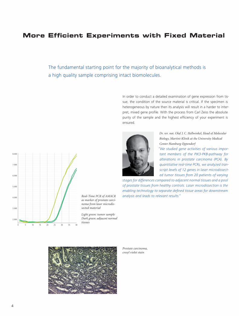
More Efficient Experiments with Fixed Material
In order to conduct a detailed examination of gene expression from tis-
sue, the condition of the source material is critical. If the specimen is
heterogeneous by nature then its analysis will result in a harder to inter-
pret, mixed gene profile. With the process from Carl Zeiss the absolute
purity of the sample and the highest efficiency of your experiment is
ensured.
The fundamental starting point for the majority of bioanalytical methods is
a high quality sample comprising intact biomolecules.
“ We studied gene activities of various impor-
tant members of the PIK3-PKB-pathway for
alterations in prostate carcinoma (PCA). By
quantitative real-time PCRs, we analyzed tran-
script levels of 12 genes in laser microdissect-
ed tumor tissues from 20 patients of varying
stages for differences compared to adjacent normal tissues and a pool
of prostate tissues from healthy controls. Laser microdissection is the
enabling technology to separate defined tissue areas for downstream
analysis and leads to relevant results.”
Dr. rer. nat. Olaf J. C. Hellwinkel, Head of Molecular
Biology, Martini-Klinik at the University Medical
Center Hamburg-Eppendorf
Prostate carcinoma, cresyl violet stain
Real-Time PCR of AMACR as marker of prostate carci- noma from laser microdis- sected material
Light green: tumor sample Dark green: adjacent normal tissues
5
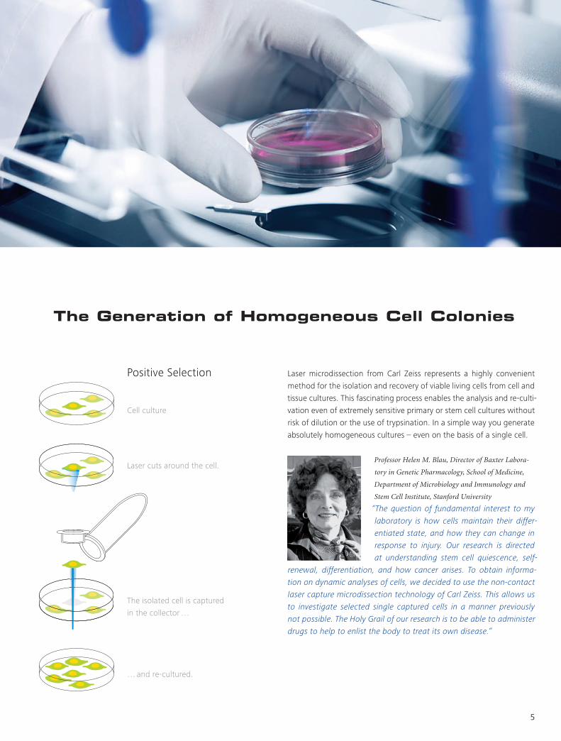
The Generation of Homogeneous Cell Colonies
Laser microdissection from Carl Zeiss represents a highly convenient
method for the isolation and recovery of viable living cells from cell and
tissue cultures. This fascinating process enables the analysis and re-culti-
vation even of extremely sensitive primary or stem cell cultures without
risk of dilution or the use of trypsination. In a simple way you generate
absolutely homogeneous cultures – even on the basis of a single cell.
“The question of fundamental interest to my
laboratory is how cells maintain their differ-
entiated state, and how they can change in
response to injury. Our research is directed
at understanding stem cell quiescence, self-
renewal, differentiation, and how cancer arises. To obtain informa-
tion on dynamic analyses of cells, we decided to use the non-contact
laser capture microdissection technology of Carl Zeiss. This allows us
to investigate selected single captured cells in a manner previously
not possible. The Holy Grail of our research is to be able to administer
drugs to help to enlist the body to treat its own disease.”
Professor Helen M. Blau, Director of Baxter Labora-
tory in Genetic Pharmacology, School of Medicine,
Department of Microbiology and Immunology and
Stem Cell Institute, Stanford University
… and re-cultured.
in the collector …
The isolated cell is captured
Laser cuts around the cell.
Cell culture
Positive Selection
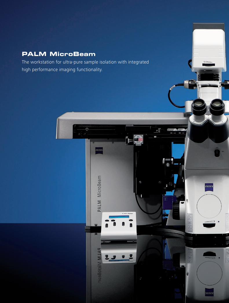
PALM MicroBeamThe workstation for ultra-pure sample isolation with integrated
high performance imaging functionality.
PALM MicroTweezersThe dedicated instrument for contact-free micromanipulation
with laser optical trapping.

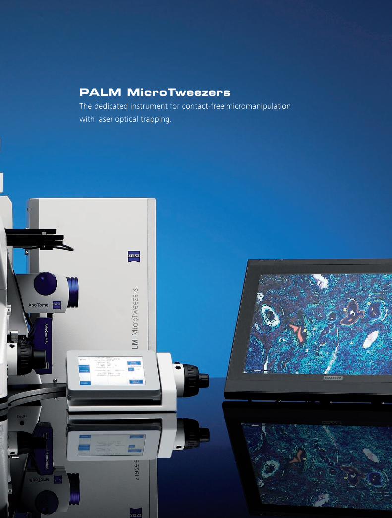
PALM MicroTweezersThe dedicated instrument for contact-free micromanipulation
with laser optical trapping.
PALM CombiSystemThe flexible all-in-one platform for laser microdissection, laser optical trapping,
or a combination of both.
PALM LaboratoriesLeading-edge technology, first-class technical and applied know-how
and a deep understanding of the demands of modern research.

8
Contact–free Micromanipulation
Using the force field of a highly focused laser beam (optical trap), ma-
nipulation functions are conducted without mechanical contact. Living
cells can be manipulated either in isolation or amongst other cells
without damage to their structure or function. The wavelength of the
applied infra-red laser is 1064 nm and therefore within medical tech-
nology’s “therapeutic window” of 600 nm to 1200 nm for preventing
damage to living systems.
Optical Tweezers, also known as laser tweezers, are a highly effective
and non-invasive method to manipulate cells as well as trap, move,
and sort microscopic particles.
The laser traps the selected cell. The cell can be moved within the medium… …towards a desired target.
Literature
Ashkin, Arthur: Optical trapping and manipulation of neutral particles using lasers, Proc. Natl. Acad. Sci. Volume 94, pp. 4853–4860 (1997)
Kuyper, Christopher L. and Chiu, Daniel T.: Optical trapping: A versatile technique for biomanipulation, Applied Spectroscopy, Volume 56, Number 11: 300A–312A (2002)
9
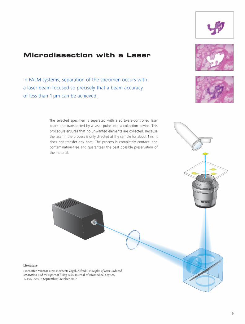
Microdissection with a Laser
In PALM systems, separation of the specimen occurs with
a laser beam focused so precisely that a beam accuracy
of less than 1 μm can be achieved.
The selected specimen is separated with a software-controlled laser
beam and transported by a laser pulse into a collection device. This
procedure ensures that no unwanted elements are collected. Because
the laser in the process is only directed at the sample for about 1 ns, it
does not transfer any heat. The process is completely contact- and
contamination-free and guarantees the best possible preservation of
the material.
Literature
Horneffer, Verena; Linz, Norbert; Vogel, Alfred: Principles of laser-induced separation and transport of living cells, Journal of Biomedical Optics, 12 (5), 054016 September/October 2007

10
Optimal Solutions for Your Research
Our aim is to provide you with the best possible solutions for your research.
This begins with the proven high-quality processes and
systems from Carl Zeiss. Beyond that, we offer you a spe-
cial service with RentalLab: to save time, money, and re-
sources you can rent our excellently equipped research
facilities for your projects or allow our experts to perform
the experiments completely.
Through the efficiency of our lab, we make it possible for
you to reduce your workload to a minimum. Furthermore,
through the practical work in our labs we gain knowledge
that directly influences the continued development of our
products and services as, for instance, in the constant op-
timization of the expendable materials for our systems.
Accessories and consumables for laser microdissection:
MembraneSlides – Improve the efficiency of laser microdissection, we
have the right slide for your application: PEN,PET & nuclease-free.
Sample Collection – A range of capture consumables designed to fit
your needs. Choose from AdhesiveCaps for dry collection to eight cap
strips or even multititer plate format for high content experiments.
Live Cell Applications – A versatile range of dedicated cell culture plastic-
ware is available to suit the demands of live cell culture, imaging, and laser
microdissection. The LiveCell Collector is an accessory for the isolation of
adherent live cells without additional trypsinization and under sterile
conditions. Specialized DuplexDishes and MembraneRings are supplied in
two sizes (50 mm and 35 mm). For greater capacity we also offer an
insert for up to six Lumox™ dishes.

11
Automated Sample Extraction
CapMover
The CapMover locates a microfuge tube lid over the capture position of a specimen
slide or cell culture dish. The special advantage of the CapMover lies in its ease of
use: with a mouseclick it is positioned exactly over the specimen, with another click
of the mouse the CapCheck function can be conducted.
CapMover and RoboMover are the fully automated and computer controlled
collection devices of the PALM systems. They enable reliable and user friendly
collection of specimens.
RoboMover
The RoboMover is the advanced form of the CapMover. It enables, for example, the
exact division of several selected elements into different Caps or even in the re-
cesses of a microtiter plate. The RoboMover automatically sorts the color-coded
elements into the pre-set collection devices. In this manner, hundreds of samples
can be precisely sorted and collected.
Positioning of the CapMover is made simple by the intuitive control window.
RoboMover inserts will be automatically recognized so that predefined capture sequences can be initiated from the element list.
12
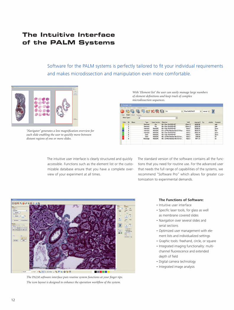
The Intuitive Interface of the PALM Systems
Software for the PALM systems is perfectly tailored to fit your individual requirements
and makes microdissection and manipulation even more comfortable.
The intuitive user interface is clearly structured and quickly
accessible. Functions such as the element list or the custo-
mizable database ensure that you have a complete over-
view of your experiment at all times.
The PALM software interface puts routine system functions at your finger tips.
The icon layout is designed to enhance the operation workflow of the system.
‘Navigator’ generates a low magnification overview for each slide enabling the user to quickly move between distant regions of one or more slides.
With ‘Element list’ the user can easily manage large numbers of element definitions and keep track of complex microdissection sequences.
The Functions of Software:
•Intuitiveuserinterface
•Specificlasertools,forglassaswell
as membrane covered slides
•Navigationoverseveralslidesand
serial sections
•Optimizedusermanagementwithele-
ment lists and individualized settings
•Graphictools:freehand,circle,orsquare
•Integratedimagingfunctionality:multi-
channel fluorescence and extended
depth of field
•Digitalcameratechnology
•Integratedimageanalysis
The standard version of the software contains all the func-
tions that you need for routine use. For the advanced user
that needs the full range of capabilities of the systems, we
recommend “Software Pro” which allows for greater cus-
tomization to experimental demands.

13
Colibri
Instead of a conventional white light source, Colibri
uses high-power LEDs for contrast-rich images
with high dynamic range.
From Microdissection System to Research Platform
With the built-in inverted research microscope Axio Observer, the PALM systems offer
not only highly specialized microdissection technology, but also the proven imaging
functionality from Carl Zeiss. Furthermore PALM systems are compatible with countless
other ZEISS components and allow expansion to a multi-faceted research platform.
AxioCam Series
Ranging from 1.4 to 13 megapixels, the AxioCam series
has the optimal solution for all requirements in digital
camera technology.
Incubation
The Incubator XL PALM S1 with software
controlled thermostat guarantees a stable
environment for living cells.
ApoTome
The innovative plug-in module for the fluorescence
beam path with “grid projection” for improved
image quality and 3D microscopy.
Contrast Methods from Carl Zeiss
For example PlasDIC, the first polarization-
optical differential interference contrast
that allows the use of plastic dishes.

Patents
US Patents: 5.998.129, 5.689.109, 6.930.764,
Other Patents: EP 879408 B1, JP 3311757, EP 679325 B1,
DE 102 54 229.5
PALM Systems use laser devices and comply with the
international safety requirements. Please mind your
local safety regulations when working with lasers.
For assistance ask your sales representative.
• Absolute purity in sample extraction
• Maximum preservation of the material
• Combined microdissection and high-
end imaging technology
• Intuitive user interface
• Digital camera technology
• Application support and comprehensive
service
The PALM Family :

Info
rmat
ion
subj
ect t
o ch
ange
. Pr
inte
d on
env
ironm
enta
lly fr
iend
ly p
aper
bl
each
ed w
ithou
t chl
orin
e.
60- 3
-000
1/e
– is
sued
04.
10
Carl Zeiss Microscopy GmbH07745 Jena, Germany [email protected]/microdissection
Laser Technology from Carl Zeiss :Success Factor in Modern Science