the prevalence of sickle cell trait among …
TRANSCRIPT

i
THE PREVALENCE OF SICKLE CELL TRAIT AMONG ADOLESCENTS IN
BUNGOMA COUNTY, KENYA.
EINSTEIN KIBET WATENGA
RESEARCH THESIS SUBMITTED IN PARTIAL FULFILLMENT OF THE
REQUIREMENT OF THE DEGREE OF MASTER OF MEDICINE IN CHILD
HEALTH AND PAEDIATRICS OF MOI UNIVERSITY.
© 2020

ii
DECLARATION:
Student’s declaration:
This thesis is my original work and has not been presented to any other
university/institution. No part of this thesis may be reproduced without prior written
permission from the author and/or Moi University.
Dr. Einstein Kibet Watenga.
SM/PGCHP/08/15.
Signature………………………………..Date……………………
Declaration by Supervisors:
This research thesis has been submitted for examination with our approval as
University supervisors.
1. Prof. Constance Nalianya Tenge.
Associate Professor, Department of Child Health and Paediatrics
Signature………………………………... Date………………….
2. Dr. Julia Jerono Songok.
Lecturer, Department of Child Health and Paediatrics.
Signature………………………………….Date……………………

iii
DEDICATION
I dedicate this work to my father, the late Mr. Stephen Watenga Chemengu for
encouraging me to work on my dreams and make the world a better place.

iv
ACKNOWLEDGEMENT.
I would like to express my sincere gratitude to the following: First to God almighty,
secondly to my supervisors: Prof. Constance Tenge and Dr. Julia Songok for the
mentorship and unrelenting support in the development of this thesis. Thirdly, to my
research assistants Mr. Duke Omari and Ms. Faith Aato. I also appreciate the input
from my statistician, Mr. Gerald Lwande.
Many thanks to members of the department of Child Health and Paediatrics including
the lecturers and fellow postgraduate students for their highly valued opinions and
comments that contributed to the development of this document.
I also express my appreciation to the National research fund for funding this study.
Finally, I thank all my family members for their moral support and words of
encouragement.

v
ABBREVIATIONS, ACRONYMS.
Hb Haemoglobin.
HbAA Normal haemoglobin.
HbAC Carrier state of Haemoglobin C disease.
HbAS Carrier state of sickle cell disease also known as the sickle cell trait.
HbCC Haemoglobin C disease.
HBE Hb Electrophoresis.
HbF Foetal Haemoglobin.
HbSC Milder form of haemoglobinopathy compared to sickle cell with both
haemoglobin C and S.
HbSS Abnormal variant of haemoblobin responsible for sickle cell disease.
HPLC High performance liquid chromatography.
IEF Isoelectric Focusing.
SCD Sickle Cell disease.
SCT Sickle cell trait.
WHO The World Health Organization.

vi
DEFINITIONS.
Adolescent- Any person aged between 10 and 19 years.
Sickle cell trait- Carrier state of sickle cell disease, where there is one normal gene
and one sickle cell gene. Also documented as HbAS.
Haemoglobin variants- refers to HbAA,HbAS,HbAC,HbSS,HbSC,HbCC.
Sickle cell disease- refers to HbSS, HbSC variants.

vii
TABLE OF CONTENTS.
DECLARATION: .......................................................................................................... ii
DEDICATION ...............................................................................................................iii
ACKNOWLEDGEMENT. ........................................................................................... iv
ABBREVIATIONS, ACRONYMS............................................................................... v
DEFINITIONS.............................................................................................................. vi
TABLE OF CONTENTS..............................................................................................vii
LIST OF TABLES ........................................................................................................ xi
LIST OF FIGURES ......................................................................................................xii
ABSTRACT................................................................................................................. xiii
CHAPTER ONE ............................................................................................................ 1
INTRODUCTION ......................................................................................................... 1
1.1 Background information ...................................................................................... 1
1.2 Research problem statement................................................................................. 4
1.3 Justification .......................................................................................................... 5
1.4 Research question................................................................................................. 6
1.5 Broad objective. ................................................................................................... 6
1.6 Specific objectives................................................................................................ 6
CHAPTER TWO ........................................................................................................... 7
LITERATURE REVIEW .............................................................................................. 7
2.1 General information on sickle cell disease........................................................... 7
viii
2.2 Prevalence of HbAS. .......................................................................................... 10
2.3 Prevalence of HbSS............................................................................................ 12
2.4 Prevalence of other haemoglobin variants ......................................................... 13
2.5Association between demographic characteristics and sickle cell trait ............... 13
2.6 Laboratory diagnosis of sickle cell gene status. ................................................. 14
2.7 Control of sickle cell disease. ............................................................................. 16
CHAPTER THREE ..................................................................................................... 19
METHODOLOGY....................................................................................................... 19
3.1 Study site ............................................................................................................ 19
3.2 Study Population ................................................................................................ 19
3.3 Study design ....................................................................................................... 19
3.4 Sample size......................................................................................................... 20
3.5 Eligibility............................................................................................................ 20
3.5.1 Inclusion criteria .......................................................................................... 20
3.6 Sampling procedure............................................................................................ 20
3.7 Execution of the study ........................................................................................ 21
3.8 Data collection instruments ................................................................................ 22
3.9 Data management. .............................................................................................. 22
3.10 Data analysis .................................................................................................... 22
3.11 Ethical considerations. ..................................................................................... 22
CHAPTER FOUR........................................................................................................ 24

ix
RESULTS .................................................................................................................... 24
4.1 Demographic characteristics. ............................................................................. 24
4.2 Prevalence of sickle cell trait among adolescents in Bungoma County. ............ 25
4.3Haemoglobin variants among adolescents in Bungoma County. ........................ 25
4.4. Association between demographic characteristics and sickle cell trait ............. 26
4.4.1 Gender.......................................................................................................... 26
4.4.2Geographical Area (Constituency) ............................................................... 27
4.4.3 Family history and Sickle Cell trait ............................................................. 28
CHAPTER FIVE ......................................................................................................... 29
DISCUSSION .............................................................................................................. 29
5.1 Prevalence of sickle cell trait among adolescents in Bungoma County. ............ 30
5.2 Haemoglobin variants among adolescents in Bungoma County. ....................... 31
5.3. Association of Demographic characteristicswith sickle cell trait among
adolescents in Bungoma County. ............................................................................. 33
5.3.1 Gender.......................................................................................................... 33
5.3.2 Geographical area (constituency): ............................................................... 34
5.3.3 Family history: ............................................................................................. 35
5.4 Study Limitations ............................................................................................... 35
CHAPTER SIX ............................................................................................................ 36
CONCLUSIONS AND RECOMMENDATIONS ...................................................... 36
6.1 Conclusions ........................................................................................................ 36
6.2 Recommendations .............................................................................................. 36

x
REFERENCES. ........................................................................................................... 37
APPENDICES ............................................................................................................. 43
Appendix I: Bungoma County. ................................................................................ 43
Appendix II: Data Collection Form ......................................................................... 44
Appendix III: Swahili Consent Form. ...................................................................... 45
Appendix IV: English Consent Form....................................................................... 48
Appendix V: Assent Form........................................................................................ 52
Appendix VI: Swahili Assent Form............................................................................. 53
Appendix VII: The Counseling Process. ..................................................................... 54
Appendix VIII: The Sickle Scan™ Test Protocol. ...................................................... 55
Appendix IX: sickle scan™ validation certificate. ...................................................... 59
Appendix X: certificate of registration for medomix concept ltd. ............................... 60
Appendix XI: IREC Approval ..................................................................................... 61

xi
LIST OF TABLES
Table 1: Demographic characteristics. ......................................................................... 24
Figure 1: HbAS Constituency Prevalence ................................................................... 25
Table 2: Hemoglobin Variants among adolescents in Bungoma County ................... 26
Table 3: Gender versus Sickle Cell Test Results ......................................................... 26
Table 4: Test of Association of Gender and Sickle Cell Trait .................................... 27
Table 5: Association between Constituency and Sickle Cell trait ............................... 27
Table 6: Family History of Sickle Cell Disease versus Sickle Cell Test Results ........ 28
Table 7: Test of association for Family History of Sickle Cell Disease and Sickle Cell
Trait .............................................................................................................................. 28

xii
LIST OF FIGURES
Figure 1: HbAS Constituency Prevalence ................................................................... 25

xiii
THE PREVALENCE OF SICKLE CELL TRAIT AMONG ADOLESCENTS IN
BUNGOMA COUNTY, KENYA
ABSTRACT
Background: Sickle cell disease (SCD) is a genetic disorder inherited in autosomal recessive fashion and affects the haemoglobin (Hb) molecule. The homozygous state (HbSS) is the severest form of SCD and the heterozygous state(HbAS) is the Sickle
cell trait or carrier state (SCT). Health education, genetic counseling, testing and identifying persons with the sickle cell gene and advising them on how to prevent
birth of children with SCD are important primary intervention strategies for control of SCD. Objectives: To determine the prevalence of sickle cell trait among adolescents
attending mixed day secondary schools in Bungoma County. To determine the haemoglobin variants among adolescents attending mixed day secondary schools in
Bungoma County. To test the association between the demographic characteristics and sickle cell trait among adolescents attending mixed day secondary schools in Bungoma County.
Methods cross-sectional study was done on adolescents attending mixed day secondary schools in Bungoma County of Western Kenya between January 2017 and
July 2017. Stratified random sampling was applied; the county was stratified into 9 constituencies; a mixed day secondary school was selected randomly from each constituency. Public health education on SCD control was offered to 2389 students in
9 schools, 2248 met eligibility criteria. A total of 225 eligible participants were selected randomly from the school registers and consented. Pretest counseling was done followed by testing for sickle cell gene status using a point of care (POC) device
(The Sickle scan™). Post test counseling was offered. Demographics and test results were documented. Proportions and Percentages was used to describe categorical data;
Mean and Median for continuous data. Logistic regression was done and risk ratios (RR) generated, Pearson chi square was used to test for associations. Level of significance was set at p-value <0.05 with Confidence interval at 95%.
Results: All 225 participants consented for testing; Males: 116/225(51.6%), Females: 109/225(48.4%). The median age was 17 years. HbAS prevalence was 18.7%
(42/225) (CI 0.14-0.24). Proportion of males with HbAS was 24/116(20.7%); proportion of females with HbAS was 18/109(16.5%). Proportion of HbAS among students with known family history of SCD was 6/12 (50%);proportion of HbAS
among those with no known family history of SCD was 36/213 (16.9%).Of those with family history of SCD; 1/12(8.3%)reported a sibling,9/12(75%) reported a cousin and
2/12(16.6%)reported other relatives. Hb variants were HbAA (183/225,81.3%) and HbAS (42/225,18.7%), there were no other Hb variants. There was no invalid result. Those with Family History of SCD were three times more likely to have HbAS(RR-
2.958, p-value= 0.004,CI:1.561-5.607). Conclusion: Prevalence of HbAS among adolescents is high. Hb variants found were
HbAA and HbAS, there was no HbSS or HbC variants found. Having a Known Family History of SCD was significantly associated with having HbAS. Recommendations: Public Health Education on SCD and SCT to Bungoma County
residents. Setting up of genetic counseling and testing centers for SCD and SCT in Bungoma County.

1
CHAPTER ONE
INTRODUCTION
1.1 Background information
Sickle cell disease (SCD) was first described by James B. Herrick in 1910. It is a
genetic disorder that affects the haemoglobin molecule.
The haemoglobin molecule is constituted by four globin chains; Genes in the α-globin
and β-globin clusters on chromosome 16 and 11respectively control the production of
globin chains(Iyer, Sakhare, Sengupta, &Velumani, 2015). Point mutation result in
haemoglobin gene variants which fall into two broad groups – structured variants in
which there is a change in the sequence of amino acid producing unusual
haemoglobin (haemoglobinopathy) and thalassaemia in which there is a lower or
abolished production of globin chains. Sickle cell anaemia is caused by a point
mutation in the β-globin chain of haemoglobin causing the hydrophilic amino acid
glutamate to be replaced by the hydrophobic amino acid valine at the sixth position
(Malowany& Butany, 2012). Fetal haemoglobin (HbF) has two alpha and two gamma
chains (a2y2) and Adult haemoglobin (HbA) has two alpha and two beta chains
(a2β2)(Domínguez, Zurita, Calvopiña, Villacís, & Mora, 2013).
SCD is inherited in an autosomal recessive fashion. A person who has two normal
genes has normal haemoglobin (HbAA)(Malowany J.I., 2012). A person who receives
a gene for sickle cell disease from one parent and a normal gene from the other has a
condition called sickle cell trait (heterozygous state HbAS). Two genes of the sickle
hemoglobin, each one inherited from both parents results in the disease(homozygous
state HbSS). If both parents are heterozygous for the HbS mutation (HbAS), there is a
25% chance per pregnancy of having an offspring with Sickle cell disease(HbSS) and
50% chance of the offspring being a carrier (HbAS) and a 25% chance of the

2
offspring having the normal Haemoglobin (HbAA). If one parent is homozygous
(HbSS) and the other parent is heterozygous (HbAS), there is a 50% chance for every
pregnancy of having an offspring with Sickle cell disease (HbSS) and a 50 % chance
of having a carrier (HbAS). If one parent is homozygous (HbSS) and the other is
normal (HbAA), then every pregnancy is likely to result into an offspring who is a
carrier (HbAS). If both parents are homozygous (HbSS) then every pregnancy is
likely to result into an offspring with sickle cell disease (HbSS). It is advisable that
persons who have the gene for sickle cell (HbAS or HbSS) should have offsprings
with persons with normal genes(HbAA) in order to avoid the likelihood of getting
offsprings with HbSS.
Other compound heterozygous states include sickle-haemoglobin C (HbSC), sickle
beta-zero thalasaemia (HbSβ°), sickle beta-plusthalasaemia (HbSβ+), sickle-
haemoglobin D-Punjab (HbSD-) among others. (Malowany J.I., 2012)
Sickle cell trait is the carrier state of the disease. Individuals with sickle cell trait are
mostly healthy, live a normal life (John, 2010) but contribute towards transmission of
the sickle cell gene. They have potential of giving birth to children with sickle cell
disease.
Persons with homozygous state of the disease(HbSS) and compound heterozygous
states such as sickle-haemoglobin C (HbSC) are symptomatic. Symptoms usually
begin after six months of life relating to decline of fetal haemoglobin levels. They
present with signs and symptoms of anaemia, infection and vaso-occlusive crisis such
as jaundice, pallor, fever and severe pain. (Lazarus, 2011)
Sickle cell disease poses a significant public health burden in sub-Saharan Africa, and
especially in the malaria endemic regions where the gene frequency is highest. In

3
these regions, it is a significant cause of morbidity and mortality and control programs
become essential. Despite the need for control programs, most nations in the affected
areas do not have control programs.(WHO, 2006.)
In Kenya, the sickle cell gene is found mostly in the lake regions of western Kenya
and the Coastal areas, these are malaria endemic regions.(Aluoch JR, 1993)
In terms of control strategies, it is paramount to note that at the 59th World Health
Assembly, having recognized the public health burden associated with sickle cell
disease, recommendations were proposed for control of the disease. First, that national
programs for prevention and management of sickle cell anaemia be established by
member states with the aim of reducing morbidity and mortality associated with sickle
cell anaemia. National programs need to focus on: surveillance, information
dissemination, genetic counseling and testing. Other measures proposed include:
offering appropriate, affordable and easily accessible emergency care to people living
with sickle cell anaemia, developing capacity to evaluate impact of sickle cell
anaemia programs, training of health professionals in high prevalent areas, developing
holistic care within existing primary health care systems offering medical genetics
services in partnership with various levels of governments and Nongovernmental
organizations, promotion of relevant community education (including health
counseling), promotion of effective international cooperation in combating sickle cell
anaemia and support of research on sickle cell anaemia (WHO, 2006.).
Recently, in the country there has been increased public sensitization about sickle cell
disease from the media and the theme for the world sickle cell day held in Kenya in
2015 was “know your sickle cell status” which is a step in controlling the perpetuation
of the gene.
4
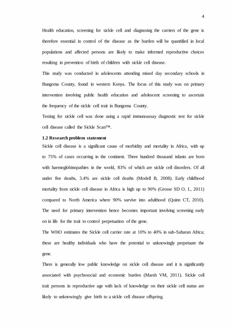
Health education, screening for sickle cell and diagnosing the carriers of the gene is
therefore essential in control of the disease as the burden will be quantified in local
populations and affected persons are likely to make informed reproductive choices
resulting in prevention of birth of children with sickle cell disease.
This study was conducted in adolescents attending mixed day secondary schools in
Bungoma County, found in western Kenya. The focus of this study was on primary
intervention involving public health education and adolescent screening to ascertain
the frequency of the sickle cell trait in Bungoma County.
Testing for sickle cell was done using a rapid immunoassay diagnostic test for sickle
cell disease called the Sickle Scan™.
1.2 Research problem statement
Sickle cell disease is a significant cause of morbidity and mortality in Africa, with up
to 75% of cases occurring in the continent. Three hundred thousand infants are born
with haemoglobinopathies in the world, 83% of which are sickle cell disorders. Of all
under five deaths, 3.4% are sickle cell deaths (Modell B, 2008). Early childhood
mortality from sickle cell disease in Africa is high up to 90% (Grosse SD O. I., 2011)
compared to North America where 90% survive into adulthood (Quinn CT, 2010).
The need for primary intervention hence becomes important involving screening early
on in life for the trait to control perpetuation of the gene.
The WHO estimates the Sickle cell carrier rate at 10% to 40% in sub-Saharan Africa;
these are healthy individuals who have the potential to unknowingly perpetuate the
gene.
There is generally low public knowledge on sickle cell disease and it is significantly
associated with psychosocial and economic burden (Marsh VM, 2011). Sickle cell
trait persons in reproductive age with lack of knowledge on their sickle cell status are
likely to unknowingly give birth to a sickle cell disease offspring.

5
The average cost for managing a patient with the disease in an outpatient setting in
rural Kenya is estimated to be KES 13,800 (USD 138) annually. In settings where
majority of population live under abject poverty, the cost of managing a sickle cell
disease patient becomes significant in the household budget and is likely to affect the
survival of an affected individual. (Amendah DD, 2013).
There is limited epidemiological data on sickle cell disease around the globe and most
nations with significant burden of sickle cell disease do not have control programs,
Kenya included (WHO, 2006.).
Adolescents in secondary schools are just about to become adults and likely to plan a
reproductive lifestyle. There is therefore need to educate and test this group.
1.3 Justification
Transmission of the sickle cell disease is autosomal recessive. Those with the trait are
mostly healthy individuals who can unknowingly contribute to the birth of the
children with homozygous state who present with complications that result in
increased morbidity and mortality with high treatment costs and psychological
burden. Knowledge on sickle cell status is likely to inform decision making on
reproductive lifestyle with resultant control of transmission of the sickle gene.
In countries where primary intervention strategies have been adopted, for example in
North America, there has been improved survival in the population with reduction of
sickle cell related deaths. (Chakravorty S, 2015)
Prevalence studies for sickle cell are therefore important to quantify burden of disease
so that strategies can be put in place to control the transmission of the gene and more
so the SS combination (homozygous state) which is fatal.(Piel FB H. S., 2013)
Adolescents in secondary school are able to understand information, are about to
become adults and are accessible. They are therefore a suitable group to screen as
they approach adulthood so that they can make decisions in future regarding their

6
reproductive life basing on their sickle cell status. This will contribute towards
primary prevention of sickle cell disease by reduction in the number of children born
with the homozygous combination.
Knowledge on sickle cell disease and status among adolescents is likely to inform
relationship decisions and eventual spouse choice in their future where they will be
adults and are likely to reproduce. Persons with sickle cell trait and knowledge about
sickle cell disease are likely to pair with persons who have normal haemoglobin and
prevent the possibility of giving birth to children with sickle cell disease.
1.4 Research question
What is the prevalence of sickle cell trait among adolescents in Bungoma County?
1.5 Broad objective.
To determine the prevalence of sickle cell trait among adolescents in Bungoma
County.
1.6 Specific objectives.
1. To determine the prevalence of sickle cell trait among adolescents attending
mixed day secondary schools in Bungoma County.
2. To determine the haemoglobin variants among adolescents attending mixed day
secondary schools in Bungoma County.
3. To test the association between the demographic characteristics and sickle cell
trait among adolescents attending mixed day secondary schools in Bungoma
County.

7
CHAPTER TWO
LITERATURE REVIEW
This chapter describes the general information on sickle cell disease, the prevalence
of the trait, haemoglobin variants and how to determine them using various tests and
the demographic associations of the Sickle cell trait. The WHO intervention strategies
for SCD will also be highlighted.
2.1 General information on sickle cell disease
Sickle cell disease is a genetic disorder of the haemoglobin molecule whose
proximate cause is a mutation of the β-globin chain of hemoglobin. (Ansong, Akoto,
Ocloo, & Ohene-Frempong, 2013; Kreuels et al., 2009).
The haemoglobin molecule is constituted by four globin chains; fetal haemoglobin
(HbF) which has two alpha and two gamma chain (a2y2) and adult haemoglobin
(HbA) has two alpha and two beta chains (a2β2)(Domínguez et al., 2013). Genes in
the α-globin and β-globin clusters on chromosome 16 and 11 respectively and control
the production of globin chains(Iyer, Sakhare, Sengupta, &Velumani, 2015). Point
mutation result in haemoglobin gene variants which fall into two broad groups –
structured variants in which there is a change in the sequence of amino acid producing
unusual haemoglobin (haemoglobinopathy) and thalassaemia in which there is a lower
or abolished production of globin chains. Sickle cell anaemia is caused by a point
mutation in the β-globin chain of haemoglobin causing the hydrophilic amino acid
glutamate to be replaced by the hydrophobic amino acid valine at the sixth
position(Malowany& Butany, 2012).

8
More than 300,000 children are born annually with hemoglobinopathies globally, of
which 83% are sickle cell disorders (Modell & Darlison, 2008).
Also known as sickle cell anemia; it is characterized by an abnormality in oxygen
carrying hemoglobin molecule in red blood cells (Bowers, Pepple, & Reid, 2008). It is
one of the most common hemoglobinopathies in Africa, Middle East and India.
However, it is now found globally with an increasing incidence in Europe and North
America due to increasing migration rates from areas that have high prevalence
(Stuart & Nagel, 2004).
Sickle cell disease is part of the broader haemoglobinopathies which are disorders of
haemoglobin structure. In this disease, red blood cells assume the abnormal sickle
shape and become rigid under conditions of low oxygen such as dehydration, acidosis
and infection (Malowany& Butany, 2012).
There are a variety of genotypes of the sickle cell syndrome. There is homozygosity
for the mutation that cause HbS – sickle cell anaemia (SCA) that is also referred to as
„HbSS‟ or „SS disease‟. In the heterozygous state, where there is one sickle gene and
one normal haemoglobin gene, the result is a condition referred to as „Hb AS‟ or
sickle cell trait (SCT). Other forms of sickle cell disease are compound heterozygous
states where there is a copy of the mutation that cause HbS and a copy of another
abnormal haemoglobin allele. Examples include sickle-haemoglobin C (HbSC),
sickle beta-zero thalasaemia (HbSβ°), sickle beta-plusthalasaemia (HbSβ+), sickle-
haemoglobin D-Punjab (HbSD-) among others. The different types of sickle cell
syndromes exhibit varying degrees of severity of the clinical features(Malowany&
Butany, 2012).

9
The allele responsible for sickle cell anaemia is autosomal recessive and is found on
the short arm of chromosome 11.Usually, the parents of an individual with HbSS are
heterozygote and therefore carry one HbS allele (HbAS) and are asymptomatic
thereby being referred to as carriers or have the sickle cell trait (Malowany J.I., 2012).
However,in certain populations, because of the high rate of HbS carriers(HbAS), it is
possible that the parent may be homozygous (HbSS) or a compound heterozygote
(HbSC). If both parents are heterozygous for the HbS mutation (HbAS), there is a
25% chance per pregnancy of having an offspring with Sickle cell disease (HbSS) and
50% chance of the offspring being a carrier (HbAS) and a 25% chance of the
offspring having the normal Haemoglobin (HbAA). If one parent is homozygous
(HbSS) and the other parent is heterozygous (HbAS), there is a 50% chance for every
pregnancy of having an affected offspring (HbSS) and a 50 % chance of having a
carrier (HbAS). If one parent is homozygous (HbSS) and the other is normal
(HbAA), then every pregnancy is likely to result into carrier state (HbAS). If both
parents are homozygous (HbSS) then every pregnancy is likely to result into a sickle
cell disease offspring (HbSS). Persons with the trait (HbAS) or the disease (HbSS)
should pair with normal persons (HbAA) in order to avoid the likelihood of getting an
affected offspring (HbSS).
Various complex interactions of the sickle red cells with the endothelium and other
blood cells have been described that could contribute to the pathophysiology of sickle
cell disease which involve adhesion molecules and production of pro-inflammatory
mediators(Manwani& Frenette, 2013).
The clinical manifestation of sickle cell disease is varied with any organ being
affected. These are classified as: Vaso-occlusive and hematologic. Vaso-occlusive

10
manifestations include pain, acute chest syndrome, splenic infarction, stroke and
avascular necrosis of joints being prominent. Hematologic manifestations include
episodes of severe anemia, leg ulcers and pulmonary hypertension (Ansong et al.,
2013; Kato, Gladwin, & Steinberg, 2007).
With early diagnosis and treatment, it is possible to ameliorate the course of the
disease and ultimately improve patient survival. Better survival rates and improved
quality of life have been attributed to prophylactic penicillin, immunizations,
comprehensive care and parental education about the serious sickle cell disease
complications (Ndefo, Maxwell, Nguyen, & Chiobi, 2008; Tsaras, Owusu-Ansah,
Boateng, & Amoateng-Adjepong, 2009).
Although persons with sickle cell trait are usually asymptomatic, they could
potentially be predisposed to complications such as: gross hematuria, increased
urinary tract infection rates in women, complications of hyphema, splenic infarction
with altitude hypoxia or exercise and life threatening complications of exercise
(Eichner, 2007). Hematuria has been recognized as the most common manifestation
of sickle cell trait (Ahmed & Ibrahim, 2006).
2.2 Prevalence of HbAS.
Globally, sickle cell trait is present among 300 million individuals with the highest
prevalence of 30-40% in sub-Saharan Africa (Kato et al., 2007). According to the
WHO, the sickle cell trait is widespread reaching its highest prevalence in parts of
Africa as well as people with origins from equatorial Africa, the Mediterranean basin
and Saudi Arabia. In Africa the prevalence of the sickle cell trait is highest between
latitudes 15 degrees North and 20 degrees South, ranging between 10% and 40%of
the population in some areas. The prevalence decreases to 1% and 2% in north Africa
and less than 1% in southern Africa. Where the prevalence is above 20%, the disease
affects up to 2% of the population.
11
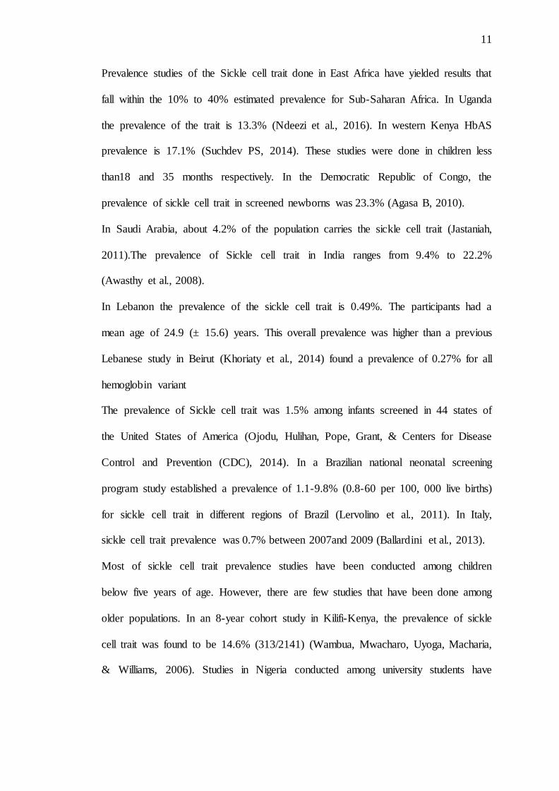
Prevalence studies of the Sickle cell trait done in East Africa have yielded results that
fall within the 10% to 40% estimated prevalence for Sub-Saharan Africa. In Uganda
the prevalence of the trait is 13.3% (Ndeezi et al., 2016). In western Kenya HbAS
prevalence is 17.1% (Suchdev PS, 2014). These studies were done in children less
than18 and 35 months respectively. In the Democratic Republic of Congo, the
prevalence of sickle cell trait in screened newborns was 23.3% (Agasa B, 2010).
In Saudi Arabia, about 4.2% of the population carries the sickle cell trait (Jastaniah,
2011).The prevalence of Sickle cell trait in India ranges from 9.4% to 22.2%
(Awasthy et al., 2008).
In Lebanon the prevalence of the sickle cell trait is 0.49%. The participants had a
mean age of 24.9 (± 15.6) years. This overall prevalence was higher than a previous
Lebanese study in Beirut (Khoriaty et al., 2014) found a prevalence of 0.27% for all
hemoglobin variant
The prevalence of Sickle cell trait was 1.5% among infants screened in 44 states of
the United States of America (Ojodu, Hulihan, Pope, Grant, & Centers for Disease
Control and Prevention (CDC), 2014). In a Brazilian national neonatal screening
program study established a prevalence of 1.1-9.8% (0.8-60 per 100, 000 live births)
for sickle cell trait in different regions of Brazil (Lervolino et al., 2011). In Italy,
sickle cell trait prevalence was 0.7% between 2007and 2009 (Ballardini et al., 2013).
Most of sickle cell trait prevalence studies have been conducted among children
below five years of age. However, there are few studies that have been done among
older populations. In an 8-year cohort study in Kilifi-Kenya, the prevalence of sickle
cell trait was found to be 14.6% (313/2141) (Wambua, Mwacharo, Uyoga, Macharia,
& Williams, 2006). Studies in Nigeria conducted among university students have

12
reported that the prevalence of sickle cell trait ranges between 19-24% (Adeyemo &
Soboyejo, 2006; Jeremiah, 2006).
2.3 Prevalence of HbSS.
From previous studies, majority of HbSS are witnessed among children below five
years (Suchdev, Ruth, Earley, Macharia, & Williams, 2014). However, studies among
older age groups show little or no HbSS prevalence. This is evidenced by both
Kenyan (Wambua, Mwangi, et al., 2006) and Nigerian studies (Adeyemo &
Soboyejo, 2006; Jeremiah, 2006). In Western Kenya, the prevalence of HbSS among
children between 6-35 months was 1.6% (Suchdev et al., 2014); however, in Kilifi–
Kenya, the prevalence was 0.001% among older children(Wambua, Mwacharo, et al.,
2006). In Nigeria, the prevalence of HbSS among older participants ranged between
0-1.3% (Adeyemo & Soboyejo, 2006; Jeremiah, 2006).
A study in Uganda reported the prevalence of HbSS as 0.7%(Ndeezi et al.,
2016).This was done in children less than18 months.
In a study done in the Democratic Republic of Congo, the prevalence of HbSS in
screened newborns was reported as 0.96% (Agasa B, 2010).
In the United Kingdom, one baby in every 2000 is born with Sickle cell disease
according to the National Health Service (NHS). In France, the overall birth
prevalence was found to be 1 in 2415 (Bardakjian et al., 2000).In Saudi Arabia, about
0.26% of the population has sickle cell disease(Jastaniah, 2011).
From these studies it is clear the sickle cell disease burden is heaviest in Africa.
Reducing prevalence with increasing age is an indication of mortality of those with
the SCD.

13
2.4 Prevalence of other haemoglobin variants
Whereas the sickle cell gene is the most common abnormal haemoglobin variant,
there exists other abnormal haemoglobin variants. The commonest abnormal
haemoglogin variant other than the HbS is the HbC. HbC is more limited than HbS to
geographical areas in the West and North West Africa with a prevalence of up to 15%
(Piel et al., 2013). However, there has been a low prevalence along the corridor
between West Africa and Egypt due to human migration(Williams & Weatherall,
2012). In a West African study; HbC is thought to have originated along Niger river
as a result of the founders‟ effect (Cataldo, 2012; Williams & Weatherall, 2012). The
prevalence was as high as 40% in Northern Ghana, up to 50% in Northern Ivory
Coast, 20% in Togo and Benin, less than 1% in Central Africa and nonexistent in East
Africa (Bachir & Galacteros, 2004)
From studies described above, all forms of HbC gene combinations namely HbAC,
HbCC, HbSC were grouped together to give the HbC variants prevalence
There is paucity of data on prevalence of other described haemoglobin variants like
the HbD- Punjab variant.
2.5Association between demographic characteristics and sickle cell trait
The prevalence of the sickle cell trait is highest between latitudes 15 degrees North
and 20 degrees South. The explanation as to why the sickle cell trait is highly
prevalent in the tropics has been linked to its association with protection against
malaria(Piel et al., 2013). In Western Uganda, it was observed that the prevalence of
sickle cell trait was significantly higher in Bundibugyo (13.4%) compared to
Mbarara/Ntugumo (3%) regions (Okwi et al., 2010). This was attributed to a high
prevalence of malaria in Bundibugyo and less intermarriage (consangunity).

14
Being an inherited disease, the likelihood of being a carrier, or having the sickle cell
disease increases with known family history of sickle cell disease (Malowany&
Butany, 2012). The sickle cell gene frequency is beginning to affect other parts of the
world previously not known to have sickle cell disease as a result of migration and
intermarriage. Previous studies have shown that there is no significant association
between sickle cell trait and gender (Adeyemo & Soboyejo, 2006; El Ariss, Younes,
Matar, & Berjaoui, 2016; Jeremiah, 2006; Piel et al., 2013; Wambua, Mwacharo, et
al., 2006).
2.6 Laboratory diagnosis of sickle cell gene status.
Laboratory diagnosis is based on identification of HbS and the absence or reduction in
the quantity of HbA in red blood cells. Screening tests available commonly in
laboratories in Africa include the Sodium metabisulphite sickling tests and sickle
solubility tests. Confirmatory tests confirm the phenotype. Possible phenotypes in
sickle cell disease include SS,AS,SC,S𝛽− thalassemia. Tests commonly used in
describing the phenotypes include Hb electrophoresis (HBE), Isoelectric
focusing(IEF) and High performance liquid chromatography (HPLC)(J. Makani,
2013).A newer point of care rapid diagnostic immunoassay test has been developed
which has comparable sensitivity to HBE,IEF and HPLC(Julie Kanter, 2015).
The sickling test for screening is based on the principle that sodium metabisulphite
creates hypoxic states for red blood cells and causes them to sickle and this can then
be visualized under a microscope. The sickling solubility test is based on the relative
insoluble state of HbS when combined with a reducing agent such as sodium
dithionite.The sensitivity of the sickling test is 65% and that of solubility test is
45%.(Okwi A.L, 2010). The sickling test will be positive in persons with both HbAS
and HbSS. However, it cannot differentiate HbAS from HbSS.

15
Confirmatory tests are based on the principle that different Hb isoforms have different
ionic charge which makes them migrate with different velocities in an electric field.
High performance liquid chromatography uses cation exchange chromatography to
identify haemoglobin variants and has the advantage of the ability to quantify Hb
variants. Isoelectric focusing is a qualitative test used to separate proteins based on
their isoelectric points which refers to where the net charge of the protein is zero, the
method is similar to Hb cellulose electrophoresis in principle only that it forms
sharper bands with better resolution. Isoelectric focusing has advantage over Hb
electrophoresis in that it can detect more Hb variants and requires no chemical
reagents. However. Isoelectric focusing cannot quantify Hb variants whereas Hb
electrophoresis can. In terms of sensitivity; High performance liquid chromatography
and Isoelectric focusing are comparable to Hb Electrophoresis with sensitivities more
than 99%. Important to note is that High performance liquid chromatography,
Isoelectric focusing and Hb Electrophoresis have a long turn around time, require
machines for analysis and highly skilled trained personnel to operate them(J. Makani,
2013).
An emerging diagnostic tool is the rapid immunoassay testing that is based on antigen
antibody interaction, the currently available kit based on this principle is the Sickle
Scan™ which only requires about five microlitres of blood put in a buffer
pretreatment to lyse red blood cells and release haemoglobin. The released
haemoglobin is then allowed to travel on a special cartridge and react with specific
antibodies producing blue lines for specific Hb variants A, S and C. A control line is
also visualized. This test has comparable sensitivity to HBE, IEF and HPLC at more
than 99%. This test kit was designed for low income settings, requires minimal
training, does not require specialized machines, is not labour intensive and results are

16
available within five minutes at the point of care. Its pitfall is that it is limited to
diagnosing HbA, HbS and HbC variants only and is not recommended in infancy
because of the possibility of interference of results by fetal haemoglobin.(Julie
Kanter, 2015).
Prevalence studies for the trait can therefore be conducted using any of the mentioned
tests with sensitivity above 99%. This study used the rapid immunoassay method for
testing participants.
2.7 Control of sickle cell disease.
The WHO intervention strategy for sickle cell disease lists primary intervention
strategies as public health education and genetic counseling together with making
appropriate marriage choices to avoid giving birth to children with sickle cell disease.
Secondary intervention involves early diagnosis through newborn screening and
offering immunizations, good nutrition, folate administration and prophylaxis against
infections to prevent complications. Tertiary intervention involves management of
complications of sickle cell disease and rehabilitation including analgesia, hydration
and blood transfusion.
Globally, most intervention strategies are focused on newborn screening. In Africa the
program is being established in Ghana, Liberia, Nigeria and Tanzania in collaboration
with the Association of public health laboratories (APHL) and Centers for Disease
Control(CDC). Majority of other countries in sub-saharan Africa with high sickle cell
burden have not taken this important secondary intervention strategy. In the United
States, the National sickle cell control act of 1972 provided a platform for setting up
sickle cell disease clinics that conduct testing, genetic counseling and education on
sickle cell. This strategy has improved survival in the United States (Chakravorty S,
2015). Other secondary interventions include immunizations against encapsulated
organisms, prophylaxis against malaria and other infections by penicillins, folate is

17
also recommended because of the high turnover of red blood cells and hydroxyurea
administration to increase levels of fetal haemoglobin which leads to prevention of
crises and improves survival. Continuous health education regarding sickle cell
disease and triggers of crisis is also important. These strategies are essential in
improving survival of an affected individual. Tertiary control involves treatment of
crises and sickle cell disease complications when they arise.
Primary intervention is usually accompanied by ethical concerns especially in married
couples and pregnant mothers; involving whether to annul a marriage or opt for other
options like birth control and adoption. In pregnancies with the fetus affected by
sickle cell disease, termination of pregnancy raises lots of ethical questions. A study
in Cameroon found the acceptability of prenatal diagnosis in doctors to be 78.7% and
in parents who previously had an affected child to be 89.8%. Despite termination of
pregnancy option not being readily available, up to 62.5% would terminate the
affected pregnancy if given the option (Ambroise Wonkam, 2015.). In Nigeria, a
study on premarital couples found that among couples, up to two thirds would call off
the marriage if there was risk of their offspring inheriting sickle cell anaemia (Nnaji
G.A, 2013).
The best timing for primary intervention seems to be at premarital level involving
counseling couples before marriage and at secondary school level to help affected
individuals choose appropriate partners in their reproductive life. A good example of
screening is in Jamaica where secondary school students were tested to know their
sickle cell status. The screening program targeted 15 to 19 year olds in the fifth and
sixth forms. Education on sickle cell in this group increased demand for testing over a
six year academic period beginning 2007/8 from 56% to 92% (Karlene Mason, 2015).

18
Overall, newborn screening may contribute to prevention of birth of children with
HbSS; there is a probability of the affected children making informed spouse choices
when they grow up into adults with knowledge of their sickle cell gene status. It is
therefore important to have genetic counseling and screening programs accompanied
with public education on sickle cell disease so that control of the disease can be
successful through a community strategy.

19
CHAPTER THREE
METHODOLOGY
3.1 Study site
This study was conducted in Bungoma County, which is county number 39 in the
Republic of Kenya. It covers an area of 3,032 Km2. Bungoma county borders Trans-
Nzoia County to the Northeast, Kakamega County to the East and Southeast and
Busia County to the West and Southwest. It also borders the republic of Uganda to the
Northwest.
Bungoma county is made up of nine constituencies (geopolitical administrative
regions)namely:Bumula, Kanduyi, Kabuchai, Sirisia, Kimilili, Tongaren, Mt. Elgon,
Webuye East and Webuye West.The population as at 2009 census was 1,374,627, of
these Males were 671,222(48.8%) and Females were 703,405 (51.2%). Those in the
10 to 20-year age bracket were 347,875, of these, 173,570 were Females(49.9%)
and174,305 were Male(50.1%).
The County has 275 public and 12 private secondary schools, with a secondary
eligible age population of 150,738, of which 75,597(50.2%) are boys and
75,141(49.8%)are girls. There are 189 mixed day secondary schools in Bungoma
County. The county has 136 health facilities of which 11 are hospitals, 4 nursing
homes, 16 health centers, 78 dispensaries, 27 clinics and 134 community units.
(County, 2015)
3.2 Study Population
The study populations were adolescents attending mixed day secondary schools in
Bungoma County.
3.3 Study design
This was a descriptive cross-sectional study.

20
3.4 Sample size
The Sample size were calculated using the Fischer‟s formula. Prevalence of sickle cell
trait used in this study was 17.1% (Suchdev PS, 2014) among a paediatric population
aged less than 35 months. This fell within the World Health Organization of 10 to
40% range, and it was done in Western Kenya of which Bungoma is part of. Since
sickle cell trait children are likely to have a normal healthy life this could still be
representative of older age groups in particular the adolescents who were the target of
this study. The prevalence of the trait in this study was similar to the prevalence in
Eastern Uganda which borders western Kenya (Andrew L. Okwi, 2010).
Confidence interval at 95%,
Z of 1.96
Margin of error (e) at 5 %(0.05) .
Application of formulae:N = Z2P(1-P) = 1.962 X 0.171 X(1- 0.171)
e2 (0.05)2
Sample size from formulae application yields 218 participants.
3.5 Eligibility
3.5.1 Inclusion criteria
i. Students attending mixed day secondary schools in Bungoma County.
ii. Aged between 10 and 19 years.
3.6 Sampling procedure
Stratified simple random sampling was adopted. Bungoma County was stratified into
the nine constituencies. A mixed day secondary school was then picked by simple
random sampling (by a neutral party) from each constituency, giving a total of nine
secondary schools. Twenty five students per school who met the eligibility criteria
were then randomly selected from the school registers of the randomly selected
schools representing the nine constituencies. Adolescents were sourced from mixed

21
day secondary schools to ensure that both male and female gender was equitably
represented.
3.7 Execution of the study
The study was conducted over a six month (January to March 2017, May to July
2017) period that coincided with the school calendar. The research assistants included
a nurse and a laboratory technician who were trained on counseling and testing for
sickle cell disease using the sickle scanTM rapid test. The Research assistants were
under the supervision of the Principal investigator. The 9 randomly selected schools
were Bungoma Muslim Secondary School(Kanduyi constituency), St. Mark‟s
Kipchiria Secondary School(Mt.Elgon constituency), D.E.B Lunyu Secondary
School(Tongaren costituency), Hon. Moses Wetangula Mikayu Friends Secondary
School(Kabuchai constituency), Sitikho Friends Secondary School(Webuye West
constituency), Lugusi Secondary School (Webuye East constituency), Sikhendu
Mixed Secondary School (Kimilili constituency), Chebukuyi ACK Secondary School
(Sirisia constituency) and Chiliba Secondary School (Bumula constituency). Public
health education on SCD control was offered to 2389 students in the 9 schools by the
Principal Investigator assisted by the Research Assistants, 2248 met eligibility
criteria. A total of 225 eligible participants were selected randomly from the school
registers and consented. Pretest counseling was done followed by testing for sickle
cell gene status using a point of care(POC) device (The Sickle scan™) where
capillary blood was obtained from a finger prick under sterile conditions, mixed with
buffer and introduced to a cartridge, results were ready within five minutes. Post test
counseling was then offered. Counselling was offered in make shift screened rooms.
Demographics and test results were documented.

22
3.8 Data collection instruments
A data collection tool (Appendix II) collecting demographic characteristics and
screening test results were used. The demographic data of interest were age, gender,
ethnicity/sub-ethnicity, constituency, sub-county, location, village and family history
of sickle cell disease. The screening test results was documented as the various
haemoglobin variants.
3.9 Data management.
Data collected was cleaned, entered into Microsoft Excel and exported to SPSS
version 23 and presented in cross tabulation and histograms.
The data was backed up to prevent data loss.
3.10 Data analysis
Mean and Median was used to describe continuous data and proportions/percentages
for categorical data.
Logistic regression was applied to generate risk ratios and associations were tested
using the pearson chi square test.
Confidence interval set at 95% and level of significance p-value <0.05.
3.11 Ethical considerations.
Approval for the study was obtained from the Moi University School of Medicine and
Moi Teaching and Referral Hospital Institutional Research and Ethics Committee.
Permission for community entry was sought from the Bungoma County Director of
medical services and the Bungoma County Director of Education. Permission from
the participating schools was sought from the Parents Teachers Association and Board
of Governors through the school principals.

23
Health talks were given to the participating schools to inform them about sickle cell
disease, its transmission and importance of getting tested and knowing their sickle cell
status.
Informed written consent and assent (Appendix III, IV, V and VI) from parents of
adolescents less than eighteen years old was obtained. This was facilitated on a
convenient day organized by the participating schools through the school principals.
For parents unable to be met at school but having access to a mobile phone, consent
forms were delivered to them by their children and a follow up explanation through
mobile phone communication was done by the research team on what the study was
all about. Those above eighteen years of age gave informed written consent by
themselves. Those under eighteen years required parental consent and they gave
assent before participating in the study.
Adequate pre and post test counseling to offer reassurance and allay any anxieties,
psychological support and medical advice was offered to participants. This counseling
was offered by trained research assistants. (Appendix VII)
There was strict adherence to laboratory standard operating protocols for carrying out
the screening test (Appendix VIII). Biohazard waste was safely disposed. This was
achieved through training of the research assistants who were recruited in this study.
Those who were not sampled but wished to be tested were directed on how to get
access to counseling and testing.
Confidentiality was maintained when handling participants, their results and any other
data collected. This was achieved through data coding, encryption and saving them in
password protected computers.

24
CHAPTER FOUR
RESULTS
4.1 Demographic characteristics.
A total of 225 adolescents from mixed-day secondary schools in 9 constituencies from
Bungoma County, Kenya were enrolled into this study. Each constituency contributed
25 participants. All of the sampled participants agreed to participate. Of these, 116
(51.6%) were male while 109 (48.4%) were female. The participants‟ age ranged
between 13 to 19 years with a median age of 17 years and a mean age of 16.75 years
(SD±1.485) as shown in Table 1. Of the 225 participants 12(5.3%) had a known
family history of sickle cell disease; one had a sibling affected, nine had an affected
cousin and two had non-specified relatives with sickle cell disease as shown in Table
1.
Table 1: Demographic characteristics.
Variable Values
Age (years) Minimum
Maximum Median Mean
13
19 17 16.75 (SD 1.485)
Gender
Male Female
116 (51.6%) 109 (48.4%)
Family History of SCD
Yes No
12 (5.3%) 213 (94.7%)
If yes to Family History of SCD
Sibling Cousin Others
1 (8.3%) 9 (75%) 2 (16.6%)

25
4.2 Prevalence of sickle cell trait among adolescents in Bungoma County.
The study found that the prevalence of sickle cell trait (HbAS) in Bungoma county
between January and July, 2017 was 18.7% (95% CI: 0.14-0.24); whereby 42 of the
225 adolescents sampled had a sickle cell trait.
The study further found that Bumula constituency had the highest (36%; 9/25) sickle
cell trait burden, almost double the average prevalence. Other constituencies that had
higher than overall prevalence were Webuye West (28%; 7/25), Mt. Elgon (20%;
5/25) and Kabuchai (20%; 5/25). The least burden was observed in Tongaren
constituency (4%; 1/25) as shown in Figure 1.
Figure 1: HbAS Constituency Prevalence
4.3 Haemoglobin variants among adolescents in Bungoma County.
The haemoglobin variants determined among adolescents attending mixed day
secondary schools in Bungoma County were: HbAA (81.3%; 183/225),HbAS (18.7%;
42/225). The other variants HbSS, HbAC, HbCC and HbSC were 0% (0/225) with no
invalid results as shown in table 2.

26
Table 2: Hemoglobin Variants among adolescents in Bungoma County
Hb Variant Number/225 Percentage (%)
HbAA 183 81.3
HbAS 42 18.7
HbSS 0 0
HbAC 0 0
HbCC 0 0
HbSC 0 0
Invalid 0 0
Total 225 100%
4.4. Association between demographic characteristics and sickle cell trait
The study tested the association of the following demographic characteristics: gender,
geographical area (constituency) and family history of sickle cell disease with sickle
cell trait.
4.4.1 Gender
Of the male participants (n= 116) enrolled into the study; 20.7% (n=24) had a sickle
cell trait while among the female participants (n=109), 16.5% (n=18) had a sickle cell
trait as shown in table 3.
Table 3: Gender versus Sickle Cell Test Results
Sickle Cell Test Results Total
HbAA HbAS
Gender Male 92(79.3%) 24(20.7%) 116(100%)
Female 91(83.5%) 18(16.5%) 109(100%)
Total 183(81.3%) 42(18.7%) 225(100%)
Being of the male gender conferred a 1.253 (95% CI: 0.721-2.177) relative risk of
sickle cell trait, compared to being female. However, this was not statistically
significant (p-value = 0.422) as shown in table 4.

27
Table 4: Test of Association of Gender and Sickle Cell Trait
Gender (Male / Female)
RR
(Relative
Risk)
95% Confidence
Interval
Pearson Chi-
square Test of
association
Lower Upper p-value
For cohort Sickle Cell Test Results =
HbAS
1.253 .721 2.177 0.422
N of Valid Cases 225
4.4.2Geographical Area (Constituency)
Residents of Bumula constituency had a two-times likelihood of having sickle cell
trait compared to non-Bumula constituency residents, with a risk ratio of 2.182 (95%
CI: 1.187-4.010). This was statistically significant (p-value=0.018) as shown in table
5.
Table 5: Association between Constituency and Sickle Cell trait
Sickle Cell Test
Results
HbAA HbAS RR (95% CI) P-value
Constituency Bumula 16 9 2.182 (1.187-4.010) 0.018
Kanduyi 21 4 0.842 (0.382-2.161) 0.717
Kabuchai 20 5 1.081 (0.468-2.495) 0.856
Sirisia 21 4 0.842 (0.382-2.161) 0.717
Kimilili 22 3 0.615 (0.205-1.845) 0.364
Tongaren 24 1 0.195 (0.028-1.356) 0.046
Mt. Elgon 20 5 1.081 (0.468-2.495) 0.856
Webuye East 21 4 0.842 (0.382-2.161) 0.717
Webuye West 18 7 1.600 (0.797-3.212) 0.204 Total 183 42

28
4.4.3 Family history and Sickle Cell trait
Among all the study participants, 5.3% (n=12) had a family history of sickle cell
disease. Of those with family history of sickle cell disease, 50% (n=6) had sickle cell
trait. Of those without a family history of sickle cell disease, 16.9% (n=36) had sickle
cell trait, as shown in table 6.
Table 6: Family History of Sickle Cell Disease versus Sickle Cell Test Results
Sickle Cell Test Results
Total HbAS HbAA
Family History of Sickle
Cell Disease Yes 6 (50%) 6 (50%) 12 (100%)
No 36 (16.9%) 177 (83.1%) 213 (100%)
Total 42 (18.7%) 183 (81.3%) 225 (100%)
Adolescents with a family history of sickle cell disease were three times more likely
to have the sickle cell trait with a relative risk (RR) of 2.958 (95% CI: 1.561-5.607)
than those without a family history, this was statistically significant (p-value = 0.004)
as shown in table 7.
Table 7: Test of association for Family History of Sickle Cell Disease and Sickle
Cell Trait
History of Sickle Cell Disease
(Yes / No)
RR
(Relative
Risk)
95% Confidence
Interval
Pearson Chi-
square Test of
association
Lower Upper p-value
For cohort Sickle Cell Test
Results = HbAS 2.958 1.561 5.607
0.004
N of Valid Cases 225

29
CHAPTER FIVE
DISCUSSION
Inherited haemoglobin disorders (sickle cell disorders and thalassaemia) were
originally characteristic of the tropics and subtropics but are now common worldwide
due to migration. However, globally they are still common among people whose
ancestors lived in tropical and sub-tropical sub-Saharan regions.
The world health organization (WHO) recognized sickle cell disease as global public
health problem and urged member states to come up with national control programs
for sickle cell disease among them supporting community awareness programs and
research on sickle cell disease; this study is a step towards the implementation of
WHO primary intervention strategies which include public health education on sickle
cell disease, genetic counseling and testing and advice on reproductive choices
(World Health Organization, 2005). Where Programmes for Sickle cell disease
control exist, there has been notable success with increased survival of persons with
Sickle cell disease and reduced birth of children with Sickle cell disease (Quinn CT,
2010). Primary interventions are aimed at preventing birth of children with sickle cell
disease while secondary and tertiary interventions are designed to improve the quality
of care and life of those born with sickle cell disease.
It is high time this region of Africa embraces Sickle cell control interventions and this
study is a step in the right direction since it quantifies the burden in this region.
This chapter will discuss the findings from this study which can then be used to direct
resources for necessary interventions.

30
5.1 Prevalence of sickle cell trait among adolescents in Bungoma County.
In this study, the prevalence of sickle cell trait (HbAS) among mixed-day secondary
school students from Bungoma County between January and July, 2017 was 18.7%.
This is almost similar to the findings of 17.1%; found in Western Kenya villages of
Nyando, Kisumu County among 858 children aged 6-35 months (Suchdev et al.,
2014). This could be attributed to geographical similarities of the study settings (as
both were conducted in Western Kenya‟s - malaria holo-endemic region) with a
similar study design (cross-sectional study design). The slight difference in the
prevalence values could be attributed to the target population (children below 3 years)
and a larger study population (n=858). However, findings were higher than a Kenyan
8-year cohort study in Kilifi which found a HbAS frequency of 14.6% (313/2141)
(Wambua, Mwacharo, et al., 2006). This could be attributed to the differences in
study objectives, sample size and study methodology. In the Kilifi study, the main
objective was to determine the proportion of co-inheritance of αthalassaemia and
sickle cell trait among children who survived after being monitored for hospital
admission from malaria and other diseases (Wambua, Mwacharo, et al., 2006).
The sickle cell trait prevalence in this study is higher than in Eastern Uganda (Okwi
et al., 2010). In Eastern Uganda, which borders Bungoma County in Western Kenya;
the prevalence of sickle cell trait was found to be 17.5%. This difference could be due
to study methodological differences such as sample size (n=286), study population
age differences (6-60 months) (Okwi et al., 2010).
The findings of this study are different from a retrospective study conducted in
Beirut-Lebanon between 2002 and 2014 from laboratory records of 184,105 study
subjects, 899 of them were found to have sickle cell trait with a prevalence of 0.49%
(95% CI, 0.46-0.52%). This could be attributed to differences in study settings and

31
geographical locations. Sickle cell trait has been associated with protective benefits
against falciparum malaria (Williams et al., 2005) Many malaria endemic regions
have equally high sickle cell prevalence; however, Lebanon eradicated malaria
disease in the 1950s, hence the possible explanation to the very low prevalence of
HbAS(Khoury, Asmar, Yeretzian, & Nassif, 2012).
It is important to note that despite the methodological differences, all the East African
studies cited above reported prevalence values falling within the WHO estimates of
10% to 40% for Equatorial Africa. This region of Africa also has a high burden of the
homozygous state (HbSS) which usually presents with lifelong symptoms (Grosse et
al., 2011). In this region the WHO recommends heightened interventions for Sickle
cell disease control. The absence of primary prevention strategies in this group will
lead to continued unknowing perpetuation of the sickle cell gene; with chances of
unknowingly getting offsprings with sickle cell disease if persons with the sickle cell
trait pair up unknowingly and have biological children (Modell B, 2008).
5.2 Haemoglobin variants among adolescents in Bungoma County.
The study determined a prevalence of 81.3% (183/225) for HbAA(normal
haemoglobin). This is consistent with a Nigerian study among students of African
ancestry which found a HbAA prevalence of 80.32% (Jeremiah, 2006). However, our
study findings were higher than another Nigerian study conducted among students in
a single university of Lagos department; which found a HbAA prevalence of 70%
(Adeyemo & Soboyejo, 2006). This difference could be attributed to localization of
sampling to a single university department as opposed to representative sampling of
the entire university students.

32
The knowledge that there is naturally a high proportion of persons with normal
haemoglobin (HbAA) should drive public health education and control programmes
in encouraging pairing up between persons with normal haemoglobin and those with
the sickle cell gene (HbAS and HbSS) so as to prevent birth of offsprings with the
homozygous state (HbSS).
This study found no participant with HbSS (Sickle Cell Disease -SCD). This is
consistent with a Kenyan study which found 0.001% (2/2143) (Wambua, Mwacharo,
et al., 2006). The study is also consistent with a Nigerian study conducted among
students that found no study participant with a sickle cell gene in homozygous state
(HbSS) (Jeremiah, 2006). A comparison of these findings to early child studies like
the one done in Nyando, western Kenya, which reported HbSS prevalence at
1.6%(Suchdev et al., 2014) demonstrates that as children grow up the prevalence of
HbSS declines. This drop could be because of deaths from complications of Sickle
cell disease. Thus the lack of adolescents with HbSS could possibly be attributed to
the high mortality rate of over 90% among children under 5 years of age with a
diagnosis of sickle cell disease (Grosse et al., 2011). This could point towards a poor
quality of care for persons with sickle cell disease. Therefore, there is need to have
primary intervention strategies for sickle cell disease put in place even as we continue
to improve secondary and tertiary interventions. Secondary and tertiary interventions
will improve quality of care of those affected while primary interventions will prevent
occurrence of new cases of the symptomatic homozygous state.
In this study, HbC variants were non-existent. This is consistent with previous studies
which found that; the geographic range of HbC is more limited than HbS as it is more
centered on the West and North West Africa. However, there has been a low-
prevalence along the corridor between West Africa and Egypt due to human

33
migration. In a West African study; HbC is thought to have originated along Niger
river as a result of the founders‟ effect. The prevalence was as high as 40% in
Northern Ghana and Burkina Faso, up to 50% in Northern Ivory Coast, 20% in Togo
and Benin, less than 1% in Central Africa and nonexistent in East Africa (Bachir &
Galacteros, 2004).
5.3. Association of Demographic characteristicswith sickle cell trait among
adolescents in Bungoma County.
5.3.1 Gender
This study determined that the male participants had a higher proportion of sickle cell
trait at 20.7% (n=24) than the female participants at 16.5% (n=18). This is different
from a Lebanese study (El Ariss et al., 2016) conducted in Beirut, where the
proportion of sickle cell trait was lower among males (0.35%) compared to females
(0.62%) despite equal number of male and female study participants. This difference
in proportions could be first attributed to the very low prevalence of sickle cell trait in
Lebanon(Khoriaty et al., 2014). Both studies did not find gender as a significant
association with the sickle cell trait.
This study‟s findings also differed from a Nigerian study. In the Nigerian study
(Adeyemo & Soboyejo, 2006), the proportion of males with sickle cell trait
(20.8%;n=11) was lower compared to females (28.9%; n=28). This difference could
be attributed to gender variations in study participants sampling proportions. In the
Nigerian study, the female participants accounted for 64.7% (n=97) of the entire study
population while in this study females accounted for 48.4% (n=109) of all study
participants. Among the male participants, the Nigerian study had a lower proportion
of male participants (25.3%; n=53) while in this study the males accounted for 51.6%
(n=116).

34
From this study and all the studies cited in the preceding two paragraphs, gender has
no association with the Sickle cell trait.
5.3.2 Geographical area (constituency):
In this study, the prevalence of sickle cell trait ranged between 4% (Tongaren
Constituency) and 36% (Bumula Constituency) with an overall prevalence of 18.7%
in Bungoma County. Previous studies have observed similar variations of sickle cell
trait within the same geographical area. In a Ugandan study, the prevalence of sickle
cell trait varied in both Western and Eastern Uganda (Okwi et al., 2010). In Eastern
Uganda, sickle cell trait was highest at 17.5% (N=286) in Mbale/Sironko area.
However in Western Uganda, the prevalence ranged between 3% (N=370) in
Mbarara/Ntungamo and 13.4% (N=201) in Bundibugyo(Okwi et al., 2010). The
authors argued that the prevalence of Bundigubyo reduced from 45% in 1949 to
13.4% in 2010 possibly due to biological (malaria control) and socio-cultural
(intermarriage) factors. The stratified differences of sickle cell trait prevalence in the
current study could be possibly explained by both biological and sociocultural factors.
This study further found that coming from Bumula was significantly associated (p-
value= 0.018) with having the sickle cell trait compared to coming from other
constituencies in Bungoma county. This was similar to the Ugandan study where in
Western Uganda. Coming from Bundigubyo was significantly associated (p-
value=0.001) with having the sickle cell trait compared to coming from
Mbarara/Ntungamo area (Okwi et al., 2010). This could be possibly attributed to high
malaria transmission in Bundigubyo and Bumula constituency; or the high levels of
consanguinity and the founders‟ effect.

35
An understanding of the uniqueness in variation of local burdens of the sickle cell
gene can direct resource allocation for intervention strategies.
5.3.3 Family history:
The study observed that having a family history of sickle cell disease was
significantly associated (p-value = 0.004) with having a sickle cell trait. This is in line
with genetic studies of the hereditary nature of sickle cell trait (Ndila et al., 2018;
Tsaras et al., 2009).
This study also noted that despite having no family history of sickle cell disease,
16.9% of this group still had HbAS. This is a huge percentage. It therefore follows
that interventions for sickle cell disease control should not only be restricted to those
with a family history but also cut across all other populations of western Kenya.
5.4 Study Limitations
1. The point of care test kit was limited to diagnosing hemoglobin variants HbA,
HbS and HbC.
2. The study missed out on younger adolescents (10 to 12years old) by virtue of
it being done in secondary schools where most adolescents are above 12 years.

36
CHAPTER SIX
CONCLUSIONS AND RECOMMENDATIONS
6.1 Conclusions
The study concludes that adolescent sickle cell trait prevalence in Bungoma county is
high (18.7%) with the most common hemoglobin variant being HbAA. There was no
HbSS. There was no HbC variant. Residing in Bumula Constituency and having a
family history of sickle cell disease were significantly associated with the sickle cell
trait.
6.2 Recommendations
1. Sensitization on sickle cell disease and Sickle cell trait should be done in
Bungoma county to encourage residents to get tested and know their sickle
cell gene status.
2. There is need for Bungoma County through the Ministry of Health to set up
genetic counseling and testing centers for sickle cell disease and sickle cell
trait where residents can get tested and be advised on how to prevent birth of
children with sickle cell disease through informed reproductive choices.

37
REFERENCES.
Adeyemo, O. A., & Soboyejo, O. B. (2006). Frequency distribution 0f ABO, RH
blood groups and blood genotypes among the cell biology and genetics students of University of Lagos, Nigeria. African Journal of Biotechnology, 5(22).
Agasa, B., Bosunga, K., Opara, A., Tshilumba, K., Dupont, E., Vertongen, F., ... & Gulbis, B. (2010). Prevalence of sickle cell disease in a northeastern region of the Democratic Republic of Congo: what impact on transfusion
policy?. Transfusion medicine, 20(1), 62-65..
Ahmed, S. G., & Ibrahim, U. A. (2006). Haemoglobin-S in sickle cell trait with papillary necrosis. British Journal of Haematology, 135(3), 415–416.
Akinsheye, I., Alsultan, A., Solovieff, N., Ngo, D., Baldwin, C. T., Sebastiani, P., ... & Steinberg, M. H. (2011). Fetal hemoglobin in sickle cell anemia. Blood, The
Journal of the American Society of Hematology, 118(1), 19-27.
Aluoch, J. R., & Aluoch, L. H. (1993). Survey of sickle disease in Kenya. Tropical
and geographical medicine, 45(1), 18-21.
Ambroise Wonkam, M. V. (2015.). Perspectives in genetics and sickle cell disease
prevention in Africa: beyond the preliminary data from Cameroon. Public Health Genomics.
Amendah, D. D., Mukamah, G., Komba, A., Ndila, C., & Williams, T. N. (2013). Routine paediatric sickle cell disease (SCD) outpatient care in a rural Kenyan
hospital: utilization and costs. PloS one, 8(4).
Ana Ferreira, I. M. (2011). Sickle Hemoglobin Confers Tolerance to Plasmodium
Infection. Cell.
Ansong, D., Akoto, A. O., Ocloo, D., & Ohene-Frempong, K. (2013). Sickle cell disease: management options and challenges in developing countries. Mediterranean Journal of Hematology and Infectious Diseases, 5(1), e2013062.
Awasthy, N., Aggarwal, K. C., Goyal, P. C., Prasad, M. S., Saluja, S., & Sharma, M. (2008). Sickle cell disease: Experience of a tertiary care center in a nonendemic
area. Annals of Tropical Medicine and Public Health, 1(1), 1. Bachir, D., & Galacteros, F. (2004). Hemoglobin C. Orphanet, (November), 1–4.
Ballardini, E., Tarocco, A., Marsella, M., Bernardoni, R., Carandina, G., Melandri, C., ... & Bigoni, S. (2013). Universal neonatal screening for sickle cell disease and
other haemoglobinopathies in Ferrara, Italy. Blood Transfusion, 11(2), 245.
Ballas, S. K., Gupta, K., & Adams-Graves, P. (2012). Sickle cell pain: a critical reappraisal. Blood, The Journal of the American Society of Hematology, 120(18), 3647-3656.
Bardakjian, J., Benkerrou, M., Bernaudin, F., Briard, M. L., Ducrocq, R., Lambilliotte, A., ... & Elion, J. (2000). Neonatal screening of sickle cell anemia
in metropolitan France. Archives de pediatrie: organe officiel de la Societe francaise de pediatrie, 7(12), 1261.

38
Behrman, K. J.(2006). Paediatric acute peritoneal dialysis in southern Nigeria. Postgraduate medical journal, 82(965), 228-230. (2006.). Nelsons textbook of
Paediatrics.
Beutler, E. (2001). Hemolytic anemia due to chemical and physical agents. Williams Hematology, 6th edition, New York, 629-632.
Booth, C., Inusa, B., & Obaro, S. K. (2010). Infection in sickle cell disease: a review. International Journal of Infectious Diseases, 14(1), e2-e12.
Bowers, A. S., Pepple, D. J., & Reid, H. L. (2008). Oxygen delivery index in subjects with normal haemoglobin (HbAA), sickle cell trait (HbAS) and homozygous
sickle cell disease (HbSS). Clinical Hemorheology and Microcirculation, 40(4), 303–309.
Brown, B. J., Jacob, N. E., Lagunju, I. A., & Jarrett, O. O. (2013). Morbidity and mortality pattern in hospitalized children with sickle cell disorders at the
University College Hospital, Ibadan, Nigeria. Nigerian Journal of Paediatrics, 40(1), 34-39.
Castro, O., Brambilla, D. J., Thorington, B., Reindorf, C. A., Scott, R. B., Gillette, P., ... & Levy, P. S. (1994). The acute chest syndrome in sickle cell disease: incidence and risk factors. The Cooperative Study of Sickle Cell Disease.
Cataldo, F. (2012). Immigration and changes in the epidemiology of hemoglobin
disorders in Italy : an emerging public health burden. Italian Journal of Pediatrics, 38, 32.
Chakravorty, S., & Williams, T. N. (2015). Sickle cell disease: a neglected chronic disease of increasing global health importance. Archives of disease in childhood, 100(1), 48-53.
Clarkson, J. G. (1992). The ocular manifestations of sickle-cell disease: a prevalence
and natural history study. Transactions of the American Ophthalmological Society, 90, 481.
County, B. (2015). First Bungoma County Integrated Development Plan 2013- 2017.
David C Rees, T. N. (2010). Sickle Cell disease. Lancet., 2018-31.
Domínguez, Y., Zurita, C., Calvopiña, D., Villacís, J., & Mora, M. (2013). Prevalence
of common hemoglobin variants in an afro-descendent Ecuadorian population. Bio Medical Central research notes, 6(1), 132.
Ebert, E. C., Nagar, M., & Hagspiel, K. D. (2010). Gastrointestinal and hepatic complications of sickle cell disease. Clinical gastroenterology and hepatology,
8(6), 483-489.
Eichner, E. R. (2007). Sickle cell trait. Journal of Sport Rehabilitation, 16(3), 197–
203. Retrieved from http://www.ncbi.nlm.nih.gov/pubmed/17923725
El Ariss, A. B., Younes, M., Matar, J., & Berjaoui, Z. (2016). Prevalence of Sickle
Cell Trait in the Southern Suburb of Beirut, Lebanon. Mediterranean Journal of Hematology and Infectious Diseases, 8(1),

39
Gladwin, M. T., Sachdev, V., Jison, M. L., Shizukuda, Y., Plehn, J. F., Minter, K., ... & Hunter, L. A. (2004). Pulmonary hypertension as a risk factor for death in
patients with sickle cell disease. New England Journal of Medicine, 350(9), 886-895.
Grosse, S. D., Odame, I., Atrash, H. K., Amendah, D. D., Piel, F. B., & Williams, T. N. (2011). Sickle cell disease in Africa: a neglected cause of early childhood
mortality. American journal of preventive medicine, 41(6), S398-S405.
Iyer, S., Sakhare, S., Sengupta, C., & Velumani, A. (2015). Hemoglobinopathy in
India. Clinica Chimica Acta, 444, 229-233.
J. Makani, S. F.-A.-F. (2013). Sickle Cell Disease: New Opportunities and Challenges in Africa. The ScientificWorld Journal.
Jastaniah, W. (2011). Epidemiology of sickle cell disease in Saudi Arabia. Annals of Saudi Medicine, 31(3), 289–293.
Jeremiah, Z. A. (2006). Abnormal haemoglobin variants, ABO and Rh blood groups among student of African descent in Port Harcourt, Nigeria. African Health
Sciences, 6(3), 177–181.
John, N. (2010). A review of clinical profile in sickle cell traits. Oman medical
journal, 25(1), 3.
Jonathan C. Goldsmith, V. L. (2012.). Framing the research agenda for sickle cell trait: building on the current understanding of clinical events and their potential implications. American Journal of hematology.
Joseph E Maakaron, E. C. (2015). Medscape. Retrieved from www.http://emedicine.medscape.com/article/205926-workup#showall
Kanter, J., Telen, M. J., Hoppe, C., Roberts, C. L., Kim, J. S., & Yang, X. (2015).
Validation of a novel point of care testing device for sickle cell disease. BMC medicine, 13(1), 225.
Kato, G. J., Gladwin, M. T., & Steinberg, M. H. (2007). Deconstructing sickle cell disease: reappraisal of the role of hemolysis in the development of clinical subphenotypes. Blood Reviews, 21(1), 37–47.
Khoriaty, E., Halaby, R., Berro, M., Sweid, A., Abbas, H. A., & Inati, A. (2014).
Incidence of sickle cell disease and other hemoglobin variants in 10,095 Lebanese neonates. PloS One, 9(9), e105109.
Khoury, W. el, Asmar, M. K., Yeretzian, J. S., & Nassif, H. (2012). National Health Statistics Report in Lebanon in collaboration with.
Kreuels, B., Ehrhardt, S., Kreuzberg, C., Adjei, S., Kobbe, R., Burchard, G. D., … May, J. (2009). Sickle cell trait (HbAS) and stunting in children below two years
of age in an area of high malaria transmission. Malaria Journal, 8, 16.
Kwaku Ohene-Frempong, S. J. (1998.). Cerebrovascular Accidents in Sickle Cell
Disease: Rates and Risk Factors. Blood, 288-294.

40
Lamarre, Y., Romana, M., Waltz, X., Lalanne-Mistrih, M. L., Tressières, B., Divialle-Doumdo, L., ... & Maillard, F. (2012). Hemorheological risk factors of acute
chest syndrome and painful vaso-occlusive crisis in children with sickle cell disease. Haematologica, 97(11), 1641-1647.
Lazarus, H. M. (2011). Concise Guide to Haematology. Wiley – Blackwell., P.8. ISBN 1-4051-9666-1.
Lervolino, L. G., Baldin, P. E. A., Picado, S. M., Calil, K. B., Viel, A. A., & Campos, L. A. F. (2011). Prevalence of sickle cell disease and sickle cell trait in
national neonatal screening studies. Revista brasileira de hematologia e hemoterapia, 33(1), 49-54.
Malowany, J. I., & Butany, J. (2012, February). Pathology of sickle cell disease. In Seminars in diagnostic pathology (Vol. 29, No. 1, pp. 49-55). WB Saunders.
Manwani, D., & Frenette, P. S. (2013). Vaso-occlusion in sickle cell disease:
pathophysiology and novel targeted therapies. Blood, The Journal of the American Society of Hematology, 122(24), 3892-3898.
Manwani, D., & Frenette, P. S. (2013). Vaso-occlusion in sickle cell disease: pathophysiology and novel targeted therapies. Blood, 2013.
Marsh, V. M., Kamuya, D. M., & Molyneux, S. S. (2011). „All her children are born that way‟: gendered experiences of stigma in families affected by sickle cell
disorder in rural Kenya. Ethnicity & health, 16(4-5), 343-359.
Mason, K., Gibson, F., Higgs, D., Fisher, C., Thein, S. L., Clark, B., ... & Serjeant, G.
(2016). Haemoglobin Variant Screening in Jamaica: Meeting Student's Request. British journal of haematology, 172(4), 634.
Miller, S. T., Sleeper, L. A., Pegelow, C. H., Enos, L. E., Wang, W. C., Weiner, S. J., ... & Kinney, T. R. (2000). Prediction of adverse outcomes in children with
sickle cell disease. New England Journal of Medicine, 342(2), 83-89.
Modell, B., & Darlison, M. (2008). Global epidemiology of haemoglobin disorders
and derived service indicators. Bulletin of the World Health Organization, 86, 480-487.
Ndeezi, G., Kiyaga, C., Hernandez, A. G., Munube, D., Howard, T. A., Ssewanyana, I., … Aceng, J. R. (2016). Burden of sickle cell trait and disease in the Uganda
Sickle Surveillance Study (US3): a cross-sectional study. The Lancet Global Health, 4(3).
Ndefo, U. A., Maxwell, A. E., Nguyen, H., & Chiobi, T. L. (2008). Pharmacological management of sickle cell disease. P & T : A Peer-Reviewed Journal for
Formulary Management, 33(4), 238–243.
Ndila, C. M., Uyoga, S., Macharia, A. W., Nyutu, G., Peshu, N., Ojal, J., ... &
Sepúlveda, N. (2018). Human candidate gene polymorphisms and risk of severe malaria in children in Kilifi, Kenya: a case-control association study. The Lancet
Haematology, 5(8), e333-e345.

41
Nnaji, G. A., Ezeagwuna, D. A., Nnaji, I. J. F., Osakwe, J. O., Nwigwe, A. C., & Onwurah, O. W. (2013). Prevalence and pattern of sickle cell disease in
premarital couples in Southeastern Nigeria. Nigerian journal of clinical practice, 16(3), 309-314.
Ojodu, J., Hulihan, M. M., Pope, S. N., Grant, A. M., & Centers for Disease Control and Prevention (CDC). (2014). Incidence of sickle cell trait--United States, 2010.
MMWR. Morbidity and Mortality Weekly Report, 63(49), 1155–1158.
Okwi, A. L., Byarugaba, W., Ndugwa, C. M., Parkes, A., Ocaido, M., & Tumwine, J.
K. (2010). An up-date on the prevalence of sickle cell trait in Eastern and Western Uganda. Bio Medical Central Hematology, 10(1), 5.
Piel, F. B., Hay, S. I., Gupta, S., Weatherall, D. J., & Williams, T. N. (2013). Global burden of sickle cell anaemia in children under five, 2010–2050: modelling
based on demographics, excess mortality, and interventions. PLoS medicine, 10(7).
Piel, F. B., Howes, R. E., Patil, A. P., Nyangiri, O. A., Gething, P. W., Bhatt, S., … Hay, S. I. (2013). The distribution of haemoglobin C and its prevalence in
newborns in Africa. Scientific Reports, 3(1671).
Piel, F. B., Patil, A. P., Howes, R. E., Nyangiri, O. A., Gething, P. W., Williams, T.
N., ... & Hay, S. I. (2010). Global distribution of the sickle cell gene and geographical confirmation of the malaria hypothesis. Nature communications,
1(1), 1-7.
Piel, F. B., Patil, A. P., Howes, R. E., Nyangiri, O. A., Gething, P. W., Dewi, M., ... &
Hay, S. I. (2013). Global epidemiology of sickle haemoglobin in neonates: a contemporary geostatistical model-based map and population estimates. The
Lancet, 381(9861), 142-151.
Piel, F. B., Rees, D. C., & Williams, T. N. (2014). Piel, F. B., Rees, D. C., &
Williams, T. N. (2014). Managing the burden of sickle-cell disease in Africa. The Lancet Haematology, 1(1), e11-e12.
Platt, O. S. (1982). Exercise-Induced Hemolysis in Sickle Cell Anemia:Shear Sensitivity and Erythrocyte Dehydration. Blood.
Powars, D. R., Chan, L., & Schroeder, W. A. (1989). The influence of fetal hemoglobin on the clinical expression of sickle cell anemia. Annals of the New
York Academy of Sciences, 565, 262-278.
Quinn, C. T., Rogers, Z. R., McCavit, T. L., & Buchanan, G. R. (2010). Improved
survival of children and adolescents with sickle cell disease. Blood, The Journal of the American Society of Hematology, 115(17), 3447-3452.
Salman, Z. A., & Hassan, M. K. (2015). Hospitalization events among children and adolescents with sickle cell disease in Basra, Iraq. Anemia, 2015.
Shapiro, B. S., Dinges, D. F., Orne, E. C., Bauer, N., Reilly, L. B., Whitehouse, W. G., ... & Orne, M. T. (1995). Home management of sickle cell-related pain in
children and adolescents: natural history and impact on school attendance. Pain, 61(1), 139-144.

42
Stuart, M. J., & Nagel, R. L. (2004). Sickle-cell disease. The Lancet, 364(9442), 1343-1360.
Suchdev, P. S., Ruth, L. J., Earley, M., Macharia, A., & Williams, T. N. (2014). The
burden and consequences of inherited blood disorders among young children in western K enya. Maternal & child nutrition, 10(1), 135-144.
Tsaras, G., Owusu-Ansah, A., Boateng, F. O., & Amoateng-Adjepong, Y. (2009). Complications associated with sickle cell trait: a brief narrative review. The American journal of medicine, 122(6), 507-512.
Wambua, S., Mwacharo, J., Uyoga, S., Macharia, A., & Williams, T. N. (2006).
Co‐ inheritance of α+‐ thalassaemia and sickle trait results in specific effects on haematological parameters. British journal of haematology, 133(2), 206-209.
Wambua, S., Mwangi, T. W., Kortok, M., Uyoga, S. M., Macharia, A. W., Mwacharo, J. K., … Williams, T. N. (2006). The Effect of a þ-Thalassaemia on the
Incidence of Malaria and Other Diseases in Children Living on the Coast of Kenya PL o S MEDICINE. PLoS Medicine | Www, 3.
Warkentin, T. E., Barr, R. D., Ali, M. A., & Mohandas, N. (1990). Recurrent acute splenic sequestration crisis due to interacting genetic defects: hemoglobin SC disease and hereditary spherocytosis.
Williams, T. N., & Weatherall, D. J. (2012). World distribution, population genetics,
and health burden of the hemoglobinopathies. Cold Spring Harbor perspectives in medicine, 2(9), a011692.
Williams, T. N., Mwangi, T. W., Wambua, S., Alexander, N. D., Kortok, M., Snow, R. W., & Marsh, K. (2005). Sickle cell trait and the risk of Plasmodium falciparum malaria and other childhood diseases. The Journal of infectious
diseases, 192(1), 178-186.
World Health Organization. (2005) Fifty-Ninth World Health Assembly. Resolutions and decisions annexes. Resolution WHA59. 20. Sicklecell anaemia [Internet]. Geneva: WHO; 22-27 May 2006 [cited 04 Apr 2013].

43
APPENDICES
Appendix I: Bungoma County.
Administrative and political units, Bungoma County
Constituencies Sub-
counties
Area
(km2)
No. of
wards
No. of
divisions
locations
No. of
sub
locations
Bumula Bumula 347.8 7 1 10 24
Kanduyi Bungoma
South
318.5 9 1 5 14
Kabuchai Bungoma
Central
232.3 4 4 12 18
Sirisia Bungoma
West
213.2 3 4 7 15
Kimilili Kimilili 181.2 4 2 8 18
Tongaren Bungoma
North
378.4 6 2 12 23
Mt. Elgon Mt. Elgon 956.6 6 4 16 40
Webuye East Webuye
East
161.8 3 2 6 13
Webuye West Webuye
West
242.6 3 1 5 14
Total 3,032.4 45 21 81 176

44
Appendix II: Data Collection Form
1. Participant No.
2. Age in years.
3. Gender. Male Female
4. Constituency
Bumula Kanduyi Kabuchai Sirisia Kimilili Tongaren Mt.Elgon
Webuye East Webuye west
5. Sub County
Bumula Bungoma south Bungoma central Bungoma west Kimilili
Bungoma North Mt,Elgon Webuye east Webuye west
6. Location……………………………………………………………
7. Village……………………………………………………………..
8. Ethnicity……………………………………………………………
9. Sub-Ethnicity………………………………………………………
10. Family History of Sickle Cell disease:
Yes No.
11. If Yes to No. 9,Specify
Sibling
Cousin
Others
12. Test results.
HbAA
HbAS
HbAC
HbSS
HbSC
HbCC
Invalid
Not clear

45
Appendix III: Swahili Consent Form.
IDHINI YA KUHUSIKA KWENYE UTAFITI WA KISAYANSI UNAOKUSUDIA
KUJUA KIWANGO CHA “SICKLE CELL TRAIT” KWA VIJANA
WALIOBALEGHE KAUNTI YA BUNGOMA.
Fomu hii imegawanywa katika sehemu mbili:
Ujumbe kuhusu madhumuni ya utafitihuu.
Cheti cha kukubali kuhusika kwenye utafitihuu.
Utapewa nakala ya fomuhii.
SEMEMU YA 1: UJUMBE KUHUSU UTAFITI.
Utangulizi.
Unaulizwa kuhusika kwa mwanao kwenye utafiti unaokusudiwa kubain ikiwango cha
Sickle cell trait kwa vijana waliobaleghe kaunti ya Bungoma.Kuchagua kuhusika ni
kwa hiari yako napia uko nauhuru wakujiondoa kwenye utafiti wakati wowote.
Pia unahaki wakuuliza ujumbe Zaidi kuhusu utafiti huu.
Ujumbe kuhusu ugonjwa wa sickle cell.
Ugonjwa wa sickle cell ni ugonjwa wa kuridhiwa ambayo inasababisha chembe
chembe nyekundu ya damu kupata umbo ambayo inasababisha kuharibika haraka
kwa chembe chembe nyekundu hatimaye mtu hupungukiwa na damu.Mtu
huridhijeni.Mtu akiwa na jeni mbili huwa anakuwa na ugonjwa huu hatari ambayo
inaweza hatarisha maisha, baadhi ya dalili ya ugonjwani macho ya manjano, uchungu
mwilini na upungufu wa damu mwilini.Wale ambao wana jeni moja wako buheri wa
afya lakini wanaweza zaa mtoto aliye na sickle cell iwapo mzazi mwengine pia
akonajeni moja au mbili.
Madhumuni ya utafiti huu.
Utafiti huu unakusudia kujua kiwango cha watu katika jamii ambao wanabeba jeniya
sickle cell.Huuutafitiutasaidiaserikalikutengenezamipangoyakudhibitiugonjwawa
sickle cell.Ni vizuri pia wananchi kujua hali yao ya sickle cell kwani hii itasaidia
katika kuzuia kueneakwa sickle cell.

46
Ujumbe kuhusu utafiti utakavyofanyika.
Vijana walio baleghe akiwemo mwanao wamechaguliwa na njia ya kisayansi, kitaka
chofua tani kuwaeleza ujumbe kuhusu sickle cell, baadaya hapo watafiti watafanya
kipimo cha damu kujua haliya sickle cell kwa mwanao. Kipimo chenyewa kina
uchungu kiasi tu, damu itachukuliwa kwenye kidole.
Kwa nini mwanao?
Mwanao amechaguliwa kwa kuzingatia mbinu zakisayansi zitumiwazo kwenye
utafiti.
Muda wautafiti:
Utafiti utafanywa kati ya mwezi wa januari hadi Julai 2017.
Kitakachofanyika kwa mwanao wakatiwa utafiti huu:
Mwanao atatarajiwa kutupa ujumbe kuhusu sehemu anayoishi na kabila, ujumbe huo
utatumika kwa madhumuni ya utafiti pekee. Kisha atafanyiwa kipimo iliyotajwa
mbeleni
Je,kuna madhara?
Hakunamadharayakuhusikakwenyeutafitihuu,
mbalinauchungukiasiwakatiwakutoadamu, kilakitukitakuwasawa.
Je, kuna uzuriwa kuhusika kwenye utafiti?
Ndio, kujua hali yako ya sickle cell nimwanzo wakuzuia kuzaliwa kwa watoto ambao
wako na ugonjwa wa sickle cell nakuendeleza jamii yenye afya bora.
Hakutakuwa na malipo kwa kuhusika kwenye utafiti.
Je, ukitaka kuuliza maswali zaidi?
Piga nambari ifuatayo: 0723810824 ambayo ni nambari ya mtafiti mkuu (Dkt.
Einstein KIbet Watenga) napia 05333471-3008 ambayo ni namba ya tume kinacho
toa kibali utafiti ufanyike,(IREC)

47
Je, ujumbe utakoyopeana utalindwa na mnagani?
Ujumbe wote utawekwa kwa usiri, Iwapo kutakuwa na umuhimu wakueleza
mashirika mengine yanayokusudia mema kwa jamii, ujumbe huo utapeanwa lakini
kwa namna ambayo inalinda haki zako .Kwa mfano tunaweza kuondoa jina na
chochote kilekingine kinachoweza kukutambulishawa kati wakueleza mashirika
mengine kuhusu matokeo ya utafiti.
SEHEMU YA 2: CHETI CHA KUKUBALI KUHUSIKA KWENYE UTAFITI.
Nimesoma pia nimeelezwa hadi nikaelewa yote ambayo hii utafiti inakusudia
kufanya na nimekubali kwa hiari kujisa jili.
____________________ ______________________ __________
Jina la Mshiriki Sahihi/Kidole Tarehe
__________________________ __________________
Jina la Shahidi Uhusiano.Tarehe
_______________________ ___________________ __________
Jina la mwenye kuchukua kibali sahihi Tarehe
_______________________ _____________________ __________
Jina la Mtafiti, Sahihi Tarehe

48
Appendix IV: English Consent Form.
INFORMED CONSENT FORM FOR THE STUDY TITLED “PREVALENCE
OF THE SICKLE CELL TRAIT AMONG ADOLESCENTS IN BUNGOMA
COUNTY”
This Informed Consent Form has two parts:
• Information Sheet (to share information about the study with you)
• Certificate of Consent (for signatures if you choose to participate)
You will be given a copy of the signed Informed Consent Form
Part I: Information Sheet
Introduction:
You are being requested to allow your child to take part in this research study as a
participant. This information is provided to inform you about the study. Please read
this form carefully. You will be given a chance to ask questions. If you decide to let
your child be in the study, you will be given a copy of this consent form for your
records.
Taking part in this research study is voluntary. You are also free to have your child
withdrawn from this study at any time. If after data collection you choose to quit, you
can request that the information provided by you and your child be destroyed and thus
would not be used in the research study. You have a right to information about the
study or any issue of concern.
Information on Sickle cell disease:
Sickle cell disease is a genetic or hereditary blood disorder of the haemoglobin
molecule which is part of the red blood cells that transport blood throughout the body
of a human being. It is inherited in an autosomal recessive fashion. Any person
carrying the sickle cell gene can transmit it to his or her offspring. Two categories of
such persons exist:
Those who have inherited the gene from one parent, and are referred to as
sickle cell carriers or have the sickle cell trait. They lead normal lives and therefore do
not show any signs of the disease and are unaware of they carry the gene.
Those who have inherited the gene from both parents and referred to as having
the sickle cell anaemia. They have lifelong signs of sickness which include anaemia
(lack of blood), occlusion of blood vessels leading to painful episodes, stroke, chronic
damage to various organs like the lungs, heart, joint and kidneys.
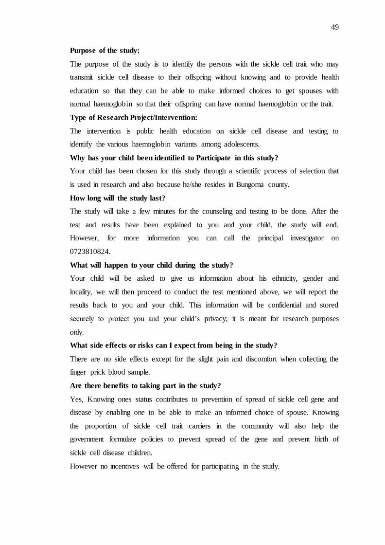
49
Purpose of the study:
The purpose of the study is to identify the persons with the sickle cell trait who may
transmit sickle cell disease to their offspring without knowing and to provide health
education so that they can be able to make informed choices to get spouses with
normal haemoglobin so that their offspring can have normal haemoglobin or the trait.
Type of Research Project/Intervention:
The intervention is public health education on sickle cell disease and testing to
identify the various haemoglobin variants among adolescents.
Why has your child been identified to Participate in this study?
Your child has been chosen for this study through a scientific process of selection that
is used in research and also because he/she resides in Bungoma county.
How long will the study last?
The study will take a few minutes for the counseling and testing to be done. After the
test and results have been explained to you and your child, the study will end.
However, for more information you can call the principal investigator on
0723810824.
What will happen to your child during the study?
Your child will be asked to give us information about his ethnicity, gender and
locality, we will then proceed to conduct the test mentioned above, we will report the
results back to you and your child. This information will be confidential and stored
securely to protect you and your child‟s privacy; it is meant for research purposes
only.
What side effects or risks can I expect from being in the study?
There are no side effects except for the slight pain and discomfort when collecting the
finger prick blood sample.
Are there benefits to taking part in the study?
Yes, Knowing ones status contributes to prevention of spread of sickle cell gene and
disease by enabling one to be able to make an informed choice of spouse. Knowing
the proportion of sickle cell trait carriers in the community will also help the
government formulate policies to prevent spread of the gene and prevent birth of
sickle cell disease children.
However no incentives will be offered for participating in the study.

50
Who do I call if I have questions about the study?
Questions about the study: Dr. Einstein Kibet 0723810824
Questions about your rights as a research subject: You may contact Institutional
Review Ethics Committee (IREC) 053 33471 Ext.3008. IREC is a committee that
reviews studies for safety and to protect the rights of study participants.
Will the information you provide be kept private?
All reasonable efforts will be made to keep your protected information (private and
confidential. Protected Information is information that is, or has been, collected or
maintained and can be linked back to you or your child. Using or sharing
(“disclosure”) of such information must follow National privacy guidelines. By
signing the consent document for this study, you are giving permission
(“authorization”) for the uses and disclosures of your child‟s personal information. A
decision to take part in this research means that you agree to let the research team use
and share the child‟s Protected Information as described below.
As part of the study, Dr Einstein Kibet Watenga and his study team may share the
results of your child‟s information e.g. laboratory tests, risk factors. These may be
study or non-study related. They may also share portions of your child‟s medical
record, with the groups named below:
The National Bioethics. Committee,
The Institutional Review and Ethics Committee,
[Add others as appropriate, e.g., national institutes of health, representatives of
{sponsor name},etc].
National privacy regulations may not apply to these groups; however, they have their
own policies and guidelines to assure that all reasonable efforts will be made to keep
your personal information private and confidential.
The sponsor may give your child‟s personal health information, not containing his/her
name, to others or use it for research purposes other than those listed in this form. In
handling your child‟s personal information, the sponsor, [PI] and associated staff will
keep your child‟s information in strict confidence, and shall comply with any and all
applicable laws regarding the confidentiality of such information.
The study results will be retained in your child‟s research record for at least six years
after the study is completed. At that time, the research information not already in

51
your child‟s medical record will be deleted. Any research information entered into
your child‟s medical record will be kept indefinitely.
Unless otherwise indicated, this permission to use or share your child‟s Personal
Information does not have an expiration date. If you decide to withdraw your
permission, we ask that you contact Dr Einstein Kibet Watenga, telephone
0723810824 in writing and let him know that you are withdrawing your permission.
The mailing address is, email: [email protected], P.o box 4606, 30100 Eldoret.
At that time, we will stop further collection of any information about you. However,
the health information collected before this withdrawal may continue to be used for
the purposes of reporting and research quality.
You have the right to see and copy your child‟s personal information related to the
research study for as long as the study doctor or research institution holds this
information. However, to ensure the scientific quality of the research study, you will
not be able to review some of your child‟s research information until after the
research study has been completed.
Your child‟s treatment, payment or enrollment in any health plans or eligibility for
benefits will not be affected if you decide not to take part. You will receive a copy of
this form after it is signed.
Part II: Consent of Subject:
I have read or have had read to me the description of the research study. The
investigator or his/her representative has explained the study to me and has answered
all of the questions I have at this time. I have been told of the potential risks,
discomforts and side effects as well as the possible benefits (if any) of the study. I
freely volunteer to take part in this study.
______________________ ______________________ __________________
Name of Participant Signature of subject/thumbprint Date &
Time
(Witness to print if the
Subject is unable to write
__________________________ ____________________________________
Name of Representative/Witness Relationship to Subject
________________________ ____________________ __________
Name of person Obtaining Consent Signature of person Date
Obtaining Consent
______________________ ___________________________
Printed name of Investigator Signature of Investigator Date

52
Appendix V: Assent Form.
ASSENT FORM FOR PARTICIPATION IN A STUDY TO DETERMINE THE
PREVALENCE OF SICKLE CELL TRAIT AMONG ADOLESCENTS IN
BUNGOMA COUNTY.
The researcher has explained to me about the study.
I understand that the researcher will ask questions about me.
I understand that they will take a little blood from me.
They need this sample so that they can determine my haemoblobin type and that
my sickle cell status can be determined.
My parent/guardian has agreed that I should participate in the study, I also
agree to the following. I voluntarily agree that I will participate as follows:
1. BE INTERVIEWED
2. FOR SAMPLES TO BE OBTAINED
Signature/mark of the child………………………………………
Name of child…………………………………………………….
Date………………………………………………………………

53
Appendix VI: Swahili Assent Form.
RUHUSA YA KUHUSIKA KWAKO KWENYE UTAFITI WA KISAYANSI
KUHUSU “SICKLE CELL TRAIT” KAUNTI YA BUNGOMA.
Nimeelezwa kuhusu utafiti huu.
Najua nitaulizwa maswali kunihusu.
Nafahamu kwamba nitatolewa damu kiasi.
Najua kwamba damu hiyo niyakubaini hali yangu ya sickle cell.
Mzazi wangu abamekubali nihusike kwenye utafiti huu ,mimi pia nimekubali
kwa hiari kuhusika kwa namna ifuatayo:
1. KUHOJIWA.
2. KUCHUKULIWA DAMU
Sahihi/Alama ya mtoto……………………………………………………………
Jina la mtoto……………………………………………………………………….
Tarehe……………………………………………………………………………..

54
Appendix VII: The Counseling Process.
Pretesting counseling.
1. Welcome participant.
2. Reassure participant.
3. Ask what they know about sickle cell disease.
4. Go through basic information on sickle cell disease including transmission and
severity of homozygous state.(sickle cell anaemia)
5. Emphasise on importance of screening and knowing status for prevention
purposes.
6. Discuss implication of results.
7. Explain to participant how the rapid immunoassay using sickle scan test works
and how results are read.
8. Get informed consent from participant.
9. Collect demographic data on questionnaire.
10. Test participant.(see appendix V on how to test)
Post testing counseling.
1. Reassure participant.
2. Go through the results together.
3. Discuss implications of result obtained.
4. Offer medical advice to participants.

55
Appendix VIII: The Sickle Scan™ Test Protocol.
STEP 1.
Do not open pouch until ready to use.
Prepare necessary materials:
A: Sickle SCAN™ cartridge
B: PreTreatment Buffer Module
C: Capillary Sampler (5 µL volume)
*Label PreTreatment Module and Test cartridge with patient ID.
STEP 2.
Obtain a fingerstick specimen using standard laboratory protocols.
*For heelstick or intravenous sampling follow standard laboratory protocols.

56
STEP 3.
Using Capillary Sampler, obtain 5 µL of fingerprick blood specimen.
*Take care to draw sample by capillary action; do not squeeze dispensing bulb.
STEP 4.
Open PreTreatment Module and immerse the Sampler tip into the Module. Dispense
the specimen into the buffer.
*Take care in opening the PreTreatment Module, as it contains a premeasured volume
of extraction buffer.
STEP 5.
Replace and tightly screw the two-piece cap onto the Module.

57
STEP 6.
Invert the module and mix 3 times, allowing complete treatment of the specimen with
buffer.
STEP 7.
Remove colored cap of the PreTreatment Module.
*Dispense 3 drops into waste to allow removal of any air bubbles in the dropper.
Immediately dispense 5 drops into the Sickle SCAN™ cartridge.
*Test on a level surface at room
STEP 8.
Allow test to run for 5 minutes.
*Test results that have run over 24 hours are invalid.
STEP 9.
Read the results of the Sickle SCAN™ by viewing the detection window.

58
POSSIBLE RESULTS.

59
Appendix IX: sickle scan™ validation certificate.

60
Appendix X: certificate of registration for medomix concept ltd.

61
Appendix XI: IREC Approval