the rna-binding protein hud: a regulator of neuronal differentiation, maintenance and plasticity
TRANSCRIPT

The RNA-binding protein HuD:a regulator of neuronal differentiation,maintenance and plasticityJulie Deschenes-Furry,1 Nora Perrone-Bizzozero,2 and Bernard J. Jasmin1,3*
SummarymRNA stability is increasingly recognized as beingessential for controlling the expression of a wide varietyof transcripts during neuronal development and synapticplasticity. In this context, the role of AU-rich elements(ARE)containedwithin the30 untranslated region (UTR)oftranscripts has nowemerged as key because of their highincidence in a large number of cellular mRNAs. Thisimportant regulatory element is known to significantlymodulate the longevity of mRNAs by interacting withavailable stabilizing or destabilizing RNA-binding pro-teins (RBP). Thus, in parallel with the emergence of ARE,RBP are also gaining recognition for their pivotal role in
regulating expression of a variety of mRNAs. In thenervous system, the member of the Hu family of ARE-binding proteins known as HuD, has recently beenimplicated in multiple aspects of neuronal functionincluding the commitment anddifferentiation of neuronalprecursors as well as synaptic remodeling in matureneurons. Through its ability to interact with ARE andstabilize multiple transcripts, HuD has now emerged asan important regulator of mRNA expression in neurons.The present review is designed to provide a comprehen-sive and updated view of HuD as an RBP in the nervoussystem. Additionally, we highlight the role of HuD inmultiple aspects of a neuron’s life from early differentia-tion to changes in mature neurons during learningparadigms and in response to injury and regeneration.Finally, we describe the current state of knowledgeconcerning the molecular and cellular events regulatingthe expression and activity of HuD in neurons.BioEssays28:822–833, 2006. � 2006 Wiley Periodicals, Inc.
Introduction
In recent years, the role of the 30untranslated region (30UTR)
has become increasingly recognized in modulating mRNA
stability, localization and translation in several cell biological
aspects. More specifically, the 30UTR can control mRNA
expression at the earliest stages of oogenesis, embryogen-
esis and tissue development (see for review Refs 1,2). Post-
transcriptional events have been implicated in the regulation of
short-lived mRNAs encoding specific cytokines, growth-
factors and immediate-early response genes (see for review
Ref. 3). Importantly, post-transcriptional mechanisms are not
limited to short-lived transcripts since they are also important
for the control of variousmRNAs encoding proteins involved in
metabolic activities including for example, the regulation of
parathyroid hormone mRNA stability by calcium and phos-
phate.(4) In parallel to the discovery of additionalmRNAs being
regulated via post-transcriptional mechanisms, there is also
an increase in the number of diseases of the cardiovascular,
immune and nervous systems that are associated with
mutations in the 30UTR of specific mRNAs or with misregula-
tion of RNA-binding proteins (RBP) that interact with 30UTRs
(see for review Refs 3,5–8).
Within the nervous system, the role of RBP in all aspects of
neuronal function is not only becoming evident but is also
1Department of Cellular and Molecular Medicine and Centre for
Neuromuscular Disease, Faculty of Medicine, University of Ottawa,
Ottawa, Ontario, Canada.2Department of Neurosciences, University of New Mexico, School of
Medicine, Albuquerque, New Mexico.3Molecular Medicine Program, Ottawa Health Research Institute,
Ottawa, Ontario, Canada.
Funding agencies. Work in the Jasmin laboratory is supported by the
Canadian Institutes of Health Research, the Muscular Dystrophy
Association of America and the Association Francaise contre les
Myopathies. Work in Perrone-Bizzozero’s laboratory is supported by
the National Institutes of Health; Grant number: NS30255.
*Correspondence to: Bernard J. Jasmin, Department of Cellular and
Molecular Medicine, University of Ottawa, 451 Smyth Road, Ottawa,
ON, K1H 8M5. E-mail: [email protected]
DOI 10.1002/bies.20449
Published online in Wiley InterScience (www.interscience.wiley.com).
822 BioEssays 28.8 BioEssays 28:822–833, � 2006 Wiley Periodicals, Inc.
Abbreviations: 30UTR, 30 untranslated region; AChE, acetylcholines-
terase; ARE, AU-rich element; CARM1, co-activator associated
methyltransferase 1; CPEB, cytoplasmic polyadenylation element
binding; DRG, dorsal root ganglion; ELAV, embryonic lethal abnor-
mal-vision; FMRP, Fragile-X mental retardation protein; hnRNP A1 or
K, heteronuclear ribonucleoprotein A1 or K; IMP1, insulin-like growth
factor mRNA binding protein 1; KSRP, K homology-type splicing
regulatory protein; MARCKS, myristoylated alanine-rich C kinase
substrate; PABP, poly(A)-binding protein; RBP, RNA-binding protein;
RNP, ribonucleoprotein; RRM, RNA-recognition motif; SCG, superior
cervical ganglion; SNP, single nucleotide polymorphism; ZBP, zipcode-
binding protein.
Review articles
gaining importance. This is likely related to the fact that
amongst cell types, neurons present an architectural chal-
lenge because of their relatively small cell body size and
extensive network of projections and connections. Aswe learn
more about the basic mechanisms underlying neurogenesis,
neurite elongation, synapse formation and plasticity, we find
that RNA-binding proteins are not only involved in all these
events but that they are in fact essential and, hence, act as
key components, to ensure their appropriate unfolding and
completion. Consequently, it is not surprising to find direct
associations between neurodegenerative diseases and con-
ditions such as spinal muscular atrophy, amyotrophic lateral
sclerosis and Fragile X syndrome, and perturbation in RNA
regulation and/or RBP activity (see for review Refs 3,5,6,8,9).
There are a number of well-characterized RBP that have
been shown to assume specific roles in normal neuronal
development and function (see Table 1).(10–13) Among
these RBP, the cytoplasmic polyadenylation element binding
(CPEB) protein, zipcode-binding protein (ZBP), Fragile-Xmental
retardation protein (FMRP) and Staufen are important for
mRNA transport along dendrites or axons.(14–19) In addition to
transporting specific target transcripts, CPEB, ZBPandFMRP
all have translational regulatory roles such that translation is
inhibited during transport and activated at the final destination
by specific signals.(20–24) Besides regulating transport and
translation, RBP are known to control mRNA degradation.
To date, the majority of these, including specifically AUF1
(seeTable 1), havebeen shown to stimulate or facilitatemRNA
decay.(25,26) In comparison, theHu family of RBP, ofwhichHuD
is the focus of this review, are among the few known proteins
that stabilize transcripts in the cytoplasm.
Over the last 15 years, there has therefore been grow-
ing interest in the function and regulation of the Hu family of
RBP, which is directly involved in development of paraneo-
plastic encephalomyelitis and sensory neuronopathy syn-
dromes.(31,33,36) These syndromes result from the expression
of neuronal proteins by tumor cells in the body, typically small-
cell lung cancer tumors. Ectopic expression of these ordinarily
immune-privileged proteins results in production of onconeur-
al antibodies that attack the nervous system and lead to the
development of autoimmune neurologic diseases. The anti-
body produced in the above disorder was termed anti-Hu
based on the name of the patient in which the antibody was
discovered.(31,37) The Hu antibody was used to identify HuD
and other Hu proteins (see further).(33)
Amongst the different family members, HuD is recognized
as oneof the earliestmarkers of neuronal cells aswell as being
an essential regulator of neuronal differentiation and survi-
val.(8,27,38–43) Of particular interest and of potential clinical
relevance is the recent association between polymorphisms
in the HuD gene and age-at-onset of Parkinson’s disease.(44)
A total of nine single nucleotide polymorphisms (SNP) within
the human HuD locus region were genotyped in this study,
of which two, one in intron 2 and one in exon 8, showed
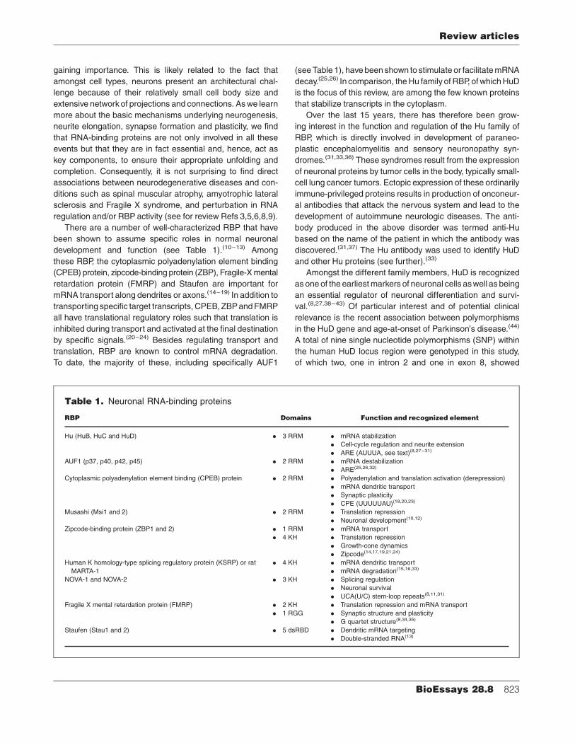
Table 1. Neuronal RNA-binding proteins
RBP Domains Function and recognized element
Hu (HuB, HuC and HuD) . 3 RRM . mRNA stabilization
. Cell-cycle regulation and neurite extension
. ARE (AUUUA, see text)(8,27–31)
AUF1 (p37, p40, p42, p45) . 2 RRM . mRNA destabilization
. ARE(25,26,32)
Cytoplasmic polyadenylation element binding (CPEB) protein . 2 RRM . Polyadenylation and translation activation (derepression)
. mRNA dendritic transport
. Synaptic plasticity
. CPE (UUUUUAU)(18,20,23)
Musashi (Msi1 and 2) . 2 RRM . Translation repression
. Neuronal development(10,12)
Zipcode-binding protein (ZBP1 and 2) . 1 RRM . mRNA transport
. 4 KH . Translation repression
. Growth-cone dynamics
. Zipcode(14,17,19,21,24)
Human K homology-type splicing regulatory protein (KSRP) or rat . 4 KH . mRNA dendritic transport
MARTA-1 . mRNA degradation(15,16,33)
NOVA-1 and NOVA-2 . 3 KH . Splicing regulation
. Neuronal survival
. UCA(U/C) stem-loop repeats(8,11,31)
Fragile X mental retardation protein (FMRP) . 2 KH . Translation repression and mRNA transport
. 1 RGG . Synaptic structure and plasticity
. G quartet structure(8,34,35)
Staufen (Stau1 and 2) . 5 dsRBD . Dendritic mRNA targeting
. Double-stranded RNA(13)
Review articles
BioEssays 28.8 823
moderately to strongly significant effects on age-at-onset of
Parkinson’s disease. Here, we therefore present a compre-
hensive and updated view of HuD and its role in multiple
aspects of a neuron’s life including the initial phenotypic
commitment and differentiation, as well as synaptic remodel-
ing during learning paradigms and in response to injury and
regeneration. Additionally, we describe the current state of
knowledge concerning the molecular and cellular events
regulating the expression, activity and function of HuD in
neurons.
The HuD Gene, its transcripts and
encoded proteins
As mentioned, HuD was the first of the Hu proteins to be
identified as theanti-Huantigen in patientswith paraneoplastic
encephalomyelitis and sensory neuronopathy.(31,33,36) The Hu
protein family also includes HuB (Hel-N1), HuC (Hel-N3) and
HuR (HuA).(33) Whereas HuR is ubiquitously expressed
and HuB is found in neurons and gonads, expression of HuC
and HuD is restricted to neurons. Due to the presence of three
highly conserved RNA-recognition motifs (RRM) (see below
and for review 45,46), HuD shares a high degree of homology
with the Drosophila proteins ELAV (responsible for the
Embryonic Lethal Abnormal Vision phenotype; see Fig. 1)
that is required for the normal development and maintenance
of the nervous system.(33,47)
HuD is encoded by a relatively large gene (�146 Kb)
located on chromosome1 in humans (1p34) and chromosome
4 in mice (49.5 cM). As shown in Fig. 2, some of the exons are
separatedby large intronic regions. The localizationof theHuD
gene in the mouse genome was based on syntenic regions
between human and mouse, suggesting that the flanking
genes are conserved between species.(48) The gene consists
of three putative non-coding exon 1 variants, termed 1a, 1b
and1c, andseveral commoncodingexons (2, 3, 4, 5and8; see
Fig. 2).(30,48) The first and second RRM are encoded by exons
2 and 3, and exons 4 and 5, respectively, while the third RRM is
encodedbyexon 8. TheHuDgene is also subject to alternative
splicing of exons 6 and 7 which affects the length of the
hinge region linking the second and third RRM. This results in
the three different transcripts and molecular forms, termed
HuDpro, HuD and HuDmex(30) of which HuDpro and HuD are the
major variants.(47–50) The resulting proteins have molecular
masses ranging between37and43 kDaandare characterized
by the three RRMs and unique nuclear export signals located
within the hinge region.(40)With theexceptionof theN-terminal
domain and the hinge region, the protein sequences between
the different Hu proteins are very similar (70 to 85% identity;
see Fig. 1).(47)
HuD’s RNA-recognition motifs (RRM)
and target transcripts
As shown in Figs 1 and 2, all Hu proteins contain three RRM.
RRMs are found in various proteins involved in RNA proces-
sing and turnover, including hnRNPA1 involved in splicing and
transport of RNA, poly(A)-binding protein (PABP) involved in
transcript stability and translation, and the Drosophila ELAV
protein involved in RNA splicing (see for review 45,46). The
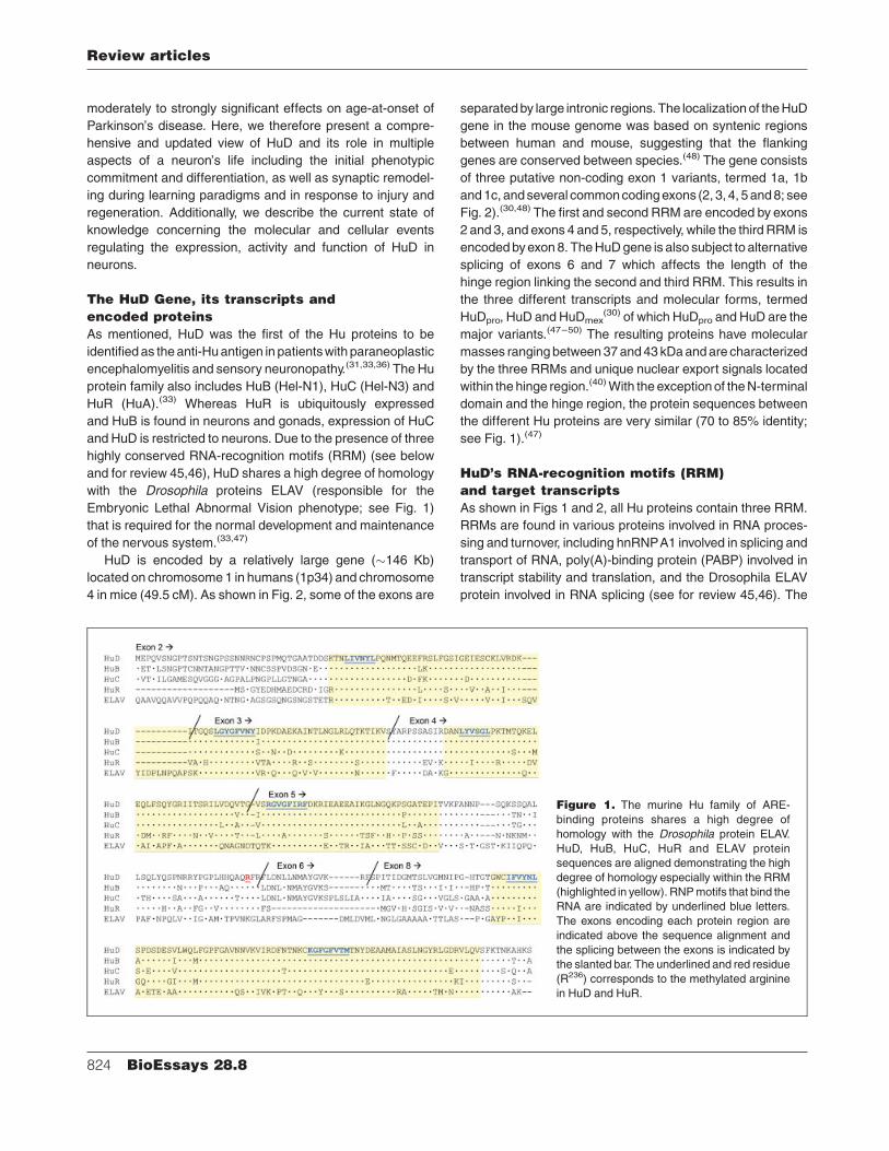
Figure 1. The murine Hu family of ARE-
binding proteins shares a high degree of
homology with the Drosophila protein ELAV.
HuD, HuB, HuC, HuR and ELAV protein
sequences are aligned demonstrating the high
degree of homology especially within the RRM
(highlighted in yellow). RNPmotifs that bind the
RNA are indicated by underlined blue letters.
The exons encoding each protein region are
indicated above the sequence alignment and
the splicing between the exons is indicated by
the slanted bar. The underlined and red residue
(R236) corresponds to the methylated arginine
in HuD and HuR.
Review articles
824 BioEssays 28.8

RRM is a conserved structure of�80–90 residues containing
two consensus ribonucleoprotein (RNP) motifs separated by
25–35 amino acids that interact directly with the RNA. The
RNP motifs, octameric RNP-1 and hexameric RNP-2, each
contain three conserved aromatic residues that are implicated
in RNA interactions in a number of different RBP including
proteins that bind pre-mRNA, mRNA, pre-rRNA as well as
small nuclear and heteronuclear RNAs. The consensus
structure of RRM consists of b1-a1-b2-b3-a2-b4 and the
location of the RNPmotifs in the first and third b-strands of theRRM is highly conserved.(45,51) Although the structure of RRM
iswell preserved, only a few residues foundmostly in the RNP
motifs are highly conserved. Consequently, it is the variable
regions between the RNP motifs and RRM that tend to lend
sequence specificity to the RBP. Similarly to other RRM-
containing RBP, the first and second HuD RRM consist of the
consensus RRM structure and form a cleft between them
where the RNA is bound specifically between the b-sheets.(52)
Crystal structureexperiments revealed that the residueswithin
the first and second HuD RRM that interact with the RNA, are
conserved between HuD and ELAV proteins.(52)
Hu proteins recognize and bind specifically to well-
described AU-rich elements (ARE) found within the 30UTR of
approximately 1 in 20 human genes(53,54) and directly
implicated in RNA turnover.(30,53,55,56) The AREs are sepa-
rated into three classes based on their sequence and
structure. Class I AREs consist of one to three dispersed
AUUUAmotifs separated byU-rich regionswhile class II AREs
consist of multiple clusters of AUUUA motifs. Class III AREs,
although less-well defined, consist mainly of U-rich sequences
and do not contain the AUUUA pentamer.(57) The different
classes of AREs appear to instruct distinct modes of mRNA
decay. Class I and III ARE-directed mRNA degradation
involves simultaneous deadenylation of mRNAs resulting in
a pool of transcripts of homogeneous lengths, termed
distributive deadenylation, which is followed by degradation
of the mRNA body. In contrast, Class II ARE-directed mRNA
degradation consists of complete deadenylation of one
transcript resulting in a pool of mRNAs of heterogeneous
lengths, termed processive deadenylation.(58) Recently, HuD-
RNA crystal structure and in vitro binding assays have
determined that the consensus sequence recognized by
RRM1 and RRM2 is X-U/C-U-X-X-U/C?-U-U/C, where the
‘‘?’’ denotes questionable binding of theC in this location.(52,59)
Consequently, HuD has been demonstrated to bind and
stabilize transcripts with CU- and U-rich sequences as well
as class II and III AU-rich elements. The structure of the RRM
allows for the binding of any number of domains and RNA
conformations.(52) Similarly to HuR, HuD, while able to bind all
classes of ARE, retains some sequence specificity and
preference such that it will not bind a transcript based solely
on the presence of an ARE.(60)
Unlikemany other ARE-binding proteins that principally act
to destabilize transcripts, such as AUF1 family members
(Table 1), Hu proteins act to stabilize ARE-containing
transcripts thereby significantly prolonging their half-
life.(39,42,61,62) In vitro binding studies performed with HuD
have demonstrated that the RNA is bound primarily by the first
RRM while the second RRM, which also binds the RNA,
functions mostly to stabilize the RNA–protein complex.(52,63)
Figure 2. HuD gene, transcript and protein organization. A: HuD exon and intron organization is depicted. Green boxes correspond to
three putative alternative exon1 variants, blue boxes correspond to coding region exons and red boxes correspond to alternatively spliced
coding region exons. Note the large intronic regions separating exons 1 through 4. B: Alternative splicing of exons 6 and 7 results in the
production of three alternative transcripts (HuDpro, HuD and HuDmex) which have untranslated regions (UTR and exon 1 shown in green) of
varying and unknown lengths. The promoter region driving gene transcription and the predominant exon 1 variant alongwith the resulting 50
untranslated region, remain unknown.C:Protein organization of the differentHuD transcripts. Thevarying length of the hinge region linking
the second and third RRM results from alternative splicing of exons 6 and 7.
Review articles
BioEssays 28.8 825
The third RRM, in addition to participating in maintaining
RNA–protein complex stability, binds to poly(A) tails.(64–67) In
the latter case, the length of the poly(A) tail correlates with the
overall binding efficiency of Hu proteins.(64) In contrast, the
third RRM of mouse HuB and HuC could not bind to poly(A)
ribohomopolymers and generally showed very little RNA-
binding activity.(68) There are, however, some inconsistencies
between studies as another study performed with mouse HuC
demonstrated that the third RRM could specifically bind to
poly(A)-sepharose beads.(66) The different results obtained
with each Hu family member may simply reflect different
technical approaches. In that context, the studies performed
with HuD used complete in vitro transcribed GAP-43 tran-
scripts with differing lengths of poly(A) tails as opposed to
poly(A) nucleotide chains, and could consequently be con-
sidered a more accurate representation of the binding activity
of the third RRM.(64) Alternatively, it is possible that the third
RRM is also involved in protein–protein interactions rather
than direct RNA-binding, as suggested by Kasashima et al.
(2002), who showed that HuD, HuB and HuC could form
dimers via the third RRM.
As the variety of cis-acting elements recognized by the
RRM have increased, so have the number of transcripts that
interact with HuD. Accordingly, HuD has been shown to bind
and stabilize several developmentally regulated transcripts
including c-fos, c-myc, N-myc, p21waf1, neuroserpin and
MARCKS.(69–73) Interestingly, and in agreement with its role
in neuronal differentiation, many of the transcripts bound by
HuD play a key role in the formation of neuronal processes
such as GAP-43 and tau.(39,62,74) In this context, our studies
have recently shown that HuD also binds and stabilizes
acetylcholinesterase (AChE) transcripts during neuronal
differentiation.(42) Since in addition to its well-recognized
function in neurotransmission, AChE can also stimulate
neurite outgrowth (Refs 42,75,76 and references therein), it
appears therefore that HuD binds and regulates the stability of
a subset of transcripts with similar functional roles that is key
for neuronal development and plasticity.
Although the principal function of HuD is to stabilize its
target transcripts and increase their half-life, several studies
performed with HuD or other members of the Hu family
suggest that HuD is also involved in other aspects of mRNA
regulation (see Figure 3). The variable linker domain sepa-
rating the second and third RRMs has been suggested
to contain a novel nuclear export signal that allows HuD
to shuttle target transcripts out of the nucleus into the
cytoplasm.(29,40,77) In addition, the first and second RRM and
part of the linker domain have been shown to interact with the
primary mRNA export receptor TAP/NXF1 in cultured neuro-
nal cells.(78) In fact, previous studies have shown that neurite
elongation is severely impeded when shuttling of HuD is
blocked, suggesting that HuD plays an important role in
localizing its target transcripts to the cytoplasm.(40) Once in the
cytoplasm, HuD and its cargo are targeted to axons, in the
case of tau mRNA, and growth cones, in the case of GAP-43
mRNA.(79,80)
Specific subcellular localization of the HuD-mRNA gran-
ules appears to involve the KIF3A microtubule-associated
motor protein.(79) Along these lines, HuDwas shown to bind in
an RNA-dependent manner to insulin-like growth factor
mRNA-binding protein 1 (IMP1).(40,81) IMP1 is the mouse
ortholog of chicken ZBP1 which is known to have a role in
mRNA transport and translation regulation (see Table 1).(82)
Furthermore, HuD has also been shown to colocalize with
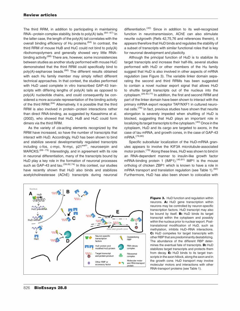
Figure 3. HuD function and regulation within
neurons. A: HuD gene transcription within
neurons may be controlled by neuron-specific
transcription factors. HuD transcript may also
be bound by itself. B: HuD binds its target
transcript within the cytoplasm and possibly
within the nucleus prior to nuclear export. Post-
translational modification of HuD, such as
methylation, inhibits HuD–RNA interactions.
C: HuD competes for target transcripts with
otherRBP that are predominantly destabilizing.
The abundance of the different RBP deter-
mines the eventual fate of transcripts. D: HuDstabilizes target transcripts and protects them
from decay. E: HuD binds to its target tran-
scripts in the axon hillock, along the axonand in
the growth cone. HuD transport may involve
molecular motors and interactions with other
RNA-transport proteins (see Table 1).
Review articles
826 BioEssays 28.8
ribosomes in dendrites of pyramidal neurons of the CA1 and
CA3 regions of the hippocampus.(83) Finally, HuD-bound
mRNAs are also targeted to ribosomes and polysomes,
similarly to HuB and neurofilament-M mRNA.(80,81,84) These
studies suggest therefore that HuD could also be involved,
either directly or indirectly through binding with other RBP
with translational functions, in stimulating translation of its
target transcripts. In this context, a recent study has shown
that HuB can interact with hnRNP K and both can bind to
p21 mRNA 30UTR.(85) This later work also demonstrated
that hnRNP K alone decreased the translation of this
transcript, whereas HuB could block this effect and indirectly
increase translation. Given the similarities in Hu protein
function and target transcripts, it is possible that HuD has
similar functions.
Functional role of HuD in neurons
The neuronal Hu proteins are known to be some of the earliest
markers of the neuronal phenotype since they appear to
suppress neuroblast proliferation and promote neuronal
differentiation.(47,68,86–88) In situ hybridization experiments
performed in developing and adult mouse and rat cerebral and
cerebellar cortex revealed a distinct expression pattern for
mRNAs encodingHuB, HuC andHuD.(47,89) During embryonic
development, HuD expression can be localized to cells exiting
the cell cycle in the ventricular zone, and to those migrating in
the intermediate zones and undergoing terminal differentia-
tion.(41,47,89) In the adult brain, HuD-positive neurons corre-
spond primarily to projection neurons found in the neocortex,
hippocampus, entorhinal cortex and cerebellum. Similarly,
HuD is very strongly expressed in the ventral motoneurons of
the spinal cord, the sensory neurons of the dorsal root ganglia
(DRG) and sympathetic neurons in their ganglia.(41,89) At the
subcellular level, HuD is detected in the cell body, axon and
growth cones.(47,79,80) Generally, HuB and HuD have a similar
pattern of expression which is somewhat different from HuC.
Although expression of HuB and HuD overlap, they appear to
have similar(68,85) and opposing(90,91) functions during neuro-
nal development depending on the context. For example, they
can both stimulate neuronal differentiation and neurite out-
growth but have opposing roles in progenitor cell self-renewal,
such that HuB positively and HuD negatively affects this
capacity.
Studies in which the expression level of HuD has been
manipulated byeither overexpression or downregulation, have
highlighted the significance of HuD in stabilizing various
mRNA in neurons. Specifically, HuD binds a subset of
transcripts that encode proteins involved in neuronal develop-
ment. Several recent studies have shown that overexpression
of HuD increases the rate and length of neurite outgrowth in
several different types of neuronal cells, including in both
primary cultures and neuronal cell lines. By contrast,
decreased HuD expression results in an inhibition of neurite
extension.(38,39,41,62) Together, these results indicate that HuD
is an important regulator of neurite elongation and morpholo-
gical differentiation.(8,92) Expression of HuD in non-neuronal
cells does not stimulate the development of processes,
suggesting that pluripotent cells must already be engaged
along a neuronal cell fate in order for HuD to be effective in
promoting morphological differentiation.(38,41) This effect is
specific to neuronal Hu proteins, as HuB and HuC can also
stimulatemorphological differentiation, while HuR is unable to
stimulate neurite extension in neural crest cells.(41,68,84)
Recently, HuD transgenic and HuD knockout mice have
beengeneratedand characterized.(91,93) HuD transgenicmice
specifically expressed higher total amounts of HuD in the
forebrain, particularly in all subregions of the hippocampus
including in dentate granule cells that do not normally express
HuD.(47,83,89) Interestingly, in both HuD-overexpressing and
-deficient mice, there are no apparent morphological or
structural defects in the adult brain. However, extension of
some cranial nerves is transiently impaired during embry-
ogenesis in HuD-deficient mice. In comparison, loss-of-
function alleles of Drosophila elav are embryonic-lethal and
hypomorphic mutations result in morphological and structural
defects of the eye.(94,95) Given that there are three neuronal Hu
proteins with similar functions and one ELAV, it is possible that
expression of the other members of the Hu family are
compensating for the loss of HuD in knockout animals.
Although there are no apparent morphological defects, HuD-
deficient mice display motor/sensory defects such as an
abnormal clasping reflex of the hindlimbs when suspended by
the tail.(91) The abnormal clasping reflex suggests a defect in
sensory and motor functions which is also confirmed by poor
performance in rotarod experiments.(91) Both of these mouse
models of aberrant HuD expression have initially been used to
confirm some of the in vitro and cell culture data and to further
elucidate the importance of HuD to neuronal development and
function.
During early developmental stages, HuD appears to be
involved at multiple levels of cell lineage commitment,
differentiation and survival. Studies using HuD knockout mice
showed that, during embryogenesis, the absence of HuD is
related to an increase in the progenitor cells’ ability to renew,
and to a decrease in their ability to exit the cell cycle and
morphologically differentiate into neurons.(91) Cells that are
not leaving the cell cycle are also more likely to undergo
apoptosis since levels of apoptosis are elevated in HuD-
deficient mice. In adult mice, the number of subventricular
zone progenitor cells is greater in the HuD knockout mice.
These studies suggest therefore, that HuD is important for
promoting the exit from the cell cycle, for negative regulation of
proliferation and stimulation of the differentiation process.(91)
In addition, an increased level of HuD in cultures of neural crest
cells precipitates neurotrophin dependence.(41,96) Similarly,
HuD overexpression in mouse embryonic stem cells can
Review articles
BioEssays 28.8 827

double the number of cells with long neurites and expressing
GAP-43 only when the cells are induced to differentiate with
retinoic acid.(38) The results obtained with HuD knockout mice
and from cultured neuronal cell lines, together with the
preferential expression of HuD in projection neurons, strongly
suggest that HuD through its stabilizing effect on several
transcripts, is essential for the proper development and
maintenance of neuronal extensions.
The significance of HuD in the regulated expression of
mRNAs and proteins involved in the growth and formation of
neuronal projections, suggests that it is also important to other
aspects of axonal and dendritic functions in the adult nervous
system.A fewdistinct andcomplementary studieshaveclearly
demonstrated that spatial learning tasks can enhance HuD
expression in hippocampal neurons resulting in a concomitant
increase in GAP-43 transcript levels.(97,98) Another experi-
mental paradigm, single trial contextual fear conditioning, also
demonstrated an association between HuD expression and
learning.(83) In the latter study, HuD expression was increased
in different regions of the hippocampus namely, in the hilar
region of the dentate gyrus, as well as in the CA3 and CA1
regions. Along similar lines, increased expression of HuD in
the hippocampus of transgenic mice overexpressing HuD,
stimulates GAP-43 mRNA expression and stabilization(93) as
well as AChE mRNA expression.(99) More specifically, over-
expression of HuD in dentate granule cells, where HuD is not
normally expressed, results in a marked increase in GAP-43
mRNA levels via post-transcriptional mRNA stabilization of
newly synthesized, but normally degraded, transcripts.(93)
Consequently, atypical expression of HuD in neurons may
have important regulatory effects on the expression of its
target transcripts and, thus, a significant downstreameffect on
neuronal function.
Given its significant role in morphological differentiation,
HuD has also been suggested to have a role in axonal
regeneration. Recent findings have shown that following nerve
crush of DRG sensory neurons and facial motoneurons, HuD
protein and transcript levels increasewithin 7 days of the injury
and remain elevated for up to 21 days.(100,101) This increase in
HuD expression is accompanied by a dramatic increase in the
mRNA levels of GAP-43, a known target of HuD. In
regenerating neurons of the DRG, HuD protein colocalized
with GAP-43 transcripts as well as with ribosomal RNA.(100) A
recent study has also demonstrated the co-localization of HuD
and GAP-43 mRNA in growth cones.(80) In this context, we
have found that exogenousexpressionof humanHuD in the rat
superior cervical ganglion (SCG) results in themaintenance of
AChE and GAP-43 mRNA levels following axotomy (unpub-
lished data). Together, these findings demonstrate that,
in addition to its role during neuronal development, HuD
expression and function correlate with plasticity of the nervous
system both in response to injury and during learning. Further
studies performed with the HuD-transgenic and -deficient
mice are necessary to strengthen these correlations into direct
or indirect involvement of HuD.
Molecular events regulating expression
of HuD and its function
Despite the well-established involvement of HuD in the control
of a subset of neuronal transcripts involved in neuronal
differentiation and plasticity, there appears to be a lack of
information concerning thenatureof themolecular andcellular
mechanisms that regulate expression of HuD in neurons. This
is important since a number of recent studies point to mRNA
stability as a consequence of functional antagonism between
stabilizing and destabilizing proteins (see for reviewRef. 102).
This is best exemplified by studies performed with the
ubiquitous Hu family member HuR, whose tissue expression
overlaps with AUF1, a family of four isoforms that are
predominantly destabilizing (Table 1).(32,103) In addition, many
transcripts that havebeen identifiedas targets forHuRarealso
AUF1 targets, such as p21, c-fos, TNF-alpha and cyclin
D1.(102,104) The antagonistic effects of these proteins have
been demonstrated using siRNA to individually decrease the
expression of HuR or AUF1, resulting in increased association
of the target transcript with the opposite protein and
subsequent decreased or increased stability, respectively.(104)
Using a similar approach, competition for transcript-binding
siteswas also demonstratedwithin the AUF1 family.(105) In this
context, it is important to note that AUF1 has also been
reported to have mRNA-stabilizing activity.(106,107) Thus,
it appears that competition for target transcripts and the
eventual outcome for their longevity are largely determined by
the relative abundance of the different RBP.(102,104) Accord-
ingly, it becomes important to gain a better understanding of
the events presiding over HuD expression in neurons.
Transcriptional and post-transcriptionalregulatory mechanisms.The HuD gene is divided into eight coding exons beginning
with exon 2 (see Figs 1 and 2).(48) A comparison of the
available cDNA sequences in GenBank (NM_010488,
BC052451 and BC052451) shows the presence of three
alternate non-coding exon1 splice variants termed exon1a, 1b
and 1c (see Fig. 2).(48) Presently, it is unclear whether a single
promoter is responsible for transcription of this large gene
with splicing of exon1 variants occurring subsequently or
whether there are multiple promoter choices driving specific
expression of exon 1 variants. Although the HuD 50 regulatory
region of the mammalian gene has yet to be isolated and
characterized, studies performed with the Xenopus laevis
HuD homologue, elrD, have shown that there are two possible
promoter regions upstream of exon 2.(108) The most 50
promoter region is upstream of a non-coding exon 1 and the
second promoter region is located within the first intron. Both
Review articles
828 BioEssays 28.8
promoter regions have been shown to drive transcription in a
neuronal-dependent fashion and to contain neuron-specific
transcription factor binding sites (E-box and neuron-restrictive
silencer factor site). Similarly, characterization of the human
HuB 50 regulatory region demonstrates the presence of cis-
acting elements that can direct tissue-specific expression in
neurons.(109) Analysis of the zebrafish HuC 50 regulatory
region resulted in the identification of multiple E-box se-
quences, a putative MyT1-binding site and two GC boxes that
could also drive neuron-specific expression of HuC.(110,111)
The Drosophila ELAV 50 flanking region including enhancer
domains within an intronic region, also confers neural-specific
expression patterns to this gene.(112) Accordingly, the specific
and early neuronal expression of HuD alongwith the presence
of alternate 50 regulatory regions strongly suggest that
expression of HuD is regulated by transcriptional mechanisms
involving selective cis-acting elements and trans-acting
factors that are specific to neurons (seeRef. 87). In agreement
with this view, a recent study demonstrated that the transcrip-
tional rate of HuD is significantly altered in neurons subjected
to changes in the levels of thyroid hormone.(113)
In addition to transcriptional regulatory events, the relative
abundance of HuD may also be regulated by post-transcrip-
tional mechanisms (see Figure 3). Northern blot analysis has
revealed the presence of two HuD transcripts of �3.7 and
4.4 kb in neurons.(114) These are thought to originate from
alternative polyadenylation or splicing to a putative down-
stream non-coding exon (exon 9).(48) Alignment of available
cDNA sequences indeed reveals the presence of alternate
30UTRs. Amongst the different Hu proteins, the 30UTRs
are unique to each transcript.(47) In contrast, specific se-
quences coding for stem-loop structures and putative ARE
within the HuD 30UTR are highly conserved between different
species.(28) In addition, the 30UTR of the Drosophila homo-
logues (ELAV and SxL) are important for the appropriate
regulation of these transcripts.(115,116) More specifically, HuB,
ELAVand SxL have been shown to autoregulate their expres-
sion through interactions with their own 30UTR.(116–118)
Recent studies have shown that ELAV expression is depen-
dent on the presence of a long UTR at its 30 end.(115) An
ELAV-binding site was in fact found within the elav mRNA
located within an alternative non-coding 30exon.(118) The
authors of this study proposed that ELAV protein binds to
elav mRNA 30UTR when protein levels reach a certain
threshold and, in this case, block further protein production.
Such a similar negative autoregulatory loop also can be
assigned to SxL protein, which can downregulate its own
expression by binding to specific regions within the
sxl 30UTR.(116) Together, these observations are coherent
with the notion that, in addition to transcriptional events
(see paragraph above), post-transcriptional mechanisms
operating through the 30UTR are also important for control-
ling HuD expression.
Post-translational regulatory mechanismsAlthough the abundance of RBP is important to ultimately
control the fate of specific transcripts, other factors such as the
RBPaffinity and function are also implicated. In a recent study,
wehaveshown thatHuDdidnot actively bind tooneof its target
transcripts until neuronal differentiation was stimulated, even
though significant levels of HuD protein were present in
undifferentiated PC12 cells and these levels did not change
upon differentiation (see Refs 42,49,62). This suggests that
additional factors affect the ability of HuD to stabilize target
transcripts. In this context, several recent studies have begun
to elucidate the pathways regulating post-translational mod-
ification of the Hu family of RNA-binding proteins including
phosphorylation andmethylation, and to determine the impact
of these modifications on their activity.(54,62,119) Notably, NGF-
induced differentiation and stress-related stimuli can indirectly
regulate HuD and HuR function, respectively, by a PKC-
mediated signaling cascade.(62,120) Recently, a series of
indirect experiments led to the suggestion that threonine
phosphorylation of the neuronal Hu proteins (HuB, HuC and
HuD) by PKCa isozyme results in the re-distribution of the
proteins and in an increased stabilization of GAP-43 mRNA,
a well-known HuD target.(54)
In addition to phosphorylation, a recent study has shown
that arginine residues in the HuR hinge region are methylated
by the methyltransferase CARM1 (coactivator-associated
arginine methyltransferase 1) in response to lipopolysacchar-
ide.(119) Arginine methylation is another common modification
of some RBP that results in modification in the pattern of
protein–protein interactions. Along those lines, it has also
recently been demonstrated in PC12 cells that HuD is
methylated on the corresponding arginine (Arg236, see Fig. 1)
by the same methyltransferase, and that methylation results
in decreased RNA-binding activity of HuD.(121) In these
studies, methylation-resistant mutant HuD increased expres-
sion of some target transcripts such as p21cip1/waf1, and
resulted in increased neuronal differentiation of PC12 cells.
Although these cell culture studies suggest that phosphoryla-
tion and methylation can regulate HuD function directly or
indirectly, it should be noted that these studies have been
performed in vitro. Whether post-translational modifications
are also occurring in vivo still remains to be determined.
Nonetheless, these studies indicate that post-translational
modifications are also likely to be important in regulating the
binding and functional activity of HuD on target transcripts.
Finally, the fate of target transcripts may also depend on
other factors that might modulate the binding activity and
specificity of HuD. These include the structure and sequence
of the target transcripts and the interaction with additional
RBPs. The transcript itself can, in many cases, modify the
function of RBP (see for review Refs 52,122). An example of
this was given above with the poly(A) tail since the presence
and length of the poly(A) tail can alter the binding of theRRM to
Review articles
BioEssays 28.8 829
the ARE.(64,67) The presence of HuD in RNP granules and the
suggested multimerization of HuD implies that interactions
with other proteins or RBP can modulate the activity of HuD
and its specificity in a manner analogous to the interactions of
transcription factors on promoter elements.(47,79,85,123) This
may explain in part the identification of numerous RBPs
interacting with a single transcript such asGAP-43, AChE and
the early-response genes c-myc and c-fos (see for review
102).(42,61,62,124)
Conclusion and perspective
To date, there is compelling evidence indicating that, through
its ability to bind and stabilize a specific subset of transcripts,
HuD plays an essential role in the development and
maintenance of neuronal phenotype. The downstream effects
of this specific activity include the stimulation of morphological
differentiation of newly committed and developing neurons.
Additionally, HuDexpression andbindingactivity have recently
been shown to increase during nerve regeneration as well as
with learning andmemory. The ability of HuD to directly control
these key molecular and cellular events indicates that HuD
may act as an important regulator of neuronal differentiation
and function. Therefore, a thorough understanding of the
mechanisms affecting the expression, activity and function of
HuD in neurons appears warranted in order to increase our
basic knowledge of the events regulating neuronal differentia-
tion, maintenance and plasticity. Ultimately, this knowledge
could prove useful for the development of novel therapeutic
strategies aimed at manipulating, pharmacologically or
through gene therapy approaches, the levels of RBP that
may be beneficial for the treatment of several neurological
diseases and conditions.
Acknowledgments
We thank members of the Jasmin laboratory for fruitful
discussions.
References1. Bashirullah A, Cooperstock RL, Lipshitz HD. 1998. RNA localization in
development. Annu Rev Biochem 67:335–394.
2. King ML, Zhou Y, Bubunenko M. 1999. Polarizing genetic information
in the egg: RNA localization in the frog oocyte. Bioessays 21:546–
557.
3. Hollams EM, Giles KM, Thomson AM, Leedman PJ. 2002. MRNA
stability and the control of gene expression: implications for human
disease. Neurochem Res 27:957–980.
4. Naveh-Many T, Bell O, Silver J, Kilav R. 2002. Cis and trans acting
factors in the regulation of parathyroid hormone (PTH) mRNA stability
by calcium and phosphate. FEBS Lett 529:60–64.
5. Conne B, Stutz A, Vassalli JD. 2000. The 30 untranslated region of
messenger RNA: a molecular ‘hotspot’ for pathology? Nat Med 6:637–
641.
6. Mendell JT, Dietz HC. 2001. When the message goes awry: disease-
producing mutations that influence mRNA content and performance.
Cell 107:411–414.
7. Misquitta CM, Iyer VR, Werstiuk ES, Grover AK. 2001. The role of 30-
untranslated region (30-UTR) mediated mRNA stability in cardiovascular
pathophysiology. Mol Cell Biochem 224:53–67.
8. Perrone-Bizzozero N, Bolognani F. 2002. Role of HuD and other RNA-
binding proteins in neural development and plasticity. J Neurosci Res
68:121–126.
9. Gallo JM, Jin P, Thornton CA, Lin H, Robertson J, et al. 2005. The role of
RNA and RNA processing in neurodegeneration. J Neurosci 25:10372–
10375.
10. Imai T, Tokunaga A, Yoshida T, Hashimoto M, Mikoshiba K, et al. 2001.
The neural RNA-binding protein Musashi1 translationally regulates
mammalian numb gene expression by interacting with its mRNA. Mol
Cell Biol 21:3888–3900.
11. Jensen KB, Dredge BK, Stefani G, Zhong R, Buckanovich RJ, et al.
2000. Nova-1 regulates neuron-specific alternative splicing and is
essential for neuronal viability. Neuron 25:359–371.
12. Okano H, Imai T, Okabe M. 2002. Musashi: a translational regulator of
cell fate. J Cell Sci 115:1355–1359.
13. Roegiers F, Jan YN. 2000. Staufen: a common component of mRNA
transport in oocytes and neurons? Trends Cell Biol 10:220–224.
14. Bassell GJ, Oleynikov Y, Singer RH. 1999. The travels of mRNAs
through all cells large and small. FASEB J 13:447–454.
15. Rehbein M, Wege K, Buck F, Schweizer M, Richter D, et al. 2002.
Molecular characterization of MARTA1, a protein interacting with the
dendritic targeting element of MAP2 mRNAs. J Neurochem 82:1039–
1046.
16. Gherzi R, Lee KY, Briata P, Wegmuller D, Moroni C, et al. 2004. A KH
domain RNA binding protein, KSRP, promotes ARE-directed mRNA
turnover by recruiting the degradation machinery. Mol Cell 14:571–583.
17. Farina KL, Singer RH. 2002. The nuclear connection in RNA transport
and localization. Trends Cell Biol 12:466–472.
18. Huang YS, Carson JH, Barbarese E, Richter JD. 2003. Facilitation of
dendritic mRNA transport by CPEB. Genes Dev 17:638–653.
19. Zhang HL, Eom T, Oleynikov Y, Shenoy SM, Liebelt DA, et al. 2001.
Neurotrophin-induced transport of a beta-actin mRNP complex in-
creases beta-actin levels and stimulates growth cone motility. Neuron
31:261–275.
20. Mendez R, Richter JD. 2001. Translational control by CPEB: a means to
the end. Nat Rev Mol Cell Biol 2:521–529.
21. Ross AF, Oleynikov Y, Kislauskis EH, Taneja KL, Singer RH. 1997.
Characterization of a beta-actin mRNA zipcode-binding protein. Mol
Cell Biol 17:2158–2165.
22. Si K, Giustetto M, Etkin A, Hsu R, Janisiewicz AM, et al. 2003. A
neuronal isoform of CPEB regulates local protein synthesis and
stabilizes synapse-specific long-term facilitation in aplysia. Cell 115:
893–904.
23. Wu L, Wells D, Tay J, Mendis D, Abbott MA, et al. 1998. CPEB-mediated
cytoplasmic polyadenylation and the regulation of experience-dependent
translation of alpha-CaMKII mRNA at synapses. Neuron 21:1129–1139.
24. Huttelmaier S, Zenklusen D, Lederer M, Dictenberg J, Lorenz M, et al.
2005. Spatial regulation of beta-actin translation by Src-dependent
phosphorylation of ZBP1. Nature 438:512–515.
25. Guhaniyogi J, Brewer G. 2001. Regulation of mRNA stability in
mammalian cells. Gene 265:11–23.
26. Wilson GM, Brewer G. 1999. The search for trans-acting factors
controlling messenger RNA decay. Prog Nucleic Acid Res Mol Biol
62:257–291.
27. Antic D, Keene JD. 1997. Embryonic lethal abnormal visual RNA-
binding proteins involved in growth, differentiation, and posttranscrip-
tional gene expression. Am J Hum Genet 61:273–278.
28. Good PJ. 1995. A conserved family of elav-like genes in vertebrates.
Proc Natl Acad Sci USA 92:4557–4561.
29. Keene JD. 1999. Why is Hu where? Shuttling of early-response-gene
messenger RNA subsets. Proc Natl Acad Sci USA 96:5–7.
30. Liu J, Dalmau J, Szabo A, Rosenfeld M, Huber J, et al. 1995.
Paraneoplastic encephalomyelitis antigens bind to the AU-rich elements
of mRNA. Neurology 45:544–550.
31. Musunuru K, Darnell RB. 2001. Paraneoplastic neurologic disease
antigens: RNA-binding proteins and signaling proteins in neuronal
degeneration. Annu Rev Neurosci 24:239–262.
32. Gouble A, Morello D. 2000. Synchronous and regulated expression of
two AU-binding proteins, AUF1 and HuR, throughout murine develop-
ment. Oncogene 19:5377–5384.
Review articles
830 BioEssays 28.8

33. Szabo A, Dalmau J, Manley G, Rosenfeld M, Wong E, et al. 1991. HuD,
a paraneoplastic encephalomyelitis antigen, contains RNA-binding
domains and is homologous to Elav and Sex-lethal. Cell 67:325–333.
34. Antar LN, Bassell GJ. 2003. Sunrise at the synapse: the FMRP mRNP
shaping the synaptic interface. Neuron 37:555–558.
35. Bardoni B, Mandel JL. 2002. Advances in understanding of fragile X
pathogenesis and FMRP function, and in identification of X linked
mental retardation genes. Curr Opin Genet Dev 12:284–293.
36. Dalmau J, Furneaux HM, Gralla RJ, Kris MG, Posner JB. 1990.
Detection of the anti-Hu antibody in the serum of patients with small
cell lung cancer—a quantitative western blot analysis. Ann Neurol 27:
544–552.
37. Graus F, Elkon KB, Cordon-Cardo C, Posner JB. 1986. Sensory
neuronopathy and small cell lung cancer. Antineuronal antibody that
also reacts with the tumor. Am J Med 80:45–52.
38. Anderson KD, Sengupta J, Morin M, Neve RL, Valenzuela CF, et al.
2001. Overexpression of HuD accelerates neurite outgrowth and
increases GAP-43 mRNA expression in cortical neurons and retinoic
acid-induced embryonic stem cells in vitro. Exp Neurol 168:250–258.
39. Aranda-Abreu GE, Behar L, Chung S, Furneaux H, Ginzburg I. 1999.
Embryonic lethal abnormal vision-like RNA-binding proteins regulate
neurite outgrowth and tau expression in PC12 cells. J Neurosci 19:
6907–6917.
40. Kasashima K, Terashima K, Yamamoto K, Sakashita E, Sakamoto H.
1999. Cytoplasmic localization is required for the mammalian ELAV-like
protein HuD to induce neuronal differentiation. Genes Cells 4:667–683.
41. Wakamatsu Y, Weston JA. 1997. Sequential expression and role of Hu
RNA-binding proteins during neurogenesis. Development 124:3449–3460.
42. Deschenes-Furry J, Belanger G, Perrone-Bizzozero N, Jasmin BJ. 2003.
Post-transcriptional regulation of Acetylcholinesterase mRNAs in nerve
growth factor-treated PC12 cells by the RNA-binding protein HuD. J Biol
Chem 278:5710–5717.
43. Figueroa A, Cuadrado A, Fan J, Atasoy U, Muscat GE, et al. 2003. Role
of HuR in skeletal myogenesis through coordinate regulation of muscle
differentiation genes. Mol Cell Biol 23:4991–5004.
44. Noureddine MA, Qin XJ, Oliveira SA, Skelly TJ, van der WJ, et al. 2005.
Association between the neuron-specific RNA-binding protein ELAVL4
and Parkinson disease. Hum Genet 117:27–33.
45. Burd CG, Dreyfuss G. 1994. Conserved structures and diversity of
functions of RNA-binding proteins. Science 265:615–621.
46. Kenan DJ, Query CC, Keene JD. 1991. RNA recognition: towards
identifying determinants of specificity. Trends Biochem Sci 16:214–220.
47. Okano HJ, Darnell RB. 1997. A hierarchy of Hu RNA binding proteins in
developing and adult neurons. J Neurosci 17:3024–3037.
48. Inman MV, Levy S, Mock BA, Owens GC. 1998. Gene organization and
chromosome location of the neural-specific RNA binding protein Elavl4.
Gene 208:139–145.
49. Steller U, Kohls S, Muller B, Soller R, Muller R, et al. 1996. The RNA
binding protein HuD: rat cDNA and analysis of the alternative spliced
mRNA in neuronal differentiating cell lines P19 and PC12. Brain Res Mol
Brain Res 35:285–296.
50. Tora M, Barbera VM, Real FX. 2000. Detection of HuD transcripts by
means of reverse transcriptase and polymerase chain reaction:
implications for the detection of minimal residual disease in patients
with small cell lung cancer. Cancer Lett 161:157–164.
51. Birney E, Kumar S, Krainer AR. 1993. Analysis of the RNA-recognition
motif and RS and RGG domains: conservation in metazoan pre-mRNA
splicing factors. Nucleic Acids Res 21:5803–5816.
52. Wang X, Tanaka Hall TM. 2001. Structural basis for recognition of AU-
rich element RNA by the HuD protein. Nat Struct Biol 8:141–145.
53. Bakheet T, Williams BR, Khabar KS. 2003. ARED 2.0: an update of AU-
rich element mRNA database. Nucleic Acids Res 31:421–423.
54. Pascale A, Amadio M, Scapagnini G, Lanni C, Racchi M, et al. 2005.
Neuronal ELAV proteins enhance mRNA stability by a PKCalpha-
dependent pathway. Proc Natl Acad Sci USA 102:12065–12070.
55. Chung S, Jiang L, Cheng S, Furneaux H. 1996. Purification and
properties of HuD, a neuronal RNA-binding protein. J Biol Chem
271:11518–11524.
56. Bakheet T, Williams BR, Khabar KS. 2006. ARED 3.0: the large and
diverse AU-rich transcriptome. Nucleic Acids Res 34:D111–D114.
57. Chen CY, Shyu AB. 1995. AU-rich elements: characterization and
importance in mRNA degradation. Trends Biochem Sci 20:465–470.
58. Xu N, Chen CY, Shyu AB. 1997. Modulation of the fate of cytoplasmic
mRNA by AU-rich elements: key sequence features controlling mRNA
deadenylation and decay. Mol Cell Biol 17:4611–4621.
59. Park-Lee S, Kim S, Laird-Offringa IA. 2003. Characterization of the
interaction between neuronal RNA-binding protein HuD and AU-rich
RNA. J Biol Chem 278:39801–39808.
60. Toba G, Qui J, Koushika SP, White K. 2002. Ectopic expression of
Drosophila ELAV and human HuD in Drosophila wing disc cells reveals
functional distinctions and similarities. J Cell Sci 115:2413–2421.
61. Deschenes-Furry J, Belanger G, Mwanjewe J, Lunde JA, Parks RJ, et al.
2005. The RNA-binding protein HuR binds to acetylcholinesterase
transcripts and regulates their expression in differentiating skeletal
muscle cells. J Biol Chem 280:25361–25368.
62. Mobarak CD, Anderson KD, Morin M, Beckel-Mitchener A, Rogers SL,
et al. 2000. The RNA-binding protein HuD is required for GAP-43 mRNA
stability, GAP-43 gene expression, and PKC-dependent neurite out-
growth in PC12 cells. Mol Biol Cell 11:3191–3203.
63. Park S, Myszka DG, Yu M, Littler SJ, Laird-Offringa IA. 2000. HuD RNA
Recognition Motifs Play Distinct Roles in the Formation of a Stable
Complex with AU-Rich RNA. Mol Cell Biol 20:4765–4772.
64. Beckel-Mitchener AC, Miera A, Keller R, Perrone-Bizzozero NI. 2002.
Poly(A) Tail Length-dependent Stabilization of GAP-43 mRNA by the
RNA-binding Protein HuD. J Biol Chem 277:27996–28002.
65. Anderson KD, Morin MA, Beckel-Mitchener A, Mobarak CD, Neve RL,
et al. 2000. Overexpression of HuD, but not of its truncated form HuD
IþII, promotes GAP-43 gene expression and neurite outgrowth in PC12
cells in the absence of nerve growth factor. J Neurochem 75:1103–
1114.
66. Abe R, Sakashita E, Yamamoto K, Sakamoto H. 1996. Two different RNA
binding activities for the AU-rich element and the poly(A) sequence of
the mouse neuronal protein mHuC. Nucleic Acids Res 24:4895–4901.
67. Ma WJ, Chung S, Furneaux H. 1997. The Elav-like proteins bind to AU-
rich elements and to the poly(A) tail of mRNA. Nucleic Acids Res
25:3564–3569.
68. Akamatsu W, Okano HJ, Osumi N, Inoue T, Nakamura S, et al. 1999.
Mammalian ELAV-like neuronal RNA-binding proteins HuB and HuC
promote neuronal development in both the central and the peripheral
nervous systems. Proc Natl Acad Sci USA 96:9885–9890.
69. Chagnovich D, Cohn SL. 1996. Binding of a 40-kDa protein to the N-
myc 30-untranslated region correlates with enhanced N-myc expression
in human neuroblastoma. J Biol Chem 271:33580–33586.
70. Cuadrado A, Navarro-Yubero C, Furneaux H, Kinter J, Sonderegger P,
et al. 2002. HuD binds to three AU-rich sequences in the 30-UTR of
neuroserpin mRNA and promotes the accumulation of neuroserpin
mRNA and protein. Nucleic Acids Res 30:2202–2211.
71. Joseph B, Orlian M, Furneaux H. 1998. p21(waf1) mRNA contains a
conserved element in its 30-untranslated region that is bound by the
Elav-like mRNA-stabilizing proteins. J Biol Chem 273:20511–20516.
72. Manohar CF, Short ML, Nguyen A, Nguyen NN, Chagnovich D, et al.
2002. HuD, a neuronal-specific RNA-binding protein, increases the in
vivo stability of MYCN RNA. J Biol Chem 277:1967–1973.
73. Wein G, Rossler M, Klug R, Herget T. 2003. The 30-UTR of the mRNA
coding for the major protein kinase C substrate MARCKS contains a
novel CU-rich element interacting with the mRNA stabilizing factors HuD
and HuR. Eur J Biochem 270:350–365.
74. Chung S, Eckrich M, Perrone-Bizzozero N, Kohn DT, Furneaux H. 1997.
The Elav-like proteins bind to a conserved regulatory element in the 30-
untranslated region of GAP-43 mRNA. J Biol Chem 272:6593–6598.
75. Layer PG, Willbold E. 1995. Novel functions of cholinesterases in
development, physiology and disease. Prog Histochem Cytochem 29:
1–94.
76. Soreq H, Seidman S. 2001. Acetylcholinesterase—new roles for an old
actor. Nat Rev Neurosci 2:294–302.
77. Fan XC, Steitz JA. 1998. HNS, a nuclear-cytoplasmic shuttling
sequence in HuR. Proc Natl Acad Sci USA 95:15293–15298.
78. Saito K, Fujiwara T, Katahira J, Inoue K, Sakamoto H. 2004. TAP/NXF1,
the primary mRNA export receptor, specifically interacts with a neuronal
RNA-binding protein HuD. Biochem Biophys Res Commun 321:291–297.
Review articles
BioEssays 28.8 831

79. Aronov S, Aranda G, Behar L, Ginzburg I. 2002. Visualization of
translated tau protein in the axons of neuronal P19 cells and
characterization of tau RNP granules. J Cell Sci 115:3817–3827.
80. Smith CL, Afroz R, Bassell GJ, Furneaux HM, Perrone-Bizzozero NI,
et al. 2004. GAP-43 mRNA in growth cones is associated with HuD and
ribosomes. J Neurobiol 61:222–235.
81. Atlas R, Behar L, Elliott E, Ginzburg I. 2004. The insulin-like growth
factor mRNA binding-protein IMP-1 and the Ras-regulatory protein
G3BP associate with tau mRNA and HuD protein in differentiated P19
neuronal cells. J Neurochem 89:613–626.
82. Nielsen J, Christiansen J, Lykke-Andersen J, Johnsen AH, Wewer UM,
et al. 1999. A family of insulin-like growth factor II mRNA-binding
proteins represses translation in late development. Mol Cell Biol 19:
1262–1270.
83. Bolognani F, Merhege MA, Twiss J, Perrone-Bizzozero NI. 2004.
Dendritic localization of the RNA-binding protein HuD in hippocampal
neurons: association with polysomes and upregulation during con-
textual learning. Neurosci Lett 371:152–157.
84. Antic D, Lu N, Keene JD. 1999. ELAV tumor antigen, Hel-N1,
increases translation of neurofilament M mRNA and induces
formation of neurites in human teratocarcinoma cells. Genes Dev
13:449–461.
85. Yano M, Okano HJ, Okano H. 2005. Involvement of Hu and
heterogeneous nuclear ribonucleoprotein K in neuronal differentiation
through p21 mRNA post-transcriptional regulation. J Biol Chem 280:
12690–12699.
86. Barami K, Iversen K, Furneaux H, Goldman SA. 1995. Hu protein
as an early marker of neuronal phenotypic differentiation by sub-
ependymal zone cells of the adult songbird forebrain. J Neurobiol 28:
82–101.
87. Marusich MF, Furneaux HM, Henion PD, Weston JA. 1994. Hu neuronal
proteins are expressed in proliferating neurogenic cells. J Neurobiol 25:
143–155.
88. Weickert CS, Webster MJ, Colvin SM, Herman MM, Hyde TM, et al.
2000. Localization of epidermal growth factor receptors and putative
neuroblasts in human subependymal zone. J Comp Neurol 423:359–372.
89. Clayton GH, Perez GM, Smith RL, Owens GC. 1998. Expression of
mRNA for the elav-like neural-specific RNA binding protein, HuD, during
nervous system development. Brain Res Dev Brain Res 109:271–280.
90. Okano H, Akamatsu W, Yano M, Mitsuhashi T, Fujihara H, et al. 2005.
RNA-Binding Protein Hu Family Regulates Neural Stem Cell Proliferation
and Differentiation. Abstract viewer/Itinerary Planner. Washington, DC:
Society for Neuroscience Program No. 598.13.
91. Akamatsu W, Fujihara H, Mitsuhashi T, Yano M, Shibata S, et al. 2005.
The RNA-binding protein HuD regulates neuronal cell identity and
maturation. Proc Natl Acad Sci USA 102:4625–4630.
92. Malter JA. 2001. Regulation of mRNA Stability in the Nervous System
and Beyond. J Neurosci Res 66:311–316.
93. Bolognani F, Tanner DC, Merhege M, Deschenes-Furry J, Jasmin B,
et al. 2006. In vivo post-transcriptional regulation of GAP-43 mRNA by
overexpression of the RNA-binding protein HuD. J Neurochem 96:790–
801.
94. Campos AR, Grossman D, White K. 1985. Mutant alleles at the locus
elav in Drosophila melanogaster lead to nervous system defects. A
developmental-genetic analysis. J Neurogenet 2:197–218.
95. Jimenez F, Campos-Ortega JA. 1987. Genes in subdivision 1B of the
Drosophila melanogaster X-chromosome and their influence on neural
development. J Neurogenet 4:179–200.
96. Dalmau J, Furneaux HM, Rosenblum MK, Graus F, Posner JB. 1991.
Detection of the anti-Hu antibody in specific regions of the nervous
system and tumor from patients with paraneoplastic encephalomyelitis/
sensory neuronopathy. Neurology 41:1757–1764.
97. Pascale A, Gusev PA, Amadio M, Dottorini T, Govoni S, et al. 2004.
Increase of the RNA-binding protein HuD and posttranscriptional up-
regulation of the GAP-43 gene during spatial memory. Proc Natl Acad
Sci USA 101:1217–1222.
98. Quattrone A, Pascale A, Nogues X, Zhao W, Gusev P, et al. 2001.
Posttranscriptional regulation of gene expression in learning by the
neuronal ELAV-like mRNA-stabilizing proteins. Proc Natl Acad Sci USA
98:11668–11673.
99. Deschenes-Furry JL, Mousavi K, Smith LE, Parks RJ, Perrone-Bizzonero
N, et al. 2003. Post-transcriptional regulation of acetylcholinesterase
mRNAs in akotomized neurons. Abstract viewer/Itinerary Planner.
Washington, DC: Society for Neuroscience Program No. 471.15.
100. Anderson KD, Merhege MA, Morin M, Bolognani F, Perrone-Bizzozero
NI. 2003. Increased expression and localization of the RNA-binding
protein HuD and GAP-43 mRNA to cytoplasmic granules in DRG
neurons during nerve regeneration. Exp Neurol 183:100–108.
101. Anderson KD, Steward O. 2003. In vivo Expression of the Neuronal-
specific RNA-binding Protein HuD in Reference to a Potential Target
mRNA (GAP-43) in the Mouse Facial Nucleus During Regeneration.
Abstract viewer/Itinerary Planner. Washington, DC: Society for Neu-
roscience Program No. 78.17.
102. Barreau C, Paillard L, Osborne HB. 2005. AU-rich elements and
associated factors: are there unifying principles? Nucleic Acids Res
33:7138–7150.
103. Lu JY, Schneider RJ. 2004. Tissue distribution of AU-rich mRNA-binding
proteins involved in regulation of mRNA decay. J Biol Chem 279:
12974–12979.
104. Lal A, Mazan-Mamczarz K, Kawai T, Yang X, Martindale JL, et al. 2004.
Concurrent versus individual binding of HuR and AUF1 to common
labile target mRNAs. EMBO J 23:3092–3102.
105. Raineri I, Wegmueller D, Gross B, Certa U, Moroni C. 2004. Roles of
AUF1 isoforms, HuR and BRF1 in ARE-dependent mRNA turnover
studied by RNA interference. Nucleic Acids Res 32:1279–1288.
106. Kiledjian M, DeMaria CT, Brewer G, Novick K. 1997. Identification of
AUF1 (heterogeneous nuclear ribonucleoprotein D) as a component of
the alpha-globin mRNA stability complex. Mol Cell Biol 17:4870–4876.
107. Sela-Brown A, Silver J, Brewer G, Naveh-Many T. 2000. Identification of
AUF1 as a parathyroid hormone mRNA 30-untranslated region-binding
protein that determines parathyroid hormone mRNA stability. J Biol
Chem 275:7424–7429.
108. Nassar F, Wegnez M. 2001. Characterization of two promoters of the
Xenopus laevis elrD gene. Biochem Biophys Res Commun 283:392–
398.
109. King PH. 1996. Cloning the 50 flanking region of neuron-specific Hel-N1:
evidence for positive regulatory elements governing cell-specific
transcription. Brain Res 723:141–147.
110. Park HC, Kim CH, Bae YK, Yeo SY, Kim SH, et al. 2000. Analysis of
upstream elements in the HuC promoter leads to the establishment of
transgenic zebrafish with fluorescent neurons. Dev Biol 227:279–293.
111. Zhao C, He X, Tian C, Meng A. 2006. Two GC-rich boxes in huC
promoter play distinct roles in controlling its neuronal specific
expression in zebrafish embryos. Biochem Biophys Res Commun
342:214–220.
112. Yao KM, White K. 1994. Neural specificity of elav expression: defining a
Drosophila promoter for directing expression to the nervous system.
J Neurochem 63:41–51.
113. Cuadrado A, Navarro-Yubero C, Furneaux H, Munoz A. 2003. Neuronal
HuD gene encoding a mRNA stability regulator is transcriptionally
repressed by thyroid hormone. J Neurochem 86:763–773.
114. Abe R, Uyeno Y, Yamamoto K, Sakamoto H. 1994. Tissue-specific
expression of the gene encoding a mouse RNA binding protein
homologous to human HuD antigen. DNA Res 1:175–180.
115. Samson ML. 1998. Evidence for 30 untranslated region-dependent
autoregulation of the Drosophila gene encoding the neuronal nuclear
RNA-binding protein ELAV. Genetics 150:723–733.
116. Yanowitz JL, Deshpande G, Calhoun G, Schedl PD. 1999. An N-terminal
truncation uncouples the sex-transforming and dosage compensation
functions of sex-lethal. Mol Cell Biol 19:3018–3028.
117. Abe R, Yamamoto K, Sakamoto H. 1996. Target specificity of neuronal
RNA-binding protein, Mel-N1: direct binding to the 30 untranslated
region of its own mRNA. Nucleic Acids Res 24:2011–2016.
118. Borgeson CD, Samson ML. 2005. Shared RNA-binding sites for
interacting members of the Drosophila ELAV family of neuronal proteins.
Nucleic Acids Res 33:6372–6383.
119. Li H, Park S, Kilburn B, Jelinek MA, Henschen-Edman A, et al. 2002.
Lipopolysaccharide-induced methylation of HuR, an mRNA-stabilizing
protein, by CARM1. Coactivator-associated arginine methyltransferase.
J Biol Chem 277:44623–44630.
Review articles
832 BioEssays 28.8

120. Wang W, Fan J, Yang X, Furer-Galban S, Lopez dS I, et al. 2002. AMP-
activated kinase regulates cytoplasmic HuR. Mol Cell Biol 22:3425–3436.
121. Fujiwara T, Mori Y, Chu DL, Koyama Y, Miyata S, et al. 2006. CARM1
Regulates Proliferation of PC12 Cells by Methylating HuD. Mol Cell Biol
26:2273–2285.
122. Williamson JR. 2000. Induced fit in RNA-protein recognition. Nat Struct
Biol 7:834–837.
123. Kasashima K, Sakashita E, Saito K, Sakamoto H. 2002. Complex
formation of the neuron-specific ELAV-like Hu RNA-binding proteins.
Nucleic Acids Res 30:4519–4526.
124. Irwin N, Chao S, Goritchenko L, Horiuchi A, Greengard P, et al.
2002. Nerve growth factor controls GAP-43 mRNA stability via the
phosphoprotein ARPP-19. Proc Natl Acad Sci USA 99:12427–
12431.
Review articles
BioEssays 28.8 833