the role of serotonin in the control of locomotor ... · pdf file1 laboratory of neuromuscular...
TRANSCRIPT

Review Acta Neurobiol Exp 2014, 74: 172–187
© 2014 by Polish Neuroscience Society - PTBUN, Nencki Institute of Experimental Biology
ABBREVIATIONS:
5-HT – 5-hydroxytryptamine; serotonin5HT1A, 5-HT2A, 5-HT2C, 5-HT7 – serotonergic receptors 8-OHDPAT – (±)-8-hydroxy-2-(dipropylamino)tetralin
hydrobromideB1, B2, B3 – caudal populations of serotonergic cellsChR2 – channelrhodopsin2CPG – Central Pattern GeneratorDREADDS – designer receptors exclusively activated
by designer drugs
E14 – 14th day of fetal development; the day following mating designed as E0
EMG – electromyographyIML – intermediolateral cell columnLhx3, Chx10 – transcription factorsNMDA – N-Methyl-D-aspartic acidNkx2.2, Pet-1 – transcription factorsPPR – the parapyramidal regionSol – soleus muscleSCI – spinal cord injuryS2 – sacral 2 spinal cord segmentT12 – thoracic 12 spinal cord segmentTA – tibialis anterior muscleVglut2 – vesicular glutamate transporter 2YFP – yellow fluorescent protein
The role of serotonin in the control of locomotor movements and strategies for restoring locomotion after spinal cord injury
Urszula Sławińska1*, Krzysztof Miazga1 and Larry M. Jordan2
1 Laboratory of Neuromuscular Plasticity, Department of Neurophysiology, Nencki Institute of Experimental Biology PAS, Warsaw, Poland, *Email: [email protected]; 2 Department of Physiology, Spinal Cord Research Centre,
University of Manitoba, Winnipeg MB, Canada
In this review we will discuss different ways for re-establishing serotonergic activity that can enhance recovery of coordinated plantar stepping after spinal cord injury in adult rats. It is well known that serotoninergic neurons located in the medulla are able to initiate locomotor activity. This effect is exerted by actions on motoneurons and on neurons of the locomotor CPG (Central Pattern Generator). Motoneuron and interneuron excitability is increased, and putative CPG interneurons display oscillatory behaviour in response to serotonin receptor activation. The medullary serotonergic nuclei play multiple roles in the control of locomotion, and they terminate on specific target neurons with different types of serotonergic receptors in the spinal cord. Activation of these serotonergic receptors can restore locomotor movements after spinal cord injury. Specifically, using defined serotonergic agonists the 5-HT2 receptors can be stimulated to control CPG activation as well as motoneuron output, while 5-HT7 receptors to control activity of the locomotor CPG. These results are consistent with the roles for these receptors during locomotion in intact rodents and in rodent brainstem–spinal cord in vitro preparations. The other possibility to encourage the remaining spinal cord circuitry below the total transection to control recovery of plantar hindlimb stepping is restoration of serotonergic innervation by intraspinal grafting of embryonic 5-HT neurons. Our data show that grafting of different populations of 5-HT neurons dissected from embryonic brainstem provides differential control over multiple components of the spinal locomotor circuitry through specific serotonin receptors. Moreover, we demonstrated that the best effect of motor recovery is obtained after grafting of neurons destined to form the B1, B2 and B3 descending 5-HT systems. Using only one of the subpopulations for intraspinal grafting, for example, B3 or the lateral group of 5-HT neurons, induces only partial recovery of plantar stepping with a clear lack of proper interlimb coordination. This confirms the hypothesis that transplantation of 5-HT neurons from specific embryonic sources is necessary to obtain optimal recovery of locomotor hindlimb movement.
Key words: locomotion, spinal cord injury, 5-HT, intraspinal grafting
Correspondence should be addressed to U. Sławińska Email: [email protected]
Received 20 February 2014, accepted 28 April 2014

Replacing 5-HT locomotor control after SCI 173
INTRODUCTION
The effects of spinal cord injury on the control of locomotion can be attributed in large part to the loss of specific descending neural pathways that normally serve to initiate and control locomotion. Chief among these in rodents is the descending serotonergic (5-hy-droxytryptamine, 5-HT) pathway, that originates in cells of the medulla and terminates at all levels of the spinal cord. The importance of serotonergic descend-ing neurons for the control of locomotion has been reviewed (Gimenez y Ribotta et al. 2000, Schmidt and Jordan 2000, Jordan et al. 2008, Boulenguez and Vinay 2009, Jordan and Sławińska 2011, Pearlstein et al. 2011). Restoration of serotonergic control after spi-nal cord injury has been a goal of numerous studies. And despite the fact that serotonergic fibers have a greater propensity for regeneration than other descend-ing pathways (Hawthorne et al. 2011), strategies to improve 5-HT neuron regeneration have not led to very extensive recovery. A combination of techniques for activating the locomotor CPG have been devel-oped, including epidural and intraspinal stimulation (Mushahwar et al. 2002, Courtine et al. 2009, Tator et al. 2012) as well as application of drugs that serve to replace the actions of transmitters normally released from descending locomotor pathways to control the CPG (Orsal et al. 2002), and training (Barbeau et al. 1999, Edgerton et al. 2008, Rossignol and Frigon 2011). Here we will review the effects of systemic drug appli-cation that mimics the action of serotonin and the effects of grafting of 5-HT neurons from specific embryonic sources that are necessary to obtain opti-mal recovery of well-coordinated locomotor hindlimb movements in paraplegic rats.
PLANTAR STEPPING AFTER SPINAL CORD INJURY
Stepping movements are well known to occur after transection of the spinal cord in several species, but in rats spinalized as adults, locomotor activity is difficult to elicit without some form of exteroceptive stimula-tion, such as mechanical stimulation of the tail (see Fig. 1). This effect appears to result from the sensory activation of neurons of the spinal central pattern gen-erator (CPG) for locomotion, but after complete transection it falls short of producing well-developed plantar stepping, because the rhythmic output is unco-
ordinated (Jordan and Sławińska 2011). Tail stimula-tion has been recognized as a means to promote loco-motor function in adult rats with a spinal cord transec-tion (Meisel and Rakerd 1982), and it has been used in our studies on recovery of treadmill locomotion (Sławińska et al. 2000, 2012b, Majczyński et al. 2005). It has also been used to allow treadmill locomotor training in adult spinal rats (Macias et al. 2009) and mice (Leblond et al. 2003). Tail stimulation and electri-cal stimulation of the cauda equina have been known for some time to be effective means of eliciting fictive locomotion in isolated rodent spinal cord preparations (Smith et al. 1988, Lev-Tov et al. 2000, Whelan et al. 2000, Delvolve et al. 2001, Norreel et al. 2003, Gordon and Whelan 2006). Tail nerve electrical stimulation is now a standard procedure to induce body weight-sup-ported stepping in adult rats with contusion injury (Zhang et al. 2010). After total transection, however, there is apparent “paralysis” of the limb, but rhythmic muscle bursts of activity that are not coordinated per-sist, resulting often in ineffective or little actual move-ment, without placement of the plantar surface of the paw on the substrate, “dragging” of the limb (see Fig. 1E) on the dorsal surface of the paw, and little weight support. According to Kaegi and coworkers (2001), “Using EMG recordings … after a SCI in adult rats, we gained detailed insight into various changes occur-ring in the stepping pattern, with many of them unde-tectable for behavioural tests or kinematic analysis.” (p. 249). The advantage of EMG recording is demon-strated when an apparently paralyzed limb displays well-developed rhythmic activity, but the “paralysis” is actually a failure of coordinated activity among the various muscles of the hindlimb to allow proper place-ment of the paw on the plantar surface (Majczyński et al. 2007, Liu et al. 2009). After various interventions, this uncoordinated activity can be transformed into muscle synergies that can give rise to effective place-ment of the paw on the plantar surface, with weight support throughout the stance phase of the locomotor cycle.
Our analysis of locomotor activity under various conditions in spinal rats (e.g., drug-induced locomo-tion, locomotion after intraspinal transplants) led us to suggest a means of defining plantar walking based largely upon EMG recordings, as illustrated in Figure 1. Here we compare typical data from a spinal rat two months after spinal cord injury during locomotor activity on a treadmill induced by tail stimulation (Fig.

174 U. Sławińska et al.
1, left side) with normal treadmill locomotion in an intact rat (Fig. 1A–D, right side) and a rat that received a graft to restore serotonin control of the CPG (Fig. 1E, right side). Figure 1E2 presents a photo of a walking spinal rat that received an intraspinal graft of seroton-ergic neurons, which recovered the plantar stepping pattern resembling natural walking of intact rats. The EMG activity recorded from the ankle extensor mus-
cles (soleus, Sol) and the ankle flexor muscles (tibialis anterior, TA) of the left (l) and right (r) hindlimbs dur-ing an episode of treadmill locomotion is shown in Figure 1A. The disorganized activity seen in an untreated spinal animal is evident, and the pattern observed in an intact rat is clearly different. The place-ment of the plantar surface of the paw on the treadmill is a key feature of recovery toward the normal condi-
Fig. 1. Criteria for plantar stepping based upon EMG analysis. Tests were conducted with the experimenter manually placing the animal in the horizontal posture on the treadmill at speeds of 5 and 10 cm/s. Hindlimb movement was induced by tail pinching. In the left panels (A1–E1) are illustrated typical results obtained in spinal rats without any intervention. In the panels on the right (A2–E2) are results from intact (A2–D2) or repaired (E2) animals. The criteria that we use for detecting plantar walking are stated. (A) EMG – electromyography; (B) Linear envelops of rectified and integrated EMG; (C) Burst duration/cycle duration analysis (D) Inter- and intralimb coordination, 0 – onset of r TA; (E) Single frame from treadmill locomotion trials of experimental rats (left – spinal; right – grafted)
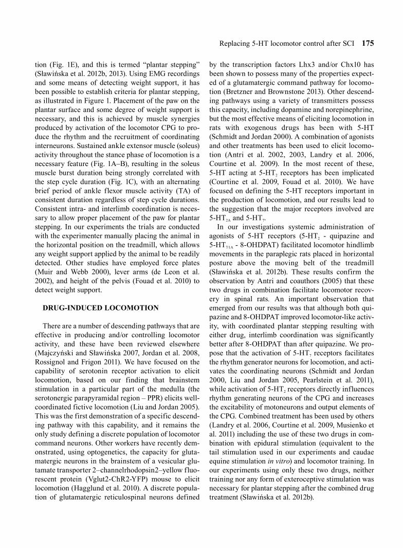
Replacing 5-HT locomotor control after SCI 175
tion (Fig. 1E), and this is termed “plantar stepping” (Sławińska et al. 2012b, 2013). Using EMG recordings and some means of detecting weight support, it has been possible to establish criteria for plantar stepping, as illustrated in Figure 1. Placement of the paw on the plantar surface and some degree of weight support is necessary, and this is achieved by muscle synergies produced by activation of the locomotor CPG to pro-duce the rhythm and the recruitment of coordinating interneurons. Sustained ankle extensor muscle (soleus) activity throughout the stance phase of locomotion is a necessary feature (Fig. 1A–B), resulting in the soleus muscle burst duration being strongly correlated with the step cycle duration (Fig. 1C), with an alternating brief period of ankle flexor muscle activity (TA) of consistent duration regardless of step cycle durations. Consistent intra- and interlimb coordination is neces-sary to allow proper placement of the paw for plantar stepping. In our experiments the trials are conducted with the experimenter manually placing the animal in the horizontal position on the treadmill, which allows any weight support applied by the animal to be readily detected. Other studies have employed force plates (Muir and Webb 2000), lever arms (de Leon et al. 2002), and height of the pelvis (Fouad et al. 2010) to detect weight support.
DRUG-INDUCED LOCOMOTION
There are a number of descending pathways that are effective in producing and/or controlling locomotor activity, and these have been reviewed elsewhere (Majczyński and Sławińska 2007, Jordan et al. 2008, Rossignol and Frigon 2011). We have focused on the capability of serotonin receptor activation to elicit locomotion, based on our finding that brainstem stimulation in a particular part of the medulla (the serotonergic parapyramidal region – PPR) elicits well-coordinated fictive locomotion (Liu and Jordan 2005). This was the first demonstration of a specific descend-ing pathway with this capability, and it remains the only study defining a discrete population of locomotor command neurons. Other workers have recently dem-onstrated, using optogenetics, the capacity for gluta-matergic neurons in the brainstem of a vesicular glu-tamate transporter 2–channelrhodopsin2–yellow fluo-rescent protein (Vglut2-ChR2-YFP) mouse to elicit locomotion (Hagglund et al. 2010). A discrete popula-tion of glutamatergic reticulospinal neurons defined
by the transcription factors Lhx3 and/or Chx10 has been shown to possess many of the properties expect-ed of a glutamatergic command pathway for locomo-tion (Bretzner and Brownstone 2013). Other descend-ing pathways using a variety of transmitters possess this capacity, including dopamine and norepinephrine, but the most effective means of eliciting locomotion in rats with exogenous drugs has been with 5-HT (Schmidt and Jordan 2000). A combination of agonists and other treatments has been used to elicit locomo-tion (Antri et al. 2002, 2003, Landry et al. 2006, Courtine et al. 2009). In the most recent of these, 5-HT acting at 5-HT2 receptors has been implicated (Courtine et al. 2009, Fouad et al. 2010). We have focused on defining the 5-HT receptors important in the production of locomotion, and our results lead to the suggestion that the major receptors involved are 5-HT2A and 5-HT7.
In our investigations systemic administration of agonists of 5-HT receptors (5-HT2 - quipazine and 5-HT7/1A - 8-OHDPAT) facilitated locomotor hindlimb movements in the paraplegic rats placed in horizontal posture above the moving belt of the treadmill (Sławińska et al. 2012b). These results confirm the observation by Antri and coauthors (2005) that these two drugs in combination facilitate locomotor recov-ery in spinal rats. An important observation that emerged from our results was that although both qui-pazine and 8-OHDPAT improved locomotor-like activ-ity, with coordinated plantar stepping resulting with either drug, interlimb coordination was significantly better after 8-OHDPAT than after quipazine. We pro-pose that the activation of 5-HT7 receptors facilitates the rhythm generator neurons for locomotion, and acti-vates the coordinating neurons (Schmidt and Jordan 2000, Liu and Jordan 2005, Pearlstein et al. 2011), while activation of 5-HT2 receptors directly influences rhythm generating neurons of the CPG and increases the excitability of motoneurons and output elements of the CPG. Combined treatment has been used by others (Landry et al. 2006, Courtine et al. 2009, Musienko et al. 2011) including the use of these two drugs in com-bination with epidural stimulation (equivalent to the tail stimulation used in our experiments and caudae equine stimulation in vitro) and locomotor training. In our experiments using only these two drugs, neither training nor any form of exteroceptive stimulation was necessary for plantar stepping after the combined drug treatment (Sławińska et al. 2012b).

176 U. Sławińska et al.

Replacing 5-HT locomotor control after SCI 177
It is well known that the action of quipazine could be medicated by 5-HT2A, 5-HT2B or 5-HT2C receptors. The predominance of 5-HT2A receptors in control of locomotor movement after quipazine application was demonstrated in adult rats (Ung et al. 2008). The role of 5-HT2A receptors in the production of locomotion by stimulation of brainstem serotonergic neurons have been also implicated in neonatal rats (Liu and Jordan 2005). On the other hand, Murray with colleagues (Murray et al. 2010) claimed that for recovery of loco-motor function after staggered hemisection the consti-tutively active 5-HT2C receptors are responsible. In their study 5-HT2C receptors were upregulated in the spinal cord below the level of injury. However, below a total spinal cord transection, in contrast to signifi-cant upregulation of 5-HT2A receptor mRNA, there were no changes in 5-HT2C receptor mRNA expression detected (Navarrett et al. 2012). The upregulation of 5-HT2A receptors after complete spinal cord injury was described in motoneurons (Jordan et al. 2010, Kong et al. 2010, 2011, Navarrett et al. 2012). We also have preliminary evidence using intrathecal drug applica-tion of serotonergic antagonists that in intact adult rats 5-HT2A receptors but not 5-HT2C receptors are involved in locomotor control (Sławińska et al. 2012a). Thus, except for the staggered hemisection preparation, the consensus seems to be that quipazine acts through 5-HT2A receptors.
Regarding the effectiveness of 8-OHDPAT for induc-ing locomotor movement the importance of both 5-HT7 and 5-HT1A receptors, was also implicated since locomo-tion could be blocked by antagonists of either of them and application of 8-OHDPAT could facilitate locomo-tion in 5-HT7 receptor knockout mice or in the presence of a selective 5-HT7 receptor antagonist (Landry et al. 2006). On the other hand, there is growing evidence showing that 5-HT1A receptors control the activity of motoneurons (reviewed in Perrier et al. 2013) as well as locomotor interneurons (Noga et al. 2009). Thus, it is possible that the actions of 8-OHDPAT that do not require 5-HT7 receptors are exerted on motoneurons rather than on locomotor CPG neurons. It was demon-strated that the decrease caused by 5-HT in NMDA-induced periodicity in fictive locomotion of the neonatal rat spinal cord in vitro was attenuated by pharmacologi-cal agonists of the 5-HT1 receptor family (Beato and Nistri 1998). Moreover, it was shown that locomotor activity evoked by brainstem stimulation was depressed by effects of serotonin exerted on 5-HT1A receptors, while in the absence of these receptors the effect of increasing available 5-HT resulting in initiation of loco-motion was mediated by 5-HT7 receptors (Dunbar et al. 2010). So, since the effects of 5-HT1A receptor activation are normally a decrease in neuronal activity, while those mediated by 5-HT7 are excitatory (Beato and Nistri 1998, Schmidt and Jordan 2000, Hochman et al. 2001,
Fig. 2. Grafts of 5-HT neurons of different populations (B1–B3, B3 and lateral) are differentially effective for improving hindlimb locomotor movements of spinal rats. (A) Immunolabeling of a horizontal section of a brainstem showing 5-HT positive populations of neurons in an E14 embryo from which the tissue was dissected for intraspinal grafting of spinalized rats. (B1, C1, D1) Bilaterally well-coordinated EMG patterns in the soleus (Sol) and tibialis anterior (TA) muscles are seen in a spinal rat grafted with B1–B3 neurons (B1, top panel) vs. less coordinated EMG activity in a spinalized rat grafted with lateral or B3 only 5-HT populations (C1, D1). The same raw EMG records rectified, filtered, and normalized to the step cycle (onset of activity in the right TA considered the onset of the cycle), show left-right and flexor-extensor coordination in a rat grafted with B1–B3 neurons (B1, lower panel) vs. a rat grafted with lateral or B3 only 5-HT populations (C1 and D1, lower panels). Regression lines showing the relationship between step cycle durations and burst durations for the left and right TA and Sol muscles in three groups of grafted rats are illustrated (B2, C2, D2). Polar plots of the relationships between the onset of r TA burst activity and either the contralateral TA showing interlimb coordination and (B4, C4, D4) or the ipsilateral right Sol showing intralimb coordination. The 0 position on the polar plots corresponds to the onset of activity in the right TA muscle, and the positions of the filled circles (black) indicate the times of onset of activity in the left TA (B3, C3, D3). The polar plots in B4, C4, D4 show intralimb coordination, with the times of onset of ipsilateral Sol activity plotted in relation to the times of onset of the ipsilateral right TA. E1 provides bar diagrams showing the mean slopes (±SD) for the regressions in three groups of grafted rats (B3, C3, D3). E2 presents bar diagrams showing the means (±SD) of the angles of the left-right TA (l-r) and of the right Sol–right TA (r S-T) relationships (relative timing of the onsets for the two muscles) represented in each polar plot in three groups of grafted rats animals, Bar diagrams in E3 show the means (±SD) of r values between the times of onset of activity in both left-right and flexor-extensor EMG comparisons in three groups of grafted rats. Statistical significance (Student’s t-test) comparing the animals grafted with various populations of 5-HT neurons is indicated by red stars: * P<0.05; ** P<0.005. The comparisons include rats with the B1–B3 transplants (n=7), rats grafted with the lateral group of 5-HT neurons (n=5) or rats grafted with only the B3 population (n=4).

178 U. Sławińska et al.
Heckman et al. 2009, Dunbar et al. 2010), we have focused on the latter as the most likely mediator of 5-HT activation of the locomotor CPG.
In general, therefore, interpretation of the effective-ness of these two drugs, quipazine and 8-OHDPAT, requires consideration of both the type of preparation and the various sites of action of the drugs that are likely to be involved in enhancing locomotor hindlimb movement.
We have argued that the problem in spinal rats that impairs locomotion is the loss of some descending system that controls coordination (Jordan and Sławińska 2011). This is based on the observation that without descending serotonin control, there is rhyth-mic activity in the hindlimbs, but it is not sufficient to produce coordinated plantar stepping. The rhythm generator is functional, but its output is disturbed such that co-contractions of antagonists occur, and interlimb coordination is impaired. Addition of 5-HT or its agonists appears to restore coordinated muscle activity (Pearlstein et al. 2005, Liu et al. 2009, Jordan and Sławińska 2011). We have shown that spinal loco-motion improved by quipazine is disrupted by strych-nine, which confirms a role of glycinergic interneu-rons in reciprocal inhibition and interlimb coordina-tion (Pearlstein et al. 2005, Boulenguez and Vinay 2009, Jordan and Sławińska 2011). Other factors con-trolling these same cells can be expected to emerge.
LIMITATIONS OF DRUG-INDUCED LOCOMOTION
Even though the serotonergic agonists are effective in eliciting coordinated hindlimb locomotor move-ments, in our experience it is difficult to obtain a sus-tained effect using this approach. Thus, the use of these drugs to elicit well-coordinated plantar stepping indicates that activation of this system can be effective, but systemic administration is unlikely to be a solution for restoring functional stepping. Some of the limita-tions of the systemic drug application approach are discussed below.
As pointed out below, serotonin can interfere with the natural sensory feedback processes that promote coordinated locomotion. It is also the case that these drugs can have other unwanted side-effects. Tachyphylaxis, defined as the rapid appearance of a progressive decrease in the response to a given dose after repetitive administration of a pharmacologically
or physiologically active substance, is an obvious drawback to the repeated use of drugs to elicit locomo-tion (definition provided by Stedman’s Medical dic-tionary 2000). If this occurs, increasingly large doses are required to achieve the desired effect. This may be due to alterations in the receptors over prolonged treat-ment or produced by the injury. For example, work in Edmonton has provided evidence that certain 5-HT receptors become constitutively active after injury (Murray et al. 2010). Some such effects are likely to occur with repeated doses of 5-HT agonists, making the efficacy of this approach limited.
Side effects of serotonin treatment can also occur, and the most dramatic of these is the Serotonin Syndrome, which can include: agitation or restless-ness, confusion, rapid heart rate and high blood pres-sure, dilated pupils, loss of muscle coordination or twitching muscles, heavy sweating, diarrhea, head-ache, shivering, goose bumps (Volpi-Abadie et al. 2013). Severe serotonin syndrome can be a life-threat-ening condition.
As the effects of the Serotonin Syndrome symptoms suggest, there are peripheral effects of systemic admin-istration of 5-HT and its agonists. These include effects on the heart, respiratory system, gut, vessels and the immune system. The effects of systemic actions of 5-HT and its agonists are therefore widespread, and the possible effects of sustained applications of drugs to induce locomotion are unknown. It seems likely that the efficacy of this approach may be limited by such side effects.
LIMITATIONS OF COMBINATORIAL APPROACH
Recent efforts to restore locomotion after spinal cord injury have incorporated a number of different approaches in combination, including systemic drug application, and at least one has resulted in potent pro-tection for the procedures followed (Guertin et al. 2011). An example is the use of systemic drug applica-tion in combination with electrical stimulation of the surface of the spinal cord below the level of the lesion, and locomotor training with the animals held in a jacket over a treadmill or walkway in the upright pos-ture, with only the hindlimbs available to support the animal’s weight. Pharmacological enhancement of locomotor recovery after SCI has been coupled with the benefits of training and epidural stimulation (Fong

Replacing 5-HT locomotor control after SCI 179
et al. 2009). Courtine and coworkers (2009) reported the use of specific combinations of pharmacological and electrical stimulation interventions, together with locomotor training, could induce full weight-bearing locomotion in the upright posture. Their pharmaco-logical treatment consisted of a combination of 5-HT2 and 5-HT1A/7 agonists. Subsequently this same group has expanded the pharmacological treatment to include pharmacological modulation of serotonergic, dop-aminergic and noradrenergic receptors to facilitate specific features of locomotion (Musienko et al. 2011). They describe facilitation of consistent locomotor movements with 5-HT1A/7 agonists, and they improved weight bearing abilities with 5-HT2A/C agonists. The results of these combinatorial approaches can be dra-matic, but there are drawbacks that need to be pointed out.
First, the system of neurons responsible for coordi-nation can also be activated from the plantar surface of the paw (Sławińska et al. 2012b). In this case placing the animal in the upright posture and allowing the paws to be artificially placed so that cutaneous recep-tors detect the presence of weight support induces well-coordinated stepping similar to that provided by 5-HT agonists or transplants. This effect can be blocked by anaesthesia of the foot. A balance exists between the 5-HT effect and the afferent feedback effect however, because in some cases the combination of the two leads to disordered coordination (Sławińska et al. 2012b). Recognition of this effect of paw afferent feedback should be taken into account when interpret-ing the data that has arisen from experimental para-digms where rats are placed in an upright posture to detect their recovery from spinal cord injury. This pos-ture alone clearly facilitates locomotor activity in the absence of any other intervention (Sławińska et al. 2012c).
The ability of epidural electrical stimulation of the spinal cord to elicit locomotion is a promising interven-tion that is now being tested clinically (Harkema et al. 2011). It was first demonstrated in spinal cord injured patients (Dimitrijevic et al. 1998). It was subsequently shown to be effective in the cat spinal cord, and various animals models have allowed the search for the most effective sites and modes of delivery to persist. In rats and mice the effects are also encouraging, as recently reviewed (Gerasimenko et al. 2008, Boulenguez and Vinay 2009). Afferent feedback is important for epidu-ral stimulation evoked locomotion, because contact of
the plantar surface of the foot with the treadmill and partial weight support is necessary (Ichiyama et al. 2005). After T12–S2 unilateral deafferentation (Lavrov et al. 2008), epidural stimulation is less effective for eliciting stepping on the deafferented side. Recent experiments have confirmed the efficacy of epidural stimulation in humans (Harkema et al. 2011), and have demonstrated that the procedure can influence other functions besides locomotion. The epidural stimulation approach is likely to continue to be developed for restor-ing function after spinal cord injury, but its combination with repeated systemic drug applications to achieve well-coordinated locomotion has the limitations described above. It is possible that some form of electri-cal stimulation can be used in combination with cell replacement therapy to some advantage, however.
RESTORATION OF LOCOMOTOR FUNCTION BY INTRASPINAL GRAFTING OF SEROTONINERGIC EMBRYONIC CELLS
Various methods for local intraspinal application of monoaminergic drugs are being explored, as pointed out above. One promising approach is the grafting of embryonic cells destined to release monoamines taken from the brainstem into the spinal cord below the site of the lesion. It has been demonstrated that these embryonic cells can survive and release monoamines in the host environment. The results of first homolo-gous fetal brain grafts were published almost 40 years ago (Nygren et al. 1977). The development of the transplantation approach has been reviewed in detail (Vrbová and Sławińska 2012). The improved hindlimb locomotor function restoration brought about by graft-ed serotonin-releasing cells was shown for the first time to be mediated in part by 5-HT receptors, since cyproheptadine (5-HT2 antagonist) IP application dis-rupted the hindlimb movement restored by the graft (Majczyński et al. 2005).
In our recent research we have extended these find-ings by demonstrating the effectiveness of grafting of different populations of serotonergic cells in adult rats. There are three recognized populations of 5-HT neu-rons (B1, B2, B3) that send their axons into different parts of spinal cord grey matter. The axons of neurons located in raphe obscurus and raphe pallidus (B1 and B2) terminate in the ventral horn. Raphe magnus neu-rons (B3) send axons to the dorsal horn and to the ventral horn as well. The precise termination of neu-

180 U. Sławińska et al.
rons located in the parapyramidal region (PPR), classi-fied as a part of B3, are unknown. However, there is information that shows the more laterally placed 5-HT neurons are derived in part from more caudal descend-ing 5-HT groups (Tork 1990). Moreover the sources of serotonergic fibers present in laminae VII and X in the spinal cord are not yet known. The molecular genetics of the brainstem 5-HT system is now emerging (Cordes 2005, Jensen et al. 2008, Bang et al. 2012). We pre-sume that different cell population can play different roles in control of locomotor movements, and we explored this possibility in our recent publication (Sławińska et al. 2013), where we used all three popu-lations of 5-HT neurons (B1, B2 and B3).
In our recent paper (Sławińska et al. 2013) we dem-onstrated that for better recovery of hindlimb locomo-tor movements in paraplegic rats simultaneous supple-mentation of various populations of 5-HT neurons (B1, B2, B3) that innervate different parts of the spinal cord network is required. We have now extended this research to include a comparison of B1–B3 grafts with grafts of neurons from a more lateral region of the fetal brainstem and from the B3 region only (Fig. 2A), The effects of these grafts on locomotion are illustrated in Figure 2 (panels C and D).These effects were com-pared with those obtained from transplants of the B1, B2 and B3 regions (Fig. 2B). The results show that the lateral transplants give rise to plantar stepping with extensor muscle activity related to the step cycle dura-tion. This is clear in the regression lines comparing burst duration to cycle duration (Fig. 2). However, coordination is impaired compared to the B1–B3 trans-planted animals, as illustrated in the polar plots in Figure 2B for the B1–B3 grafted animals and Figure 2C for the animals with grafts taken from the lateral group of 5-HT neurons in the fetal brainstem. Figure 2D shows the locomotor activity produced in animals with grafts from the B3 region only. The quality of plantar walking in these animals was significantly less than in the other animals. Significant differences were observed between the three groups of animals when intra- and interlimb coordination was compared (Fig. 2E). We conclude from these observations that the lateral group of neurons is capable of restoring some features of plantar walking, but the restoration of intra- and interlimb coordination is incomplete. The poor locomotion observed in the B3 grafted animals in comparison to other groups of grafted rats may be associated with the absence of 5-HT innervation of the
area around the central canal and lamina VII. Comparisons of the walking produced by each of these grafts with control spinal animals that received no grafts reveal that all the grafted animals that received a piece of lateral brainstem or the B1–B3 serotonergic populations were significantly better than control spi-nal animals. Improvement of locomotion in those that received only B3 serotonergic cells was not statisti-cally significant (to see control rats see Sławińska et al. 2013).
Figure 3 shows examples of the distribution of 5-HT fibers found in the B1–B3 and lateral graft animals. It was a consistent observation that the B1–B3 grafts provided robust input to the area around the central canal, as well as cells in lamina VII and the dorsal horn in the area near the graft (Fig. 3A). Furthermore, there was often substantial innervation of motoneu-rons in lamina IX of the lumbar spinal cord (Fig. 3B). The animals that received the grafts from the lateral region of the fetal brainstem only showed consistent innervation of the intermediolateral cell column (IML), the central canal, and some innervation of motoneu-rons in the area of the spinal cord nearest the transplant (Fig. 3C). The innervation was consistently more sparse in the lateral graft cases than in the B1–B3 cases. In more caudal areas of the spinal cord, there was innervation of the central canal region and some sparse innervation of lamina VII, but little innervation of the caudal motor nuclei (Fig. 3D). The B3 grafted animals illustrated in Figure 2D did not reveal any 5-HT innervation in the central canal area (data not shown).
The innervation provided by the grafted cells appears to be guided to some degree by the target cells they were originally intended to innervate. This is consistent with the observations of Rajaofetra and col-leagues (1992), who showed discrete innervation pat-terns for different groups of 5-HT neurons derived from different parts of the fetal brainstem. One issue that arises from these observations is whether merely the restoration of some 5-HT in the lesioned spinal cord is sufficient to facilitate plantar stepping, rather than reinnervation of specific spinal neurons, so that any 5-HT neurons can produce the effect. We argue that the innervation of certain groups of neurons is necessary, however, because recovery of some aspects of plantar walking is associated in all cases with inner-vation of the area around the central canal, a region where many neurons active during locomotion are

Replacing 5-HT locomotor control after SCI 181
located (Dai et al. 2005). Neurons in this area of the cat spinal cord that are active during locomotion receive 5-HT input and are also rich in 5-HT2A and 5-HT7 receptors (Noga et al. 2009). Locomotor cells in this region have been shown to be excited by 5-HT (Dai et al. 2009). It is noteworthy that left-right coordination is consistently improved by the grafts, because commis-sural neurons have been shown to be excited by 5-HT as well (Carlin et al. 2006, Zhong et al. 2006). In our experiments, interlimb coordination is impaired in animals with sparse or no 5-HT innervation of the area around the central canal. Furthermore, plantar step-ping is poorly developed in the B3 transplanted ani-mals that had no 5-HT innervation of the central canal area. Thus, there is an abundance of evidence implicat-ing neurons of this area in the production of locomotor activity in both cat and rat preparations. The neurons responsible for this effect have not yet been identi-fied.
In our recent investigations we made an effort to determine the relative contributions of 5-HT2 and 5-HT7 receptors to transplant-induced locomotor hindlimb movement recovery (Sławińska et al. 2013).The crucial role of 5-HT2 receptors was confirmed using systemic application of cyproheptadine, an antagonist at 5-HT2A/2B/2C receptors, which altered left-right coordination and the cycle duration of hindlimb locomotor movement in grafted spinal rats. Moreover, cyproheptadine application shortened soleus burst duration and decreased its peak amplitude. Thus, our results showed that blockage of 5-HT2 receptors inter-fered with the ability of the graft-derived intraspinal serotonin to activate components of the CPG as well the motoneurons. In addition we established that blockade of 5-HT7 receptors using SB269970 (an antagonist of 5-HT7 receptors) impairs inter- and intralimb coordination in locomotor hindlimb move-ments of grafted spinal rats. It is important to note that
Fig. 3. Immunolabeling of transverse spinal cord sections showing 5-HT-positive neurons and fibers in rats grafted with dif-ferent populations of 5-HT neurons. (A) shows the 5-HT positive neurons present in a large graft occupying the central region of the gray matter and numerous 5-HT positive fibers within and emerging from the graft of the B1–B3 serotonergic populations. (B) shows example of 5-HT fibers in close proximity to 5-HT2A receptor positive motoneurons in the ventral horn at the L3 spinal cord level of spinal grafted rat with the B1–B3 serotonergic populations. (C) and (D) show 5-HT-positive neurons and fibers present in the ventral horn, around the central canal and in the intermedilateral zone emerging from a graft of the lateral population of serotonergic brainstem neurons. We found that in the lateral and B3 only (data not shown) grafted spinal rats there are fewer 5-HT positive fibers in the ventral horn and intermediolateral zone compared with B1–B3 grafted rats.

182 U. Sławińska et al.
in contrast to cyproheptadine, SB269970 did not alter muscle activity (burst duration or amplitude). Thus, our results show that blockade of 5-HT7 receptors alters activity of interneurons responsible for generat-ing the reciprocal pattern of flexor and extensor motoneuron activity, as well as neurons responsible for coordinating activity in the two hindlimbs during hindlimb plantar stepping of grafted rats but does not influence motoneuron excitability directly. Our results allow us to conclude that both receptors, 5-HT2 and 5-HT7, mediate the improved locomotor movements in spinal rats after grafting, but through actions on differ-ent populations of spinal locomotor neurons. We are persuaded that 5-HT2 receptors control CPG activation as well as motoneuron output, while 5-HT7 receptors contribute primarily to activity of the locomotor CPG and associated interneurons involved in intra- and interlimb coordination.
FUTURE PROSPECTS FOR CELL REPLACEMENT THERAPY
One promising idea for the cell replacement strategy is the use of defined populations of neurons for replacement therapy. There is evidence showing that grafted embryonically defined motoneurons leads to significant functional improvement following ventral root avulsion. It has been shown that in adult rats the missing motoneurons can be replaced by embryonic grafted neurons and their axons can be guided to den-ervated muscles via a reimplanted ventral root so the grafted motoneurons contribute to the innervation and functional recovery of the denervated muscles (Nógrádi and Vrbová 1996, Gal et al. 2012). As we described above the transplantation of discrete populations of embryonic serotonergic cells into the spinal cord below the total transection of adult rats give rise to improvement of plantar stepping (Sławińska et al. 2000, Majczyński et al. 2005, Sławińska et al. 2013). Other sources of defined cell populations are now being developed, particularly the use of stem cells directed to a particular lineage. As reviewed by Rossi and Keirstead (2009), transplantation of cells with an oligodendroglial lineage (Totoiu et al. 2004, Keirstead et al. 2005), Schwann cells (Kohama et al. 2001), olfac-tory ensheathing cells (Imaizumi et al. 1998), and various stem cell types (Brustle et al. 1999, Keirstead et al. 2005, Nistor et al. 2005) remyelinates axons in animal models with demyelination. Restoration of
motor neuron circuitry occurs following transplanta-tion of motor neuron-primed human neural stem cells (Gao et al. 2005, 2007) or mouse embryonic stem cell derived motoneurons (Deshpande et al. 2006) into models of motoneuron disease. Cholinergic processes exit the ventral root and travel through the sciatic nerve to form physiologically active neuromuscular junctions. This raises the question of whether cell replacement therapy using stem cells might be devel-oped for replacement of serotonergic neurons. Recently a new method allowing induction of serotonergic neu-rons from embryonic stem and pluripotent stem cells has been described (Shimada et al. 2012). It was found that noggin, a known antagonist of bone morphogenic protein, induced embryonic stem cells to express genes involved in serotonergic differentiation, such as Nkx2.2, Pet-1, Sonic hedgehog, tryptophan hydroxy-lase 2, and serotonin transporter. These cells could also demonstrate high potassium-induced release of serotonin. Thus, this method might provide an option for serotonergic differentiation of pluripotent stem cells and can open some new therapeutic perspectives for developing a strategy aiming to replace missing serotonergic innervation in various pathological situa-tions including spinal cord injury.
CLINICAL IMPLICATIONS
The results of experiments on animals can be used as a basis for development of clinical approaches for restoring locomotion. Locomotor training is now being subjected to clinical trials. An early report consolidat-ing data from a number of centers has been published (Dobkin et al. 2007), but the human locomotor training approach has only produced anecdotal evidence in favour of a beneficial effect, in contrast to the animals studies. Nevertheless, these approaches are still being applied clinically. For example, a recent case report (Harkema et al. 2011) has incorporated epidural stimu-lation of the lumbosacral spinal cord and demonstrated effects on voluntary movement, standing, and assisted stepping after motor complete paraplegia. It can be expected that other combinations of approaches that have had positive reports in animals will continue to be applied in humans, including systemic and/or epi-dural drug applications.
Could interventions such as replacement of seroton-ergic neurons be a viable clinical approach for func-tional recovery after spinal cord injury? Pearse and

Replacing 5-HT locomotor control after SCI 183
Bunge (2006) have reviewed the problems associated with designing cell- and gene-based regeneration strat-egies to repair the injured spinal cord. The use of Schwann cells to promote regeneration (Wiliams and Bunge 2012) is now in the clinical trial stage, and U.S. Food and Drug Administration approval has been awarded. This is a promising first step in the develop-ment of cell replacement therapies for spinal cord injury. In the case of replacing serotonergic neurons, the positive preclinical data is all from fetal cell trans-plants, although 5-HT secreting cell lines injected intrathecally have been attempted (Eaton et al. 2011). This approach did not result in significant improve-ment in locomotor control, however. As the data reported here suggest, it will be necessary to develop ways to create not only 5-HT neurons, but those des-tined to become specific descending 5-HT neurons, including neurons from the B1, B2 and B3 groups. Further refinement of the tools to recognize and guide the development of these specific neurons is necessary. Furthermore, methods to control the transplanted cells have yet to be developed, and a means of providing a supply of appropriately differentiated 5-HT neurons for transplantation into the spinal cord has not been developed. The cells also must be in the right stage of development, as is the case with the E14 embryonic cells used in our experiments, otherwise they will not be able to grow and find appropriate targets. This is an area of research with considerable potential, however, because even without any means to increase the activ-ity of the command neurons, they provide facilitation of locomotion. It will be advantageous to develop methods to allow for voluntary control of the activity of these transplanted cells as well. The transplanted cells have specific receptors that allow them to be selectively activated after transplantation using intrath-ecally applied drugs that have minimal effects on the spinal cord below the lesion but can activate the trans-planted neurons. Less specific activation tools include epidural and intraspinal stimulation. The potential for these approaches to selectively activate the transplant-ed neurons is not yet known. The electrophysiological and pharmacological tools that are standard ways of activating spinal cord neurons need to be developed in the laboratory. It is now possible to selectively activate genetically defined transplanted neurons. One such approach is optogenetics, were identified 5-HT neu-rons can be engineered to express ChR2 so that a light stimulus can activate the transplanted cells.
Alternatively, it is possible to use systemic application of a designer drug to activate “designer receptors exclusively activate designer drugs” (DREADDS) (Dong et al. 2010).
CONCLUSION
In conclusion, we argue that replacement of 5-HT neurons responsible for the initiation and control of locomotion is a worthy research goal in the spinal cord injury field. What is left to do is to further define the 5-HT neurons suitable for this approach, ones that can survive and innervate the appropriate targets, particu-larly the area around the central canal and the motoneu-ron area. The cells must be at a stage of development equivalent to the E14 neurons used in successful fetal cell transplants, and they must be destined to develop into the cells of the B1–B3 groups that are likely to provide maximal functional recovery. Finally, methods of controlling the activity of these cells need to be developed.
ACKNOWLEDGEMENTS
The authors thank Maria Setterbom, Gilles Detillieux, Zhicheng Zhou, Anna M. Cabaj, Henryk Majczyński and Yue Dai for their contributions to these experiments. This work was supported by grants from the Polish Ministry of Science and High Education (N N404 134439) and the Nencki Institute statutory donation to US and grants from the Canadian Institutes of Health Research (CIHR) and the Manitoba Spinal Cord Injury Research Committee (MSCIRC) to LMJ.
REFERENCES
Antri M, Barthe JY, Mouffle C, Orsal D (2005) Long-lasting recovery of locomotor function in chronic spinal rat fol-lowing chronic combined pharmacological stimulation of serotonergic receptors with 8-OHDPAT and quipazine. Neurosci Lett 384: 162–167.
Antri M, Mouffle C, Orsal D, Barthe JY (2003) 5-HT1A receptors are involved in short- and long-term processes responsible for 5-HT-induced locomotor function recov-ery in chronic spinal rat. Eur J Neurosci 18: 1963–1972.
Antri M, Orsal D, Barthe JY (2002) Locomotor recovery in the chronic spinal rat: effects of long-term treatment with a 5-HT2 agonist. Eur J Neurosci 16: 467–476.

184 U. Sławińska et al.
Bang SJ, Jensen P, Dymecki SM, Commons KG (2012) Projections and interconnections of genetically defined serotonin neurons in mice. Eur J Neurosci 35: 85–96.
Barbeau H, McCrea DA, O’Donovan MJ, Rossignol S, Grill WM, Lemay MA (1999) Tapping into spinal circuits to restore motor function. Brain Res Brain Res Rev 30: 27–51.
Beato M, Nistri A (1998) Serotonin-induced inhibition of locomotor rhythm of the rat isolated spinal cord is medi-ated by the 5-HT1 receptor class. Proc Biol Sci 265: 2073–2080.
Boulenguez P, Vinay L (2009) Strategies to restore motor functions after spinal cord injury. Curr Opin Neurobiol 19: 587–600.
Bretzner F, Brownstone RM (2013) Lhx3-Chx10 reticu-lospinal neurons in locomotor circuits. J Neurosci 33: 14681–14692.
Brustle O, Jones KN, Learish RD, Karram K, Choudhary K, Wiestler OD, Duncan ID, McKay RD (1999) Embryonic stem cell-derived glial precursors: a source of myelinat-ing transplants. Science 285: 754–756.
Carlin KP, Dai Y, Jordan LM (2006) Cholinergic and sero-tonergic excitation of ascending commissural neurons in the thoraco-lumbar spinal cord of the neonatal mouse. J Neurophysiol 95: 1278–1284.
Cordes SP (2005) Molecular genetics of the early develop-ment of hindbrain serotonergic neurons. Clin Genet 68: 487–494.
Courtine G, Gerasimenko Y, van den Brand R, Yew A, Musienko P, Zhong H, Song B, Ao Y, Ichiyama RM, Lavrov I, Roy RR, Sofroniew MV, Edgerton VR (2009) Transformation of nonfunctional spinal circuits into func-tional states after the loss of brain input. Nat Neurosci 12: 1333–1342.
Dai X, Noga BR, Douglas JR, Jordan LM (2005) Localization of spinal neurons activated during locomotion using the c-fos immunohistochemical method. J Neurophysiol 93: 3442–3452.
Dai Y, Carlin KP, Li Z, McMahon DG, Brownstone RM, Jordan LM (2009) Electrophysiological and pharmaco-logical properties of locomotor activity-related neurons in cfos-EGFP mice. J Neurophysiol 102: 3365–3383.
de Leon RD, Kubasak MD, Phelps PE, Timoszyk WK, Reinkensmeyer DJ, Roy RR, Edgerton VR (2002) Using robotics to teach the spinal cord to walk. Brain Res Brain Res Rev 40: 267–273.
Delvolve I, Gabbay H, Lev-Tov A (2001) The motor output and behavior produced by rhythmogenic sacrocaudal net-works in spinal cords of neonatal rats. J Neurophysiol 85: 2100–2110.
Deshpande DM, Kim YS, Martinez T, Carmen J, Dike S, Shats I, Rubin LL, Drummond J, Krishnan C, Hoke A, Maragakis N, Shefner J, Rothstein JD, Kerr DA (2006) Recovery from paralysis in adult rats using embryonic stem cells. Ann Neurol 60: 32–44.
Dimitrijevic MR, Gerasimenko Y, Pinter MM (1998) Evidence for a spinal central pattern generator in humans. Ann N Y Acad Sci 860: 360–376.
Dobkin B, Barbeau H, Deforge D, Ditunno J, Elashoff R, Apple D, Basso M, Behrman A, Harkema S, Saulino M, Scott M (2007) The evolution of walking-related out-comes over the first 12 weeks of rehabilitation for incom-plete traumatic spinal cord injury: the multicenter ran-domized Spinal Cord Injury Locomotor Trial. Neurorehabil Neural Repair 21: 25–35.
Dong S, Allen JA, Farrell M, Roth BL (2010) A chemical-genetic approach for precise spatio-temporal control of cellular signaling. Mol Biosyst 6: 1376–1380.
Dunbar MJ, Tran MA, Whelan PJ (2010) Endogenous extra-cellular serotonin modulates the spinal locomotor net-work of the neonatal mouse. J Physiol 588: 139–156.
Eaton MJ, Widerstrom-Noga E, Wolfe SQ (2011) Subarachnoid transplant of the human neuronal hNT2.19 serotonergic cell line attenuates behavioral hypersensi-tivity without affecting motor dysfunction after severe contusive spinal cord injury. Neurol Res Int 2011: 891605.
Edgerton VR, Courtine G, Gerasimenko YP, Lavrov I, Ichiyama RM, Fong AJ, Cai LL, Otoshi CK, Tillakaratne NJ, Burdick JW, Roy RR (2008) Training locomotor net-works. Brain Res Rev 57: 241–254.
Fong AJ, Roy RR, Ichiyama RM, Lavrov I, Courtine G, Gerasimenko Y, Tai YC, Burdick J, Edgerton VR (2009) Recovery of control of posture and locomotion after a spinal cord injury: solutions staring us in the face. Prog Brain Res 175: 393–418.
Fouad K, Rank MM, Vavrek R, Murray KC, Sanelli L, Bennett DJ (2010) Locomotion after spinal cord injury depends on constitutive activity in serotonin receptors. J Neurophysiol 104: 2975–2984.
Gal L, Pajer K, Nógrádi A, Sławińska U (2012) Grafted embryonic motoneurons contribute to the coordinated functional improvement of denervated hindlimbs after motoneuron loss induced by ventral root avulsion. FENS Abstract, Vol. 6, p.132.05
Gao J, Coggeshall RE, Chung JM, Wang J, Wu P (2007) Functional motoneurons develop from human neural stem cell transplants in adult rats. Neuroreport 18: 565–569.

Replacing 5-HT locomotor control after SCI 185
Gao J, Coggeshall RE, Tarasenko YI, Wu P (2005) Human neural stem cell-derived cholinergic neurons innervate muscle in motoneuron deficient adult rats. Neuroscience 131: 257–262.
Gerasimenko Y, Roy RR, Edgerton VR (2008) Epidural stimulation: comparison of the spinal circuits that gener-ate and control locomotion in rats, cats and humans. Exp Neurol 209: 417–425.
Gimenez y Ribotta M, Provencher J, Feraboli-Lohnherr D, Rossignol S, Privat A, Orsal D (2000) Activation of loco-motion in adult chronic spinal rats is achieved by trans-plantation of embryonic raphe cells reinnervating a pre-cise lumbar level. J. Neurosci. 20: 5144–5152.
Gordon IT, Whelan PJ (2006) Monoaminergic control of cauda-equina-evoked locomotion in the neonatal mouse spinal cord. J Neurophysiol 96: 3122–3129.
Guertin PA, Ung RV, Rouleau P, Steuer I (2011) Effects on locomotion, muscle, bone, and blood induced by a com-bination therapy eliciting weight-bearing stepping in nonassisted spinal cord-transected mice. Neurorehabil Neural Repair 25: 234–242.
Hagglund M, Borgius L, Dougherty KJ, Kiehn O (2010) Activation of groups of excitatory neurons in the mam-malian spinal cord or hindbrain evokes locomotion. Nat Neurosci 13: 246–252.
Harkema S, Gerasimenko Y, Hodes J, Burdick J, Angeli C, Chen Y, Ferreira C, Willhite A, Rejc E, Grossman RG, Edgerton VR (2011) Effect of epidural stimulation of the lumbosacral spinal cord on voluntary movement, stand-ing, and assisted stepping after motor complete paraple-gia: a case study. Lancet 377: 1938–1947.
Hawthorne AL, Hu H, Kundu B, Steinmetz MP, Wylie CJ, Deneris ES, Silver J (2011) The unusual response of sero-tonergic neurons after CNS Injury: lack of axonal dieback and enhanced sprouting within the inhibitory environ-ment of the glial scar. J Neurosci 31: 5605–5616.
Heckman CJ, Mottram C, Quinlan K, Theiss R, Schuster J (2009) Motoneuron excitability: the importance of neuro-modulatory inputs. Clin Neurophysiol 120: 2040–2054.
Hochman S, Garraway SM, Machacek DW, Shay BL (2001) 5-HT receptors and the neuromodualatory control of spinal cord function. In: Motor Neurobiology of the Spinal Cord (Cope TC, Ed). CRC Press, New York, NY, p. 48–87.
Ichiyama RM, Gerasimenko YP, Zhong H, Roy RR, Edgerton VR (2005) Hindlimb stepping movements in complete spinal rats induced by epidural spinal cord stimulation. Neurosci Lett 383: 339–344.
Imaizumi T, Lankford KL, Waxman SG, Greer CA, Kocsis JD (1998) Transplanted olfactory ensheathing cells remy-
elinate and enhance axonal conduction in the demyeli-nated dorsal columns of the rat spinal cord. J Neurosci 18: 6176–6185.
Jensen P, Farago AF, Awatramani RB, Scott MM, Deneris ES, Dymecki SM (2008) Redefining the serotonergic system by genetic lineage. Nat Neurosci 11: 417–419.
Jordan LM, Liu J, Hedlund PB, Akay T, Pearson KG (2008) Descending command systems for the initiation of loco-motion in mammals. Brain Res Rev 57: 183–191.
Jordan LM, Fabczak H, Kisielnicka E, Leszczyńska A, Majczyński H, Nagy JI, Sławińska U (2010) Segmental distribution of 5-HT2A and 5-HT2C receptor up-regula-tion one month after complete spinal cord injury. Society for Neuroscience Meeting 2010, San Diego, CA. Program No. 259.20.
Jordan LM, Sławińska U (2011) Chapter 12--modulation of rhythmic movement: control of coordination. Prog Brain Res 188: 181–195.
Kaegi S, Schwab ME, Dietz V, Fouad K (2001) Electromyographic activity associated with spontaneous functional recovery after spinal cord injury in rats. Eur J Neurosci 16: 249–258.
Keirstead HS, Nistor G, Bernal G, Totoiu M, Cloutier F, Sharp K, Steward O (2005) Human embryonic stem cell-derived oligodendrocyte progenitor cell transplants remy-elinate and restore locomotion after spinal cord injury. J Neurosci 25: 4694–4705.
Kohama I, Lankford KL, Preiningerova J, White FA, Vollmer TL, Kocsis JD (2001) Transplantation of cryopreserved adult human Schwann cells enhances axonal conduction in demyelinated spinal cord. J Neurosci 21: 944–950.
Kong XY, Wienecke J, Hultborn H, Zhang M (2010) Robust upregulation of serotonin 2A receptors after chronic spi-nal transection of rats: an immunohistochemical study. Brain Res 1320: 60–68.
Kong XY, Wienecke J, Chen M, Hultborn H, Zhang M (2011) The time course of serotonin 2A receptor expres-sion after spinal transection of rats: an immunohis-tochemical study. Neuroscience 177: 114–126.
Landry ES, Lapointe NP, Rouillard C, Levesque D, Hedlund PB, Guertin PA (2006) Contribution of spinal 5-HT1A and 5-HT7 receptors to locomotor-like movement induced by 8-OH-DPAT in spinal cord-transected mice. Eur. J. Neurosci. 24: 535–546.
Lavrov I, Courtine G, Dy CJ, van den Brand R, Fong AJ, Gerasimenko Y, Zhong H, Roy RR, Edgerton VR (2008) Facilitation of stepping with epidural stimulation in spi-nal rats: role of sensory input. J Neurosci 28: 7774–7780.

186 U. Sławińska et al.
Leblond H, L’Esperance M, Orsal D, Rossignol S (2003) Treadmill locomotion in the intact and spinal mouse. J Neurosci 23: 11411–11419.
Lev-Tov A, Delvolve I, Kremer E (2000) Sacrocaudal affer-ents induce rhythmic efferent bursting in isolated spinal cords of neonatal rats. J Neurophysiol 83: 888–894.
Liu J, Jordan LM (2005) Stimulation of the parapyramidal region of the neonatal rat brain stem produces locomotor-like activity involving spinal 5-HT7 and 5-HT2A recep-tors. J Neurophysiol 94: 1392–1404.
Liu J, Akay T, Hedlund PB, Pearson KG, Jordan LM (2009) Spinal 5-HT7 receptors are critical for alternating activity during locomotion: in vitro neonatal and in vivo adult studies using 5-HT7 receptor knockout mice. J Neurophysiol 102: 337–348.
Macias M, Nowicka D, Czupryn A, Sulejczak D, Skup M, Skangiel-Kramska J, Czarkowska-Bauch J (2009) Exercise-induced motor improvement after complete spi-nal cord transection and its relation to expression of brain-derived neurotrophic factor and presynaptic mark-ers. BMC Neurosci 10: 144.
Majczyński H, Maleszak K, Cabaj A, Sławińska U (2005) Serotonin-related enhancement of recovery of hind limb motor functions in spinal rats after grafting of embryonic raphe nuclei. J. Neurotrauma 22: 590–604.
Majczyński H, Maleszak K, Górska T, Sławińska U (2007) Comparison of two methods for quantitative assessment of unrestrained locomotion in the rat. J Neurosci Methods 163: 197–207.
Majczyński H, Sławińska U (2007) Locomotor recovery after thoracic spinal cord lesions in cats, rats and humans. Acta Neurobiol Exp (Wars) 67: 235–257.
Meisel RL, Rakerd B (1982) Induction of hindlimb stepping movements in rats spinally transected as adults or as neo-nates. Brain Res 240: 353–356.
Muir GD, Webb AA (2000) Mini-review: assessment of behavioural recovery following spinal cord injury in rats. Eur J Neurosci 12: 3079–3086.
Murray KC, Nakae A, Stephens MJ, Rank M, D’Amico J, Harvey PJ, Li X, Harris RL, Ballou EW, Anelli R, Heckman CJ, Mashimo T, Vavrek R, Sanelli L, Gorassini MA, Bennett DJ, Fouad K (2010) Recovery of motoneu-ron and locomotor function after spinal cord injury depends on constitutive activity in 5-HT2C receptors. Nature Medicine 16: 694–700.
Mushahwar VK, Gillard DM, Gauthier MJ, Prochazka A (2002) Intraspinal micro stimulation generates locomo-tor-like and feedback-controlled movements. IEEE Trans Neural Syst Rehabil Eng 10: 68–81.
Musienko P, van den Brand R, Marzendorfer O, Roy RR, Gerasimenko Y, Edgerton VR, Courtine G (2011) Controlling Specific Locomotor Behaviors through Multidimensional Monoaminergic Modulation of Spinal Circuitries. J. Neurosci. 31: 9264–9278.
Navarrett S, Collier L, Cardozo C, Dracheva S (2012) Alterations of serotonin 2C and 2A receptors in response to T10 spinal cord transection in rats. Neurosci Lett 506: 74–78.
Nistor GI, Totoiu MO, Haque N, Carpenter MK, Keirstead HS (2005) Human embryonic stem cells differentiate into oligodendrocytes in high purity and myelinate after spinal cord transplantation. Glia 49: 385–396.
Noga BR, Johnson DM, Riesgo MI, Pinzon A (2009) Locomotor-activated neurons of the cat. I. Serotonergic innervation and co-localization of 5-HT7, 5-HT2A, and 5-HT1A receptors in the thoraco-lumbar spinal cord. J Neurophysiol 102: 1560–1576.
Nógrádi A, Vrbová G (1996) Improved motor function of denervated rat hindlimb muscles induced by embryonic spinal cord grafts. Eur J Neurosci 8: 2198–2203.
Norreel JC, Pflieger JF, Pearlstein E, Simeoni-Alias J, Clarac F, Vinay L (2003) Reversible disorganization of the locomotor pattern after neonatal spinal cord transec-tion in the rat. J Neurosci 23: 1924–1932.
Nygren LG, Olson L, Seiger A (1977) Mnoaminergic rein-nervation of the transected spinal cord by homologous fetal brain grafts. Brain Res 129: 227–235.
Orsal D, Barthe JY, Antri M, Feraboli-Lohnherr D, Yakovleff A, Gimenez y Ribotta M, Privat A, Provencher J, Rossignol S (2002) Locomotor recovery in chronic spinal rat: long-term pharmacological treatment or transplanta-tion of embryonic neurons? Prog Brain Res 137: 213–230.
Pearlstein E, Ben Mabrouk F, Pflieger JF, Vinay L (2005) Serotonin refines the locomotor-related alternations in the in vitro neonatal rat spinal cord. Eur J Neurosci 21: 1338–1346.
Pearlstein E, Bras H, Deneris ES, Vinay L (2011) Contribution of 5-HT to locomotion - the paradox of Pet-1(-/-) mice. Eur J. Neurosci 33: 1812–1822.
Pearse DD, Bunge MB (2006) Designing cell- and gene-based regeneration strategies to repair the injured spinal cord. J Neurotrauma 23: 438–452.
Perrier JF, Rasmussen HB, Christensen RK, Petersen AV (2013) Modulation of the intrinsic properties of motoneu-rons by serotonin. Curr Pharm Des 19: 4371–4384.
Rajaofetra N, Konig N, Poulat P, Marlier L, Sandillon F, Drian MJ, Geffard M, Privat A (1992) Fate of B1-B2 and

Replacing 5-HT locomotor control after SCI 187
B3 rhombencephalic cells transplanted into the transected spinal cord of adult rats: light and electron microscopic studies. Exp Neurol 117: 59–70.
Rossi SL, Keirstead HS (2009) Stem cells and spinal cord regeneration. Curr Opin Biotechnol 20: 552–562.
Rossignol S, Frigon A (2011) Recovery of locomotion after spinal cord injury: some facts and mechanisms. Annu Rev Neurosci 34: 413–440.
Schmidt BJ, Jordan LM (2000) The role of serotonin in reflex modulation and locomotor rhythm production in the mammalian spinal cord. Brain Res Bull 53: 689–710.
Shimada T, Takai Y, Shinohara K, Yamasaki A, Tominaga-Yoshino K, Ogura A, Toi A, Asano K, Shintani N, Hayata-Takano A, Baba A, Hashimoto H (2012) A simpli-fied method to generate serotonergic neurons from mouse embryonic stem and induced pluripotent stem cells. J Neurochem 122: 81–93.
Sławińska U, Jordan LM, Cabaj AM, Burger B, Fabczak H, Majczyński H (2012a) Locomotion of intact adult rats is controlled by 5-HT2A and 5-HT7 but not 5-HT2C recep-tors. Society for Neuroscience Meeting, New Orleans, LA. Program No. 85.09
Sławińska U, Majczyński H, Dai Y, Jordan LM (2012b) The upright posture improves plantar stepping and alters responses to serotonergic drugs in spinal rats. J Physiol (Lond.) 590: 1721–1736.
Sławińska U, Majczyński H, Djavadian R (2000) Recovery of hindlimb motor functions after spinal cord transection is enhanced by grafts of the embryonic raphe nuclei. Exp Brain Res 132: 27–38.
Sławińska U, Miazga K, Cabaj AM, Leszczyńska AN, Majczyński H, Nagy JI, Jordan LM (2013) Grafting of fetal brainstem 5-HT neurons into the sublesional spinal cord of paraplegic rats restores coordinated hindlimb locomotion. Exp Neurol 247: 572–581.
Sławińska U, Rossignol S, Bennett DJ, Schmidt BJ, Frigon A, Fouad K, Jordan LM (2012c) Comment on “Restoring voluntary control of locomotion after para-lyzing spinal cord injury”. Science 338: 328. [author reply p. 328]
Smith JC, Feldman JL, Schmidt BJ (1988) Neural mecha-nisms generating locomotion studied in mammalian brain stem-spinal cord in vitro. Faseb J 2: 2283–2288.
Stedman’s Medical Dictionary (2000) Lippincott, Williams, and Wilkins, Philadelphia, PA.
Tator CH, Minassian K, Mushahwar VK (2012) Spinal cord stimulation: therapeutic benefits and movement genera-tion after spinal cord injury. Handb Clin Neurol 109: 283–296.
Tork I (1990) Anatomy of the serotonergic system. Ann N Y Acad Sci 600: 9–34; discussion 34–35.
Totoiu MO, Nistor GI, Lane TE, Keirstead HS (2004) Remyelination, axonal sparing, and locomotor recovery following transplantation of glial-committed progenitor cells into the MHV model of multiple sclerosis. Exp Neurol 187: 254–265.
Ung RV, Landry ES, Rouleau P, Lapointe NP, Rouillard C, Guertin PA (2008) Role of spinal 5-HT2 receptor sub-types in quipazine-induced hindlimb movements after a low-thoracic spinal cord transection. Eur J Neurosci 28: 2231–2242.
Volpi-Abadie J, Kaye AM, Kaye AD (2013) Serotonin Syndrome. Ochsner J 13: 533–540.
Vrbová G, Sławińska U (2012) Summary of strategies used to repair the injured spinal cord. In: Restorative Neurology of Spinal Cord Injury (Dimitrijevic MR, Kakulas BA, McKay WB, Vrbová G, Eds). Oxford University Press, Oxford, UK.
Whelan P, Bonnot A, O’Donovan MJ (2000) Properties of rhythmic activity generated by the isolated spinal cord of the neonatal mouse. J Neurophysiol 84: 2821–2833.
Wiliams RR, Bunge MB (2012) Schwann cell transplanta-tion: a repair strategy for spinal cord injury? Prog Brain Res 201: 295–312.
Zhang SX, Huang F, Gates M, White J, Holmberg EG (2010) Tail nerve electrical stimulation induces body weight-supported stepping in rats with spinal cord injury. J Neurosci Methods 187: 183–189.
Zhong G, Diaz-Rios M, Harris-Warrick RM (2006) Serotonin modulates the properties of ascending commissural interneurons in the neonatal mouse spinal cord. J Neurophysiol 95: 1545–1555.