the role of sleep on inhibitory control in young …
TRANSCRIPT

University of Massachusetts Amherst University of Massachusetts Amherst
ScholarWorks@UMass Amherst ScholarWorks@UMass Amherst
Doctoral Dissertations Dissertations and Theses
November 2017
THE ROLE OF SLEEP ON INHIBITORY CONTROL IN YOUNG THE ROLE OF SLEEP ON INHIBITORY CONTROL IN YOUNG
CHILDREN WITH ATTENTION-DEFICIT/HYPERACTIVITY CHILDREN WITH ATTENTION-DEFICIT/HYPERACTIVITY
DISORDER (ADHD) DISORDER (ADHD)
Amanda Cremone University of Massachusetts Amherst
Follow this and additional works at: https://scholarworks.umass.edu/dissertations_2
Part of the Cognitive Neuroscience Commons, and the Developmental Neuroscience Commons
Recommended Citation Recommended Citation Cremone, Amanda, "THE ROLE OF SLEEP ON INHIBITORY CONTROL IN YOUNG CHILDREN WITH ATTENTION-DEFICIT/HYPERACTIVITY DISORDER (ADHD)" (2017). Doctoral Dissertations. 1029. https://doi.org/10.7275/10669770.0 https://scholarworks.umass.edu/dissertations_2/1029
This Open Access Dissertation is brought to you for free and open access by the Dissertations and Theses at ScholarWorks@UMass Amherst. It has been accepted for inclusion in Doctoral Dissertations by an authorized administrator of ScholarWorks@UMass Amherst. For more information, please contact [email protected].

THE ROLE OF SLEEP ON INHIBITORY CONTROL IN YOUNG CHILDREN WITH
ATTENTION-DEFICIT/HYPERACTIVITY DISORDER (ADHD)
A Dissertation Presented
by
AMANDA CREMONE
Submitted to the Graduate School of the
University of Massachusetts Amherst in partial fulfillment
of the requirements for the degree of
DOCTOR OF PHILOSOPHY
September 2017
Neuroscience and Behavior Program

©Copyright by Amanda Cremone 2017
All Rights Reserved

THE ROLE OF SLEEP ON INHIBITORY CONTROL IN YOUNG CHILDREN WITH
ATTENTION-DEFICIT/HYPERACTIVITY DISORDER (ADHD)
A Dissertation Presented
By
AMANDA CREMONE
Approved as to style and content by:
_______________________________________
Rebecca Spencer, Chair
_______________________________________
Jennifer McDermott, Member
_______________________________________
Elizabeth Harvey, Member
_______________________________________
Sara Whitcomb, Member
_______________________________________
Youngbin Kwak, Member
_______________________________________
Rebecca Spencer, Graduate Program Director
Neuroscience and Behavior Program
_______________________________________
John Lopes, Interim Director
Interdisciplinary Graduate Programs

iv
ABSTRACT
THE ROLE OF SLEEP ON INHIBITORY CONTROL IN YOUNG CHILDREN WITH
ATTENTION-DEFICIT/HYPERACTIVITY DISORDER (ADHD)
SEPTEMBER 2017
AMANDA CREMONE, B.S., MERRIMACK COLLEGE
Ph.D., UNIVERSITY OF MASSACHUSETTS AMHERST
Directed by: Professor Rebecca Spencer
Alongside the hallmark symptoms of hyperactivity and inattention, children with
attention-deficit/hyperactivity disorder (ADHD) often report having sleep problems.
Although sleep deficits are consistently found when evaluated subjectively, impairments
in sleep physiology are inconsistent. Compared to typically developing (TD) children,
children with ADHD have greater spectral power in the delta (0.5 to 4 Hz) and theta
frequency bands (4 to 7 Hz). Moreover, activity in these bands is differentially related to
cognitive outcomes in ADHD and TD populations. As such, this dissertation sought to
examine relations between sleep physiology and inhibitory control, a primary deficit of
ADHD, in young children with and without ADHD. In the first study, children completed
a Go/No-Go task before and after polysomnography-monitored overnight sleep.
Inhibitory control was improved with overnight sleep in TD children but not in children
with ADHD. Morning inhibitory control was positively correlated with rapid eye
movement (REM) theta activity in TD children. Although theta activity was greater in the
ADHD group, it was not associated with subsequent behavior. In the second study,
separate groups of children, with and without ADHD, participated in a sleep-based
intervention to determine whether extending overnight sleep duration would reduce theta

v
activity and, in turn, improve inhibitory control. Again, inhibitory control was gauged via
a Go/No-Go task and overnight sleep physiology measured with polysomnography. The
results of this second study indicate that children with and without ADHD were able to
extend overnight sleep duration when bedtime was advanced. In the ADHD group,
inhibitory control was improved only when sleep duration was extended. Inhibitory
control was improved following overnight sleep in the TD group (regardless of sleep
extension), consistent with the results of the first study. In contrast to the results of the
first study, however, morning inhibitory control was associated with SWA but not theta
activity (recorded during sleep or wake). Specifically, less SWA was related to greater
morning inhibitory control in children with ADHD when overnight sleep duration was
extended. Collectively, the results of this dissertation suggest that markers of sleep
physiology are uniquely related to inhibitory functioning in children with and without
ADHD.

vi
TABLE OF CONTENTS
Page
ABSTRACT .....................................................................................................................iv
LIST OF TABLES ...........................................................................................................viii
LIST OF FIGURES .........................................................................................................ix
CHAPTER
1. BACKGROUND AND SIGNIFICANCE ...........................................................1
1.1 Attention-Deficit/Hyperactivity Disorder (ADHD) .................................1
1.2 Deficits Associated with ADHD ..............................................................1
1.3 Developmental Trajectories of Inhibitory Control and Sleep
Electroencephalography (EEG) ...............................................................4
1.4 Relations between Inhibitory Control and Sleep in Typically
Developing Children ................................................................................5
1.5 Overarching Goal of Dissertation Research ............................................6
2. REM THETA ACTIVITY ENHANCES INHIBITORY CONTROL IN
TYPICALLY DEVELOPING CHILDREN BUT NOT CHILDREN WITH
ADHD SYMPTOMS ...........................................................................................8
2.1 Introduction ..............................................................................................8
2.2 Methods....................................................................................................10
2.3 Results ......................................................................................................15
2.4 Discussion ................................................................................................20
3. EFFECTS OF SLEEP EXTENSION ON INHIBITORY CONTROL AND
SLEEP PHYSIOLOGY IN CHILDREN WITH AND WITHOUT ADHD .......32
3.1 Introduction ..............................................................................................32
3.2 Methods....................................................................................................34
3.3 Statistical Analyses ..................................................................................43
3.4 Results ......................................................................................................45
3.5 Discussion ................................................................................................55
4. THETA ACTIVITY IN CHILDREN WITH AND WITHOUT ADHD
ACROSS WAKE AND SLEEP ..........................................................................76
vii
4.1 Introduction ..............................................................................................76
4.2 Methods....................................................................................................78
4.3 Statistical Analyses ..................................................................................79
4.4 Results ......................................................................................................80
4.5 Discussion ................................................................................................82
5. GENERAL DISCUSSION ..................................................................................86
5.1 Sleep and Inhibitory Control ....................................................................86
5.2 Relations between Sleep Physiology and Inhibitory Control ..................87
5.3 Differences in Sleep and Wake Theta Activity........................................88
5.4 Summary ..................................................................................................89
5.5 Future Directions .....................................................................................90
APPENDIX: TIPS TO HELP YOUR CHILD FALL ASLEEP ......................................91
BIBLIOGRAPHY ............................................................................................................92

viii
LIST OF TABLES
Page
Table
2.1 Participant demographics and behaviors .............................................................25
2.2 Sleep macrostructure and microstructure (F4) .....................................................26
3.1 Participant demographics .....................................................................................63
3.2 Information about ADHD diagnoses ...................................................................64
3.3 Differences in sleep between conditions and groups (actigraphy) ......................65
3.4 Differences in sleep between conditions and groups (PSG) ................................66
3.5 Differences in behavior between conditions and groups .....................................67
4.1 Differences in wake and sleep theta activity (μV2/Hz) in each group .................85

ix
LIST OF FIGURES
Page
Figure
2.1 Order of stimulus presentation during the Go/No-Go task ..................................27
2.2 Topographic distributions of REM theta activity for ADHD and TD children ...28
2.3 Group differences in inhibitory control and sustained attention ..........................29
2.4 Correlations between frontal theta activity (F4; in μV2/Hz) and morning
inhibitory control .................................................................................................30
2.5 Full power curves for REM and nREM sleep at frontal electrode F4 ..................31
3.1 Order of stimulus presentation during the Go/No-Go task ..................................68
3.2 Outline of study protocol .....................................................................................69
3.3 Day-by-day plot of change in total sleep time between conditions in the
ADHD and TD groups .........................................................................................70
3.4 Topographic group differences in REM theta activity and SWS SWA ...............71
3.5 Differences in inhibitory control between conditions and groups .......................72
3.6 Correlations between frontal theta activity (F4; in μV2/Hz) and morning
inhibitory control during each condition for the ADHD and TD groups ............73
3.7 Correlations between frontal SWA (F4; in μV2/Hz) and morning inhibitory
control during each condition for the ADHD and TD groups .............................74
3.8 Correlations between the change in total sleep time (PSG) and the change in
behavior in each group .........................................................................................75

1
CHAPTER 1
BACKGROUND AND SIGNIFICANCE
1.1 Attention-Deficit/Hyperactivity Disorder (ADHD)
Attention-deficit/hyperactivity disorder (ADHD) is a commonly diagnosed
neurobehavioral condition, affecting an estimated 7.2% (approximately 129 million) of
children 18 years of age and younger (Thomas, Sanders, Doust, Beller, & Glasziou,
2015). The prevalence of childhood ADHD has increased nearly three-fold from 1977 to
2013. Importantly, symptoms of ADHD that manifest during childhood persist
throughout adolescence and adulthood, and are linked to heightened risk for maladaptive
outcomes throughout development (Harpin, 2005; Wilens, Faraone, & Biederman, 2004).
For example, ADHD symptomology is associated with academic underachievement in
adolescents. In adulthood, ADHD impedes personal relationships, as evidenced by higher
rates of separation and divorce, as well as delayed professional development. Given the
widespread prevalence and severity of problems associated with ADHD, opportunities
for early diagnosis and intervention are needed.
1.2 Deficits Associated with ADHD
According to the Diagnostic Interview Schedule for Children (DISC-IV; Shaffer,
Fisher, Lucas, Dulcan, & Schwab-Stone, 2000), ADHD symptoms are grouped into two
distinct domains: hyperactivity/impulsivity and inattention. Subtypes of ADHD are
determined by the categorization of each individual’s symptoms and are used to inform
treatment strategies. If an individual presents with six symptoms of excessive activity,
particularly motor activity, he or she is assigned the Hyperactive Impulsive subtype.
Conversely, the Inattentive subtype is characterized by the presence of six symptoms that

2
reflect an inability to sustain or modulate attention. If symptoms span both the
hyperactive/impulsive and inattentive domains, the individual is classified as having the
Combined subtype. Because symptoms of inattention do not typically emerge until the
school years, the Inattentive and Combined subtypes are not common in young children
(Applegate et al., 1997; Barkley, 1997). Symptoms of hyperactivity/impulsivity, on the
other hand, manifest during early childhood.
Theoretical models of ADHD suggest that symptoms, particularly those reflecting
the Hyperactive Impulsive subtype, emerge as a consequence of primary deficits in
inhibitory control (Barkley, 1997; Doyle, 2006; Nigg, 2000; Oosterlaan, Logan, &
Sergeant, 1998). Inhibitory control is defined as the ability to voluntarily withhold a
prepotent response (Cavanagh & Frank, 2014; Durston et al., 2002). Inhibitory deficits
are associated with secondary cognitive impairments in self-regulation, working memory,
abstract thinking, and creativity (Barkley, 1997). Not surprisingly, relative to typically
developing (TD) children, children with ADHD have impaired inhibitory control
(Castellanos et al., 2000; Doyle, 2006; Durston et al., 2003; Oosterlaan, Logan, &
Sergeant, 1998; Schachar, Tannock, Marriott, & Logan, 1995; Yong-Liang et al., 2000).
Taken together, these findings suggest that treatments targeting inhibitory deficits may
improve symptoms and cognitive outcomes in ADHD children.
In addition to inhibitory deficits, many children with ADHD have insufficient
sleep. Both subjective (i.e., caregiver report) and objective (i.e., actigraphy) assessments
of sleep indicate that children with ADHD have longer sleep latency, reduced sleep
duration, and lower sleep efficiency than TD children (Weiss, Craig, Davies, Schibuk, &
Stein, 2015; Yoon, Jain, & Shapiro, 2012). Despite these reported differences in sleep

3
timing, studies utilizing polysomnography, the gold standard of human sleep
measurement, indicate no consistent difference in sleep macrostructure (the proportion of
time spent in distinct sleep stages; Cohen-Zion & Ancoli-Israel, 2004; Herman, 2015;
Sadeh, Pergamin, & Bar-Haim, 2006). However, recent evidence shows that sleep
microstructure (the dynamic characteristics of the sleep electroencephalography) differs
between ADHD and TD children (Ringli, Souissi, Kurth, Brandeis, Jenni, & Huber,
2013; Saletin, Coon, & Carskadon, 2016).
Slow wave activity (SWA; the spectral power of the delta frequency band) and
theta activity (the spectral power of the theta frequency band) are two components of
sleep microstructure that differ between children with and without symptoms of ADHD.
Compared to TD children, SWA is greater in children with ADHD (Ringli et al., 2013).
Preliminary evidence suggests that theta activity is likewise elevated in ADHD children
during sleep (Saletin, Coon, & Carskadon, 2016). Wake theta activity is also greater in
individuals with ADHD (Barry, Clarke, & Johnston, 2003; Hermens, Soei, Clarke, Kohn,
Gordon, & Williams, 2005; Snyder & Hall, 2006).
Both SWA and theta activity have been linked to cognitive functioning in TD
populations. For example, SWA is positively correlated with emotional attention
(Cremone, Kurdziel, Fraticelli-Torres, McDermott, & Spencer, 2016) and memory
consolidation (e.g., Benedict, Scheller, Rose-John, Born, & Marshall, 2009; Prehn-
Kristensen, Munz, Molzow, Wilhelm, Wiesner, & Baving, 2013; Walker, 2009). Theta
activity is also associated with memory consolidation (e.g., Hutchinson & Rathore, 2015;
Nishida, Pearsall, Buckner, & Walker, 2009; Schrenier, Lehmann, & Rasch, 2015) as
4
well as decision-making (Seeley, Smith, MacDonald, & Beninger, 2016) and cognitive
control (Cavanagh & Frank, 2014).
Although these data highlight connections between sleep microstructure and
cognition in TD populations, these relations are understudied in individuals with ADHD.
Recent evidence indicates that low frequency SWA (< 1 Hz) is positively correlated with
declarative memory consolidation in TD children but not in children with ADHD (Prehn-
Kristensen et al., 2011). Similarly, relations between theta activity, recorded during rapid
eye movement (REM) sleep, and cognition differ in ADHD and TD children. Greater
REM theta activity is associated with enhanced emotional memory consolidation in TD
children but poorer memory in children with ADHD (Prehn-Kristensen, Munz, Molzow,
Wilhelm, Wiesner, & Baving, 2013). In sum, these data indicate that SWA and theta
activity are differentially related to cognitive outcomes in TD children and children with
ADHD. As SWA and theta activity are altered in children with ADHD, differences in
these sleep components may exacerbate cognitive impairments and ADHD
symptomology.
1.3 Developmental Trajectories of Inhibitory Control and Sleep
Electroencephalography (EEG)
From 5 to 7 years of age, significant maturation of the frontal lobe supports the
development of executive functions, including inhibitory control (Tao, Wang, Fan, &
Gao, 2015). By approximately 7 years of age, maturation of the executive attention
network (e.g., prefrontal cortex and cingulate) supports efficient inhibitory control
(Anderson, 2002). Imaging data indicate that activity in this network predicts
performance on inhibitory tasks such as the Go/No-Go task (Durston et al., 2002).

5
Similarly, caregiver’s subjective assessments of their child’s self-control (e.g.,
impulsivity, distractibility, persistence), improve significantly between 5 and 6 years of
age (Tao, Wang, Fan, & Gao, 2015).
The developmental trajectory of inhibitory functioning coincides with a shift in
sleep EEG activity. Longitudinal data indicate both SWA and theta activity decline
during childhood and adolescence (Campbell & Feinberg, 2009). Specifically, there is a
steady, linear decline in SWA from birth until approximately 6 years of age. Levels of
SWA plateau thereafter until adolescence. Theta activity, on the other hand, declines
significantly between 6 and 11 years of age. As SWA and theta activity are strongly
associated with neural development (Campbell & Feinberg, 2009), it is important to
understand relations between SWA and theta activity and inhibitory control in children
with ADHD, particularly during early childhood when inhibitory control develops and
changes in sleep EEG occur.
1.4 Relations between Inhibitory Control and Sleep in Typically Developing Children
Evidence in TD children indicates that inhibitory control is compromised by sleep
loss. In a sample of 7- to 11-year-old children, teachers reported that child impulsivity
was greater when overnight sleep was shortened by one hour for one week (Gruber,
Cassoff, Frenette, Wiebe, & Carrier, 2012). Likewise, 9- to 12-year-old children
committed more errors on a Continuous Performance Task, a task used to gauge attention
and inhibition, following three days of experimental sleep restriction (Sadeh, Gruber, &
Raviv, 2003). In TD children, insufficient sleep is also linked to secondary cognitive
impairments associated with reduced inhibitory control (Barkley, 1997). Specifically,
self-regulation (Dahl, 1996; Miller, Seifer, Crossin, & LeBourgeois, 2014) and working

6
memory (Kopasz, Loessl, Hornyak, Reimann, Nissen, Piosczyk, & Voderholzer, 2010;
Sadeh, Gruber, & Raviv, 2003; Steenari, Vuontela, Paavonen, Carlson, Fjallberg, &
Aronen, 2003) are compromised by sleep loss in TD children. As these cognitive
impairments stem from both impaired inhibitory control and insufficient sleep, improving
inhibitory control via sleep-targeted interventions should result in reduction of these
deficits.
In TD children, experimental interventions that extend sleep length improve
cognitive functioning (Gruber et al., 2012; Sadeh, Gruber, & Raviv, 2003; Vriend et al.,
2013). For example, a 27-minute increase of overnight sleep duration was associated with
reduced daytime sleepiness, emotional lability, and restless/impulsive behaviors in 7- to
11-year-old children (Gruber et al., 2012). Similarly, a 28-minute increase in sleep
duration was associated with improved emotional regulation, attention, and working
memory in 8- to 12-year-old children (Vriend et al., 2013). Although this growing body
of literature indicates that sleep extension improves cognitive functioning in TD children,
the effects of sleep extension on cognitive outcomes in children with ADHD remain
unexplored.
1.5 Overarching Goal of Dissertation Research
The overarching goal of this dissertation was to determine whether sleep
contributes to inhibitory control in children with ADHD. Three studies were designed to
test the hypothesis that sleep-related processes are altered in children with ADHD and, in
turn, exacerbate inhibitory deficits in this population. The aims of these studies were as
follows:

7
1. Determine whether there was sleep-dependent enhancement of inhibitory control
in TD children and children with ADHD symptoms (Chapter 2)
2. Determine if a sleep-targeted intervention (i.e., sleep extension) improved
inhibitory control in children with and without ADHD (Chapter 3)
3. Determine whether levels of theta activity were similar across sleep and wake in
children with and without ADHD and whether inhibitory control was better
predicted by theta activity during sleep or wake (Chapter 4)

8
CHAPTER 2
REM THETA ACTIVITY ENHANCES INHIBITORY CONTROL IN TYPICALLY
DEVELOPING CHILDREN BUT NOT CHILDREN WITH ADHD SYMPTOMS
The aim of this study was to determine whether differences in sleep physiology
were related to inhibitory control in typically developing children and children with
symptoms of attention-deficit/hyperactivity disorder (ADHD). To test this, children with
and without symptoms of ADHD completed a Go/No-Go task to gauge inhibitory control
before and after overnight sleep (monitored with polysomnography). The results of this
study are published in Experimental Brain Research. The publication is provided below.
2.1 Introduction
Inhibitory control, the ability to suppress prepotent responses, is compromised by
sleep deficits (Chuah et al. 2006; Drummond et al. 2006; Goel et al. 2009). Individuals
with attention-deficit/hyperactivity disorder (ADHD) have impaired inhibitory control
(Schachar et al. 1995; Oosterlaan et al. 1998; Castellanos et al. 2000; Yong-Liang et al.
2000; Durston et al. 2003) and commonly experience sleep disturbances (Cohen-Zion and
Ancoli-Israel 2004; Owens 2005; Yoon et al. 2012). However, it is unknown whether
sleep disturbances are related to cognitive impairments in this population. If so, sleep
may be a target for early diagnosis and treatment of ADHD.
Individuals with ADHD have longer sleep latency and reduced sleep duration and
efficiency relative to typically developing (TD) controls (Yoon et al. 2012; Weiss et al.
2015). Studies utilizing polysomnography indicate no consistent differences in sleep
macrostructure (i.e., sleep stages; Cohen-Zion and Ancoli-Israel 2004; Sadeh et al. 2006;
Herman 2015). However, sleep microstructure differs between children with ADHD and

9
TD children: slow wave activity (SWA; the spectral power of the delta frequency band) is
reported to be greater in children with ADHD (Ringli et al. 2013). Preliminary evidence
indicates that theta activity (the spectral power of the theta frequency band) is marginally
greater during non-rapid eye movement sleep (nREM) in ADHD children 10-12 years of
age (Saletin et al. 2016). During wakefulness, theta activity is likewise elevated in young
adults with ADHD compared to TD young adults (Barry et al. 2003; Hermens et al. 2005;
Snyder and Hall 2006). Whether theta activity differs in early childhood when most
ADHD symptoms emerge (Applegate et al. 1997; American Academy of Pediatrics 2011)
is unknown.
Slow wave and theta activity decline across childhood into adolescence
(Campbell and Feinberg 2009). Developmental changes in SWA and theta activity reflect
changes in cortical plasticity and brain maturation (Cajochen et al. 1999; Kurth et al.
2010; Leemburg et al. 2010; Ringli et al. 2013). Supporting this pattern, the rates of
decline for SWA and theta activity across development parallel the rate of cortical
thinning (Shaw et al. 2008; Campbell and Feinberg 2009). Slow wave and theta activity
are both associated with cognitive functioning. For example, consolidation of memories
over an interval of sleep correlates with SWA (Benedict et al. 2009; Walker 2009) and
theta activity (Nishida et al. 2009; Prehn-Kristensen et al. 2013; Hutchinson and Rathore
2015; Schreiner et al. 2015) in the sleep bout. Prefrontal rapid eye movement (REM)
sleep theta activity is also positively correlated with decision-making in young adults
(Seeley et al. 2016). Likewise, wake theta activity is linked to inhibitory control in TD
populations (Cavanagh and Frank 2014).

10
These studies pose the hypothesis that differences in sleep microstructure may
contribute to reduced inhibitory control, a core deficit in individuals with ADHD. To test
this hypothesis, children completed a Go/No-Go task (see Figure 2.1) to gauge inhibitory
control and sustained attention before (baseline session) and after (morning session)
overnight sleep. High-density polysomnography was used to measure sleep macro- and
microstructure. We hypothesized that TD children would exhibit sleep-dependent
enhancement of inhibitory control and sustained attention whereas children with ADHD
symptoms would not. Moreover, we hypothesized that group differences in inhibitory
control and sustained attention, observed after sleep, would be associated with sleep
microstructure, specifically SWA and theta activity.
2.2 Methods
2.2.1 Participants
Children, 4-8 years of age, were recruited through community advertisements and
the Child Studies Database at the University of Massachusetts Amherst. Caregivers
completed a pre-screening phone interview to determine their child’s eligibility and
group placement (ADHD or TD control) using the ADHD section of the Diagnostic
Interview Schedule for Children IV (DISC-IV; Shaffer et al. 2000). The DISC-IV is a
structured, diagnostic interview used to assess pediatric psychiatric disorders in children
4 years of age and older (Shaffer et al. 2000; Rolon-Arroyo et al. 2016). The ADHD
section of the DISC-IV has adequate test-retest reliability (Kappa = 0.79). As
Oppositional Defiant Disorder (ODD) is highly comorbid with childhood ADHD
(Waschbusch 2002), the ODD scale of the DISC-IV was used to determine whether
symptoms of ODD contributed to behavioral outcomes in our sample. All interviews

11
were conducted by a masters-level graduate student (C.I. Lugo-Candelas), supervised by
a licensed clinician (E.A. Harvey).
Exclusion criteria included a current diagnosis or history of intellectual
disabilities, hearing or visual disabilities, receptive language delay, cerebral palsy,
epilepsy, autism, or psychosis. Children (both ADHD and TD) with a current diagnosis or
history of sleep disorders (i.e., sleep apnea, sleep disordered breathing, or restless leg
syndrome) were not included in this study as these disorders may confound results. The
ADHD group was composed of children who had at least six symptoms of
hyperactivity/impulsivity, at least three of which were present in two settings, listed in
the ADHD section of the DISC-IV. Hyperactive/impulsive symptoms and not inattentive
symptoms were used to determine ADHD status because the presentation of
predominately inattentive symptoms typically has later age of onset and is thought to be
distinct from presentations involving hyperactivity/impulsivity (Applegate et al. 1997).
As ADHD is not typically diagnosed until children enroll in formal schooling, children in
this sample were not required to have a physician’s formal diagnosis of the disorder
(Applegate et al. 1997; American Academy of Pediatrics 2011). Importantly,
accumulating evidence indicates that an ADHD diagnosis can be reliably assigned during
the preschool years (Rolon-Arroyo et al. 2016). Typically developing controls were
defined as having three or fewer symptoms on the ADHD section of DISC-IV.
Thirty-three children (9 F; Mage = 6.71, SD = 0.91 years) were tested. Eighteen
children (5 F; Mage = 6.70, SD = 1.07 years) were placed in the ADHD group. Fifteen
children (4 F; Mage = 6.73, SD = 0.71 years) were classified as TD controls.

12
Seven children in the ADHD group (0 F; Mage = 6.79, SD = 1 year) had a prior
diagnosis of ADHD whereas 10 (5 F; Mage = 6.61, SD = 1.23 years) did not (diagnosis
data missing from 1 child). Only two enrolled children were taking medication for
ADHD (1 Tenex, 1 Adderall). As these medications may alter sleep physiology,
participants were asked to abstain from using them 48 hours prior to the overnight visit
(Konofal et al. 2010). Statistical outcomes (i.e., behavior and sleep physiology) did not
differ when the two children with a history of medication use were excluded from
analyses.
According to caregiver report, 72.7% of the children tested were white/Caucasian,
6.1% were Latino/Hispanic, 3.0% were black/African American, 3.0% were Asian, and
15.2% were biracial/mixed race. Of the caregivers for enrolled children, 12.1% earned a
high school diploma, 6.1% earned an Associate’s Degree, 27.3% earned a Bachelor’s
Degree, 48.5% earned a Master’s Degree, and 6.1% earned a Doctorate.
2.2.2 Sleep Physiology
Polysomnography recordings of overnight sleep were obtained using customized
high-density polysomnography electrode caps (EasyCap). These caps had 24 EEG
electrodes assigned to O1, O2, C3, C4, CP1, CP2, CP5, CP6, F3, F4, Fz, FCz, FC1, FC2, FC5,
FC6, F7, F8, P3, P4, P7, P8, Pz, and POz. The montage also included two electrooculogram
leads and two electromyogram leads (affixed to the chin). Data were recorded relative to
mid-forehead ground placed at FPz. EEG data were recorded referenced to Cz and
contralateral mastoids (A1 and A2).
Polysomnography was scored according to the revised American Academy of
Sleep Medicine manual (American Academy of Sleep Medicine 2007) by a trained

13
researcher. Scoring was confirmed against a second trained researcher, who was unaware
of the participant’s group status (ADHD versus TD). On average, 84% of the sleep stages
scored were the same between the two scorers (ranging from 80% to 93%). Importantly,
inter-rater reliability did not differ for groups. As such, results are based on staging from
the initial scorer.
Spectral analysis was conducted using Brain Analyzer 2 software (Version 2.4;
Brain Products). Previous studies have identified links between frontal theta activity and
inhibitory control (Cavanagh and Frank 2014). Consistent with these studies and others,
spectral power was drawn from F4 (Mann et al. 1992). Spectral power is reported in
power density (μV2/Hz). Slow wave activity was characterized as activity between 0.5
and 4 Hz (delta) recorded during slow wave sleep (SWS) and nREM stage 2 and SWS
combined (Benedict et al. 2009; Prehn-Kristensen et al. 2013). Theta activity is defined
as activity between 4 and 7 Hz recorded during REM and nREM sleep (Nishida et al.
2009; Prehn-Kristensen et al. 2013). Analysis of sleep stages and spectral power was
averaged across all participants within each group.
2.2.3 Behavioral Measures
To assess inhibitory control and sustained attention, children completed a Go/No-
Go task. The Go/No-Go task is a valid and reliable measure of inhibition and attention in
young children (Kindlon et al. 1995; Bezdjian et al. 2009). Stimuli used in the Go/No-Go
task were 10 images of animals. Go trials (75% of trials) featured images of various
animals (e.g., giraffe, elephant, panda). In remaining trials, No-Go trials (25% of trials), a
chimpanzee was presented (see Figure 2.1). The order of No-Go and Go trials varied with
the exception that No-Go trials were separated by 0, 2, or 4 Go trials (to prevent children

14
from learning this pattern of trial presentation). Displayed images were 3 inches in height
and 4 inches in length; each centered on a 14-inch computer screen positioned
approximately 15 inches from the child.
Each trial began with the presentation of an animal image for 700 ms. Children
were instructed to respond, via a button press on a mouse, for all of the animals (Go
trials), except for the chimpanzee for which they were to inhibit their response (No-Go
trials). A blank screen was presented for 500 ms between trials. Two pseudo-random trial
orders were used for all participants (for baseline and morning sessions, trial order
counterbalanced across participants).
2.2.4 Procedure
Procedures were approved by the Institutional Review Board at the University of
Massachusetts Amherst. Caregivers consented to their child’s participation and child
verbal assent was obtained before commencing with experimental procedures. Children
followed a self-selected sleep schedule prior to the experimental procedures performed in
the lab.
Caregivers and children were scheduled to arrive at the sleep lab approximately 1
hour before the child’s typical bedtime. After acclimating to the sleep lab, children
completed the Go/No-Go task (baseline session). To begin, children were given 12
practice trials to ensure that they understood task instructions. Subsequently, children
were presented with test trials in 2 blocks of 60 trials each (total of 120 test trials). The
task took approximately 10 minutes to complete.
Following completion of the task and prior to bedtime, children were fitted with a
polysomnography cap. Children and caregivers slept in separate beds within the same

15
room overnight. The following morning, the cap was removed. Approximately 30
minutes after wake onset (to mitigate sleep inertia), children completed the Go/No-Go
task once more (morning session). Caregivers were provided monetary compensation
and children were given an age-appropriate prize for their participation.
2.3 Results
Demographic information is presented in Table 2.1. Child age (t(31) = -0.08, p =
0.937), gender (X2 (1, N = 33) = 0.01, p = 0.943), average sleep duration (from caregiver
report; t(30) = 0.91, p = 0.372; data missing from 1 child), and ethnicity (X2 (4, N = 33) =
6.25, p = 0.182) were not significantly different between groups.
2.3.1 REM theta activity is greater in children with ADHD symptoms
Four children in the ADHD group were omitted from sleep physiology analyses
due to recording error (n = 3) and noncompliance (n = 1).1 Thus, results pertaining to
sleep physiology are presented for 14 children in the ADHD group (4 F; Mage = 6.77, SD
= 1.05 years), with 7.29 symptoms of hyperactivity (SD = 0.91) and 6.29 symptoms of
inattention (SD = 1.73) on average, and 15 TD controls (4 F; Mage = 6.73, SD = 0.71
years).
Independent samples t-tests were used compare sleep microstructure between
groups. Theta activity recorded during REM was significantly greater in the ADHD
group compared to the TD group (Table 2.2). Theta activity recorded during nREM sleep
did not differ between groups, supporting REM-specific elevation of theta activity in the
ADHD group. To determine the specificity of REM theta elevation in this sample, full
power curves were evaluated (see Figure 2.5). In addition to REM theta activity, SWA
1 Results were unchanged when the children without usable sleep physiology data (n = 4)
were omitted from analyses.

16
recorded during REM sleep was elevated in ADHD children. However, nREM SWA and
SWS-specific SWA did not differ between groups. Collectively, these findings indicate
that low frequency spectral activity (SWA and theta activity) was significantly elevated
in ADHD children during REM but not nREM sleep.
Exploratory independent samples t-tests were used to confirm that sleep
macrostructure (sleep stages) did not differ between groups. Consistent with prior studies
(Cohen-Zion and Ancoli-Israel 2004; Sadeh et al. 2006; Herman 2015), there were no
group differences in sleep macrostructure (Table 2.2).2 Sleep physiology did not differ
between children with or without a prior diagnosis of ADHD (ps > 0.133), with the
exception that children with a prior diagnosis had less nREM stage 1 (M = 7.13, SD =
2.15) than those who were not diagnosed (M = 11.32, SD = 3.80; t(11) = -2.50, p = 0.030,
95% CI [-7.88, -0.50]).
Given the significant difference in REM theta activity at the a priori chosen
frontal electrode site (F4; Table 2.2), we examined whether there were region-specific
differences in theta activity between the ADHD and TD groups. In addition to F4, theta
activity was greater in the ADHD group at F8 (t(25) = 2.19, p = 0.038) and marginally
greater at central electrodes C3 (t(26) = 2.02, p = 0.054) and C4 (t(26) = 1.82, p = 0.081;
Figure 2.2), indicating region-specific enhancement.
2.3.2 Inhibitory Control and Sustained Attention are Improved Following Sleep in TD
Children
Whether inhibitory control and sustained attention are modified by sleep in TD
children is unknown. To assess the effect of sleep on these measures, we computed
2 Group differences in sleep physiology were not different when ODD symptoms were
controlled for.

17
accuracy (% correct) for No-Go and Go trials. Greater accuracy on No-Go trials reflects
greater inhibitory control whereas greater accuracy on Go trials corresponds to greater
sustained attention (O’Connell et al. 2009; McDermott et al. 2012). Paired samples t-tests
were used to assess within-group changes in inhibitory control and sustained attention
between the baseline (before sleep) and morning (after overnight sleep) sessions.
Inhibitory control improved in the morning relative to baseline (t(14) = -3.57, p = 0.003,
95% CI [ -0.16, -0.04]), such that morning performance was significantly greater than
baseline performance (Figure 2.3). Similarly, sustained attention was significantly greater
in the morning, relative to baseline (t(14) = -3.25, p = 0.026, 95% CI [ -0.18, -0.01]).3
Improved inhibitory control and sustained attention following sleep could reflect
circadian variation in performance or practice effects that are independent of sleep per se.
Alternatively, changes in performance may reflect sleep-specific mechanisms. Partial
correlations (controlling for baseline scores) between morning inhibitory control and total
sleep time (r = 0.33, p = 0.255) and morning sustained attention and total sleep time (r =
-0.13, p = 0.664) were not significant. Morning inhibitory control was significantly
positively associated with REM theta activity at frontal electrode site F4 (r = 0.61, p =
0.021; Figure 2.4), whereas sustained attention was not (r = -0.10, p = 0.727). Consistent
with this finding, morning inhibitory control was significantly positively correlated with
average frontal REM theta activity recorded at F3, F4, and FZ combined (r = 0.65, p =
0.013), indicating that these relations are bilateral. Moreover, baseline inhibitory control
was not associated with REM theta activity (r = -0.01, p = 0.971), supporting this sleep-
dependent effect. Neither morning inhibitory control nor sustained attention were
3 Behavioral findings were unchanged when controlling for ODD symptoms.

18
associated with nREM SWA (rs between -0.29 and -0.09, ps ≥ 0.322). Although SWA
recorded during REM sleep was elevated in ADHD children (see Figure 2.5), it was
functionally insignificant; unlike REM theta activity, REM SWA was not correlated with
morning inhibitory control in TD children (r = 0.03, p = 0.909). Moreover, morning
inhibitory control was not associated with the percentage of time spent in nREM stage 2,
SWS, or REM sleep (rs between –0.01 and 0.18, ps ≥ 0.534), supporting a theta-specific
enhancement of inhibitory control for the TD children.
To determine whether variables other than REM theta activity contributed to
morning inhibitory control, a linear regression model was used. Baseline inhibitory
control, child age and gender, hyperactive and inattentive symptoms, total sleep time, and
REM theta activity (F4) were simultaneously entered as predictor variables in a model
evaluating morning inhibitory control in TD children. Consistent with the results of the
correlation, theta activity significantly predicted morning inhibitory control in TD
children (β = 0.01, p = 0.034). All other variables were not significant (ps ≥ 0.124).
2.3.3 Inhibitory Control and Sustained Attention are Unchanged Following Sleep in
Children with ADHD Symptoms
In contrast to results in TD children, neither inhibitory control (t(17) = -0.89, p =
0.386) nor sustained attention (t(17) = 0.71, p = 0.488) changed in the morning compared
to baseline in the ADHD group (Figure 2.3).4 These null findings are unlikely due to low
power in the ADHD group given the high power observed in the TD group (achieved
power = 0.905). Moreover, inhibitory control and sustained attention did not differ for
4 Behavioral findings were unchanged when controlling for ODD symptoms.

19
ADHD children with or without a prior diagnosis of the disorder during the baseline or
morning testing sessions (ps ≥ 0.655).
Partial correlations indicated that morning inhibitory control (r = -0.15, p = 0.617)
and sustained attention (r = -0.32, p = 0.282) were not associated with total sleep time.
Interestingly, although children with ADHD symptoms had greater theta activity, neither
morning inhibitory control (r = -0.21, p = 0.489; Figure 2.4) nor sustained attention (r = -
0.31, p = 0.310) were associated with REM theta activity in this group. Similarly, the
correlation between morning inhibitory control and average REM theta activity recorded
at F3, F4, and FZ (combined) was not significant (r = -0.40, p = 0.182). Baseline inhibitory
control was not associated with REM theta activity in this group (r = -0.22, p = 0.457).
Relations between these behaviors and nREM SWA were also not significant (rs between
-0.07 and 0.05, ps ≥ 0.828). REM SWA was also not associated with morning inhibitory
control in the ADHD group (r = -0.29, p = 0.362). Likewise, morning inhibitory control
was not associated with the percentage of time spent in nREM stage 2, SWS, or REM
sleep (rs between -0.25 and 0.15, ps ≥ 0.409).
Theta activity did not significantly predict morning inhibitory control (β = -0.01,
p = 0.456) in a linear regression model, suggesting that the mechanism underlying
enhanced morning inhibitory control in TD children is absent in ADHD children.
Baseline inhibitory control, child age and gender, hyperactive and inattentive symptoms,
and total sleep time did not predict morning inhibitory control (ps ≥ 0.294), consistent
with findings in TD children.
A Fisher r-to-z-transformation was used to compare the difference between
correlation coefficients (morning inhibitory control and REM theta activity) in the TD

20
and ADHD groups. The results of this analysis indicate that the correlation between
morning inhibitory control and REM theta activity in the TD group (r = 0.61) was
marginally greater than that of the ADHD group (r = -0.21; z = -1.89, p = 0.058).
2.4 Discussion
We report evidence that differences in REM sleep microstructure contribute to
impairments in daytime inhibition in children with symptoms of ADHD. Typically
developing children had overnight enhancement of inhibitory control and sustained
attention. Moreover, REM theta activity was positively associated with morning
inhibitory control in TD children but not in children with ADHD in spite of overall
greater REM theta activity in the ADHD group.
Inhibitory control was improved following overnight sleep in TD children.
Although circadian processes influence inhibitory control (Sagaspe et al. 2012), our data
support an active role of sleep in improving inhibition. Morning inhibitory control was
specifically associated with REM theta activity during the overnight sleep bout,
suggesting overnight improvement is likely a REM theta-dependent process. The non-
significant associations between baseline inhibitory control and REM theta activity in the
TD and ADHD groups further qualified this sleep-dependent effect. Additionally, the
results of linear regression analyses suggest that REM theta activity predicts morning
inhibitory control in TD children, even when accounting for child age, gender,
symptomology, and total sleep time.
Not surprisingly, inhibitory control was lower overall in ADHD children
(Schacher et al. 1995; Barkley 1997; Oosterlaan et al. 1998; Castellanos et al. 2000;
Yong-Liang et al. 2000; Durston et al. 2003). Strikingly, however, inhibitory control was

21
unchanged following overnight sleep in the ADHD group. Here too, a circadian
explanation is unlikely. ADHD is associated with a shortening of the circadian cycle
(Baird et al. 2012), which would predict performance improvements in the morning
relative to the evening. To the contrary, performance was unchanged. Rather, we posit
that the REM theta-dependent process that supports improvements in inhibitory control in
TD children is altered in ADHD. Even in the presence of elevated REM theta activity, a
significant correlation between REM theta and behavior, which was observed in TD
children, was not present in the ADHD group. As the difference between correlation
coefficients in the TD and ADHD groups was only marginally significant, this
interpretation should be taken with caution. We speculate that differential associations
between REM theta and behavior may reflect impairments in theta modulation in
individuals with ADHD (Hermens et al. 2005). To a certain point, theta activity may
increase inhibitory control; however, past this point, elevated theta activity may impair
inhibitory control. This concept is consistent with work in young adults where both low
and high levels of cortical activity are indicative of performance difficulties (see Haier et
al, 1988). Similarly, having low or high levels of REM theta activity may be detrimental
to subsequent inhibitory processes.
Work in primates suggests that wake theta activity coordinates neural interactions
between structures responsible for cognitive control (for review, see Womelsdorf et al.
2011). Specifically, theta oscillations in the anterior cingulate cortex modulate excitation
of post-synaptic neuronal groups in other structures in the cognitive control network (e.g.,
hippocampus, frontal and sensory cortices). These interactions are phase-locked to task-
related events that require cognitive control, including inhibition. We posit that this same
22
mechanism may underlie REM theta-dependent enhancement of inhibitory control: REM
theta activity may enhance communication between neural structures that support
inhibitory control. Provided that children with ADHD have increased REM theta activity,
these structures may be over stimulated and, consequently, less efficient during
subsequent assessments of inhibition. Additional studies utilizing neuroimaging
techniques are needed to test this hypothesis directly.
Elevated REM theta activity in the ADHD group may also reflect a maturational
lag in this population compared to the TD group. Topographic assessment of theta
activity supports this hypothesis: the greatest difference in theta activity between groups
was found in frontal and central regions, areas that lag in the posterior-anterior trajectory
of cortical development (Shaw et al. 2008). Theta activity during REM may be a
particularly important marker for identifying developmental delays, as REM sleep
processes direct brain maturation throughout early development (Marks et al. 1995). As
such, although individuals with ADHD have more theta activity than TD controls, these
children may require additional theta activity to facilitate sleep-dependent enhancement
of inhibitory processes. Alternatively, elevated REM theta may reflect an increased sleep
need for ADHD children compared to TD controls. Theta activity is known to increase
with sleep deprivation as has been shown in both animal and human paradigms (Borbely
et al. 1984; Cajochen et al. 1999). Thus, elevated REM theta activity in the ADHD group,
in the absence of a difference in total sleep time on the experimental night (see Table 2.2)
or average sleep duration (assessed via caregiver report), may suggest a greater sleep
need for children with ADHD. Additional studies targeting theta activity in children with
ADHD are needed to explore both hypotheses further. Given that children with ADHD

23
commonly experience sleep disruptions (Cohen-Zion and Ancoli-Israel 2004; Owens
2005; Yoon et al. 2012), future studies take into account the prior sleep history of TD and
ADHD children and assess sleep physiology following an optimized or stabilized sleep
schedule.
Counter to Ringli and colleagues (2013), we did not find group differences in
nREM SWA. In a cross-sectional study, Campbell and Feinberg (2009) reported that
SWA decline is not evident until late childhood (9-12 years of age). Theta decline, on the
other hand, is evident earlier in development (6-9 years of age). As such, the lack of
group differences in SWA in the present study may reflect the fact that children were 4-8
years of age, younger than those tested in previous studies (Ringli et al. 2013).
Longitudinal assessments of sleep EEG trajectories are needed to better understand
developmental differences in the trajectories of SWA and theta decline in children with
ADHD. Alternatively, differences between EEG measures in Ringli’s study and our own
may have contributed to differences in SWA findings. Ringli and colleagues (2013)
normalized spectral power in order to compare topographical differences in SWA in TD
and ADHD children. As the primary aim of this study was to assess group differences in
spectral power in frontal regions associated with inhibitory control, non-normalized
power density was compared between the TD and ADHD groups.
Notably, sustained attention was also improved following overnight sleep in TD
children but not children with ADHD symptoms. However, morning sustained attention
in TD children was not associated with increases in REM theta activity or any other
aspect of sleep physiology. Sustained attention did not correlate with inhibitory control
during the baseline or morning assessments, suggesting these processes are independent
24
(ps ≥ 0.292; Schachar et al. 1995). Importantly, consistent with our baseline measures,
sustained attention is not a core deficit in ADHD (Castellanos et al. 2006). In fact, the
ADHD group tended to do better than the TD group at baseline leaving less room for
overnight change in performance in the ADHD group compared to the TD group. As
hyperactive/impulsive children were sampled in the current study, additional research
assessing the role of sleep on behavior in children with the predominantly inattentive
symptoms are needed.
In summary, these results suggest that increased REM theta activity may be
functionally related to ADHD symptomology, providing a target for intervention.
Identifying and treating symptoms in early childhood is particularly important given that
symptoms typically persist throughout development and are related to maladaptive
outcomes such as poor academic performance and interpersonal skills (Ingram et al.
1999). Regarding treatment, sleep extension and sleep hygiene interventions could be
implemented as a means of enhancing sleep quality and, in turn, alleviating symptoms
(Hiscock et al. 2015).

25
Table 2.1. Participant demographics and behaviors.
ADHD
Mean (SD)
TD
Mean (SD)
p-value
Participant Demographics
Age (years) 6.70 (1.07) 6.73 (0.71) 0.937
Gender (Females: Males) 5:13 4:11 0.943
Hyperactive Symptoms 7.28 (1.02) 0.27 (0.80) < 0.001
Inattentive Symptoms 6.06 (2.13) 0.67 (1.59) < 0.001
ODD Symptoms 4.33 (2.09) 1.47 (1.96) < 0.001
Average Sleep Duration (Hours) 10.59 (0.81) 10.28 (1.09) 0.372
Average Bedtime 8:46 PM (38.24
min)
8:18 PM (43.35
min)
0.254
Behaviors (%)
Baseline Inhibitory Control 70.74 (16.39) 74.44 (10.44) 0.456
Morning Inhibitory Control 74.63 (15.04) 84.44 (8.79) 0.033
Baseline Sustained Attention 77.84 (16.88) 70.74 (21.15) 0.292
Morning Sustained Attention 76.67 (17.05) 80.52 (14.42) 0.494
Note: In the ADHD group, n = 18. In the TD group, n = 15.

26
Table 2.2. Sleep macrostructure and microstructure (F4).
ADHD
Mean (SD)
TD
Mean (SD)
p-value
Macrostructure
TST (minutes) 554.71 (70.28) 552.62 (53.82) 0.929
SOL (minutes) 52.93 (44.89) 49.33 (22.32) 0.785
WASO (minutes) 13.59 (10.52) 21.45 (20.44) 0.209
Sleep efficiency (%) 97.36 (1.88) 94.80 (5.20) 0.094
nREM stage 1 (%) 9.06 (3.47) 10.66 (3.59) 0.233
nREM stage 2 (%) 52.58 (10.67) 49.06 (8.64) 0.336
SWS (%) 22.03 (6.80) 22.92 (4.14) 0.671
REM (%) 16.33 (7.13) 17.32 (7.56) 0.720
Microstructure (μV2/Hz)
SWA (SWS) 503.70 (110.38) 447.76 (146.35) 0.258
SWA (nREM) 277.21 (77.51) 239.13 (89.01) 0.231
Theta (REM) 24.75 (9.06) 17.44 (5.55) 0.014
Theta (nREM) 28.49 (9.08) 29.27 (11.54) 0.842
Note: TST = total sleep time; SOL = sleep onset latency; WASO = wake after sleep
onset; nREM = non-rapid eye movement sleep; SWS = slow wave sleep; REM = rapid
eye movement; SWA = slow wave activity; SWA and theta activity recorded from frontal
electrode (F4).

27
Figure 2.1. Order of stimulus presentation during the Go/No-Go task. Go trials were
those in which images of animals including a giraffe, elephant, and panda (shown above)
were presented. No-Go trials were those in which an image of a chimpanzee (shown
above) was presented.

28
Figure 2.2. Topographic distributions of REM theta activity for ADHD (left) and TD
children (middle). Group difference in theta activity (ADHD minus TD) plotted on the
right. Note: Electrodes where group differences are statically significant are marked; *p <
0.05.
* *

29
Figure 2.3. Group differences in inhibitory control and sustained attention. Note: Means
represent those from paired samples t-tests; Error bars represent standard error; *p ≤ 0.05.

30
Figure 2.4. Correlations between frontal theta activity (F4; in μV2/Hz) and morning
inhibitory control.

31
Figure 2.5. Full power curves for REM and nREM sleep at frontal electrode F4. Raw
values were used in statistical analyses; however, log-transformed values are displayed in
this figure to aid in interpretation of group differences in activity across REM and nREM
sleep.

32
CHAPTER 3
EFFECTS OF SLEEP EXTENSION ON INHIBITORY CONTROL AND SLEEP
PHYSIOLOGY IN CHILDREN WITH AND WITHOUT ADHD
3.1 Introduction
As reviewed in Chapters 1 and 2, children with ADHD are reported to have
deficits in inhibitory control (Barkley, 1997) and insufficient sleep (Weiss, Craig, Davies,
Schibuk, & Stein, 2015; Yoon, Jain, & Shapiro, 2012). Provided evidence highlighting
the positive association between inhibitory control and sleep in TD children (Chapter 2),
the aim of the current study was to determine whether extending sleep duration would
improve inhibitory control in children with ADHD.
Insufficient sleep is associated with a variety of cognitive deficits in TD children
(Astill, Van der Heijden, Van IJzendoorn, & Van Someren, 2012; Fallone, Acebo,
Arnedt, Seifer, & Carskadon, 2001; Randazzo, Muehlback, Schweitzer, & Walsh, 1998;
Sadeh, Gruber, & Raviv, 2003; Vriend, Davidson, Corkum, Rusak, Chambers, &
McLaughlin, 2013) as well as children with ADHD (Gruber, Wiebe, Montecalvo,
Brunetti, Amsel, & Carrier, 2011). In TD children, impulsivity and inattention are
heightened when sleep is restricted (e.g., Gruber, Cassoff, Frenette, Wiebe, & Carrier,
2012; Sadeh, Gruber, & Raviv, 2003). Recent data suggest that sleep restriction is also
related to cognitive impairments in children with ADHD. When instructed to delay their
bedtime by one hour for six consecutive nights, children with ADHD subsequently
experienced reduced vigilance and attention (Gruber et al., 2011).
In TD populations, sleep-targeted interventions improve cognition (e.g., Fallone,
Acebo, Arnedt, Seifer, & Carskadon, 2001; Sadeh, Gruber, & Raviv, 2003). Specifically,
33
the extension of nocturnal sleep duration was associated with reduced daytime sleepiness,
emotional lability, and impulsivity in 7- to 11-year-old children (Gruber et al., 2012). In
chronically sleep-deprived adolescents, sleep extension lead to earlier sleep onset,
increased time spent in bed, increased sleep duration, and, importantly, improved
cognitive functioning (Dewald-Kaufmann, Oort & Meijer, 2014). However, despite this
recent work in TD populations, no experimental study has evaluated the effect of sleep
extension on cognition in young children with ADHD – a population consistently
reported to have insufficient sleep and cognitive deficits (e.g., Barkley, 1997; Owens,
2005; Weiss, Craig, Davies, Schibuk, & Stein, 2015; Yoon et al., 2012).
Moreover, the physiological mechanism supporting the cognitive benefits of sleep
extension in young children, with or without ADHD symptoms, is unknown. Theta
activity (the spectral power of the theta frequency band) and slow wave activity (SWA;
the spectral power in the delta frequency band) are physiological markers of sleep
pressure (Campbell & Feinberg, 2009) that are heightened after extended wakefulness
and sleep loss (Borbely, Tobler, & Hanagasioglu, 1984; Dijk, Brunner & Borbely, 1990).
Studies in adults indicate that extending sleep duration reduces sleep pressure (Arnal et
al., 2015). As such, extending sleep duration may also reduce sleep pressure, and the
physiological correlates of sleep pressure such as theta activity, in young children.
Consequently, cognitive outcomes may be improved, particularly among children with
ADHD who have greater SWA and theta activity (Ringli et al., 2013; Saletin et al., 2016;
Chapter 2).
Cognitive outcomes are altered when these sleep components are manipulated.
For example, when REM theta activity is inhibited, memory was impaired in mice

34
(Boyce, Glasgow, Williams, & Adamantidis, 2016). Similarly, experimentally increasing
SWA improved memory consolidation in healthy adults (Benedict, Scheller, Rose-John,
Born, & Marshall, 2009; Marshall, Helgadottir, Molle, & Born, 2006). As sleep pressure
is reduced by sleep extension (Arnal et al., 2015), it is likely that changes in theta and
SWA are associated with improved cognition.
The aim of this study was to determine whether sleep extension improved
inhibitory control in young children with and without ADHD. Based on data in TD
children (Gruber et al., 2012), it was hypothesized that inhibitory control would be
improved by nocturnal sleep extension. A second aim of this study was to understand the
mechanism through which sleep extension supports cognitive enhancement in young
children. Provided the positive correlation between inhibitory control and REM theta
activity in TD children (Chapter 2), it was predicted that decreasing theta activity, by
extending nocturnal sleep duration, would enhance subsequent inhibitory control. As
SWA is strongly liked to cognitive outcomes in young children with and without ADHD,
it was also hypothesized that improved inhibitory control could be associated with SWA.
3.2 Methods
3.2.1 Participants
Participants were 12 children with ADHD (2 F; Mage = 8.17, SD = 1.11 years;
Table 1) and 15 TD children (5 F; Mage = 8.23 years, SD = 1.10 years) between 6 and 9
years of age. Children were recruited from the previous study (Chapter 2, n = 7), the
University of Massachusetts Amherst’s Child Studies Database (IRB protocol #2010-
0029), advertisements in child-oriented establishments (e.g., pediatrician offices and
schools), and active recruitment during community events.

35
Children were eligible to participate if they slept less than or equal to 10 hours (on
average weeknights) and had a bedtime after 8 PM (on average weeknights). The
National Sleep Foundation recommends that 6- to 13-year-old children obtain 9 to 11
hours of sleep per night (Hirshkowitz et al., 2015). Provided this recommendation, it is
likely that children sleeping more than 10 hours a night, on average, would have
difficulty extending sleep duration further. However, it was expected that children
sleeping 10 hours or less would have the ability to extend to the 11 hour sleep duration.
As experimental manipulations targeted bedtime (see Protocol), the requirement for
children to have a bedtime after 8 PM was intended to prevent the sleep extension
manipulation from interfering with evening activities (e.g., dinner time).
Children in the ADHD group were required to have a current diagnosis of ADHD.
A current diagnosis was required to confirm that eligible children were formally screened
and diagnosed with the disorder. Caregivers were asked who diagnosed their child (e.g.,
pediatrician) and when that diagnosis was assigned (Table 3.2). Information regarding
medication use was also collected (Table 3.2). Diagnosis was confirmed by evaluating
ADHD symptomology and impairment rating using the ADHD Rating Scale (Barkley &
Murphy, 2006). Exclusion criteria for both groups included: (1) current diagnosis or
history of intellectual disabilities or developmental delay, (2) current diagnosis or history
of a sleep disorder such as sleep apnea, sleep disordered breathing, or restless leg
syndrome, and (3) hearing or visual impairments.
Preliminary data from TD children were used to estimate the sample size needed
to measure the effect of sleep extension on theta activity and inhibitory control in
children with ADHD. A two-tailed power analysis comparing theta activity between the

36
baseline and extension conditions in 8 TD children (power set at 0.8, alpha set at 0.05,
and an effect size of 0.94) indicated that the estimated sample size for this study was 11
participants. Likewise, an estimated sample of 11 participants was derived when
comparing inhibitory control between the baseline and extension conditions in 10 TD
children from the same dataset (power of 0.8, alpha of 0.05, and effect size of 0.4). Due
to the higher prevalence of ADHD in males (e.g., Boyle et al., 2011), a greater male to
female ratio was expected. However, recruitment efforts were not limited to males, or by
race or ethnicity.
3.5.1 Sleep Measures
Actigraphy. An Actiwatch Spectrum (Spectrum 2; Philips Respironics, Bend,
OR), a wrist-worn device with off-wrist detection and triaxial accelerometer, was used to
measure sleep and wake onset times and assure the experimental protocol was followed
(Acebo et al., 2005). Enrolled children were instructed to wear the Actiwatch on their
non-dominant wrist continuously for the 10-day testing period.
The Actiwatch samples activity at 32 Hz, with a sensitivity of <0.01g. Activity
was stored in 15-second epochs. Actigraphy is a reliable index of time spent at rest,
asleep, and awake in developmental populations, with 94% agreement with
videosomnography (sensitivity = 97%; Sitnick, Goodlin-Jones, & Anders, 2008).
Polysomnography (PSG). Sleep physiology was measured via PSG.
Polysomnography was obtained using customized, high-density PSG electrode caps
(EasyCap). These caps had 24 EEG electrodes assigned to O1, O2, C3, C4, CP1, CP2, CP5,
CP6, F3, F4, Fz, FCz, FC1, FC2, FC5, FC6, F7, F8, P3, P4, P7, P8, Pz, and POz. The montage
also included two electrooculogram leads and two electromyogram leads (affixed to the

37
chin). Data were referenced to Cz and the contralateral mastoids (A1 and A2). All
channels were recorded relative to ground, placed at FPz.
Sleep Diary. During the 10-day testing period, caregivers recorded their child’s
sleep patterns in a daily sleep diary, logging overnight sleep latency, sleep onset time,
and morning wake onset time each day. These logs were used to validate scoring of
actigraphy data.
Questionnaires. The Child Sleep Habits Questionnaire (CSHQ) was used to
assess each child’s normative sleep habits and sleep health. This assessment is reliable (
= 0.88) and validated for detecting disordered sleep in young children (Goodlin-Jones,
Sitnick, Tang, Liu & Anders, 2008; Owens, Spirito & McGuinn, 2000).
Bedtime routines are associated with improved sleep quality in young children
(Mindell, Li, Sadeh, Kwon, & Goh, 2015). As such, the Bedtime Routines Questionnaire
(BRQ) was used to quantify (1) the types of activities performed prior to nocturnal sleep
and (2) the consistency of bedtime routine performance on weekdays and weekends. The
BRQ is a reliable assessment of bedtime routines ( = 0.69 to 0.90) in children 2 to 8
years of age (Henderson & Jordan, 2010).
3.5.2 Behavior
Go/No-Go Task. A Go/No-Go task was used to assess inhibitory control. In Go
trials (75% of trials), images of various animals (e.g., giraffe, elephant, panda) were
presented. In the remaining trials, No-Go trials (25% of trials), a chimpanzee was
presented (Figure 3.1). Displayed images were 3 inches in height and 4 inches in length;
each centered on a 14-inch computer screen that was positioned approximately 15 inches
from participants.
38
Each trial began with the presentation of an animal image for 500 ms. Children
were instructed to respond, via a button press on a mouse, for all of the animals (Go
trials), except for the chimpanzee for which they were to inhibit their response (No-Go
trials). A blank screen was presented for 500 ms between trials. Children were given 12
practice trials to ensure that they understand task instructions. Subsequently, test trials
were presented in 2 blocks of 60 trials each (total of 120 test trials). Two pseudo-random
trial orders were used for all participants (for evening and morning sessions). Trial order
was counterbalanced across sessions (morning and evening), conditions (baseline and
extension), and participants.
Youth Balloon Analog Risk Task (BART-Y). The BART-Y is a valid and reliable
assessment of impulsivity in young children (Lejuez et al., 2007; Lahat et al., 2012). The
task used was purchased from www.millisecond.com and administered with Inquisit
software. In the BART-Y, children were instructed to inflate computer-generated
balloons without popping them in order to earn points. Children accumulated points for
each pump, but if a balloon exploded then all points accrued for that balloon were lost.
Children were informed that they had ability to stop pumping the balloon at any time,
prior to explosion, to collect all points earned.
Questionnaires. The ADHD Rating Scale (parent-report) is a valid and reliable
assessment of ADHD symptomology in school-aged children (internal consistency: =
0.86 to 0.92, test-retest reliability r = 0.49 to 0.61; Pelham, Fabiano, & Massetti, 2005)
and was used to evaluate symptomology in the ADHD and TD groups (Barkley &
Murphy, 2006). This scale was scored in accordance with the Disruptive Behavior Rating
Scales (DBRS; Barkley & Murphy, 2006). The Impairment Scale, also adapted from the

39
DBRS, was used to determine whether ADHD symptomology interfered with daily
functioning in the ADHD group. As Oppositional Defiant Disorder (ODD) is highly
comorbid with childhood ADHD (Waschbusch, 2002), the ODD scale of the DBRS was
used to evaluate ODD in the ADHD group. In addition to assessing symptomology within
the last six months, caregivers of children in the ADHD group completed these scales at
the end of both the baseline and extension conditions to determine if subjective
assessments of child symptomology changed between conditions.
The Child Behavior Checklist (6-18 years of age) was used as a general
assessment of childhood behavior (Achenbach & Rescorla, 2001). The CBCL/6-18 is a
widely used, validated, and reliable (test-retest, = 0.63 to 0.97) assessment of behavior
problems in school-aged children (Achenbach & Rescorla, 2001). The Child Behavior
Questionnaire (CBQ) Short Form was used to assess emotional reactivity. The CBQ is a
reliable assessment of emotional reactivity in young children, 3 to 8 years of age, and
provides reliable measures of temperament (Putnam & Rothbart, 2006). It is
recommended that The Temperament in Middle Childhood Questionnaire (TMCQ) be
used for children older than 8 years of age (https://research.bowdoin.edu/rothbart-
temperament-questionnaires/frequently-asked-questions/). As such, CBQ outcomes in the
older children in this sample should be interpreted with caution.
An in-house Health and Demographics form was used to acquire information
regarding children’s age, gender, race/ethnicity, health, and home life as well as the
caregiver’s education, employment status, socioeconomic status, and sleep health. An in-
house Post-Study Questionnaire was used to assess techniques and strategies that
caregivers used to help children adhere to the study protocol (particularly sleep

40
extension). Caregivers were asked to indicate whether they noticed any changes to their
child’s behavior following sleep extension. Children were also asked to provide feedback
regarding their experience participating in the study.
3.5.3 Procedure
Participants were recruited through the means described above. After screening
children for inclusion and exclusion criteria, the researcher scheduled an in-home visit
with the caregivers of eligible children to discuss and complete the consent form. During
this initial visit, the sleep diary and questionnaire packet (used to assess the child’s
normative sleep patterns, temperament, and behavior) were given to the caregiver. After
obtaining the child’s assent, the Actiwatch was fitted to the child’s non-dominant wrist.
The child and caregiver were shown how to use the Actiwatch and an instruction sheet
was provided for future reference. The caregiver was asked to oversee the child’s use of
the Actiwatch and complete the sleep diary, as accurately as possible, each day of the 10-
day testing period (instructions provided). The caregiver was asked to return the sleep
diary and questionnaire packet by the end of the 10-day testing period. The Actiwatch
was collected at the end of each 5-day testing period (see Procedure).
Researchers provided the caregiver their contact information. Caregivers were
encouraged to contact the researchers if they had any questions or concerns. If the
Actiwatch was malfunctioning, the caregiver was asked to contact the researcher as soon
as possible so that the device could be replaced. The caregiver was also informed that the
researcher would be contacting them (via phone or email) each day of the both the
baseline and extension conditions to assure the Actiwatch was working properly and that
all experimental procedures were being followed.

41
The study protocol is outlined in Figure 3.2. Five days of the study were
considered the baseline condition while the other five days were the extension condition.
During the baseline condition, the child followed their normal bedtime schedule for five
consecutive nights. On the last night of the baseline condition, the child, accompanied by
a caregiver, participated in an in-lab overnight visit in the Cognition & Action Lab’s
sleep facility (the Life Sciences Laboratory, UMass Amherst) to have inhibitory control
and nocturnal sleep physiology measured. During the extension condition, the child was
asked to advance their bedtime 90 minutes earlier for five consecutive nights. That is, if
the child’s bedtime was normally 9 PM (baseline bedtime), he or she was instructed to go
to sleep at 7:30 PM each night of the extension condition. The extension paradigm
targeted bedtime, rather than wake time, as a child’s wake time is often constrained by
bus schedules and school start times (https://sleepfoundation.org/sleep-news/eight-major-
obstacles-delaying-school-start-times). Similarly, a nap intervention would likely be
unsuccessful as naps are uncommon in this age group (Iglowstein, Jenni, Molinari, &
Largo, 2003).
On the last night of the extension condition, the child participated in a second in-
lab overnight visit in the sleep facility. The caregiver was provided a list of tips for
helping their child fall asleep earlier during the extension condition (see Appendix).
There was approximately 1 week with no experimental manipulations or restrictions
between the baseline and extension conditions, and the order of conditions was
counterbalanced across participants.
Although napping is uncommon in this age group (Iglowstein et al., 2003),
children were instructed to abstain from napping during the two 5-day testing periods.

42
As expected, the children in this sample did not nap regularly (Mnaps = 0 according to
caregiver report).5
Additionally, the caregiver and child were instructed that the child must maintain
a consistent rise time (e.g., wake time) across the two 5-day testing periods. Maintaining
a consistent wake time was intended to ensure that sleep extension was a product of
earlier sleep onset time and not delayed wake onset time. Actigraphy data indicate that
this protocol was enforced, however, average wake time was significantly earlier during
the extension, relative to the baseline, condition for children in both the ADHD (t(10) =
2.29, p = 0.045, 95% CI [0.46, 34.60]) and TD groups (t(13) = 2.21, p = 0.045, 95% CI
[0.32, 26.16]; Table 3.3).
During the overnight visits, participants were asked to arrive at the sleep lab
approximately 1 hour before their habitual (baseline condition) or extended (extension
condition) bedtime. After settling in, the child was asked to complete a baseline
assessment of the Go/No-Go task (~5 minutes). Following completion of the Go/No-Go
task, children in the ADHD group were then asked to compete the BART-Y to gauge
impulsivity, as impulsivity is prevalent in ADHD and strongly correlated with inhibitory
control (~5 minutes). The BART-Y was only administered in the ADHD group as the
task was added to the protocol after most of the TD children were tested. The child was
then fitted with the polysomnography (PSG) cap (~30 minutes), which recorded sleep
physiology during the entire nocturnal sleep bout.
The following morning, the child woke up at their normal rise time. The sleep
monitoring equipment was removed and the child was given time to complete their
5 Data available from 6/11children in the ADHD group and 4/15 children in the TD
group.

43
normal morning routine (e.g., breakfast, bath/shower, etc.). Following their morning
activities (~20 minutes), the child completed the morning assessments of the Go/No-Go
(~5 minutes) and BART-Y (~5 minutes; ADHD group only).
During the overnight visits, caregivers of children in the ADHD group were asked
to complete the ADHD, ODD, and Impairment Scales to evaluate their perceived change
in their child’s behavior between the experimental conditions. Thus, these assessments
gauged the child’s behavior for the previous 5 days when sleep was manipulated (as
opposed to child’s normative behaviors expressed in the past six months). The Actiwatch
worn for the previous 5-day testing period was collected. This concluded the overnight
visit.
At the end of the two 5-day testing periods, the sleep diaries and questionnaires
were collected. The caregiver and child completed the Post-Study Questionnaire to gauge
feasibility of the sleep intervention and index the strategies used to extend sleep. The
caregiver was provided monetary compensation for their time and the child chose an age-
appropriate prize. Participants (both the child and their caregiver) could withdraw from
the study at any time.
3.3 Statistical Analyses
3.3.1 Sleep
Actigraphy data was evaluated to determine whether experimental manipulations
were followed. A repeated-measures ANOVA was used to compare sleep timing
variables between the baseline and extension conditions across groups. In these models,
sleep timing variables were independently entered as outcomes variables. Condition
(baseline and extension) was entered as a within-subjects factor and group (ADHD and

44
TD) was entered as a between-subjects factor.
To determine whether sleep quality differed between the baseline and extension
conditions, repeated-measures ANOVAs were used to compare PSG outcome variables
(i.e., total sleep time, time spent in distinct sleep stages, spectral power of REM theta
activity and SWS SWA) between conditions and groups. In these models, PSG measures
were independently entered as outcomes variables, condition (baseline and extension)
was entered as a within-subjects factor, and group (ADHD and TD) was entered as a
between-subjects factor.
3.3.2 Behavior
To determine whether sleep extension improved inhibitory control in children with
and without ADHD, a repeated-measures ANOVA was used to compare inhibitory
control following the baseline and extension conditions in each group (ADHD and TD).
In these models, inhibitory control (as measured by accuracy (% correct) on No-Go trials)
was entered as the outcome variable. Condition (baseline and extension) and time
(evening and morning) were entered as within-subject factors and group (ADHD and TD)
was entered as a between-subjects factor. Repeated-measures ANOVAs were then run
independently for each group to assess group specific changes in inhibitory control
following the sleep manipulation.
Provided relations between impulsivity and inhibitory control (Logan, Schachar, &
Tannock, 1997), performance on the BART-Y was compared before and after overnight
sleep and between the baseline and extension conditions, to assess the role of sleep and
sleep extension on impulsivity in the ADHD group. In accordance with previous studies
(Lahat et al., 2012), the dependent measure in this analysis was the adjusted average

45
number of pumps. The adjusted number of pumps reflects performance that is not
constrained by the explosion point of the balloon (randomized across trials). That is, a
balloon that pops only after a few pumps is not weighted as heavily as a balloon that pops
after dozens of pumps, as riskiness in the former trial is constrained by the low number of
pumps until explosion (Lejuez et al., 2007). With this adjustment, a greater value in this
measure reflects a greater level of impulsivity, on average.
Finally, paired-samples t-tests were used to compare caregiver ratings on the
ADHD, ODD, and Impairment Scales following the baseline and extension conditions to
determine if the sleep manipulation altered the caregiver’s subjective assessment of their
child’s behavior. This measure was collected only in the ADHD group.
3.3.3 Sleep and Behavior
Partial correlations (controlling for evening inhibitory control) between morning
inhibitory control and REM theta activity were run to determine whether changes in
inhibitory control were associated with REM theta activity following the baseline and
extension conditions in each group (ADHD and TD). As inhibitory control was not
associated with theta activity, relations between inhibitory control and SWA were
assessed in the same manner.
3.4 Results
At the group level, children in both the TD and ADHD groups met formal
screening criteria, as they slept less than 10 hours on average and had bedtimes after 8
PM (Table 3.1). Children in the ADHD group had significantly more symptoms of
ADHD than children in the TD group (t(24) = 7.03, p 0.001, 95% CI [16.47, 30.17]).
Child age (t(24) = 0.11, p = 0.917) and gender (X2 (1, N = 26) = 0.74, p = 0.390) did not

46
differ between groups.
One child was excluded from statistical analyses in the ADHD group as this child
became ill during the first overnight sleep visit and did not complete the testing protocol.
Thus, results are presented for 11 children with ADHD (2 F; Mage = 8.27, SD = 1.10
years) who had complete datasets.
Due to recording error, five participants in the TD group did not have usable PSG
data. Consequently, data for sleep architecture and physiology is presented for 10 TD
children (2 F, Mage = 7.95 years, SD = 0.96 years). Additionally, data could not be
retrieved from one participant’s Actiwatch. As such, actigraphy data in the TD group is
averaged across 14 TD children (5 F; Mage = 8.18, SD = 1.13 years).
3.4.1 Sleep
In the repeated-measures ANOVA comparing sleep onset time (measured by
actigraphy) between conditions and groups, the main effect of condition was significant
(F(1,23) = 258.71, p 0.001, p2 = 0.918; Table 3.3) such that children advanced sleep
onset time from 9:11PM to 8:36PM during the extension condition, on average. The main
effect of group and condition by group interaction were not significant (p’s 0.180).
When comparing total sleep time (measured by actigraphy), the main effect of
condition was again significant: children slept 52 minutes longer during the extension
condition relative to the baseline condition, on average (F(1,23) = 59.76, p 0.001, p2 =
0.722; Table 3.3). Here too, the main effect of group and condition by group interaction
were not significant (p’s 0.336).
An exploratory repeated-measures ANOVA was run to determine whether there
were significant differences in total sleep time between days of the experimental

47
manipulations between the TD and ADHD groups. In this model, the main effect of
condition was significant such that total sleep time was longer during the extension
condition relative to the baseline condition, overall (F(1,21) = 55.87, p 0.001, p2 =
0.727; Figure 3.3). The main effect of group was not significant (F(1,21) = 0.69, p =
0.416). The main effect of day was significant (F(4,84) = 2.89, p = 0.027, p2 = 0.121).
Interactions between condition, day, and group were not significant (p’s ≥ 0.124).
Follow up paired samples t-tests indicated that, within the ADHD group, total
sleep time increased marginally between Days 2 and 5 during the extension condition
(t(10) = -2.01, p = 0.072, 95% CI [-40.16, 2.09]; Figure 3.3A). Differences in total sleep
time between other days during the manipulations were not significant (p’s ≥ 0.094). In
the TD group, total sleep time was reduced between Days 1 and 2 (t(13) = 2.87, p =
0.013, 95% CI [4.85, 34.24]) and between Days 1 and 3 (t(13) = 3.11, p = 0.008, 95% CI
[7.61, 42.28]; Figure 3.3B) during the extension condition. All other differences were not
significant (p’s ≥ 0.100). These data indicate that, among TD children, total sleep time is
reduced during the initial stages of sleep extension (between Days 1 and 3, Figure 3.3B).
Children with ADHD, on the other hand, tended to have increased sleep duration toward
the end of the 5-day extension manipulation (between Days 2 and 5), although these
findings were not significant (Figure 3.3A).
Data from PSG indicate that total sleep time was significantly longer on the
extension experimental overnight as compared to the baseline experimental night across
all children (main effect of condition: F(1,19) = 17.26, p = 0.001, p2 = 0.476; Table 3.4).
Sleep efficiency was reduced during the extension overnight across both groups (F(1,19)
= 4.98, p = 0.038, p2 = 0.208; Table 3.4). The main effect of group and condition by

48
group interaction were not significant in either analysis (p’s 0.117).
The time spent in Stages 1 and 2 did not significantly differ between the baseline
and extension experimental nights or between the ADHD and TD groups (p’s 0.175;
Table 3.4). Overall, children with ADHD had more SWS than TD children (main effect
of group: F(1,19) = 5.10, p = 0.036, p2 = 0.212; Table 3.4). The main effect of condition
was not significant (F(1,19) = 0.65, p = 0.431). Time spent in REM sleep did not differ
between conditions or groups (p’s 0.420; Table 3.4). The condition by group
interactions were not significant (p’s 0.126).
Theta activity recorded during REM sleep also did not differ between conditions
or groups (p’s 0.429; Table 3.4). The condition by group interaction was also not
significant (F(1,19) = 0.40, p = 0.537). SWA recorded during SWS also did not differ
between conditions (main effect of condition: F(1,19) = 0.05, p = 0.818; Table 3.4).
However, children with ADHD had more SWA than TD children (main effect of group:
F(1,19) = 6.48, p = 0.020, p2 = 0.254; Table 3.4). The condition by group interaction
was not significant (F(1,19) = 0.51, p = 0.484).6 Collectively, these findings suggest that
although total sleep time differed between conditions, sleep physiology was unchanged
by sleep extension in either group.
To determine whether there were topographical differences in spectral activity
between conditions and groups, an exploratory repeated-measures ANOVA was run with
condition and electrode entered as within-subjects factors and group as a between-
subjects factor. In the model assessing differences in theta activity, the main effects of
6 Results were unchanged when child age was entered as a covariate in this model. The
main effect of age and interactions with age were also not significant (p’s 0.339).

49
condition and group were not significant (p’s 0.407). The main effect of electrode
(F(23, 322) = 25.72, p 0.001, p2 = 0.648) and the interaction between condition and
electrode (F(23, 322) = 2.72, p 0.001, p2 = 0.163) were significant. In the model
assessing differences in SWA, the main effects of condition and group were not
significant (p’s 0.512). Here too, the main effect of electrode was significant (F(23,
322) = 4.57, p 0.001, p2 = 0.246). All other interactions in these models (for both theta
and SWA) were not significant (p’s 0.100). To follow up on significant main effects
and interactions, separate exploratory ANOVAs comparing spectral activity at each
electrode (1) between conditions and (2) between groups are included below.
Exploratory whole brain analyses indicate that, consistent with results at the a
priori chosen frontal electrode site (F4), REM theta activity did not differ between
conditions at any other electrode in the ADHD group (p’s 0.100). In the TD group,
however, REM theta activity was greater during the baseline condition relative to the
extension condition at frontal sites FC1 and FC5, central site C3, central parietal site
CP5, and parietal sites P3 and P7 (p’s 0.04). SWA did not significantly differ between
conditions at any electrode in either group (p’s 0.075). These data indicate that REM
theta activity was reduced with sleep extension in the TD but not the ADHD group.
Although REM theta activity did not differ between groups at the a priori chosen
frontal electrode site (F4), exploratory whole brain analyses indicated that, collapsed
across conditions, theta activity was significantly greater in the ADHD group relative to
the TD group at frontal site F8, central parietal sites CP1, CP5 and CP6, parietal sites P3
and P4, and occipital sites O1 and O2 (p’s 0.05; Figure 3.4A). In addition to frontal site
F4, SWA was greater in the ADHD group relative to the TD group at frontal site FCz

50
(t(15) = 2.52, p = 0.024, 95% CI [10.87, 129.87]; Figure 3.4B) when collapsed across
conditions. Taken together, these data indicate that low frequency EEG is elevated in
children with ADHD.
3.4.2 Behavior
A repeated-measures ANOVA was used to compare sleep-dependent changes in
inhibitory control between the ADHD and TD groups. Expectedly, inhibitory control was
greater in TD children overall (main effect of group: F(1,24) = 4.32, p = 0.048, p2 =
0.153; Table 3.5 and Figure 3.5). The main effect of time was also significant, such that
inhibitory control was greater in the morning relative to the evening (F(1,24) = 8.39, p =
0.008, p2 = 0.259). The main effect of condition was not significant (F(1,24) = 2.78, p =
0.109). The two-way interaction between condition and group was significant (F(1,24) =
9.23, p = 0.006, p2 = 0.278). Similarly, the two-way interaction between time and group
was significant (F(1,24) = 5.75, p = 0.025, p2 = 0.193). The two-way interaction
between condition and time was not significant (F(1,24) = 1.61, p = 0.216). The three-
way interaction between condition, time, and group was also not significant (F(1,24) =
0.25, p = 0.620).7 Taken together, the results of this omnibus ANOVA indicate that
inhibitory control is improved by sleep in all children. Follow-up ANOVAs were used to
assess interactions within each group.
To determine whether order effects influenced outcomes in this task, the model
was re-run with condition order entered as a between-subjects factor. The main effect of
group remained significant when controlling for the order of conditions (F(1,22) = 5.05, p
7 When child age was entered as a covariate in this model, the main effect of time was no
longer significant (F(1,23) = 0.002, p = 0.926). All other results were unchanged. The
main effect of age and interactions with age were not significant (p’s 0.209).

51
= 0.035, p2 = 0.187). The main effect of time also remained significant (F(1,22) = 7.66,
p = 0.011, p2 = 0.258). The main effect of condition became marginally significant, such
that performance during the extension condition was greater than performance during the
baseline condition (F(1,22) = 3.46, p = 0.076, p2 = 0.136). The two-way interaction
between condition and group remained significant (F(1,22) = 12.39, p = 0.002, p2 =
0.360). Similarly, the two-way interaction between time and group remained significant
(F(1,22) = 5.24, p = 0.032, p2 = 0.192). The two-way interaction between condition and
time was not significant (F(1,22) = 2.02, p = 0.169). The three-way interaction between
condition, time, and group was also not significant (F(1,22) = 0.56, p = 0.461).
The main effect of order was not significant (F(1,22) = 2.62, p = 0.120). The two-
way interactions between condition and order, time and order, and group and order were
also not significant (p’s 0.254). The three-way interaction between condition, group
and order was significant (F(1,22) = 6.68, p = 0.017, p2 = 0.233). The three-way
interactions between time, group and order, and condition, time, and order were not
significant (p’s 0.925). The four-way interaction between condition, time, group, and
order was significant (F(1,22) = 6.55, p = 0.018, p2 = 0.229). To understand the
significant interactions with order, order effects were evaluated independently for each
group in subsequent follow-up analyses.
Behavior in the ADHD Group
To assess the significant two-way interactions reported in the omnibus ANOVA,
separate repeated-measures ANOVAs were run for each group, independently. In the
ADHD group, the main effect of condition was significant: children with ADHD had
greater inhibitory control during the extension condition (F(1,10) = 7.20, p = 0.023, p2 =

52
0.419; Table 3.5 and Figure 3.5). Consistent with the results of Chapter 2, the main effect
of time was not significant, suggesting that inhibitory control was comparable during the
pre- (evening) and post-sleep (morning) assessments (F(1,10) = 0.105, p = 0.752). The
condition by time interaction was also not significant (F(1,10) = 2.79, p = 0.126).
Consistent with the omnibus ANOVA, order effects were also assessed in the
separated repeated-measures ANOVAs run for each group. Among the ADHD group, the
main effect of condition remained significant when controlling for order effects (F(1,9) =
8.80, p = 0.016, p2 = 0.494). The main effect of time was still not significant (F(1,9) =
0.10, p = 0.764). However, the condition by time interaction became significant (F(1,9) =
6.58, p = 0.030, p2 = 0.422), such that morning inhibitory control was greater than
evening inhibitory control during the extension condition. The three-way interaction
between time, condition, and order was also significant (F(1,9) = 9.83, p = 0.012, p2 =
0.522), suggesting that morning inhibitory control was greatest during the extension
condition when the extension condition occurred first (prior to the baseline condition).
The main effect of order and other interactions with order were not significant (p’s
0.146).
Children with ADHD also completed the BART-Y to assess change in impulsivity
between conditions. In the repeated-measures ANOVA used to compare sleep-dependent
changes in impulsivity (as measured by performance on the BART-Y), the main effects
of condition (F(1,10) = 1.27, p = 0.286) and time (F(1,10) = 0.13, p = 0.723) were not
significant (Table 3.5). Similarly, the condition by time interaction was not significant
(F(1,10) = 0.13, p = 0.728). These results were unchanged when controlling for order
effects. Moreover, the main effect of order and interactions with order were not

53
significant (p’s 0.080).8
In the ADHD group, caregiver’s subjective assessments of their child’s ADHD
and ODD symptoms did not differ between the baseline and extension conditions (p’s
0.395; Table 3.5). Likewise, the caregiver’s subjective assessments of their child’s
impairment did not differ between conditions (t(10) = 0.62, p = 0.549).
Behavior in the TD Group
In the TD group, the main effect of condition was not significant (F(1,14) = 1.46,
p = 0 .247; Table 3.5 and Figure 3.5). Consistent with the results of Chapter 2, however,
the main effect of time was significant such that, inhibitory control was significantly
greater in the morning, relative to the evening, during both the baseline and extension
conditions (F(1,14) = 16.80, p = 0.001, p2 = 0.546). The condition by time interaction
was not significant (F(1,14) = 0.26, p = 0.621).
The main effect of condition was still not significant when controlling for order
effects (F(1,13) = 2.31, p = 0.152). The main effect of time remained significant (F(1,13)
= 15.56, p = 0.002, p2 = 0.545). The condition by time interaction was still not
significant (F(1,13) = 0.18, p = 0.678). However, the two-way interaction between
condition and order was significant (F(1,13) = 5.08, p = 0.042, p2 = 0.281), such that
inhibitory control was greater during the extension condition if the baseline condition
8 When child age was entered as a covariate, the main effects of condition and time were
still not significant (p’s 0.663). However, the two-way interaction between condition
and time became significant (F(1,9) = 4.89, p = 0.054, p2 = 0.352) such that performance
was improved following overnight sleep, particularly during the baseline condition. The
main effect of age was marginally significant (F(1,9) = 3.84, p = 0.082, p2 = 0.299)
suggesting that older children had better performance, overall. The three-way interaction
between condition, time, and age was also significant (F(1,9) = 5.22, p = 0.048, p2 =
0.367). All other interactions with age were not significant (p’s 0.692).
54
occurred first (prior to the extension condition). The main effect of order and other
interactions with order were not significant (p’s 0.143).
3.4.3 Sleep and Behavior
In the ADHD group, REM theta activity was not significantly related to
subsequent morning inhibitory control (controlling for evening inhibitory control) during
either the baseline (r = 0.347, p = 0.326) or extension conditions (r = -0.255, p = 0.477;
Figure 3.6A).9 Similarly, REM theta activity was also not related to morning inhibitory
control during either the baseline (r = 0.336, p = 0.337) or extension conditions (r = -
0.199, p = 0.607) in the TD group (Figure 3.6B).10
SWA recorded during SWS was significantly, positively related to subsequent
morning inhibitory control (controlling for evening inhibitory control) during the baseline
condition (r = 0.731, p = 0.016; Figure 3.7A) in the ADHD group. This finding was no
longer significant when three outliers were removed (n = 8, r = 0.546, p = 0.205; Figure
3.5C). During the extension condition, SWA was not related to morning inhibitory
control (r = -0.133, p = 0.715; Figure 3.7A). However, SWA was significantly,
negatively associated with subsequent morning inhibitory control (controlling for evening
inhibitory control) during the extension condition when the outliers were removed (n = 8,
r = -0.812, p = 0.027; Figure 3.7C). This result suggests that when children with ADHD
sleep more than usual, lower amounts of SWA are associated with greater subsequent
inhibitory control. Additionally, SWA was not significantly correlated with evening
inhibitory control during either condition with or without outlier removed (r’s ≤ -0.299,
p’s 0.473), supporting this sleep-dependent effect. In the TD group, SWA was not
9 Results were unchanged when outliers were removed (p’s 0.182). 10 Results were unchanged when outliers were removed (p’s 0.633).

55
related to subsequent morning inhibitory control during either the baseline (r = -0.415, p
= 0.267) or the extension conditions (r = -0.275, p = 0.475; Figure 3.7B).11
Because SWA was differentially associated with inhibitory control during the
baseline and extension conditions in the ADHD group, relations between the change in
SWA and the change in inhibitory control (collapsed across evening and morning
assessments) between conditions were evaluated. When the outliers in SWA were
removed, this relationship was not significant (n = 8, r = -0.405, p = 0.320). To determine
whether the change in inhibitory control was related to the change in total sleep time
rather than sleep physiology, relations between the change in total sleep time (as
measured by PSG) and inhibitory control were also assessed. This relationship was also
not significant in either the ADHD (n = 11, r = 0.413, p = 0.207) or TD groups (n = 10, r
= -0.221, p = 0.539; Figure 3.8).
3.4.4 Trait-Like Differences in Sleep and Behavior
Partial correlations were used to determine whether trait-like differences may
have influenced relations between sleep-dependent changes in behavior in each group
(ADHD and TD). Correlations between morning inhibitory control and outcomes on the
ADHD Rating Scale, CBCL, CBQ, and CSHQ (controlling for evening inhibitory
control) were not significant in the ADHD group (p’s 0.152). In the TD group, higher
scores on the ADHD Rating Scale were associated with lower morning inhibitory control
(as measured by the CBQ) during the baseline condition (r = -0.557, p = 0.038). All other
correlations in the TD group were not significant (p’s 0.172).
3.5 Discussion
11 Results were unchanged when outliers were removed (p’s 0.456; Figure 3.7D).

56
This chapter examined the effect of sleep extension on inhibitory control in
children with and without ADHD. Both children with and without ADHD were able to
extend overnight sleep duration. Inhibitory control improved more than 13% from
baseline when children with ADHD extended overnight sleep duration. Inhibitory control
was not improved by sleep extension in the TD group. However, morning inhibitory
control was 10% greater than evening inhibitory control in the TD group, consistent with
the results of Chapter 2. Improvement in inhibitory control was not associated with REM
theta activity as hypothesized. Rather, decreased SWA was associated with improved
inhibition following sleep extension in children with ADHD.
3.5.1 Changes in Sleep Timing and Physiology with Sleep Extension
Consistent with data from TD children in this sample and other studies (e.g.,
Gruber et al., 2012), this study was the first to successfully demonstrate sleep extension
in children with ADHD – a population consistently reported to have reduced sleep
duration and bedtime resistance (Owens, Maxim, Nobile, McGuinn, & Msall, 2000;
Weiss et al., 2015). Specifically, when instructed to advance their bedtime by 1.5 hours,
children with ADHD extended overnight sleep duration by approximately 48 minutes, on
average.
Although sleep duration was extended, theta and SWA did not differ between
conditions, counter to the primary hypotheses. PSG was collected only on the last night
of each condition. As such, it is possible that physiological changes in sleep EEG
occurred earlier during the extension condition (Days 1-4), when children were
transitioning into the new sleep schedule. If such is the case, physiological changes in
EEG may not have been as prominent by Day 5.

57
Relatedly, the lack of change in sleep physiology may also have been a
consequence of the magnitude of sleep extension throughout the experimental
manipulation. Among children with ADHD, the greatest change in total sleep time
occurred during the last few days sleep extension (Figure 3.3A). In contrast, total sleep
time was reduced during the first few days of sleep extension in the TD sample (Figure
3.3B). Additionally, the ADHD group had greater variability in the change in total sleep
time across both conditions. Taken together, these findings suggest that response to sleep
extension varies both between and within groups. As sleep physiology changes with sleep
duration (Arnal et al., 2015; Dewald-Kaufmann, Oort & Meijer, 2014), the variability in
total sleep time across conditions may have contributed to the non-significant differences
in sleep physiology between conditions. Prolonged periods of sleep extension may be
needed to facilitate stabilization of total sleep time in order to detect significant
differences in sleep physiology after extension.
Although sleep physiology did not differ between conditions, theta and SWA
differed between groups. An exploratory whole brain analysis indicated that theta activity
was elevated among ADHD children, relative to TD children, at central, parietal, and
occipital sites (Figure 3.4A). Additionally, frontal SWA was greater in the ADHD group
(at F4 and FCz; Figure 3.4B) regardless of condition. Collectively, these results indicate
that low frequency EEG is elevated in children with ADHD. As heightened theta and
SWA are associated with the accumulation of sleep pressure, these data support the
hypothesis that sleep pressure is greater in children with ADHD as compared to TD
children.
3.5.2 Changes in Behavior with Sleep Extension

58
ADHD is associated with a shortening of the circadian cycle (Baird et al. 2012),
which predicts performance improvements in the morning relative to the evening.
However, inhibitory control was not improved by overnight sleep in this group,
consistent with the results from Chapter 2. As such, it is unlikely that group differences in
circadian rhythmicity contributed to behavioral differences in this study. Rather,
inhibitory control was improved more than 13% when children with ADHD extended
their overnight sleep duration.
In contrast to the findings in the ADHD group, sleep extension did not alter
behavior in TD children. In accordance with the data reported in Chapter 2, inhibitory
control was improved 10% following overnight sleep regardless of sleep duration. Taken
together, these results indicate that sleep extension improved inhibitory control in
children with ADHD but not in TD children.
Deficits in inhibitory control contribute to primary symptoms of ADHD
(Barkley, 1997; Doyle, 2006; Nigg, 2000; Oosterlaan, Logan, & Sergeant, 1998) as well
as secondary cognitive impairments in self-regulation and working memory (Barkley,
1997). Provided that inhibitory control was improved with extended sleep duration in the
ADHD group, these findings suggest that extending sleep duration may improve
symptoms and cognitive outcomes in this population. Importantly, these effects were
achieved by targeting bedtime, as opposed to wake time. Altering bedtime is more
practical than adjusting wake time, as work/school start times and morning bus schedules
are often inflexible. Although many caregivers reported that the earlier bedtime made
them feel “rushed” in the evening, these data suggest that earlier bedtimes may benefit
clinical outcomes in this population. Notably, the beneficial effect of sleep extension on

59
inhibition was robust and the effect size comparable to those of many stimulants used to
treat ADHD (effect size approximately 0.7; Faraone, Biederman, Spencer, & Aleardi,
2006), suggesting that sleep-based interventions may be an effective means of symptom
management. Importantly, this intervention assessed changes in inhibitory control after
only five days of experimental manipulation. As many interventions with this population
span several weeks (e.g., Herbert, Harvey, Roberts, Wichowski, and Lugo-Candelas,
2012), it is important to assess prolonged changes to behavior in future studies.
Inhibitory control is highly correlated with impulsivity (Logan, Schachar, &
Tannock, 1997). As such, the effects of sleep extension on impulsivity were evaluated in
the ADHD group using the BART-Y. Although inhibitory control and impulsivity were
highly correlated (CBQ subscales: r = -0.718, p = 0.013), sleep extension did not improve
impulsivity in children with ADHD. This finding contrasts work in TD children, who
were rated as being less impulsive following 5-days of sleep extension, relative to an
equivalent period of sleep restriction (Gruber et al., 2012). These contrasting findings
may have been a consequence of the difference in control conditions (sleep restriction
versus normal sleep) or the task used to gauge impulsivity (caregiver report versus
objective, task-based measurement).
In the BART-Y, children were awarded “points” based on their performance.
Children were told that the points earned would be used to dictate the size of the prize the
child would receive when they completed the study (note: children were allowed to pick
any size prize they wanted at the conclusion of the study). As this task was not
constrained by time and required children to utilize reward-based decision-making,
children may have deliberately engaged inhibitory control when making responses in an

60
effort to gain more points. Therefore, the outcomes derived from this task may have been
less sensitive to effects of sleep and, consequently, sleep extension. Responses to the
Go/No-Go task, on the other hand, were time sensitive: children were required to make a
response as soon as the stimuli appeared on the screen. Thus, inhibition in this sense was
automatic and may have been more sensitive to sleep-related benefits.
In the ADHD group, caregiver-report of child symptomology also did not differ
between conditions. The lack of statistical differences in symptomology was likely a
consequence of the measurement tool used. The ADHD Rating Scale is subjective and
has limited range (Likert scale items ranged from 0-3). Consequently, this scale does not
provide much room for caregivers to change their ratings between conditions.
3.5.3 Relations between Sleep Physiology and Behavior During Sleep Extension
Counter to the primary hypothesis, inhibitory control was not associated with
REM theta activity in the samples tested.12 An alternative mechanism through which
sleep physiology may have impacted subsequent inhibition was via reduction in sleep
pressure, as measured by SWA. This hypothesis was supported in the ADHD group but
not in the TD group. During the extension condition, a reduction in SWA was associated
with improved morning inhibitory control among children with ADHD. The non-
significant associations between SWA and evening inhibitory control further qualified
this sleep-dependent effect. Collectively, these data suggest that lengthening sleep time
reduces sleep pressure, via a reduction in SWA, in children with ADHD. SWA reflects
synaptic downscaling or depotentiation (Tononi & Cirelli, 2014). As SWA is elevated in
children with ADHD, neural networks may be over stimulated and thus, less efficient.
12 For discussion of these findings relative to those reported in Chapter 2, please see the
General Discussion.

61
When sleep pressure is reduced, however, these networks may be more rested and
subsequent functions such as inhibitory control may be improved as a consequence.
Although this finding presents a possible mechanism through which sleep
extension may improve inhibitory control in children with ADHD, there are notable
limitations to be discussed. First, correlations between SWA and morning inhibitory
control were sensitive to outliers in the ADHD sample tested (n = 3). Including these
outliers in analyses eliminated the aforementioned correlation during the extension
condition and contributed to a significant, positive correlation between SWA and
morning inhibitory control during the baseline condition. Second, the effect sizes of other
correlations, such as those with REM theta activity and the change in total sleep time,
were large although not significant. As such, data targeting the mechanism underlying the
beneficial effect of sleep extension on behavior in children with ADHD is inconclusive.
Additional data collection and replication is needed to strengthen the interpretation of
results related to physiology.
Second, relations between inhibitory control and SWA were detected although
SWA did not differ between the baseline and extension conditions. As discussed
previously, differences in sleep physiology may have been reduced by the end of the
testing periods when PSG was collected. Thus, future studies should evaluate incremental
changes in sleep physiology during the course of the sleep extension period to better
understand how sleep EEG changes in response to extended periods of prolonged sleep
duration.
3.5.4 Conclusions
In summary, the results of this chapter indicate that children with and without

62
ADHD are capable of significantly extending their overnight sleep duration when
instructed to advance their bedtime. In children with ADHD, the extension of overnight
sleep duration improved inhibitory control – a primary deficit in this population that is
strongly associated with symptom severity. Conversely, inhibitory control was improved
by overnight sleep, regardless of sleep duration, in TD children. Collectively, these
findings suggest that targeting sleep improves behavioral outcomes in young children
with and without ADHD.

63
Table 3.1. Participant demographics.
ADHD
Mean (SD)
TD
Mean (SD) p-value
Participant Demographics
Age (years) 8.17 (1.11) 8.23 (1.10) 0.917
Gender (Females: Males) (2:10) (5:10) 0.390
Hyperactive symptoms 15.09 (6.70) 3.13 (3.80) 0.001
Inattentive symptoms 13.36 (5.33) 2.00 (2.70) 0.001
ODD symptoms 2.18 (2.35) - -
Average sleep duration (hours) 9.21 (1.10) 9.98 (0.80) 0.161
Average bedtime (caregiver report; PM) 8:50 (37.99 minutes) 8:32 (28.27 minutes) 0.044
Note: In the ADHD group, n = 12 with the exception of ADHD symptomology ratings in
which n = 11. In the TD group, n = 15.

64
Table 3.2. Information about ADHD diagnoses.
Mean (SD)
Information about Diagnoses
Diagnosis assigned by pediatrician (%) 50%
Diagnosis assigned by psychiatrist (%) 16.67%
Diagnosis assigned by psychologist (%) 16.67%
Diagnosis assigned by “other” (%) 16.67%
Diagnosed within 3 years of testing (%) 33.33%
Diagnosed within 2 years of testing (%) 25%
Diagnosed within 1 year of testing (%) 41.67%
Information about ADHD Medications
Participants not taking medication (%) 33.33%
Participants taking stimulants (%) 50%
Participants taking other medications (%) 16.67%
Note: n = 12.

65
Table 3.3. Differences in sleep between conditions and groups (actigraphy).
ADHD TD p-value
Mean (SD) Mean (SD) (Condition)
Total sleep time (minutes)
Baseline 579.57 (46.02) 588.94 (26.02)
Extension 627.53 (46.46) 644.51 (30.48) 0.001
p-value (Group) 0.336
Sleep onset time (PM)
Baseline 9:21 (39.05
minutes)
9:01 (33.60
minutes)
Extension 8:15 (55.49
minutes)
7:52 (30.06
minutes) 0.001
p-value (Group) 0.180
Wake onset time (AM)
Baseline 7:06 (54.00
minutes)
6:50 (25.35
minutes)
Extension 6:43 (45.03
minutes)
6:37 (20.73
minutes) 0.004
p-value (Group) 0.570
Note: In ADHD group, n = 11. In TD group, n = 14.

66
Table 3.4. Differences in sleep between conditions and groups (PSG).
ADHD TD p-value
Mean (SD) Mean (SD) (Condition)
Total sleep time (minutes)
Baseline 547.83 (41.40) 581.80 (32.99)
Extension 601.92 (63.50) 610.44 (36.17) 0.001
p-value (Group) 0.233
Sleep efficiency (%)
Baseline 93.78 (3.04) 96.02 (5.00)
Extension 90.80 (5.12) 94.57 (5.63) 0.038
p-value (Group) 0.117
nREM Stage 1 (%)
Baseline 11.39 (4.79) 13.50 (7.58)
Extension 9.26 (2.65) 12.82 (7.66) 0.404
p-value (Group) 0.175
nREM Stage 2 (%)
Baseline 52.33 (4.41) 55.13 (6.37)
Extension 53.31 (4.98) 55.52 (7.49) 0.712
p-value (Group) 0.184
SWS (%)
Baseline 21.11 (4.52) 18.52 (3.63)
Extension 21.69 (5.09) 16.76 (2.89) 0.431
p-value (Group) 0.036
REM (%)
Baseline 15.16 (4.72) 12.87 (5.27)
Extension 15.74 (4.09) 14.89 (6.01) 0.256
p-value (Group) 0.420
Theta Activity (μV2/Hz)
Baseline 17.25 (7.19) 15.77 (5.60)
Extension 17.77 (5.34) 15.08 (6.85) 0.926
p-value (Group) 0.429
SWA (μV2/Hz)
Baseline 590.20 (119.99) 473.26 (155.34)
Extension 571.07 (131.81) 510.98 (48.81) 0.818
p-value (Group) 0.020
Note: In ADHD group, n = 11. In TD group n = 10.

67
Table 3.5. Differences in behavior between conditions and groups.
ADHD TD p-value
Mean (SD) Mean (SD) (Condition)
Inhibitory Control (% Correct No-Go Trials)
Baseline
Evening 59.09 (16.61) 71.33 (13.26)
Morning 56.67 (19.03) 79.56 (15.78)
Extension
Evening 69.09 (14.76) 66.00 (12.49)
Morning 73.33 (15.06) 77.11 (10.07) 0.109
p-value (Group) 0.048
p-value (Time) 0.008
Impulsivity (Adjusted Average Pump Count)*
Baseline
Evening 24.39 (8.99) -
Morning 26.58 (9.85) -
Extension
Evening 28.90 (13.35) -
Morning 29.25 (9.94) - 0.723
p-value (Time) 0.286
Caregiver Report of Symptoms*
ADHD symptoms
Baseline 20.00 (9.71) -
Extension 18.91 (11.62) - 0.694
ODD symptoms
Baseline 0.91 (1.51) -
Extension 1.36 (2.25) - 0.395
Impairment rating
Baseline 7.91 (4.85) -
Extension 7.09 (4.44) - 0.549
Notes: In ADHD, n = 11. In TD group, n = 15. *Data from ADHD group only.

68
Figure 3.1. Order of stimulus presentation during the Go/No-Go task. Go trials are those
in which images of animals including a giraffe, elephant, and panda (shown above) were
presented. No-Go trials are those in which an image of a chimpanzee (shown above) was
presented.

69
Figure 3.2. Outline of study protocol. Each child completed a 5-day baseline condition
and a 5-day extension condition. At the end of each condition, each child participated in
an in-lab overnight visit. The order of conditions was counterbalanced across
participants.

70
Figure 3.3. Day-by-day plot of change in total sleep time between conditions in the (A)
ADHD and (B) TD groups. Note: Error bars represent standard error; *p’s ≤ 0.05, #p ≤
0.08.
B
A
#
* *

71
A B
REM Theta Activity SWA
0 μV2/Hz 5 μV2/Hz -100 μV2/Hz 100 μV2/Hz
Figure 3.4. Topographic group differences (ADHD minus TD) in (A) REM theta activity
and (B) SWS SWA. Note: Analyses collapsed across the baseline and extension
conditions; *p’s ≤ 0.05.
*
* * * * *
* *
*
*

72
Figure 3.5. Difference in inhibitory control between conditions and groups. Note: Error
bars represent standard error; *p’s ≤ 0.05.
*

73
Figure 3.6. Correlations between frontal theta activity (F4; in μV2/Hz) and morning
inhibitory control during each condition in the (A) ADHD and (B) TD groups.
A
B

74
Figure 3.7. Correlations between frontal SWA (F4; in μV2/Hz) and morning inhibitory
control during each condition in the (A) ADHD and (B) TD groups. Data is also
presented with outliers removed for the (C) ADHD and (D) TD groups.
B
C D
A
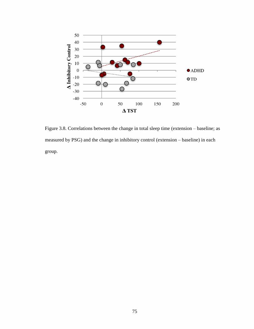
75
Figure 3.8. Correlations between the change in total sleep time (extension – baseline; as
measured by PSG) and the change in inhibitory control (extension – baseline) in each
group.
76
CHAPTER 4
THETA ACTIVITY IN CHILDREN WITH AND WITHOUT ADHD ACROSS WAKE
AND SLEEP
4.1 Introduction
Compared to TD controls, individuals with ADHD are reported to have elevated
theta activity (neural activity in the 4 to 7 Hz frequency range) during wake and sleep. A
meta-analysis assessing quantitative EEG during wake reported a 32% increase in theta
activity in individuals with ADHD, 6 to 42 years of age (Snyder & Hall, 2006). Wake
theta activity is particularly greater among children and adolescents with ADHD
compared to TD individuals (Hermens et al., 2005). Preliminary evidence indicates that
theta activity is also elevated in children with ADHD during sleep (Saletin, Coon, &
Carskadon, 2016), consistent with the findings reported in Chapter 2.
Theta activity, recorded during sleep and wake, is associated with a variety of
cognitive functions in TD populations. Accumulating evidence highlights the beneficial
role that sleep theta activity has on memory in school-aged children and young adults
(Benedict, Scheller, Rose-John, Born, & Marshall, 2009; Prehn-Kristensen, Munz,
Molzow, Wilhelm, Wiesner, & Baving, 2013; Walker, 2009). Sleep theta activity is also
positively associated with decision-making in young adults (Seeley, Smith, MacDonald,
& Beninger, 2016). Similarly, data from Chapter 2 indicate a positive association
between REM theta activity and inhibitory control in TD children 4 to 8 years of age.
Prior to the study described in Chapter 2, inhibitory control was predominately
associated with wake theta activity (Cavanagh & Frank, 2014). Specifically, EEG
components that index inhibitory control (e.g., N2) are strongly correlated with frontal

77
theta activity recorded during wakefulness in children, adolescents, and adults (Cavanagh
& Frank, 2014; Liu, Woltering, & Lewis, 2014). Wake theta activity also predicts
symptom severity in children and adolescents with ADHD (Hermens et al., 2005). As
these results attribute inhibitory control to theta activity recorded during both sleep and
wake, additional studies are needed to determine whether state-dependent characteristics
of EEG better predict inhibitory deficits in children with and without ADHD. A better
understanding of relations between inhibitory control and theta activity (during sleep
versus wake) may create opportunities for intervention in populations with inhibitory
deficits.
The goal of this study was two-fold. The first aim of this study was to compare
theta activity between wake and sleep in young children with and without ADHD.
Provided evidence that EEG is similar between wake and REM sleep in middle-aged
adults (Benca et al., 1999), it was hypothesized that the differences in sleep and wake
theta activity would be small (less than 0.3 standard deviations, representing a small-to-
medium effect size) in both groups. This null hypothesis was tested by evaluating the
effect size of the difference in theta activity recorded during sleep and wake for both TD
and ADHD children. If the difference between sleep and wake theta activity was less than
0.3 standard deviations within each group (ADHD and TD), the null hypothesis would
not be rejected and it would be determined that wake and sleep theta activity were similar
across states (see Streiner, 2003).
The second aim of this study was to determine whether wake or sleep theta
activity better predicted inhibitory control in these populations. Based on the prevalence
of sleep problems in children with ADHD (e.g., Weiss, Craig, Davies, Schibuk, & Stein,

78
2015; Yoon, Jain, & Shapiro, 2012) and the positive association between REM theta
activity and inhibitory control in TD children (Chapter 2), it was hypothesized that sleep
theta activity would account for more variability in inhibitory control than wake theta
activity. Alternatively, if variance in morning inhibitory control were better predicted by
(or equally predicted by) wake theta activity, the data would suggest that theta activity
underlies inhibitory control in young children, regardless of state (i.e., wake versus
sleep).
4.2 Methods
4.2.1 Participants
Participants were those described in Chapter 3.
4.2.2 Measures
Theta activity was recorded via EEG electrodes in the PSG cap montage (see
Chapter 3).
4.2.3 Procedure
During data collection for the sleep extension paradigm (see Chapter 3), ten
minutes of wake EEG was recorded prior to nocturnal sleep onset. Children with ADHD
(particularly those with hyperactive/impulsive symptoms) may find the standard resting
paradigm especially challenging, as these children characteristically ‘fidget/squirm’ and
have ‘difficultly sitting quietly’ (Shaffer, Fisher, Lucas, Dulcan, & Schwab-Stone, 2000).
To reduce these confounds, ten minutes of wake EEG was recorded while the child
quietly read a book (or was read to by a caregiver). Typically, 2-3 minutes of EEG is
used to gauge waking neural activity (e.g., Thompson, Woodruff Carr, White-Schwoch,
Tierney, Nicol, & Kraus, 2016). A 10-minute recording was used to assure enough

79
artifact-free data was obtained for statistical analyses. The beginning and end of this
recording period was noted on the participant’s datasheet and marked in the
polysomnogram using digital event markers. Events that may have disrupted the EEG
recording (e.g., excessive movement or talking) were noted and omitted from analyses.
Following the wake EEG recording, the lights were turned out and the child was
encouraged to go to sleep. Sleep theta activity was computed from EEG recorded during
the nocturnal sleep bout (see Chapter 3). Specifically, sleep theta activity was sampled
from epochs characterized as REM sleep, as theta activity is prominent during this sleep
stage (Hobson & Pace-Schott, 2002). In the ADHD group, ten minutes of wake EEG was
also collected the morning following overnight sleep to explore homeostatic differences
in wake theta activity.
4.3 Statistical Analyses
4.3.1 Wake Versus Sleep Theta Activity
Paired samples t-tests were used to compare average wake theta activity against
average sleep theta activity recorded during REM sleep within each group (ADHD and
TD). To test the difference in sleep and wake theta activity, the effect size of the
difference in theta activity recorded during sleep and wake was evaluated in terms of
standard deviations, independently for each group.
In accordance with the a priori hypothesis, the null hypothesis would not be
rejected if the difference between sleep and wake theta activity was less than 0.3 standard
deviations (see Streiner, 2003). If the difference between sleep and wake theta activity
was less than 0.3 standard deviations, this would indicate that wake and sleep theta
activity were similar in children with and without ADHD. Average wake theta activity

80
was also compared against random 10-minute samples of REM theta activity to control
for differences in wake and sleep EEG recording times.
Finally, an exploratory paired samples t-test was used to compare morning theta
activity to evening theta activity to assess homeostatic differences in theta activity in
children with ADHD.
4.3.2 Theta Activity and Inhibitory Control
To determine whether wake or sleep theta activity better predicted inhibitory
control in children with and without ADHD, linear regression models were used. In these
models, average morning inhibitory control (collapsed across baseline and extension
conditions) was entered as the outcome variable. Group (ADHD and TD) and average
wake and sleep theta activity were entered as predictor variables, simultaneously.
4.4 Results
Consistent with the data comparing sleep theta activity in Chapter 3, evening
wake theta activity did not differ between conditions in either group (F(1,18) = 2.27, p =
0.149; Table 4.1). As such, measurements sampled during the baseline and extension
conditions were collapsed into average measures to be used in subsequent analyses.
Evening wake theta activity was marginally greater in the ADHD group overall (F(1,18)
= 3.87, p = 0.065, p2 = 0.177; Table 4.1). The two-way interaction between condition
and group was not significant (F(1,18) = 0.06, p = 0.803).
4.4.1 Wake Versus Sleep Theta Activity
Although positively related to one another, average sleep and wake theta activity
were not significantly correlated in either TD or ADHD children (r’s ≤ 0.291, p’s ≥
0.415). In children with ADHD, the paired samples t-test used to compare average wake

81
theta activity against average sleep theta activity indicated that theta activity was not
statistically different across wake and sleep (t(10) = -0.44, p = 0.672; Table 4.1). The
mean difference in theta activity between wake and sleep (-1.11 μV2/Hz) and the standard
error of the mean difference (2.54 μV2/Hz) were then used to calculate a 95% confidence
interval for this group (lower bound = -6.19, upper bound = 3.97). The effect size of the
difference between wake and sleep theta activity was computed as 0.3 standard
deviations of the pooled standard deviation for wake and sleep theta activity (5.83 + 6.57
/ 2 = 6.2; 6.2 * 0.3 = 1.86). This effect size was not significantly smaller than a small-to-
medium effect size, as it fell within our 95% confidence interval. The effect size of
difference was also smaller than a large effect size (3.97 / 1.86 = 2.13). Thus, the null
hypothesis was not rejected.13
These findings were similar in TD children, as average wake theta activity did not
statistically differ from average sleep theta activity (t(9) = 1.05, p = 0.319; Table 4.1).
Here too, the mean difference (2.11 μV2/Hz) and the standard error of the mean
difference (2.00 μV2/Hz) were used to compute a 95% confidence interval for the TD
group (lower bound = -1.89, upper bound = 6.11). The effect size of the difference was
computed as described above (1.56) but was not significantly smaller than a small-to-
medium effect size.14
13 Results were unchanged when wake theta activity was compared against random 10-
minute samples of REM theta activity (p = 0.568, 95% confidence interval [-7.09, 3.87],
effect size of difference = 1.88). 14 Wake theta activity was significantly greater than the 10-minute segment of theta
activity randomly sampled from REM sleep (t(5) = -4.40, p =0.007, 95% CI [-1.35, -
0.35]). The effect size of difference was small (1.46), but was outside of the 95%
confidence interval computed for this group [-1.23, -0.47]. These data should be
interpreted with caution, as complete wake theta datasets were only available for 6 TD
children.

82
In the ADHD group, an exploratory paired samples t-test revealed that, collapsed
across conditions, wake theta activity measured prior to overnight sleep (M = 18.62, SD =
6.57 μV2/Hz) was significantly greater than wake theta activity measured following
overnight sleep (M = 14.07, SD = 2.49 μV2/Hz; t(10) = 3.26, p = 0.009, 95% CI [1.44,
7.65]). This finding suggests that there may be homeostatic differences in theta activity in
children with ADHD.
4.4.2 Theta Activity and Inhibitory Control
Consistent with the results of Chapter 3, the linear regression model indicated that
neither wake (B = -0.001, p = 0.767) nor sleep (B = 0.005, p = 0.307) theta activity
predicted morning inhibitory control. The main effect of group also did not significantly
predict morning inhibitory control (B = 0.116, p = 0.062). Controlling for evening
inhibitory control, age, and symptomology did not alter these statistical outcomes in
either group (p’s 0.210).
4.5 Discussion
The primary aim of this chapter was to examine whether levels of theta activity
were similar across intervals of sleep and wake in children with and without ADHD.
Sleep and wake theta activity did not statistically differ in either group. However, the
effect size of the difference between activity recorded during wake and sleep was too
small to conclude activity was statistically similar across states. The secondary aim of
this chapter was to determine whether inhibitory control was better predicted by theta
activity during sleep or wake in children with or without ADHD. Neither sleep nor wake
theta activity significantly predicted morning inhibitory control in either group.
4.5.1 Differences in Wake and Sleep Theta Activity

83
Although inconclusive, data from this chapter suggest that cortical activity in the
theta frequency band is similar across sleep and wake in young children with and without
ADHD. These results support the hypothesis that EEG is trait-like (Benca et al., 1999;
Tarokh, Carskadon, & Achermann, 2011) and stable across sleep and wake. If such is the
case, endophenotypes evident during sleep may also be identified during resting
wakefulness.
4.5.2 Relations between Theta Activity and Inhibitory Control
The results of this chapter also suggest that theta activity, recorded during sleep or
wake, was not functionally related to inhibitory control in children with or without
ADHD. As morning inhibitory control was associated with SWA in children with ADHD
(see Chapter 3), this finding is unsurprising. However, REM theta activity was positively
associated with inhibitory control in TD children in Chapter 2. As such, additional studies
are needed to better understand the contributions of theta activity to inhibitory
functioning during typical development.
4.5.3 Homeostatic Differences in Theta Activity in Children with ADHD
Exploratory analyses suggest that theta activity recorded prior to overnight sleep
was greater than that recorded following overnight sleep. This finding is consistent with
work in animal models which suggests that theta activity is elevated following prolonged
wakefulness and reduced after sleep (Leemburg, Vyazovskiy, Olcese, Bassetti, Tononi, &
Cirelli, 2010). Data from these animal studies also indicate that theta activity recorded
during wake predicts SWA recorded during subsequent sleep. As ADHD children had
elevated wake theta activity in the evening and more SWA than TD children, the results
of this dissertation suggest that children with ADHD need more sleep in order to

84
sufficiently reduce sleep pressure. Unfortunately, data from this dissertation do not
support this hypothesis.15
4.5.4 Conclusions
Taken together, the results of this chapter suggest that cortical activity in the theta
frequency band may be stable across sleep and wake, consistent with the notion that EEG
is trait-like (Benca et al., 1999; Tarokh, Carskadon, & Achermann, 2011). However, as
the confidence intervals were large, these data should be interpreted with caution.
Exploratory analysis of differences in wake theta activity before and after overnight sleep
indicate that, among children with ADHD, theta activity is higher in the evening than in
the morning. This finding supports animal studies which show that theta activity is
elevated following prolonged wakefulness and reduced after sleep (Leemburg et al.,
2010). Finally, neither wake nor sleep theta activity were found to predict variability in
subsequent assessments of inhibitory control among children with or without ADHD.
Data from the TD sample tested in Chapter 2 indicate that inhibitory control was related
to sleep theta activity, suggesting that the functional significance of sleep, and potentially
wake, theta activity may differ between typically and atypically developing children.
15 An exploratory linear regression was used to determine whether evening wake theta
activity (Chapter 4) and SWA (Chapter 3) interacted to predict morning inhibitory control
in the ADHD group. The interaction term did not significantly predict morning inhibitory
control during either the baseline or extension condition (p’s = 0.926 and 0.124,
respectively).

85
Table 4.1. Differences in wake and sleep theta activity (μV2/Hz) in each group.
ADHD TD
Mean (SD) Mean (SD)
Baseline condition
Wake
Evening 18.24 (6.41) 12.96 (4.42)
Morning 14.08 (3.87) -
Sleep 17.25 (7.19) 15.77 (5.60)
Extension condition
Wake
Evening 18.99 (7.13) 13.79 (4.78)
Morning 14.06 (2.99) -
Sleep 17.77 (5.34) 15.08 (6.85)
Note: In ADHD group, n = 15. In TD group, n = 10.

86
CHAPTER 5
GENERAL DISCUSSION
The aim of this dissertation was to examine the role of sleep on inhibitory control
in young children with ADHD. To assess these relations, children with and without
ADHD participated in overnight sleep studies in which sleep physiology was monitored
with PSG and inhibitory control gauged via a Go/No-Go task. Collectively, the results of
this dissertation indicate that inhibitory control is enhanced by overnight sleep in TD
children but not in children with ADHD. However, when children with ADHD were
instructed to advance their bedtime in an effort to extend the amount of time they slept at
night, inhibitory control was improved. Moreover, the physiological mechanisms
underlying the benefits of sleep on inhibitory control differed between TD children and
children with ADHD, suggesting that the developmental trajectory of sleep EEG may be
altered in atypical development.
5.1 Sleep and Inhibitory Control
The role of sleep on inhibitory control differed between TD and ADHD children.
As presented in Chapters 2 and 3, TD children had sleep-dependent enhancement of
inhibitory control whereas children with ADHD did not. Specifically, inhibitory control
was improved following overnight sleep in TD children but not in children with ADHD.
Although average sleep duration did not differ between these groups, the results of
Chapter 3 indicate that lengthening sleep duration improves inhibitory control in ADHD
children. Given that inhibitory control is a primary deficit in ADHD, and is strongly
associated with core symptoms such as hyperactivity and impulsivity (Barkley, 1997), the
data from the current set of studies suggest that sleep-related interventions should be

87
strongly considered to improve outcomes in this population. Relative to pharmacological
treatments, sleep-based interventions are cost-effective and have not been linked to
adverse side effects.
5.2 Relations between Sleep Physiology and Inhibitory Control
The mechanisms underlying the benefits of sleep on subsequent inhibitory control
also differed between TD children and children with ADHD. Enhancement of inhibitory
control was positively associated with theta activity recorded during REM sleep in TD
children (Chapter 2). Although REM theta activity was elevated in children with ADHD,
it was not associated with inhibitory functioning in this group (Chapter 2). Rather,
enhanced inhibitory control was associated with reduced SWA, when sleep duration was
extended, in children with ADHD (Chapter 3).
Dissimilarities in the neural mechanisms supporting inhibitory control in children
with and without ADHD are likely a consequence of developmental differences in EEG
trajectories. Longitudinal data indicate both SWA and theta activity decline during
childhood, and are strongly associated with neural development (Campbell & Feinberg,
2009). Specifically, SWA declines linearly from birth until approximately 6 years of age.
Levels of SWA plateau thereafter until adolescence. Theta activity, on the other hand,
declines significantly between 6 and 11 years of age. As changes in SWA (birth to 6
years of age) occur earlier than changes in theta activity (6 to 11 years of age) in TD
children, these findings suggest that the developmental trajectories of sleep EEG in
children with ADHD are delayed.
Correlations with behavioral outcomes support the hypothesis that children with
ADHD have delayed cortical development (Rubia, 2007), as theta activity was related to

88
inhibitory control in TD children (Chapter 2) and SWA associated with the same
outcome in children with ADHD (Chapter 3). However, longitudinal studies of sleep
EEG trajectories are needed to directly test this hypothesis.
In addition to potential differences in EEG trajectories, the samples tested in each
study differed in age. The children tested in Chapter 2 (4 to 8 years of age; Mage = 6.7
years) were younger than those tested in Chapter 3 (6 to 9 years of age; Mage = 8.3 years).
Although the children in Chapter 2 met the clinical criteria necessary to diagnosis
ADHD, the children in this sample were not required to have a physician’s formal
diagnosis of the disorder. ADHD is not typically diagnosed until 7 years of age
(Applegate et al., 1997). Thus, the age range for children recruited in Chapter 3 was
extended to include older children. As EEG trajectories change across early
development, age-related differences may have contributed to the mechanistic differences
reported between groups in Chapters 2 and 3. Similarly, the inconsistent correlations
between inhibitory control and sleep physiology (specifically, REM theta activity in the
TD groups and SWA in the ADHD groups) may have been a consequence of age
differences.
5.3 Differences in Sleep and Wake Theta Activity
Although theta activity was not associated with inhibitory functioning in children
with ADHD, accumulating evidence suggests that theta activity is elevated during both
wake (Snyder and Hall, 2006) and sleep (Chapter 2; Saletin et al., 2016) and is
functionally related to cognitive outcomes in this population. Thus, the aim of the final
study in this dissertation was to determine whether theta activity was stable across states
and better understand the role of sleep and wake theta activity in inhibitory control in
89
children with and without ADHD. Consistent with data suggesting that EEG is trait-like
(Benca et al., 1999; Tarokh, Carskadon, & Achermann, 2011), theta activity recorded
during sleep and wake was not statistically different. However, the data collected to
assess stability across states was inconclusive, as the effect size of the difference between
the two measures was small in both groups. Furthermore, neither sleep nor wake theta
activity were functionally related to inhibitory control in children with or without ADHD.
Given that theta activity was not associated with inhibitory control among the ADHD
children tested (in Chapters 2 or 3), this finding is unsurprising. However, as REM theta
activity was positively associated with inhibitory control in TD children (Chapter 2),
additional studies are needed to better understand the contributions of theta activity to
inhibitory functioning during typical development.
Exploratory analyses indicate that wake theta activity was elevated in the evening,
relative to the morning, in the ADHD group. Animal studies suggest that greater wake
theta activity predicts greater sleep pressure (Leemburg et al., 2010), suggesting that theta
and SWA may interact to predict subsequent performance. However, when the data from
Chapters 3 and 4 were evaluated together, the interaction between evening wake theta
activity and sleep SWA did not predict variability in morning inhibitory control during
either condition. Importantly, this data was drawn from a small sample. Additional data
collection may be needed to support this hypothesis.
5.4 Summary
Overall, the results of this dissertation support the widespread hypothesis that
children with ADHD have insufficient sleep. The current studies also indicate that the
developmental trajectory of low frequency sleep EEG is delayed in this population,

90
relative to TD controls. As such, sleep EEG may be used to index cortical development
and identify children at risk for ADHD (Chapters 2 and 3). Similarly, endophenotypes in
sleep EEG may be used to predict response to the sleep-based intervention associated
with improved inhibitory function in this sample (Chapter 3).
5.5 Future Directions
Based on the results of current studies, future research into the role of sleep and
inhibitory control is needed to inform treatment strategies in atypical development.
Although the behavioral findings in this dissertation were robust, the sample tested in
Chapters 3 and 4 was small. As such, additional data collection is needed to infer
generalizability and increase the statistical power needed to detect significant correlations
between physiology and behavior. Additional studies are also needed to evaluate
incremental changes in sleep physiology with changes in sleep duration (see Discussion
of Chapter 3). As sleep quality is associated with cognitive functioning, it is particularly
important to better understand how short- and long-term changes to sleep physiology
affect outcomes in young children both with and without ADHD.
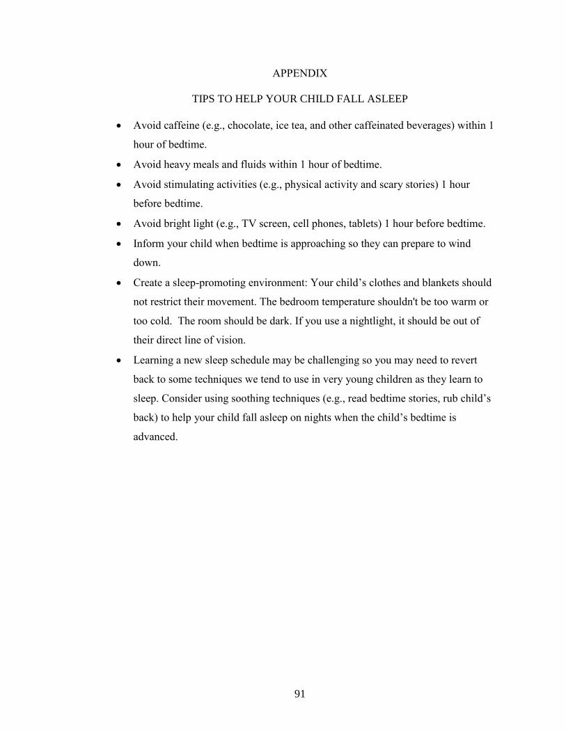
91
APPENDIX
TIPS TO HELP YOUR CHILD FALL ASLEEP
• Avoid caffeine (e.g., chocolate, ice tea, and other caffeinated beverages) within 1
hour of bedtime.
• Avoid heavy meals and fluids within 1 hour of bedtime.
• Avoid stimulating activities (e.g., physical activity and scary stories) 1 hour
before bedtime.
• Avoid bright light (e.g., TV screen, cell phones, tablets) 1 hour before bedtime.
• Inform your child when bedtime is approaching so they can prepare to wind
down.
• Create a sleep-promoting environment: Your child’s clothes and blankets should
not restrict their movement. The bedroom temperature shouldn't be too warm or
too cold. The room should be dark. If you use a nightlight, it should be out of
their direct line of vision.
• Learning a new sleep schedule may be challenging so you may need to revert
back to some techniques we tend to use in very young children as they learn to
sleep. Consider using soothing techniques (e.g., read bedtime stories, rub child’s
back) to help your child fall asleep on nights when the child’s bedtime is
advanced.

92
BIBLIOGRAPHY
Acebo, C., Sadeh, A., Seifer, R., Tzischinsky, O., Hafer, A., & Carskadon, M. A. (2005).
Sleep/wake patterns derived from activity monitoring and maternal report for
healthy 1-to 5-year-old children. Sleep, 28(12), 1568.
Achenbach, T., & Rescorla, L. (2001). ASEBA school-age forms & profiles.
American Academy of Pediatrics: Management steering committee on quality
improvement and subcommittee on attention-deficit/hyperactivity disorder. (2011).
ADHD: Clinical practice guideline for the diagnosis, evaluation, and treatment of
attention-deficit/hyperactivity disorder in children and adolescents. Pediatrics, 128,
1007-1022.
American Academy of Sleep Medicine, Iber, C, (2007), The AASM manual for the
scoring of sleep and associated events: Rules, terminology, and technical
specifications. American Academy of Sleep Medicine.
Anderson, P. (2002). Assessment and development of executive function (EF) during
childhood. Child Neuropsychology: A Journal on Normal and Abnormal
Development in Childhood and Adolescence, 8(2), 71–82.
Applegate, B., Lahey, B., Hart, E. L., Biederman, J., Hynd, G., Barkley, R., Ollendick,
T., Frick, P. J., Greenhill, L., McBurnett, K., Newcorn, J. H., Kerdyk, L., Garfinkel,
B., Waldman, I., & Shaffer, D. (1997). Validity of the age-of-onset criterion for
ADHD: A report from the DSM-IV field trials. Journal of the American Academy
of Child & Adolescent Psychiatry, 36(9), 1211–1221.
Arnal, P. J., Sauvet, F., Leger, D., van Beers, P., Bayon, V., Bougard, C., Rabat, A.,
Millet, G. Y., & Chennaoui, M. (2015). Benefits of sleep extension on sustained
attention and sleep pressure before and during total sleep deprivation and recovery.
Sleep, 38(12), 1935–1943.
Astill, R. G., Van der Heijden, K. B., Van IJzendoorn, M. H., & Van Someren, E. J. W.
(2012). Sleep, cognition, and behavioral problems in school-age children: A century
of research meta-analyzed. Psychological Bulletin, 138(6), 1109–1138.
Baird, A. L., Coogan, A. N., Siddiqui, A., Donev, R. M., & Thome, J. (2012). Adult
attention-deficit hyperactivity disorder is associated with alterations in circadian
rhythms at the behavioural, endocrine and molecular levels. Molecular Psychiatry,
17, 988–995.
Barkley, R. (1997). Behavioral inhibition, sustained attention, and executive functions:
Constructing a unifying theory of ADHD. Psychological Bulletin, 121(1), 65–94.
Barkley, R., & Murphy, K. (2006). Attention-deficit hyperactivity disorder: A clinical
workbook (3rd Edition). Guilford Press.

93
Barry, R. J., Clarke, A. R., & Johnstone, S. J. (2003). A review of electrophysiology in
attention-deficit/hyperactivity disorder: I. Qualitative and quantitative
electroencephalography. Clinical Neurophysiology, 114(2), 171–183.
Benca, R. M., Obermeyer, W. H., Larson, C. L., Yun, B., Dolski, I., Kleist, K. D.,
Weber, S. M., & Davidson, R. J. (1999). EEG alpha power and alpha power
asymmetry in sleep and wakefulness. Psychophysiology, 36(4), 430–436.
Benedict, C., Scheller, J., Rose-John, S., Born, J., & Marshall, L. (2009). Enhancing
influence of intranasal interleukin-6 on slow-wave activity and memory
consolidation during sleep. The FASEB Journal, 23(10), 3629–3636.
Bezdjian, S., Baker, L. A., Lozano, D. I., & Raine, A. (2009). Assessing inattention and
impulsivity in children during the Go/NoGo task. British Journal of Developmental
Psychology, 27, 365–383.
Borbely, A., Tobler, I., & Hanagasioglu, M. (1984). Effect of sleep deprivation on sleep
and EEG power spectra in the rat. Behavioral Brain Research, 14, 171–182.
Boyce, R., Glasgow, S. D., Williams, S., & Adamantidis, A. (2016). Causal evidence for
the role of REM sleep theta rhythm in contextual memory consolidation. Science,
352(6287), 812–816.
Boyle, C. A., Boulet, S., Schieve, L. A., Cohen, R. A., Blumberg, S. J., Yeargin-Allsopp,
M., Visser, S., & Kogan, M. D. (2011). Trends in the prevalence of developmental
disabilities in US children, 1997-2008. Pediatrics, 127(6), 1034–1042.
Cajochen, C., Foy, R., & Dijk, D. J. (1999). Frontal predominance of a relative increase
in sleep delta and theta EEG activity after sleep loss in humans. Sleep Research
Online, 2, 65–69.
Campbell, I. G., & Feinberg, I. (2009). Longitudinal trajectories of non-rapid eye
movement delta and theta EEG as indicators of adolescent brain maturation.
Proceedings of the National Academy of Sciences, 106(13), 5177–5180.
Castellanos, F., Marvasti, F., Ducharme, J., Walter, J., Israel, M., Krain, A., Pavlovsky,
C., & Hommer, D. (2000). Executive function oculomotor tasks in girls with
ADHD. Journal of the American Academy of Child & Adolescent Psychiatry, 39(5),
644–650.
Castellanos, F. X., Sonuga-Barke, E. J. S, Milham, M.P., & Tannock, R. (2006).
Characterizing cognition in ADHD: Beyond executive dysfunction. Trends in
Cognitive Sciences, 10, 117–123.
Cavanagh, J. F., & Frank, M. J. (2014). Frontal theta as a mechanism for cognitive
control. Trends in Cognitive Sciences, 18(8), 414–421.
94
Chuah, Y. M. L., Venkatraman, V., Dinges, D. F., & Chee, M. W. L. (2006). The neural
basis of interindividual variability in inhibitory efficiency after sleep deprivation.
Journal of Neuroscience, 26, 7156–7162.
Cohen-Zion, M., & Ancoli-Israel, S. (2004). Sleep in children with attention-deficit
hyperactivity disorder (ADHD): A review of naturalistic and stimulant intervention
studies. Sleep Medicine Reviews, 8(5), 379–402.
Cremone, A., Kurdziel, L. B. F., Fraticelli-Torres, A., McDermott, J. M., & Spencer, R.
M. C. (2016). Napping reduces emotional attention bias during early childhood.
Developmental Science.
Dahl, R. E. (1996). The regulation of sleep and arousal: Development and
psychopathology. Development and Psychopathology, 8(1), 3–27.
Dewald-Kaufmann, J. F., Oort, F. J., & Meijer, A. M. (2014). The effects of sleep
extension and sleep hygiene advice on sleep and depressive symptoms in
adolescents: A randomized controlled trial. Journal of Child Psychology and
Psychiatry, 55(3), 273–283.
Dijk, D. J., Brunner, D. P., & Borbély, A. A. (1990). Time course of EEG power density
during long sleep in humans. The American Journal of Physiology, 258(3 Pt 2),
R650–661.
Doyle, A. (2006). Executive functions in attention-deficit/hyperactivity disorder. Journal
of Clinical Psychiatry, 67, 21–26.
Drummond, S., Paulus, M. P., Tapert, S. F. (2006). Effects of two nights sleep
deprivation and two nights recovery sleep on response inhibition. Journal of Sleep
Research, 15, 261–265.
Durston, S., Thomas, K. M., Yang, Y., Ulug, A. M., Zimmerman, R. D., & Casey, B. J.
(2002). A neural basis for the development of inhibitory control. Developmental
Science, 5(4), F9–F16.
Durston, S., Tottenham, N. T., Thomas, K. M., Davidson, M. C., Eigsti, I.-M., Yang, Y.,
Ulug, A. Z., & Casey, B. J. (2003). Differential patterns of striatal activation in
young children with and without ADHD. Biological Psychiatry, 53(10), 871–878.
Fallone, G., Acebo, C., Arnedt, J. T., Seifer, R., & Carskadon, M. A. (2001). Effects of
acute sleep restriction on behavior, sustained attention, and response inhibition in
children. Perceptual and Motor Skills, 93(1), 213–229.
Faraone, S. V., Biederman, J., Spencer, T. J., & Aleardi, M. (2006). Comparing the
efficacy of medications for ADHD using meta-analysis. Medscape General
Medicine, 8(4).

95
Goel, N., Rao, H., Durmer, J., & Dinges, D. (2009). Neurocognitive consequences of
sleep deprivation. Seminars in Neurology, 29, 320–339.
Goodlin-Jones, B. L., Sitnick, S. L., Tang, K., Liu, J., & Anders, T. F. (2008). The
Children’s Sleep Habits Questionnaire in toddlers and preschool children. Journal
of Developmental & Behavioral Pediatrics, 29(2), 82–88.
Gruber, R., Cassoff, J., Frenette, S., Wiebe, S., & Carrier, J. (2012). Impact of sleep
extension and restriction on children’s emotional lability and impulsivity.
Pediatrics, 130(5), e1155–e1161.
Gruber, R., Wiebe, S., Montecalvo, L., Brunetti, B., Amsel, R., & Carrier, J. (2011).
Impact of sleep restriction on neurobehavioral functioning of children with attention
deficit hyperactivity disorder. Sleep, 34(3), 315–323.
Haier, R. J., Siegel, B. V., Nuechterlein, K. H., Hazlett, E., Wu, J. C., Paek, J.,
Browning, H. L., & Buchsbaum, M. S. (1988). Cortical glucose metabolic rate
correlates of abstract reasoning and attention studied with positron emission
tomography. Intelligence, 12, 199–217.
Harpin, V. A. (2005). The effect of ADHD on the life of an individual, their family, and
community from preschool to adult life. Archives of Disease in Childhood, 90, i2–
i7.
Herbert, S. D., Harvey, E. A., Roberts, J. L., Wichowski, K., and Lugo-Candelas, C. L.
(2012). A randomized controlled trial of a parent training and emotion socialization
program for families of hyperactive preschool-aged children.
Henderson, J. A., & Jordan, S. S. (2010). Development and preliminary evaluation of the
bedtime routines questionnaire. Journal of Psychopathology and Behavioral
Assessment, 32(2), 271–280.
Herman, J. H. (2015). Attention deficit/hyperactivity disorder and sleep in children.
Sleep Medicine Clinics, 10(2), 143–149.
Hermens, D. F., Soei, E. X. C., Clarke, S. D., Kohn, M. R., Gordon, E., & Williams, L.
M. (2005). Resting EEG theta activity predicts cognitive performance in attention-
deficit hyperactivity disorder. Pediatric Neurology, 32(4), 248–256.
Hirshkowitz, M., Whiton, K., Albert, S. M., Alessi, C., Bruni, O., DonCarlos, L., Hazen,
N., Herman, J., Katz, E. S., Kheirandish-Gozal, L., Neubauer, D. N., O’Donnell, A.
E., Ohayon, M., Peever, J., Rawding, R., Sachdeva, R. C., Setters, B., Vitiello, M.
V., Catesby Ware, J., & Adams Hillard, P. J. (2015). National Sleep Foundation’s
sleep time duration recommendations: Methodology and results summary. Sleep
Health, 1(1), 40–43.

96
Hiscock, H., Sciberras, E., Mensah, F., Gerner, B., Efron, D., Khano, S., & Oberklaid, F.
(2015). Impact of a behavioural sleep intervention on symptoms and sleep in
children with attention deficit hyperactivity disorder, and parental mental health:
randomised controlled trial. BMJ, 350, h68–h68.
Hobson, J. A., & Pace-Schott, E. F. (2002). The cognitive neuroscience of sleep:
Neuronal systems, consciousness and learning. Nature Reviews Neuroscience, 3(9),
679–693.
Hutchison, I. C., & Rathore, S. (2015). The role of REM sleep theta activity in emotional
memory. Frontiers in Psychology, 6.
Iglowstein, I., Jenni, O. G., Molinari, L., & Largo, R. H. (2003). Sleep duration from
infancy to adolescence: Reference values and generational trends. Pediatrics,
111(2), 302–307.
Ingram, S., Hechtman, L., & Morgenstern, G. (1999). Outcome issues in ADHD:
Adolescent and adults long-term outcome. Developmental Disabilities Research
Reviews, 5, 243–250.
Kindlon, D., Mezzacappa, E., & Earls, F. (1995). Psychometric properties of impulsivity
measures: Temporal stability, validity and factor structure. Journal of Child
Psychology and Psychiatry, 36, 645–661.
Konofal, E., Lecendreux, M., & Cortese, S. (2010). Sleep and ADHD. Sleep Medicine,
11, 652–658.
Kopasz, M., Loessl, B., Hornyak, M., Riemann, D., Nissen, C., Piosczyk, H., &
Voderholzer, U. (2010). Sleep and memory in healthy children and adolescents – A
critical review. Sleep Medicine Reviews, 14(3), 167–177.
Kurth, S., Ringli, M., Geiger, A., LeBourgeois, M., Jenni, O. G., & Huber, R. (2010).
Mapping of cortical activity in the first two decades of life: A high-density sleep
electroencephalogram study. Journal of Neuroscience, 30, 13211–13219.
Lahat, A., Degnan, K. A., White, L. K., McDermott, J. M., Henderson, H. A., Lejuez, C.
W., & Fox, N. A. (2012). Temperamental exuberance and executive function
predict propensity for risk taking in childhood. Development and Psychopathology,
24(03), 847–856.
Leemburg, S., Vyazovskiy, V.V., Olcese, U., Bassetti, C.L., Tononi, G., & Cirelli, C.
(2010). Sleep homeostasis in the rat is preserved during chronic sleep restriction.
Proceedings of the National Academy of Sciences, 107, 15939–15944.

97
Lejuez, C. W., Aklin, W., Daughters, S., Zvolensky, M., Kahler, C., & Gwadz, M.
(2007). Reliability and validity of the youth version of the Balloon Analogue Risk
Task (BART-Y) in the assessment of risk-taking behavior among inner-city
adolescents. Journal of Clinical Child and Adolescent Psychology: The Official
Journal for the Society of Clinical Child and Adolescent Psychology, American
Psychological Association, Division 53, 36(1), 106–111.
Liu, Z.-X., Woltering, S., & Lewis, M. D. (2014). Developmental change in EEG theta
activity in the medial prefrontal cortex during response control. NeuroImage, 85,
873–887.
Logan, G. D., Schachar, R. J., & Tannock, R. (1997). Impulsivity and inhibitory control.
Psychological Science, 8(1), 60–64.
Mann, C., Lubar, .J., Zimmerman, A., Miller, C., & Muenchen, R. (1992). Quantitative
analysis of EEG in boys with attention-deficit-hyperactivity disorder: Controlled
study with clinical implications. Pediatric Neurology, 8, 30–36.
Marks, G., Shaffery, J., Oksenberg, A., Speciale, S., & Roffwarg, H. (1995). A
functional role of REM sleep in brain maturation. Behavioural Brain Research, 69,
1–11.
Marshall, L., Helgadóttir, H., Mölle, M., & Born, J. (2006). Boosting slow oscillations
during sleep potentiates memory. Nature, 444(7119), 610–613.
McDermott, J. M., Westerlund, A., Zeanah, C. H., Nelson, C. A., & Fox, N. A. (2012).
Early adversity and neural correlates of executive function: Implications for
academic adjustment. Developmental Cognitive Neuroscience, 2, S59–S66.
Miller, A. L., Seifer, R., Crossin, R., & Lebourgeois, M. K. (2014). Toddler’s self-
regulation strategies in a challenge context are nap-dependent. Journal of Sleep
Research, 24(3), 279–287.
Mindell, J. A., Li, A. M., Sadeh, A., Kwon, R., & Goh, D. Y. T. (2015). Bedtime
routines for young children: A dose-dependent association with sleep outcomes.
Sleep.
Nigg, J. T. (2000). On inhibition/disinhibition in developmental psychopathology: Views
from cognitive and personality psychology and a working inhibition taxonomy.
Psychological Bulletin, 126(2), 220–246.
Nishida, M., Pearsall, J., Buckner, R. L., & Walker, M. P. (2009). REM sleep, prefrontal
theta, and the consolidation of human emotional memory. Cerebral Cortex, 19(5),
1158–1166.

98
O’Connell, R. G., Dockree, P. M., Bellgrove, M. A., Turin, A., Ward, S., Foxe, J. J., &
Robertson, I. H. (2009). Two types of action error: Electrophysiological evidence
for separable inhibitory and sustained attention neural mechanisms producing error
on go/no-go tasks. Journal of Cognitive Neuroscience, 21, 93–104.
Oosterlaan, J., Logan, G. D., & Sergeant, J. A. (1998). Response inhibition in AD/HD,
CD, comorbid AD/HD+ CD, anxious, and control children: A meta-analysis of
studies with the stop task. Journal of Child Psychology and Psychiatry, 39(3), 411–
425.
Owens, J. (2005). The ADHD and sleep conundrum: A review. Journal of
Developmental & Behavioral Pediatrics, 26(4), 312–322.
Owens, J. A., Maxim, R., Nobile, C., McGuinn, M., & Msall, M. (2000). Parental and
self-report of sleep in children with attention-deficit/hyperactivity disorder.
Archives of Pediatric and Adolescent Medicine, 154, 549-555.
Owens, J. A., Spirito, A., & McGuinn, M. (2000). The Children’s Sleep Habits
Questionnaire (CSHQ): Psychometric properties of a survey instrument for school-
aged children. Sleep, 23(8), 1043–1052.
Pelham, Jr., W. E., Fabiano, G. A., & Massetti, G. M. (2005). Evidence-based
assessment of attention deficit hyperactivity disorder in children and adolescents.
Journal of Clinical Child & Adolescent Psychology, 34(3), 449–476.
Prehn-Kristensen, A., Göder, R., Fischer, J., Wilhelm, I., Seeck-Hirschner, M.,
Aldenhoff, J., & Baving, L. (2011). Reduced sleep-associated consolidation of
declarative memory in attention-deficit/hyperactivity disorder. Sleep Medicine,
12(7), 672–679.
Prehn-Kristensen, A., Munz, M., Molzow, I., Wilhelm, I., Wiesner, C. D., & Baving, L.
(2013). Sleep promotes consolidation of emotional memory in healthy children but
not in children with attention-deficit hyperactivity disorder. PLoS ONE, 8(5),
e65098.
Putnam, S. P., & Rothbart, M. K. (2006). Development of short and very short forms of
the Children’s Behavior Questionnaire. Journal of Personality Assessment, 87(1),
102–112.
Randazzo, A. C., Muehlbach, M. J., Schweitzer, P. K., & Walsh, J. K. (1998). Cognitive
function following acute sleep restriction in children ages 10-14. Sleep, 21(8), 861–
868.
Ringli, M., Souissi, S., Kurth, S., Brandeis, D., Jenni, O. G., & Huber, R. (2013).
Topography of sleep slow wave activity in children with attention-
deficit/hyperactivity disorder. Cortex, 49(1), 340–347.

99
Rolon-Arroyo, B., Arnold, D. H., Harvey, E. A., & Marshall, N. (2016). Assessing
attention and disruptive behavior symptoms in preschool-age children: The utility
of the Diagnostic Interview Schedule for Children. Journal of Child and Family
Studies, 25. 65–76.
Rubia, K. (2007). Neuro-anatomic evidence for the maturational delay hypothesis of
ADHD. Proceedings of the National Academy of Sciences, 104(50), 19663-19664.
Sadeh, A., Gruber, R., & Raviv, A. (2003). The effects of sleep restriction and extension
on school-age children: What a difference an hour makes. Child Development,
74(2), 444–455.
Sadeh, A., Pergamin, L., & Bar-Haim, Y. (2006). Sleep in children with attention-deficit
hyperactivity disorder: A meta-analysis of polysomnographic studies. Sleep
Medicine Reviews, 10(6), 381–398.
Sagaspe, P., Taillard, J., Amiéva, H., Beck, A., Rascol, O., Dartigues, J. F., Capelli, A.,
& Philip, P. (2012). Influence of age, circadian and homeostatic processes on
inhibitory motor control: A Go/Nogo task study. PLoS One, 7, e39410.
Saletin, J. M., Coon, W. G., & Carskadon, M. A. (2016). Stage 2 sleep EEG sigma
activity and motor learning in childhood ADHD: A pilot study. Journal of Clinical
Child & Adolescent Psychology, 1–10.
Schachar, R., Tannock, R., Marriott, M., & Logan, G. (1995). Deficient inhibitory
control in attention deficit hyperactivity disorder. Journal of Abnormal Child
Psychology, 23(4), 411–437.
Schreiner, T., Lehmann, M., & Rasch, B. (2015). Auditory feedback blocks memory
benefits of cueing during sleep. Nature Communications, 6, 8729.
Seeley, C. J., Smith, C. T., MacDonald, K. J., & Beninger, R. J. (2016). Ventromedial
prefrontal theta activity during rapid eye movement sleep is associated with
improved decision-making on the Iowa Gambling Task. Behavioral Neuroscience,
130(3), 271–280.
Shaffer, D., Fisher, P., Lucas, C., Dulcan, M., & Schwab-Stone, M. (2000). NIMH
Diagnostic Interview Schedule for Children Version IV (NIMH DISC-IV):
Description, differences from pervious versions, and reliability of some common
diagnoses. Journal of the American Academy of Child & Adolescent Psychiatry, 39,
28–38.
Shaw, P., Kabani, N. J., Lerch, J. P., Eckstrand, K., Lenroot, R., Gogtay, N., Greenstein,
D., Clasen, L., Evans, A., Rapoport, J. L., Giedd, J. N., & Wise, S.P. (2008).
Neurodevelopmental trajectories of the human cerebral cortex. Journal of
Neuroscience, 28, 3586–3594.

100
Sitnick, S. L., Goodlin-Jones, B. L., & Anders, T. F. (2008). The use of actigraphy to
study sleep disorders in preschoolers: Some concerns about detection of nighttime
awakenings. Sleep, 31(3), 395.
Snyder, S. M., & Hall, J. R. (2006). A meta-analysis of quantitative EEG power
associated with attention-deficit hyperactivity disorder. Journal of Clinical
Neurophysiology, 23(5), 441–456.
Steenari, M.-R., Vuontela, V., Paavonen, E. J., Carlson, S., Fjällberg, M., & Aronen, E.
T. (2003). Working memory and sleep in 6-to 13-year-old schoolchildren. Journal
of the American Academy of Child & Adolescent Psychiatry, 42(1), 85–92.
Streiner, D. L. (2003). Unicorns Do Exist: A tutorial on “proving” the null hypothesis.
The Canadian Journal of Psychiatry, 48(11), 756–761.
Tao, T., Wang, L., Fan, C., & Gao, W. (2015). Development of self-control in children
aged 3 to 9 years: Perspective from a dual-systems model. Scientific Reports, 4(1).
Tarokh, L., Carskadon, M. A., & Achermann, P. (2011). Trait-like characteristics of the
sleep EEG across adolescent development. Journal of Neuroscience, 31(17), 6371-
6378.
Thomas, R., Sanders, S., Doust, J., Beller, E., & Glasziou, P. (2015). Prevalence of
attention-deficit/hyperactivity disorder: A systematic review and meta-analysis.
Pediatrics, 135(4), e994–e1001.
Thompson, E. C., Woodruff Carr, K., White-Schwoch, T., Tierney, A., Nicol, T., &
Kraus, N. (2016). Hemispheric asymmetry of endogenous neural oscillations in
young children: Implications for hearing speech in noise. Scientific Reports, 6,
19737.
Tononi, G. & Cirelli, C. (2014). Sleep and the price of plasticity: From synaptic and
cellular homeostasis to memory consolidation and integration. Neuron, 1(81), 12-
34.
Vriend, J. L., Davidson, F. D., Corkum, P. V., Rusak, B., Chambers, C. T., &
McLaughlin, E. N. (2013). Manipulating sleep duration alters emotional functioning
and cognitive performance in children. Journal of Pediatric Psychology, 38(10),
1058–1069.
Walker, M. P. (2009). The role of sleep in cognition and emotion. Annals of the New
York Academy of Sciences, 1156(1), 168–197.
Waschbusch, D. A. (2002). A meta-analytic examination of comorbid hyperactive-
impulsive-attention problems and conduct problems. Psychological Bulletin,
128(1), 118–150.

101
Weiss, M. D., Craig, S. G., Davies, G., Schibuk, L., & Stein, M. (2015). New research
on the complex interaction of sleep and ADHD. Current Sleep Medicine Reports,
1(2), 114–121.
Wilens, T. E., Faraone, S. V., & Biederman, J. (2004). Attention-deficit/hyperactivity
disorder in adults. JAMA, 292(5), 619–623.
Womelsdorf, T., Johnston, K., Vinck, M., & Everling, S. (2010). Theta-activity in
anterior cingulate cortex predicts task rules and their adjustments following errors.
Proceedings of the National Academy of Sciences, 107, 5248-5253.
Yong-Liang, G., Robaey, P., Karayanidis, F., Bourassa, M., Pelletier, G., & Geoffroy, G.
(2000). ERPs and behavioral inhibition in a Go/No-Go task in children with
attention-deficit/hyperactivity disorder. Brain and Cognition, 43, 215-220.
Yoon, S. Y. R., Jain, U., & Shapiro, C. (2012). Sleep in attention-deficit/hyperactivity
disorder in children and adults: Past, present, and future. Sleep Medicine Reviews,
16(4), 371–388.