the rut pathway for pyrimidine degradation: novel ... · cleaved between n-3 and c-4 by the ruta...
TRANSCRIPT
JOURNAL OF BACTERIOLOGY, Aug. 2010, p. 4089–4102 Vol. 192, No. 160021-9193/10/$12.00 doi:10.1128/JB.00201-10Copyright © 2010, American Society for Microbiology. All Rights Reserved.
The Rut Pathway for Pyrimidine Degradation: NovelChemistry and Toxicity Problems�†
Kwang-Seo Kim,1‡ Jeffrey G. Pelton,2§‡ William B. Inwood,1 Ulla Andersen,2¶Sydney Kustu,1�* and David E. Wemmer2�
Departments of Plant and Microbial Biology1 and Chemistry,2 University of California, Berkeley, California 94720
Received 26 February 2010/Accepted 2 April 2010
The Rut pathway is composed of seven proteins, all of which are required by Escherichia coli K-12 to growon uracil as the sole nitrogen source. The RutA and RutB proteins are central: no spontaneous suppressorsarise in strains lacking them. RutA works in conjunction with a flavin reductase (RutF or a substitute) tocatalyze a novel reaction. It directly cleaves the uracil ring between N-3 and C-4 to yield ureidoacrylate, asestablished by both nuclear magnetic resonance (NMR) spectroscopy and mass spectrometry. Althoughureidoacrylate appears to arise by hydrolysis, the requirements for the reaction and the incorporation of 18Oat C-4 from molecular oxygen indicate otherwise. Mass spectrometry revealed the presence of a small amountof product with the mass of ureidoacrylate peracid in reaction mixtures, and we infer that this is the directproduct of RutA. In vitro RutB cleaves ureidoacrylate hydrolytically to release 2 mol of ammonium, malonicsemialdehyde, and carbon dioxide. Presumably the direct products are aminoacrylate and carbamate, both ofwhich hydrolyze spontaneously. Together with bioinformatic predictions and published crystal structures,genetic and physiological studies allow us to predict functions for RutC, -D, and -E. In vivo we postulate thatRutB hydrolyzes the peracid of ureidoacrylate to yield the peracid of aminoacrylate. We speculate that RutCreduces aminoacrylate peracid to aminoacrylate and RutD increases the rate of spontaneous hydrolysis ofaminoacrylate. The function of RutE appears to be the same as that of YdfG, which reduces malonic semial-dehyde to 3-hydroxypropionic acid. RutG appears to be a uracil transporter.
The rut (pyrimidine utilization) operon of Escherichia coliK-12 contains seven genes (rutA to -G) (31, 38). A divergentlytranscribed gene (rutR) codes for a regulator. The RutR reg-ulator is now known to control not only pyrimidine degrada-tion but also pyrimidine biosynthesis and perhaps a number ofother things (44, 45). In the presence of uracil, RutR repres-sion of the rut operon is relieved.
Superimposed on specific regulation of the rut operon byRutR is general control by nitrogen regulatory protein C(NtrC), indicating that the function of the Rut pathway is torelease nitrogen (31, 59). The rut operon was discovered in E.coli K-12 as one of the most highly expressed operons underNtrC control. In vivo it yields 2 mol of utilizable nitrogen permol of uracil or thymine and 1 mol of 3-hydroxypropionic acidor 2-methyl 3-hydroxypropionic acid, respectively, as a wasteproduct (Fig. 1). Waste products are excreted into the medium.(Lactic acid is 2-hydroxypropionic acid.) Wild-type E. coli K-12can use uridine as the sole nitrogen source at temperatures upto 22°C but not higher. It is chemotactic to pyrimidine bases by
means of the methyl-accepting chemoreceptor TAP (taxis to-ward dipeptides), but this response is not temperature depen-dent (30).
In the known reductive and oxidative pathways for degrada-tion of the pyrimidine ring (22, 48, 52), the C-5–C-6 doublebond is first altered to decrease the aromatic character of thering, and it is then hydrolyzed between N-3 and C-4 (Fig. 1).We here show that in the Rut pathway the ring is immediatelycleaved between N-3 and C-4 by the RutA protein withoutprior manipulation and hence that RutA is an unusual oxygen-ase of a type not previously described. We determine the prod-ucts of the RutB reaction and show that RutA/F and RutB aresufficient to release both moles of ammonium from the pyrim-idine ring in vitro. Together with the known short-chain dehy-drogenase YdfG (18), they yield all of the Rut products ob-tained in vivo.
We use a variety of approaches other than biochemical as-says to explore the functions of RutC, -D, and -E. Althoughthese proteins are not required in vitro, they are required invivo for growth on uridine as the sole nitrogen source andappear to accelerate removal of toxic intermediates in the Rutpathway or their by-products. We present genetic and physio-logical evidence that the toxicity of the last Rut intermediate,malonic semialdehyde, rather than the rate of release of am-monium, limits growth on pyrimidines as the sole nitrogensource at high temperatures.
MATERIALS AND METHODS
Bacterial strains. The three strain backgrounds we worked in were wildtype (NCM3722), ntrB(Con) (ntrB [constitutive]; NCM3876), and UpBCon1(NCM4384). Strains carrying in-frame deletions in each rut gene and ydfG wereconstructed in each background by first introducing the appropriate kanamycin
* Corresponding author. Mailing address: Department of Plant &Microbial Biology, University of California, Berkeley, 111 KoshlandHall, Berkeley, CA 94720-3102. Phone: (510) 643-9308. Fax: (510)642-4995. E-mail: [email protected].
§ Present address: QB3 Institute, University of California at Berke-ley, Berkeley, CA 94720.
¶ Present address: QB3/Chemistry Mass Spectrometry Facility, Uni-versity of California, Berkeley, CA 94720.
‡ These authors contributed equally to this work.� These authors contributed equally to this work.† Supplemental material for this article may be found at http://jb
.asm.org/.� Published ahead of print on 16 April 2010.
4089
on April 29, 2020 by guest
http://jb.asm.org/
Dow
nloaded from
on April 29, 2020 by guest
http://jb.asm.org/
Dow
nloaded from
on April 29, 2020 by guest
http://jb.asm.org/
Dow
nloaded from

(Kan) insertion by bacteriophage P1-mediated transduction and then deleting itby site-specific recombination (11) (Table 1). The correctness of all deletions wasconfirmed by sequencing. Due to the absence of four bases in the forward primerfor rutD (4), RutC extends an additional 36 amino acids in the initial rutDdeletion strains (Table 1). We do not know how this affects RutC activity. Theinadvertent change to RutC was corrected by cloning the rutC rutD::Kan frag-ment from NCM4075 (derived by P1-mediated transduction from NCM4053;Table 1) into the pGEM-T Easy vector and correcting the sequence of theforward primer by site-directed mutagenesis (from ATATCGCAAGTGGGCGCGAGATTCCGGGATC [incorrect] to ATATCGCGAAGTGAGGCCGCGATGATTCCGGGATC [correct]). After the change to rutC was corrected,rutD::Kan was introduced into a �rutC strain (Table 1) (11) and used to generatecorrect rutD deletions in different backgrounds. The rutF deletion extended anadditional 12 bp beyond its predicted C-terminal end but remained in-frame andwithin rutF. Since ydfG is the only gene in its operon, it was not necessary todelete it unless kanamycin sensitivity was required.
Identifications of mutations in strains NCM4139, NCM4299, NCM4300, andNCM4384. An 11-kbp deletion beginning in the mioC gene was first found instrain NCM4384 based on tiling microarray data from Roche Nimblegen (23).The extent of the deletion was verified by PCR amplification and sequencing, andthe same deletion was found to have occurred independently when a rutE::Kanmutation was introduced into the ntrB(Con) background by phage P1-mediatedtransduction (Table 1). Roche 454 deep sequencing (20- to 30-fold coverage)allowed us to identify the remaining mutations. Using E. coli K-12 strainMG1655 as a reference strain for the assembly of each of the four genomes,Roche provided tables of differences between each strain and MG1655. Thesetables were used as a starting point to find mutational changes. By manualinspection of raw sequence data, sequence differences between strains, andcontig breaks, we found independent single-base-pair changes associated withnemR in strains NCM4139, NCM4299, and NCM4300 and sroG in strainNCM4384 (see Results). We then identified the IS186 insert in the lon promoter
in strains NCM4139 and NCM4384 by looking for new occurrences of the smallinsertion elements (IS1, IS2, IS3, IS4, IS5, IS30, IS150, and IS186).
Growth and toxicity studies. Growth studies were done in N� C� minimalmedium containing uridine or thymidine as the sole nitrogen source with 0.4%glycerol as the carbon source (31). Studies on solid medium were done with 5mM uridine or thymidine, as were standard studies on liquid medium. Unlessstated otherwise, they were done at room temperature (�22°C). To measuretoxicity, uridine or thymidine (5 mM) was added to ammonium (5 mM) as anitrogen source and growth was monitored at 37°C.
Cell extracts of UpBCon1 (NCM4384). Cells were grown on minimal mediumwith glycerol (0.5%) as a carbon source and uridine (5 mM) as the sole nitrogensource at 37°C. They were harvested, washed in 20 mM potassium phosphatebuffer (pH 7), and frozen at �80°C. Cells (�0.1 g wet weight/ml) were suspendedin potassium phosphate buffer (pH 7) and were disrupted in a French pressurecell (SLM Aminco Instruments, Inc., Rochester, NY) at 6,000 lb/in2.
Partial purification of His-tagged proteins. ASKA strains JW0997, JW5138,and JW1532, which overproduce His-tagged RutA, RutF, and YdfG protein,respectively (26), were grown on Luria broth containing chloramphenicol (25�g/ml) to an optical density at 600 nm (OD600) of �0.5, and expression wasinduced for 3 to 4 h by addition of IPTG (isopropyl-�-D-thiogalactopyranoside)(to 100 �M). Cells were harvested and frozen at �80°C until use. They weresuspended at �0.1 g wet weight/ml in 20 mM phosphate buffer (pH 7) and weredisrupted as described above. His-tagged RutA protein was then partially puri-fied using Ni�-nitrilotriacetic acid (NTA) agarose (Qiagen, Valencia, CA) ac-cording to the manufacturer’s instructions and was finally dialyzed four timesagainst 500 ml of 20 mM phosphate buffer (pH 7) at 4°C and stored in smallaliquots at �80°C (see Fig. S1 in the supplemental material). The RutF proteinwas not well overexpressed and was assayed only in extracts. Protein concentra-tions were determined using the Micro bicinchoninic acid (BCA) protein assaykit (Thermo Scientific, IL). The Fre flavin reductase, a kind gift from LuyingXun, Washington State University, Pullman, was purified as described previously(58) and was stored at �80°C. Before use, it was diluted in 20 mM phosphatebuffer (pH 7) containing 1 mM dithiothreitol (DTT). The RutB protein was usedfrom extracts of JW5139. His-tagged RutB protein was a kind gift from Tatha-gata Mukherjee and Tadhg Begley, Cornell University and Texas A and MUniversity, respectively.
Materials for the RutA/F reaction. [14C-2]- and [14C-6]uracil were purchasedfrom MP Biomedicals (Solon, OH). [14C-methyl]thymine was purchased fromMoravek Biochemicals and Radiochemicals (Brea, CA). Uniformly labeled[13C, 15N]uracil (99%, 98%), [15N]uracil (98%), [13C-4, 13C-5]uracil (99%),and 18O2 (97%) were purchased from Cambridge Isotope Laboratories, Inc.(Andover, MA).
Assay for the RutA protein. Reaction mixtures contained 40 mM Tris buffer(pH 8.2) or 40 mM phosphate buffer (pH 7), 20 �M flavin mononucleotide(FMN), 4 mM NADH, and 0.4 mM uracil. For standard reactions, we used 18 �gof His6-RutA, 6 �g of Fre, and [14C]uracil (radiolabeled at position 2 or 6 at 2 �107 cpm) in a total volume of 120 �l. Reaction mixtures were incubated at roomtemperature with agitation for 20 min, and reactions were stopped by puttingthem on ice and then freezing them at �20°C. Products were analyzed oncellulose F thin-layer chromatography (TLC) plates (Merck, Germany) in de-veloping solution containing isopropyl alcohol-water (3:1).
For 18O2 labeling of the RutA product, the total volume of reaction mixtureswas increased 5-fold. All components except enzymes were mixed, and reactionmixtures were bubbled with N2 for 5 min. They were then bubbled with 18O2 or16O2 for 1 min. During bubbling with O2, His6-RutA and Fre were added andbubbling was continued for an additional 5 min. For analysis of 50:50 mixtures of18O2- and 16O2-labeled products, equal volumes of separate reaction mixtureswere combined.
Synthesis of Z-3-ureido-2-propenoic acid. Z-3-Ureido-2-propenoic acid (ure-idoacrylate) was synthesized by adding 3 ml of 4 M ammonium hydroxide to 100mg of 2,3-dihydro-1,3-6H-oxazine-2,6-dione (3-oxauracil) (Research Organics,Inc., Cleveland, OH) on ice as described previously (16). The reaction wasallowed to proceed for 12 h at room temperature, after which 1 ml of 1 M NaOHwas added, and the solution was lyophilized to dryness. The product was ex-tracted by adding 1 ml of methanol to the dried powder. The product wasobtained by lyophilizing the methanol fraction. The presence of the correctproduct was confirmed by comparing 1H-nuclear magnetic resonance (NMR)chemical shifts and coupling constants in dimethyl sulfoxide (DMSO) with pub-lished values (16).
1D 13C NMR spectra. For NMR studies, the RutA reaction was performedusing uniformly 13C/15N-labeled uracil (Cambridge Isotope Labs, Andover, MA)as the substrate, unless stated otherwise. Reaction mixtures prepared and frozenas described above were used as such or were lyophilized and dissolved in
FIG. 1. Comparison of Rut pathway products (E. coli K-12) tothose of other pyrimidine catabolic pathways. (A) The Rut pathway,which has been studied only in vivo in E. coli K-12 (31); (B) knownreductive (52) and oxidative (22, 28, 48) pathways for catabolism ofpyrimidine rings (upper and lower pathways, respectively). Althoughthe enzyme that initiates the oxidative pathway was originally calleduracil oxidase, it is a classical monooxygenase (28). An additionalpathway (not shown) has recently been proposed in Saccharomyceskluyveri (1).
4090 KIM ET AL. J. BACTERIOL.
on April 29, 2020 by guest
http://jb.asm.org/
Dow
nloaded from

DMSO. One-dimensional (1D) 13C spectra were recorded on a Bruker Avance600-MHz spectrometer equipped with a CPTXI cryoprobe in 4 to 16 h and aBruker Avance 800-MHz spectrometer equipped with a TXI probe in 24 to 48 h.In all cases, 1H decoupling was applied during acquisition, and data were zero-
filled once before analysis. Spectra recorded in H2O were referenced to 4,4-dimethyl-4-silapentane-1 sulfonic acid (DSS; 0 ppm), and samples dissolved inDMSO were referenced to tetramethylsilane (TMS; which replaces the DMSOresonance at 39.5 ppm). For complete 13C spectra of the product, the carrier and
TABLE 1. Strains used in this study
Strain Relevant genotype or description Construction or reference
NCM3722 E. coli K-12 prototroph 49NCM3876 glnL2302 �ntrB(Con) 21NCM4047 �rutG750::Kan (JW5137)a 4NCM4049 �rutF751::Kan (JW5138) 4NCM4051 �rutE752::Kan (JW0993) 4NCM4053 �rutD753::Kan (JW0994) 4NCM4055 �rutC754::Kan (JW0995) 4NCM4057 �rutB755::Kan (JW5139) 4NCM4059 �rutA756::Kan (JW0997) 4NCM4072 �rutG750::Kan P1b(NCM4047) 3 NCM3722NCM4074 �rutE752::Kan P1(NCM4051) 3 NCM3722NCM4088 �rutDc P1(NCM4053) 3 NCM3722, FLPd
NCM4090 �rutD ntrB(Con) P1(NCM4053) 3 NCM3876, FLPNCM4092 �rutB P1(NCM4057) 3 NCM3722, FLPNCM4094 �rutB ntrB(Con) P1(NCM4057) 3 NCM3876, FLPNCM4101 �rutFe P1(NCM4049) 3 NCM3722, FLPNCM4103 �rutE P1(NCM4051) 3 NCM3722, FLPNCM4105 �rutC P1(NCM4055) 3 NCM3722, FLPNCM4107 �rutA P1(NCM4059) 3 NCM3722, FLPNCM4111 �rutG ntrB(Con) P1(NCM4047) 3 NCM3876, FLPNCM4113 �rutFe ntrB(Con) P1(NCM4049) 3 NCM3876, FLPNCM4115 �rutE ntrB(Con) P1(NCM4051) 3 NCM3876, FLPNCM4117 �rutC ntrB(Con) P1(NCM4055) 3 NCM3876, FLPNCM4119 �rutA ntrB(Con) P1(NCM4059) 3 NCM3876, FLPNCM4139 ntrB(Con) UpBCon2 Spont..f, growth on uridine at 37°CNCM4230 �ydfG760::Kan (JW1532) 4NCM4299 ntrB(Con) �rutE sup1g Spont., growth on uridine at room tempNCM4300 ntrB(Con) �rutE sup2 Spont., growth on uridine at room tempNCM4384 ntrB(Con) UpBCon1 Spont., growth on uridine at 37°CNCM4507 �rutG ntrB(Con) UpBCon1 P1(NCM4047) 3 NCM4384, FLPNCM4508 �rutF ntrB(Con) UpBCon1 P1(NCM4049) 3 NCM4384, FLPNCM4509 �rutE ntrB(Con) UpBCon1 P1(NCM4051) 3 NCM4384, FLPNCM4510 �rutDc ntrB(Con) UpBCon1 P1(NCM4053) 3 NCM4384, FLPNCM4511 �rutC ntrB(Con) UpBCon1 P1(NCM4055) 3 NCM4384, FLPNCM4512 �rutB ntrB(Con) UpBCon1 P1(NCM4057) 3 NCM4384, FLPNCM4513 �rutA ntrB(Con) UpBCon1 P1(NCM4059) 3 NCM4384, FLPNCM4713 �ydfG760::Kan P1(NCM4230) 3 NCM3722NCM4714 �ydfG760::Kan ntrB(Con) P1(NCM4230) 3 NCM3876NCM4715 �ydfG760::Kan ntrB(Con) UpBCon1 P1(NCM4230) 3 NCM4384NCM4852 �ydhM727::Kan (JW5874) 4NCM4853 rutD::Kanh Fragmenti electroporation 3 NCM4105NCM4861 �rutDh NCM4853, FLPNCM4904 �nemR ntrB(Con) P1(NCM4852) 3 NCM3876, FLPNCM4905 �rutE �nemR ntrB(Con) P1(NCM4852) 3 NCM4115, FLPNCM4906 �rutE ntrB(Con) UpBCon2 P1(NCM4074) 3 NCM4139, FLPNCM4913 �rutD::Kanh ntrB(Con) P1(NCM4853) 3 NCM3876NCM4915 �rutD::Kanh ntrB(Con) UpBCon1 P1(NCM4853) 3 NCM4384NCM4916 �ydfG760::Kan ntrB(Con) UpBCon2 P1(NCM4713) 3 NCM4139NCM4918 �rutDh ntrB(Con) NCM4913, FLPNCM4920 �rutDh ntrB(Con) UpBCon1 NCM4915, FLPNCM4969 �ydfG760::Kan �nemR ntrB(Con) P1(NCM4713) 3 NCM4904
a Strains with JW numbers were obtained directly from the Mori collection in Japan; later changes in their designation upon transfer to the E. coli Genetic StockCenter are not indicated here. In addition to the relevant mutation, the genotype of these strains is F� �(araD-araB)567 �lacZ4787(::rrnB-3) � rph-1 �(rhaD-rhaB)568hsdR514.
b Phage P1-mediated transduction.c The sequence for RutC extends an additional 36 amino acids in the rutD gene due to the absence of four bases in the forward primer designed for rutD (4). We
do not know how this affects RutC activity. The strain also carries a suppressor mutation.d Insertion removed by site-specific recombination using the FLP recombinase (4, 11).e The rutF deletion extended an additional 12 bp beyond its predicted C-terminal end but remained in-frame and within rutF.f Spont., spontaneous (see the text).g sup, suppressor.h RutC is normal.i The fragment carried rutC� rutD::Kan (see Materials and Methods).
VOL. 192, 2010 Rut PATHWAY FOR PYRIMIDINE DEGRADATION 4091
on April 29, 2020 by guest
http://jb.asm.org/
Dow
nloaded from
spectral width were set to 127 ppm and 130 ppm, respectively, and the finaldigital resolution was approximately 6 Hz/point. For identification of 16O-to-18Oisotope shifts, RutA reactions were performed with either 16O2 or 18O2 (Cam-bridge Isotope Labs, Andover, MA) and [13C-4, C-5]uracil (Cambridge IsotopeLabs, Andover, MA); equal amounts of the two reaction mixtures were com-bined. Spectra were taken in DMSO, and data were recorded at 800 MHz. The13C carrier frequency and spectral width were set to 164 ppm and 25 ppm,respectively. The final digital resolution was 1.2 Hz/point.
2D NMR spectra. A 2D 1H-13C heteronuclear single quantum coherence(HSQC) spectrum (43) of the product was recorded at 800 MHz in D2O bylyophilizing the sample from H2O and redissolving it in 100% D2O. A total of1,024 and 512 points were collected in the 1H and 13C dimensions, respectively.The carrier frequencies were set to 5.2 ppm (1H) and 118 ppm (13C), and thespectral widths were set to 13 ppm (1H) and 80 ppm (13C). The spectrum wasrecorded in 18 h and was processed with NMRPipe software (12). After zero-filling twice in the 13C dimension, the digital resolution was 16 Hz/point. Chem-ical shifts were indirectly referenced to DSS (57).
The number of protons attached to the product N-3 resonance was determinedby examining the 1H-15N coupling pattern in a 2D 13C-detected 15N-13C HSQCexperiment (6) on the reaction mixture in H2O. The spectrum was recorded onan Avance II 900-MHz instrument equipped with a CPTXI cryoprobe using theC_CO_N HSQC pulse sequence supplied by Bruker-Biospin, Inc. However, no1H decoupling was applied in the 15N dimension, and no 15N decoupling wasapplied during detection of 13C. Totals of 8,192 points and 120 points werecollected in the 13C and 15N dimensions, respectively. The carrier frequencieswere set to 170 ppm (13C) and 95 ppm (15N), and spectral widths were set to 80ppm (13C) and 87.5 ppm (15N). The total experiment time was 14 h. The datawere processed with NMRPipe software (12). Data in the 15N dimension wereincreased to 256 points by linear prediction and subsequently to 512 points byzero-filling. The final resolutions were 4.4 Hz and 15.6 Hz in the 13C and 15Ndimensions, respectively.
MS. Liquid chromatography-mass spectrometry (LC/MS) data were obtainedusing an Agilent 1200 liquid chromatograph coupled to an LTQ Orbitrap massspectrometer. The Orbitrap mass spectrometer was operated in positive-modeelectrospray ionization with a mass resolution of 30,000. A Phenomenex CapcellC18 column (5�, 120 Å, 150 by 4.6 mm) with a flow rate of 0.2 ml/min was usedfor optimum chromatographic separation. For the ureidoacrylate compounds, anisocratic gradient of 10% acetonitrile, 89% water, and 1% formic acid was used.For the peroxy form, a gradient of 0 to 50% methanol over 20 min was used.
Assay for the RutB protein. The RutB protein was assayed using 14C-labeledRutA product as the substrate and monitoring by TLC as described above orusing chemically synthesized ureidoacrylate (16) and monitoring the decrease inabsorbance at 266 nm with extinction coefficients of 17,800 M� Cm� in 0.025 MHCl (16) and 18,300 in 40 mM Tris buffer (pH 8.2). Formation of ammonia wasmonitored by coupling to the glutamate dehydrogenase reaction and measuringNADPH oxidation (ammonia assay kit; Sigma, St. Louis, MO). Formation ofmalonic semialdehyde was assayed by coupling to YdfG (18). Reaction mixturescontained 40 mM Tris (pH 8.2), 0.25 mM ureidoacrylate, 0.8 mM NADPH, 28 �gof RutB, and 4.5 �g of YdfG in a total volume of 400 �l. They were incubatedat room temperature for 3 h. Consumption of ureidoacrylate and of NADPH wasdetermined using their extinction coefficients, ammonium was determined asdescribed above, and 3-hydroxypropionic acid was identified and quantified byGC/MS as described previously (31). We verified that YdfG oxidized serine and3-hydroxypropionic acid as described previously (18). Reaction mixtures con-tained 200 mM Tris (pH 8.5), 1 mM NADP, 12 �g YdfG, and 0.5 M L-serine or3-hydroxypropionic acid in a total volume of 400 �l. NADPH formation wasmonitored at 340 nm at room temperature. The 3-hydoxypropionic acid (NH4
�
salt; 138 mg/ml) was kindly provided by Hans Liao (Cargill Corporation, Min-neapolis, MN).
RESULTS
Isolation of strains that utilize pyrimidines as the sole ni-trogen source at 37°C. In a wild-type E. coli K-12 strain, the rutoperon allows growth on pyrimidines as the sole nitrogensource at temperatures up to �22°C but not higher (31). Ex-pression of the operon is elevated in a strain that carries anntrB(Con) (ntrB [constitutive]) mutation, which increases tran-scription of all genes under NtrC control (59). In an ntrB(Con)background (NCM3876), we isolated two strains that could
grow on uridine at 37°C, UpBCon1 (NCM4384) and UpBCon2(NCM4139). The former, which grew fastest, excreted a yellowcompound into the medium and grew poorly at room temper-ature even on enriched medium. At 37°C, it obtained bothnitrogens from uridine in usable form and excreted 1 mol/molof 3-hydroxypropionic acid into the medium (see Table S1 inthe supplemental material) (31).
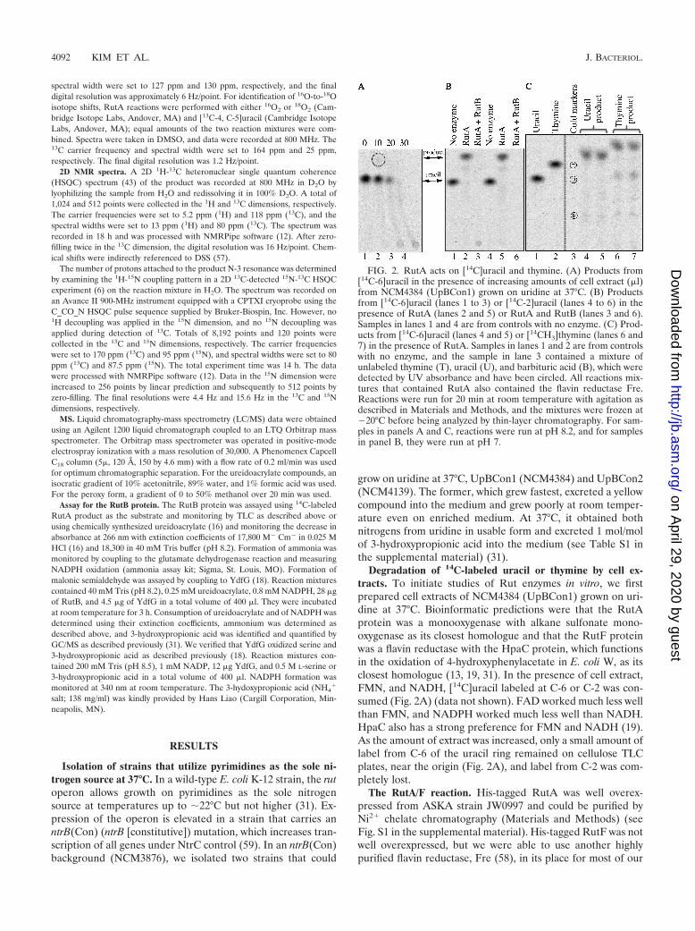
Degradation of 14C-labeled uracil or thymine by cell ex-tracts. To initiate studies of Rut enzymes in vitro, we firstprepared cell extracts of NCM4384 (UpBCon1) grown on uri-dine at 37°C. Bioinformatic predictions were that the RutAprotein was a monooxygenase with alkane sulfonate mono-oxygenase as its closest homologue and that the RutF proteinwas a flavin reductase with the HpaC protein, which functionsin the oxidation of 4-hydroxyphenylacetate in E. coli W, as itsclosest homologue (13, 19, 31). In the presence of cell extract,FMN, and NADH, [14C]uracil labeled at C-6 or C-2 was con-sumed (Fig. 2A) (data not shown). FAD worked much less wellthan FMN, and NADPH worked much less well than NADH.HpaC also has a strong preference for FMN and NADH (19).As the amount of extract was increased, only a small amount oflabel from C-6 of the uracil ring remained on cellulose TLCplates, near the origin (Fig. 2A), and label from C-2 was com-pletely lost.
The RutA/F reaction. His-tagged RutA was well overex-pressed from ASKA strain JW0997 and could be purified byNi2� chelate chromatography (Materials and Methods) (seeFig. S1 in the supplemental material). His-tagged RutF was notwell overexpressed, but we were able to use another highlypurified flavin reductase, Fre (58), in its place for most of our
FIG. 2. RutA acts on [14C]uracil and thymine. (A) Products from[14C-6]uracil in the presence of increasing amounts of cell extract (�l)from NCM4384 (UpBCon1) grown on uridine at 37°C. (B) Productsfrom [14C-6]uracil (lanes 1 to 3) or [14C-2]uracil (lanes 4 to 6) in thepresence of RutA (lanes 2 and 5) or RutA and RutB (lanes 3 and 6).Samples in lanes 1 and 4 are from controls with no enzyme. (C) Prod-ucts from [14C-6]uracil (lanes 4 and 5) or [14CH3]thymine (lanes 6 and7) in the presence of RutA. Samples in lanes 1 and 2 are from controlswith no enzyme, and the sample in lane 3 contained a mixture ofunlabeled thymine (T), uracil (U), and barbituric acid (B), which weredetected by UV absorbance and have been circled. All reactions mix-tures that contained RutA also contained the flavin reductase Fre.Reactions were run for 20 min at room temperature with agitation asdescribed in Materials and Methods, and the mixtures were frozen at�20°C before being analyzed by thin-layer chromatography. For sam-ples in panels A and C, reactions were run at pH 8.2, and for samplesin panel B, they were run at pH 7.
4092 KIM ET AL. J. BACTERIOL.
on April 29, 2020 by guest
http://jb.asm.org/
Dow
nloaded from
studies. In the presence of both RutA and Fre (or an extractcontaining RutF) and the necessary flavin and pyridine nucle-otide cofactors, [14C]uracil labeled at C-2 or C-6 was convertedto a product with faster mobility on TLC plates (Fig. 2B). Theproduct appeared to be more stable at pH 7 than 8.2 (data notshown). Addition of catalase to reaction mixtures to removeany H2O2 generated by the flavin reductase did not affect thebehavior of the RutA product on TLC plates but did clear thebackground. [Methyl-14C]thymine was also converted to aproduct with faster mobility (Fig. 2C).
To identify the product produced from uracil in the RutA/Freaction, we prepared it from 13C- and 15N-enriched uracil. A2D NMR 1H-13C correlation spectrum in D2O confirmed thata single product was produced (see Fig. S2A in the supplemen-tal material). A 1D carbon spectrum showed that splitting ofthe 13C-4 signal by 15N-3 was lost in the product, whereassplitting by 13C-5 was retained. This indicated that the uracilring had been cleaved between N-3 and C-4 (Fig. 3A). Wewere unable to obtain 13C, 15N-enriched thymine commer-cially. However, we showed that the product from unlabeledthymine had only one new H-6–H-5 methyl correlation in a 2D1H total correlation spectroscopy (TOCSY) spectrum, indicat-ing that a single product was produced and that the C-5–C-6bond was intact (data not shown). The magnitudes of thechemical shift changes for this product were similar to those
for the product from uracil, providing evidence that the twoproducts were analogous.
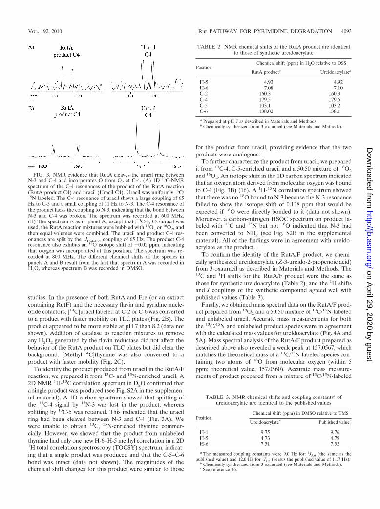
To further characterize the product from uracil, we preparedit from 13C-4, C-5-enriched uracil and a 50:50 mixture of 18O2
and 16O2. An isotope shift in the 1D carbon spectrum indicatedthat an oxygen atom derived from molecular oxygen was boundto C-4 (Fig. 3B) (16). A 1H-15N correlation spectrum showedthat there was no 18O bound to N-3 because the N-3 resonancefailed to show the isotope shift of 0.138 ppm that would beexpected if 18O were directly bonded to it (data not shown).Moreover, a carbon-nitrogen HSQC spectrum on product la-beled with 13C and 15N but not 18O indicated that N-3 hadbeen converted to NH2 (see Fig. S2B in the supplementalmaterial). All of the findings were in agreement with ureido-acrylate as the product.
To confirm the identity of the RutA/F product, we chemi-cally synthesized ureidoacrylate (Z-3-ureido-2-propenoic acid)from 3-oxauracil as described in Materials and Methods. The13C and 1H shifts for the RutA/F product were the same asthose for synthetic ureidoacrylate (Table 2), and the 1H shiftsand J couplings of the synthetic compound agreed well withpublished values (Table 3).
Finally, we obtained mass spectral data on the RutA/F prod-uct prepared from 18O2 and a 50:50 mixture of 13C/15N-labeledand unlabeled uracil. Accurate mass measurements for boththe 13C/15N and unlabeled product species were in agreementwith the calculated mass values for ureidoacrylate (Fig. 4A and5A). Mass spectral analysis of the RutA/F product prepared asdescribed above also revealed a weak peak at 157.0567, whichmatches the theoretical mass of a 13C/15N-labeled species con-taining two atoms of 18O from molecular oxygen (within 5ppm; theoretical value, 157.0560). Accurate mass measure-ments of product prepared from a mixture of 13C/15N-labeled
FIG. 3. NMR evidence that RutA cleaves the uracil ring betweenN-3 and C-4 and incorporates O from O2 at C-4. (A) 1D 13C-NMRspectrum of the C-4 resonances of the product of the RutA reaction(RutA product C4) and uracil (Uracil C4). Uracil was uniformly 13C/15N labeled. The C-4 resonance of uracil shows a large coupling of 65Hz to C-5 and a small coupling of 11 Hz to N-3. The C-4 resonance ofthe product lacks the coupling to N-3, indicating that the bond betweenN-3 and C-4 was broken. The spectrum was recorded at 600 MHz.(B) The spectrum is as in panel A, except that [13C-4, C-5]uracil wasused, the RutA reaction mixtures were bubbled with 18O2 or 16O2, andthen equal volumes were combined. The uracil and product C-4 res-onances are split by the 1JC-4–C-5 coupling of 65 Hz. The product C-4resonance also exhibits an 18O isotope shift of �0.02 ppm, indicatingthat oxygen was incorporated at this position. The spectrum was re-corded at 800 MHz. The different chemical shifts of the species inpanels A and B result from the fact that spectrum A was recorded inH2O, whereas spectrum B was recorded in DMSO.
TABLE 2. NMR chemical shifts of the RutA product are identicalto those of synthetic ureidoacrylate
PositionChemical shift (ppm) in H2O relative to DSS
RutA producta Ureidoacrylateb
H-5 4.93 4.92H-6 7.08 7.10C-2 160.3 160.3C-4 179.5 179.6C-5 103.1 103.2C-6 138.02 138.1
a Prepared at pH 7 as described in Materials and Methods.b Chemically synthesized from 3-oxauracil (see Materials and Methods).
TABLE 3. NMR chemical shifts and coupling constantsa ofureidoacrylate are identical to the published values
PositionChemical shift (ppm) in DMSO relative to TMS
Ureidoacrylateb Published valuec
H-1 9.75 9.76H-5 4.73 4.79H-6 7.31 7.32
a The measured coupling constants were 9.0 Hz for: 1J5,6 (the same as thepublished value) and 12.0 Hz for 1J1,6 (versus the published value of 11.7 Hz).
b Chemically synthesized from 3-oxauracil (see Materials and Methods).c See reference 16.
VOL. 192, 2010 Rut PATHWAY FOR PYRIMIDINE DEGRADATION 4093
on April 29, 2020 by guest
http://jb.asm.org/
Dow
nloaded from

and unlabeled uracil but with 16O2 confirmed the presence ofthis species, which appeared to be the peracid of ureidoacry-late (Fig. 4B and 5A). In the latter case, peaks were strongenough that both the 13C/15N-labeled and unlabeled speciescontaining two atoms of 16O were observed. We return to thesignificance of this in the Discussion.
The RutB reaction. RutB was initially predicted to be anisochorismatase and later a homologue of N-carbamoylsar-cosine amidohydrolase (31, 32). To see whether RutB wouldhydrolyze ureidoacrylate, the product we obtained from theRutA/F reaction, we first used RutA and the substitute flavinreductase Fre to prepare radiolabeled ureidoacrylate from ura-cil. When [14C]ureidoacrylate was treated with His-taggedRutB, 14C label originating from C-2 of uracil was lost fromTLC plates, as was standard [14C]HCO3
� (data not shown).Label from C-6 smeared near the origin. The same result wasobtained if the RutA, Fre, and RutB proteins were added toradiolabeled uracil simultaneously (Fig. 2B, lanes 3 and 6).Likewise, label from uracil was largely lost when cell extractsrather than purified enzymes were added to [14C-2] (data notshown) or [14C-6]uracil (Fig. 2A): at most, traces of RutA/Fproduct were observed. When chemically synthesized ure-idoacrylate was used as the substrate for RutB, approximately2 mol of ammonium was released per mol of uracil consumed(see Fig. S3 in the supplemental material).
Hydrolytic cleavage of ureidoacrylate between N-1 and C-2would release carbamate and aminoacrylate (Fig. 5B). Thecarbamate would in turn hydrolyze spontaneously to ammo-nium and CO2, thus accounting for production of 1 mol ofammonium and loss of label from C-2 (46, 47). The aminoac-rylate would hydrolyze spontaneously to ammonium and malo-nic semiadehyde, accounting for the second mole of ammo-nium.
To determine whether the RutB reaction released carbons 4to 6 of the uracil ring as malonic semialdehyde, we made use of
FIG. 4. Mass spectrometric evidence that RutA yields ureidoacry-late and a trace of its peracid. (A) Ureidoacrylate m/z 133.0491 cor-responds to [C4H6N2
16O218O � H]� (calculated value, 133.0494); m/z
139.0565 corresponds to [13C4H615N2
16O218O � H]� (calculated value,
139.0569). (B) Peracid of ureidoacrylate. m/z 147.0396 corresponds to[C4H6N2O4 � H]� (calculated value, 147.0400); m/z 153.0471 corre-sponds to [13C4H6
15N2O4 � H]� (calculated value, 153.0475).
FIG. 5. In vitro reactions catalyzed by RutA/F, RutB, and the short-chain dehydrogenase YdfG. (A) RutA/F reaction. The RutA/F reac-tion yields ureidoacrylate in vitro. However, there also appears to be asmall amount of ureidoacrylate peracid in reaction mixtures (Fig. 4B),and we infer that this is the product of the RutA/F reaction. Mukherjeeet al. (37) have evidence that the peracid is quickly reduced to ure-idoacrylate by NADH spontaneously (Spont.) under conditions similarto ours (see Discussion and Fig. 6) (B) RutB reaction. The RutBreaction yields 2 mol of ammonium, HCO3
�, and malonic semialde-hyde (3-oxopropionate) from ureidoacrylate. Carbamate and amino-acrylate, which hydrolyze spontaneously, are the presumed intermedi-ates. (C) YdfG reaction. The known short-chain dehydrogenase YdfG(18) reduces malonic semialdehyde to 3-hydroxypropionic acid. In ourcase, malonic semialdehyde was generated from ureidoacrylate by theRutB reaction, which was run simultaneously.
4094 KIM ET AL. J. BACTERIOL.
on April 29, 2020 by guest
http://jb.asm.org/
Dow
nloaded from
E. coli K-12 YdfG protein, a short-chain dehydrogenase that isknown to oxidize 3-hydroyxypropionate and inferred to act inthe reductive direction in vivo (18). (YdfG also oxidizes serine,and its best substrate is L-allo-threonine.) When RutB, YdfG,and NADPH were added to chemically synthesized ureido-acrylate simultaneously, approximately 1 mol of 3-hydroxypro-pionate was produced per mol of ureidoacrylate consumed,indicating that RutB did indeed release malonic semialdehyde(Table 4 and Fig. 5C). One mole of NADPH was oxidized and,as expected, approximately 2 mol of NH4
� was released. If theRutB reaction was allowed to proceed first and then YdfG wasadded later, the yield of NH4
� remained the same but the yieldof 3-hydroxypropionate was greatly reduced (Table 4), possiblydue to the formation of adducts by malonic semialdehyde (17).This might also account for the smearing and loss of malonicsemialdehyde from TLC plates.
Requirement for the RutA, -B, and -F proteins in vivo. Weconstructed strains carrying nonpolar deletions in the rut genesin three backgrounds: an otherwise wild-type background; thentrB(Con) background, in which expression of the rut operon isincreased (31); and the UpBCon1 background, in which pyri-midines can be used as the sole nitrogen source at 37°C (seeabove). The UpBCon 1 strain (NCM4384) grows poorly atroom temperature, and hence we generally studied its ability tocatabolize pyrimidines at 37°C.
Strains carrying lesions in rutA or -F in any of the threebackgrounds failed to grow on uridine as the sole nitrogensource (Table 5). Based on our in vitro results, this was ex-pected for strains carrying lesions in rutA but not necessarilyfor strains carrying rutF lesions because RutF can be replacedin vitro by the flavin reductase Fre. Apparently no other flavinreductase can substitute for RutF in vivo. Whether this isbecause other flavin reductases are not present in sufficientamounts and/or do not have access to RutA or whether thereis another explanation remains to be determined. In the ntrB-(Con) background, where levels of Rut enzymes are elevated,addition of uridine (5 mM) to the medium inhibited growth onammonium (5 mM) at 37°C (doubling time increased from 2 to3 h), indicating that a toxic intermediate(s) of the Rut pathwayprobably accumulated. Growth inhibition persisted in an ntrB-
(Con) rutF strain (doubling time increased from 2 to 3 h).Uridine was not inhibitory in an ntrB(Con) rutA strain (dou-bling time remained 2 h in the presence of uridine), in agree-ment with the view that RutA is absolutely required to initiateuracil degradation. We obtained no suppressors of rutA in anybackground. However, we did obtain suppressors of rutF in thentrB(Con) background. We showed that two such suppressors,which grew slowly on uridine and at different rates, releasedthe usual 2 mol of nitrogen in utilizable form and that theyexcreted the usual 1 mol/mol of 3-hydroxypropionic acid intothe medium (see Table S1 in the supplemental material) (31).Although we have not identified the suppressor lesions, theireffects were as expected if they increased the amount or avail-ability of another flavin reductase.
As was true of strains carrying rutA or rutF lesions, strainscarrying a lesion in rutB in any of the three backgrounds wetested failed to grow on uridine as the sole nitrogen source(Table 5). As was the case for rutA strains, strains carrying arutB lesion also failed to yield spontaneous suppressor muta-tions. In the ntrB(Con) background, where levels of Rut en-zymes are elevated, addition of uridine to ammonium-contain-ing medium markedly inhibited growth of the rutB strain at37°C (data not shown), indicating that it probably accumulateda toxic intermediate(s). Based on what is known about theRutA/F and RutB reactions, this intermediate(s) would beureidoacrylate and/or the peracid of ureidoacrylate (Fig. 5Aand see the Discussion section).
Requirement for YdfG in vivo. We constructed strains car-rying nonpolar deletions in ydfG in the three backgroundsdescribed above. A strain with an insertion in ydfG in a wild-type background grew poorly on uridine as the sole nitrogensource, and an ntrB(Con) strain carrying the insertion did notgrow at all (Table 5; see Fig. S4 in the supplemental material).The UpBCon1 strain carrying a ydfG insertion grew poorly onuridine at 37°C. Based on the fact that YdfG reduces malo-nic semialdehyde to 3-hydroxypropionic acid (see above)and therefore acts after both moles of NH4
� have been re-leased from the pyrimidine ring, we infer that failure of strains
TABLE 4. Production of 3-OH propionate from ureidoacrylate byRutB and YdfG
Substrate or productand activity
Amt (�M) of substrate or productconsumed, produced, or oxidized in:
Expt 1a Expt 2b Expt 3c
Ureidoacrylate consumed 2253-OH propionate producedd 210 384 168NADPH oxidized 173e
NH4� producedf 474 918 884
a The initial ureidoacrylate concentration was 250 �M. RutB and YdfG wereadded simultaneously and incubated for 3 h at room temperature.
b The initial ureidoacrylate concentration was 450 �M. RutB and YdfG wereadded simultaneously and incubated for 6 h at room temperature.
c The initial ureidoacrylate concentration was 450 �M. RutB was added first.After 3 h at room temperature, YdfG was added for an additional 3 h.
d A total of 380 �l of the total 400-�l reaction mixture was used, and 3-OHpropionate was measured as described previously (31) using the standard de-scribed in Materials and Methods.
e In an additional experiment, 226 �M NADPH was oxidized.f Twenty microliters of the total reaction mixture was used.
TABLE 5. Growth of rut strains on uridinea
Deletionb
Background
Wild type(NCM3722),
growth atroom temp
ntrB(Con)(NCM3876),
growth atroom temp
UpBCon1(NCM4384), growth
at 37°C
rutA � � �rutB � � �rutC � (suppressors) �/�c �rutDd � (suppressors) � (suppressors) NDf
rutE � � (suppressors) �/�rutFe � � (suppressors) �ydfG �/� � (suppressors) �/� (suppressors)
a Strains were grown on minimal agar plates with glycerol as the carbon sourceand 5 mM uridine as the sole nitrogen source (31).
b Nonpolar deletions confirmed by sequencing.c �/�, poor growth.d A rutD deletion mutant that did not affect RutC was constructed late in the
study, as described in Materials and Methods and Table 1.e The rutF deletion extended an additional 12 bp beyond its predicted C-
terminal end but remained in-frame and within rutF.f ND, not determined.
VOL. 192, 2010 Rut PATHWAY FOR PYRIMIDINE DEGRADATION 4095
on April 29, 2020 by guest
http://jb.asm.org/
Dow
nloaded from

carrying ydfG insertions to grow well on uridine is due totoxicity of malonic semialdehyde in vivo.
Role of the RutG protein in vivo. In agreement with thebioinformatic prediction that the RutG protein was a nucleo-base transporter (3, 32, 41), an otherwise wild-type strain car-rying a rutG deletion failed to grow on the nucleobase uracil(0.5 mM or 2 mM) as the sole nitrogen source but used thenucleoside uridine normally. An ntrB(Con) rutG strain grewslowly on pyrimidine bases (uracil and thymine) and obtainedboth nitrogens from the ring. Residual growth may be ac-counted for by the fact that the ntrB(Con) lesion activatestranscription of the gene(s) for other transporter(s) that cancarry pyrimidine nucleosides/bases (59). Alternatively, or inaddition, elevated expression of the rut operon in an ntrB(Con)strain may allow it to rely on a constitutively expressed trans-porter(s). An UpBCon1 rutG strain (NCM4384) grew well onpyrimidine bases at 37°C.
In the �20 occurrences of the rut operon outside the Entero-bacteriaceae, rutG is retained only in Acinetobacter (see TableS2 in the supplemental material). Occasionally as in severalmethylobacteria, rutG is replaced by genes for a multisubunittransporter (ABC type) that appears to be a pyrimidine trans-porter and is, in other bacteria, associated with the operon forthe reductive pathway of pyrimidine degradation (32, 38).
Requirement for the RutC to -E proteins in vivo. AlthoughRutC was not required for release of ammonium in vitro,strains carrying rutC lesions in the wild-type or UpBCon1 back-ground failed to grow on uridine as the nitrogen source (Table5). This indicated that they probably accumulated a toxic in-termediate that prevented their growth on the ammoniumreleased from the pyrimidine ring. The ntrB(Con) rutC straingrew very slowly on uridine: it released both moles of nitrogenin utilizable form, but in contrast to its parental strain, releasedmuch less than 1 mol/mol of 3-hydroxypropionic acid into themedium (see Table S1 in the supplemental material). Thelatter finding indicated that RutC did not act on carbamate butprobably acted on the 3-carbon intermediate released from theuracil ring (or on this portion of the molecule before hydrolysisby RutB). As explained in the Discussion, we speculate thattoxicity is due to accumulation of the peracid of aminoac-rylate. In the absence of RutC, cells apparently form lessthan the normal amount of malonic semialdehyde—andhence less 3-hydroxypropionic acid than usual—because a por-tion of the 3-carbon intermediate is diverted out of the Rutpathway. Although we obtained suppressors of rutC in thewild-type background, we did not identify them. Based onbiochemical evidence, RutC was originally predicted to be anendoribonuclease (31, 36), but recently this has been ques-tioned (32; see Discussion).
Like RutC, RutD was not required for release of ammoniumin vitro but was required for growth on pyrimidines as the solenitrogen source in vivo in the two backgrounds we tested (Ta-ble 5). The rutD::Kan insertion from which the original non-polar rutD deletion was constructed may also have caused adecrease in RutC activity (see Materials and Methods) and wassufficiently toxic, even on enriched medium, that we inadver-tently picked up suppressors when we introduced it into thewild-type and ntrB(Con) backgrounds. We studied one strainwith a mutation that suppressed rutD in each background(NCM4088 and NCM4090, respectively). Both strains released
the normal 2 mol of utilizable nitrogen from uridine but ex-creted much less than 1 mol/mol of 3-hydroxypropionic acidinto the medium (see Table S1 in the supplemental material).This indicated that RutD, like RutC, did not act on carbamatebut rather on the 3-carbon intermediate released from theuracil ring. The rutD suppressor strain NCM4088 excreted nodetectable malonic acid into the growth medium (data notshown; examined as described by Loh et al. [31]), and NMRanalysis of medium components failed to identify anything elseexcreted when it was grown on 13C, 15N-enriched uracil (datanot shown). The rutD suppressor strain grew faster on uridineas the sole nitrogen source than its parental strain. Neither ofthe suppressor lesions was identified because we were notaware that they were present until we reconstructed a correctrutD deletion (rutC�) in the wild-type and ntrB(Con) back-grounds late in the study (see Materials and Methods). Asexplained in the Discussion, we speculate that toxicity of a rutDdeletion is due to the accumulation of aminoacrylate, eventhough it can hydrolyze spontaneously. As is the case for RutC,we think cells lacking RutD form less than the normal amountof malonic semialdehyde because a portion of the 3-carbonintermediate is diverted out of the Rut pathway.
Like RutC and RutD, RutE was required for growth onuridine in vivo, although it was not required for release ofammonium from pyrimidine rings in vitro. Wild-type or ntrB-(Con) strains with a nonpolar deletion in rutE failed to grow onuridine (Table 5; see Fig. S4 in the supplemental material).Addition of uridine to ammonium-containing medium inhib-ited growth of the ntrB(Con) rutE strain at 37°C, confirmingthat this strain probably accumulated a toxic intermediate. Weobtained suppressors of rutE in the ntrB(Con) background butnot in the wild-type background. The two that we studiedreleased both moles of utilizable nitrogen from uridine andexcreted 1 mol/mol of 3-hydroxypropionic acid into the me-dium (see Table S1 in the supplemental material). The latterdistinguished them from the ntrB(Con) rutC strain and fromsuppressors of rutD and provided evidence that they formed anormal amount of malonic semialdehyde. As explained in theDiscussion, we think that the function of RutE is the same asthat of YdfG: i.e., reduction of malonic semialdehyde to 3-hy-droxypropionic acid (see below for identification of the lesionsin rutE suppressors and the logic for this argument).
Identification of rutE suppressors and lesions that allowgrowth on pyrimidines at 37°C. We obtained whole-genomesequence for strains carrying rutE suppressors or lesions thatallowed growth on pyrimidines at 37°C and assembled andanalyzed it as described in Materials and Methods. One of therutE suppressors (NCM4299) had a frameshift lesion early inthe nemR gene that should result in truncation of the NemRprotein after 65 amino acids (Table 6). (Intact NemR is 171amino acids.) The second rutE suppressor (NCM4300), whichhad the same growth rate on uridine as the first, had a lesionthat disrupts the inverted repeat in the binding site for NemR/RutR in the promoter-regulatory region for the nemRAoperon. Finally, the UpBCon2 strain (NCM4139), which wasselected spontaneously to grow on pyrimidines at 37°C butwhich we studied very little, also had what appeared to be adamaging lesion in nemR that converted G141 to S. TheUpBCon2 strain retains good ability to grow on pyrimidines atroom temperature. Hence, we were able to introduce a nonpolar
4096 KIM ET AL. J. BACTERIOL.
on April 29, 2020 by guest
http://jb.asm.org/
Dow
nloaded from

rutE deletion into this strain and confirm that the NemR(G141S)lesion suppresses the loss of RutE at room temperature (Table 7;see Fig. S4 in the supplemental material). Likewise, we were ableto introduce a nonpolar nemR deletion into the �rutE strainNCM4115 and show that it suppressed the loss of RutE at roomtemperature.
The NemR protein is a repressor of nemRA transcription;relief of repression apparently requires alkylation of one ormore of its cysteine residues (51). The NemA gene codes forthe flavoprotein N-ethylmaleimide reductase, also referred toas the “old yellow enzyme” of E. coli (55). The fact that inac-tivation of NemR or inactivation of its binding site at nemRA—which would increase the amount of NemR—suppressed arutE null lesion equally well (Table 7; see Fig. S4 in the sup-plemental material) indicates that suppression is likely to bedue to increased expression of NemA, as does the finding thatNemR apparently controls only nemRA transcription, despitethe fact that E. coli contains very large amounts of it (51).Presumably, high levels of N-ethylmaleimide reductase cansubstitute for RutE. Although both of the rutE suppressors alsocarried a large deletion around mioC, which encodes a mysteriousFMN binding protein (Table 6) (7), this deletion was not presentin the UpBCon2 strain. We found that the mioC deletion hadapparently been acquired when the rutE::Kan lesion was intro-duced into the ntrB(Con) background (but not the wild-typebackground) by phage P1-mediated transduction. Based on theresults presented above, the mioC deletion is not central torutE suppression. Using markers linked to nemR by phageP1-mediated transduction, we were able to show that theNemR(G141S) lesion in the UpBCon2 strain was both neces-sary and sufficient for growth on pyrimidines at 37°C in thentrB(Con) background (K.-S. Kim and W. B. Inwood, unpub-lished observation). However, the robust growth of UpBCon2also required a second mutation, which we identified as aninsertion of IS186 in the promoter region for the lon gene(Table 6). This insertion occurred in a hot spot and is known toeliminate Lon protease activity (42). We do not know its sig-nificance to our phenotype.
TABLE 6. Identification of mutations that suppress �rutE and/or allow growth on uridine at 37°C
Strain Phenotype Type and site of mutationa Effect of mutation
NCM4299 Suppression of �rutE Single base change C1724300 deleted nemR frameshift to NemR(1-65)Deletion of 11 kbp from 3924220 to
3935295Deletion of all or part of mioC, asnC, asnA,
viaA, ravA, kup, rbsD, rbsA, rbsC, andrbsBb
NCM4300 Suppression of �rutE Single base change G1724029A nemR; loss of dyad symmetry in NemRbinding sitec
Deletion of 11 kbp from 3924220 to3935295
Deletion of all or part of mioC, asnC, asnA,viaA, ravA, kup, rbsD, rbsA, rbsC, andrbsBb
NCM4139 (UpBcon2) Growth on uridine at 37°C Single base change G1724552A NemR(G141S)lon::IS186 lon promoter disruption by IS insertion and
duplication at 458014-458020
NCM4384 (UpBcon1) Growth on uridine at 37°C Single base change C3182707T Alters structure of sroG riboswitch for ribBDeletion of 11 kbp from 3924220 to
3935295Deletion of all or part of mioC, asnC, asnA,
viaA, ravA, kup, rbsD, rbsA, rbsC, andrbsBb
lon::IS186 lon promoter disruption by IS insertion andduplication at 458014-458020
a Genomic locations are given relative to E. coli K-12 strain MG1655 (NC_000913).b This deletion was present in the �rutE strain from which suppressors were selected (NCM4115; Table 1) and was present in the ntrB(Con) rutE::Kan strain from
which NCM4115 was constructed by FLPing. It was not present in the ntrB(Con) strain (NCM3876; Table 1) from which NCM4384 (UpBcon1) was selectedspontaneously, and it was not present in NCM4103, NCM4101, or NCM4113.
c The NemR binding site (�13 to �28 relative to the sigma 70 promoter) for the nemRA operon is altered:5� TAGACCGACTGGTCTA 3� to 5� TAGACCGACTGATCTA 3�3� ATCTGGCTGACCAGAT 5� 3� ATCTGGCTGACTAGAT 5�.
TABLE 7. Suppression of rutE and ydfG by increased expression ofNemA or RibB
Strain Relevantgenotypea
Growth onuridine at: Other
lesion(s)Roomtemp 37°C
NCM4115b �rutE � � mioC�NCM4299b �rutE nemR � � mioC�NCM4300b �rutE pnemRc � � mioC�NCM4905b �rutE �nemR � � mioC�NCM4906d �rutE nemR � �/�e lonNCM4714 �ydfG � � NoneNCM4916d �ydfG nemR �f � lonNCM4904g �nemR �� � NoneNCM4969h ydfG::Kan �nemR �f � NoneNCM4509i �rutE sroG � �/� mioC� lonNCM4715i �ydfG sroG � � mioC� lon
a All strains carry the same ntrB(Con) lesion. See Table 1 for construction andTable 6 for sequence changes.
b Congenic.c Lesion in the promoter-regulatory region for nemRA that is predicted to
prevent NemR repression (Table 6).d Congenic with NCM4139 (UpBCon2).e �/�, poor growth (see Fig. S4 in the supplemental material).f Suppressors arise.g Congenic with NCM3876.h Congenic with NCM4904.i Congenic with NCM4384 (UpBCon1).
VOL. 192, 2010 Rut PATHWAY FOR PYRIMIDINE DEGRADATION 4097
on April 29, 2020 by guest
http://jb.asm.org/
Dow
nloaded from
The UpBCon1 strain, which excreted a yellow compound thatwas identified as riboflavin (T. Mukherjee and T. Begley, personalcommunication), had a change in the riboswitch (called sroG)preceding the ribB (riboflavin B) gene (Table 6). TheUpBCon1 strain had also acquired the deletion around mioCdiscussed above. Using markers linked to ribB by phage P1-me-diated transduction, we showed that the sroG lesion was necessaryand sufficient for growth on pyrimidines at 37°C (Kim and In-wood, unpublished). However, the robust growth of UpBCon1also required the IS186 insertion in the lon promoter describedabove. It did not require the deletion around mioC.
Changes in the riboswitch for the rib operon of Bacillussubtilis resulted in riboflavin excretion by increasing transcrip-tion of the operon (35, 56). Although the effects of changingthe ribB riboswitch in E. coli are less clear, we presume that thelesion we have identified increases expression of ribB at eitherthe transcriptional or translational level. Whether directly orindirectly, this appears to result in synthesis of excess riboflavinand its excretion, although the mechanism is not obvious.
Overlap in function of RutE and YdfG. To test whetherinactivation of NemR, which suppressed a rutE deletion, alsosuppressed a ydfG lesion, we constructed a strain carrying bothnemR and ydfG lesions as described in Materials and Methods.This strain, NCM4916 [NemR(G141S) lon ntrB(Con) �ydfG],grew faster on uridine at 37°C than a corresponding strainwithout the nemR mutation, NCM4714 [ntrB(Con) �ydfG](Table 7; see Fig. S4 in the supplemental material), providingevidence that high levels of N-ethylmaleimide reductase cansubstitute for the short-chain dehydrogenase YdfG (18). Like-wise, strain NCM4969 [�nemR �ydfG ntrB(Con)] grew fasterthan strain NCM4714, with which it was congenic. Thus, highlevels of NemA can apparently substitute for either YdfG orRutE. (Suppression of ydfG was better at 37°C, and suppres-sion of rutE was better at room temperature.) Suppression ofboth rutE and ydfG lesions by nemR lesions in turn links RutEto YdfG, whose function is known in vitro, and leads to thepostulate mentioned above, namely, that RutE also reducesmalonic semialdehyde to 3-hydroxypropionic acid.
The requirement for YdfG function or RutE function forutilization of uridine at 37°C was decreased in the UpBCon1background (i.e., in the presence of the sroG lesion) (Table 7;see Fig. S4 in the supplemental material). This hints that largeamounts of reduced flavin may also be able to drive reductionof malonic semialdehyde in vivo, either per se or through anunidentified enzyme(s).
DISCUSSION
In conjunction with a flavin reductase, RutA uses molecularoxygen to cleave the uracil ring between N-3 and C-4 (Fig. 5Aand Fig. 6). NMR spectroscopic evidence that 18O from O2 wasincorporated at C-4 (Fig. 3B) indicated that the product is notN-hydroxyureidoacrylate, as did evidence that N-3 was con-verted to NH2 (see Fig. S2B in the supplemental material).The latter finding indicated that the product is not a 7-memberring compound in which oxygen is inserted between N-3 andC-4 (analogous to the Baeyer-Villiger rearrangement observedwith cyclohexanone [40]), as did its mass (Fig. 4A). That 18Owas incorporated at C-4 indicated that the product was notobtained by hydrolysis of the 7-member ring compound to
N-hydroxyureidoacrylate. The observed accurate masses of m/z133.0491 and m/z 139.0565 (Fig. 4A) provided evidence forureidoacrylate as the product. Finally, NMR spectroscopy in-dicated that the product obtained from the RutA/F (Fre) re-action in vitro is identical to chemically synthesized ureido-acrylate (Tables 2 and 3 and Fig. 5 and 6).
In both the reductive and oxidative pathways for pyrimidinecatabolism described previously (22, 48, 52) the N-3–C-4 bondis cleaved hydrolytically after the C-5–C-6 double bond hasbeen altered to decrease the aromaticity of the ring (Fig. 1).Although the product of the RutA/F reaction appears to resultfrom hydrolytic cleavage at the same position, this is not con-sistent with the requirements for the reaction or with transferof oxygen to C-4 from molecular O2. Hence, we sought evi-dence for incorporation of both moles of oxygen from O2 intothe uracil ring. Mass spectrometry indicated the presence of asmall amount of the peracid of ureidoacrylate in RutA reactionmixtures (Fig. 4B, 5, and 6). Work in a related article (37)shows that chemically synthesized ureidoacrylate peracid israpidly reduced to ureidoacrylate under in vitro reaction con-ditions similar to ours (20 mM NADH rather than 4 mM andphosphate buffer at pH 8 rather than 7) and presents a plau-sible mechanism for the formation of ureidoacrylate peracid byRutA. This greatly strengthens the view that the peracid is theproduct of the RutA/F reaction and hence that RutA is anunusual oxygenase of a type not previously described (33, 34).We propose that it be called pyrimidine oxygenase.
The RutB protein, which has all the signatures of a cysteinehydrolase (32), hydrolyzes ureidoacrylate to yield 2 mol NH3,CO2, and malonic semialdehyde (Fig. 5B). Presumably, theinitial products are carbamate and aminoacrylate, which areknown to hydrolyze spontaneously. RutB is homologous to theureidopropionase enzyme of the reductive pathway for pyrim-idine ring degradation (39, 54), and its closest homologue iscarbamoylsarcosine amidohydrolase (25, 32): both release CO2
and NH3 via carbamate. For reasons given below, we pro-pose that RutB be called peroxyureidoacrylate/ureidoacry-late amido hydrolase.
There are several reasons we think the RutB protein hydro-lyzes not only ureidoacrylate (Fig. 5B) but also its peracid (Fig.6). First, the apparent half-life for reduction of the peracid invitro is 5 min at 20 mM NADH at pH 8.0, and it is predicted tobe at least this long in vivo because the concentrations ofNADH and NADPH in E. coli are �0.2 mM each (2, 5, 20) andthe total concentration of glutathione is on the order of 10to 20 mM (5, 15). If reduction of the peracid in vivo is slow,some spontaneous decomposition—to ureidoacrylate, ura-cil, and undefined by-products (37)—could occur if RutB didnot hydrolyze the peracid rapidly. Second, cell extracts of var-ious E. coli strains yielded at most a trace of RutA/F productin vitro (Fig. 2A and B) (data for other strains not shown):rather, as radiolabeled uracil (C-2 or C-6) was consumed, theproducts of the RutB reaction appeared, indicating that hydro-lysis by RutB was much faster than the RutA/F reaction. Third,as discussed below, RutC may catalyze reduction of aminoac-rylate peracid, a product of the RutB reaction. Together, theevidence available indicates that ureidoacrylate peracid, theproduct of the RutA/F reaction, is probably the major sub-strate for RutB in vivo (Fig. 6), although clearly RutB can also
4098 KIM ET AL. J. BACTERIOL.
on April 29, 2020 by guest
http://jb.asm.org/
Dow
nloaded from

hydrolyze ureidoacrylate (Fig. 5B and Table 4; see Fig. S3 inthe supplemental material).
In conjunction with RutA/F and RutB, the short-chain de-hydrogenase YdfG (18) completes the Rut pathway in vitro byreducing malonic semialdehyde to 3-hydroxypropionic acid(Fig. 5C and 6). In vivo the absence of YdfG results in a growthdefect or failure to grow on uridine as the sole nitrogen sourcein different genetic backgrounds, indicating that E. coli K-12requires YdfG despite the fact that both moles of ammoniumhave already been released from the pyrimidine ring before itacts. Malonic semialdehyde appears to be toxic. Like otheraldehydes, it can form adducts to free amino groups, and thismay be the basis for its toxicity and the need to reduce it to thealcohol.
Evidence that RutE and YdfG have the same function. TheRutE protein is predicted to belong to nitroreductase-like sub-family 5, which contains proteins of unknown function (9, 27,32). Like members of the greater nitroreductase family, RutEis believed to use FMN as a cofactor. It is required for growthon uridine as the sole nitrogen source in both the wild-type andntrB(Con) backgrounds (Table 7; see Fig. S4 in the supple-
mental material). Genetic evidence indicates that RutE has thesame function as YdfG: i.e., both reduce malonic semialde-hyde to 3-hydroxypropionic acid, although presumably by dif-ferent mechanisms. Furthermore, the evidence indicates thattoxicity of malonic semialdehyde, not the rate of release ofammonium, limits growth of E. coli K-12 on pyrimidines as thesole nitrogen source at high temperatures. The reasoning forthese conclusions is as follows. First, relief of transcriptionalrepression of nemA, which codes for N-ethylmaleimide reduc-tase, the “old yellow” enzyme of E. coli, suppresses the absenceof either RutE or YdfG in vivo (Table 7; see Fig. S4 in thesupplemental material). This would be expected if all three hadthe same biochemical function. In agreement with this view,overproduction of N-ethylmaleimide reductase in a rutE nullstrain results in excretion of the usual 1 mol/mol of 3-hydro-propionic acid into the growth medium (see Table S1 in thesupplemental material). Finally, overexpression of N-ethyl-maleimide reductase allows an ntrB(Con) strain, which expressesthe rut operon at high levels, to grow on pyrimidines as the solenitrogen source at 37°C (although not as well as the UpBCon2strain; see Results). Additional lines of bioinformatic evidence
FIG. 6. Proposed in vivo pathway for pyrimidine ring degradation in E. coli K-12 (A) and possible handling of ureidoacrylate (B). (A) Rutpathway. RutG appears to be a pyrimidine nucleobase transporter. We infer that RutA catalyzes synthesis of ureidoacrylate peracid (see text).Although our work did not address the specific role of FMN, it is plausible that flavin hydroperoxide, a well-known intermediate in related reactions(40), would participate (37). We postulate that ureidoacrylate peracid is the primary substrate for RutB (see text). Activities of RutC, -D, and -E,which have not yet been studied biochemically, were inferred by a variety of other means. Whereas YdfG uses NADPH as a cofactor, RutE ispredicted to be a flavoprotein (9, 27). Proposed names for Rut enzymes are in the inset. (B) Formation and use of ureidoacrylate. If someureidoacrylate is formed by spontaneous reduction of ureidoacrylate peracid (37), RutB can hydrolyze it. We believe this auxiliary pathway, whichwas prominent in vitro (Fig. 5), plays a minor role in vivo (see text).
VOL. 192, 2010 Rut PATHWAY FOR PYRIMIDINE DEGRADATION 4099
on April 29, 2020 by guest
http://jb.asm.org/
Dow
nloaded from

support the view that RutE catalyzes reduction of malonicsemialdehyde to 3-hydropropionate. First, the rutE gene isoften absent from the rut operon (see Table S2 in the supple-mental material), in agreement with the view that RutE actsafter the nitrogens have been extracted from pyrimidine rings.Second, the five rut operons in Acinetobacter genomes (18genomes total), all of which lack rutE, have a gene that codesfor an enzyme in the same superfamily as YdfG and is pre-dicted to reduce malonic semialdehyde and 2-methyl malonicsemialdehyde to their corresponding alcohols (32). Finally, therut operons in the two Alteromonas species for which wholegenome sequences are available contain not only rutE but alsothe gene for an additional enzyme predicted to detoxifymalonic semialdehyde by oxidizing it rather than reducing it(malonate semialdehyde/methyl malonate semialdehyde de-hydrogenase, which would oxidize malonic semialdehyde toacetyl-S-coenzyme A [CoA]) (32, 50) (see Table S2 in thesupplemental material). Neither genome carries a ydfG gene.Biochemical studies of RutE will be particularly interestingbecause flavoenzymes generally participate in oxidation of al-cohols rather than reduction of aldehydes (24).
Speculations on RutD and RutC function. Like other Rutpathway proteins, both RutD and RutC are required for growthon uridine as the sole nitrogen source, despite the fact thatthey are not required for release of ammonium in vitro. Wespeculate that RutD, a hypothetical /�-hydrolase with noclose relatives (32), increases the rate of spontaneous hydro-lysis of aminoacrylate to malonic semialdehyde (Fig. 6). Thiswould be analogous to the role of carbonic anhydrases inaccelerating the rate of spontaneous hydration of CO2.
Finally, we speculate that RutC, a member of a family ofproteins without a clearly defined function (32), reduces theperacid of aminoacrylate to aminoacrylate, the substrate forRutD (Fig. 6). Members of the RutC family appear to bindtoxic metabolic intermediates (10, 14). Both of the other mem-bers of the family in E. coli, TdcF (threonine deaminase cata-bolic F) and YjgF, appear to be involved in metabolism of thetoxic intermediate 2-ketobutyrate (8, 10, 14, 29). Structurally,RutC family proteins are trimers with binding clefts for smallligands at monomer interfaces (53). In the clefts, they carry aninvariant R that is often followed by XC. The structure of theE. coli TdcF protein has been determined with 2-ketobutyratebound: oddly, it was bound as the rare enol tautomer, with itscarboxylate group doubly hydrogen bonded to the guanidiniumgroup of the invariant R and its enol OH group bonded to boththe backbone amide of the conserved C and the carboxyl groupof the conserved E120 in the adjacent subunit (8) (see Fig. S5in the supplemental material). The side chain of the corre-sponding C in the E. coli YjgF protein—whose structure hasbeen determined only in its unliganded form—was derivatizedwith what appeared to be a thiophosphate or a thiosulfate (53).By analogy to what is known about TdcF and YjgF, we postu-late that RutC binds the peracid form of aminoacrylate (seeFig. S5 in the supplemental material) and that it may use theXC107XXC110 motif adjacent to its invariant R105 to reduce theperacid to the carboxylic acid (aminoacrylate). If RutC bindsaminoacrylate peracid in its stable amine form—as would bepredicted—it would also inhibit spontaneous hydrolysis of theamino group. When aminoacrylate was released by RutC,RutD could then increase its spontaneous rate of hydrolysis by
catalyzing formation of the rare imine tautomer. The roles ofRutC and RutD would be to insure that reduction of theperacid form of aminoacrylate and hydrolysis of aminoacrylateoccur rapidly and in a particular order. These admittedly spec-ulative ideas provide a framework for further biochemical andgenetic studies. In the latter connection, it will be interesting todetermine the identities of rutC and rutD suppressors, forwhich tools are now available (see Materials and Methods),and to understand why a rutC strain and rutD suppressors gooff the pathway and excrete less than the usual amount of3-hydroxypropionic acid into the medium (see Table S1 in thesupplemental material). Apparently, they generate less thanthe usual amount of toxic malonic semialdehyde (see Results).
Conclusions. In summary, Rut pathway enzymes oxidativelycleave the pyrimidine ring to produce a series of reactive, toxicintermediates that includes strong oxidizing agents (peracids)and compounds known to polymerize readily (ureidoacrylatesand aminoacrylates) or form adducts (malonic semialdehyde)(Fig. 6). Only half of the Rut enzymes (RutA, RutF, and RutB)are required in vitro to release both nitrogens from the pyrim-idine ring as NH4
� (Fig. 5). The other half (RutC, RutD, andRutE) are nonetheless required in vivo, apparently to preventaccumulation of toxic intermediates and by-products. We pos-tulate that they act in order on the 3-carbon intermediatereleased by RutB (Fig. 6). The function of RutE overlaps withthat of the short-chain dehydrogenase YdfG (Tables 6 and 7;see Fig. S4 in the supplemental material), and both are re-quired in vivo: apparently neither alone has sufficient activity,and the two together are still not sufficient for growth at 37°C.
Although E. coli does not grow on pyrimidines as the solenitrogen source at 37°C, it transcribes the rut operon veryhighly at this temperature under nitrogen-limiting conditions(31, 59). Presumably the Rut pathway allows E. coli to usepyrimidines, which are readily available degradation productsof RNA, as part of the nitrogen source at 37°C. (The YdfGprotein, which is coded for outside the rut operon, may not berequired under these circumstances.) Forcing their use as thesole nitrogen source at any temperature is a trick of the experi-mentalist. The rut operon is highly expressed, even in the absenceof exogenous pyrimidines. Whether the Rut pathway is also usedto decrease the internal free pool concentrations of pyrimidinesunder nitrogen-limiting conditions and/or to generate toxic inter-mediates that help slow the growth of E. coli in a coordinated wayare intriguing possibilities that remain to be explored.
ACKNOWLEDGMENTS
We thank the National BioResource Project of the National Insti-tute of Genetics, Japan, for E. coli Keio strains and ASKA strains. Wethank Michael Coyle for initiating studies of RutG and for attemptingto determine the fate of carbons 4 to 6 of uracil in a rutD suppressorstrain, Rebecca Fong for help with strain construction, and ZhongruiZhou for 3-hydroxypropionic acid determinations. We thank HansLiao for the gift of 3-hydroxypropionic acid and for alerting us to thepossible role of the YdfG protein in its formation, and we thank ChrisWalsh for alerting us to a mechanism by which RutD could increasethe rate of aminoacrylate hydrolysis. We are grateful to Luying Xun,Washington State University, for gifts of purified Fre enzyme and astrain that overexpresses Fre and for continued interest in the work.Finally, we are indebted to Tadhg Begley for suggesting that the smallamount of RutA product with a mass indicating that both atoms of O2had been incorporated might be ureidoacrylate peracid.
This work was supported by NIH grant GM38361 to S.K. TheCentral California 900-MHz facility was supported by NIH grant
4100 KIM ET AL. J. BACTERIOL.
on April 29, 2020 by guest
http://jb.asm.org/
Dow
nloaded from
GM68933. We thank the NSF (BBS 01-19304) and NIH (RR15756)for funding for the 800-MHz NMR and BBS 87-20134 for funding forthe 600-MHz NMR.
REFERENCES
1. Andersen, G., O. Bjornberg, S. Polakova, Y. Pynyaha, A. Rasmussen, K.Møller, A. Hofer, T. Moritz, M. P. Sandrini, A. M. Merico, C. Compagno,H. E. Akerlund, Z. Gojkovic, and J. Piskur. 2008. A second pathway todegrade pyrimidine nucleic acid precursors in eukaryotes. J. Mol. Biol. 380:655–666.
2. Andersen, K. B., and K. von Meyenburg. 1977. Charges of nicotinamideadenine nucleotides and adenylate energy charge as regulatory parametersof the metabolism in Escherichia coli. J. Biol. Chem. 252:4151–4156.
3. Andersen, P. S., D. Frees, R. Fast, and B. Mygind. 1995. Uracil uptake inEscherichia coli K-12: isolation of uraA mutants and cloning of the gene. J.Bacteriol. 177:2008–2012.
4. Baba, T., T. Ara, M. Hasegawa, Y. Takai, Y. Okumura, M. Baba, K. A.Datsenko, M. Tomita, B. L. Wanner, and H. Mori. 2006. Construction ofEscherichia coli K-12 in-frame, single-gene knockout mutants: the Keio col-lection. Mol. Syst. Biol. 2:2006.0008.
5. Bennett, B. D., E. H. Kimball, M. Gao, R. Osterhout, S. J. Van Dien, andJ. D. Rabinowitz. 2009. Absolute metabolite concentrations and impliedenzyme active site occupancy in Escherichia coli. Nat. Chem. Biol. 5:593–599.
6. Bertini, I., I. C. Felli, L. Gonnelli, R. Pierattelli, Z. Spyranti, and G. A.Spyroulias. 2006. Mapping protein-protein interaction by 13C�-detected het-eronuclear NMR spectroscopy. J. Biomol. NMR 36:111–122.
7. Birch, O. M., K. S. Hewitson, M. Fuhrmann, K. Burgdorf, J. E. Baldwin,P. L. Roach, and N. M. Shaw. 2000. MioC is an FMN-binding protein that isessential for Escherichia coli biotin synthase activity in vitro. J. Biol. Chem.275:32277–32280.
8. Burman, J. D., C. E. Stevenson, R. G. Sawers, and D. M. Lawson. 2007. Thecrystal structure of Escherichia coli TdcF, a member of the highly conservedYjgF/YER057c/UK114 family. BMC Struct. Biol. 7:30.
9. Choi, J. W., J. Lee, K. Nishi, Y. S. Kim, C. H. Jung, and J. S. Kim. 2008.Crystal structure of a minimal nitroreductase, ydjA, from Escherichia coliK12 with and without FMN cofactor. J. Mol. Biol. 377:258–267.
10. Christopherson, M. R., G. E. Schmitz, and D. M. Downs. 2008. YjgF isrequired for isoleucine biosynthesis when Salmonella enterica is grown onpyruvate medium. J. Bacteriol. 190:3057–3062.
11. Datsenko, K. A., and B. L. Wanner. 2000. One-step inactivation of chromo-somal genes in Escherichia coli K-12 using PCR products. Proc. Natl. Acad.Sci. U. S. A. 97:6640–6645.
12. Delaglio, F., S. Grzesiek, G. W. Vuister, G. Zhu, J. Pfeifer, and A. Bax. 1995.NMRPipe: a multidimensional spectral processing system based on UNIXpipes. J. Biomol. NMR 6:277–293.
13. Eichhorn, E., J. R. van der Ploeg, and T. Leisinger. 1999. Characterizationof a two-component alkanesulfonate monooxygenase from Escherichia coli.J. Biol. Chem. 274:26639–26646.
14. Enos-Berlage, J. L., M. J. Langendorf, and D. M. Downs. 1998. Complexmetabolic phenotypes caused by a mutation in yjgF, encoding a member ofthe highly conserved YER057c/YjgF family of proteins. J. Bacteriol. 180:6519–6528.
15. Fahey, R. C., W. C. Brown, W. B. Adams, and M. B. Worsham. 1978.Occurrence of glutathione in bacteria. J. Bacteriol. 133:1126–1129.
16. Farkas, J., J. Hapala, O. Jindrova, and J. Skoda. 1982. Reaction of 2,3-dihydro-1,3-6H-oxazine-2,6-dione with aliphatic amines and amino acidsCollect. Czech Chem. Commun. (Camb.) 47:2932–2945.
17. Fox, C. H., F. B. Johnson, J. Whiting, and P. P. Roller. 1985. Formaldehydefixation. J. Histochem. Cytochem. 33:845–853.
18. Fujisawa, H., S. Nagata, and H. Misono. 2003. Characterization of short-chain dehydrogenase/reductase homologues of Escherichia coli (YdfG) andSaccharomyces cerevisiae (YMR226C). Biochim. Biophys. Acta 1645:89–94.
19. Galan, B., E. Díaz, M. A. Prieto, and J. García. 2000. Functional analysis ofthe small component of the 4-hydroxyphenylacetate 3-monooxygenase ofEscherichia coli W: a prototype of a new flavin:NAD(P)H reductase subfam-ily. J. Bacteriol. 182:627–636.
20. Grose, J. H., L. Joss, S. F. Velick, and J. R. Roth. 2006. Evidence thatfeedback inhibition of NAD kinase controls responses to oxidative stress.Proc. Natl. Acad. Sci. U. S. A. 103:7601–7606.
21. Gyaneshwar, P., O. Paliy, J. McAuliffe, D. L. Popham, M. I. Jordan, and S.Kustu. 2005. Sulfur and nitrogen limitation in Escherichia coli K-12: specifichomeostatic responses. J. Bacteriol. 187:1074–1090.
22. Hayaishi, O., and A. Kornberg. 1952. Metabolism of cytosine, thymine,uracil, and barbituric acid by bacterial enzymes. J. Biol. Chem. 197:717–732.
23. Inwood, W. B., J. A. Hall, K.-S. Kim, L. Demirkhanyan, D. Wemmer, H.Zgurskaya, and S. Kustu. 2009. Epistatic effects of the protease/chaperoneHflB on some damaged forms of the Escherichia coli ammonium channelAmtB. Genetics 183:1327–1340.
24. Kalliri, E., S. B. Mulrooney, and R. P. Hausinger. 2008. Identification ofEscherichia coli YgaF as an L-2-hydroxyglutarate oxidase. J. Bacteriol. 190:3793–3798.
25. Kim, J. M., S. Shimizu, and H. Yamada. 1986. Purification and character-
ization of a novel enzyme, N-carbamoylsarcosine amidohydrolase, fromPseudomonas putida 77. J. Biol. Chem. 261:11832–11839.
26. Kitagawa, M., T. Ara, M. Arifuzzaman, T. Ioka-Nakamichi, E. Inamoto, H.Toyonaga, and H. Mori. 2005. Complete set of ORF clones of Escherichiacoli ASKA library (a complete set of E. coli K-12 ORF archive): uniqueresources for biological research. DNA Res. 12:291–299.
27. Koike, H., H. Sasaki, T. Kobori, S. Zenno, K. Saigo, M. E. Murphy, E. T.Adman, and M. Tanokura. 1998. 1.8 Å crystal structure of the majorNAD(P)H:FMN oxidoreductase of a bioluminescent bacterium, Vibrio fischeri:overall structure, cofactor and substrate-analog binding, and comparisonwith related flavoproteins. J. Mol. Biol. 280:259–273.
28. Kornberg, A. 2006. Osamu Hayaishi: pioneer first of the oxygenases, then themolecular basis of sleep and throughout a great statesman of science.IUBMB Life 58:253.
29. LaRossa, R. A., and T. K. Van Dyk. 1987. Metabolic mayhem caused by2-ketoacid imbalances. Bioessays 7:125–130.
30. Liu, X., and R. E. Parales. 2008. Chemotaxis of Escherichia coli to pyrimi-dines: a new role for the signal transducer Tap. J. Bacteriol. 190:972–979.
31. Loh, K. D., P. Gyaneshwar, E. Markenscoff Papadimitriou, R. Fong, K.-S.Kim, R. Parales, Z. Zhou, W. Inwood, and S. Kustu. 2006. A previouslyundescribed pathway for pyrimidine catabolism. Proc. Natl. Acad. Sci.U. S. A. 103:5114–5119.
32. Marchler-Bauer, A., J. B. Anderson, F. Chitsaz, M. K. Derbyshire, C. De-Weese-Scott, J. H. Fong, L. Y. Geer, R. C. Geer, N. R. Gonzales, M. Gwadz,S. He, D. I. Hurwitz, J. D. Jackson, Z. Ke, C. J. Lanczycki, C. A. Liebert, C.Liu, F. Lu, S. Lu, G. H. Marchler, M. Mullokandov, J. S. Song, A. Tasneem,N. Thanki, R. A. Yamashita, D. Zhang, N. Zhang, and S. H. Bryant. 2009.CDD: specific functional annotation with the Conserved Domain Database.Nucleic Acids Res. 37:D205–D210.
33. Massey, V. 1994. Activation of molecular oxygen by flavins and flavoproteins.J. Biol. Chem. 269:22459–22462.
34. Massey, V. 2000. The chemical and biological versatility of riboflavin. Bio-chem. Soc. Trans. 28:283–296.
35. Mironov, A. S., I. Gusarov, R. Rafikov, L. E. Lopez, K. Shatalin, R. A.Kreneva, D. A. Perumov, and E. Nudler. 2002. Sensing small molecules bynascent RNA: a mechanism to control transcription in bacteria. Cell 111:747–756.
36. Morishita, R., A. Kawagoshi, T. Sawasaki, K. Madin, T. Ogasawara, T. Oka,and Y. Endo. 1999. Ribonuclease activity of rat liver perchloric acid-solubleprotein, a potent inhibitor of protein synthesis. J. Biol. Chem. 274:20688–20692.
37. Mukherjee, T., Y. Zhang, S. Abdelwahed, S. Ealick, and T. Begley. 2010.Catalysis of flavoenzyme-mediated amide hydrolysis. J. Am. Chem. Soc.132:5550–5551.
38. Osterman, A. 2006. A hidden metabolic pathway exposed. Proc. Natl. Acad.Sci. U. S. A. 103:5637–5638.
39. Romao, M. J., D. Turk, F. X. Gomis-Ruth, R. Huber, G. Schumacher, H.Mollering, and L. Russmann. 1992. Crystal structure analysis, refinementand enzymatic reaction mechanism of N-carbamoylsarcosine amidohydro-lase from Arthrobacter sp. at 2.0 A resolution. J. Mol. Biol. 226:1111–1130.
40. Ryerson, C. C., D. P. Ballou, and C. Walsh. 1982. Mechanistic studies oncyclohexanone oxygenase. Biochemistry 21:2644–2655.
41. Saier, M. H., Jr., B. H. Eng, S. Fard, J. Garg, D. A. Haggerty, W. J.Hutchinson, D. L. Jack, E. C. Lai, H. J. Liu, D. P. Nusinew, A. M. Omar, S. S.Pao, I. T. Paulsen, J. A. Quan, M. Sliwinski, T. T. Tseng, S. Wachi, and G. B.Young. 1999. Phylogenetic characterization of novel transport protein fam-ilies revealed by genome analyses. Biochim. Biophys. Acta 1422:1–56.
42. SaiSree, L., M. Reddy, and J. Gowrishankar. 2001. IS186 insertion at a hotspot in the lon promoter as a basis for lon protease deficiency of Escherichiacoli B: identification of a consensus target sequence for IS186 transposition.J. Bacteriol. 183:6943–6946.
43. Schleucher, J., M. Schwendinger, M. Sattler, P. Schmidt, O. Schedletzky, S.Glaser, O. W. Sørensen, and C. Griesinger. 1994. A general enhancementscheme in heteronuclear multidimensional NMR employing pulsed fieldgradients. J. Biomol. NMR 4:301–306.
44. Shimada, T., K. Hirao, A. Kori, K. Yamamoto, and A. Ishihama. 2007. RutRis the uracil/thymine-sensing master regulator of a set of genes for synthesisand degradation of pyrimidines. Mol. Microbiol. 66:744–757.
45. Shimada, T., A. Ishihama, S. J. Busby, and D. C. Grainger. 2008. TheEscherichia coli RutR transcription factor binds at targets within genes aswell as intergenic regions. Nucleic Acids Res. 36:3950–3955.
46. Simaga, S., and E. Kos. 1978. Uracil catabolism by Escherichia coli K12S. Z.Naturforsch. C 33:1006–1008.
47. Simaga, S., and E. Kos. 1981. Properties and regulation of pyrimidine ca-tabolism in Escherichia coli. Int. J. Biochem. 13:615–619.
48. Soong, C. L., J. Ogawa, E. Sakuradani, and S. Shimizu. 2002. Barbiturase, anovel zinc-containing amidohydrolase involved in oxidative pyrimidine me-tabolism. J. Biol. Chem. 277:7051–7058.
49. Soupene, E., W. C. van Heeswijk, J. Plumbridge, V. Stewart, D. Bertenthal,H. Lee, G. Prasad, O. Paliy, P. Charernnoppakul, and S. Kustu. 2003.Physiological studies of Escherichia coli strain MG1655: growth defects andapparent cross-regulation of gene expression. J. Bacteriol. 185:5611–5626.
VOL. 192, 2010 Rut PATHWAY FOR PYRIMIDINE DEGRADATION 4101
on April 29, 2020 by guest
http://jb.asm.org/
Dow
nloaded from

50. Stines-Chaumeil, C., F. Talfournier, and G. Branlant. 2006. Mechanisticcharacterization of the MSDH (methylmalonate semialdehyde dehydroge-nase) from Bacillus subtilis. Biochem. J. 395:107–115.
51. Umezawa, Y., T. Shimada, A. Kori, K. Yamada, and A. Ishihama. 2008. Theuncharacterized transcription factor YdhM is the regulator of the nemAgene, encoding N-ethylmaleimide reductase. J. Bacteriol. 190:5890–5897.
52. Vogels, G. D., and C. Van der Drift. 1976. Degradation of purines andpyrimidines by microorganisms. Bacteriol. Rev. 40:403–468.
53. Volz, K. 1999. A test case for structure-based functional assignment: the 1.2A crystal structure of the yjgF gene product from Escherichia coli. ProteinSci. 8:2428–2437.
54. Walsh, T. A., S. B. Green, I. M. Larrinua, and P. R. Schmitzer. 2001.Characterization of plant beta-ureidopropionase and functional overexpres-sion in Escherichia coli. Plant Physiol. 125:1001–1011.
55. Williams, R. E., and N. C. Bruce. 2002. ‘New uses for an Old Enzyme’—
the Old Yellow Enzyme family of flavoenzymes. Microbiology 148:1607–1614.
56. Winkler, W. C., S. Cohen-Chalamish, and R. R. Breaker. 2002. An mRNAstructure that controls gene expression by binding FMN. Proc. Natl. Acad.Sci. U. S. A. 99:15908–15913.
57. Wishart, D. S., C. G. Bigam, J. Yao, F. Abildgaard, H. J. Dyson, E. Oldfield,J. L. Markley, and B. D. Sykes. 1995. 1H, 13C and 15N chemical shiftreferencing in biomolecular NMR. J. Biomol. NMR 6:135–140.
58. Xun, L., and E. R. Sandvik. 2000. Characterization of 4-hydroxyphenyl-acetate 3-hydroxylase (HpaB) of Escherichia coli as a reduced flavin adeninedinucleotide-utilizing monooxygenase. Appl. Environ. Microbiol. 66:481–486.
59. Zimmer, D. P., E. Soupene, H. L. Lee, V. F. Wendisch, A. B. Khodursky, B. J.Peter, R. A. Bender, and S. Kustu. 2000. Nitrogen regulatory protein C-controlled genes of Escherichia coli: scavenging as a defense against nitrogenlimitation. Proc. Natl. Acad. Sci. U. S. A. 97:14674–14679.
4102 KIM ET AL. J. BACTERIOL.
on April 29, 2020 by guest
http://jb.asm.org/
Dow
nloaded from

JOURNAL OF BACTERIOLOGY, Jan. 2011, p. 326 Vol. 193, No. 10021-9193/11/$12.00 doi:10.1128/JB.01302-10Copyright © 2011, American Society for Microbiology. All Rights Reserved.
ERRATUM
The Rut Pathway for Pyrimidine Degradation: Novel Chemistryand Toxicity Problems
Kwang-Seo Kim, Jeffrey G. Pelton, William B. Inwood,Ulla Andersen, Sydney Kustu, and David E. Wemmer
Departments of Plant and Microbial Biology and Chemistry, University of California, Berkeley, California 94720
Volume 192, no. 16, p. 4089–4102, 2010. Supplemental material: In Figure S5 and its legend, “aminoacrylate” should read“aminoacrylate peracid.” Revised supplemental material is posted at http://jb.asm.org/cgi/content/full/192/16/4089/DC1.
326