the skeletal system - cabrillo collegepdarcey/bio 13a/bio13afall08/bio 13a classnotes... · 1 the...
TRANSCRIPT
1
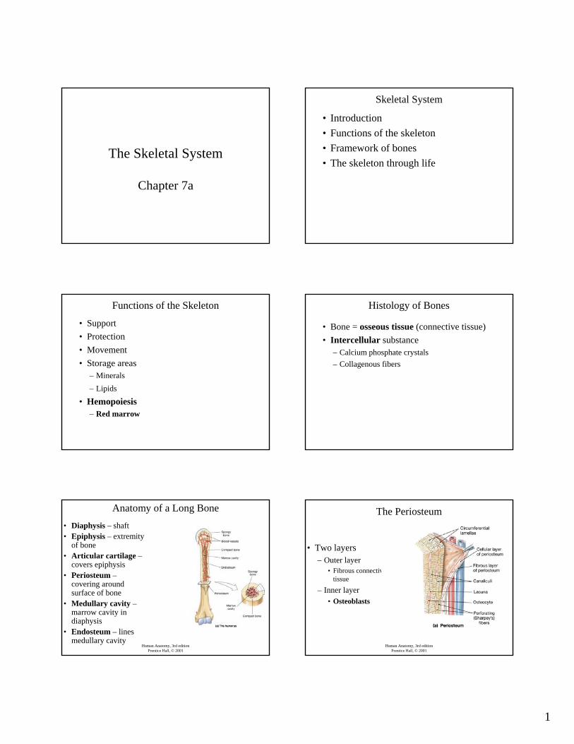
The Skeletal System
Chapter 7a
Skeletal System
• Introduction• Functions of the skeleton• Framework of bones• The skeleton through life
Functions of the Skeleton
• Support• Protection• Movement• Storage areas
– Minerals– Lipids
• Hemopoiesis– Red marrow
Histology of Bones
• Bone = osseous tissue (connective tissue)• Intercellular substance
– Calcium phosphate crystals– Collagenous fibers
Human Anatomy, 3rd editionPrentice Hall, © 2001
Anatomy of a Long Bone• Diaphysis – shaft• Epiphysis – extremity
of bone• Articular cartilage –
covers epiphysis• Periosteum –
covering around surface of bone
• Medullary cavity –marrow cavity in diaphysis
• Endosteum – lines medullary cavity
Human Anatomy, 3rd editionPrentice Hall, © 2001
The Periosteum
• Two layers– Outer layer
• Fibrous connective tissue
– Inner layer• Osteoblasts

2
Human Anatomy, 3rd editionPrentice Hall, © 2001
The Endosteum
• Single layer– Osteoclasts– Osteoblasts
Human Anatomy, 3rd editionPrentice Hall, © 2001
Structure of Bone Tissue
• Pores– Living cells– Channels for blood vessels– Decrease weight of bone
• Degree of porosity– Spongy bone– Compact bone
Human Anatomy, 3rd editionPrentice Hall, © 2001
Compact Bone
• Haversian system(Osteon)– Haversian canals– Lacunae– Canaliculi
Human Anatomy, 3rd editionPrentice Hall, © 2001
Spongy Bone• Composed of trabeculae• Penetrated by blood vessels from periosteum
Ossification
• Embryo skeleton– Begins as cartilage & membrane– Bone formation begins about 6 weeks after
fertilization
Ossification
• 1st stage – embryonic cells migrate into future bone sites– Become chondroblasts or– Become osteoblasts

3
Endochondral Ossification• Occurs within a hyaline cartilage model• Occurs in most bones of the body• Periosteum forms at about week 8• Calcification begins in center of diaphysis
– Primary ossification center• Secondary ossification centers at
epiphyses• Medullary cavity forms
Endochondral Ossification
Endochondral Ossification Fetus. 10 weeks
Fetus, 16 weeks Remaining Cartilage• Articular cartilage• Epiphyseal plate
– Bone grows in length
4
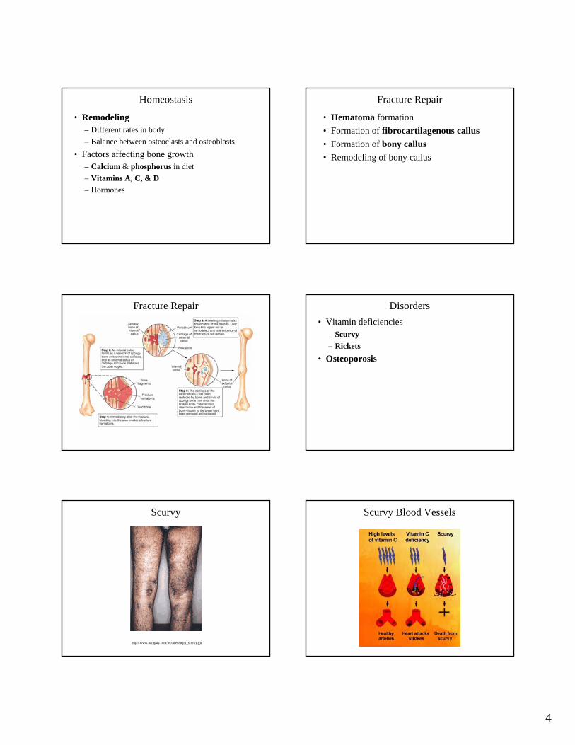
Homeostasis
• Remodeling– Different rates in body– Balance between osteoclasts and osteoblasts
• Factors affecting bone growth– Calcium & phosphorus in diet– Vitamins A, C, & D– Hormones
Fracture Repair
• Hematoma formation• Formation of fibrocartilagenous callus• Formation of bony callus• Remodeling of bony callus
Fracture Repair Disorders• Vitamin deficiencies
– Scurvy– Rickets
• Osteoporosis
Scurvy
http://www.pathguy.com/lectures/nejm_scurvy.gif
Scurvy Blood Vessels

5
Rickets
http://bioe.eng.utoledo.edu/adms_staffs/akkus/2003_WEB_PROJECTS/hormone/vitamin_d.htm
Osteoporosis
Human Anatomy, 3rd editionPrentice Hall, © 2001
Organization of the Skeletal SystemAxial skeleton
SkullVertebral columnThoracic cage
Appendicular skeletonPectoral girdleUpper limbsPelvic girdleLower limbs
Human Anatomy, 3rd editionPrentice Hall, © 2001
The Human Skeleton
Human Anatomy, 3rd editionPrentice Hall, © 2001
Skull
– Cranium• Protects brain
– Facial bones• Protect sense organs
Human Anatomy, 3rd editionPrentice Hall, © 2001
Vertebral Column– Vertebrae
• Cervical• Thoracic• Lumbar• Sacral
– Sacrum– Coccyx– Function
• Protect spinal cord and nerves

6
Human Anatomy, 3rd editionPrentice Hall, © 2001
Intervertebral Discs
• Vertebrae– Separated by
intervertebral discs
Human Anatomy, 3rd editionPrentice Hall, © 2001
The Atlas and Axis
Human Anatomy, 3rd editionPrentice Hall, © 2001
A Typical Vertebra
Human Anatomy, 3rd editionPrentice Hall, © 2001
The Sacrum and Coccyx• Sacrum
– Several fused vertebrae
• Coccyx– Rudimentary tailbone
Human Anatomy, 3rd editionPrentice Hall, © 2001
Thoracic Cage– Ribs (12 pairs)
• Articulate with vertebrae
– Sternum(breastbone)
– Function• Protect underlying
organs
Human Anatomy, 3rd editionPrentice Hall, © 2001
Pectoral Girdle– Scapula (shoulder
blade)– Clavicle (collar bone)– Attached to posterior
ribs and sternum– Function
• Connects bones of arms to axial skeleton
• Aids in arm movements

7
Human Anatomy, 3rd editionPrentice Hall, © 2001
Upper Limbs– Humerus– Radius– Ulna– Carpals– Metacarpals– Phalanges
Human Anatomy, 3rd editionPrentice Hall, © 2001
The Radius and Ulna
Human Anatomy, 3rd editionPrentice Hall, © 2001
Bones of the Wrist and Hand
Human Anatomy, 3rd editionPrentice Hall, © 2001
Pelvic Girdle
– Coxal (hip) bones• Ileum• Ischium• Pubis
– Attached to sacrum and coccyx
– Function• Connect bones of
legs to axial skeleton
Human Anatomy, 3rd editionPrentice Hall, © 2001
The Pelvic Girdle
Human Anatomy, 3rd editionPrentice Hall, © 2001
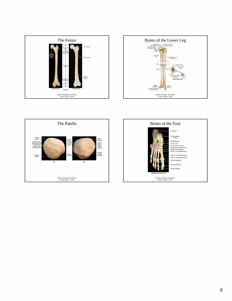
Lower Limbs– Femur– Tibia– Fibula– Patella– Tarsals– Metatarsals– Phalanges
8
Human Anatomy, 3rd editionPrentice Hall, © 2001
The Femur
Human Anatomy, 3rd editionPrentice Hall, © 2001
Bones of the Lower Leg
Human Anatomy, 3rd editionPrentice Hall, © 2001
The Patella
Human Anatomy, 3rd editionPrentice Hall, © 2001
Bones of the Foot