the thymidine phosphorylase imaging agent 123i...
TRANSCRIPT
1
The Thymidine Phosphorylase Imaging Agent 123I-IIMU Predicts the Efficacy of
Capecitabine
Nobuya Kobashi1, Hiroki Matsumoto1*, Songji Zhao2, Shunsuke Meike1, Yuki Okumura1, Tsutomu Abe1,
Hiromichi Akizawa3, Kazue Ohkura4, Ken-ichi Nishijima2, Nagara Tamaki2, Yuji Kuge2,5
1. Research Center, Nihon Medi-Physics Co., Ltd., 299-0266 Sodegaura, Japan
2. Graduate School of Medicine, Hokkaido University, 060-8638 Sapporo, Japan
3. Showa Pharmaceutical University, 194-8543, Machida, Japan
4. Faculty of Pharmaceutical Sciences, Health Sciences University of Hokkaido, 061-0293
Ishikari-Tobetsu, Japan
5. Central Institute of Isotope Science, Hokkaido University, 060-0815 Sapporo, Japan
*Corresponding author:
Hiroki Matsumoto
Research Center, Nihon Medi-Physics Co., Ltd.
299-0266 Sodegaura, Japan
Telephone: +81 438 62 7611
Fax: +81 438 62 5911
E-mail: [email protected]
Journal of Nuclear Medicine, published on April 7, 2016 as doi:10.2967/jnumed.115.165811by on June 7, 2018. For personal use only. jnm.snmjournals.org Downloaded from
2
First author:
Nobuya Kobashi
Research Center, Nihon Medi-Physics Co., Ltd.
299-0266 Sodegaura, Japan
Telephone: +81 438 62 7611
Fax: +81 438 62 5911
Word count: 4911
Financial Support: This work was supported by the Creation of Innovation Centers for Advanced
Interdisciplinary Research Areas Program of the Ministry of Education, Culture, Sports, Science, and
Technology of Japan.
Short running title: 123I-IIMU and capecitabine efficacy
by on June 7, 2018. For personal use only. jnm.snmjournals.org Downloaded from
3
Abstract
Recently, companion diagnostics with nuclear medicine techniques have been anticipated as more suitable
means than biopsy for predicting treatment efficacy. The anti-cancer effect of capecitabine, an orally
administered chemotherapeutic agent activated by thymidine phosphorylase (TP), is positively associated
with tumor TP expression levels. This study aimed to assess whether TP imaging using a radiolabeled
uracil derivative, 123I-5-iodo-6-[(2-iminoimidazolidinyl)methyl]uracil (123I-IIMU), could predict the
efficacy of capecitabine treatment. Methods: Sensitivity to doxifluridine, a metabolite of capecitabine and
direct substrate for TP, was assessed by WST assays in vitro for three human colon cancer cell lines with
different TP expression profiles. The intracellular uptake and retention of 123I-IIMU were evaluated. Mice
inoculated with each cell line were treated with capecitabine for 2 weeks, and tumor growth was compared.
In vivo distribution studies and single photon emission computed tomography/computed tomography
imaging of 123I-IIMU were performed in inoculated mice. Results: In vitro experiments showed a positive
relation between TP expression levels and doxifluridine sensitivity. In vitro studies revealed that
intracellular uptake and retention of 123I-IIMU were dependent on TP expression levels. In vivo
experiments in inoculated mice showed that 123I-IIMU accumulation in tumor tissue was in line with TP
expression levels and susceptibility to capecitabine treatment. Moreover, single photon emission computed
tomography/computed tomography imaging of 123I-IIMU in tumor-inoculated mice showed that 123I-IIMU
by on June 7, 2018. For personal use only. jnm.snmjournals.org Downloaded from

4
reflects TP expression levels in tumor tissues. Conclusion: 123I-IIMU could be used as an in vivo
companion diagnostic for predicting the efficacy of capecitabine treatment.
Keywords: companion diagnostics, doxifluridine, single photon emission computed tomography, uracil
derivative.
by on June 7, 2018. For personal use only. jnm.snmjournals.org Downloaded from
5
Introduction
Molecularly targeted drugs such as gefitinib and trastuzumab have been widely used in cancer
treatment. To select patients expected to respond to these medicines, in vitro companion diagnostics have
been used in clinical practice to assess gene mutations or protein expression before administration.
Companion diagnostics also decrease unnecessary adverse drug reactions while enabling patient
stratification and facilitating drug development. Currently, biopsy samples or surgical specimens are used
for in vitro companion diagnostics in clinical practice. However, several studies have evaluated companion
diagnostics with imaging modalities (1-3). The folate receptor imaging agent 99mTc-etarfolatide was
developed as a companion radiopharmaceutical agent for vintafolide, a conjugate of folic acid and a vinca
alkaloid, for targeting folate receptors in cancer cells (1-3). 99mTc-etarfolatide had higher sensitivity and
specificity for the non-invasive detection of vintafolide-susceptible metastatic cancer foci than folate
receptor immunohistochemistry using biopsy samples, suggested to result from changes in folate receptor
expression over time or the heterogeneity of folate receptor expression among cancer lesions. Therefore, to
avoid repeated biopsies and correctly evaluate the expression of target proteins difficult to examine with
limited samples (e.g., the folate receptor), radiopharmaceutical companion diagnostics are more suitable
than in vitro companion diagnostics. Additionally, other imaging agents such as 18F-FAC for gemcitabine
(4) and 18F-misonidazole for tirapazamine (5) have been reported to predict the effect of anticancer drugs.
by on June 7, 2018. For personal use only. jnm.snmjournals.org Downloaded from
6
Capecitabine, an orally bioavailable drug and the prodrug of 5-fluorouracil, is a broadly used
anticancer drug for colorectal, breast, and stomach cancer. It produces serious adverse reactions including
hand-foot syndrome, diarrhea, and bone marrow suppression (6,7), and tumor response rates vary from
20–50% (8-10). Thymidine phosphorylase (TP), which is overexpressed in various tumors, catalyzes the
reversible conversion of thymidine to thymine and 2-deoxy-D-ribose-1-phosphate (11). Capecitabine is
absorbed through the intestine and metabolized to doxifluridine by carboxylesterases and cytidine
deaminases in the liver. Doxifluridine is metabolized to active forms by TP in the liver and tumor tissues
(12) (Suppl. Fig. 1). Capecitabine-based chemotherapies have been reported to be more effective in tumors
expressing high TP levels (13-16). However, in these studies, tumor TP expression levels were determined
immunohistochemically in surgical specimens or biopsy samples. TP expression is heterogeneous even in
primary tumors (17), differs between tumor and stromal cells and between the primary lesion and
metastatic foci (18), and is affected by chemotherapy (e.g., taxanes, cyclophosphamide, anthracycline, and
platinum) and radiotherapy (19-23). Based on these previous reports, TP imaging should be more suitable
for predicting capecitabine efficacy than biopsy, similar to 99mTc-etarfolatide for vintafolide-susceptible
tumors. Furthermore, if TP imaging could predict capecitabine response, non-responder patients could be
identified earlier without unnecessary adverse effects and have an opportunity to receive alternative
medications.
by on June 7, 2018. For personal use only. jnm.snmjournals.org Downloaded from
7
We previously designed, synthesized, and evaluated the radiolabeled TP inhibitor
125I-5-iodo-6-[(2-iminoimidazolidinyl)methyl]uracil (125I-IIMU) as a non-invasive TP imaging probe (24).
125I-IIMU accumulated in cancer cells and tumor tissues depending on TP expression levels (25-27),
suggesting that radiolabeled IIMU enables TP-specific image acquisition. Because TP is responsible for
capecitabine activation, we hypothesized that 123I-IIMU, as an imaging probe for single photon emission
computed tomography (SPECT), could be used to predict capecitabine efficacy in cancer patients. To test
this hypothesis, we examined relations among TP expression levels, capecitabine sensitivity, and 123I-IIMU
accumulation in human colorectal cancer cell lines.
Materials and Methods
Cell cultures
The human colorectal cancer cell lines HCT116, WiDr, and DLD-1 were obtained from
American Type Culture Collection and cultured in McCoy's 5A, MEM, and RPMI1640 culture media,
respectively, containing 10% fetal bovine serum and penicillin/streptomycin/neomycin at 37°C in 5% CO2.
All cell culture regents were purchased from Life Technologies Corporation (Carlsbad, CA).
by on June 7, 2018. For personal use only. jnm.snmjournals.org Downloaded from
8
Cell viability assay
Cells were seeded at a density of 2×10³ (HCT116), 8×10³ (WiDr), and 3 ×10³ (DLD-1) cells/well
in 96-well plates and treated with doxifluridine (Santa Cruz Biotechnology, Santa Cruz, CA), a metabolite
of capecitabine, at concentrations of 391 nM to 200 μM for 48 h at 37°C. After incubation, viable cells
were assessed using Cell Counting Kit-8 (WST-8 colorimetric method, Dojindo Laboratories, Kumamoto,
Japan) according to the manufacturer's protocol. Absorbance at 450 nm was measured using a VersaMax
microplate reader (Molecular Devices, Sunnyvale, CA). Cell viability was expressed as the absorbance
relative to the absorbance of untreated controls in each experiment and calculated as a percentage. The
survival curves for each doxifluridine-treated cell line were constructed using GraphPad Prism v5.0
(GraphPad Software. San Diego, CA), and the half maximal inhibitory concentration (IC50) value of
doxifluridine was calculated accordingly.
Transient transfection with small interference RNA
TP small interference RNA (siRNA) was synthesized by Japan Bio Services (Asaka, Japan). The
siRNA sequences were 5'-AUAGACUCCAGCUUAUCCAAGGUGC-3' (sense) and
5'-GCACCUUGGAUAAGCUGGAGUCUAU-3' (antisense) (28). Silencer Negative Control siRNA was
by on June 7, 2018. For personal use only. jnm.snmjournals.org Downloaded from
9
purchased from Life Technologies Corporation. HCT116 cells were transfected with 20 nM siRNA using
Lipofectamine RNAiMAX (Life Technologies Corporation). After 72-h incubation, cells were collected
for cell viability assay and western blot.
Intracellular uptake and retention studies
HCT116 and DLD-1 cells were seeded at a density of 5×10⁵ cells/well in 6-well plates, washed
twice with 0.01 M phosphate-buffered saline (phosphate-buffered saline, 0.0027 M KCl, 0.137 M NaCl),
and placed in serum-free medium containing 123I-IIMU (1 mL). For cellular uptake assay, cells were
incubated for 0.5, 1, and 2 h at 37°C. For cellular efflux assay, cells were incubated with 123I-IIMU for 2 h
and then washed twice with ice-cold phosphate-buffered saline. After the tracer solution was removed,
serum-free medium (1 mL) was added, and the cells were further incubated for 0.5, 1, and 2 h. Following
incubation for uptake or efflux, the cells were washed twice with ice-cold phosphate-buffered saline and
lysed in 0.5 M NaOH (0.5 mL). Radioactivity in each aliquot was measured using a gamma counter
(ARC-7001, Hitachi Aloka Medical, Mitaka, Japan) and normalized against the total protein concentration.
Animal model
by on June 7, 2018. For personal use only. jnm.snmjournals.org Downloaded from
10
Female Balb/c-nu/nu mice (5–8 weeks old) were purchased from CLEA (Tokyo, Japan). All
animal studies were approved by the Laboratory Animal Care and Use Committee of Hokkaido University
or Nihon Medi-Physics Research Center and conducted in accordance with the institutional guidelines of
each institution. Tumor cells (2.0×10⁶ cells) were suspended in serum-free culture medium, mixed with an
equal volume of Matrigel (Becton, Dickinson and Company, Franklin Lakes, NJ), and subcutaneously
inoculated in the right flank of mice. For SPECT/CT imaging, HCT116 and DLD-1 cells were inoculated
in the right and left flank of mice, respectively. Experiments started when the average tumor volume was
250–400 mm3.
Capecitabine treatment
Capecitabine (Santa Cruz Biotechnology) was suspended in distilled water and orally
administered (539 mg/kg/day) to tumor-inoculated mice for 5 days per week, as previously reported (29).
Control tumor-inoculated mice were left untreated. To evaluate capecitabine antitumor effect, tumor size
and body weight were measured twice per week. Tumor volume was calculated using a caliper according
to the following equation: volume = height × width × depth × (π / 6). Relative tumor size was calculated by
dividing the tumor volume on any given day by that on the first day of treatment.
by on June 7, 2018. For personal use only. jnm.snmjournals.org Downloaded from
11
Biodistribution studies
These studies were performed 15 days after inoculation. Under isoflurane/air anesthesia, saline
containing 123I-IIMU (667 kBq/0.1 mL) was injected through the tail vein. At 30 min post-injection, tumor
and control tissues were collected and weighed, and their radioactivity was measured using a single
channel gamma counter (Ohyo Koken Kogyo, Fussa, Japan). Radioactivity was expressed as a percentage
of the injected dose per gram of tissue (%ID/g).
Single photon emission computed tomography/computed tomography
SPECT/CT imaging was performed using an Inveon SPECT/CT scanner (Siemens Medical
Solutions, Munich, Germany) with a double head detector. Each head contained a 68×68 pixelated
scintillator array. Each pinhole collimator had an aperture of 2.0 mm. The radius of rotation was 35 mm.
Studies were performed 12 days after inoculation. A saline solution of 123I-IIMU (25 MBq/0.1 mL) was
injected through the tail vein under isoflurane anesthesia. At 45 min after administration, data were
acquired for 30 min.
Immunohistochemistry
by on June 7, 2018. For personal use only. jnm.snmjournals.org Downloaded from
12
After SPECT/CT scanning, the mice were euthanized by exsanguination under deep isoflurane
anesthesia, and tumor tissues were excised. Tumor tissues were fixed in 15% formalin for 48 h, paraffin
embedded, and sectioned at 4 μm. The sections were mounted on slides, deparaffinized, and rehydrated.
Antigen retrieval was performed by heating the slides at 95°C in pH 9.0 ethylenediaminetetraacetic acid
solution for 20 min. Endogenous peroxidase activity was blocked by treatment with 0.3% H2O2 for 10 min.
The slides were incubated with Mousestain kit blocking reagent A (Nichirei Biosciences, Tokyo, Japan)
and then with a mouse monoclonal anti-TP antibody (GF40-100UGCN, Merck, Darmstadt, Germany)
overnight at 4°C. Sections were then incubated with Mousestain kit blocking reagent B (Nichirei),
followed by incubation with Mousestain kit simple stain mouse MAX-PO (M) at room temperature. The
sections were developed using diaminobenzidine (Dako, Japan) and counterstained with hematoxylin.
Additionally, some sections were stained with hematoxylin-eosin using a standard protocol.
Statistical analysis
Data are presented as the mean ± standard error of the mean (SEM). One-way or two-way
analysis of variance and Tukey's multiple comparison tests were used to analyze capecitabine efficacy in
vivo and in biodistribution experiments. Student's t test was used for other experiments. P < 0.05 was
considered statistically significant.
by on June 7, 2018. For personal use only. jnm.snmjournals.org Downloaded from
13
Results
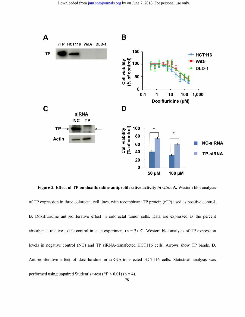
Antiproliferative activity of doxifluridine in cancer cell lines
Because capecitabine is converted to doxifluridine in the liver and then to 5-fluorouracil by TP in
tumor cells (Suppl. Fig. 1), we used doxifluridine in our in vitro assay. TP expression levels in HCT116
cells were higher than those in WiDr or DLD-1 cells (Fig. 2A). HCT116 cells were more sensitive to
doxifluridine treatment than WiDr and DLD-1 cells (Fig. 2B). The doxifluridine IC50 values for HCT116,
WiDr, and DLD-1 cells were 26.4, 74.3, and 77.9 µM, respectively, suggesting that TP expression levels
parallel the antiproliferative activity of doxifluridine in vitro. There was no statistically significant
difference among IC50 values for the three cell types. TP siRNA transfection dramatically downregulated
TP expression in HCT116 cells (Fig. 2C). After 50 μM doxifluridine treatment for 48 h, the viability of TP
siRNA-transfected cells and negative control cells was 70.3 and 40.5%, respectively (P < 0.01) (Fig. 2D).
Thus, downregulation of TP significantly inhibited doxifluridine anti-cancer activity.
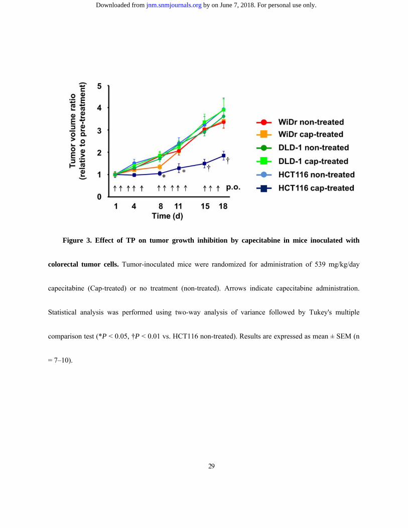
Effect of capecitabine in transplanted tumors
Capecitabine in vivo antiproliferative activity was evaluated in mice inoculated with HCT116,
WiDr, or DLD-1 cells. Capecitabine inhibited the growth of tumors formed from HCT116 cells, while no
by on June 7, 2018. For personal use only. jnm.snmjournals.org Downloaded from
14
significant change was observed in relative tumor size in mice inoculated with WiDr and DLD-1 cells
compared to control (Fig. 3).
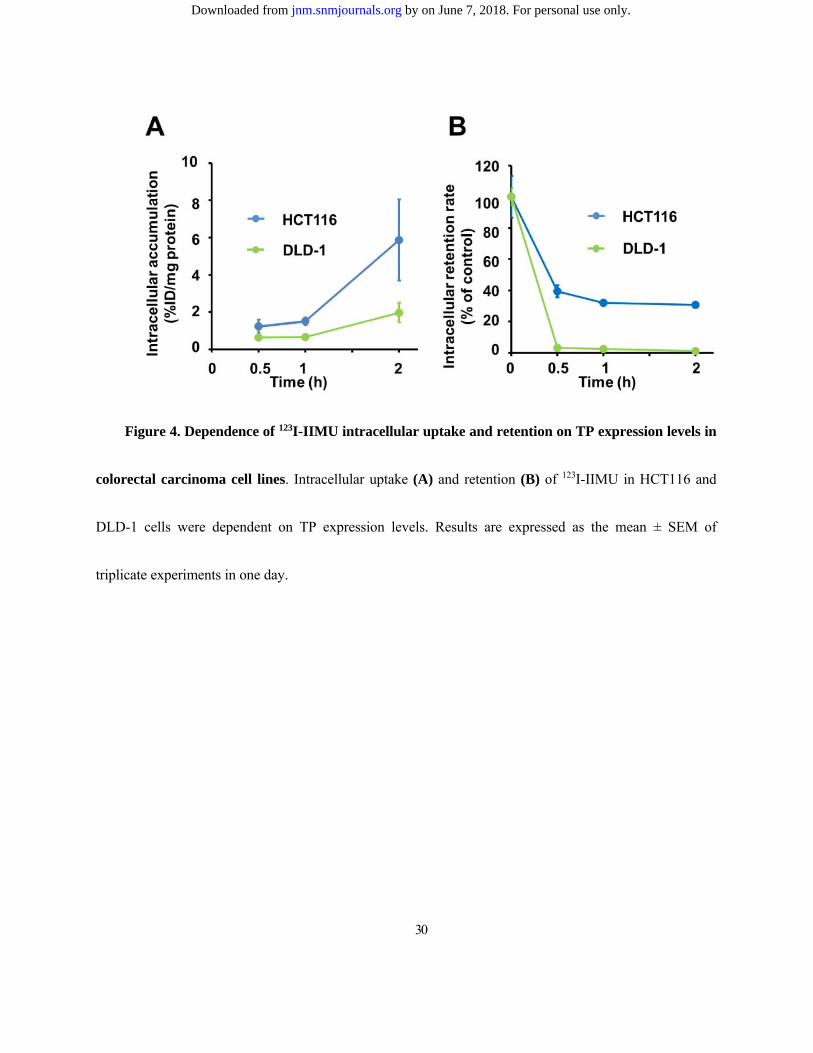
Intracellular 123I-IIMU uptake and retention
To assess whether 123I-IIMU could reflect TP expression differences among these cell lines, we
performed intracellular uptake and retention studies. In HCT116 cells, 123I-IIMU intracellular uptake
increased with incubation time and was significantly higher than that in DLD-1 cells (Fig. 4A). In the
efflux assay, 30.7% of the radioactivity prior to removing the tracer solution was retained by HCT116 cells
at 2 h after removal, whereas only 1.23% was retained by DLD-1 cells (Fig. 4B).
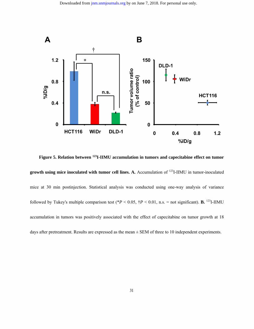
123I-IIMU uptake by transplanted tumors
We further examined the biodistribution of 123I-IIMU in mice carrying xenografts of the three cell
lines. Radioactivity in HCT116, WiDr, and DLD-1 tumors at 30 min postinjection was 0.99, 0.38, and
0.22 %ID/g, respectively (Fig. 5A). Radiotracer levels in other tissues were similar across groups (Suppl.
Table 1). Additionally, radioactivity levels in thyroid gland and stomach, an indicator of in vivo
deiodization, were low in these mice, as previously reported (24). These data indicate a positive relation
between 123I-IIMU accumulation and tumor expression levels of TP. Fig. 5B shows that capecitabine
by on June 7, 2018. For personal use only. jnm.snmjournals.org Downloaded from
15
antiproliferative activity in tumor-bearing mice is consistent with 123I-IIMU accumulation in tissues from
each tumor cell line.
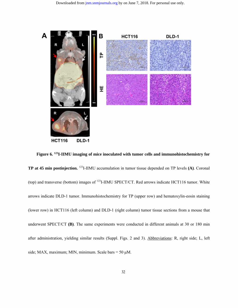
SPECT/CT imaging of 123I-IIMU and immunohistochemical detection of TP
To assess whether 123I-IIMU could detect high TP expression in tumors in vivo, we performed a
SPECT/CT study (Fig. 6A). 123I-IIMU accumulated in xenografts of capecitabine-sensitive HCT116 cells,
but not in DLD-1-inoculated xenografts. In HCT116 tumors, 123I-IIMU showed a high tumor/muscle ratio
(Suppl. Table 1) and clearly enabled the detection of high TP expression in SPECT images. However, a
large amount of 123I-IIMU was distributed in the liver and small intestine (Suppl. Fig. 2). To confirm the
TP expression levels in HCT116 and DLD-1 cells, HCT116 and DLD-1 tumors were excised after
SPECT/CT imaging, sectioned, and immunohistochemically stained (Fig. 6B). High TP expression levels
were observed in HCT116 tumors. Little TP expression was observed in DLD-1 tumors.
Discussion
Antiproliferative activity of doxifluridine in vitro was higher in HCT116 cells, which have higher
TP expression levels, than in WiDr and DLD-1 cells (Figs. 2A and 2B). TP downregulation significantly
decreased sensitivity to doxifluridine (Figs. 2C and 2D). A previous study showed that the antiproliferative
by on June 7, 2018. For personal use only. jnm.snmjournals.org Downloaded from
16
activity of doxifluridine in vitro was higher in HCT116 cells than in DLD-1cells (30). Our results are
consistent with this finding. We further investigated the relationship between the efficacy of capecitabine
and TP expression levels in vivo (Fig. 3). The efficacy of capecitabine has been found to correlate with TP
mRNA levels and TP activity in previous studies (31,32). These studies showed that HCT116 cells were
susceptible to capecitabine treatment, but neither WiDr nor DLD-1 cells were. Additionally, HCT116 had
the highest TP activity among the three cell types, and DLD-1 had the lowest. Our results also correspond
to these findings in vivo.
In vitro, intracellular uptake and retention of 123I-IIMU were higher in HCT116 cells than in
DLD-1 cells with low TP expression (Figs. 4A and 4B). In our previous studies, we found high
accumulation of 125I-IIMU in high TP-expressing A431 human epithelial carcinoma cells (25,26), and
125I-IIMU accumulation was inhibited by adding unlabeled IIMU. These results showed that the uptake of
radiotracer in tumor cells corresponded to TP expression levels. Additionally, in vivo biodistribution
experiments showed higher uptake of 123I-IIMU in HCT116 tumors than in the other tumors (Fig. 5A), and
the antiproliferative effect of capecitabine against tumor growth in mice was associated with the
accumulation of 123I-IIMU in each cell line (Fig. 5B). In our previous studies, we investigated the
biodistribution of 125I-IIMU and 123I-IIMU in mice (25,27). The radiolabelled tracers mainly accumulated
in liver and small intestine, consist with our present results. Furthermore, we confirmed mRNA and protein
by on June 7, 2018. For personal use only. jnm.snmjournals.org Downloaded from
17
levels of TP in various mouse tissues (27). We observed high TP expression in liver and intestine, which
corresponded to the observed high accumulation of radiolabeled IIMU (Suppl. Table. 1). Taken together,
our results show an association between 123I-IIMU accumulation in tumor cells and capecitabine efficacy
both in vitro and in vivo with the same cancer cell lines. However, it was not clear whether
123I-IIMU-SEPCT/CT can detect differences between high and low TP expressing tumor. Therefore, we
performed a SPECT/CT study, in which 123I-IIMU clearly detected HCT116 tumors with high TP
expression levels (Figs. 6A and 6B), while the accumulation of 123I-IIMU in DLD-1 tumors was negligible,
indicating that 123I-IIMU can discriminate tumor TP expression levels non-invasively. However, liver and
small intestine metastasis may be difficult to visualize because we observed high physiological
accumulations of 123I-IIMU in liver and small intestine (Suppl. Fig. 2). This result was consistent with the
biodistribution study (Suppl. Table. 1).
99mTc-etarfolatide images as a biomarker to predict the antiproliferative activity of vintafolide did
not always reflect immunohistochemical results because most surgical specimens for pathological
diagnosis had been obtained months or years prior. Moreover, folate receptor expression levels in
metastatic lesions differed from those in the primary tumor. However, in practice, technical and ethical
issues prevent a pathological diagnosis being performed in all surgical specimens and metastases to predict
drug response. Additionally, similar studies have reported that TP expression levels in tumors affect
by on June 7, 2018. For personal use only. jnm.snmjournals.org Downloaded from
18
capecitabine efficacy (13-16). Our results show that 123I-IIMU has potential as a prognostic imaging
biomarker for capecitabine efficacy. Because radiation and chemotherapy alter TP expression, whole-body
TP measurement in real time using 123I-IIMU would enable the more accurate prediction of treatment
outcomes.
A limitation of our study is that we only used human colorectal cell lines. To further assess the
potential use of 123I-IIMU imaging, further experiments with cell lines derived from cancer in other organs
such as breast, head and neck are needed. Additionally, PET imaging with 124I-IIMU could provide more
informative images concerning quantification of TP. However, 124I has a longer half-life time and
numerous higher-energy gamma emissions. With regard to commercial availability, 123I is extensively used.
Therefore, 123I-IIMU would be more acceptable for initial clinical study. TP activates not only capecitabine
but also 5-fluorouracil, doxifluridine, and S-1 (33-37). Therefore, 123I-IIMU could likely predict the effect
of treatment using all of these drugs. In vivo companion diagnostics using 123I-IIMU and SPECT may
provide optimized treatments and better quality of life for individual cancer patients.
by on June 7, 2018. For personal use only. jnm.snmjournals.org Downloaded from
19
Conclusion
We showed an association between TP expression levels determined non-invasively using
123I-IIMU in tumor cells and the efficacy of capecitabine in vitro and in vivo, suggesting that 123I-IIMU is a
predictive imaging biomarker for the outcome of capecitabine treatment.
Disclosure
This work was supported by the Creation of Innovation Centers for Advanced Interdisciplinary Research
Areas Program of the Ministry of Education, Culture, Sports, Science, and Technology of Japan.
Conflicts of Interest: Nobuya Kobashi, Shunsuke Meike, Yuki Okumura, Tsutomu Abe, and Hiroki
Matsumoto are employees of Nihon Medi-Physics Co., Ltd. Hiromichi Akizawa, Kazue Ohkura, Ken-ichi
Nishijima, Songji Zhao, Yuji Kuge, Hokkaido University, and Health Sciences University of Hokkaido
have patent rights for 123I-IIMU.
Acknowledgments
The authors thank Ms. Miho Ikenaga for performing the cell viability assay.
by on June 7, 2018. For personal use only. jnm.snmjournals.org Downloaded from
20
References
1. Fisher RE, Siegel BA, Edell SL, et al. Exploratory study of 99mTc-EC20 imaging for identifying
patients with folate receptor-positive solid tumors. J Nucl Med. 2008;49:899–906.
2. Morris RT, Joyrich RN, Naumann RW, et al. Phase II study of treatment of advanced ovarian cancer
with folate-receptor-targeted therapeutic (vintafolide) and companion SPECT-based imaging agent
(99mTc-etarfolatide). Ann Oncol. 2014;25:852–858.
3. Maurer AH, Elsinga P, Fanti S, et al. Imaging the folate receptor on cancer cells with
99mTc-etarfolatide: properties, clinical use, and future potential of folate receptor imaging. J Nucl Med.
2014;55:701–704.
4. Laing RE, Walter MA, Campbell DO, et al. Noninvasive prediction of tumor responses to
gemcitabine using positron emission tomography. Proc Natl Acad Sci U S A. 2009;106:2847–2852.
5. Rischin D, Hicks RJ, Fisher R, et al. Prognostic significance of 18F-misonidazole positron emission
tomography-detected tumor hypoxia in patients with advanced head and neck cancer randomly
assigned to chemoradiation with or without tirapazamine: a substudy of trans-tasman radiation
oncology group study 98.02. J Clin Oncol. 2006;24:2098–2104.
6. Bang YJ, Van Cutsem E, Feyereislova A, et al. Trastuzumab in combination with chemotherapy
versus chemotherapy alone for treatment of HER2-positive advanced gastric or gastro-oesophageal
by on June 7, 2018. For personal use only. jnm.snmjournals.org Downloaded from
21
junction cancer (ToGA): a phase 3, open-label, randomised controlled trial. Lancet.
2010;376:687–697.
7. Kang YK, Kang WK, Shin DB, et al. Capecitabine/cisplatin versus 5-fluorouracil/cisplatin as first-line
therapy in patients with advanced gastric cancer: a randomised phase III noninferiority trial. Ann
Oncol. 2009;20:666–673.
8. Saeki T, Kimura T, Toi M, Taguchi T. A pilot phase II study of capecitabine in advanced or recurrent
breast cancer. Breast Cancer. 2006;13:49–57.
9. Blum JL, Jones SE, Buzdar AU, et al. Multicenter phase II study of capecitabine in
paclitaxel-refractory metastatic breast cancer. J Clin Oncol. 1999;17:485–493.
10. Van Cutsem E, Findlay M, Osterwalder B, et al. Capecitabine, an oral fluoropyrimidine carbamate
with substantial activity in advanced colorectal cancer: results of a randomized phase II study. J Clin
Oncol. 2000;18:1337–1345.
11. Takebayashi Y, Yamada K, Miyadera K, et al. The activity and expression of thymidine
phosphorylase in human solid tumours. Eur J Cancer. 1996;32A:1227–1232.
12. Bonotto M, Bozza C, Di Loreto C, et al. Making capecitabine targeted therapy for breast cancer:
which is the role of thymidine phosphorylase? Clin Breast Cancer. 2013;13:167–172.
by on June 7, 2018. For personal use only. jnm.snmjournals.org Downloaded from
22
13. Schüller J, Cassidy J, Dumont E, et al. Preferential activation of capecitabine in tumor following oral
administration to colorectal cancer patients. Cancer Chemother Pharmacol. 2000;45:291–297.
14. Lee SJ, Choi YL, Park YH, et al. Thymidylate synthase and thymidine phosphorylase as predictive
markers of capecitabine monotherapy in patients with anthracycline- and taxane-pretreated metastatic
breast cancer. Cancer Chemother Pharmacol. 2011;68:743–751.
15. Koizumi W, Okayasu I, Hyodo I, et al. Prediction of the effect of capecitabine in gastric cancer by
immunohistochemical staining of thymidine phosphorylase and dihydropyrimidine dehydrogenase.
Anticancer Drugs. 2008;19:819–824.
16. Petrioli R, Bargagli G, Lazzi S, et al. Thymidine phosphorylase expression in metastatic sites is
predictive for response in patients with colorectal cancer treated with continuous oral capecitabine and
biweekly oxaliplatin. Anticancer Drugs. 2010;21:313–319.
17. Naruke A, Azuma M, Takeuchi A, et al. Comparison of site-specific gene expression levels in primary
tumors and synchronous lymph node metastases in advanced gastric cancer. Gastric Cancer.
2015;18:262–270.
18. Vallböhmer D, Kuramochi H, Shimizu D, et al. Molecular factors of 5-fluorouracil metabolism in
colorectal cancer: analysis of primary tumor and lymph node metastasis. Int J Oncol.
2006;28:527–533.
by on June 7, 2018. For personal use only. jnm.snmjournals.org Downloaded from

23
19. Toi M, Atiqur Rahman M, Bando H, Chow LW. Thymidine phosphorylase (platelet-derived
endothelial-cell growth factor) in cancer biology and treatment. Lancet Oncol. 2005;6:158–166.
20. Griffiths L, Dachs GU, Bicknell R, et al. The influence of oxygen tension and pH on the expression of
platelet-derived endothelial cell growth factor/thymidine phosphorylase in human breast tumor cells
grown in vitro and in vivo. Cancer Res. 1997;57:570–572.
21. Puglisi F, Andreetta C, Valent F, et al. Anthracyclines and taxanes induce the upregulation of
thymidine phosphorylase in breast cancer cells. Anticancer Drugs. 2007;18:883–888.
22. Endo M, Shinbori N, Fukase Y, et al. Induction of thymidine phosphorylase expression and
enhancement of efficacy of capecitabine or 5'-deoxy-5-fluorouridine by cyclophosphamide in
mammary tumor models. Int J Cancer. 1999;83:127–134.
23. Zhang SH, Zhang H, He HW, et al. Lidamycin up-regulates the expression of thymidine
phosphorylase and enhances the effects of capecitabine on the growth and pulmonary metastases of
murine breast carcinoma. Cancer Chemother Pharmacol. 2013;72:777–788.
24. Takahashi M, Seki K, Nishijima K, et al. Synthesis of a radioiodinated thymidine phosphorylase
inhibitor and its preliminary evaluation as a potential SPECT tracer for angiogenic enzyme expression.
J Label Comp Radiopharm. 2008;51:384–387.
by on June 7, 2018. For personal use only. jnm.snmjournals.org Downloaded from
24
25. Akizawa H, Zhao S, Takahashi M, et al. In vitro and in vivo evaluations of a radioiodinated thymidine
phosphorylase inhibitor as a tumor diagnostic agent for angiogenic enzyme imaging. Nucl Med Biol.
2010;37:427–432.
26. Li H, Zhao S, Jin Y, et al. Radiolabeled uracil derivative as a novel SPECT probe for thymidine
phosphorylase: suppressed accumulation into tumor cells by target gene knockdown. Nucl Med
Commun. 2011;32:1211–1215.
27. Zhao S, Li H, Nishijima K, et al. Relationship between biodistribution of a novel thymidine
phosphorylase (TP) imaging probe and TP expression levels in normal mice. Ann Nucl Med.
2015;29:582–587.
28. Thanasai J, Limpaiboon T, Jearanaikoon P, et al. Effects of thymidine phosphorylase on tumor
aggressiveness and 5-fluorouracil sensitivity in cholangiocarcinoma. World J Gastroenterol.
2010;16:1631–1638.
29. Ishikawa T, Fukase Y, Yamamoto T, et al. Antitumor activities of a novel fluoropyrimidine,
N4-pentyloxycarbonyl-5'-deoxy-5-fluorocytidine (capecitabine). Biol Pharm Bull. 1998;21:713–717.
30. Miwa M, Ura M, Nishida M, et al. Design of a novel oral fluoropyrimidine carbamate, capecitabine,
which generates 5-fluorouracil selectively in tumours by enzymes concentrated in human liver and
cancer tissue. Eur J Cancer. 1998;34:1274–1281.
by on June 7, 2018. For personal use only. jnm.snmjournals.org Downloaded from
25
31. Yasuno H, Kurasawa M, Yanagisawa M, et al. Predictive markers of capecitabine sensitivity
identified from the expression profile of pyrimidine nucleoside-metabolizing enzymes. Oncol Rep.
2013;29:451–458.
32. Ishikawa T, Sekiguchi F, Fukase Y, et al. Positive correlation between the efficacy of capecitabine
and doxifluridine and the ratio of thymidine phosphorylase to dihydropyrimidine dehydrogenase
activities in tumors in human cancer xenografts. Cancer Res. 1998;15:685–690.
33. Yang Q, Barbareschi M, Mori I, et al. Prognostic value of thymidine phosphorylase expression in
breast carcinoma. Int J Cancer. 2002;97:512–517.
34. Ogawa M, Watanabe M, Mitsuyama Y, et al. Thymidine phosphorylase mRNA expression may be a
predictor of response to post-operative adjuvant chemotherapy with S-1 in patients with stage III
colorectal cancer. Oncol Lett. 2014;8:2463–2468.
35. Ishii R, Takiguchi N, Oda K, et al. Thymidine phosphorylase expression is useful in selecting
adjuvant chemotherapy for stage III gastric cancer. Int J Oncol. 2001;19:717–722.
36. Kikuyama S, Inada T, Shimizu K, et al. p53, bcl-2 and thymidine phosphorylase as predictive markers
of chemotherapy in patients with advanced and recurrent gastric cancer. Anticancer Res.
2001;21:2149–2153.
by on June 7, 2018. For personal use only. jnm.snmjournals.org Downloaded from

26
37. Yamamoto Y, Toi M, Tominaga T. Prediction of the effect of 5'-deoxy-5-fluorouridine by the status
of angiogenic enzyme thymidine phosphorylase expression in recurrent breast cancer patients. Oncol
Rep. 1996;3:863–865.
by on June 7, 2018. For personal use only. jnm.snmjournals.org Downloaded from
27
Figures
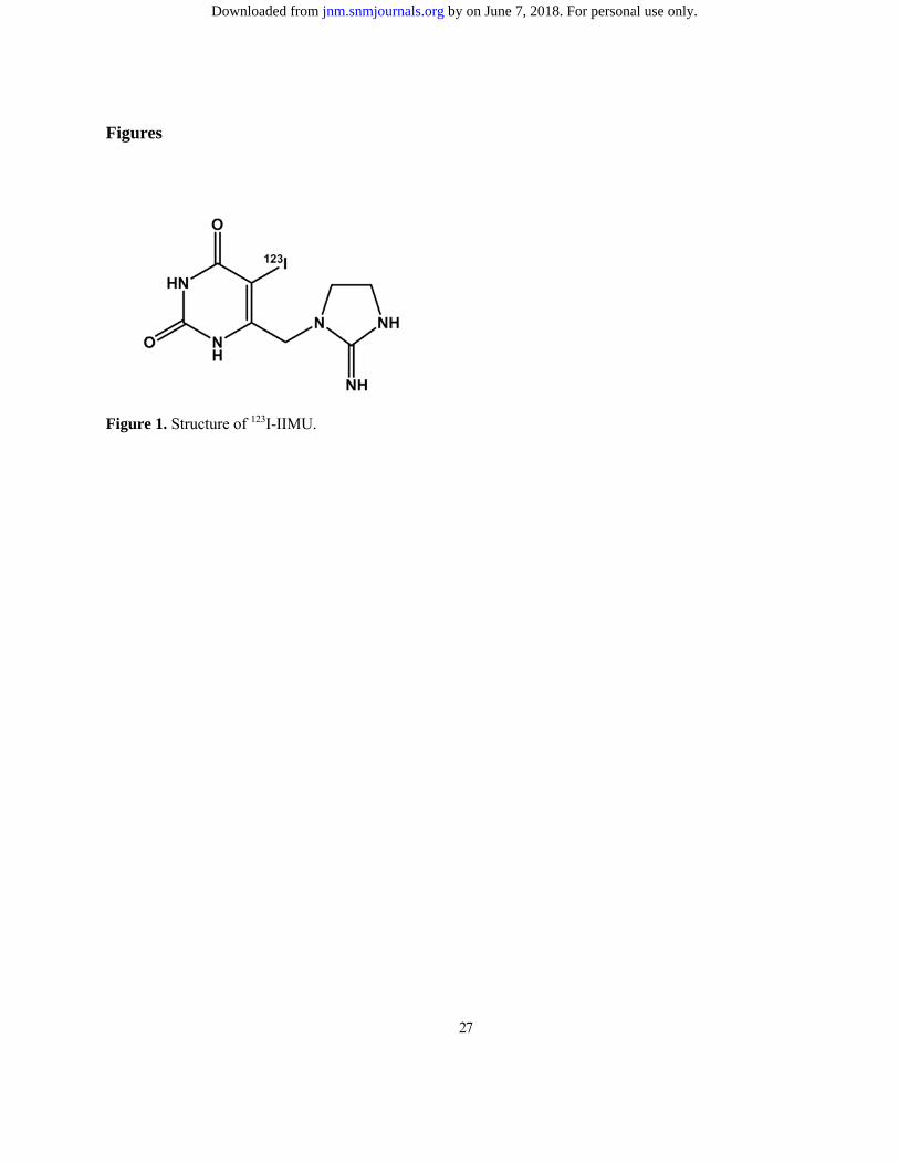
Figure 1. Structure of 123I-IIMU.
by on June 7, 2018. For personal use only. jnm.snmjournals.org Downloaded from
28
Figure 2. Effect of TP on doxifluridine antiproliferative activity in vitro. A. Western blot analysis
of TP expression in three colorectal cell lines, with recombinant TP protein (rTP) used as positive control.
B. Doxifluridine antiproliferative effect in colorectal tumor cells. Data are expressed as the percent
absorbance relative to the control in each experiment (n = 3). C. Western blot analysis of TP expression
levels in negative control (NC) and TP siRNA-transfected HCT116 cells. Arrows show TP bands. D.
Antiproliferative effect of doxifluridine in siRNA-transfected HCT116 cells. Statistical analysis was
performed using unpaired Student’s t-test (*P < 0.01) (n = 4).
by on June 7, 2018. For personal use only. jnm.snmjournals.org Downloaded from
29
Figure 3. Effect of TP on tumor growth inhibition by capecitabine in mice inoculated with
colorectal tumor cells. Tumor-inoculated mice were randomized for administration of 539 mg/kg/day
capecitabine (Cap-treated) or no treatment (non-treated). Arrows indicate capecitabine administration.
Statistical analysis was performed using two-way analysis of variance followed by Tukey's multiple
comparison test (*P < 0.05, †P < 0.01 vs. HCT116 non-treated). Results are expressed as mean ± SEM (n
= 7–10).
by on June 7, 2018. For personal use only. jnm.snmjournals.org Downloaded from
30
Figure 4. Dependence of 123I-IIMU intracellular uptake and retention on TP expression levels in
colorectal carcinoma cell lines. Intracellular uptake (A) and retention (B) of 123I-IIMU in HCT116 and
DLD-1 cells were dependent on TP expression levels. Results are expressed as the mean ± SEM of
triplicate experiments in one day.
by on June 7, 2018. For personal use only. jnm.snmjournals.org Downloaded from
31
Figure 5. Relation between 123I-IIMU accumulation in tumors and capecitabine effect on tumor
growth using mice inoculated with tumor cell lines. A. Accumulation of 123I-IIMU in tumor-inoculated
mice at 30 min postinjection. Statistical analysis was conducted using one-way analysis of variance
followed by Tukey's multiple comparison test (*P < 0.05, †P < 0.01, n.s. = not significant). B. 123I-IIMU
accumulation in tumors was positively associated with the effect of capecitabine on tumor growth at 18
days after pretreatment. Results are expressed as the mean ± SEM of three to 10 independent experiments.
by on June 7, 2018. For personal use only. jnm.snmjournals.org Downloaded from
32
Figure 6. 123I-IIMU imaging of mice inoculated with tumor cells and immunohistochemistry for
TP at 45 min postinjection. 123I-IIMU accumulation in tumor tissue depended on TP levels (A). Coronal
(top) and transverse (bottom) images of 123I-IIMU SPECT/CT. Red arrows indicate HCT116 tumor. White
arrows indicate DLD-1 tumor. Immunohistochemistry for TP (upper row) and hematoxylin-eosin staining
(lower row) in HCT116 (left column) and DLD-1 (right column) tumor tissue sections from a mouse that
underwent SPECT/CT (B). The same experiments were conducted in different animals at 30 or 180 min
after administration, yielding similar results (Suppl. Figs. 2 and 3). Abbreviations: R, right side; L, left
side; MAX, maximum; MIN, minimum. Scale bars = 50 μM.
by on June 7, 2018. For personal use only. jnm.snmjournals.org Downloaded from
Doi: 10.2967/jnumed.115.165811Published online: April 7, 2016.J Nucl Med. Kazue Ohkura, Nishijima Ken-ichi, Nagara Tamaki and Yuji KugeNobuya Kobashi, Hiroki Matsumoto, Songji Zhao, Shunsuke Meike, Yuki Okumura, Tsutomu Abe, Hiromichi Akizawa, Capecitabine
I-IIMU Predicts the Efficacy of123The Thymidine Phosphorylase Imaging Agent
http://jnm.snmjournals.org/content/early/2016/04/06/jnumed.115.165811This article and updated information are available at:
http://jnm.snmjournals.org/site/subscriptions/online.xhtml
Information about subscriptions to JNM can be found at:
http://jnm.snmjournals.org/site/misc/permission.xhtmlInformation about reproducing figures, tables, or other portions of this article can be found online at:
and the final, published version.proofreading, and author review. This process may lead to differences between the accepted version of the manuscript
ahead of print area, they will be prepared for print and online publication, which includes copyediting, typesetting,JNMcopyedited, nor have they appeared in a print or online issue of the journal. Once the accepted manuscripts appear in the
. They have not beenJNM ahead of print articles have been peer reviewed and accepted for publication in JNM
(Print ISSN: 0161-5505, Online ISSN: 2159-662X)1850 Samuel Morse Drive, Reston, VA 20190.SNMMI | Society of Nuclear Medicine and Molecular Imaging
is published monthly.The Journal of Nuclear Medicine
© Copyright 2016 SNMMI; all rights reserved.
by on June 7, 2018. For personal use only. jnm.snmjournals.org Downloaded from