the triassic thecodontian reptile desmatosuchus
TRANSCRIPT
THE TRIASSIC THECODONTIAN REPTILE DESMATOSUCHUS
OSTEOLOGY AND RELATIONSHIPS
by
BRYAN JOHN SMALL, B.S.
A THESIS
IN
MUSEUM SCIENCE
Submitted to the Graduate Faculty of Texas Tech University in
Partial Fulfillment of the Requirements for
the Degree of
MASTER OF ARTS
Approved
December, 198 5

f-^^
.;z^
ACKNOWLEDGMENTS
This study was made possible through a series of
grants by the National Geographic Society, and a Research
Assistantship through the Museum.
I am grateful to the members of my committee, Robert
Baker and Clyde Jones for their encouragement and helpful
comments. I am especially grateful to Sankar Chatterjee
for his invaluable encouragement, interest, help and total
support. This thesis would not have been possible without
him. My thanks to Mikael W. Nickell for his valuable
guidance with my illustrations and layout. I also thank
Robert Long of the University of California at Berkeley
for his helpful comments and discussion. I am especially
thankful to my family for their total support and
encouragement during my graduate studies.
11

TABLE OF CONTENTS
ACKNOWLEDGMENTS ii
LIST OF FIGURES v
I. INTRODUCTION 1
II. GEOLOGICAL SETTING 4
III. MATERIALS AND METHODS 9
IV. SYSTEMATIC PALEONTOLOGY 14
V. DESCRIPTION OF DESMATOSUCHUS 16
Skull 16
Dermal Bones of the
Skull Roof 19
Palatal Complex 25
Braincase 30
Lower Jaw 3 6
Dentition 41
Shoulder Girdle 42
Forelimb 4 2
Pelvic Girdle 48
Femur 5 3
Dermal Armor 5 6
V I . STANCE AND GAIT 58
VII. MODE OF LIFE 64
VIII. TAXONOMY 67
IX. GENERA OF STAGONOLEPIDIDAE 70
111
X. EVOLUTION AND RELATIONSHIPS 74
XI. CONCLUSIONS 77
LITERATURE CITED 81
IV
LIST OF FIGURES
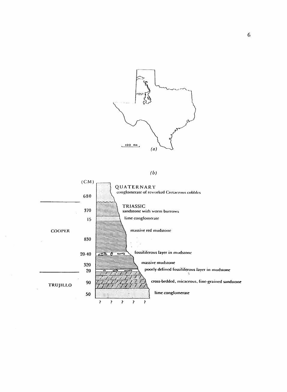
1. Dockum Formation in Texas and Type section of Cooper Creek Member at Post quarry, Garza County, Texas 5
2. Desmatosuchus haplocerus, composite restoration of the skull in (a) dorsal view; (b) lateral view with lower jaw 17
3. Premaxilla of TTUP 9024 20
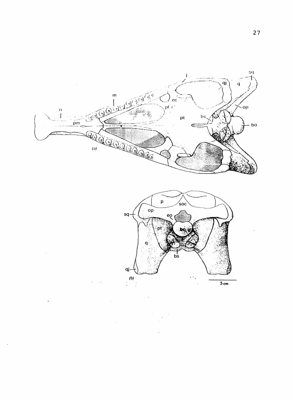
4. Composite restoration of skull in: (a) palatal view; (b) occipital view 26
5. Composite view of the braincase in: (a) lateral view; (b) anterior view 31
6. TTUP 9024, right mandible in: (a) lateral viev;; (b) medial view; (c) TTUP 9025, isloated tooth 37
7. TTUP 9023, right shoulder girdle in:
(a) lateral view; (b) posterior view 43
8. TTUP 9170, right humerus and ulna 45
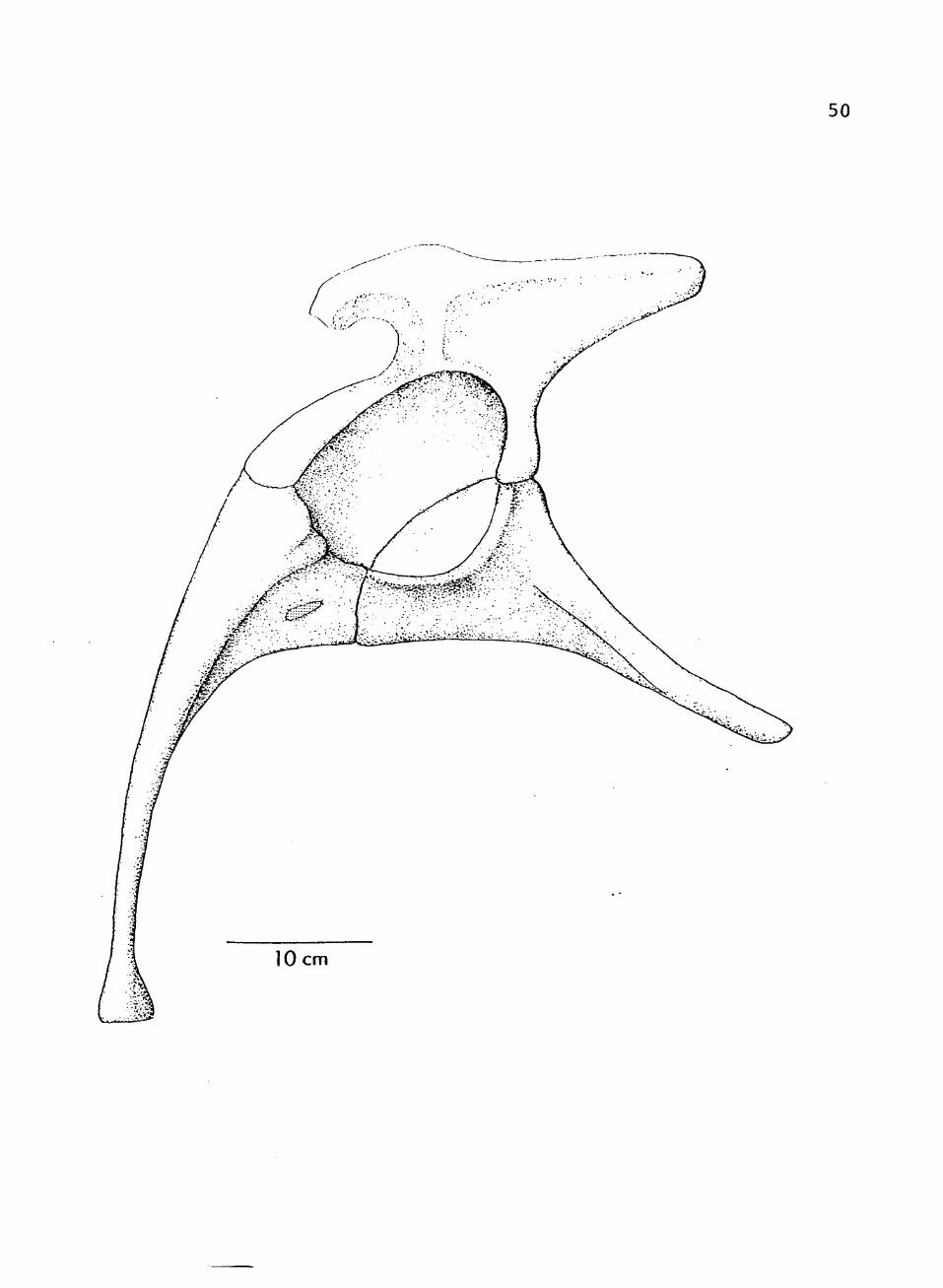
9. Composite restoration of pelvis, lateral view 49
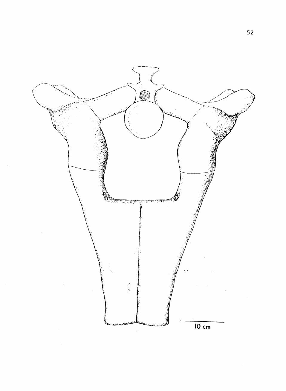
10. Composite restoration of pelvis, anterior view 51
11. TTUP 9024, right femur in: (a) anterior view; (b) posterior view 54
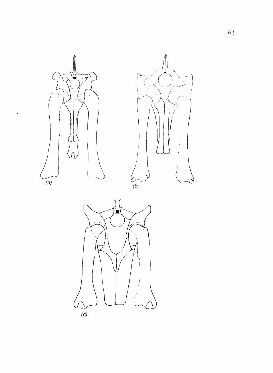
12. Pelvic girdles and femora in posterior view of: (a) Postosuchus; (b) Fasolosuchus (after Bonaparte, 1981); (c) Desmatosuchus 60
V
CHAPTER I
INTRODUCTION
The aetosaurs (Family StagonolGpididac) were larae,
armored quadrupeds of the Late Tria.ssic. They are the
only known herbivorous thecodonts, occurring in North
America, South America, Europe, India, and possibly China.
The North American genera are Desmatosuchus, Typothorax,
Paratypothorax, Calyptosuchus, and Stegomus. The three
genera of South America are Aetosauroides,
Argentinosuchus, and Neoaetosauroides. Two genera are
represented in Europe, Aetosaurus and Stagonolepis. There
is one undescribed aetosaur from India, based on some
dermal armor. One problematic aetosaur is known from.
China.
The North American genus Desmatosuchus is known from
the Late Triassic Dockum Formation of West Texas and
Eastern New Mexico, and the Chinle Formation of Arizona
and New Mexico. Desmatosuchus was first described in part
by Cope in 1887, and later by Case in 1920. It is
distinguished from other aetosaurs by the large recurved
shoulder spines, and by its overall large size.
In 1982 through 1984, field parties from Texas Tech
University led by Sankar Chatterjee discovered abundant
2
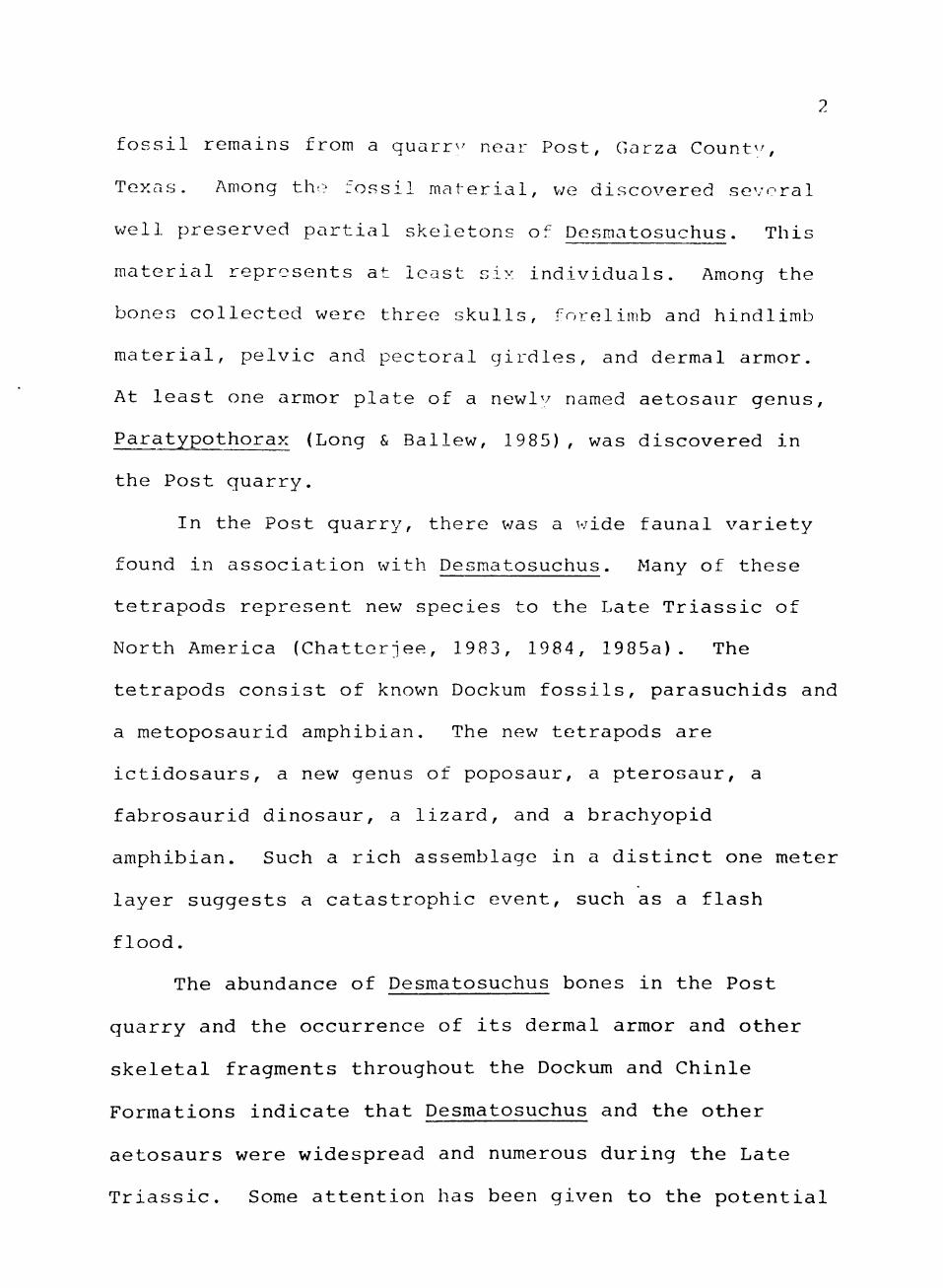
fossil remains from a quarrv^ near Post, Garza Count^^,
Texas. Among the fossil material, v;e discovered several
well preserved partial skeletons of Desmatosuchus. This
material represents at least six individuals. Among the
bones collected were three skulls, forelimb and hindlimb
material, pelvic and pectoral girdles, and dermal armor.
At least one armor plate of a newly named aetosaur genus,
Paratypothorax (Long & Ballew, 1985) , was discovered in
the Post quarry.
In the Post quarry, there was a wide faunal variety
found in association with Desmatosuchus. Many of these
tetrapods represent new species to the Late Triassic of
North America (Chatterjee, 1983, 1984, 1985a). The
tetrapods consist of known Dockum fossils, parasuchids and
a metoposaurid amphibian. The new tetrapods are
ictidosaurs, a new genus of poposaur, a pterosaur, a
fabrosaurid dinosaur, a lizard, and a brachyopid
amphibian. Such a rich assemblage in a distinct one meter
layer suggests a catastrophic event, such as a flash
flood.
The abundance of Desmatosuchus bones in the Post
quarry and the occurrence of its dermal armor and other
skeletal fragments throughout the Dockum and Chinle
Formations indicate that Desmatosuchus and the other
aetosaurs were widespread and numerous during the Late
Triassic. Some attention has been given to the potential
stratigraphic correlation possibilities of parasuchids
(Chatterjee, 1978; Gregory, 1957; Shelton, 1984).
Recently, Long and Ballew (1985) has suggested that
aetosaurs could possibly be used as correlative tools for
further subdivisions of the Late Triassic.
Until the Post finds, the anatomy of Desmatosuchus
was imperfectly known. This study offers the first
detailed description of the skull and post cranial
skeleton. There are many advancements in the skull
(edentuluous premaxilla and further closing of lower
temporal openings) previously unknown, and the attainment
of an erect posture. In the skeletal morphology,
Desmatosuchus shows an uncanny resemblance to later
ankylosaur dinosaurs. The similarities may be attributed
to convergent evolution rather than any phylogenetic
relationships, although the ankylosaurs may be related to
some unknown sister group of the stagonolepidids.
CHAPTER II
GEOLOGICAL SETTING
The Upper Triassic Dockum Formation of the
Southwestern United States occurs in Northwest Texas and
Eastern New Mexico (Fig. 1). The Dockum "red beds" are
unconformably overlain by Cretaceous, Tertiary, or
Quaternary sediments, and unconformably overlay the
Permian Quartermaster Group. The Dockum Formation is
characterized by a variety of lithologies. These are
cross-bedded sandstones, conglomerates, siltstones, and
mudstones.
The Dockum has been interpreted as a series of
depositional, relict Paleozoic basins. Sediments
deposited in these basins came from the Amarillo Uplift,
Bravo Dome, and Matodor arch in the North, and the
Ouachita Uplift to the Southeast (McGowen et al., 1983).
The depositional environment consisted of braided and
meandering streams, alluvial fans, fan deltas, high
constructive elongate fans, lacustrine, and valley fill.
There has been much confusion over the status of the
Dockum. Cummins (1890) first described the Dockum and
gave it Formation status. He derived the name from
Dockum, Dickens County, Texas. Gould (1907) upgraded the
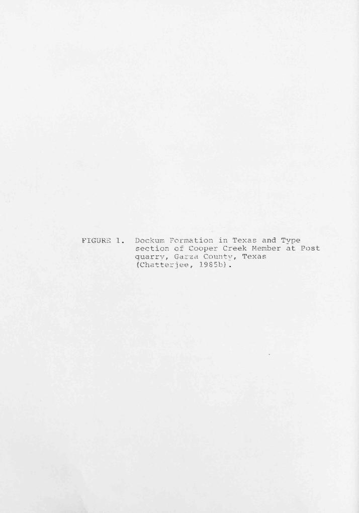
FIGURE 1. DockuiF: Forroation in Texas and Type section of Cooper Creek Member at Post quarry, Gai'sa County, Texas (Chatterjee, 19S5b).
COOPKR
T R U J I L L O
(b)
Q U A T E R N A R Y Lonsloinciate of reworked Crci;i< eous cobbles
T R I A S S I C sandstone with worm burrows
lime conglomerate
massive red mudstone
20-40 ^ to=o N fossiliferoiis layer in iniidsione
massive mudstone
poorly defined fossiliferoiis layer in mudstone
90 / / '^J^'^ /.'/., ^ ^ ^"A cross-bedded, micaceous, fine-grained sandstone /• .f..f..f
lime conglomerate
Dockum to Group status, naming two formations. The lower
Tecovas Formation consisted mainly of shales ant!
siltstones. The upper Trujillo Formation consisted of
sandstones and conglomerates.
Reedside et al. (1957) added a middle Formation, the
Santa Rosa, and named the upper unit the Chinle.
Recently, a benthosuchid (=Eocyclotosaurus) amphibian has
been discovered in the Santa Rosa sandstones of New
Mexico. This amphibian is known from the Middle Triassic
Moenkopi Formation of Arizona. This makes it impossible
for the the Santa Rosa to be placed in the Dockum time
frame.
Recently, Chatterjee (1985b) has proposed a major
revision of the Dockum to formation status. He retains
three units, reducing them to member status, and dropped
the Santa Rosa entirely. He named a new upper member the
Cooper Creek Member (Fig. 1).
The new Cooper Creek Member is possibly Mid to Late
Norian in age, and the Dockum Formation ranges from Late
Carnian to Late Norian. This would make the upper part of
the Dockum Formation correlative with the Chinle
Formation. This conflicts with previous works (Colbert &
Gregory, 1957; Murry, 1982).
The evidence for a Norian age is based on the faunal
assemblage from the Post quarry. The ictidosaur and
fabrosaur are closely related to Southern Hemisphere forms
8
of Late Triassic to Lower Jurassic in age (Chatterjo-,
1983, 1984). The new poposaur Postosuchus is closelv
related to Teratosaurus of the UDper Norian of Gernuinv
(Chatterjee, 1985a).
Parasuchids are also useful correlative tools. The
parasuchid genus Nicrosaurus occurs in the Post quarry.
Nicrosaurus is restricted to Gtubensandstein (Norian) in
age. Two other genera occur in other parts of the Dockum.
Parasuchus and Angistorhinus are restricted to the
Carnian.
A recent paper by Long and Ballew (1985) appears to
dispute a Norian age for the Cooper Creek Member. Long
and Ballew (1985) claim that aetosaurs can be used as
correlative tools in the Chinle and Dockum Formations.
The occurrence of Desmatosuchus indicates a Carnian age
according to their findings. They also state that there
are no true Typothorax specimens in the Dockum..
Typothorax is supposedly restricted to the Norian.
Undescribed Typothorax scutes (TTUP 545) from the Texas
Tech collection are from the Dockum. This should make
parts of the Dockum Norian in age. Desmatosuchus from the
Post quarry shows many advanced traits, but due to a lack
of comparative material, its stratigraphic use cannot be
determined. All of the above evidence indicates that the
Post quarry faunal assemblage is younger than Carnian, and
is equivalent to the Chinle Formation of Arizona.
CHAPTER III
MATERIALS AND METHODS
Six of the specimens Texas Tech University usod in
this study came from the Post quarry, Garza Cou^ tv, TOM.IS
(TTUP 9023-9025, 6169-9171). One specimen (TT[;P 9172)
came from Home Creek, Crosby County, Texas. Manv of tho
individuals from the Post quarry were disarticulatoci, but
associated. The bones were taken from a hard red clav
matrix. Acetone was used to soften the clay for easy
removal of the bones without moisture damage. A
calcareous crust occurred on some of the bones. A weak
solution of hydrochloric acid was carefully used to remove
the crust and identify the sutures. Some of the tools
used were pin-vice, ice-pick, and brush. A "glyptal"
solution was sometimes applied to the bones to strengthen
them.
The specimen repostories used in this study are
identified by the following abbreviations:
TMM - Texas Memorial Museum
TTUP - The Museum, Texas Tech University
UCMP - University of California Museum of Paleontology
10
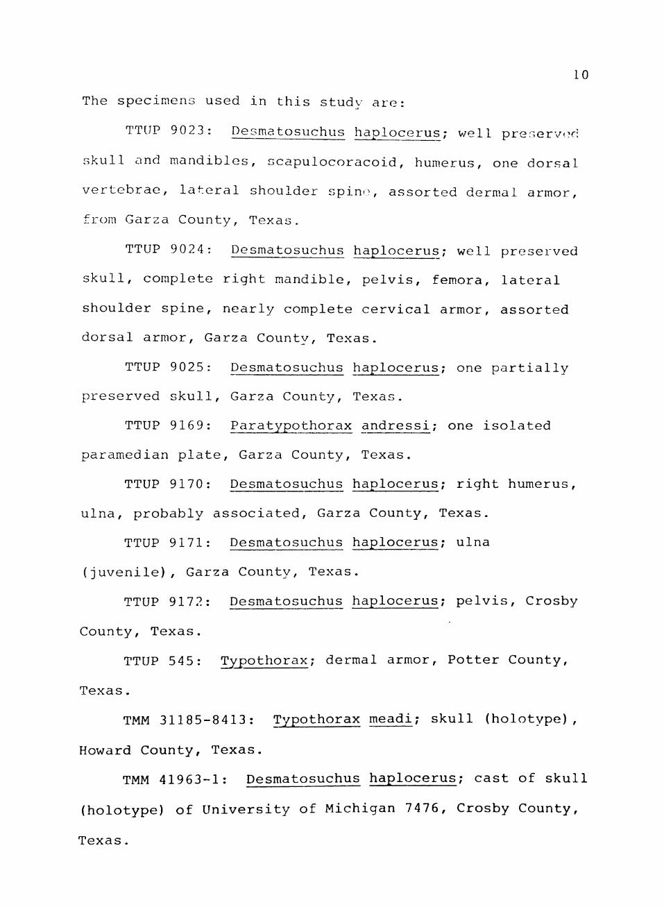
The specimens used in this study are:
TTUP 902 3: Desmatosuchus haplocerus; well preserv<-d
skull and mandibles, scapulocoracoid, hum.erus, one dorsal
vertebrae, lateral shoulder spine, assorted dermal armor,
from Garza County, Texas.
TTUP 9024: Desmatosuchus haplocerus; well preserved
skull, complete right mandible, pelvis, femora, lateral
shoulder spine, nearly complete cervical armor, assorted
dorsal armor, Garza County, Texas.
TTUP 9025: Desmatosuchus haplocerus; one partially
preserved skull, Garza County, Texas.
TTUP 9169: Paratypothorax andressi; one isolated
paramedian plate, Garza County, Texas.
TTUP 9170: Desmatosuchus haplocerus; right humerus,
ulna, probably associated, Garza County, Texas.
TTUP 9171: Desmatosuchus haplocerus; ulna
(juvenile), Garza County, Texas.
TTUP 9172: Desmatosuchus haplocerus; pelvis, Crosby
County, Texas.
TTUP 545: Typothorax; dermal armor. Potter County,
Texas.
TMM 31185-8413: Typothorax meadi; skull (holotype),
Howard County, Texas.
TMM 41963-1: Desmatosuchus haplocerus; cast of skull
(holotype) of University of Michigan 7476, Crosby County,
Texas.

11
UCMP 25989: Desmatosuchus haplocerus; partial ilium,
St. Johns, Arizona.
UCMP 27408: Desmatosuchus haplocerus; braincase, St.
Johns, Arizona.
UCMP 27988: Desmatosuchus haplocerus; femur, St.
Johns, Arizona.
UCMP 32166: Desmatosuchus haplocerus; partial pubis,
St. Johns, Arizona.
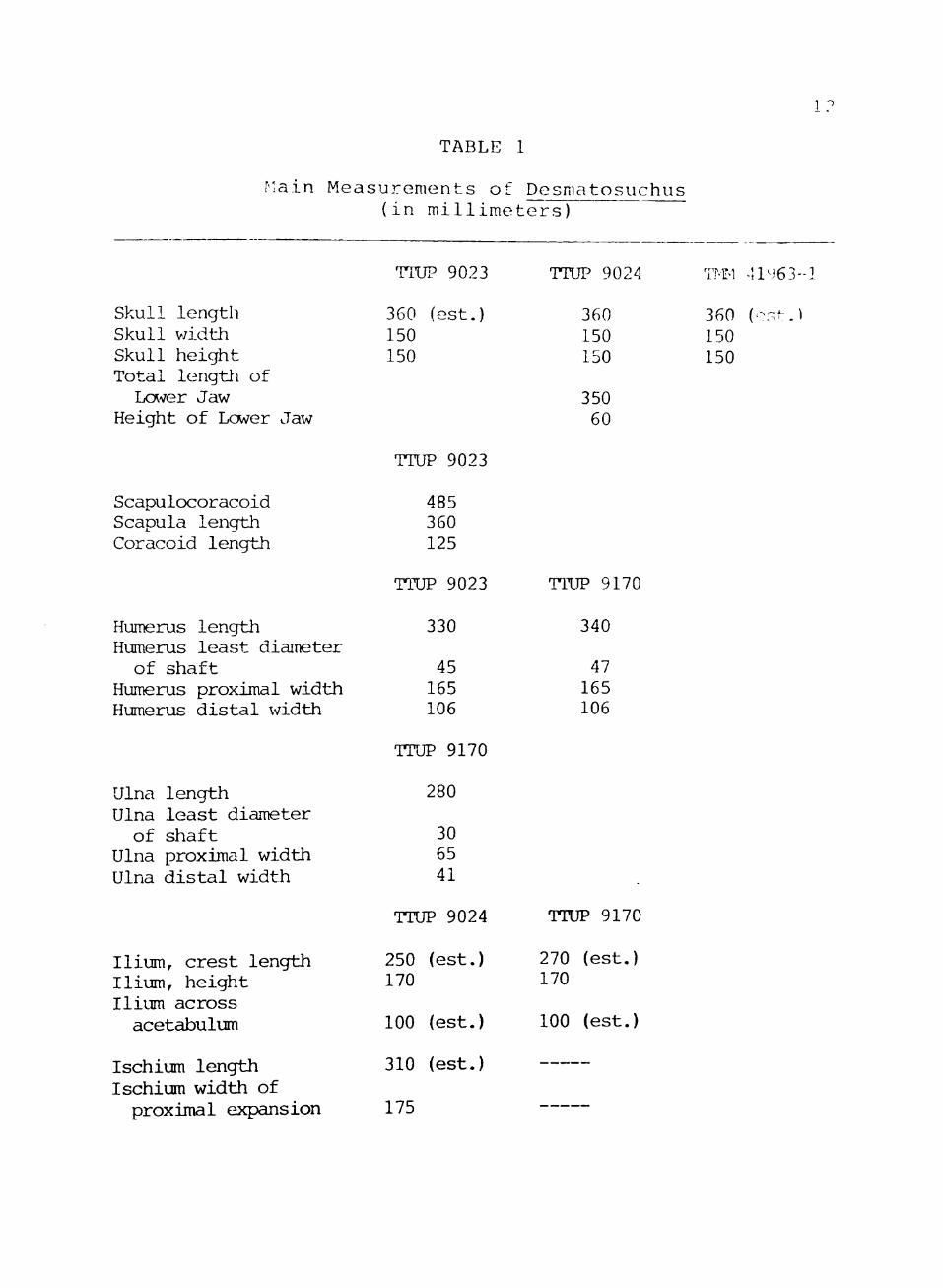
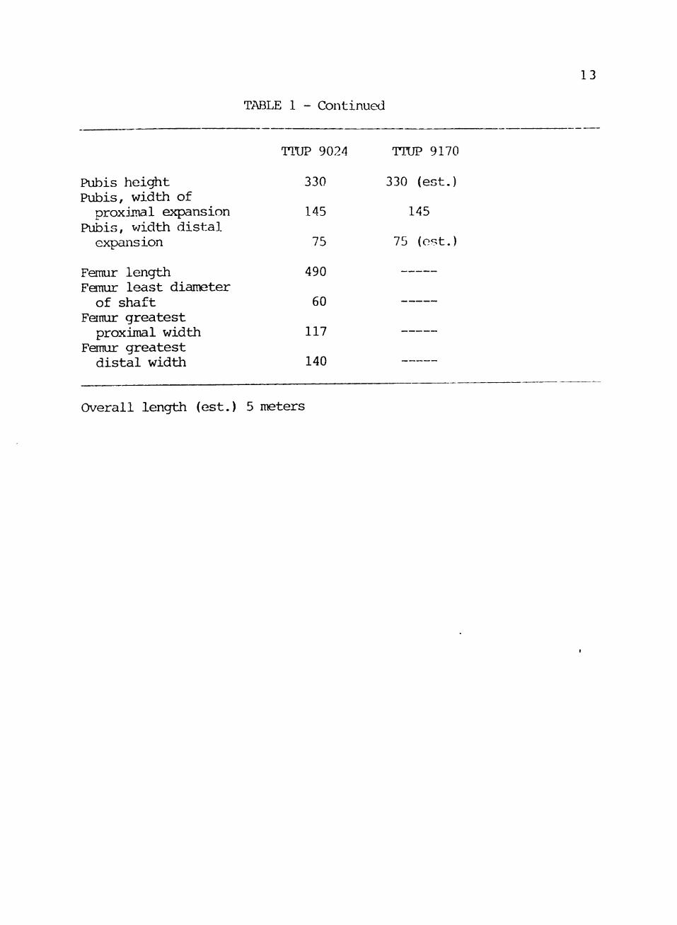
The main measurements of Desmatosuchus are listed in
Table 1.
TABLE
Main Measurements of
Skull length Skull v/idth Skull height Total lengi±i of LcAver Jaw
Height of Lower Jaw
Scapulocoracoid Scapula length Coracoid length
Humerus length Humerus least diameter of shaft
Humerus proximal width Humerus distal width
Ulna length Ulna least diameter of shaft
Ulna proximal width Ulna distal width
Ilium, crest length Ilium, height Ilium across acetabulum
Ischium length Ischium width of
proximal expansion
(in millime
TTUP 9023
360 (est.) 150 150
TTUP 9023
485 360 125
TTUP 9023
330
45 165 106
TTUP 9170
280
30 65 41
TTUP 9024
250 (est.) 170
100 (est.)
310 (est.)
175
1
Desmatosuchus ters)
TTUP 9024
360 150 150
350 60
TTUP 9170
340
47 165 106
TTUP 9170
270 170
100
(est.)
(est.)
T?'n 41463-1
360 (-St.) 150 150
13
TABLE 1 - Continued
Pubis height Pubis, width of proxjjnal expansion
Pubis, width distal expansion
Femur length Femur least diameter of shaft
Fenur greatest proximal v/idth
Femur greatest distal width
TTUP 9024
330
145
75
490
60
117
140
TTUP 9170
330 (est.)
145
75 (est.)
Overall length (est.) 5 meters
CHAPTER IV
SYSTEMATIC PALEONTOLOGY
Class Reptilia
Subclass Archosauria
Order Thecodontia
Suborder Pseudosuchia
Infraorder Aetosauria
Family Stagonolepididae Lydekker, 1887
Genus Desmatosuchus Case, 1920
Species Desmatosuchus haplocerus (Cope, 1892).
Revised diagnosis: Large quadrupedal pseudosuchian,
3 meters or more in length; skull com.paratively small
(length 37 cm), narrow, triangular with edentulous beak;
teeth blunt bulb-shaped, possibly herbivorous; external
naris longer than the antorbital fenestra; premaxilla
expanded anteriorly with upturned tip; snout pointed v/ith
nasals approximately half the length of the skull roof;
lower temporal opening almost closed off secondarily with
contact of postorbital and squamosal; upper temporal
opening large, facing laterally; quadrate fixed, jaw-joint
below the level of the alveolar margin; braincase strongly
fused with the skull roof; laterosphenoid ossified; lower
14
15
jaw slipper-shaped, edentulous in front, with long, narrow
lateral mandibular fenestra.
Centra amphicoelous, sacrals 3, co-ossified; scapula
with strong acromion process, forelimbs v/ell-developed;
ulna with prominent olecranon process; pelvis advanced,
acetabulum deep, imperforate; pubis long with strong
median symphysis, ischium plate-like; femur straight with
strong inturned head; posture "fully-improved"; ankle
joint "crocodile-normal" with a large calcaneal tuber.
Dorsal armor with four rows of scutes, with pitted, radial
sculpture; lateral plates with compressed spines; shoulder
spine very large, horn-like.
Horizon: Dockum Formation of West Texas, Late
Triassic.
CHAPTER V
DESCRIPTION OF DESMATOSUCh^US
Skull
The description and drawing of the skull are
composite, based on specimens TTUP 9023 and TTUP 9024 (Sec
Fig. 2). TTUP 9023 possesses a well-preserved and
articulated palatal region, skull roof (excluding nasal
and premaxilla), braincase and partially complete
mandibles. TTUP 9024 has a well-preserved premaxilla,
nasal, and a complete mandible, as well as skull roof and
palatal region. All specimens have a problem of a lack of
preservation of certain sutures. This could be due to
either the m.aturity of the individuals or a preservational
problem. Some cranial sutures are inferred from other
aetosaurs (Walker, 1961).
The skull of Desmatosuchus is a narrow,
triangular-shaped skull, small in proportion to the
postcranial skeleton. The average length is 37 cm, width
is 17.5, and height is 15. Some distinguishing
characteristics of this genus are an edendulous
premaxilla, and further closing off the infratemporal
fenestra.
16
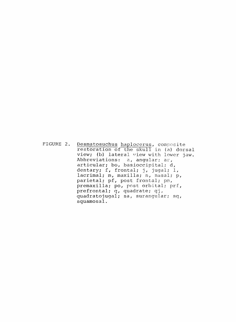
FIGURE 2. Desmatosuchus haplocerus, composite restoration of the skull in (a) dorsal viev/; (b) lateral view with lower jaw. Abbreviations: a, angular; ar, articular; bo, basioccipital; d, dentary; f, frontal; j, jugal; 1, lacrimal; m, maxilla; n, nasal; p, parietal; pf, post frontal; pm, premaxilla; po, post orbital; prf, prefrontal; q, quadrate; qj, quadratojugal; sa, surangular; sq, squamosal.

18
po
5cin.

19
Dermal Bones of the Skull Roof
The premaxilla is a slender, forked bone (See
Fig. 3) . The anterior tip is turned upv/ard and expanded
to form a shovel-shaped structure. A small, tapering
projection slopes backward from the anterior tip forming
the antero-dorsal edge of the external naris. This
projection fits into a V-shaped groove along the ventral
symphysis of the expanded anterior tip of the nasal.
Approximately half way back a small but prominent narial
protuberance occurs on the dorsal surface. The ventral
surface narrows into a bony ridge. Farther backward, the
premaxilla divides into two thin, broad branches. The
ventral branch lies against the ventral surface of the
maxilla and vomer, and extends as far back as the second
maxillary tooth. The lateral edge of this branch fits
into a groove along the medial edge of the maxilla. The
lateral branch extends backward and upward along the
lateral surface of the maxilla. It sets in a slight
depression along the ventro-posterior edge of the external
naris. The premaxilla fits very loosely with the maxilla
indicating some flexibility at this joint. This is why
the bone has never been found before in Desmatosuchus.
The premaxilla is entirely edentulous, contrary to all
other knov/n aetosaurs.
The maxilla is an elongate, triangular bone.
we 11-exposed in lateral view. Anteriorly, the bone has a

FIGURE 3. Premaxilla of TTUP 9024 (a) Left lateral view (b) Ventral view
21
fm.
(A)
5 cm
9 O
saddle-shaped notch for the reception of the premaxilla.
Posteriorly, it forks to curve around the antorbital
fenestra. Its ascending branch extends up and back to
form a foot-like projection which articulates dorsally
with the nasal and posteriorly with the lacrimal. The
anterior edge of tlie ascending branch forms part of the
posterior edge of the external naris, while the posterior
edge of the ascending branch forms the antero-dorsal edge
of the antorbital fenestra.
The postero-ventral branch of the maxilla extends
back to meet the jugal. It forms the major portion of the
ventral edge of the antorbital fenestra. The ventral edge
forms a slight convex curve as it extends back. There are
small nutrient foramina present along the lateral surface
that correspond with the number of aveoli.
The tooth count in the maxilla seems to vary among
the individuals, ranging from 10 to 12. There is a
strongly pronounced shelf on the medial surface of the
maxilla. This shelf tapers posteriorly until its junction
with the palatine. The shelf provides a stable base for
the articulation of the premaxilla and is appressed
against the anterior tip of the vomer. The shelf also
forms the lateral edge of the internal naris.
The nasal is the largest bone of the skull roof,
accounting for almost half the length of the skull.
Anteriorly, it is thickened vertically and expanded

2 3
laterally to meet the premaxilla. As the bone extends
back, it becomes thinner and narrower, forming the dorsal
edge of the external naris. As it approaches the frontal,
it curves downward to meet the maxilla. The nasal is
separated from the antorbital fenestra by the lacrimal.
Posteriorly, the nasal is bordered by the prefrontal and
frontal.
The frontal is about one-third of the length of the
nasal. It is a slightly sculptured bone bordered
ventrally by the prefrontal and the orbit. Postero-
ventrally, it forms a junction with the postfrental,
postorbital, and parietal. Posteriorly, the frontal meets
the parietal on a line that curves forward to meet the
mid-line. This suture also lies along a raised knob that
starts on the frontal and slopes upward on the parietal.
The parietal forms the postero-dorsal edge of the
supratemporal fenestra. The bone is slightly sculptured.
Posteriorly, it develops an overhanging flange which is
overlapped by the first pair of paramedian scutes. From
the posterior edge, the parietal projects downward on the
occipital surface as a flat bone which meets the
paroccipital process. Laterally, the parietal extends as
a bar to meet the squamosal.
The lacrimal is a thin, narrow bone which meets the
ascending process of the maxilla. Its ventral surface
forms the dorsal border of the antorbital fenestra.
2 4
Dorsally, the lacrimal rises to meet the nasal and
prefrontal. Posteriorly, the bone slopes downward to
receive the ascending branch of the jugal. The lacriir,al
makes the posterior rim of the orbit, and forms a
protruberance with the prefrontal.
The prefrontal is a thick bone with an exposed
surface bordered anteriorly by the nasal, and posteriorly
by the orbit. It borders the lacrimal ventrally, while
its dorsal edge meets the frontal.
The postfrontal is a small, thick, triangular bone
which tapers out ventrally against the postorbital.
Anteriorly, it borders the orbit. Posteriorly, it forms
the anterior part of the postorbital bar.
The postorbital forms the rounded bar betv/een the
orbit and supratemporal openings. As it postorbital
descends, it becomes a thin flat bone which flares out
anteriorly and posteriorly. The anterior branch borders
the jugal and orbit. The posterior branch forms part of
the ventral edge of the supratemporal fenestra, and
articulates with the squamosal. Ventrally, it borders the
infratemporal fenestra.
The jugal articulates anteriorly with the maxilla.
It sends a dorsal branch upward to make a contact with the
lacrimal. Posteriorly, the bone has two branches around
the infratemporal fenestra. The upper branch comes in
contact with the postorbital. The lower branch descends

25
slightly downward from the maxilla and joins with the
quadratojugal as a narrow tapering process.
The quadratojugal is overlapped dorsally by the
squamosal. Anteriorly, it borders the infratemporal
opening and makes a oontact with the jugal. Posteriorly
and medially, it articulates inimovably with the quadrate.
The squamosal meets the postorbital anteriorly
between supratemporal and infratemporal fenestrae. The
ventral end of the squamosal overlaps the quadratojugal.
Dorsally and anteriorly it forms part of the supratemporal
fenestra. As the squamosal continues its upward ascent,
it forms a "horn" along with the paroccipital process.
From there, the squamosal sends out a ridge that makes
contact with the dorsal and occipital branch of the
parietal. Posteriorly it makes a contact with the
quadrate.
Palatal Complex
The vomer is a thin, long bone that flairs into a
broad horizontal anterior surface to join the maxilla (See
Fig. 4). Together they form a platform which rests on the
premaxilla. The vomer extends back as a thin, laterally
compressed bone with a long median symphysis. It forms
the medial edge of the internal naris. The two vomers
diverge posteriorly at the contact with the pterygoid.
Each branch is a thin, tapering process which appresses

IIGURE 4. Composite restoration of skull in: (a) palatal view; (b) occipital veiw Abbreviations: bo, basioccipital; bs, basisphenoid; ec, ectopterygoid• eo exoccipital; j, jugal; m, maxilla; n, nasal; op, opisthotic; p, parietal; pi palatine; pm, premaxilla; pt, pterygoid; q, quadrate; aj, quadratojugal; soc, supraoccipital; sq squamosal; v, vomer.
27
5an

28
along the lateral surface of the anterior ptorvcrc id
branch.
The palatine, along with the vomer and maxilla lorris
the border for the internal naris. The palatine itself
forms a channel at the posterior end of the internal
naris. This channel tapers downward posteriorly. The
lateral edge of the palatine meets the medial edge of the
maxilla in a "foot-like" process. This process forms the
anterior border of the palatal fenestra. Posteriorly, the
palatine extends to m.eet the pterygoid.
The pterygoid is divided into three branches: the
palatal ramus, the lateral branch, and the vertical
quadrate ramus. The two pterygoids meet at the midline
with a weak symphysis, extending back to a palatal vacuity
formed under and exposing the parasphenoid.
The palatal branch is a thin, laterally compressed
bone that extends forward for the articulation with the
vomer. As the palatal branch extends back, it expands
laterally and meets the palatine at the posterior end of
the palatal channel. The lateral branch extends outv;ard
and downward from the broad pterygoid plate. The
ectopterygoid meets the anterior end of the lateral
branch. The dorso-lateral edge of the branch articulates
with the medial edge of the jugal at its junction with the
maxilla.
29
A constriction forms behind the lateral branch
opposite to the palatal vacuity. As the pterygoid flares
out laterally behind this constriction, it meets the
quadrate along the posterior edge of the skull. This is
where the quadrate ramus articulates posteriorly with the
basisphenoid. Between the basisphenoid and the quadrate,
the quadrate ramus becomes a thickened ridge of bone
before it projects upward alongside the quadrate. This
thin sheet of bone slightly overlaps the ventral and
medial surfaces of the quadrate. Medially and dorsally,
the quadrate ramus rests against the opisthotic and
basioccipital.
The ectopterygoid is a small bone that forms the
posterior edge of the palatal fenestra. From this border,
it flares sharply downward to form the anterior portion of
the lateral branch of the pterygoid. The lateral edge
passes sharply upward to articulate with the jugal and
possibly the maxilla.
The quadrate is the thick posterior bone of the
skull. Dorsally, it fits under the horn created by the
squamosal and the paroccipital process. The quadrate
makes a C-shaped curve as it descends. It meets the
squamosal and quadratojugal laterally. There is no
quadrate foramen visible. The quadrate is firmly fixed
with its neighboring bones. This is in contrast with
Stagonolepis, where the quadrate is thought to be
30
streoptostylic (Walker, 1961) . Medially and ventrally, it
makes contact with the pterygoid. Ventrally, it flares
out to articulate with the thick bony ridqe of the
quadrate ramus of the pterygoid. The glenoid surface is
basically transversely oriented with two condvles for
articulation v/ith the lower jaw.
Braincase
The braincase is firmly fused with the skull roof and
palate in all specimens examined, making a detailed study
difficult (See Fig. 5). This deficiency is remedied by an
undescribed Desmatosuchus braincase (UCMP 27408), which is
disarticulated, and beautifully preserved. There are some
differences between UCMP 27408 and the TTUP specimens.
When differences do occur, the TTUP specimens v;ill be
used, with the differences pointed out. As in the rest of
the skull, many bones are fused, obscuring the sutures.
Some sutures are inferred from the general thecodontian
pattern. The braincase is very similar to that of
Parasuchus (Chatterjee, 1978) .
The supraoccipital is a flat, triangular bone in
posterior view. It is firmly fused with the parietals.
Ventrally, it forms the dorsal border of the foramen
m.agnum. There are two prominent flanges on the border of
the foraman magnum for the reception of the atlas.
Ventro-laterally, the supraoccipital is fused to the
opisthotic. TTUP 9023 does not show an elongate pit in

31

FIGURE 5. Composite viev; of the braincase in: (a) lateral view; (b) anterior view. Abbreviations: bo, basioccipital; bs, basisphenoid; bpt, basipterygoid process; ef, epipterygoid foramen; eo, exoccipital; fo, fenestra ovalis; hf, hypophyseal fenestra; ic, internal carotid foramen; mf, metotic foramen; Is, laterosphenoid; op, opisthotic; p, parietal; pr prootic; prs, presphenoid; ps, parasphenoid; soc supraoccipital; foramina for cranial nerves in Roman numerals.

32
-^ ( mf • V - ; - -
hf J: ^ l - ^ '^^::^:-:^.^
5 cm
33
the dorsal region of the supraoccipital as in Case's
(1922) specimen (TMM 41963-1) and UCMP 27408, TTUP 9024
and 9025 are not well preserved in this area. The
anterior side of the supraoccipital is not exposed due to
overlap of the parietals.
The opisthotic is a laterally projecting, wing-like
structure that articulates with the squamosal and
quadrate. The dorsal surface is firmly fused with the
parietal and supraoccipital. Ventrally, it overlaps the
pterygoid. The ventral surface broadens medially to form
the posterior borders of the fenestra ovalis and the
metotic foramen. Anteriorly, the opisthocic is partially
overlapped by the pro-otic. Together, these bones form a
thin, prominent, ventral ridge.
The exoccipital forms the side wall of the foramen
magnum. Dorsally it is fused with the opisthotic without
any visible suture. The exoccipital is pillar-shaped at
its base. It articulates ventrally with the
basioccipital. The posterior tip of the exoccipital
extends to the occipital condyle. The lateral side of the
exoccipital contains two foramina for hypoglossal (XII)
nerve. Only one foramen is pronounced. The exoccipitals
do not meet at a mid-line in the foramen magnum in the
TTUP specimens, but meet in the UCMP specimen.
The basioccipital forms the large, distinct occipital
condyle at the base of the foramen magnum. The ventral
34
surface is constricted directly in front of the condvle.
It then slopes downward and forward to the basal tuber.
The basal tubra are large, prominent oval masses with .\
highly rugose surface. The suture between the
basioccipital and basisphenoid is probably in a deep
groove that runs through the middle of the tubera. The
dorsal surface of the basioccipital forms part of the
border of the fenestra ovalis and the metotic fenestra.
The pro-otic is a flat, slightly concave bone that
overlies the anterior portion of the opisthotic. It is
fused dorsally with the parietal. It has an anterior
articulation with the laterosphenoid. Along its anterior
margin it forms part of the borders for the two outlets of
the trigeminal (V) nerve. The ventral surface forms a
small groove on top of the bony ridge with the opisthotic
and basisphenoid. This groove is for the facialis (VII)
nerve.
The laterosphenoid is a small dorsally curved bone
that is dorsally fused with the parietal. The
antero-dorsal articulation is where the parietal meets the
frontal. Anterior to the laterosphenoid is the
presphenoid. The anterior portion of the laterosphenoid
contains a foramen for the trochlearis (IV) nerve.
Ventrally and anteriorly it meets the basisphenoid; the
suture is interrupted by the hypophyseal fenestra and the
35
foramen for oculmotor (III) nerve. Posteriorly it makes
up most of the borders of the trigeminal incisure.
The basisphenoid forms the anterior portion of the
basal tubera. The dorsal surface forms the deep groove
which houses the course of the internal carotid. A
shallow pit is present anterior to the basal tubera and
posterior to the basipterygoid process. In this region
the TTUP specimens differ from TMM 41963-1 and UCMP 27400.
In the TTUP specimens the pit is very shallow with a
substantial gap between the basal tuber and basipterygoid
process. The TMM and UCMP specimens have a very deep pit,
and there is almost no gap between the basal tubera and
basipterygoid process.
The basipterygoid process is a large knob that is
smooth and convex anteriorly for the reception of the
pterygoid. There is a tapering projection above the
basipterygoid process and the parasphenoid. It tapers to
a point between the laterosphenoid and the presphenoid.
Within this process lies the foramen for oculmotor (III)
nerve and the hypophyseal fenestra.
The parasphenoid (cultiform process) is ossified in
Desmatosuchus. It projects forward as a flat, tapering
bone above the level of the basipterygoid process. It can
be seen through the palatal vacuity.
The presphenoid is ossified. It is the smallest of
the bones of the braincase. It is a small triangular bone

36
that meets at a mid-line in the anterior-most portion of
the braincase. The large fenestra for the optic II nerve
exit is shared by both presphenoids.
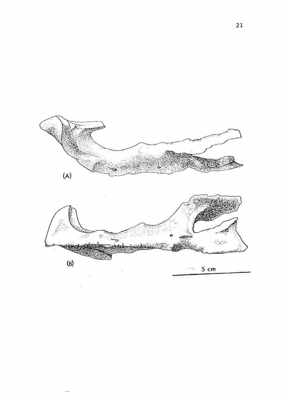
Lower Jaw
The lower jav/ is slipper shaped, with an edentulous
beak (See Fig. 6). The articular surface is below the
aveoler line. The lateral mandibular fenestra is long and
narrow. The tooth count of the lower jaw varies from five
to six.
The dentary covers about half the length of the lower
jaw. The anterior half of the dorsal surface is
edentulous. This surface slopes inward. This edentulous
area was probably covered by a horny sheath during life.
The lateral surface of the dentary extends back to
the mandibular fenestra where it forks into upper and
lov/er branches.
The upper branch extends back as a rounded
projection, overlapping the surangular. The lower branch
extends back unusually far, overlapping the angular with a
sharp point. The dorsal surface of the lower branch forms
almost half of the lower rim of the mandibular fenestra.
The medial surface of the dentary is overlapped
posteriorly by the splenial. The splenial sends forward a
single tapering projection over the dentary, starting at
the third tooth and sloping downward and forward to the

37
FIGURE 6. TTUP 9024, right mandible in: (a) lateral viev;; (b) medial viev/; (c) TTUP 9025, isolated tooth. Abbreviations: a, angular; ar, articular; d, dentary; pa, prearticular; sa, surangular; sp, splenial.

38
•,s.^'-^-' ".",a \ \ .
V nr - a >
.>^ lii-^'
' \
sp
(a)
. /
(b) 5 cm
/ ;
(c) L _i
2 cm
,"9
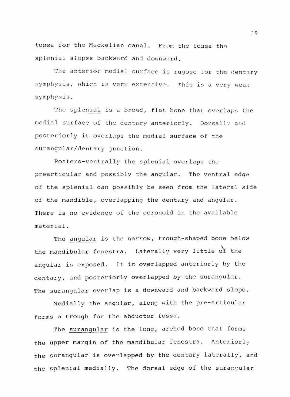
fossa for the Meckelian canal. From the fossa the
splenial slopes backward and downward.
The anterior medial surface is rugose for the (dentary
symphysis, which is very extensive. This is a very weak
sym.physis .
The splenial is a broad, flat bone that overlaps the
medial surface of the dentary anteriorly. Dorsally and
posteriorly it overlaps the medial surface of the
surangular/dentary junction.
Postero-ventrally the splenial overlaps the
prearticular and possibly the angular. The ventral edge
of the splenial can possibly be seen from the lateral side
of the mandible, overlapping the dentary and angular.
There is no evidence of the coronoid in the available
material.
The angular is the narrov;, trough-shaped bone below
the mandibular fenestra. Laterally very little or the
angular is exposed. It is overlapped anteriorly by the
dentary, and posteriorly overlapped by the surangular.
The surangular overlap is a downward and backward slope.
Medially the angular, along with the pre-articular
forms a trough for the abductor fossa.
The surangular is the long, arched bone that forms
the upper margin of the mandibular fenestra. Anteriorly
the surangular is overlapped by the dentary laterally, and
the splenial medially. The dorsal edge of the surangular
40
is missing, but is presumed to have a relatively high
arch. The surangular forms the upper margin of the
mandibular fenestra and the internal mandibular fenestra.
Posteriorly the surangular makes contact with the
prearticular and articular m.edially, and with the angular
and articular laterally. A small, prominent foramen is
present on the posterior, lateral surface, and exists in
front of the articular surface. The surangular forms the
outer half of the articular surface.
The prearticular combines v/ith the angular to form
the groove of the Meckelian canal. The dorsal edge curves
sharply upward to form the ventral margin of the internal
fenestra. The splenial overlaps the prearticular
anteriorly. Ventrally the prearticular meets the angular
which curves down and around from the lateral side. The
posterior sutures cannot be made out, but the prearticular
probably meets the articular directly beneath the
articular surface.
The articular is a short bone, forming most of the
gleniod surface. The gleniod surface is an undulating,
concave surface for the reception of the quadrate.
Medially the surface extends outward as a shelf.
Posteriorly the articular extends back as a knob-like
projection.

41
Dentition
The maxilla and dentary are characterized by closely
spaced, deep aveoli (See Fig. 6). The dental formula
varies in the TTUP specimens. This appears to be common
among the aetosaurs (Case, 1922; WalJcer, 1961). The
dental formula is premaxilla 0, maxilla 10 to 12, dentary
5 to 6. The alveoli vary in size on the maxilla, starting
small, and increasing in size to about number 6, then a
gradual decrease in size.
There is only one isolated tooth present among the
TTUP specimens (TTUP 9025). The root is long and thick.
The tooth and deep aveoli indicate a typical thecodontian
implantation. The base of the crown is constricted. The
crown has a bulbous shape and is slightly compressed front
to back. It is slightly recurved with no serrations, and
a blunt tip. Microscopic examination reveals faint
longitudinal striations. There are no replacement teeth
present, so determination of the replacement mechanism and
pattern is unknown. It can be assumed, due to the lack of
teeth preserved, that the teeth were held in life by soft
connective tissue. The blunt, bulbous teeth combined with
the edentulous premaxilla and dentary tip indicate a
herbivorous diet.
42
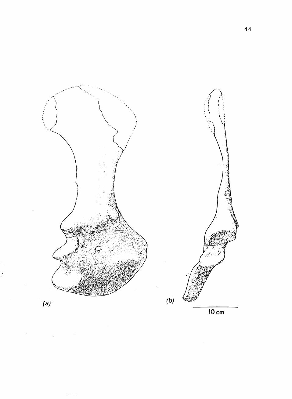
Shoulder Girdle
Only one scapulocoracoid is preserved from the Post
quarry (See Fig. 7). It is the right scapulocoracoid from
TTUP 9023. It is virtually complete (including the
curvature) except for the extreme proximal end. The
scapula and coracoid are firmly fused.
The proximal end of the scapula is flattened and
flared. The shaft is also relatively flat and
constricted, with a slight, inward curve. The posterior
edge of the shaft bears a small triceps tubercle, about
seven centimeters above the glenoid.
The glenoid is postero-laterally facing. It is a
very broad, long, U-shaped cavity. Anterior to the
glenoid and close to the anterior edge is a pronounced
trochanter. The clavicle would have articulated with the
scapulocoracoid on a flat, shelf-like area in front of
this trochanter.
The coracoid is flared with a concave medial surface.
There is no overhanging ventral lip as in Stagonolepis. A
coracoid foramen occurs about three centimeters anterior
to the glenoid. There is no clavicle or interclavicle
preserved in the Texas Tech specimens.
Forelimb
The forelimb is much shorter than the hindlimb (See
Fig. 8). The humerus is less than two-thirds the length

43
FIGURE 7. TTUP 9023, right shoulder girdle in: (a) lateral view; (b) posterior view.
44
(a) (b)
10 cm
45
FIGURE 8. TTUP 9170, right humerus and ulna. (a) and (b) posterior and anterior views of humerus, (c) and (d) lateral and anterior views of ulna.

46
/ ' \
> %>:,
~A
(d)
a
A l
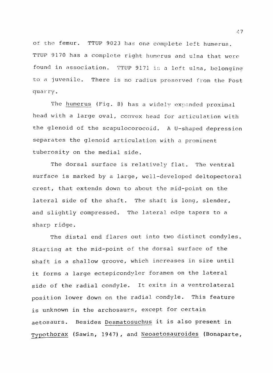
of the femur. TTUP 9023 has one complete left humerus.
TTUP 9170 has a complete right humerus and ulna that were
found in association. TTUP 9171 is a left ulna, belonging
to a juvenile. There is no radius preserved from the Post
quairy.
The humerus (Fig. 8) has a widely expanded proximal
head v/ith a large oval, convex head for articulation v/ith
the glenoid of the scapulocorocoid. A U-shaped depression
separates the glenoid articulation with a prominent
tuberosity on the medial side.
The dorsal surface is relatively flat. The ventral
surface is marked by a large, well-developed deltopectoral
crest, that extends down to about the m.id-point on the
lateral side of the shaft. The shaft is long, slender,
and slightly compressed. The lateral edge tapers to a
sharp ridge.
The distal end flares out into two distinct condyles.
Starting at the mid-point of the dorsal surface of the
shaft is a shallow groove, which increases in size until
it forms a large ectepicondyler foramen on the lateral
side of the radial condyle. It exits in a ventrolateral
position lower down on the radial condyle. This feature
is unknown in the archosaurs, except for certain
aetosaurs. Besides Desmatosuchus it is also present in
Typothorax (Sawin, 1947), and Neoaetosauroides (Bonaparte,
48
1981). This is a primitive feature found in cotylosaurs
and synapsids, and in some mammals (Romer, 1956).
The ulna is elongate with a well developed olecranon
on the proximal head. The sigmoid notch is well
developed, with two angling facets, probably for fitting
into the groove on the distal end of the humerus. There
is a pronounced protuberence on the lateral surface of the
olecranon. This forms part of the antero-lateral facet of
the sigmoid notch.
The shaft is laterally compressed with a depression
running along the upper medial part of the shaft. The
shaft is slightly curved in a medial direction at the
distal end. The distal end is small and oval shaped.
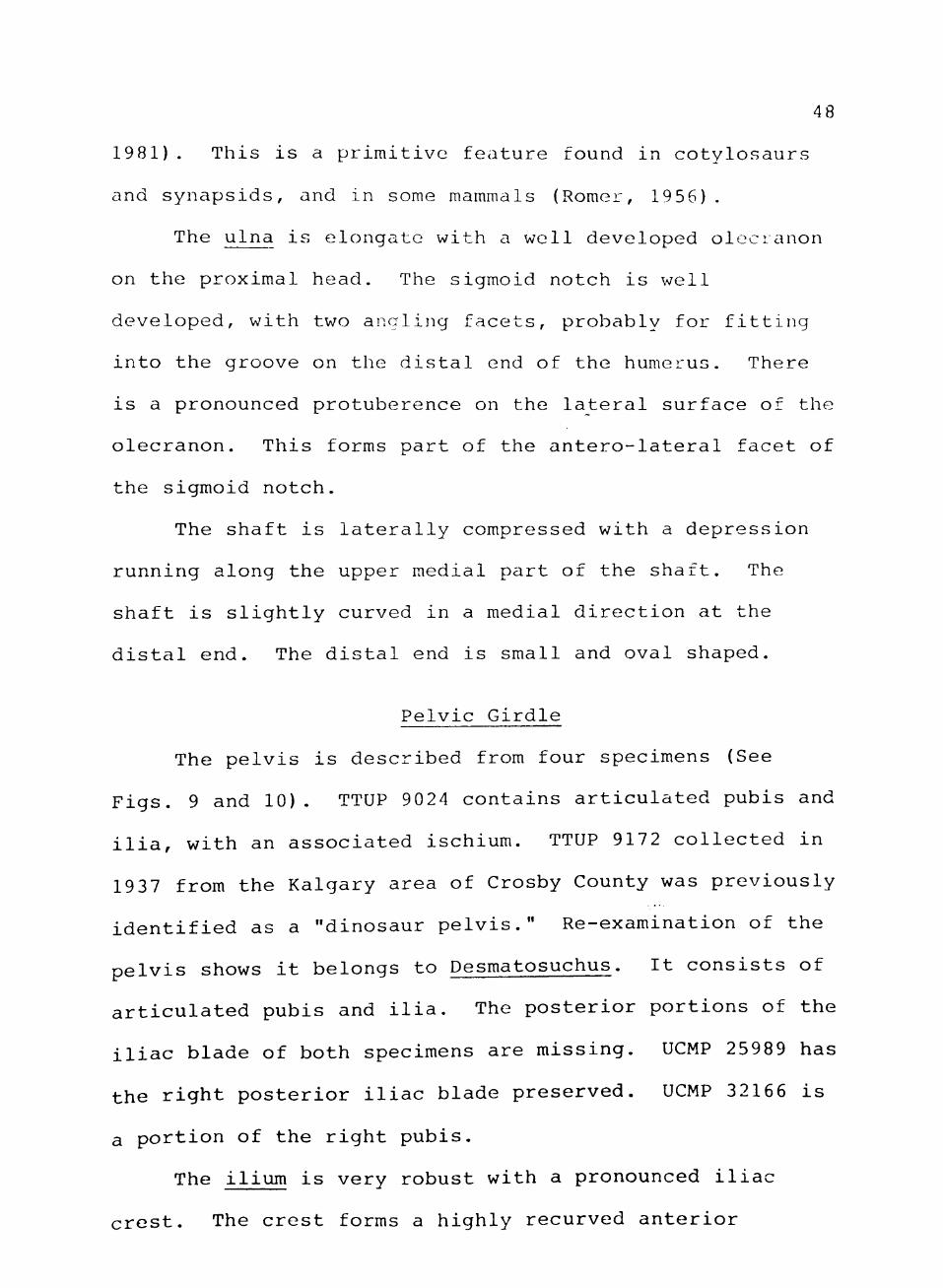
Pelvic Girdle
The pelvis is described from four specimens (See
Figs. 9 and 10). TTUP 9024 contains articulated pubis and
ilia, with an associated ischium. TTUP 9172 collected in
1937 from the Kalgary area of Crosby County was previously
identified as a "dinosaur pelvis." Re-examination of the
pelvis shows it belongs to Desmatosuchus. It consists of
articulated pubis and ilia. The posterior portions of the
iliac blade of both specimens are missing. UCMP 25989 has
the right posterior iliac blade preserved. UCMP 32166 is
a portion of the right pubis.
The ilium is very robust with a pronounced iliac
crest. The crest forms a highly recurved anterior
49
FIGURE 9. Composite restoration of pelvis, lateral view.
50
51
FIGURE 10. Composite restoration of pelvis, anterior view.
52
10 cm
53
projection. The ilium is laterally extended, but not
horizontally tilted. The acetabulum is closed, but deeply
excavated. This excavation is not only medially, but
dorsally also. This creates a deep supraacetabular crest,
which rests on top of the femur.
The ilium seems to be firmly fused with the pubis.
The ischium symphysis is definite, but weak.
The pubis forms the antero-distal portion of the
acetabulum. Posteriorly the pubis makes partial contact
with the ischium.
The vertical projection of the pubis is very
elongate. Laterally, the pubis forms a rod-like ridge.
Medially this ridge becomes plate-like. The pubies have a
strongly fused symphysis.
The lateral edges taper slightly inward on the distal
end. The pubis ends distally with a thickened foot-like
projection.
The ischium has a well defined concave area
antero-dorsally that forms part of the acetabulum. It
projects backward and downward. The ischium blade is
thickened dorsally, tapering slightly on the ventral
surface.
Femur
The femur is a long, robust bone with a relatively
straight shaft (See Fig. 11). The proximal and distal
ends are twisted about 35°-40° in relation to one another.
54
«
FIGURE 11. TTUP 9024, right femur in: (a) anterior view; (b) posterior view.

55
10 cm
56
The partially inturned head is expanded, but not full"
developed. The supraacetabular crest would have rested on
top of the femur. Laterally the head slopes downward fo a
horizontal depression at the greater trochanter. In the
niddle of the head, there is a pronounced anterior/
posterior expansion. Dorsally the head has a distinct
spherical shape.
The fourth trochanter appears about one-third of the
way down the shaft, on the posterior surface. It appears
as a large, mound-like mass that encompasses over
three-fourths of the width of the shaft.
The distal head is flared laterally and medially,
dominated by tv/o prominent condyles. The outer condyle is
a prominent termination of a rounded, outer ridge, that
runs the length of the femur. There is a broad shelf
lateral to the condyle. The inner condyle is less
prominent, but broader. There is a shallow, short
inter-condyler groove between the condyles.
Dermal Armor
Some dermal armor of TTUP 9023 and 9024 is preserved.
The first seven paramedian series are preserved in TTUP
9024, along with the second, fourth, and fifth lateral
spines. The armor of the TTUP 9024 differs slightly from
previously described Desmatosuchus armor (Case, 1922; Long
and Ballew 1985).
57
The armor forms a narrow carapace with two rows of
paramedian scutes bordered by two rows of lateral scutes.
The paramedians have a thin anterior laminae. This
laminae is overlapped by the preceding row. The dorsal
surface is characterized ly closely spaced, deep pits in a
random pattern. There is a prominent eminence in the
center of each scute that points antero-medially.
Posteriorly the scute tapers into a thin ridge. The
medial surface is vertically flat with a complex tongue
and groove articulation. The lateral surface is concave,
with a tongue and groove articulation.
The lateral scutes are characterized by well-
developed "horns" or spines. These spines are directed
laterally and dorso-posteriorly. They increase in size
from row one to row five, and then decrease in size in the
following rov/s. The fifth horn is large (28 cm. est.),
rounded, and recurved. TTUP 9024 differs from other
previously described Desmatosuchus specimens in the large
size of the fourth horn. Other specimens have a fourth
horn that is about 45% of the fifth horn. TTUP 9024 has a
fourth horn that is at least 60% of the fifth horn. It is
not known whether this is just an individual variation,
sexual dimorphism, or a different species.
CHAPTER VI
STANCE AND GAIT
The reconstruction of the pelvis and femur of
Desmatosuchus shov/s that it had an advanced locomotor
apparatus. Desmatosuchus, along with some other
thecodonts, shows some interesting adaptations in
achieving an erect posture. This contradicts previous
theories regarding the locomotor abilities of the
thecodonts.
Bakker (1981) and Charig (1972) listed three grades
of locomotion among the reptiles: sprawling,
semi-improved, and fully improved. The thecodonts were
generally classified as having a semi-improved posture.
The sem.i-improved condition consisted of: an imperforate
acetabulum, small anterior process of ilium, weak
supraacetabular crest, somewhat elongate pubis and
ischium, sigmoidal femur, poorly developed and slightly
inturned head of the femur, untwisted tibia, crocodiloid
tarsus with calcaneal tuber, and a pes with a plantigrade
posture.
The fully improved condition which was reserved for
the dinosaurs consisted of: a fenestrated acetabulum,
strongly developed anterior process of the ilium, strong
58

59
supraacetabular crest, very elongated pubis and ischium,
vertically positioned femur with a distinct, strongly
inturned head, twisted tibia, mesotarsal tarsus without
calcaneal tuber, and a pes with a digitgrade posture.
Recent authors (Bonaparte, 1981, 1984; Parrish 1984;
Chatterjee 1985a) have stated that some thecodonts did
achieve erect postures through different means than the
dinosaurs (Fig. 12). All thecodonts that achieved some
form of erect posture retained the primitive crocodiloid
tarsus and a pes with a plantigrade posture.
Bonaparte (1984) stated that the archosaurs took at
least two paths to an erect posture. The path taken by
the dinosaurs has already been stated. The thecodont
Fasolosuchus (Bonaparte, 1984) developed a unique method
of locomotion. The ilium took a m.ore horizontal position
with a low iliac blade. In this condition the acetabulum
was functionally deep, but the acetabulum was shallow when
the ilium is oriented vertically. This horizontal
position of the acetabulum created a graviportal relation
with the femur. The femur did not require an
anteromedially projecting head, or pronounced fourth and
lesser trochanters, as in the dinosaurs.
Postosuchus used another adaptation to achieve an
erect posture (Chatterjee, 1985a). The pelvis maintained
a vertical orientation and developed a projecting
60
FIGURE 12. Pelvic girdles and femora in posterior view of: (a) Postosuchus; (b) Fasolosuchus (after Bonaparte,
1981) ; (c) Desmatosuchus.
61
(c)

62
supraacetabular crest. As is Fasolosuchus, Postosuchus
retained a primitive foniur. The femur was sigmoidal with
little development of the head. The head was not
anteromedially projecting, so the acetabulum did not need
to be very deep. The weight v/as transferred through the
supracacetabular crest. Postosuchus also retained the
primitive ankle joint and pes.
Parrish (1984) mentions that the aetosaurs also
attained an erect posture, but he did not discuss how the
acetabulum was oriented. Examination of the literature
(Sawin, 1947; Casamiquela, 1960; Walker, 1961) shows that
most aetosaurs had a shallow acetabulum when oriented
vertically. To achieve an erect posture they would have
to have a more horizontally oriented ilium.. The fem.ur of
UCMP 27988 has less of an inturned head than TTUP 9024.
The UCMP specimen also lacks the dorsal notch on the
lateral end of the head as in TTUP 9024.
The TTUP specimens show a mosaic of characters in the
femur and acetabulum in attaining an erect posture. The
acetabulum was deep, unfenestrated, but the ilium has only
a slight if any horizontal inclination. The
supraacetabular crest has an exaggerated lateral
projection. Not only is the acetabulum deep, but the roof
of the supraacetabular crest is excavated to fit over the
spherical projection of the femoral head. The head is
inturned and the notch on the lateral end allows the femur
63
to clear the supraacetabular lip. The shaft is straight,
so that the proximal and distal expansions are in the same
plane; it has moderately developed fourth trochanter. The
pubis is elongate and tapers distally to prevent any
knocking with the femur. In articulation with the
acetabulum, the femur attains the vertical pose. The
expanded supraacetabular crest, and the deep acetabulum,
created a graviportal relation with the femur.
The stance and gait of some thecodonts were much more
advanced than previously thought, as they experimented
with various methods of achieving a fully improved
condition. It is quite possible that the dinosaurs could
have inherited their fully improved conditions from any
one of the thecodont adaptations.

CHAPTER VII
MODE OF LIFE
Due to its large size, advanced locomotor abilities,
specialized mastication, and abundance, Desmatosuichus v/as
the chief herbivore in the Dockum food v/eb. It:s dermal
armvOr would have offered some protection from its enemies.
A modern analogy with Desmatosuchus can be found v/ith
the armadillo. The armadillo possesses an edentulous
snout, and peg-like cheek teeth. It is also armored. The
Armadillo "roots" around for vegetation and insects.
Desmatosuchus possesses these characteristics plus a
turned-up, shovel-shaped premaxilla. Walker (1961)
suggested that the turned-up snout of Stagonolepis was
used for digging or "grubbing" amongst the soft
vegetation. Also, Walker (1961) suggested that the
premaxilla and dentary tips were covered with horny
sheaths. This would be expected if Desmatosuchus used the
premaxilla and dentary for food gathering. A horny sheath
would have protected the bone and acted as a cutting
device or for plucking or grasping objects.
The skull is essentially akinetic except for the
premaxilla. Walker (1961) theorized that the quadrate of
Stagonolepis was movable. The quadrate in Desmatosuchus
64
65
is definitely immobile. The only area of movement in the
skull was in the flexible joints between the premaxilla
and nasal, and the premaxilla and maxilla. This movement
probably assisted in digging or probing for food. Also
the two dentaries are firmly fixed by a strong median
symphysis.
The jaw articulation is reminiscent of ornithischian
dinosaurs. The articulation is below the tooth level.
This creates a "nut cracker" effect in which the
mandibular teeth can remain somewhat parallel to the
maxillary teeth and maintain an even biting force. There
is a close fit between the quadrate and articular, which
created an orthal jaw movement.
The teeth of Desmatosuchus were unspecialized. Only
one tooth was found in the Post quarry (TTUP 9025). The
teeth were presumably used for cutting or shearing soft
material.
The Dockum environment was characterized by numerous
lakes and rivers. Desmatosuchus scutes are often found in
association with parasuchids and metoposaurs. It is
possible that Desmatosuchus fed along these waterways,
digging in the soft mud for food. This is in contrast to
Stagonolepis, which seems to have lived in an arid
environment (Walker, 1961). The aetosaurs were able to
adapt to a variety of niches.
66
The dermal armor of Desmatosuchus provided it with
its only means of defense. There is a difference between
cervical lateral spines among different individuals; the
differences may indicate sexual dimorphism. TTUP 9024 has
a greatly enlarged fourth horn to go along with the
shoulder (fifth) horn. The fourth horn of Case's (1922)
specimen is greatly reduced.
Despite this protective armor, Desmatosuchus v/ould
have had difficulty in warding off an attack by
Postosuchus and other carnivores. Evidence from the Post
quarry indicates Postosuchus functioned in a family unit.
It would have been difficult for Desmatosuchus to ward off
a group of Postosuchus. Several isolated Desmatosuchus
scutes were found in a rich concentration of Postosuchus
skeletons. This could possibly indicate predation by
Postosuchus.
There is some evidence to indicate that Desmatosuchus
might have functioned also in a herd or family unit.
There is evidence of at least six individuals, including
one juvenile, in a forty square meter section. At least
14 individuals were found together in the Placerias quarry
near St. Johns, Arizona (Long and Ballew, 1985) .
CHAPTER VIII
TAXONOMY
Desmatosuchus is an aberrant form of aetosaur,
showing characteristics not known in other aetosaurs. A
comparison of the type skull of Desmatosuchus haplocerus
(TMM 41963) shows some minor differences in the braincase.
Unfortunately, D. haplocerus is incompletely known. The
type skull is missing the entire palatal region and lower
temporal region, as well as the premaxilla and mandibles.
This makes comparisons difficult. Until more comparative
material is found, the TTUP specimens will be designated
as D. haplocerus.
There has been some confusion over the taxonomy of D.
haplocerus. Gregory (1953) was able to sort this problem
out. Cope (1887) described some aetosaur material from
New Mexico which he named Episcoposaurus horridus. In
1891 he named E. haplocerus from the Dockum of Texas. It
was later determined that E. horridus was a junior synonym
of an earlier described genus Typothorax (Cope 1875).
This revision invalidated the name Episcoposaurus.
Case (1920) described a large aetosaur from Crosby
County, Texas, naming it Desmatosuchus spurensis. It was
later determined that Desmatosuchus and E. haplocerus were
67

68
synonymous (Case, 1929; Sawin, 1947). Since the type
specimen of Episcoposaurus was classified as Typothorax,
it became necessary to combine E. haplocerus with
Desmatosuchus spurensis. The name Desmatosuchus was
retained and haplocerus used instead of spurensis because
it was the original type specimen (Gregory, 1953).
Another problem over the years has arisen over the
relationship of D. haplocerus and Typothorax meadi
(Gregory, 1953; Elder, 1978; Murry, 1982). Some consider
the two genera to be synonyms. Gregory (1953) discussed
this problem and came to the conclusion that they were
separate genera. He based his conclusion on the dermal
armor. Elder and Murry contend that D. haplocerus and T.
meadi are synonyms. They also based some of their
arguments on the dermal armor. They feel that the
differences in the shoulder horn are due to sexual
dimorphism. This problem shows that armor cannot be
depended on for an accurate identification.
Other criteria such as the skull and post cranial
skeleton should be used. The skull shows some distinct
differences in the premaxilla and mandible. Examination
of T. meadi (TMM 31195-84B) reveals that T. meadi had
premaxillary teeth, a very large mandibular fenestra and a
difference in the dentary/angular suture. There is also a
difference in the dentary tooth count. Desmatosuchus has
5 to 6 dentary teeth while T. meadi has seven. It seems
69
that much of the postcranial material of T. meadi has been
incorrectly determined. On this basis a comparison of
postcranial material is difficult.
The fourth and fifth shoulder spines of TTUP 9024
differ from Case's type of Desmatosuchus. This is quite
possibly a result of sexual dimorphism, instead of the
criteria stated by Elder (1978) and Murry (1982). On the
basis of the armor and skull D. haplocerus and T. meadi
should remain separate.
CHAPTER IX
GENERA OF STAGONOLEPIDIDAE
Order Thecodontia
Suborder Pseudosuchia
Infraorder Aetosauria
Family Stagonolepididae Lydekker 1887
Armor-plated, herbivorous reptiles from the Late
Triassic Formations of North America, South America,
Europe, India, and China. Size range, 1.5 meters to
5 meters; Small skull relative to body size; tip of
snout edentolous; "slipper-shaped" jaw; reduced lower
temporal opening; 4 rows of ornate paramedian and
lateral scutes; ankle joint crocodile-normal
Genus Aetosauroides, Casamiquela 1960
A. scagliali, Casamiquela 1960 (Type)
=Arqentinosuchus bonapartei, Casamiquela 1960
Ischigualasto Formation (Late Triassic), Argentina;
Medium in size (2.5 meters); premaxilla with
teeth; teeth sharp, conical; ulna with no
olecranon; armor lacks lateral spines.
Genus Aetosaurus, 0. Fraas 1877
A. ferratus, O. Fraas 1877 (Type)
A. crassicuada, E. Fraas 1907
70

71
Stubensandstein (Late Triassic), Germany;
Small in size (1.5 meters); premaxilla with
teeth (4); no anterior expansion of premaxilla;
9-10 teeth on maxilla; 7 or 8 dentary teeth;
ulna olecranon slightly developed; narrow
carapace; no lateral spines.
Genus Calyptosuchus, Long and Ballew 1985
C. wellesi. Long and Ballev/ 1985
Dockum and Chinle Formations (Late Triassic), U.S.A.;
Known only from armor; carapace narrow; lateral
spines; no shoulder horns; pitting in armor
different from Desmatosuchus.
Genus Desmatosuchus, Case 1920
D. haplocerus, Cope 1892 (Type)
=Episcopesaurus haplocerus. Cope 18 92
=Desmatosuchus spurensis, Case 1920
Dockum and Chinle formations (Late Triassic), U.S.A.;
Large size (5 meters); premaxilla edentolous; 10
maxillary teeth; 5 to 6 dentary teeth; ulna
olecranon well developed; pelvis with deep
acetabulum, femur straight, with developed head;
carapace narrow; lateral spines with well
developed shoulder horns.
72
Genus Neoaetosauroides, Bonaparte 1969
N. rmgaeus, Bonaparte 1969 (Type)
Los Colorados Formation (Late Triassic), Argentina;
Size medium (2.5 meters?); premaxilla with 4
teeth; 6? maxillary teeth; 7 dentary teeth; jaw
very short; femur fairly straight with slightly
developed head; ulna with slightly developed
ulna; carapace narrow; no lateral spines
Genus Paratypothorax, Long and Ballew 1985
^. andressi, Long and Ballew 1985 (Type)
Stubensandstein (Late Triassic), Germany; Dockum and
Chinle (Late Triassic), U.S.A.;
Known only from armor, very wide carapace, with
small lateral horns.
Genus Stagonolepis, Agassiz 1844
S. robertsoni, Agassiz 1844 (Type)
Lossiemouth Bed (Late Triassic), Great Britain;
Medium size (2.5 meters) premaxilla with
anterior expansion; 4 to 5 premaxillary teeth;
11 to 12 maxillary teeth; dentary teeth 9 to 10;
slightly developed olecranon ulna; femur
slightly sigmoid; femoral head underdeveloped;
carapace narrow; very small lateral keels.
Genus Stegomus, Marsh 1896
S. arcuatus. Marsh 1896 (Type)
Newark Group (Late Triassic), U.S.A.;
73
Moderate size (2.5 meters); only armor is known;
narrov/ carapace; no lateral spines.
Genus Typothorax, Cope 1875
^Episcoposaurus, Cope 18 87
T. coccinarium. Cope 1875 (Type)
=Episcoposaurus horridus, Cope 18 87
Dockum and Chinle Formation (Late Triassic), U.S.A.;
Moderate in size (2 to 3 meters) premaxillary
teeth 4?; maxilla with 9 teeth; dentary with 7
teeth; olecranon moderately developed, pelvis
with fairly deep acetabulum; carapace very wide;
lateral spines.
CHAPTER X
EVOLUTION AND RELATIONSHIPS
There is no fossil evidence to link the aetosaurs
with earlier thecodonts. Their origins are obscure. The
aetosaurs have been classified as Pseudosuchians on the
basis of their tarsal anatomy (Chatterjee, 1982). All
aetosaurs (where the ankle joint is known) possessed the
CN (Crocodile-normal) joint with a well-developed
calcaneal tuber. This makes the aetosaurs a sister group
of the rauisuchids and parasuchids. The braincase also
shows strong similarities with the rauisuchids, and
parasuchids.
The aetosaurs were apparently a "dead-end" lineage.
It is interesting to note the similarities with the upper
Cretaceous ankylosaurs. They have similar adaptations in
the skull such as an edentolous snout, and the tendency to
close off certain temporal openings. There is an uncanny
resemblance in the forelimbs of the two groups. The
aetosaurs and ankylosaurs are the only archosaurs to
possess well-developed olecranon processes on the ulna
(Romer, 1956; Coombs, 1978). It is interesting to note
that the ankylosaurs were the only dinosaurs with an
imperforate acetabulum. They are also both armor-plated,
74
75
with some ankylosaurs possessing lateral spines. This is
a good example of convergent evolution. They are not
directly related, but may share common ancestr\'.
The relationships between the aetosaurs is uncertain,
mostly due to a lack of comparative material in some
species. It is possible to place them into three
loose-knit categories. One category consists of
Aetosuarus, Aetosauroides, and Stagonolepis. They seem to
possess the most primitive features in the skull, pelvis,
and armor. The second category could be the spinaceous
aetosaurs Desmatosuchus, and Calyptosuchus, both with
well-developed lateral spines. The third group could be
the wide bodies Typothorax and Paratypothorax. The
carapace width is extremely exaggerated.
As stated earlier, aetosaurs are good examples of
explosive evolution. They appeared suddenly at the start
of the Late Triassic and were extinct by the close of the
Triassic. Why did they become extinct? They had
protective armor and a relatively advanced locomotor
apparatus. Despite these attributes they were probably
victims of competitive exclusion and predation.
Evidence shows that the fabrosaurid dinosaurs
appeared during the uppermost Triassic and were
contemporaneous with the aetosaurs during that time
(Chatterjee, 1984). The fabrosaurs were fast, bipedal
dinosaurs which could outrun their enemies. Despite the
76
locomotor abilities of Desmatosuchus, its armor and bulky
size surely slowed it dov/n. Later herbivores developed
offensive weapons to ward off attack. The Uppermost
Triassic/Lov/er Jurassic plateosaurs possessed long, sharp
claws. The stegosaurs possessed spiked or clubbed tails.
The cerotopsids developed prominent horns protruding from
their skulls. Since the aetosaurs possessed no offensive
weapons, they could not fight back when attacked.
CHAPTER XI
CONCLUSIONS
The aetosaurs were a v/idelv distributed group of Late
Triassic thecodonts, recorded from North and South
America, Europe, India, and possibly China. Here I
described new material of an aetosaur, Desmatosuchus
haplocerus, from the Dockum Formation of West Texas.
The Dockum should be given a formation status, with
the new upper unit, the Cooper Creek Member, designated as
Norian in age. This is based on the new faunal assemblage
discovered near Post, Texas. Among the tetrapods from
Post are ictidosaurs, fabrosaurid dinosaurs, and an
advanced poposaur, Postosuchus. These groups of reptiles
are known from uppermost Triassic and Lower Jurassic
Formations from Europe and the Southern Hemisphere. This
is the first known occurrence of an aetosaur with such an
advanced faunal assemblage.
The discovery of well preserved skeletal material of
Desmatosuchus provides the first detailed examination of
the Desmatosuchus skull, forelim.b, and hindlimb. The
skull shows some features previously unknown in
Desmatosuchus and the family Stagonolepididae. The
premaxilla is entirely edentulous, the mandibular fenestra
77
78
is reduced, and there is a further reduction in the lower
temporal opening.
The forelimb and hindlimb also shov/ some advanced
characters. The ulna has a highly developed olecranon
process with a well developed sigmoid notch. The humerus
is very dinosaurian with a well developed delto-pectoral
crest.
The pelvis displays a very deep acetabulum on an
essentially vertical ilium, with an expanded
supraacetabular crest. The femur is vertically oriented
with a straight shaft, developed fourth trochanter, and a
highly inturned head with a dorso-lateral notch. These
features allowed Desmatosuchus to attain an erect posture.
The advancements of the Desmatosuchus locomotor
apparatus combined with the advanced characters of
rauisuchids and poposaurs indicate that some thecodonts as
a group were much more advanced than previously thought.
These locomotor advancements were near dinosaurian in
nature.
The Desmatosuchus specimens from Post show some
advanced traits and are slightly different in the
braincase, femur, and cervical armor than previously known
D. haplocerus material. Unfortunately, D. haplocerus is
poorly known; therefore, due to a lack of comparative
elements, the Post specimens shall be classified as D.
haplocerus.

79
Examination of the skull and arir.or indicates that D.
haplocerus and Typothorax meadi should remain s'-parato
taxa. The main differences are the edentolous premaxilla
of D. haplocerus compared to the tooth-bearing premaxilla
of T. meadi. There is a distinct difforenoo in the shapo
and size of the mandibular fenestra and the dentary tooth
count.
There are differences in the dermal armor between
different individuals of D. haplocerus. This can be
attributed to sexual dimorphism instead of the armor
differences between D. haplocerus and T. meadi.
The aetosaurs are good examples of explosive
evolution, appearing suddenly in the Late Triassic and
becoming extinct at the close of the Triassic. The
aetosaurs were the only known herbivorous thecodonts.
They developed some intersting adaptations for feeding
which parallel the ankylosaurs. The endentulous snout is
similar to the ornithischian dinosaurs. The premaxilla
had flexible joints with the nasal and maxilla allowing
some degree of movement for feeding. The teeth in
Desmatosuchus are reduced and could only be used as a
shearing mechanism. The jaw joint is below the level of
the tooth row. Ornithiscians used the same biting method
which allowed an even parallel biting surface with all of
the teeth.
80
Despite an improved locomotor apparatus and armor
protection, Desmatosuchus would have had difficulty in
warding off an attack by the larger Postosuchus or
rauisuchids.
From the Post quarry comes the first evidence of
aetosaurs occurring together with herbivorous dinosaurs.
Desmatosuchus could have become extinct as a result of
competitive exclusion, as the early fabrosaurs were fast,
bipedal herbivores which could outrun their enemies, and
were much more efficient at moving around in search of
food.
Despite their short time span and worldwide
distribution, there are still problems with using
aetosaurs as correlative tools because of their limited
distribution in time and space, and the relationships
among the aetosaurs is unclear.
Desmatosuchus was previously thought to be confined
to a Carnian Age, but the Post finds indicate that they
survived into the Norian. More work needs to be done on
the aetosaurs of the Chinle and Dockum in particular to
gain a better understanding of the stratigraphic
relationships among the different North American
aetosaurs.

LITERATURE CITED
Bakker, R. T., 1981. Dinosaur physiologv and the origin of the mammals. Evolution 25, 636-658.
Bonaparte, J. F., 1981. Descripcion de Fasolasuchus tenax y su Significado en la sistematica v evoluoion de Tos" thecodondia. Rev. Mus. Argentina Cien. natural. Bernar. Riva. 3, 5 5-101.
1982. Classification of the thecodonts. Geobios, memoire special 6, 99-112.
1984. Locomotion in rauisuchid thecodonts. Journal of Vert. Paleontology 3(4) 210-218.
Casamiquela, R. M. , 1960. Notica preliminar Sabre dos nuevos estagonolepoideos Argenitinos. Ameghiniana 2(1) 3-9.
Case, E. C , 1920. Preliminary description of a new suborder of phytosaurian reptiles with a description of a new species of Phytosaurus. Journal of Geologv, 28:524-535.
, 1922, New reptiles and stegocephalians from the Upper Triassic of Western Texas. Carnegie Inst. Wash., Pub. 321:7-84.
, 1929. Description of the skull of a new form of phytosaur, with notes on the characters of described North American phytosaurs. Univ. Mich. Studies, Mem. Mus. Paleont., 2:1-56.
Charig, A. J., 1972. The evolution of the archosaur pelvis and hindlimb: an explanation in functional terras. In K. A. Joysey and T. S. Kemp (eds.). Studies in Vertebrate Evolution, 121-155. Oliver and Boyd, Edinburgh.
Chatterjee, S., 1978. A primative parasuchid (phytosaur) reptile from the upper Triassic Maleri Formation of India. Paleontology, 21 (1) : 83-127.
81
82
, 1982. Phylogeny and classification of thecodontian reptiles. Nature, Lond., 295, 317-320.
, 1983. An ictidosaur fossil from North America. Science, Wash., 220, 1151-1153.
, 1984. A new ornithischian dinosaur from the Triassic of North America. Naturwissenschaften 71, S. 630.
, 1985a. Postosuchus, a new thecodontian reptile from the Triassic of Texas and the origin of tyrannosaurs. Phil. Trans. R. Soc. Lond. B, 309, 395-460.
, 1985b. The Late Triassic Dockum vertebrates: their stratigraphic and paleobiogeographic significance. (In Press) In: Padian, K. (ed.). The beginning of the age of Dinosaurs. Cambridge University Press.
Colbert, E. H. and J. T. Gregory, 1957. Correlation of Continental Triassic sediments by vertebrate fossils. Bull. Geol. Soc. Amer., 68:1456-1467.
Coombs, W. P., 1978. The families of the ornithischian dinosaur order Ankylosauria. Palaeontology 21(1):143-170.
Cope, E. D., 1875. The geology of New Mexico. Acad. Nat. Sci. Philadelphia Proc., pp. 263-267.
Cope, E. D., 1887. A contribution to the history of the vertebrata of the Tias of North America. Am. Philos. S o c , Proc, 24:209-228.
Cummins, W. F., 1890. The Permian of Texas units overlying beds. Texas Geol. Surv. 1st. Ann. Report. 183-197.
Elder, Z. L., 1978. Paleontology and Paleoecology of the Dockum Group, upper Triassic, Howard County, Texas. M.A. Thesis, University of Texas at Austin.
Gould, C. N., 1907. The geology and water resources of the western portion of the Panhandle of Texas. U.S. Geol. Surv. water supply paper. 191:1-70.
Gregory, J. T., 1953. Typothorax and Desmatosuchus. Postilla, 16:1-27.

83
Gregory, J. T., 1957. Significance of fossil vertebrates for correlation of Late Triassic continental deposits of North America. 20th Internat. Geol. Congr. Seccion I-El Mesozioco del hemisferio occidental y Sus Correlaciones Mundiales, 1956, 7-25.
Long, R. A., and K. L. Ballew, 1985. Aetosaur dermal armor from the Late Triassic of Southwestern North America, with special reference to material from the Chinle Formation of Petrified Forest National Park. Mus. N. Arizona Bull. 54:45-68.
McGowen, J. H., G. E. Granata, and S. J. Seni, 1983. Depositional Setting of the Triassic Dockum Group, Texas Panhandle and Eastern New Mexico, pp. 13-38. In Reynolds, M. W. and E. D. Dolly (eds.) Mesozoic Biostratigraphy of the West-Central United States. Soc Econ. Paleon. Min. Denver.
Murry, P., 1982. Biostratigraphy and paleoecology of the Dockum Group (Triassic) of Texas. Ph.D. Dissertation. Southern Methodist University. 1-459.
Parrish, J. M., 1984. Locomotor grades in the Thecodontia. In: Reif, W. E., and F. Westphal (eds.). Third Symposium on Mesozoic Terrestrial Ecosystems, Short Papers. Tubingen (Attempto Verlag).
Reedside, J. B., P. C. Applin, E. H. Colbert, J. T. Gregory, H. D. Hadley, B. Kummel, P. J. Lewis, J. D. Love, M. MaldonadoKoerdel, E. D. McKee, D. B. McLaughlin, S. W. Muller, J. A. Reinemund, J. Rodgers, J. Sanders, N. J. Silberling, and K. Waage, 1957. Correlation of the Triassic Formations of North America exclusive of Canada. Bull. Geol. Soc. Araer. 68:1451-1514.
Romer, A. D., 1956. Osteology of the Reptiles. University of Chicago Press.
Sawin, H. J., 1947. The pseudosuchian reptile Typothorax meadi. Jour. Paleont. 21:201-238.
Shelton. S. Y., 1984. Parasuchicd reptiles from the Triassic Dockum Group of West Texas. M.A. Thesis, Texas Tech University.
Walker, A. D., 1961. Triassic reptiles from the Elgin area: Stagonolepis, Dasygnathus and their allies. Phil. Trans. R. Soc Lond. B. 244:103-204.
