the triple helix of collagens – an ancient protein ...triple helix, a unique structure that is a...
TRANSCRIPT

OPINION
The triple helix of collagens – an ancient protein structure thatenabled animal multicellularity and tissue evolutionAaron L. Fidler1,2,*, Sergei P. Boudko1,2,*, Antonis Rokas3 and Billy G. Hudson1,2,4,5,6,7,8,9,‡
ABSTRACTThe cellular microenvironment, characterized by an extracellularmatrix (ECM), played an essential role in the transition fromunicellularity to multicellularity in animals (metazoans), and in thesubsequent evolution of diverse animal tissues and organs. A majorECM component are members of the collagen superfamily –
comprising 28 types in vertebrates – that exist in diversesupramolecular assemblies ranging from networks to fibrils. Eachassembly is characterized by a hallmark feature, a protein structurecalled a triple helix. A current gap in knowledge is understanding themechanisms of how the triple helix encodes and utilizes information inbuilding scaffolds on the outside of cells. Type IV collagen, recentlyrevealed as the evolutionarily most ancient member of the collagensuperfamily, serves as an archetype for a fresh view of fundamentalstructural features of a triple helix that underlie the diversity ofbiological activities of collagens. In this Opinion, we argue that thetriple helix is a protein structure of fundamental importance in buildingthe extracellular matrix, which enabled animal multicellularity andtissue evolution.
KEYWORDS: Cell biology, Collagen, Evolution, Extracellular matrix,Multicellularity, Triple helix
IntroductionThe extracellular matrix (ECM) played an essential role during thetransition from unicellular organisms to multicellular animals(metazoans). The ECM comprises a basement membrane (BM)that underlies epithelia cells, and an interstitial matrix (IM) that ispositioned between cells in the intercellular spaces and undergoescontinuous controlled remodeling (Hynes, 2012; Bonnans et al.,2014; Nelson and Bissell, 2006; Inman et al., 2015). Yet, a majorgap in cell biology is to understand how cells generate and interactwith the ECM (Sherwood, 2015; Jayadev and Sherwood, 2017).The collagen superfamily of proteins is a major component
of ECMs, which – in vertebrates – comprises 28 types (I–XXVIII) that
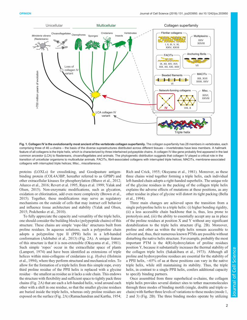
are derived from a total of 46 α-chains across the superfamily (Fig. 1)(Ricard-Blum, 2011; Kadler et al., 2007; Ricard-Blum and Ruggiero,2005). Invertebrates generally contain collagen IV, XV or XVIII,some fibrillar collagens, as well as some fibril-associated collagenswith interrupted triple helices (FACITs) (Fidler et al., 2014, 2017;Fahey andDegnan, 2010;Meyer andMoussian, 2009; Boot-Handfordand Tuckwell, 2003; Whittaker et al., 2006; Kadler et al., 2007).Among these collagens, type IV is the evolutionarily most ancient,based on recent studies of non-bilaterian animals (sponges,ctenophores, placozoans and cnidarians) and unicellular groups(Fidler et al., 2017; Grau-Bove et al., 2017) (Fig. 1).
Collagens are the most abundant protein in the human body(Kadler et al., 2007; Shoulders and Raines, 2009). They occur asdiverse supramolecular assemblies, ranging from networks to fibrils,and broadly function in structural, mechanical and organizationalroles that define tissue architecture and influence cellular behavior(Shoulders and Raines, 2009; Ricard-Blum, 2011; Ricard-Blum andRuggiero, 2005). Defects in collagens underlie the cause of almost 40human genetic diseases, affecting numerous organs and tissues inmillions of people worldwide (summarized in Table 1).
Disease pathogenesis typically involves genetic alterations of thetriple helix, a unique structure that is a hallmark feature common toall collagens. The triple helix bestows exceptional mechanicalresistance to tensile forces and a capacity to bind a plethora ofmacromolecules. Yet, there is a gap in our current knowledge inunderstanding the mechanisms of how a triple helix encodes andutilizes information in building supramolecular assemblies on theoutside of cells. Here, we present collagen IV, the most ancient ofthe collagen superfamily, and argue that it is ideally suited to serveas an archetype for investigating and describing core functions of atriple helix.
The triple helix – assembly and structural features thatencode informationThe chemical structure of the triple helix was determined throughthe seminal work of structural biologists and chemists over the lastcentury (see Box 1 in supplementary material). Its unique structurebestows upon collagens an exceptional mechanical resistance totensile forces and a plethora of organizing information for buildingan ECM (Fig. 2). The triple helix presents all residues, exceptglycine (Gly), on its surface, which is the most economical androbust way to encode binding motifs of any protein structure.Moreover, the triple helix exhibits extensive post-translationalmodifications (PTMs), such as hydroxylation, glycosylation andphosphorylation, adding – in tandem – a secondary layer ofinformation in addition to its amino acid (aa) code (Yamauchi andShiiba, 2008). These PTMs confer even more diversity with tissue-specific and disease-specific variations, even amongst identicaltypes of collagen (Pokidysheva et al., 2013). Furthermore,additional collagen modifications are mediated by specificextracellular enzymes, such as peroxidasin and lysyl oxidases-like
1Department of Medicine, Division of Nephrology and Hypertension, VanderbiltUniversity Medical Center, Nashville, TN, 37232, USA. 2Center for Matrix Biology,Vanderbilt University Medical Center, Nashville, TN, 37232, USA. 3Department ofBiological Sciences, Vanderbilt University Medical Center, Nashville, TN, 37232,USA. 4Department of Pathology, Microbiology, and Immunology, VanderbiltUniversity Medical Center, Nashville, TN, 37232, USA. 5Department of MedicalEducation and Administration, Vanderbilt University Medical Center, Nashville, TN,37232, USA. 6Department of Cell and Developmental Biology, Vanderbilt UniversityMedical Center, Nashville, TN, 37232, USA. 7Department of Biochemistry,Vanderbilt University Medical Center, Nashville, TN, 37232, USA. 8Vanderbilt-Ingram Cancer Center, Vanderbilt University Medical Center, Nashville, TN, 37232,USA. 9Vanderbilt Institute of Chemical Biology, Vanderbilt University MedicalCenter, Nashville, TN, 37232, USA.*These authors contributed equally to this work
‡Author for correspondence ([email protected])
A.L.F., 0000-0002-2519-8864; A.R., 0000-0002-7248-6551; B.G.H., 0000-0002-5420-4100
1
© 2018. Published by The Company of Biologists Ltd | Journal of Cell Science (2018) 131, jcs203950. doi:10.1242/jcs.203950
Journal
ofCe
llScience
proteins (LOXLs) for crosslinking, and Goodpasture antigen-binding protein (COL4A3BP, hereafter referred to as GPBP) andother extracellular kinases for phosphorylation (Bhave et al., 2012;Añazco et al., 2016; Revert et al. 1995, Raya et al. 1999; Yalak andOlsen, 2015). Non-enzymatic modifications, such as glycation,oxidation or chlorination, add even more complexity (Brown et al.,2015). Together, these modifications may serve as regulatorymechanisms on the outside of cells that may instruct cell behaviorand influence tissue architecture and stability (Yalak and Olsen,2015; Pedchenko et al., 2010).To fully appreciate the capacity and versatility of the triple helix,
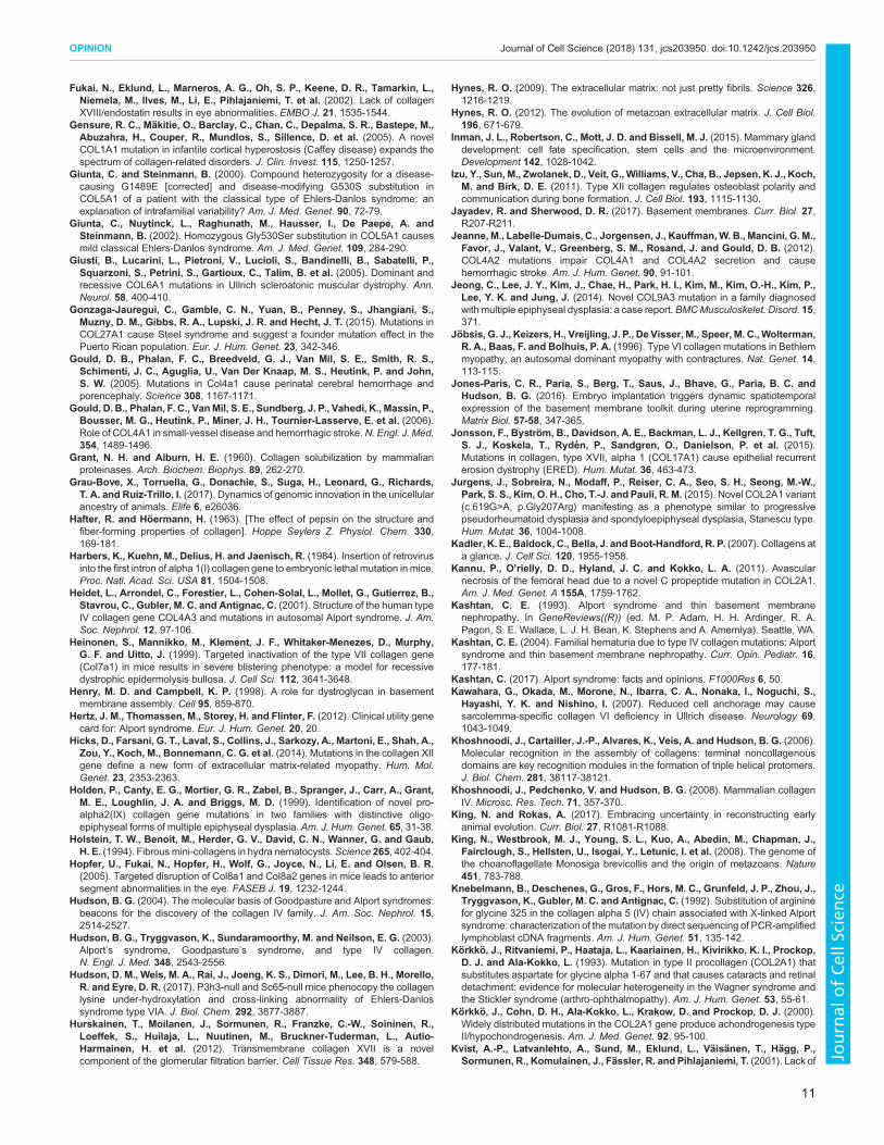
one should consider the building blocks (polypeptide chains) of thisstructure. These chains are simple ‘rope-like’ structures built fromproline residues. In aqueous solutions, such a polyproline chainadopts a polyproline type II (PPII) helix in a left-handedconformation (Adzhubei et al., 2013) (Fig. 2A). A unique featureof this structure is that it is non-extensible (Okuyama et al., 1981).Such simple ‘ropes’ occur in the extracellular space of plants(Lamport, 1974) and have been identified as extensions of triplehelices within mini-collagens of cnidarians (e.g. Hydra) (Holsteinet al., 1994), where they perform structural and mechanical roles. Toallow for the formation of a triple helix from this simple rope, everythird proline residue of the PPII helix is replaced with a glycineresidue – the smallest aa residue as it lacks a side chain. This endowsthe structurewith flexibility and sufficient space to tightly pack threechains (Fig. 2A) that are each a left-handed helix, wind around eachother with a shift in one residue, so that the smaller glycine residuesare buried inside the triple helix, whereas only proline residues areexposed on the surface (Fig. 2A) (Ramachandran and Kartha, 1954;
Rich and Crick, 1955; Okuyama et al., 1981). Moreover, as thesethree chains wind together forming a triple helix, each individualleft-handed chain adopts a right-handed superhelix. The unique roleof the glycine residues in the packing of the collagen triple helixexplains the adverse effects of mutations at these positions, as anyother residue in place of glycine will distort its tight packing (Bellaet al., 1994).
Three main changes are achieved upon the transition from asingle polyproline helix to a triple helix: (i) higher bending rigidity,(ii) a less accessible chain backbone that is, thus, less prone toproteolysis and, (iii) the ability to essentially accept any aa in placeof the proline residues at position X and Y without any significantdestabilization to the triple helix structure (Fig. 2B). Moreover,proline and other aa within the triple helix remain accessible tosolvent and, thus, their numerous known PTMs are possible withoutdisturbing the native helix structure. For example, probably the mostimportant PTM is the 4(R)-hydroxylation of proline residuesposition Y, because it substantially increases the thermal stability ofthe collagen triple helix (Sakakibara et al., 1973). Although allproline and hydroxyproline residues are essential for the stability ofa PPII helix, ∼65% of aa at these positions can vary in the nativetriple helix, while still maintaining its stability. Thus, the triplehelix, in contrast to a single PPII helix, confers additional capacityto specify binding partners.
Once assembled from three superhelical α-chains, the collagentriple helix provides several distinct sites to tether macromoleculesthrough three modes of binding motifs (single, double and triple α-chain) with further three levels of variations to these modes (mode 1,2 and 3) (Fig. 2B). The three binding modes operate by utilizing
Collagen superfamily
Triple helixT i l h li
VIII, X
Network-forming collagens
IX, XII, XIV, XVI,XIX, XX, XXI, XXII
FACITs
VII
Anchoring fibrils
I, II, III, V, XI,XXIV, XXVII
Fibrillar collagens
VI
Beaded filaments
XXVI,XXVIII
Misc.
MACITsXIII, XVII
XXIII, XXV
MultiplexinsXXV
XXVIII
IV
VertebratesInsects
CnidariansSpongesCtenophoresChoanoflagellates
>700
mill
ion
year
s of
evo
lutio
n
Unicellular Multicellular
Ministeria vibrans
Loss ofcollagen IVvariant
LCA collagen IVvariant
(filastereans)
Fig. 1. Collagen IV is the evolutionarilymost ancient of the vertebrate collagen superfamily. The collagen superfamily has 28members in vertebrates, eachcomprising three of 46 α-chains – the basis of the diverse suprastructures distributed across different tissues – invertebrates have less members. A hallmarkfeature of all collagens is the triple helix, which is characterized by three intertwined polypeptide chains. A collagen IV-like gene probably first appeared in the lastcommon ancestor (LCA) to filastereans, choanoflagellates and animals. The phylogenetic distribution suggests that collagen IV played a critical role in thetransition of unicellular organisms to multicellular animals. FACITs, fibril-associated collagens with interrupted triple helices; MACITs, membrane-associatedcollagens with interrupted triple helices; Misc., miscellaneous.
2
OPINION Journal of Cell Science (2018) 131, jcs203950. doi:10.1242/jcs.203950
Journal
ofCe
llScience

Table 1. Human genetic diseases/disorders due to mutation(s) in collagens and experimental collagen knockout studies
Collagentype Human genetic diseases/disorders due to mutation(s) in collagens Loss of protein
I (G) Osteogenesis imperfecta (Wenstrup et al., 1988; Vogel et al., 1987;Peltonen et al., 1980; Lamande et al., 1989; Labhard et al., 1988; Cohn et al.,1986; Baldwin et al., 1989)(N) Ehlers–Danlos syndrome (Wirtz et al., 1987; Malfait et al., 2006;Nuytinck et al., 2000)Caffey disease (Gensure et al., 2005) (for all, see http://www.le.ac.uk/ge/collagen/)
Embryonic lethality during organogenesis before E15, embryonicgrowth arrest at E11.5–E12 (Schnieke et al., 1983; Harbers et al.,1984)
II (G) Spondyloepiphyseal dysplasias (Lee et al., 1989; Tiller et al., 1990;Chan et al., 1993; Jurgens et al., 2015; Barat-Houari et al., 2016)(G) Spondyloepimetaphyseal dysplasias (Tiller et al., 1995; Sulko et al.,2005)(G) Achondrogenesis 2 (Vissing et al., 1989; Mortier et al., 1995, 2000;Körkkö et al., 2000; Chan et al., 1995; Barat-Houari et al., 2016)(G) Legg–Calvé–Perthes disease (Miyamoto et al., 2007)(G) Kniest dysplasia (Wilkin et al., 1994; Winterpacht et al., 1996)(G) Avascular necrosis of femoral head (Liu et al., 2005; Kannu et al.,2011)Platyspondylic lethal skeletal dysplasia, Torrance type (Nishimura et al.,2004; Zankl et al., 2005)Spondyloperipheral dysplasia (Zankl et al., 2005; Nishimura et al.,2004)(G) Stickler syndrome 1 (Richards et al., 2000, 2010; McAlinden et al.,2008; Körkkö et al., 1993; Barat-Houari et al., 2016)(N) Osteoarthritis with mild chondrodysplasia (Ala-Kokko et al., 1990)(N) Czech dysplasia (Williams et al., 1993; Tzschach et al., 2008; Matsuiet al., 2009; Barat-Houari et al., 2016)
Perinatal lethality, abnormal skeleton, abnormal cranium, abnormalrespiratory system (Li et al., 1995a).IIA isoform-specific knockout: prenatal lethality, holoprosencephalloss of head tissues and mid-facial structures (Leung et al., 2010).
III (G) Ehlers–Danlos syndrome, vascular types (Narcisi et al., 1994; Trompet al., 1989a,b; Pepin et al., 1993) (http://www.le.ac.uk/ge/collagen/)
Embryonic lethality before E9.5, pre- to mid-gestation lethality(Smith et al., 2011)
IV (α1α1α2) (G) Cerebral small vessel disease (Gould et al., 2006; Shah et al., 2012;Sibon et al., 2007)(G) Hereditary angiopathy with nephropathy aneurysms and musclecramps (HANAC) (Plaisier et al., 2007, 2010)(G) Porencephaly 1 (Breedveld et al., 2006; de Vries et al., 2009; Gouldet al., 2005; Yoneda et al., 2013)(G) Porencephaly 2 (Yoneda et al., 2012)(G) Tortuosity of retinal arteries (Zenteno et al., 2014)(G) Schizencephaly (Yoneda et al., 2013)(N) Intracerebral hemorrhage (Jeanne et al., 2012; Weng et al., 2012)
Embryonic lethality at E10.5–E11.5 (Poschl et al., 2004)
IV (α3α4α5) (G) Alport syndrome (Barker et al., 1990; Boye et al., 1998; Heidet et al.,2001; Kashtan, 2017; Knebelmann et al., 1992; Mehta and Jim, 2017;Mochizuki et al., 1994; Nagel et al., 2005; Renieri et al., 1992; Zhou et al.,1991; Van Der Loop et al., 2000; Zhou et al., 1992) (http://databases.lovd.nl/shared/genes/COL4A5)(G) Thin basement membrane nephropathy (TBMD) (Badenas et al.,2002; Buzza et al., 2003; Lemmink et al., 1996; Kashtan, 2004)
Decreased survival rate, premature death, proteinuria,glomerulosclerosis, glomerulonephritis, renal interstitial fibrosis,hearing loss (Cosgrove et al., 1996, 1998; Rheault et al., 2004;LeBleu et al., 2010)
IV (α5α5α6) (G) X-linked deafness 6 (Rost et al., 2014) No abnormal phenotype detected (Fox et al., 2007)
V (G) Ehlers–Danlos syndrome (Watanabe et al., 2016; Symoens et al.,2009; Richards et al., 1998; Michalickova et al., 1998; Malfait et al., 2005;Giunta and Steinmann, 2000; Giunta et al., 2002; De Paepe et al., 1997)(http://www.le.ac.uk/ge/collagen/)
Embryonic lethality at E10–E12 (Wenstrup et al., 2004; Park et al.,2015)
VI (G)Bethlemmyopathy 1 (Scacheri et al., 2002; Pepe et al., 1999; Pan et al.,1998; Lucioli et al., 2005; Lampe et al., 2005; Jöbsis et al., 1996; Baker et al.,2007)(G) Ullrich congenital muscular dystrophy 1 (Lampe et al., 2005;Kawahara et al., 2007; Giusti et al., 2005; Baker et al., 2005)Dystonia 27 (Zech et al., 2015)
Abnormalities in morphology of l muscle, impaired skeletal musclecontractility, myopathy (Bonaldo et al., 1998)
VII (G)Epidermolysis bullosa dystrophica,multiple types (Shinkuma, 2015;Fine et al., 1989; Christiano et al., 1993, 1994, 1995, 1996; Woodley et al.,2004)
Postnatal lethality within the first week from complications ofblistering (Heinonen et al., 1999)
VIII (N) Corneal dystrophies (Biswas et al., 2001) Dysgenesis of the anterior segment of the eye (Hopfer et al., 2005)
IX (G)Multiple epiphyseal dysplasia 2, 3 and 6 (Paassilta et al., 1999; Jeonget al., 2014; Holden et al., 1999; Czarny-Ratajczak et al., 2001)Stickler Syndrome 4 and 5 (Van Camp et al., 2006; Baker et al., 2011)(N) Intervertebral disc disease (Paassilta et al., 2001; Annunen et al.,1999b)
Osteoarthritis, abnormalities in morphology of bone and cartilage,abnormal bone healing, impaired hearing (Fassler et al., 1994;Asamura et al., 2005; Ting et al., 1999)
Continued
3
OPINION Journal of Cell Science (2018) 131, jcs203950. doi:10.1242/jcs.203950
Journal
ofCe
llScience

one, two or all three distinct chains of the triple helix in differentcombinations to directly bind a molecule (Fig. 2B; modes 1–3). Thethree levels of variation to these modes add additional diversity tobinding specificity on top of involving either one, two or threechains. The first level stems from the variation that is provided bythe ≤20 possible aa residues that can occupy the variable positionsX and Y of each tripeptide; this results in extensive structuralvariability across the three chains, as well as within each Gly-X-Ytripeptide of the same chain (Fig. 2B; level 1). The second levelconfers even more diversity because of the proclivity of collagens tobecome post-translationally modified and change their structure(Fig. 2B; level 2). The third level utilizes chain staggering thatoccurs during triple helix assembly between either two or threechains and enables new combinations of chains that can give rise toadditional binding motifs (Fig. 2B; level 3).In addition to these binding specificities, the triple helix also
possesses other structural features that underlie its biologicalfunction. It is non-stretchable along its longitudinal axis,
providing great tensile strength to withstand all physiologicalmechanical loads and stresses in our body. It also confersresistance to proteases – making collagens some of the most long-lived proteins – as well as to a wide range of pH values in order towithstand adverse conditions (Steven, 1965; Hafter andHöermann, 1963; Grant and Alburn, 1960; Drake et al., 1966;Uzawa et al., 1998; Pokidysheva et al., 2013; Hudson et al., 2017;Eyre et al., 2011). Furthermore, the triple helix exhibits variablelengths among collagen suprastructures, such as networks, fibrilsand filaments (Ricard-Blum, 2011). For example, the triple helixlength of fibrillar collagens results from multiple duplications ofexons that are either 54 or 45 base pairs in length and encode Gly-X-Y (Yamada et al., 1980; Exposito et al., 2010). Moreover, theterminal and internal incorporation of non-triple helical sequences(e.g. interruptions) in all collagen types extends their functionalcapacity (Ricard-Blum, 2011; Khoshnoodi et al., 2006).Collectively, the binding motifs (see above) together with thesefeatures of a collagen triple helix underlie the diversity of
Table 1. Continued
Collagentype Human genetic diseases/disorders due to mutation(s) in collagens Loss of protein
X Schmid metaphyseal chondrodysplasia (Wallis et al., 1994; McIntoshet al., 1994; Bateman et al., 2005)
Abnormal spleen morphology, abnormal hematopoietic system(Rosati et al., 1994)
XI (G) Stickler syndrome 2 (Annunen et al., 1999a; Richards et al., 1996,2010),Marshall syndrome (Annunen et al., 1999a)(G) Fibrochondrogenesis 1 (Tompson et al., 2010)(G)Otospondylomegaepiphyseal dysplasia (OSMED) (Melkoniemi et al.,2000; Pihlajamaa et al., 1998; Sirko-Osadsa et al., 1998; Vikkula et al., 1995)(G) Deafness (Chakchouk et al., 2015; Chen et al., 2005; McGuirt et al.,1999)Fibrochondrogenesis 2 (Tompson et al., 2012)
Neonatal lethality; abnormalities in cartilage of limbs, ribs, mandibleand trachea (Li et al., 1995b)
XII Ullrich congenital muscular dystrophy 2 (Zou et al., 2014)(G) Bethlem myopathy 2 (Zou et al., 2014; Hicks et al., 2014)
Increased perinatal lethality, skeletal abnormalities, decreased bodyweight, abnormal skeletal muscle morphology and physiology (Izuet al., 2011; Zou et al., 2014)
XIII* Congenital myasthenic syndrome type 19 (Logan et al., 2015) Deletion of cytosolic and transmembrane regions, abnormal skeletalmuscle morphology, muscle degeneration, myopathy, impairedexercise endurance, increased susceptibility to injury (Kvist et al.,2001)
XIV unknown Decreased tendon stiffness, decreased skin tensile strength(Ansorge et al., 2009)
XV unknown Abnormal muscle morphology, increased muscle regeneration,myopathy (Eklund et al., 2001)
XVI unknown unknown
XVII (G) Generalized atrophic benign epidermolysis bullosa (McGrath et al.,1996; Schumann et al., 1997; Tasanen et al., 2000a,b; Wu et al., 2002)Epithelial recurrent erosion dystrophy (Jonsson et al., 2015)
Significant postnatal lethality, sterility, postnatal growth retardation,blistering, abnormal renal glomerulus morphology (Hurskainenet al., 2012; Nishie et al., 2007)
XVIII Knobloch syndrome (Aldahmesh et al., 2013; Sertie et al., 2000) Eye abnormalities, intracranial hemorrhage, abnormalities inmorphology of brain, cranium and renal glomerulus; abnormal lipidmetabolism (Bishop et al., 2010; Fukai et al., 2002; Utriainen et al.,2004)
XIX unknown ∼95% postnatal lethality, dysphagia, abnormal esophagusmorphology, weight loss (Sumiyoshi et al., 2004)
XX to XXIV unknown unknown
XXV (G) Congenital cranial dysinnervation disorder (Shinwari et al., 2015) Neonatal lethality; abnormalities in morphology of motor neurons,skeletal muscles; respiratory failure; cyanosis (Tanaka et al., 2014)
XXVI unknown unknown
XXVII* (G) Steel syndrome (Gonzaga-Jauregui et al., 2015) 87-residue deletion of collagenous domain; neonatal lethality;chondrodysplasia; respiratory failure at birth; abnormalities inmorphology of lung and skeleton; decreased fetal size (Plumbet al., 2011)
XXVIII unknown unknown
G, Gly and non-Gly mutations reported in triple helix; N, only non-Gly mutations reported in triple helix; *deletion mutants for collagens XIII and XXVII
4
OPINION Journal of Cell Science (2018) 131, jcs203950. doi:10.1242/jcs.203950
Journal
ofCe
llScience

biological activities of the diverse supramolecular assemblies ofcollagens.
The roles of the ECM and collagen IV in the transition of theUrmetazoan to multicellular animalsThe last common ancestor to animals, the Urmetazoan, almostcertainly reproduced by gametogenesis, underwent gastrulationduring early development, had the ability for cells to differentiateboth during development and as stem cells, and comprised anepithelial layer of cells forming the body of the animal – features thatare still fundamental to extant animals (Richter and King, 2013;King and Rokas, 2017). Importantly, these cellular activitiesultimately required the invention of an ECM to provide asubstrate for attachment and signaling cues to regulate cellbehavior and function in tissue genesis and homeostasis (Abedinand King, 2010). The appearance of a specialized form of ECM, the
BM, coincided with the transition to multicellularity. The BMfunctions in several cellular activities, including migration,adhesion, delineation of apical–basal polarity and modulation ofdifferentiation during development (Petersen et al., 1992; Lukashevand Werb, 1998; Daley and Yamada, 2013; Hynes, 2009, 2012;Yurchenco, 2011; Ozbek et al., 2010; Henry and Campbell, 1998).
Importantly, understanding the makeup of BMs betweendifferent animals sheds light on the functions of proteins in theevolution of animal multicellularity and tissues. BMs are composedof numerous proteins, vary between animals. The BM of bilateriananimals (e.g. human, fly, C. elegans, sea urchin) is composed ofseveral proteins, including collagen IV, laminin, perlecan, nidogen,fibronectin, proteoglycans, peroxidasin, GPBP, and collagens XVand XVIII (Hynes, 2012; Yurchenco, 2011; Jayadev and Sherwood,2017). Potentially, there are many more components of the BM(Chew and Lennon, 2018). Among these, collagen IV is a major
GDPGSYGSPG
GPPGDPGFPGGLPGEAGIPG
A
B
Polyprolinetype-II helix
Pro
GlyPro
PPolyp
Pro
ProPro
-X-Y
-X-Y
Gly
Left-handedhelix
Le Right-handed superhelixFormed from left-handed helix
PTM
GDPGSYGSPG
GPPGDPGFPGGLPGEAGIPG
A
G
I
L
PV
F WY
DE R
H KST
CM
NQ
Three singleα-chains Binding modesNon-extensible
triple helix Variations
Gly replaces everythird residue
Non-extensiblehelix SuperhelixTriple helix
Building blocks
Collagen triple helix
Assembly ofthree chains
Pro residuescreaterigidity
Gly residuesimpartflexibility
Conformationalchange in each
chain
Simple glycinesallow chains to
tightly pack
Single-chainconformation
within triple helix
(Gly-X-Y)n
Right-handedtriple helix
Three superhelicesformed uponassembly of triple helix
Left-handed helices
Mod
e 1
Mod
e 2
Mod
e 3
One α-chain
Two α-chains
Three α-chains Chain stagger
PTM
Variable aa residues
Leve
l 2Le
vel 1
Leve
l 3
Fig. 2. Building a collagen triple helix and encoding information. (A) The building blocks (polypeptide chains) of the triple helix are left-handed polyprolinetype-II helices, i.e. non-extensible structures that have structural and mechanical roles in the ECM of plants and some animals. Replacement of every third proline(Pro) residue with a glycine Gly) residue results in increased flexibility. (B) Three left-handed superhelices wind together and pack tightly owing to these Glyresidues, thereby forming a right-handed triple helix. Once associated, the non-extensibility of the structure is restored, and the combination of three chainsresults in three bindingmodes (one, two or three chains) with three levels of variation to thesemodes, i.e. level 1: 20 variable aa residues, level 2: post-translationalmodifications (PTMs) and, level 3: chain stagger. Together, these specify the numerous possible binding motifs that are positioned along the length of the triplehelix of any of the 28 collagen types to bind various macromolecules.
5
OPINION Journal of Cell Science (2018) 131, jcs203950. doi:10.1242/jcs.203950
Journal
ofCe
llScience

component that is conserved among animal phyla (Fidler et al.,2014). In a recent study, we described collagen IV at the origins ofanimal multicellularity and in tissue evolution, as revealed by closeexamination of sponges, ctenophores and other non-bilaterallysymmetrical animals (Fidler et al., 2017). Ctenophores and spongeshave been established as the two most likely candidates to be thesister-groups to the rest of animals, based on phylogenetic analysesof genomic and transcriptomic data and cell-type evolution (Ryanet al., 2013; Moroz et al., 2014; Pisani et al., 2015; Whelan et al.,2015; Telford et al., 2016; King and Rokas, 2017; Feuda et al.,2017). Our genomic analysis of the extracellular matrix componentswithin the ctenophores and Homoscleromorph sponges revealed aBM ‘toolkit’ consisting of just collagen IV and laminin (Fidler et al.,2017). However, the demosponges, a sponge class, lack bothlaminin and classic collagen IV but do contain spongins, which areshort collagen IV variants (Exposito et al., 1991; Aouacheria et al.,2006; Fidler et al., 2017). The order in which these variants andcollagen IV first appeared is still unknown. Despite containing fewerBM proteins than bilaterians, many sponges and ctenophores formclassic BMs (Fidler et al., 2017; Boute et al., 1996; Leys et al., 2009).Importantly, comparison of BM components of animals with
those of unicellular lineages is key to determining their importanceduring the transition to multicellularity. Within choanoflagellates,
the closest relatives of animals, no complete ECM proteins exist;yet, domains that are characteristic of laminins and shortcollagenous Gly-X-Y repeats are present (King et al., 2008;Fahey and Degnan, 2012; Fidler et al., 2017). Interestingly, a recentstudy reported the discovery of a collagen IV-like gene in thefilasterean, Ministeria vibrans, a unicellular lineage that divergedprior to choanoflagellates and animals (Grau-Bove et al., 2017).This finding indicates that collagen IV has a premetazoan ancestryand a function for single cells. Collectively, these findings suggestthat collagen IV played a role in the transition from unicellularorganisms to multicellular animals (Grau-Bove et al., 2017; Fidleret al., 2017) (Fig. 1). Therefore, we consider collagen IV as anarchetype of collagens to describe the fundamental features of atriple helix that underlie biological functions.
The triple helix of collagen IV scaffoldsCollagen IV forms a network that functions as a scaffold withinBMs (Fig. 3). These scaffolds provide tensile strength, connectadjacent cells and organize supramolecular protein assemblies thatare able to influence cell behavior (Wang et al., 2008; Emsley et al.,2000; Parkin et al., 2011; Cummings et al., 2016; Vanacore et al.,2009). Network assembly begins with the intracellular formation oftriple-helical protomers comprising three α-chains (Brown et al.,
Epithelial Schwann Myocytes
Cell types without basement membraneAdipocytes Epithelial Schwann Myocytes Adipocytes
Cell types with basement membrane
ExtracellularCl–
Br–
MonomersProtomer assembly NC1 assembly
Trimer Protomer
7S assembly
Extracellular
ExtracellularIntracellular
Triplehelix
Bindingmotif B
Bindingmotif A
Bindingmotif C
GrowthfactorGrowth
factor
Macromoleculeorientation and
binding
NidogenLaminin
Smart scaffold
Triplehelix
Collagen IV ‘smart’ scaffold assembly
Triple helix
Sulfiliminecrosslinks
A B
Populated scaffold
LOXL2crosslinks
7S
NC1
Fig. 3. The triple helix of collagen IV scaffolds encodes information for the tethering of macromolecules. (A) Assembly begins with collagen IV monomers(α-chains) transcribed and assembled into protomers inside of many cell types. Protomers are composed of three intertwining α-chains that form a triple helix. Onthe outside of cells, the NC1- and 7S-domains direct the assembly of protomers into network structures of higher order crosslinked by sulfilimine and lysyl oxidase-like protein 2 (LOXL2). The higher Cl− concentration of the ECM induces NC1-domain-directed oligomerization of protomers that form ‘smart’ scaffolds. (B) Onceassembled, collagen IV networks function as smart scaffolds, bestowing BMs that underlie and surround cells of several capabilities. Vast amounts of structuralinformation is encoded in motifs located at specific sites along the surface of the triple helix to tether macromolecules. The mode of variation is based on 20variable aa residues on a single chain, or the combination of one, two or three chains, post-translational modifications (PTMs) and chain stagger (see Fig. 2). Thetethering at specific sites spatially organizes molecules along the triple helix, resulting in a populated scaffold within the BM that provides tensile strength totissues, and influences cell behavior, adhesion and migration during tissue development and regeneration. Partly modified, with permission (Protein Science)from Brown et al., 2017.
6
OPINION Journal of Cell Science (2018) 131, jcs203950. doi:10.1242/jcs.203950
Journal
ofCe
llScience

2017). Protomer assembly is directed and regulated by the non-collagenous (NC)1 recognition modules, which are located at theC-terminus of each α-chain; this is followed by the twisting togetherof the collagenous domains – the Gly-X-Y repeats – into a triplehelix. In mammals, three distinct protomers (α112, α345 and α556)are formed from six genetically distinct α-chains (α1–α6), therebyforming three distinct networks (Khoshnoodi et al., 2008). Oncesecreted into the extracellular space, protomers adjoin via their NC1domains and the N-terminal 7S domains (Fig. 3A). NC1-domainassociation is mediated by extracellular Cl−, which activates amolecular switch that enables adjacent protomers to adjoin two NC1domain trimers (Fig. 3A) (Cummings et al., 2016). 7S domainsassemble into a complex of four independent protomer 7S domains(Añazco et al., 2016). Upon association mediated through NC1- and7S-domains, the collagen IV networks are reinforced throughcovalent crosslinks at both the NC1- and 7S-domain interfaces.NC1-domain hexamers are stabilized through sulfilimine (-S=N-)double bonds, crosslinks that are induced by peroxidasin (PXDN) –an animal heme peroxidase embedded in the BM – and by Br−
(Vanacore et al., 2009; Bhave et al., 2012; McCall et al., 2014).Concurrently, 7S dodecamers are crosslinked by lysyl oxidase-likeprotein 2 (LOXL2) (Añazco et al., 2016).Collagen IV networks function as smart scaffolds, bestowing
BMs with several capabilities (Fig. 3B). Via the triple helix,scaffolds tether different extracellular molecules, i.e. laminins,proteoglycans, perlecans, nidogens, growth factors and extracellularenzymes (such as peroxidasin and lysyl oxidase-like protein 2)(Parkin et al., 2011; Hynes, 2012; Bhave et al., 2012; Añazco et al.,
2016). The information for the tethering of these molecules isencoded at sites within the triple helix, and depends on the 20variable aa residues within a single chain, or a combination of one,two or three chains, chain stagger and post-translationalmodifications (see Fig. 2). The tethering of these molecules atspecific sites along the triple helix spatially organizes bindingpartners (Fig. 3B), which – in turn – forms a diverse multi-proteincomplex that represents a BM. The distribution of binding partnerswithin a BM is not a static arrangement, and can be dynamicallyregulated throughout early development and beyond (Inman et al.,2015; Jones-Paris et al., 2016). The resulting scaffold, populatedwith macromolecules bound to the triple helix, provides tensilestrength to tissues, attaches to cells through cell-surface receptorsand influences cell behavior in tissue development, function andregeneration (Eble et al., 1993; Emsley et al., 2000; Khoshnoodiet al., 2008; Valiathan et al., 2012; Fu et al., 2013).
Evidence for essentiality of the triple helixThe biological importance of the triple helix is displayed in severalways. It is an ancient structure that is conserved between animalsand is expressed ubiquitously in their ECMs. The triple helix is acommon protein structure of numerous and distinct collagensuprastructures with diverse biological activities, including thenetwork-forming collagens (IV, VIII, X), the FACITs (IX, XII, XIV,XVI, XIX, XX, XXI, XXII), fibrils (I, II, III, V, XI, XXIV, XXVII),anchoring fibrils (VII) and beaded filaments (VI) (Fig. 1). There arealmost 40 diseases wherein mutations of glycine residues affectmultiple tissues and organs in millions of people (Fig. 4A,B,
Muscle IV HANAC syn.
VI Bethlem myopathy, UCMDXII Bethlem myopathy
VasculatureIII Ehlers-Danlos syn.IV CSVD, HANAC syn.
BoneI Osteogenesis imperfectaII dysplasias, LCPD, Achondrogenesis,
Kniest dysplasia, AVN femoral head XI Stickler syn., OSMED
XXVII Steel syn.
MD
XX
EyeI Osteogenesis imperfecta
II dysplasias, Stickler syn.IV Alport syn.XI Stickler syn.
XXV CCDD
Inner earI Osteogenesis imperfectaII Stickler syn.
IV Alport syn., X-linked deafnessXI Deafness
SkinIII Ehlers-Danlos syn.V Ehlers-Danlos syn.
VII Epidermolysis bullosaXVI Epidermolysis bullosa
CartilageII Chondrodysplasias,
AchondrogenesisIX Multiple epiphyseal dysplasiaXI Fibrochondrogenesis,
Chondrodysplasias
JointsII Stickler syn.III Ehlers-Danlos syn.V Ehlers-Danlos syn.IX Multiple epiphyseal dysplasiaXI Stickler syn.
I Osteogenesis imperfecta,Dentinogenesis imperfecta
KidneyIV Alport syn., HANAC syn.
BrainIV Porencephaly,
Schizencephaly
Teeth
X
YX
Gly
YX
Glycinemutation
Triple helixdisrupted
A Glycine mutations in triple helix B Diseases and disorders caused by glycine mutations in triple helix
Fig. 4. Mutations in the triple helix can result in genetic diseases. (A) A single missense mutation of a glycine to another residue results in disruption of thetriple helix’s function. (B) Glycine mutations are responsible for a number of genetic diseases involving the kidney, teeth, muscle, joints, cartilage, brain,skin, vasculature, bone, inner ear and eye in humans (see Table 1). (B) Diseases and disorders due to replacement of glycine residue(s) in the triple helix ofcollagens. The type of collagen is indicated in bold Roman numerals. AVN, avascular necrosis of femoral head; CCDD, congenital cranial dysinnervationdisorder; CSVD, cerebral small-vessel disease; HANAC, hereditary angiopathy with nephropathy, aneurysms and muscle cramps; UCMD, Ullrich congenitalmuscular dystrophy; LCPD, Legg–Calve–Perthes disease; OSMED, otospondylomegaepiphyseal dysplasia; syn, syndrome.
7
OPINION Journal of Cell Science (2018) 131, jcs203950. doi:10.1242/jcs.203950
Journal
ofCe
llScience

Table 1). Glycine residues are crucial for the structural integrity ofthe triple helix (see Figs 2 and 4A); therefore, mutated collagenmolecules can assemble into faulty fibrils, networks, and otherassemblies can cause tissue dysfunction.As examples for such glycine mutations, osteogenesis imperfecta
(OI; also known as brittle bone disease), and Alport syndrome are twocollagen-dependent genetic disorders that are well-studied (Fig. 4C,Table 1). For OI, over 800 mutations in collagen I have been described(Marini et al., 2007; Forlino et al., 2011; Forlino and Marini, 2016).Approximately 80% are glycine mutations that occur in the triple helix(Forlino et al., 2011). However, such mutations, depending on thenature of substitution as well as its location, lead to different degrees ofpost-translational modifications and structural destabilization of thetriple helix. For example, substitutions in the first 200 residues ofcollagen I are non-lethal, whereas there are two regions (helix positions691–823 and 910–964), in which substitutions can cause lethalitybecause they align with main ligand-binding sites for integrins, matrixmetalloproteinases, fibronectin and cartilage oligomeric matrix protein(Marini et al., 2007). In Alport syndrome, the collagen IV scaffold ismutated, which leads to progressive organ failure in kidney, ear and eye(Williamson, 1961; Hudson, 2004; Hudson et al., 2003; Cosgrove
et al., 2007; Cosgrove and Liu, 2017; Kashtan, 1993; Chew andLennon, 2018). Over 1700 mutations have been found to occur in thecollagen IV scaffold that is composed of α3, α4 and α5 chains. Ofthese,∼85% are glycine substitutions that are located in the triple helix(Hertz et al., 2012) (Table 1).
In summary, the many collagen diseases that involve glycinemutations directly demonstrate the essentiality of the triple helix fortissue architecture and function. Its essentiality is further supportedexperimentally by collagen knockout studies in mice (Table 1). Therespective mouse phenotypes include developmental lethality uponknockout of collagen types I, II, III, IV, V, VII, XII, XVII, XIX,XXVI and XXVII, muscle deformities in that of types VI, XII, XIII,XV and XXV, and bone and cartilage deformities in that of types IXand XI (see references in Table 1).
Conclusions and perspectivesThe triple helix is unique among all other protein structures –globular or fibrous – in its capacity to encode vast amounts ofinformation that is available on its surface for utilization on theoutside of cells (Fig. 5A). The triple helix, arranged in variouspatterns forming diverse supramolecular scaffolds, tethers and
Informationaccessibleat the surface
Bindingmotif
Collagentriple helix
Encoded informationin the collagen triple helix
20 variable aa per chain,one to three distinct chains,
chain stagger, PTMs
Collagens
Blood vesselFACIT
Unicellularancestor
Interstitialmatrix
B
Basementmembrane
The triple helix in animal evolution
Verterbrate animals
Collagen IVenabled
multicellularityand ECM
Non-bilaterian animals
Fundamental architectural unit Collagensuperfamily
enabled furtherECM evolution
Collagen IVLCA
collagen IVvariant
Triple helix
Collagen IV evolutionin Urmetazoan or
unicellular ancestor
Kidney MuscleVasculatureBrain
Ezym
eunraveling
Unravel toaccessinformationEncoded information
in the DNA double helix
DNAdouble helixFour variable bp
A
inforcoded informatione DNA double helix
Four variable bp
A
CT
G
A Encoded information outside/inside of the cell
C
e.g.
Fibril
Fibroblasts
Anchoring fibrils
Triple helix
Collagen IV
accessibleat the surface
Bindingmotif
Collagentriple helix
Encin the
20 vaone to
Fig. 5. The fundamental importance of a triple helix in enabling animal multicellularity and tissue evolution. (A) The unique structure and vast encodingproperties of the collagen triple helix outside the cell (left) evokes an analogy to the DNA double helix inside the cell (right). (B) The triple helix proteinstructurewas present in unicellular organisms andwas co-opted in the form of collagen IV, enabling the transition to multicellular animals. The triple helix was alsoadapted as a key feature of all members of the diverse collagen superfamily in the ECM, enabling tissue evolution. LCA, last common ancestor.
8
OPINION Journal of Cell Science (2018) 131, jcs203950. doi:10.1242/jcs.203950
Journal
ofCe
llScience

spatially organizes macromolecules, thus providing tensile strengthto tissues and influencing cell behavior. This unique structure of thetriple helix with its encoded degree of information evokes ananalogy to the DNA double helix (Fig. 5A).The biological importance of the triple helix is also evident from
the almost 40 genetic diseases and its ubiquitous presence inanimals. The triple helix was co-opted in the form of collagen IV toenable the evolutionary transition from unicellular organisms tomulticellular animals, and the triple helix was also adapted to giverise to all the other members of the diverse collagen superfamily,thereby enabling the evolution of tissues and organs (Fig. 5B).Thus, the triple helix represents a fundamental protein structure thatnature adapted for building an extracellular matrix.There are many provocative and unanswered questions regarding
the function of triple helices in suprastructures and their dysfunctionin diseases. These include: (i) What are the unknown sites encodedin the triple helix for binding partners as exemplified by those incollagen I fibrils and collagen IV networks (Di Lullo et al., 2002;Parkin et al., 2011)? (ii) How is information in the triple helix usedto assemble suprastructures (Orgel et al., 2011)? (iii) What are themechanisms of how triple helices within suprastructures influencecell function? (iv) What is the impact of PTMs on the structure andfunction of the triple helix? (v) How do genetic mutations in thetriple helix cause tissue dysfunction? (vi) How do geneticbackgrounds affect phenotype variations? (vii) What are themechanisms for the function of the triple helix in the transitionfrom unicellular organisms to multicellular animals.To answer such fundamental questions, collagen IV is an ideal
archetype because it is the most ancient of the collagens, and it ispresent in unicellular organisms and non-bilaterian animals(Fig. 5B). Furthermore, recent studies have revealed that, in someorganisms, collagen IV also occurs in the absence of a BM, such asin certain ctenophores and sponges, placozoans and the unicellularfilasterean Ministeria vibrans (Fidler et al., 2017; Grau-Bove et al.,2017; Schierwater et al., 2009). Moreover, a recent study ofDrosophila development provided evidence that, in the absence ofBM, collagen IV has a role in intercellular adhesion and pro-growthsignaling (Dai et al., 2017; Zajac and Horne-Badovinac, 2017).Together, these recent studies clearly indicate that collagen IV canhave a direct role in influencing cell behavior, outside of the BM;thus, the core functions of the collagen triple helix, as well as its rolein the transition from unicellular organisms to multicellular animals,can be addressed by comparative studies in these organisms. Suchknowledge may provide insights into unknown roles of collagens incell biology, disease pathogenesis and evolution of animals.
AcknowledgementsThis article is the culmination of a ten-year Aspirnaut expedition to the dawn of theanimal kingdom in search of the evolutionary origin of collagen IV. The expeditioninvolved over 100 middle school, high school, undergraduate and graduate studentsfrom disadvantaged backgrounds, and was championed by Aaron Fidler, JulieHudson and Billy Hudson. We are grateful for the contributions of all of thesestudents in the discovery that collagen IV occurred in all animals, and thefundamental importance of the triple helical protein structure in animal evolution aspresented in this capstone article.
Competing interestsThe authors declare no competing or financial interests.
FundingThis work was supported by National Institutes of Health DK18381 to B.G.H.,National Science Foundation DEB-1442113 to Antonis Rokas, March of DimesFoundation March of Dimes Prematurity Research Center Ohio Collaborative toAntonis Rokas, and the Aspirnaut Program to Julie K. Hudson and B.G.H. Depositedin PMC for release after 12 months.
Supplementary informationSupplementary information available online athttp://jcs.biologists.org/lookup/doi/10.1242/jcs.203950.supplemental
ReferencesAbedin, M. andKing, N. (2010). Diverse evolutionary paths to cell adhesion. Trends
Cell Biol. 20, 734-742.Adzhubei, A. A., Sternberg, M. J. E. and Makarov, A. A. (2013). Polyproline-II
helix in proteins: structure and function. J. Mol. Biol. 425, 2100-2132.Ala-Kokko, L., Baldwin, C. T., Moskowitz, R.W. and Prockop, D. J. (1990). Single
base mutation in the type II procollagen gene (COL2A1) as a cause of primaryosteoarthritis associated with a mild chondrodysplasia.Proc. Natl. Acad. Sci. USA87, 6565-6568.
Aldahmesh, M. A., Khan, A. O., Mohamed, J. Y., Levin, A. V., Wuthisiri, W.,Lynch, S., Mccreery, K. and Alkuraya, F. S. (2013). No evidence for locusheterogeneity in Knobloch syndrome. J. Med. Genet. 50, 565-566.
An azco, C., Lopez-Jimenez, A. J., Rafi, M., Vega-Montoto, L., Zhang, M.-Z.,Hudson, B. G. and Vanacore, R. M. (2016). Lysyl oxidase-like-2 cross-linkscollagen IV of glomerular basement membrane. J. Biol. Chem. 291, 25999-26012.
Annunen, S., Korkko, J., Czarny, M., Warman, M. L., Brunner, H. G., Kaariainen,H., Mulliken, J. B., Tranebjaerg, L., Brooks, D. G., Cox, G. F. et al. (1999a).Splicing mutations of 54-bp exons in the COL11A1 gene cause Marshallsyndrome, but other mutations cause overlapping Marshall/Stickler phenotypes.Am. J. Hum. Genet. 65, 974-983.
Annunen, S., Paassilta, P., Lohiniva, J., Perala, M., Pihlajamaa, T., Karppinen,J., Tervonen, O., Kroger, H., Lahde, S., Vanharanta, H. et al. (1999b). An alleleof COL9A2 associated with intervertebral disc disease. Science 285, 409-412.
Ansorge, H. L., Meng, X., Zhang, G., Veit, G., Sun, M., Klement, J. F., Beason,D. P., Soslowsky, L. J., Koch, M. and Birk, D. E. (2009). Type XIV collagenregulates fibrillogenesis: PREMATURE COLLAGEN FIBRIL GROWTH ANDTISSUE DYSFUNCTION IN NULL MICE. J. Biol. Chem. 284, 8427-8438.
Aouacheria, A., Geourjon, C., Aghajari, N., Navratil, V., Deleage, G., Lethias, C.and Exposito, J.-Y. (2006). Insights into early extracellular matrix evolution:spongin short chain collagen-related proteins are homologous to basementmembrane type IV collagens and form a novel family widely distributed ininvertebrates. Mol. Biol. Evol. 23, 2288-2302.
Asamura, K., Abe, S., Imamura, Y., Aszodi, A., Suzuki, N., Hashimoto, S.,Takumi, Y., Hayashi, T., Fassler, R., Nakamura, Y. et al. (2005). Type IXcollagen is crucial for normal hearing. Neuroscience 132, 493-500.
Badenas, C., Praga, M., Tazon, B., Heidet, L., Arrondel, C., Armengol, A.,Andres, A., Morales, E., Camacho, J. A., Lens, X. et al. (2002). Mutations intheCOL4A4 and COL4A3 genes cause familial benign hematuria. J. Am. Soc.Nephrol. 13, 1248-1254.
Baker, N. L., Morgelin, M., Peat, R., Goemans, N., North, K. N., Bateman, J. F.and Lamande, S. R. (2005). Dominant collagen VI mutations are a commoncause of Ullrich congenital muscular dystrophy. Hum. Mol. Genet. 14, 279-293.
Baker, N. L., Morgelin, M., Pace, R. A., Peat, R. A., Adams, N. E., Gardner,R. J. M. K., Rowland, L. P., Miller, G., De Jonghe, P. Ceulemans, B.et al.(2007). Molecular consequences of dominant Bethlemmyopathy collagen VImutations. Ann. Neurol. 62, 390-405.
Baker, S., Booth, C., Fillman, C., Shapiro, M., Blair, M. P., Hyland, J. C. and Ala-Kokko, L. (2011). A loss of function mutation in the COL9A2 gene causesautosomal recessive Stickler syndrome. Am. J. Med. Genet. A 155A, 1668-1672.
Baldwin, C. T., Constantinou, C. D., Dumars, K. W. and Prockop, D. J. (1989). Asingle base mutation that converts glycine 907 of the alpha 2(I) chain of type Iprocollagen to aspartate in a lethal variant of osteogenesis imperfecta. The singleamino acid substitution near the carboxyl terminus destabilizes the whole triplehelix. J. Biol. Chem. 264, 3002-3006.
Barat-Houari, M., Sarrabay, G., Gatinois, V., Fabre, A., Dumont, B., Genevieve,D. and Touitou, I. (2016). Mutation update for COL2A1 gene variants associatedwith type II collagenopathies. Hum. Mutat. 37, 7-15.
Barker, D. F., Hostikka, S. L., Zhou, J., Chow, L. T., Oliphant, A. R., Gerken, S. C.,Gregory, M. C., Skolnick, M. H., Atkin, C. L. and Tryggvason, K. (1990).Identification of mutations in the COL4A5 collagen gene in Alport syndrome.Science 248, 1224-1227.
Bateman, J. F., Wilson, R., Freddi, S., Lamande, S. R. and Savarirayan, R.(2005). Mutations of COL10A1 in Schmid metaphyseal chondrodysplasia. Hum.Mutat. 25, 525-534.
Bella, J., Eaton, M., Brodsky, B. and Berman, H. M. (1994). Crystal and molecularstructure of a collagen-like peptide at 1.9 A resolution. Science 266, 75-81.
Bhave, G., Cummings, C. F., Vanacore, R. M., Kumagai-Cresse, C., Ero-Tolliver,I. A., Rafi, M., Kang, J.-S., Pedchenko, V., Fessler, L. I., Fessler, J. H. et al.(2012). Peroxidasin forms sulfilimine chemical bonds using hypohalous acids intissue genesis. Nat. Chem. Biol. 8, 784-790.
Bishop, J. R., Passos-Bueno, M. R., Fong, L., Stanford, K. I., Gonzales, J. C.,Yeh, E., Young, S. G., Bensadoun, A., Witztum, J. L., Esko, J. D. et al. (2010).Deletion of the basement membrane heparan sulfate proteoglycan type XVIIIcollagen causes hypertriglyceridemia inmice and humans.PLoSONE 5, e13919.
9
OPINION Journal of Cell Science (2018) 131, jcs203950. doi:10.1242/jcs.203950
Journal
ofCe
llScience

Biswas, S., Munier, F. L., Yardley, J., Hart-Holden, N., Perveen, R., Cousin, P.,Sutphin, J. E., Noble, B., Batterbury, M., Kielty, C. et al. (2001). Missensemutations in COL8A2, the gene encoding the alpha2 chain of type VIII collagen,cause two forms of corneal endothelial dystrophy. Hum. Mol. Genet. 10,2415-2423.
Bonaldo, P., Braghetta, P., Zanetti, M., Piccolo, S., Volpin, D. andBressan, G.M.(1998). Collagen VI deficiency induces early onset myopathy in the mouse: ananimal model for Bethlem myopathy. Hum. Mol. Genet. 7, 2135-2140.
Bonnans, C., Chou, J. andWerb, Z. (2014). Remodelling the extracellular matrix indevelopment and disease. Nat. Rev. Mol. Cell Biol. 15, 786-801.
Boot-Handford, R. P. and Tuckwell, D. S. (2003). Fibrillar collagen: the key tovertebrate evolution? A tale of molecular incest. BioEssays 25, 142-151.
Boute, N., Exposito, J.-Y., Boury-Esnault, N., Vacelet, J., Noro, N., Miyazaki, K.,Yoshizato, K. and Garrone, R. (1996). Type IV collagen in sponges, the missinglink in basement membrane ubiquity. Biol. Cell 88, 37-44.
Boye, E., Mollet, G., Forestier, L., Cohen-Solal, L., Heidet, L., Cochat, P.,Grunfeld, J.-P., Palcoux, J.- B., Gubler, M.-C. and Antignac, C. (1998).Determination of the genomic structure of the COL4A4 gene and of novelmutations causing autosomal recessive Alport syndrome. Am. J. Hum. Genet. 63,1329-1340.
Breedveld, G., De Coo, I. F., Lequin, M. H., Arts, W. F., Heutink, P., Gould, D. B.,John, S. W., Oostra, B. and Mancini, G. M. (2006). Novel mutations in threefamilies confirm a major role of COL4A1 in hereditary porencephaly. J. Med.Genet. 43, 490-495.
Brown, K. L., Darris, C., Rose, K. L., Sanchez, O. A., Madu, H., Avance, J.,Brooks, N., Zhang, M.-Z., Fogo, A., Harris, R. et al. (2015). Hypohalous acidscontribute to renal extracellular matrix damage in experimental diabetes.Diabetes64, 2242-2253.
Brown, K. L., Cummings, C. F., Vanacore, R. and Hudson, B. (2017). Buildingcollagen IV smart scaffolds on the outside of cells. Protein Sci. 26, 2151-2161.
Buzza, M., Dagher, H., Wang, Y. Y., Wilson, D., Babon, J. J., Cotton, R. G. andSavige, J. (2003). Mutations in the COL4A4 gene in thin basement membranedisease. Kidney Int. 63, 447-453.
Chakchouk, I., Grati, M., Bademci, G., Bensaid, M., Ma, Q., Chakroun, A.,Foster, J., II, Yan, D., Duman, D., Diaz-Horta, O. et al. (2015). Novel mutationsconfirm that COL11A2 is responsible for autosomal recessive non-syndromichearing loss DFNB53. Mol. Genet. Genomics 290, 1327-1334.
Chan, D., Cole, W. G., Chow, C. W., Mundlos, S. and Bateman, J. F. (1995). ACOL2A1 mutation in achondrogenesis type II results in the replacement of type IIcollagen by type I and III collagens in cartilage. J. Biol. Chem. 270, 1747-1753.
Chan, D., Taylor, T. K. and Cole, W. G. (1993). Characterization of an arginine 789to cysteine substitution in alpha 1 (II) collagen chains of a patient withspondyloepiphyseal dysplasia. J. Biol. Chem. 268, 15238-15245.
Chen, W., Kahrizi, K., Meyer, N. C., Riazalhosseini, Y., Van Camp, G.,Najmabadi, H. and Smith, R. J. (2005). Mutation of COL11A2 causesautosomal recessive non-syndromic hearing loss at the DFNB53 locus. J. Med.Genet. 42, e61.
Chew, C. and Lennon, R. (2018). Basement membrane defects in genetic kidneydiseases. Front. Pediatr. 6, 11.
Christiano, A. M., Greenspan, D. S., Hoffman, G. G., Zhang, X., Tamai, Y., Lin,A. N., Dietz, H. C., Hovnanian, A. and Uitto, J. (1993). A missense mutation intype VII collagen in two affected siblings with recessive dystrophic epidermolysisbullosa. Nat. Genet. 4, 62-66.
Christiano, A. M., Ryynanen, M. and Uitto, J. (1994). Dominant dystrophicepidermolysis bullosa: identification of a Gly–>Ser substitution in the triple-helicaldomain of type VII collagen. Proc. Natl. Acad. Sci. USA 91, 3549-3553.
Christiano, A. M., Morricone, A., Paradisi, M., Angelo, C., Mazzanti, C.,Cavalieri, R. and Uitto, J. (1995). A glycine-to-arginine substitution in thetriple-helical domain of type VII collagen in a family with dominant dystrophicepidermolysis bullosa. J. Invest. Dermatol. 104, 438-440.
Christiano, A. M., Mcgrath, J. A. and Uitto, J. (1996). Influence of the secondCOL7A1 mutation in determining the phenotypic severity of recessive dystrophicepidermolysis bullosa. J. Invest. Dermatol. 106, 766-770.
Cohn, D. H., Byers, P. H., Steinmann, B. and Gelinas, R. E. (1986). Lethalosteogenesis imperfecta resulting from a single nucleotide change in one humanpro alpha 1(I) collagen allele. Proc. Natl. Acad. Sci. USA 83, 6045-6047.
Cosgrove, D. and Liu, S. (2017). Collagen IV diseases: a focus on the glomerularbasement membrane in Alport syndrome. Matrix Biol. 57-58, 45-54.
Cosgrove, D., Meehan, D. T., Grunkemeyer, J. A., Kornak, J. M., Sayers, R.,Hunter, W. J. and Samuelson, G. C. (1996). Collagen COL4A3 knockout: amouse model for autosomal Alport syndrome. Genes Dev. 10, 2981-2992.
Cosgrove, D., Samuelson, G., Meehan, D. T., Miller, C., Mcgee, J., Walsh, E. J.and Siegel, M. (1998). Ultrastructural, physiological, and molecular defects in theinner ear of a gene-knockout mouse model for autosomal Alport syndrome. Hear.Res. 121, 84-98.
Cosgrove, D., Kalluri, R., Miner, J.-H., Segal, Y. and Borza, D.-B. (2007).Choosing a mouse model to study the molecular pathobiology of Alportglomerulonephritis. Kidney Int. 71, 615-618.
Cummings, C. F., Pedchenko, V., Brown, K. L., Colon, S., Rafi, M., Jones-Paris,C., Pokydeshava, E., Liu, M., Pastor-Pareja, J. C., Stothers, C. et al. (2016).
Extracellular chloride signals collagen IV network assembly during basementmembrane formation. J. Cell Biol. 213, 479-494.
Czarny-Ratajczak, M., Lohiniva, J., Rogala, P., Kozlowski, K., Perala, M., Carter,L., Spector, T. D., Kolodziej, L., Seppanen, U., Glazar, R. et al. (2001). Amutation in COL9A1 causes multiple epiphyseal dysplasia: further evidence forlocus heterogeneity. Am. J. Hum. Genet. 69, 969-980.
Dai, J., Ma, M., Feng, Z. and Pastor-Pareja, J. C. (2017). Inter-adipocyte adhesionand signaling by collagen IV intercellular concentrations in Drosophila. Curr. Biol.27, 2729-2740.e4.
Daley, W. P. and Yamada, K. M. (2013). ECM-modulated cellular dynamics as adriving force for tissue morphogenesis. Curr. Opin. Genet. Dev. 23, 408-414.
De Paepe, A., Nuytinck, L., Hausser, I., Anton-Lamprecht, I. and Naeyaert, J. M.(1997). Mutations in the COL5A1 gene are causal in the Ehlers-Danlossyndromes I and II. Am. J. Hum. Genet. 60, 547-554.
De Vries, L. S., Koopman, C., Groenendaal, F., Van Schooneveld, M., Verheijen,F.W., Verbeek, E., Witkamp, T. D., VanDerWorp, H. B. andMancini, G. (2009).COL4A1 mutation in two preterm siblings with antenatal onset of parenchymalhemorrhage. Ann. Neurol. 65, 12-18.
Di Lullo, G. A., Sweeney, S. M., Korkko, J., Ala-Kokko, L. and San Antonio, J. D.(2002). Mapping the ligand-binding sites and disease-associated mutations onthe most abundant protein in the human, type I collagen. J. Biol. Chem. 277,4223-4231.
Drake, M. P., Davison, P. F., Bump, S. and Schmitt, F. O. (1966). Action ofproteolytic enzymes on tropocollagen and insoluble collagen. Biochemistry 5,301-312.
Eble, J. A., Golbik, R., Mann, K. and Kuhn, K. (1993). The alpha 1 beta 1 integrinrecognition site of the basement membrane collagen molecule [alpha 1(IV)]2alpha 2(IV). EMBO J. 12, 4795-4802.
Eklund, L., Piuhola, J., Komulainen, J., Sormunen, R., Ongvarrasopone, C.,Fassler, R., Muona, A., Ilves, M., Ruskoaho, H., Takala, T. E. S. et al. (2001).Lack of type XV collagen causes a skeletal myopathy and cardiovascular defectsin mice. Proc. Natl. Acad. Sci. USA 98, 1194-1199.
Emsley, J., Knight, C. G., Farndale, R. W., Barnes, M. J. and Liddington, R. C.(2000). Structural basis of collagen recognition by integrin alpha2beta1. Cell 101,47-56.
Exposito, J.-Y., Le Guellec, D., Lu, Q. and Garrone, R. (1991). Short chaincollagens in sponges are encoded by a family of closely related genes. J. Biol.Chem. 266, 21923-21928.
Exposito, J.-Y., Valcourt, U., Cluzel, C. and Lethias, C. (2010). The fibrillarcollagen family. Int. J. Mol. Sci. 11, 407-426.
Eyre, D. R., Weis, M., Hudson, D. M., Wu, J.-J. and Kim, L. (2011). A novel 3-hydroxyproline (3Hyp)-rich motif marks the triple-helical C terminus of tendon typeI collagen. J. Biol. Chem. 286, 7732-7736.
Fahey, B. and Degnan, B. M. (2010). Origin of animal epithelia: insights from thesponge genome. Evol. Dev. 12, 601-617.
Fahey, B. and Degnan, B. M. (2012). Origin and evolution of laminin gene familydiversity. Mol. Biol. Evol. 29, 1823-1836.
Fassler, R., Schnegelsberg, P. N., Dausman, J., Shinya, T., Muragaki, Y.,Mccarthy, M. T., Olsen, B. R. and Jaenisch, R. (1994). Mice lacking alpha 1 (IX)collagen develop noninflammatory degenerative joint disease. Proc. Natl. Acad.Sci. USA 91, 5070-5074.
Feuda, R., Dohrmann, M., Pett, W., Philippe, H., Rota-Stabelli, O., Lartillot, N.,Worheide, G. and Pisani, D. (2017). Improved modeling of compositionalheterogeneity supports sponges as sister to all other animals. Curr. Biol. 27,3864-3870.e4.
Fidler, A. L., Vanacore, R. M., Chetyrkin, S. V., Pedchenko, V. K., Bhave, G., Yin,V. P., Stothers, C. L., Rose, K. L., Mcdonald, W. H., Clark, T. A. et al. (2014). Aunique covalent bond in basement membrane is a primordial innovation for tissueevolution. Proc. Natl. Acad. Sci. USA 111, 331-336.
Fidler, A. L., Darris, C. E., Chetyrkin, S. V., Pedchenko, V. K., Boudko, S. P.,Brown, K. L., Gray Jerome, W., Hudson, J. K., Rokas, A. and Hudson, B. G.(2017). Collagen IV and basement membrane at the evolutionary dawn ofmetazoan tissues. Elife 6, e24176.
Fine, J. D., Johnson, L. and Wright, T. (1989). Epidermolysis bullosa simplexsuperficialis. A new variant of epidermolysis bullosa characterized by subcornealskin cleavage mimicking peeling skin syndrome. Arch. Dermatol. 125, 633-638.
Forlino, A. and Marini, J. C. (2016). Osteogenesis imperfecta. Lancet 387,1657-1671.
Forlino, A., Cabral, W. A., Barnes, A. M. and Marini, J. C. (2011). Newperspectives on osteogenesis imperfecta. Nat. Rev. Endocrinol. 7, 540-557.
Fox, M. A., Sanes, J. R., Borza, D.-B., Eswarakumar, V. P., Fassler, R., Hudson,B. G., John, S. W. M., Ninomiya, Y., Pedchenko, V., Pfaff, S. L. et al. (2007).Distinct target-derived signals organize formation, maturation, and maintenanceof motor nerve terminals. Cell 129, 179-193.
Fu, H.-L., Valiathan, R. R., Arkwright, R., Sohail, A., Mihai, C., Kumarasiri, M.,Mahasenan, K. V., Mobashery, S., Huang, P., Agarwal, G. et al. (2013).Discoidin domain receptors: unique receptor tyrosine kinases in collagen-mediated signaling. J. Biol. Chem. 288, 7430-7437.
10
OPINION Journal of Cell Science (2018) 131, jcs203950. doi:10.1242/jcs.203950
Journal
ofCe
llScience
Fukai, N., Eklund, L., Marneros, A. G., Oh, S. P., Keene, D. R., Tamarkin, L.,Niemela, M., Ilves, M., Li, E., Pihlajaniemi, T. et al. (2002). Lack of collagenXVIII/endostatin results in eye abnormalities. EMBO J. 21, 1535-1544.
Gensure, R. C., Makitie, O., Barclay, C., Chan, C., Depalma, S. R., Bastepe, M.,Abuzahra, H., Couper, R., Mundlos, S., Sillence, D. et al. (2005). A novelCOL1A1 mutation in infantile cortical hyperostosis (Caffey disease) expands thespectrum of collagen-related disorders. J. Clin. Invest. 115, 1250-1257.
Giunta, C. and Steinmann, B. (2000). Compound heterozygosity for a disease-causing G1489E [corrected] and disease-modifying G530S substitution inCOL5A1 of a patient with the classical type of Ehlers-Danlos syndrome: anexplanation of intrafamilial variability? Am. J. Med. Genet. 90, 72-79.
Giunta, C., Nuytinck, L., Raghunath, M., Hausser, I., De Paepe, A. andSteinmann, B. (2002). Homozygous Gly530Ser substitution in COL5A1 causesmild classical Ehlers-Danlos syndrome. Am. J. Med. Genet. 109, 284-290.
Giusti, B., Lucarini, L., Pietroni, V., Lucioli, S., Bandinelli, B., Sabatelli, P.,Squarzoni, S., Petrini, S., Gartioux, C., Talim, B. et al. (2005). Dominant andrecessive COL6A1 mutations in Ullrich scleroatonic muscular dystrophy. Ann.Neurol. 58, 400-410.
Gonzaga-Jauregui, C., Gamble, C. N., Yuan, B., Penney, S., Jhangiani, S.,Muzny, D. M., Gibbs, R. A., Lupski, J. R. and Hecht, J. T. (2015). Mutations inCOL27A1 cause Steel syndrome and suggest a founder mutation effect in thePuerto Rican population. Eur. J. Hum. Genet. 23, 342-346.
Gould, D. B., Phalan, F. C., Breedveld, G. J., Van Mil, S. E., Smith, R. S.,Schimenti, J. C., Aguglia, U., Van Der Knaap, M. S., Heutink, P. and John,S. W. (2005). Mutations in Col4a1 cause perinatal cerebral hemorrhage andporencephaly. Science 308, 1167-1171.
Gould, D. B., Phalan, F. C., VanMil, S. E., Sundberg, J. P., Vahedi, K., Massin, P.,Bousser, M. G., Heutink, P., Miner, J. H., Tournier-Lasserve, E. et al. (2006).Role of COL4A1 in small-vessel disease and hemorrhagic stroke.N. Engl. J. Med.354, 1489-1496.
Grant, N. H. and Alburn, H. E. (1960). Collagen solubilization by mammalianproteinases. Arch. Biochem. Biophys. 89, 262-270.
Grau-Bove, X., Torruella, G., Donachie, S., Suga, H., Leonard, G., Richards,T. A. and Ruiz-Trillo, I. (2017). Dynamics of genomic innovation in the unicellularancestry of animals. Elife 6, e26036.
Hafter, R. and Hoermann, H. (1963). [The effect of pepsin on the structure andfiber-forming properties of collagen]. Hoppe Seylers Z. Physiol. Chem. 330,169-181.
Harbers, K., Kuehn, M., Delius, H. and Jaenisch, R. (1984). Insertion of retrovirusinto the first intron of alpha 1(I) collagen gene to embryonic lethal mutation in mice.Proc. Natl. Acad. Sci. USA 81, 1504-1508.
Heidet, L., Arrondel, C., Forestier, L., Cohen-Solal, L., Mollet, G., Gutierrez, B.,Stavrou, C., Gubler, M. C. and Antignac, C. (2001). Structure of the human typeIV collagen gene COL4A3 and mutations in autosomal Alport syndrome. J. Am.Soc. Nephrol. 12, 97-106.
Heinonen, S., Mannikko, M., Klement, J. F., Whitaker-Menezes, D., Murphy,G. F. and Uitto, J. (1999). Targeted inactivation of the type VII collagen gene(Col7a1) in mice results in severe blistering phenotype: a model for recessivedystrophic epidermolysis bullosa. J. Cell Sci. 112, 3641-3648.
Henry, M. D. and Campbell, K. P. (1998). A role for dystroglycan in basementmembrane assembly. Cell 95, 859-870.
Hertz, J. M., Thomassen, M., Storey, H. and Flinter, F. (2012). Clinical utility genecard for: Alport syndrome. Eur. J. Hum. Genet. 20, 20.
Hicks, D., Farsani, G. T., Laval, S., Collins, J., Sarkozy, A., Martoni, E., Shah, A.,Zou, Y., Koch, M., Bonnemann, C. G. et al. (2014). Mutations in the collagen XIIgene define a new form of extracellular matrix-related myopathy. Hum. Mol.Genet. 23, 2353-2363.
Holden, P., Canty, E. G., Mortier, G. R., Zabel, B., Spranger, J., Carr, A., Grant,M. E., Loughlin, J. A. and Briggs, M. D. (1999). Identification of novel pro-alpha2(IX) collagen gene mutations in two families with distinctive oligo-epiphyseal forms of multiple epiphyseal dysplasia. Am. J. Hum. Genet. 65, 31-38.
Holstein, T. W., Benoit, M., Herder, G. V., David, C. N., Wanner, G. and Gaub,H. E. (1994). Fibrousmini-collagens in hydra nematocysts.Science 265, 402-404.
Hopfer, U., Fukai, N., Hopfer, H., Wolf, G., Joyce, N., Li, E. and Olsen, B. R.(2005). Targeted disruption of Col8a1 and Col8a2 genes in mice leads to anteriorsegment abnormalities in the eye. FASEB J. 19, 1232-1244.
Hudson, B. G. (2004). The molecular basis of Goodpasture and Alport syndromes:beacons for the discovery of the collagen IV family. J. Am. Soc. Nephrol. 15,2514-2527.
Hudson, B. G., Tryggvason, K., Sundaramoorthy, M. and Neilson, E. G. (2003).Alport’s syndrome, Goodpasture’s syndrome, and type IV collagen.N. Engl. J. Med. 348, 2543-2556.
Hudson, D. M., Weis, M. A., Rai, J., Joeng, K. S., Dimori, M., Lee, B. H., Morello,R. and Eyre, D. R. (2017). P3h3-null and Sc65-null mice phenocopy the collagenlysine under-hydroxylation and cross-linking abnormality of Ehlers-Danlossyndrome type VIA. J. Biol. Chem. 292, 3877-3887.
Hurskainen, T., Moilanen, J., Sormunen, R., Franzke, C.-W., Soininen, R.,Loeffek, S., Huilaja, L., Nuutinen, M., Bruckner-Tuderman, L., Autio-Harmainen, H. et al. (2012). Transmembrane collagen XVII is a novelcomponent of the glomerular filtration barrier. Cell Tissue Res. 348, 579-588.
Hynes, R. O. (2009). The extracellular matrix: not just pretty fibrils. Science 326,1216-1219.
Hynes, R. O. (2012). The evolution of metazoan extracellular matrix. J. Cell Biol.196, 671-679.
Inman, J. L., Robertson, C., Mott, J. D. and Bissell, M. J. (2015). Mammary glanddevelopment: cell fate specification, stem cells and the microenvironment.Development 142, 1028-1042.
Izu, Y., Sun, M., Zwolanek, D., Veit, G., Williams, V., Cha, B., Jepsen, K. J., Koch,M. and Birk, D. E. (2011). Type XII collagen regulates osteoblast polarity andcommunication during bone formation. J. Cell Biol. 193, 1115-1130.
Jayadev, R. and Sherwood, D. R. (2017). Basement membranes. Curr. Biol. 27,R207-R211.
Jeanne, M., Labelle-Dumais, C., Jorgensen, J., Kauffman,W. B., Mancini, G. M.,Favor, J., Valant, V., Greenberg, S. M., Rosand, J. and Gould, D. B. (2012).COL4A2 mutations impair COL4A1 and COL4A2 secretion and causehemorrhagic stroke. Am. J. Hum. Genet. 90, 91-101.
Jeong, C., Lee, J. Y., Kim, J., Chae, H., Park, H. I., Kim, M., Kim, O.-H., Kim, P.,Lee, Y. K. and Jung, J. (2014). Novel COL9A3 mutation in a family diagnosedwith multiple epiphyseal dysplasia: a case report. BMCMusculoskelet. Disord. 15,371.
Jobsis, G. J., Keizers, H., Vreijling, J. P., De Visser, M., Speer, M. C.,Wolterman,R. A., Baas, F. and Bolhuis, P. A. (1996). Type VI collagen mutations in Bethlemmyopathy, an autosomal dominant myopathy with contractures. Nat. Genet. 14,113-115.
Jones-Paris, C. R., Paria, S., Berg, T., Saus, J., Bhave, G., Paria, B. C. andHudson, B. G. (2016). Embryo implantation triggers dynamic spatiotemporalexpression of the basement membrane toolkit during uterine reprogramming.Matrix Biol. 57-58, 347-365.
Jonsson, F., Bystrom, B., Davidson, A. E., Backman, L. J., Kellgren, T. G., Tuft,S. J., Koskela, T., Ryden, P., Sandgren, O., Danielson, P. et al. (2015).Mutations in collagen, type XVII, alpha 1 (COL17A1) cause epithelial recurrenterosion dystrophy (ERED). Hum. Mutat. 36, 463-473.
Jurgens, J., Sobreira, N., Modaff, P., Reiser, C. A., Seo, S. H., Seong, M.-W.,Park, S. S., Kim, O. H., Cho, T.-J. and Pauli, R. M. (2015). Novel COL2A1 variant(c.619G>A, p.Gly207Arg) manifesting as a phenotype similar to progressivepseudorheumatoid dysplasia and spondyloepiphyseal dysplasia, Stanescu type.Hum. Mutat. 36, 1004-1008.
Kadler, K. E., Baldock, C., Bella, J. andBoot-Handford, R. P. (2007). Collagens ata glance. J. Cell Sci. 120, 1955-1958.
Kannu, P., O’rielly, D. D., Hyland, J. C. and Kokko, L. A. (2011). Avascularnecrosis of the femoral head due to a novel C propeptide mutation in COL2A1.Am. J. Med. Genet. A 155A, 1759-1762.
Kashtan, C. E. (1993). Alport syndrome and thin basement membranenephropathy. In GeneReviews((R)) (ed. M. P. Adam, H. H. Ardinger, R. A.Pagon, S. E. Wallace, L. J. H. Bean, K. Stephens and A. Amemiya). Seattle, WA.
Kashtan, C. E. (2004). Familial hematuria due to type IV collagen mutations: Alportsyndrome and thin basement membrane nephropathy. Curr. Opin. Pediatr. 16,177-181.
Kashtan, C. (2017). Alport syndrome: facts and opinions. F1000Res 6, 50.Kawahara, G., Okada, M., Morone, N., Ibarra, C. A., Nonaka, I., Noguchi, S.,
Hayashi, Y. K. and Nishino, I. (2007). Reduced cell anchorage may causesarcolemma-specific collagen VI deficiency in Ullrich disease. Neurology 69,1043-1049.
Khoshnoodi, J., Cartailler, J.-P., Alvares, K., Veis, A. and Hudson, B. G. (2006).Molecular recognition in the assembly of collagens: terminal noncollagenousdomains are key recognition modules in the formation of triple helical protomers.J. Biol. Chem. 281, 38117-38121.
Khoshnoodi, J., Pedchenko, V. and Hudson, B. G. (2008). Mammalian collagenIV. Microsc. Res. Tech. 71, 357-370.
King, N. and Rokas, A. (2017). Embracing uncertainty in reconstructing earlyanimal evolution. Curr. Biol. 27, R1081-R1088.
King, N., Westbrook, M. J., Young, S. L., Kuo, A., Abedin, M., Chapman, J.,Fairclough, S., Hellsten, U., Isogai, Y., Letunic, I. et al. (2008). The genome ofthe choanoflagellate Monosiga brevicollis and the origin of metazoans. Nature451, 783-788.
Knebelmann, B., Deschenes, G., Gros, F., Hors, M. C., Grunfeld, J. P., Zhou, J.,Tryggvason, K., Gubler, M. C. and Antignac, C. (1992). Substitution of argininefor glycine 325 in the collagen alpha 5 (IV) chain associated with X-linked Alportsyndrome: characterization of themutation by direct sequencing of PCR-amplifiedlymphoblast cDNA fragments. Am. J. Hum. Genet. 51, 135-142.
Korkko, J., Ritvaniemi, P., Haataja, L., Kaariainen, H., Kivirikko, K. I., Prockop,D. J. and Ala-Kokko, L. (1993). Mutation in type II procollagen (COL2A1) thatsubstitutes aspartate for glycine alpha 1-67 and that causes cataracts and retinaldetachment: evidence for molecular heterogeneity in the Wagner syndrome andthe Stickler syndrome (arthro-ophthalmopathy). Am. J. Hum. Genet. 53, 55-61.
Korkko, J., Cohn, D. H., Ala-Kokko, L., Krakow, D. and Prockop, D. J. (2000).Widely distributed mutations in the COL2A1 gene produce achondrogenesis typeII/hypochondrogenesis. Am. J. Med. Genet. 92, 95-100.
Kvist, A.-P., Latvanlehto, A., Sund, M., Eklund, L., Vaisanen, T., Hagg, P.,Sormunen, R., Komulainen, J., Fassler, R. and Pihlajaniemi, T. (2001). Lack of
11
OPINION Journal of Cell Science (2018) 131, jcs203950. doi:10.1242/jcs.203950
Journal
ofCe
llScience

cytosolic and transmembrane domains of type XIII collagen results in progressivemyopathy. Am. J. Pathol. 159, 1581-1592.
Labhard, M. E., Wirtz, M. K., Pope, F. M., Nicholls, A. C. and Hollister, D. W.(1988). A cysteine for glycine substitution at position 1017 in an alpha 1(I) chain oftype I collagen in a patient with mild dominantly inherited osteogenesisimperfecta. Mol. Biol. Med. 5, 197-207.
Lamande, S. R., Dahl, H. H., Cole, W. G. and Bateman, J. F. (1989).Characterization of point mutations in the collagen COL1A1 and COL1A2genes causing lethal perinatal osteogenesis imperfecta. J. Biol. Chem. 264,15809-15812.
Lampe, A. K., Dunn, D. M., Von Niederhausern, A. C., Hamil, C., Aoyagi, A.,Laval, S. H., Marie, S. K., Chu, M. L., Swoboda, K., Muntoni, F. et al. (2005).Automated genomic sequence analysis of the three collagen VI genes:applications to Ullrich congenital muscular dystrophy and Bethlem myopathy.J. Med. Genet. 42, 108-120.
Lamport, D. T. (1974). The role of hydroxyproline-rich proteins in the extracellularmatrix of plants. Symp. Soc. Dev. Biol. 30, 113-130.
Lebleu, V., Sund, M., Sugimoto, H., Birrane, G., Kanasaki, K., Finan, E., Miller,C. A., Gattone, V. H., II, Mclaughlin, H., Shield, C. F.III et al. (2010). Identificationof the NC1 domain of {alpha}3 chain as critical for {alpha}3{alpha}4{alpha}5 typeIV collagen network assembly. J. Biol. Chem. 285, 41874-41885.
Lee, B., Vissing, H., Ramirez, F., Rogers, D. and Rimoin, D. (1989). Identificationof the molecular defect in a family with spondyloepiphyseal dysplasia. Science244, 978-980.
Lemmink, H. H., Nillesen, W. N., Mochizuki, T., Schroder, C. H., Brunner, H. G.,Van Oost, B. A., Monnens, L. A. and Smeets, H. J. (1996). Benign familialhematuria due to mutation of the type IV collagen alpha4 gene. J. Clin. Invest. 98,1114-1118.
Leung, A.W. L.,Wong, S. Y. Y., Chan, D., Tam, P. P. L. andCheah, K. S. E. (2010).Loss of procollagen IIA from the anterior mesendoderm disrupts the developmentof mouse embryonic forebrain. Dev. Dyn. 239, 2319-2329.
Leys, S. P., Nichols, S. A. and Adams, E. D. M. (2009). Epithelia and integration insponges. Integr. Comp. Biol. 49, 167-177.
Li, S. W., Prockop, D. J., Helminen, H., Fassler, R., Lapvetelainen, T., Kiraly, K.,Peltarri, A., Arokoski, J., Lui, H., Arita, M. et al. (1995a). Transgenic mice withtargeted inactivation of the Col2 alpha 1 gene for collagen II develop a skeletonwith membranous and periosteal bone but no endochondral bone. Genes Dev. 9,2821-2830.
Li, Y., Lacerda, D. A., Warman, M. L., Beier, D. R., Yoshioka, H., Ninomiya, Y.,Oxford, J. T., Morris, N. P., Andrikopoulos, K., Ramirez, F. et al. (1995b). Afibrillar collagen gene, Col11a1, is essential for skeletal morphogenesis. Cell 80,423-430.
Liu, Y.-F., Chen, W.-M., Lin, Y.-F., Yang, R.-C., Lin, M.-W., Li, L.-H., Chang, Y.-H.,Jou, Y.-S., Lin, P.-Y., Su, J.-S. et al. (2005). Type II collagen gene variants andinherited osteonecrosis of the femoral head. N. Engl. J. Med. 352, 2294-2301.
Logan, C. V., Cossins, J., Rodriguez Cruz, P. M., Parry, D. A., Maxwell, S.,Martınez-Martınez, P., Riepsaame, J., Abdelhamed, Z. A., Lake, A. V. R.,Moran, M. et al. (2015). Congenital myasthenic syndrome type 19 is caused bymutations in COL13A1, encoding the atypical non-fibrillar collagen type XIIIalpha1 chain. Am. J. Hum. Genet. 97, 878-885.
Lucioli, S., Giusti, B., Mercuri, E., Vanegas, O. C., Lucarini, L., Pietroni, V.,Urtizberea, A., Ben Yaou, R., De Visser, M., Van Der Kooi, A. J. et al. (2005).Detection of common and private mutations in the COL6A1 gene of patients withBethlem myopathy. Neurology 64, 1931-1937.
Lukashev, M. E. and Werb, Z. (1998). ECM signalling: orchestrating cell behaviourand misbehaviour. Trends Cell Biol. 8, 437-441.
Malfait, F., Coucke, P., Symoens, S., Loeys, B., Nuytinck, L. and De Paepe, A.(2005). Themolecular basis of classic Ehlers-Danlos syndrome: a comprehensivestudy of biochemical and molecular findings in 48 unrelated patients. Hum. Mutat.25, 28-37.
Malfait, F., Symoens, S., Coucke, P., Nunes, L., De Almeida, S. andDePaepe, A.(2006). Total absence of the alpha2(I) chain of collagen type I causes a rare formof Ehlers-Danlos syndrome with hypermobility and propensity to cardiac valvularproblems. J. Med. Genet. 43, e36.
Marini, J. C., Forlino, A., Cabral, W. A., Barnes, A. M., San Antonio, J. D.,Milgrom, S., Hyland, J. C., Korkko, J., Prockop, D. J., De Paepe, A. et al.(2007). Consortium for osteogenesis imperfecta mutations in the helical domain oftype I collagen: regions rich in lethal mutations align with collagen binding sites forintegrins and proteoglycans. Hum. Mutat. 28, 209-221.
Matsui, Y., Michigami, T., Tachikawa, K., Yamazaki, M., Kawabata, H. andNishimura, G. (2009). Czech dysplasia occurring in a Japanese family.Am. J. Med. Genet. A 149A, 2285-2289.
Mcalinden, A., Majava, M., Bishop, P. N., Perveen, R., Black, G. C. M., Pierpont,M. E., Ala-Kokko, L. and Mannikko, M. (2008). Missense and nonsensemutations in the alternatively-spliced exon 2 of COL2A1 cause the ocular variantof Stickler syndrome. Hum. Mutat. 29, 83-90.
Mccall, A. S., Cummings, C. F., Bhave, G., Vanacore, R., Page-Mccaw, A. andHudson, B. G. (2014). Bromine is an essential trace element for assembly ofcollagen IV scaffolds in tissue development and architecture. Cell 157,1380-1392.
Mcgrath, J. A., Gatalica, B., Li, K., Dunnill, M. G., McMillan, J. R., Christiano,A. M., Eady, R. A. and Uitto, J. (1996). Compound heterozygosity for a dominantglycine substitution and a recessive internal duplication mutation in the type XVIIcollagen gene results in junctional epidermolysis bullosa and abnormal dentition.Am. J. Pathol. 148, 1787-1796.
Mcguirt, W. T., Prasad, S. D., Griffith, A. J., Kunst, H. P. M., Green, G. E.,Shpargel, K. B., Runge, C., Huybrechts, C., Mueller, R. F., Lynch, E. et al.(1999). Mutations in COL11A2 cause non-syndromic hearing loss (DFNA13).Nat.Genet. 23, 413-419.
Mcintosh, I., Abbott, M. H., Warman, M. L., Olsen, B. R. and Francomano, C. A.(1994). Additional mutations of type X collagen confirm COL10A1 as the Schmidmetaphyseal chondrodysplasia locus. Hum. Mol. Genet. 3, 303-307.
Mehta, L. and Jim, B. (2017). Hereditary renal diseases. Semin. Nephrol. 37,354-361.
Melkoniemi, M., Brunner, H. G., Manouvrier, S., Hennekam, R., Superti-Furga,A., Kaariainen, H., Pauli, R. M., Van Essen, T., Warman, M. L., Bonaventure, J.et al. (2000). Autosomal recessive disorder otospondylomegaepiphysealdysplasia is associated with loss-of-function mutations in the COL11A2 gene.Am. J. Hum. Genet. 66, 368-377.
Meyer, F. and Moussian, B. (2009). Drosophila multiplexin (Dmp) modulates motoraxon pathfinding accuracy. Dev. Growth Differ. 51, 483-498.
Michalickova, K., Susic, M., Willing, M. C., Wenstrup, R. J. and Cole, W. G.(1998). Mutations of the alpha2(V) chain of type V collagen impair matrixassembly and produce ehlers-danlos syndrome type I. Hum. Mol. Genet. 7,249-255.
Miyamoto, Y., Matsuda, T., Kitoh, H., Haga, N., Ohashi, H., Nishimura, G. andIkegawa, S. (2007). A recurrent mutation in type II collagen gene causes Legg-Calve-Perthes disease in a Japanese family. Hum. Genet. 121, 625-629.
Mochizuki, T., Lemmink, H. H., Mariyama, M., Antignac, C., Gubler, M.-C.,Pirson, Y., Verellen-Dumoulin, C., Chan, B., Schroder, C. H., Smeets, H. J.et al. (1994). Identification of mutations in the alpha 3(IV) and alpha 4(IV) collagengenes in autosomal recessive Alport syndrome. Nat. Genet. 8, 77-81.
Moroz, L. L., Kocot, K. M., Citarella, M. R., Dosung, S., Norekian, T. P.,Povolotskaya, I. S., Grigorenko, A. P., Dailey, C., Berezikov, E., Buckley, K. M.et al. (2014). The ctenophore genome and the evolutionary origins of neuralsystems. Nature 510, 109-114.
Mortier, G. R., Wilkin, D. J., Wilcox, W. R., Rimoin, D. L., Lachman, R. S., Eyre,D. R. and Cohn, D. H. (1995). A radiographic, morphologic, biochemical andmolecular analysis of a case of achondrogenesis type II resulting from substitutionfor a glycine residue (Gly691→Arg) in the type II collagen trimer. Hum. Mol. Genet.4, 285-288.
Mortier, G. R., Weis, M., Nuytinck, L., King, L. M., Wilkin, D. J., De Paepe, A.,Lachman, R. S., Rimoin, D. L., Eyre, D. R. andCohn, D. H. (2000). Report of fivenovel and one recurrent COL2A1 mutations with analysis of genotype-phenotypecorrelation in patients with a lethal type II collagen disorder. J. Med. Genet. 37,263-271.
Nagel, M., Nagorka, S. and Gross, O. (2005). Novel COL4A5, COL4A4, andCOL4A3 mutations in Alport syndrome. Hum. Mutat. 26, 60.
Narcisi, P., Richards, A. J., Ferguson, S. D. and Pope, F. M. (1994). A family withEhlers—Danlos syndrome type III/articular hypermobility syndrome has a glycine637 to serine substitution in type III collagen. Hum. Mol. Genet. 3, 1617-1620.
Nelson, C. M. and Bissell, M. J. (2006). Of extracellular matrix, scaffolds, andsignaling: tissue architecture regulates development, homeostasis, and cancer.Annu. Rev. Cell Dev. Biol. 22, 287-309.
Nishie, W., Sawamura, D., Goto, M., Ito, K., Shibaki, A., McMillan, J. R., Sakai,K., Nakamura, H., Olasz, E., Yancey, K. B. et al. (2007). Humanization ofautoantigen. Nat. Med. 13, 378-383.
Nishimura, G., Nakashima, E., Mabuchi, A., Shimamoto, K., Shimamoto, T.,Shimao, Y., Nagai, T., Yamaguchi, T., Kosaki, R., Ohashi, H. et al. (2004).Identification of COL2A1 mutations in platyspondylic skeletal dysplasia, Torrancetype. J. Med. Genet. 41, 75-79.
Nuytinck, L., Freund, M., Lagae, L., Pierard, G. E., Hermanns-Le, T. and DePaepe, A. (2000). Classical Ehlers-Danlos syndrome caused by a mutation intype I collagen. Am. J. Hum. Genet. 66, 1398-1402.
Okuyama, K., Okuyama, K., Arnott, S., Takayanagi, M. and Kakudo, M. (1981).Crystal and molecular structure of a collagen-like polypeptide (Pro-Pro-Gly)10.J. Mol. Biol. 152, 427-443.
Orgel, J. P. R. O., San Antonio, J. D. and Antipova, O. (2011). Molecular andstructural mapping of collagen fibril interactions. Connect. Tissue Res. 52, 2-17.
Ozbek, S., Balasubramanian, P. G., Chiquet-Ehrismann, R., Tucker, R. P. andAdams, J. C. (2010). The evolution of extracellular matrix. Mol. Biol. Cell 21,4300-4305.
Paassilta, P., Lohiniva, J., Annunen, S., Bonaventure, J., Le Merrer, M., Pai, L.and Ala-Kokko, L. (1999). COL9A3: a third locus for multiple epiphysealdysplasia. Am. J. Hum. Genet. 64, 1036-1044.
Paassilta, P., Lohiniva, J., Goring, H. H., Perala, M., Raina, S. S., Karppinen, J.,Hakala, M., Palm, T., Kroger, H., Kaitila, I. et al. (2001). Identification of a novelcommon genetic risk factor for lumbar disk disease. JAMA 285, 1843-1849.
Pan, T. C., Zhang, R. Z., Pericak-Vance, M. A., Tandan, R., Fries, T., Stajich,J. M., Viles, K., Vance, J. M., Chu, M. L. and Speer, M. C. (1998). Missense
12
OPINION Journal of Cell Science (2018) 131, jcs203950. doi:10.1242/jcs.203950
Journal
ofCe
llScience

mutation in a vonWillebrand factor type A domain of the alpha 3(VI) collagen gene(COL6A3) in a family with Bethlem myopathy. Hum. Mol. Genet. 7, 807-812.
Park, A. C., Phillips, C. L., Pfeiffer, F. M., Roenneburg, D. A., Kernien, J. F.,Adams, S. M., Davidson, J. M., Birk, D. E. and Greenspan, D. S. (2015).Homozygosity and heterozygosity for null Col5a2 alleles produce embryoniclethality and a novel classic Ehlers-Danlos syndrome-related phenotype.Am. J. Pathol. 185, 2000-2011.
Parkin, J. D., San Antonio, J. D., Pedchenko, V., Hudson, B., Jensen, S. T. andSavige, J. (2011). Mapping structural landmarks, ligand binding sites, andmissense mutations to the collagen IV heterotrimers predicts major functionaldomains, novel interactions, and variation in phenotypes in inherited diseasesaffecting basement membranes. Hum. Mutat. 32, 127-143.
Pedchenko, V., Bondar, O., Fogo, A. B., Vanacore, R., Voziyan, P., Kitching,A. R., Wieslander, J., Kashtan, C., Borza, D.-B., Neilson, E. G. et al. (2010).Molecular architecture of the Goodpasture autoantigen in anti-GBM nephritis.N. Engl. J. Med. 363, 343-354.
Peltonen, L., Palotie, A. and Prockop, D. J. (1980). A defect in the structure of typeI procollagen in a patient who had osteogenesis imperfecta: excess mannose inthe COOH-terminal propeptide. Proc. Natl. Acad. Sci. USA 77, 6179-6183.
Penades, J. R., Bernal, D., Revert, F., Johansson, C., Fresquet, V. J., Cervera,J., Wieslander, J., Quinones, S. and Saus, J. (1995). Characterization andexpression of multiple alternatively spliced transcripts of the Goodpasture antigengene region. Goodpasture antibodies recognize recombinant proteinsrepresenting the autoantigen and one of its alternative forms. Eur. J. Biochem.229, 754-760.
Pepe, G., Bertini, E., Giusti, B., Brunelli, T., Comeglio, P., Saitta, B., Merlini, L.,Chu, M. L., Federici, G. and Abbate, R. (1999). A novel de novo mutation in thetriple helix of the COL6A3 gene in a two-generation Italian family affected byBethlem myopathy. A diagnostic approach in the mutations’ screening of type VIcollagen. Neuromuscul. Disord. 9, 264-271.
Pepin, M. G., Murray, M. L. and Byers, P. H. (1993). Vascular Ehlers-Danlossyndrome. In GeneReviews((R)) (ed. M. P. Adam, H. H. Ardinger, R. A. Pagon,S. E. Wallace, L. J. H. Bean, K. Stephens and A. Amemiya). Seattle, WA.
Petersen, O. W., Ronnov-Jessen, L., Howlett, A. R. and Bissell, M. J. (1992).Interaction with basement membrane serves to rapidly distinguish growth anddifferentiation pattern of normal andmalignant human breast epithelial cells. Proc.Natl. Acad. Sci. USA 89, 9064-9068.
Pihlajamaa, T., Prockop, D. J., Faber, J., Winterpacht, A., Zabel, B., Giedion, A.,Wiesbauer, P., Spranger, J. and Ala-Kokko, L. (1998). Heterozygous glycinesubstitution in the COL11A2 gene in the original patient with the Weissenbacher-Zweymuller syndrome demonstrates its identity with heterozygous OSMED(nonocular Stickler syndrome). Am. J. Med. Genet. 80, 115-120.
Pisani, D., Pett, W., Dohrmann, M., Feuda, R., Rota-Stabelli, O., Philippe, H.,Lartillot, N. and Worheide, G. (2015). Genomic data do not support comb jelliesas the sister group to all other animals. Proc. Natl. Acad. Sci. USA 112,15402-15407.
Plaisier, E., Gribouval, O., Alamowitch, S., Mougenot, B., Prost, C., Verpont,M. C., Marro, B., Desmettre, T., Cohen, S. Y., Roullet, E. et al. (2007). COL4A1mutations and hereditary angiopathy, nephropathy, aneurysms, and musclecramps. N. Engl. J. Med. 357, 2687-2695.
Plaisier, E., Chen, Z., Gekeler, F., Benhassine, S., Dahan, K., Marro, B.,Alamowitch, S., Paques, M. and Ronco, P. (2010). Novel COL4A1 mutationsassociated with HANAC syndrome: a role for the triple helical CB3[IV] domain.Am. J. Med. Genet. A 152A, 2550-2555.
Plumb, D. A., Ferrara, L., Torbica, T., Knowles, L., Mironov, A., Jr., Kadler, K. E.,Briggs, M. D. and Boot-Handford, R. P. (2011). Collagen XXVII organises thepericellular matrix in the growth plate. PLoS ONE 6, e29422.
Pokidysheva, E., Zientek, K. D., Ishikawa, Y., Mizuno, K., Vranka, J. A.,Montgomery, N. T., Keene, D. R., Kawaguchi, T., Okuyama, K. andBachinger,H. P. (2013). Posttranslational modifications in type I collagen from differenttissues extracted from wild type and prolyl 3-hydroxylase 1 null mice. J. Biol.Chem. 288, 24742-24752.
Poschl, E., Schlotzer-Schrehardt, U., Brachvogel, B., Saito, K., Ninomiya, Y.and Mayer, U. (2004). Collagen IV is essential for basement membrane stabilitybut dispensable for initiation of its assembly during early development.Development 131, 1619-1628.
Ramachandran, G. N. and Kartha, G. (1954). Structure of collagen. Nature 174,269-270.
Raya, A., Revert, F., Navarro, S. and Saus, J. (1999). Characterization of a noveltype of serine/threonine kinase that specifically phosphorylates the humangoodpasture antigen. J. Biol. Chem. 274, 12642-12649.
Renieri, A., Seri, M., Myers, J. C., Pihlajaniemi, T., Massella, L., Rizzoni, G. andDe Marchi, M. (1992). De novo mutation in the COL4A5 gene converting glycine325 to glutamic acid in Alport syndrome. Hum. Mol. Genet. 1, 127-129.
Revert, F., Penades, J. R., Plana, M., Bernal, D., Johansson, C., Itarte, E.,Cervera, J., Wieslander, J., Quinones, S. and Saus, J. (1995). Phosphorylationof the Goodpasture antigen by type A protein kinases. J. Biol. Chem. 270,13254-13261.
Rheault, M. N., Kren, S. M., Thielen, B. K., Mesa, H. A., Crosson, J. T., Thomas,W., Sado, Y., Kashtan, C. E. and Segal, Y. (2004). Mouse model of X-linkedAlport syndrome. J. Am. Soc. Nephrol. 15, 1466-1474.
Ricard-Blum, S. (2011). The collagen family. Cold Spring Harb. Perspect. Biol. 3,a004978.
Ricard-Blum, S. and Ruggiero, F. (2005). The collagen superfamily: from theextracellular matrix to the cell membrane. Pathol. Biol. (Paris) 53, 430-442.
Rich, A. and Crick, F. H. (1955). The structure of collagen. Nature 176, 915-916.Richards, A. J., Yates, J. R., Williams, R., Payne, S. J., Pope, F. M., Scott, J. D.
and Snead, M. P. (1996). A family with Stickler syndrome type 2 has a mutation inthe COL11A1 gene resulting in the substitution of glycine 97 by valine in alpha 1(XI) collagen. Hum. Mol. Genet. 5, 1339-1343.
Richards, A. J., Martin, S., Nicholls, A. C., Harrison, J. B., Pope, F. M. andBurrows, N. P. (1998). A single base mutation in COL5A2 causes Ehlers-Danlossyndrome type II. J. Med. Genet. 35, 846-848.
Richards, A. J., Baguley, D. M., Yates, J. R., Lane, C., Nicol, M., Harper, P. S.,Scott, J. D. and Snead, M. P. (2000). Variation in the vitreous phenotype ofStickler syndrome can be caused by different amino acid substitutions in the Xposition of the type II collagen Gly-X-Y triple helix. Am. J. Hum. Genet. 67,1083-1094.
Richards, A. J., Mcninch, A., Martin, H., Oakhill, K., Rai, H., Waller, S., Treacy,B., Whittaker, J., Meredith, S., Poulson, A. et al. (2010). Stickler syndrome andthe vitreous phenotype: mutations in COL2A1 and COL11A1. Hum. Mutat. 31,E1461-E1471.
Richter, D. J. and King, N. (2013). The genomic and cellular foundations of animalorigins. Annu. Rev. Genet. 47, 509-537.
Rosati, R., Horan, G. S. B., Pinero, G. J., Garofalo, S., Keene, D. R., Horton,W. A., Vuorio, E., De Crombrugghe, B. and Behringer, R. R. (1994). Normallong bone growth and development in type X collagen-null mice. Nat. Genet. 8,129-135.
Rost, S., Bach, E., Neuner, C., Nanda, I., Dysek, S., Bittner, R. E., Keller, A.,Bartsch, O., Mlynski, R., Haaf, T. et al. (2014). Novel form of X-linkednonsyndromic hearing loss with cochlear malformation caused by a mutation inthe type IV collagen gene COL4A6. Eur. J. Hum. Genet. 22, 208-215.
Ryan, J. F., Pang, K., Schnitzler, C. E., Nguyen, A.-D., Moreland, R. T.,Simmons, D. K., Koch, B. J., Francis, W. R., Havlak, P., Program, N. C. S. et al.(2013). The genome of the ctenophore Mnemiopsis leidyi and its implications forcell type evolution. Science 342, 1242592.
Sakakibara, S., Inouye, K., Shudo, K., Kishida, Y., Kobayashi, Y. and Prockop,D. J. (1973). Synthesis of (Pro-Hyp-Gly) n of defined molecular weights. Evidencefor the stabilization of collagen triple helix by hydroxypyroline. Biochim. Biophys.Acta 303, 198-202.
Scacheri, P. C., Gillanders, E. M., Subramony, S. H., Vedanarayanan, V., Crowe,C. A., Thakore, N., Bingler, M. and Hoffman, E. P. (2002). Novel mutations incollagen VI genes: expansion of the Bethlemmyopathy phenotype.Neurology 58,593-602.
Schierwater, B., De Jong, D. andDesalle, R. (2009). Placozoa and the evolution ofMetazoa and intrasomatic cell differentiation. Int. J. Biochem. Cell Biol. 41,370-379.
Schnieke, A., Harbers, K. and Jaenisch, R. (1983). Embryonic lethal mutation inmice induced by retrovirus insertion into the alpha 1(I) collagen gene. Nature 304,315-320.
Schumann, H., Hammami-Hauasli, N., Pulkkinen, L., Mauviel, A., Kuster, W.,Luthi, U., Owaribe, K., Uitto, J. and Bruckner-Tuderman, L. (1997). Threenovel homozygous point mutations and a new polymorphism in the COL17A1gene: relation to biological and clinical phenotypes of junctional epidermolysisbullosa. Am. J. Hum. Genet. 60, 1344-1353.
Sertie, A. L., Sossi, V., Camargo, A. A., Zatz, M., Brahe, C. and Passos-Bueno,M. R. (2000). Collagen XVIII, containing an endogenous inhibitor of angiogenesisand tumor growth, plays a critical role in the maintenance of retinal structure and inneural tube closure (Knobloch syndrome). Hum. Mol. Genet. 9, 2051-2058.
Shah, S., Ellard, S., Kneen, R., Lim, M., Osborne, N., Rankin, J., Stoodley, N.,Van Der Knaap, M., Whitney, A. and Jardine, P. (2012). Childhood presentationof COL4A1 mutations. Dev. Med. Child Neurol. 54, 569-574.
Sherwood, D. R. (2015). A developmental biologist’s “outside-the-cell” thinking.J. Cell Biol. 210, 369-372.
Shinkuma, S. (2015). Dystrophic epidermolysis bullosa: a review. Clin. Cosmet.Investig. Dermatol. 8, 275-284.
Shinwari, J. M. A., Khan, A., Awad, S., Shinwari, Z., Alaiya, A., Alanazi, M., Tahir,A., Poizat, C. and Al Tassan, N. (2015). Recessive mutations in COL25A1 are acause of congenital cranial dysinnervation disorder. Am. J. Hum. Genet. 96,147-152.
Shoulders, M. D. and Raines, R. T. (2009). Collagen structure and stability. Annu.Rev. Biochem. 78, 929-958.
Sibon, I., Coupry, I., Menegon, P., Bouchet, J.-P., Gorry, P., Burgelin, I., Calvas,P., Orignac, I., Dousset, V., Lacombe, D. et al. (2007). COL4A1 mutation inAxenfeld-Rieger anomaly with leukoencephalopathy and stroke. Ann. Neurol. 62,177-184.
Sirko-Osadsa, D. A., Murray, M. A., Scott, J. A., Lavery,M. A.,Warman,M. L. andRobin, N. H. (1998). Stickler syndrome without eye involvement is caused by
13
OPINION Journal of Cell Science (2018) 131, jcs203950. doi:10.1242/jcs.203950
Journal
ofCe
llScience

mutations in COL11A2, the gene encoding the alpha2(XI) chain of type XIcollagen. J. Pediatr. 132, 368-371.
Smith, L. B., Hadoke, P. W. F., Dyer, E., Denvir, M. A., Brownstein, D., Miller, E.,Nelson, N., Wells, S., Cheeseman, M. and Greenfield, A. (2011).Haploinsufficiency of the murine Col3a1 locus causes aortic dissection: a novelmodel of the vascular type of Ehlers-Danlos syndrome. Cardiovasc. Res. 90,182-190.
Steven, F. S. (1965). The cleavage of tyrosyl peptides by pepsin from collagensolubilised by the Nishihara technique. Biochim. Biophys. Acta 97, 465-471.
Sulko, J., Czarny-Ratajczak, M., Wozniak, A., Latos-Bielenska, A. andKozlowski, K. (2005). Novel amino acid substitution in the Y-position ofcollagen type II causes spondyloepimetaphyseal dysplasia congenita.Am. J. Med. Genet. A 137A, 292-297.
Sumiyoshi, H., Mor, N., Lee, S. Y., Doty, S., Henderson, S., Tanaka, S.,Yoshioka, H., Rattan, S. andRamirez, F. (2004). Esophageal muscle physiologyandmorphogenesis require assembly of a collagen XIX-rich basement membranezone. J. Cell Biol. 166, 591-600.
Symoens, S., Malfait, F., Renard, M., Andre, J., Hausser, I., Loeys, B., Coucke,P. and De Paepe, A. (2009). COL5A1 signal peptide mutations interfere withprotein secretion and cause classic Ehlers-Danlos syndrome. Hum. Mutat. 30,E395-E403.
Tanaka, T.,Wakabayashi, T., Oizumi, H., Nishio, S., Sato, T., Harada, A., Fujii, D.,Matsuo, Y., Hashimoto, T. and Iwatsubo, T. (2014). CLAC-P/collagen type XXVis required for the intramuscular innervation of motoneurons duringneuromuscular development. J. Neurosci. 34, 1370-1379.
Tasanen, K., Eble, J. A., Aumailley, M., Schumann, H., Baetge, J., Tu, H.,Bruckner, P. andBruckner-Tuderman, L. (2000a). Collagen XVII is destabilizedby a glycine substitution mutation in the cell adhesion domain Col15. J. Biol.Chem. 275, 3093-3099.
Tasanen, K., Floeth, M., Schumann, H. and Bruckner-Tuderman, L. (2000b).Hemizygosity for a glycine substitution in collagen XVII: unfolding anddegradation of the ectodomain. J. Invest. Dermatol. 115, 207-212.
Telford, M. J., Moroz, L. L. and Halanych, K. M. (2016). Evolution: a sisterlydispute. Nature 529, 286-287.
Tiller, G. E., Rimoin, D. L., Murray, L. W. and Cohn, D. H. (1990). Tandemduplication within a type II collagen gene (COL2A1) exon in an individual withspondyloepiphyseal dysplasia. Proc. Natl. Acad. Sci. USA 87, 3889-3893.
Tiller, G. E., Polumbo, P. A., Weis, M. A., Bogaert, R., Lachman, R. S., Cohn,D. H., Rimoin, D. L. and Eyre, D. R. (1995). Dominant mutations in the type IIcollagen gene, COL2A1, produce spondyloepimetaphyseal dysplasia, Strudwicktype. Nat. Genet. 11, 87-89.
Ting, K., Ramachandran, H., Chung, K. S., Shah-Hosseini, N., Olsen, B. R. andNishimura, I. (1999). A short isoform of Col9a1 supports alveolar bone repair.Am. J. Pathol. 155, 1993-1999.
Tompson, S. W., Bacino, C. A., Safina, N. P., Bober, M. B., Proud, V. K., Funari,T., Wangler, M. F., Nevarez, L., Ala-Kokko, L., Wilcox, W. R. et al. (2010).Fibrochondrogenesis results from mutations in the COL11A1 type XI collagengene. Am. J. Hum. Genet. 87, 708-712.
Tompson, S. W., Faqeih, E. A., Ala-Kokko, L., Hecht, J. T., Miki, R., Funari, T.,Funari, V. A., Nevarez, L., Krakow, D. and Cohn, D. H. (2012). Dominant andrecessive forms of fibrochondrogenesis resulting from mutations at a secondlocus, COL11A2. Am. J. Med. Genet. A 158A, 309-314.
Tromp, G., Kuivaniemi, H., Shikata, H. and Prockop, D. J. (1989a). A single basemutation that substitutes serine for glycine 790 of the alpha 1 (III) chain of type IIIprocollagen exposes an arginine and causes Ehlers-Danlos syndrome IV. J. Biol.Chem. 264, 1349-1352.
Tromp, G., Kuivaniemi, H., Stolle, C., Pope, F. M. and Prockop, D. J. (1989b).Single base mutation in the type III procollagen gene that converts the codon forglycine 883 to aspartate in a mild variant of Ehlers-Danlos syndrome IV. J. Biol.Chem. 264, 19313-19317.
Tzschach, A., Tinschert, S., Kaminsky, E., Lusga, E., Mundlos, S. and Graul-Neumann, L. M. (2008). Czech dysplasia: report of a large family and furtherdelineation of the phenotype. Am. J. Med. Genet. A 146A, 1859-1864.
Utriainen, A., Sormunen, R., Kettunen, M., Carvalhaes, L. S., Sajanti, E.,Eklund, L., Kauppinen, R., Kitten, G. T. and Pihlajaniemi, T. (2004).Structurally altered basement membranes and hydrocephalus in a type XVIIIcollagen deficient mouse line. Hum. Mol. Genet. 13, 2089-2099.
Uzawa, K., Marshall, M. K., Katz, E. P., Tanzawa, H., Yeowell, H. N. andYamauchi, M. (1998). Altered posttranslational modifications of collagen in keloid.Biochem. Biophys. Res. Commun. 249, 652-655.
Valiathan, R. R., Marco, M., Leitinger, B., Kleer, C. G. and Fridman, R. (2012).Discoidin domain receptor tyrosine kinases: new players in cancer progression.Cancer Metastasis Rev. 31, 295-321.
Van Camp, G., Snoeckx, R. L., Hilgert, N., Van Den Ende, J., Fukuoka, H.,Wagatsuma, M., Suzuki, H., Smets, R. M., Vanhoenacker, F., Declau, F. et al.(2006). A new autosomal recessive form of Stickler syndrome is caused by amutation in the COL9A1 gene. Am. J. Hum. Genet. 79, 449-457.
Van Der Loop, F. T. L., Heidet, L., Timmer, E. D. J., Van Den Bosch, B. J. C.,Leinonen, A., Antignac, C., Jefferson, J. A., Maxwell, A. P., Monnens, L. A. H.,
Schroder, C. H. et al. (2000). Autosomal dominant Alport syndrome caused by aCOL4A3 splice site mutation. Kidney Int. 58, 1870-1875.
Vanacore, R., Ham, A.-J. L., Voehler, M., Sanders, C. R., Conrads, T. P.,Veenstra, T. D., Sharpless, K. B., Dawson, P. E. and Hudson, B. G. (2009). Asulfilimine bond identified in collagen IV. Science 325, 1230-1234.
Vikkula, M., Mariman, E. C. M., Lui, V. C. H., Zhidkova, N. I., Tiller, G. E.,Goldring, M. B., Van Beersum, S. E. C., De Waal Malefijt, M. C., Van DenHoogen, F. H., Ropers, H. H. et al.(1995). Autosomal dominant and recessiveosteochondrodysplasias associated with the COL11A2 locus. Cell 80, 431-437.
Vissing, H., D’alessio, M., Lee, B., Ramirez, F., Godfrey, M. and Hollister, D. W.(1989). Glycine to serine substitution in the triple helical domain of pro-alpha 1 (II)collagen results in a lethal perinatal form of short-limbed dwarfism. J. Biol. Chem.264, 18265-18267.
Vogel, B. E., Minor, R. R., Freund, M. and Prockop, D. J. (1987). A point mutationin a type I procollagen gene converts glycine 748 of the alpha 1 chain to cysteineand destabilizes the triple helix in a lethal variant of osteogenesis imperfecta.J. Biol. Chem. 262, 14737-14744.
Wallis, G. A., Rash, B., Sweetman, W. A., Thomas, J. T., Super, M., Evans, G.,Grant, M. E. and Boot-Handford, R. P. (1994). Amino acid substitutions ofconserved residues in the carboxyl-terminal domain of the alpha 1(X) chain of typeX collagen occur in two unrelated families with metaphyseal chondrodysplasiatype Schmid. Am. J. Hum. Genet. 54, 169-178.
Wang, X., Harris, R. E., Bayston, L. J. and Ashe, H. L. (2008). Type IV collagensregulate BMP signalling in Drosophila. Nature 455, 72-77.
Watanabe, M., Nakagawa, R., Naruto, T., Kohmoto, T., Suga, K., Goji, A.,Kagami, S., Masuda, K. and Imoto, I. (2016). A novel missense mutation ofCOL5A2 in a patient with Ehlers-Danlos syndrome. Hum. Genome Var. 3, 16030.
Weng, Y. C., Sonni, A., Labelle-Dumais, C., De Leau, M., Kauffman, W. B.,Jeanne, M., Biffi, A., Greenberg, S. M., Rosand, J. and Gould, D. B. (2012).COL4A1 mutations in patients with sporadic late-onset intracerebral hemorrhage.Ann. Neurol. 71, 470-477.
Wenstrup, R. J., Cohn, D. H., Cohen, T. and Byers, P. H. (1988). Arginine forglycine substitution in the triple-helical domain of the products of one alpha 2(I)collagen allele (COL1A2) produces the osteogenesis imperfecta type IVphenotype. J. Biol. Chem. 263, 7734-7740.
Wenstrup, R. J., Florer, J. B., Brunskill, E. W., Bell, S. M., Chervoneva, I. andBirk, D. E. (2004). Type V collagen controls the initiation of collagen fibrilassembly. J. Biol. Chem. 279, 53331-53337.
Whelan, N. V., Kocot, K. M., Moroz, L. L. andHalanych, K. M. (2015). Error, signal,and the placement of Ctenophora sister to all other animals. Proc. Natl. Acad. Sci.USA 112, 5773-5778.
Whittaker, C. A., Bergeron, K.-F., Whittle, J., Brandhorst, B. P., Burke, R. D. andHynes, R. O. (2006). The echinoderm adhesome. Dev. Biol. 300, 252-266.
Wilkin, D. J., Bogaert, R., Lachman, R. S., Rimoin, D. L., Eyre, D. R. and Cohn,D. H. (1994). A single amino acid substitution (G103D) in the type II collagen triplehelix produces Kniest dysplasia. Hum. Mol. Genet. 3, 1999-2003.
Williams, C. J., Considine, E. L., Knowlton, R. G., Reginato, A., Neumann, G.,Harrison, D., Buxton, P., Jimenez, S. and Prockop, D. J. (1993).Spondyloepiphyseal dysplasia and precocious osteoarthritis in a family with anArg75→Cys mutation in the procollagen type II gene (COL2A1). Hum. Genet. 92,499-505.
Williamson, D. A. J. (1961). Alport’s syndrome of hereditary nephritis withdeafness. Lancet 2, 1321-1323.
Winterpacht, A., Superti-Furga, A., Schwarze, U., Stoss, H., Steinmann, B.,Spranger, J. and Zabel, B. (1996). The deletion of six amino acids at the C-terminus of the alpha 1 (II) chain causes overmodification of type II and type XIcollagen: further evidence for the association between small deletions in COL2A1and Kniest dysplasia. J. Med. Genet. 33, 649-654.
Wirtz, M. K., Glanville, R. W., Steinmann, B., Rao, V. H. and Hollister, D. W.(1987). Ehlers-Danlos syndrome type VIIB. Deletion of 18 amino acids comprisingthe N-telopeptide region of a pro-alpha 2(I) chain. J. Biol. Chem. 262,16376-16385.
Woodley, D. T., Keene, D. R., Atha, T., Huang, Y., Lipman, K., Li, W. andChen, M.(2004). Injection of recombinant human type VII collagen restores collagenfunction in dystrophic epidermolysis bullosa. Nat. Med. 10, 693-695.
Wu, Y., Li, G. and Zhu, X. (2002). A novel homozygous point mutation in theCOL17A1 gene in a Chinese family with generalized atrophic benignepidermolysis bullosa. J. Dermatol. Sci. 28, 181-186.
Yalak, G. and Olsen, B. R. (2015). Proteomic database mining opens up avenuesutilizing extracellular protein phosphorylation for novel therapeutic applications.J. Transl. Med. 13, 125.
Yamada, Y., Avvedimento, V. E., Mudryj, M., Ohkubo, H., Vogeli, G., Irani, M.,Pastan, I. and De Crombrugghe, B. (1980). The collagen gene: evidence for itsevolutinary assembly by amplification of a DNA segment containing an exon of 54bp. Cell 22, 887-892.
Yamauchi, M. and Shiiba, M. (2008). Lysine hydroxylation and cross-linking ofcollagen. Methods Mol. Biol. 446, 95-108.
Yoneda, Y., Haginoya, K., Arai, H., Yamaoka, S., Tsurusaki, Y., Doi, H., Miyake,N., Yokochi, K., Osaka, H., Kato, M. et al. (2012). De novo and inherited
14
OPINION Journal of Cell Science (2018) 131, jcs203950. doi:10.1242/jcs.203950
Journal
ofCe
llScience
mutations in COL4A2, encoding the type IV collagen alpha2 chain causeporencephaly. Am. J. Hum. Genet. 90, 86-90.
Yoneda, Y., Haginoya, K., Kato, M., Osaka, H., Yokochi, K., Arai, H., Kakita, A.,Yamamoto, T., Otsuki, Y., Shimizu, S. et al. (2013). Phenotypic spectrum ofCOL4A1 mutations: porencephaly to schizencephaly. Ann. Neurol. 73, 48-57.
Yurchenco, P. D. (2011). Basement membranes: cell scaffoldings and signalingplatforms. Cold Spring Harb. Perspect. Biol. 3, a004911.
Zajac, A. L. and Horne-Badovinac, S. (2017). Tissue structure: a CIVICs lesson foradipocytes. Curr. Biol. 27, R1013-R1015.
Zankl, A., Neumann, L., Ignatius, J., Nikkels, P., Schrander-Stumpel, C.,Mortier, G., Omran, H., Wright, M., Hilbert, K., Bonafe, L. et al. (2005).Dominant negative mutations in the C-propeptide of COL2A1 causeplatyspondylic lethal skeletal dysplasia, torrance type, and define a novelsubfamily within the type 2 collagenopathies. Am. J. Med. Genet. A 133A, 61-67.
Zech, M., Lam, D. D., Francescatto, L., Schormair, B., Salminen, A. V., Jochim,A., Wieland, T., Lichtner, P., Peters, A., Gieger, C. et al. (2015). Recessivemutations in the alpha3 (VI) collagen gene COL6A3 cause early-onset isolateddystonia. Am. J. Hum. Genet. 96, 883-893.
Zenteno, J. C., Crespı, J., Buentello-Volante, B., Buil, J. A., Bassaganyas, F.,Vela-Segarra, J. I., Diaz-Cascajosa, J. and Marieges, M. T. (2014). Next
generation sequencing uncovers a missense mutation in COL4A1 as the cause of
familial retinal arteriolar tortuosity. Graefes Arch. Clin. Exp. Ophthalmol. 252,1789-1794.
Zhou, J., Barker, D. F., Hostikka, S. L., Gregory, M. C., Atkin, C. L. andTryggvason, K. (1991). Single base mutation in alpha 5(IV) collagen chain gene
converting a conserved cysteine to serine in Alport syndrome.Genomics 9, 10-18.Zhou, J., Hertz, J. M., Leinonen, A. and Tryggvason, K. (1992). Complete amino
acid sequence of the human alpha 5 (IV) collagen chain and identification of a
single-basemutation in exon 23 converting glycine 521 in the collagenous domain
to cysteine in an Alport syndrome patient. J. Biol. Chem. 267, 12475-12481.Zou, Y., Zwolanek, D., Izu, Y., Gandhy, S., Schreiber, G., Brockmann, K.,
Devoto, M., Tian, Z., Hu, Y., Veit, G. et al. (2014). Recessive and dominant
mutations in COL12A1 cause a novel EDS/myopathy overlap syndrome in
humans and mice. Hum. Mol. Genet. 23, 2339-2352.
15
OPINION Journal of Cell Science (2018) 131, jcs203950. doi:10.1242/jcs.203950
Journal
ofCe
llScience