the use of ilizarov external fixation following failed...
TRANSCRIPT
The Use of Ilizarov External Fixation Following FailedInternal Fixation
Mark R. Brinker, M.D.*, and Daniel P. O’Connor, Ph.D.†
Summary: Failure of internal fixation following treatment of a fracture or fracturenonunion presents a challenging clinical situation. In certain cases, Ilizarov externalfixation may be the preferred method to treat bony injuries that have failed to unitefollowing one or more attempts at internal fixation. This paper reviews the modes offailure following internal fixation, revision internal fixation as an option, and theapplication of the Ilizarov method following failure of internal fixation. KeyWords: Ilizarov—Nonunion—Delayed union—Revision surgery.
Failure of fracture or fracture nonunion treatmentfollowing internal fixation can be defined in many ways.The failure can be related to the mechanical construct orto the local biology at the site of injury, or both.
Mechanical instability following internal fixation re-sults in excessive motion at the site of bony injuryimpairing the fracture repair process. This instabilityoften results from, and can further potentiate, hardwareloosening and fatigue failure, which in turn lead to stillfurther instability. Biologic failure can result from inad-equate vascularity or poor bone-to-bone contact, or both.
A wide variety of options are currently available forthe treatment of a fracture or fracture nonunion that hasfailed internal fixation. In many instances, a revisionsurgery using a similar or a different type of internalfixation will lead to a successful outcome. In certaincases that have failed internal fixation, however, theIlizarov method may offer significant advantages. Exam-ples of such cases include those: 1) that have failed tounite despite multiple well-executed attempts using in-ternal fixation; 2) with bony fragments that are too smallor too numerous for revision surgery with internal fixa-
tion, as is often seen with periarticular injuries; 3) withan associated infection of bone; 4) with an associatedbony defect; 5) with osteopenic states where bony pur-chase can be problematic with internal fixation, particu-larly screw fixation; and 6) with severe irreducible de-formity at the site of a stiff (hypertrophic) nonunion.
A variety of treatment modes using the Ilizarovmethod have been described. These include monofocal,bifocal, and trifocal techniques. An Ilizarov methodtreatment mode can be chosen that addresses the specificproblems presented for a particular case of failed internalfixation. This paper reviews the modes of failure follow-ing internal fixation, the use of revision internal fixationas a treatment option, and the application of the Ilizarovmethod following failure of internal fixation.
MODES OF FAILURE FOLLOWING INTERNALFIXATION
The most basic requirements for fracture or fracturenonunion healing are: 1) mechanical stability, 2) anadequate blood supply, and 3) bone-to-bone contact. Theabsence of one or more of these factors predisposes toproblems with bone healing following internal fixation.5
The basic requirements for healing may be negativelyaffected by: 1) the severity of the injury, 2) suboptimalsurgical fixation from either a poor treatment plan or agood treatment plan carried out poorly, or 3) a combi-nation of the injury severity and the suboptimal technicalperformance of the operative procedure.
From the *Center for Problem Fractures and Limb Restoration,Fondren Orthopedic Group L.L.P. and Texas Orthopedic Hospital,Houston, Texas; *Tulane University School of Medicine, New Orleans,Louisiana; and †the Joe W. King Orthopedic Institute, Houston, Texas,U.S.A.
Address correspondence and reprint requests to Mark R. Brinker,MD, Fondren Orthopedic Group L.L.P., 7401 South Main Houston,Texas 77030 USA.
Techniques in Orthopaedics®
17(4):490–505 © 2003 Lippincott Williams & Wilkins, Inc., Philadelphia
490
Mechanical InstabilityMechanical instability can follow internal fixation and
results in excessive motion at the fracture site. Factorsproducing mechanical instability include: 1) inadequatefixation with hardware; 2) distraction at the fracture sitewith a gap between the fracture surfaces; 3) bone loss;and 4) poor bone quality for purchase. In the presence ofan adequate blood supply, excessive motion at the site ofbony injury results in abundant callus formation, widen-ing of the fracture line, failure of fibrocartilage mineral-ization, and, ultimately, a nonunion that will inevitablyresult in hardware failure.
Inadequate fixation following plate and screw stabili-zation results from implants that are too small in size ortoo few in number, or from poor technical performanceof the procedure. In general, small fragment screws (3.5mm) are more likely to loosen or fatigue than largefragment screws (4.5 mm). Shorter plates are not aseffective at resisting cantilever loads as longer plates,which benefit from both an increased moment arm andthe ability to place a greater number of screws.
Intramedullary nails function as internal splints withcontact between the implant and bone along the medul-lary canal. These devices benefit from their load-sharingcharacteristics. In general, the ultimate strength of anintramedullary nail is greater than that of plates andscrews. Therefore, early fatigue failure of a nail is lesscommon than with plate and screw fixation. However,the rigidity afforded by an intramedullary nail is consid-erably less than that of plate and screw fixation. Al-though a nail is less likely to fatigue than plates andscrews, the fixation they provide permits more motion atthe site of bony injury, which may contribute to problemswith bone healing.
Inadequate VascularityLoss of blood supply to the surfaces at a bony injury
may arise because of the severity of the injury or becauseof surgical dissection. Open injuries and high-energyclosed injuries are associated with soft tissue strippingand damage to the periosteal blood supply. These injuriescan also disrupt the nutrient vessels and thus impair theendosteal blood supply. A number of studies have showna relationship between the extent of soft tissue injury andthe rate of the fracture nonunion.6,8,10 Whatever thecause, inadequate vascularity results in necrotic bone atthe site of injury that inhibits the normal biology of bonyhealing.
Poor Bone ContactBone-to-bone contact is an important requirement for
bony repair following internal fixation. Poor bone-to-
bone contact at the site of bony injury may result from:1) soft tissue interposition, 2) malposition or malalign-ment of the fracture fragments, 3) bone loss, and 4)distraction of the fracture fragments. Whatever the eti-ology, poor bone-to-bone contact compromises mechan-ical stability and creates a defect that the repair processmust bridge. As these defects increase in size, the prob-ability of bony union following internal fixation de-creases. In addition, the likelihood of hardware failureincreases.
REVISION INTERNAL FIXATION AS ANOPTION
Two primary treatment options exist following failureof internal fixation: 1) revision internal fixation, and 2)the Ilizarov method.
Revision Internal FixationIn many cases, revision internal fixation following one
or more prior failed internal fixations may lead to asuccessful clinical outcome. The revision surgery mayuse plate and screw fixation, or intramedullary nailfixation, or, in rare instances, both.
Revision Plate and Screw FixationThe principles of revision surgery using plate and
screw fixation include: 1) stable internal fixation undercompression; 2) decortication; 3) bone grafting in non-unions associated with gaps or poor vascularity; 4) leav-ing the nonunion tissue undisturbed in cases of hyper-trophic nonunions; and 5) early return to function. Themechanical properties of the revision plate stabilizationmay be maximized using a variety of techniques, includ-ing the use of: 1) longer plates; 2) thicker plates; 3) fixedangle devices; 4) dual plating; 5) interfragmentaryscrews; 6) larger diameter screws; 7) a greater number ofscrews; and 8) screw augmentation techniques (such asthe use of screws with locking nuts, or with polymeth-ylmethacrylate).
Revision Intramedullary Nail FixationRevision surgery using an intramedullary nail following
failed internal fixation can be classified as either: 1) in-tramedullary nail fixation, or 2) exchange nailing.
Intramedullary nailing is an excellent method of pro-viding mechanical stability to a bony injury that hasfailed prior internal fixation. The method is useful fornonunions of the long bones whose injuries have previ-ously been treated by a method other than an intramed-ullary nail, such as following failed plate and screw
491ILIZAROV EXTERNAL FIXATION AFTER FAILED INTERNAL FIXATION
Techniques in Orthopaedics®, Vol. 17, No. 4, 2002
fixation. Intramedullary nail fixation is particularly use-ful for lower extremity nonunions because of the ulti-mate strength and load-sharing characteristics of in-tramedullary nails. In addition, intramedullary implantsare an excellent treatment option for patients with os-teopenic states where bone purchase may be poor.
Intramedullary nail fixation as a treatment for non-union is commonly combined with a biologic methodsuch as open grafting, intramedullary grafting, or in-tramedullary reaming. These techniques are used to stim-ulate the local biologic activity at the nonunion site, butthe intramedullary nail itself is strictly a mechanicaltreatment method.
Intramedullary nail fixation as a treatment for non-union following failed plate and screw fixation is mostcommonly used in the tibia. Here, healing rates fornonunions have been reported to exceed 90%.21,32,34
Exchange Nailing Following Failed IntramedullaryNail Fixation
In the previous section, intramedullary nail fixationfollowing failure of plate and screw fixation was dis-cussed. That method is distinguished from exchangenailing in that the latter is a method that produces bothmechanical and biologic effects. By definition, exchangenailing requires the removal of a previously placed in-tramedullary nail and the placement of a new largerdiameter nail.
Exchange nailing stimulates healing of nonunions byimproving the local mechanical environment in twoways, and by improving the local biologic environmentin two ways. Enlargement of the medullary canal viareaming allows for the placement of a larger diameternail that is stronger and stiffer (provided that the manu-facturer does not decrease the wall thickness as the naildiameter increases). The stiffer, stronger nail augmentsmechanical stability at the nonunion site, which pro-motes bony union. The second mechanical benefit ofreaming is the widening and lengthening of the isthmicportion of the medullary canal. This enhances mechani-cal stability by increasing the endosteal cortical contactarea of the nail. This effect is particularly dramatic whenexchange nailing is performed on a long bone that wasinitially treated with a small diameter nail using anunreamed technique.
Biologically, the products of reaming act as local bonegraft at the nonunion site and thus stimulate medullaryhealing. The second biologic benefit of reaming is re-lated to the resulting changes in the endosteal and peri-osteal circulation. Medullary reaming results in a sub-stantial decrease in endosteal blood flow.4,18 This loss of
endosteal blood flow following reaming is accompaniedby a dramatic increase in both periosteal flow28 andperiosteal new bone formation.11
Exchange nailing is an excellent treatment methodwhen good bone-to-bone contact is present at the non-union site. The technique is less well suited for caseswith large partial or complete segmental bone defects.
In nonunions of the tibia, exchange nailing achieveshealing in 90% to 95% of cases.10,33,38 In the femoralshaft, exchange nailing remains the treatment of choicefor nonunions, but the rate of success is probably lowerthan that seen for the tibia.9,14,23,37 In the supracondylarfemoral region, exchange nailing often produces poorresults and other treatment methods should be used.19 Inthe humeral shaft, poor results have been reported forexchange nailing for nonunions.20
THE ILIZAROV METHOD FOLLOWINGFAILED INTERNAL FIXATION
For certain fractures and fracture nonunions that havefailed internal fixation, the Ilizarov method offers manyadvantages. Some of these advantages are that the Il-izarov method: 1) is primarily percutaneous, minimallyinvasive, and typically requires only minimal soft tissuedissection; 2) can promote generation of bony tissue; 3)is versatile; 4) can be used in the presence of acute orchronic infection; 5) allows for stabilization of smallintraarticular or periarticular bone fragments; 6) allowsfor simultaneous bony healing and deformity correction;and 7) allows for immediate weightbearing and earlyjoint mobilization.
The Ilizarov construct provides mechanical strengthand stability, with resistance to shear and rotationalforces. The Ilizarov method is somewhat unique in thatthe 1.8 mm tensioned wires produce a “trampoline ef-fect” during weightbearing activities, which promotesosseous integration by mechanically stimulating the siteof bony injury. Treatment with the Ilizarov method canbe augmented through frame modification when a frac-ture or fracture nonunion fails to show progression tohealing. Generally, frame modification is not painful,does not require anesthesia, and can be performed in theoffice. Frame modification should not be consideredfailure of the Ilizarov method; rather, it is consideredcontinued treatment. By contrast, modifying plate andscrew fixation or intramedullary nail fixation requiresrepeat surgical intervention.
A variety of modes of treatment can be employedusing the Ilizarov method. These include compression,distraction, lengthening, and bone transport. Treatment
492 M.R. BRINKER AND D.P. O’CONNOR
Techniques in Orthopaedics®, Vol. 17, No. 4, 2002
may be monofocal, such as with simple compression ordistraction across the site of bony injury. Bifocal treat-ment denotes that two healing sites exist, such as in thecase of a bone transport where healing must occur at boththe distraction site (regenerate bone formation) and thedocking site (via compression). Trifocal treatment de-notes that three healing sites exist, such as in a double-level bone transport.
Many cases of fracture or fracture nonunion that havefailed internal fixation respond well to treatment with theIlizarov method. Examples of such cases include: 1)those that have failed to unite despite multiple well-executed attempts using internal fixation; 2) those withbony fragments that are too small or too numerous forrevision surgery with internal fixation, as is often seenwith periarticular injuries; 3) those with an associatedinfection of bone; 4) those with an associated bonydefect; 5) those with osteopenic states where bony pur-chase can be problematic with internal fixation, particu-larly screw fixation; and 6) those with severe irreducibledeformity at the site of a stiff (hypertrophic) nonunion.
Failure to Unite Despite Multiple Well-ExecutedAttempts Using Internal Fixation
The Ilizarov method may be successfully applied fortreatment of fracture or fracture nonunion after failure ofmultiple attempts using plates and screws or multiplefailed exchange nailings. Failure of well-executed inter-nal fixation may be associated with necrotic avascularbone segments that fail to unite despite achieving excel-lent mechanical stability.
Failure of Plate and Screw FixationFailure of plate and screw fixation may arise as a result
of failure of primary bone healing and may ultimatelydisplay bone resorption with widening at the fracture site(Figs. 1 and 2). In cases displaying bone resorption, hard-ware removal and gradual compression over the course ofseveral weeks using the Ilizarov method is often successfulin promoting bony union. In all instances (other than caseswith a hypertrophic nonunion or those with gross purulentdrainage), the adjacent bony surfaces should be preparedusing a decortication technique and the bony bed should be
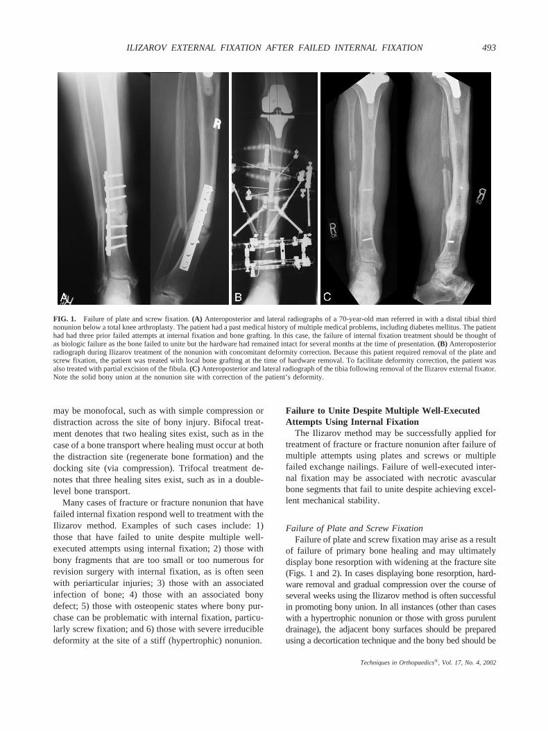
FIG. 1. Failure of plate and screw fixation. (A) Anteroposterior and lateral radiographs of a 70-year-old man referred in with a distal tibial thirdnonunion below a total knee arthroplasty. The patient had a past medical history of multiple medical problems, including diabetes mellitus. The patienthad had three prior failed attempts at internal fixation and bone grafting. In this case, the failure of internal fixation treatment should be thought ofas biologic failure as the bone failed to unite but the hardware had remained intact for several months at the time of presentation. (B) Anteroposteriorradiograph during Ilizarov treatment of the nonunion with concomitant deformity correction. Because this patient required removal of the plate andscrew fixation, the patient was treated with local bone grafting at the time of hardware removal. To facilitate deformity correction, the patient wasalso treated with partial excision of the fibula. (C) Anteroposterior and lateral radiograph of the tibia following removal of the Ilizarov external fixator.Note the solid bony union at the nonunion site with correction of the patient’s deformity.
493ILIZAROV EXTERNAL FIXATION AFTER FAILED INTERNAL FIXATION
Techniques in Orthopaedics®, Vol. 17, No. 4, 2002
grafted with autogenous cancellous bone graft at the time ofplate and screw removal.
Slow, gradual compression is generally applied at arate of 0.25 to 0.5 mm per day for a period of 2 to 4weeks. Once the bone ends are in contact, the ringsspanning the site of bony injury are moving closertogether to a greater extent than are the bone fragments.When this occurs, the wires on either side of the fractureor fracture nonunion site bow. Compression stimulatesbony healing for most fracture or fracture nonunionsfollowing failed internal fixation. In the optimal sce-
nario, the bone fragments have large, transversely ori-ented adjacent surfaces, which allow good bony contactand are stable to axial compression. Simple monofocalcompression is usually unsuccessful for fractures or fracturenonunions associated with infection with purulent drainageand large intervening segments of necrotic bone.
Failure of Exchange NailingFailed exchange nailing is an uncommon, but chal-
lenging, problem (Fig. 3). Failure of exchange nailingmay be related to either a biologic problem or micromo-
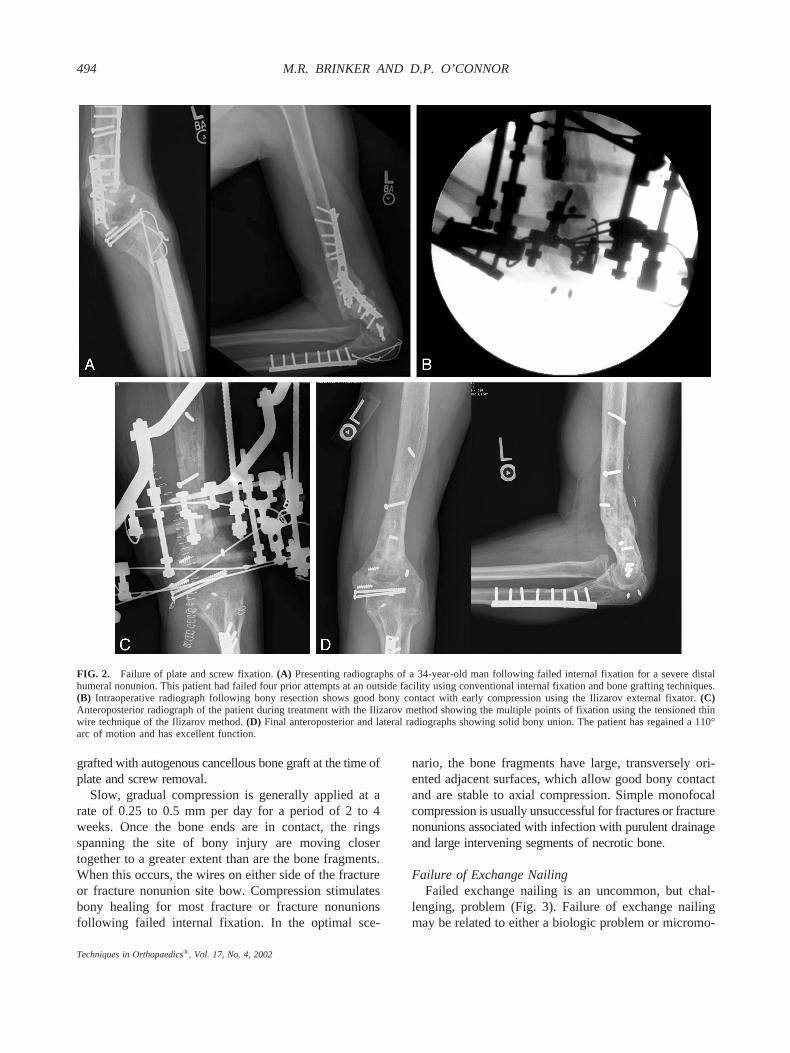
FIG. 2. Failure of plate and screw fixation. (A) Presenting radiographs of a 34-year-old man following failed internal fixation for a severe distalhumeral nonunion. This patient had failed four prior attempts at an outside facility using conventional internal fixation and bone grafting techniques.(B) Intraoperative radiograph following bony resection shows good bony contact with early compression using the Ilizarov external fixator. (C)Anteroposterior radiograph of the patient during treatment with the Ilizarov method showing the multiple points of fixation using the tensioned thinwire technique of the Ilizarov method. (D) Final anteroposterior and lateral radiographs showing solid bony union. The patient has regained a 110°arc of motion and has excellent function.
494 M.R. BRINKER AND D.P. O’CONNOR
Techniques in Orthopaedics®, Vol. 17, No. 4, 2002
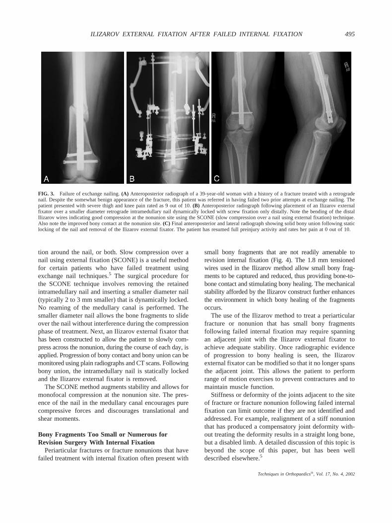
tion around the nail, or both. Slow compression over anail using external fixation (SCONE) is a useful methodfor certain patients who have failed treatment usingexchange nail techniques.5 The surgical procedure forthe SCONE technique involves removing the retainedintramedullary nail and inserting a smaller diameter nail(typically 2 to 3 mm smaller) that is dynamically locked.No reaming of the medullary canal is performed. Thesmaller diameter nail allows the bone fragments to slideover the nail without interference during the compressionphase of treatment. Next, an Ilizarov external fixator thathas been constructed to allow the patient to slowly com-press across the nonunion, during the course of each day, isapplied. Progression of bony contact and bony union can bemonitored using plain radiographs and CT scans. Followingbony union, the intramedullary nail is statically lockedand the Ilizarov external fixator is removed.
The SCONE method augments stability and allows formonofocal compression at the nonunion site. The pres-ence of the nail in the medullary canal encourages purecompressive forces and discourages translational andshear moments.
Bony Fragments Too Small or Numerous forRevision Surgery With Internal Fixation
Periarticular fractures or fracture nonunions that havefailed treatment with internal fixation often present with
small bony fragments that are not readily amenable torevision internal fixation (Fig. 4). The 1.8 mm tensionedwires used in the Ilizarov method allow small bony frag-ments to be captured and reduced, thus providing bone-to-bone contact and stimulating bony healing. The mechanicalstability afforded by the Ilizarov construct further enhancesthe environment in which bony healing of the fragmentsoccurs.
The use of the Ilizarov method to treat a periarticularfracture or nonunion that has small bony fragmentsfollowing failed internal fixation may require spanningan adjacent joint with the Ilizarov external fixator toachieve adequate stability. Once radiographic evidenceof progression to bony healing is seen, the Ilizarovexternal fixator can be modified so that it no longer spansthe adjacent joint. This allows the patient to performrange of motion exercises to prevent contractures and tomaintain muscle function.
Stiffness or deformity of the joints adjacent to the siteof fracture or fracture nonunion following failed internalfixation can limit outcome if they are not identified andaddressed. For example, realignment of a stiff nonunionthat has produced a compensatory joint deformity with-out treating the deformity results in a straight long bone,but a disabled limb. A detailed discussion of this topic isbeyond the scope of this paper, but has been welldescribed elsewhere.5
FIG. 3. Failure of exchange nailing. (A) Anteroposterior radiograph of a 39-year-old woman with a history of a fracture treated with a retrogradenail. Despite the somewhat benign appearance of the fracture, this patient was referred in having failed two prior attempts at exchange nailing. Thepatient presented with severe thigh and knee pain rated as 9 out of 10. (B) Anteroposterior radiograph following placement of an Ilizarov externalfixator over a smaller diameter retrograde intramedullary nail dynamically locked with screw fixation only distally. Note the bending of the distalIlizarov wires indicating good compression at the nonunion site using the SCONE (slow compression over a nail using external fixation) technique.Also note the improved bony contact at the nonunion site. (C) Final anteroposterior and lateral radiograph showing solid bony union following staticlocking of the nail and removal of the Ilizarov external fixator. The patient has resumed full preinjury activity and rates her pain at 0 out of 10.
495ILIZAROV EXTERNAL FIXATION AFTER FAILED INTERNAL FIXATION
Techniques in Orthopaedics®, Vol. 17, No. 4, 2002
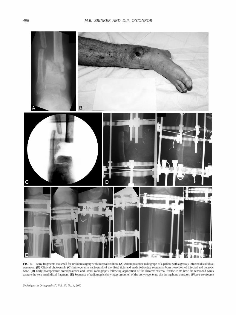
FIG. 4. Bony fragments too small for revision surgery with internal fixation. (A) Anteroposterior radiograph of a patient with a grossly infected distal tibialnonunion. (B) Clinical photograph. (C) Intraoperative radiograph of the distal tibia and ankle following segmental bony resection of infected and necroticbone. (D) Early postoperative anteroposterior and lateral radiographs following application of the Ilizarov external fixator. Note how the tensioned wirescapture the very small distal fragment. (E) Sequence of radiographs showing progression of the bony regenerate site during bone transport. (Figure continues)
496 M.R. BRINKER AND D.P. O’CONNOR
Techniques in Orthopaedics®, Vol. 17, No. 4, 2002
Associated Infection of BoneAn infection of a fracture or fracture nonunion follow-
ing failed internal fixation poses a dual challenge that ischaracterized by two of the most difficult orthopaedicentities to treat: bone infection and ununited fracture. Aninfection of a fracture or fracture nonunion followingfailed internal fixation is often accompanied by incapac-itating pain (often with narcotic dependency), soft tissueproblems, deformities, joint problems (contractures, de-formities, limited range of motion), motor and sensorydysfunction, osteopenia, poor general health, depression,and a myriad of other problems.
The goals in treating an infection of a fracture orfracture nonunion following failed internal fixation are:
1) to obtain solid bony union; 2) to eradicate the infec-tion; and 3) to maximize function of the extremity andthe patient. Before embarking on a course of treatment,the length of time required, the number of operativeprocedures anticipated, and the intensity of the treatmentplan must be discussed with the patient and family. Thecourse of treatment is difficult to predict and the possi-bility of persistent infection and nonunion despite appro-priate treatment should be discussed, and the possibilityof future amputation should be considered.
The treatment strategy for bone infection followingfailed internal fixation is dependent on the nature of theinfection; specifically, whether the infection is draining,nondraining-active, or nondraining-quiescent.29 Treatment
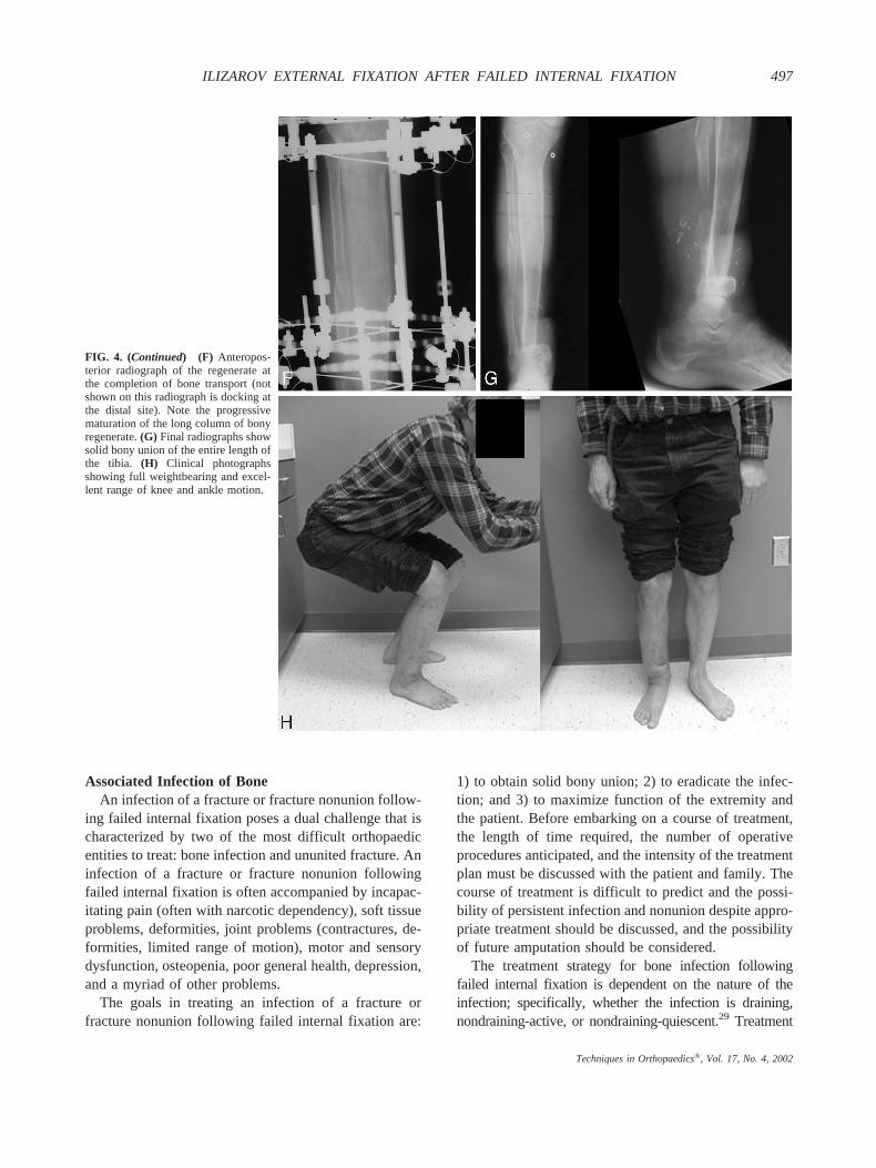
FIG. 4. (Continued) (F) Anteropos-terior radiograph of the regenerate atthe completion of bone transport (notshown on this radiograph is docking atthe distal site). Note the progressivematuration of the long column of bonyregenerate. (G) Final radiographs showsolid bony union of the entire length ofthe tibia. (H) Clinical photographsshowing full weightbearing and excel-lent range of knee and ankle motion.
497ILIZAROV EXTERNAL FIXATION AFTER FAILED INTERNAL FIXATION
Techniques in Orthopaedics®, Vol. 17, No. 4, 2002
involves both a biologic and a mechanical approach, both ofwhich are addressed with the Ilizarov method.
Active Draining InfectionsWhen purulent drainage is ongoing, the injury site will
take longer and be more difficult to heal. An activelydraining infection following failed internal fixation ne-cessitates serial radical debridements to eliminate theinfection (Fig. 5). The first debridement should includeremoval of all orthopaedic hardware in the zone of theinfection. In addition, deep cultures should be obtained,including specimens of soft tissues and bone. Perioper-ative antibiotics should be stopped for at least 1 weekprior to obtaining deep intraoperative cultures. Excisionof all necrotic soft tissues (e.g., fascia, muscle, abscesscavities, and sinus tracts), bone, and foreign bodies,should be performed. The soft tissue sinus tract should besent for pathologic specimen to rule out carcinoma.
Following debridement of an actively draining boneinfection, a dead space is commonly present. The initialtreatment typically involves insertion of antibiotic-im-pregnated polymethylmethacrylate beads, and a beadexchange is performed at the time of each serial debride-ment. The dead space can subsequently be managed in anumber of ways. Currently, the most widely used methodinvolves filling the soft tissue dead space with a rota-
tional vascularized muscle pedicle flap (e.g., gastrocne-mius or soleus27) or a microvascularized free flap (e.g.,latissimus dorsi, rectus, others).35,36 Another method ofmanaging the dead space involves open wound care withmoist dressings, as in the Papineau technique,26 untilgranulation occurs and skin grafting can be performed.
Generally, a consulting infectious disease specialistdirects systemic antibiotic therapy. Following procure-ment of deep surgical cultures, the patient is placed onbroad-spectrum intravenous antibiotics as the cultureresults are pending. Antibiotic coverage is later directedat the infecting organisms when the culture results areavailable.
After elimination of infection, the resulting bony de-fects can be reconstructed using a variety of techniquesavailable with the Ilizarov method, as will be discussedin the section that follows on Associated Bony Defects.
Active Nondraining InfectionsNondraining but active bone infections following
failed internal fixation present with swelling, tenderness,and local erythema (Fig. 6). The history often includesepisodes of fever or other constitutional symptoms.These bone conditions are treated using similar princi-ples to those described for actively draining bone infec-tions: debridement, intraoperative cultures, soft tissue
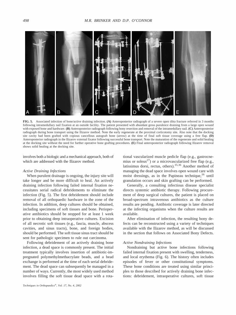
FIG. 5. Associated infection of bone/active draining infection. (A) Anteroposterior radiograph of a severe open tibia fracture referred in 3 monthsfollowing intramedullary nail fixation at an outside facility. The patient presented with abundant gross purulence draining from a large open woundwith exposed bone and hardware. (B) Anteroposterior radiograph following bony resection and removal of the intramedullary nail. (C) Anteroposteriorradiograph during bone transport using the Ilizarov method. Note the early regenerate at the proximal corticotomy site. Also note that the dockingsite cavity had been grafted with copious cancellous autograft bone (arrow) at the time of final soft tissue coverage using a free flap. (D)Anteroposterior radiograph in the Ilizarov external fixator following successful bone transport. Note the maturation of the regenerate and solid healingat the docking site without the need for further operative bone grafting procedures. (E) Final anteroposterior radiograph following Ilizarov removalshows solid healing at the docking site.
498 M.R. BRINKER AND D.P. O’CONNOR
Techniques in Orthopaedics®, Vol. 17, No. 4, 2002
management, mechanical stabilization, bone healingstimulation, and systemic antibiotic therapy. These casestypically require hardware removal, incision and drain-age of an abscess, and excision of only small amounts ofbone and soft tissues. Nondraining bone infections arefrequently managed with primary closure following in-cision and drainage or may be managed with a closedsuction-irrigation drainage system until the infection be-comes quiescent.
Nondraining Quiescent InfectionsNondraining quiescent infections following failed in-
ternal fixation are those in patients with a history ofinfection but without drainage or symptoms for 3 ormore months29 or without a history of infection but withpositive nuclear medicine studies (Fig. 7). In these cases,the Ilizarov method may be used to promote bony heal-ing by applying compression across the fracture sitewithout requiring open debridement or bone grafting.31
Associated Bony DefectsSegmental bone defects associated with fractures or
fracture nonunions result from: 1) high energy openfractures; 2) surgical debridement of devitalized bone
fragments; 3) surgical debridement for bone infection; 4)surgical excision of necrotic bone; and 5) surgical trim-ming at a fracture or fracture nonunion site to improvethe surface characteristics.5
Segmental bone defects associated with fractures orfracture nonunions may have circumferential (complete)bone loss or partial (incomplete) bone loss. These defectsmay be managed using a variety of treatments. Thetreatment methods fit into three broad categories includ-ing: 1) static methods, 2) acute compression methods,and 3) gradual compression methods.
Circumferential Bone Loss: Static Treatment MethodsStatic treatment methods fill the defect between the
bone ends. When using static methods, the proximal anddistal ends of the fracture or nonunion site are fixed usingorthopaedic hardware (internal or external fixation).Static methods for treating bone defects include the useof: 1) autogenous cancellous or cortical bone grafts; 2)vascularized autografts; 3) bulk or strut cortical allo-grafts; 4) mesh cage-bone graft constructs; and 5) syn-ostosis techniques. A variety of static treatment methodsusing internal and external fixation have been well de-scribed elsewhere.5
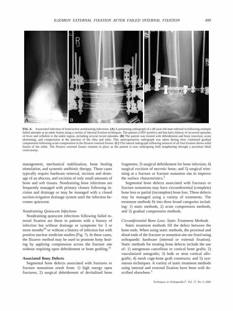
FIG. 6. Associated infection of bone/active nondraining infections. (A) A presenting radiograph of a 48-year-old man referred in following multiplefailed attempts at an ankle fusion using a variety of internal fixation techniques. The patient is HIV-positive and has had a history of recurrent episodesof fever and cellulitis to the ankle region, including several recent episodes. (B) The patient was treated with debridement and bony resection, acuteshortening, and compression at the junction of the tibia and talus. This anteroposterior radiograph was taken during slow continued gradualcompression following acute compression in the Ilizarov external fixator. (C) This lateral radiograph following removal of all foot fixation shows solidfusion of the ankle. The Ilizarov external fixator remains in place as the patient is now undergoing limb lengthening through a proximal tibialcorticotomy.
499ILIZAROV EXTERNAL FIXATION AFTER FAILED INTERNAL FIXATION
Techniques in Orthopaedics®, Vol. 17, No. 4, 2002
Circumferential Bone Loss: Acute CompressionMethods
Acute compression methods obtain immediate bone-to-bone contact at the fracture or fracture nonunion siteby acutely shortening the extremity. The extent of acuteshortening that is possible is limited by the soft tissues(soft tissue compliance, surgical and open wounds, andneurovascular structures). Some authors13,17,31 have sug-gested that greater than 2 to 2.5 cm of acute shortening ata nonunion site may lead to soft tissue problems, al-though others have reported that acute shortening isappropriate for defects up to 7 cm in length.25 In limbswith paired bones, partial excision of the unaffected boneis needed to allow compression across the affected bone.For example, partial excision of the fibula shaft (whenthe fibula is intact) is necessary to allow compressionand shortening of the tibia.
Immediate bone-to-bone contact with acute compres-sion across a segmental defect begins the process ofhealing as early as possible. A disadvantage of acutecompression at segmental defects is the resulting func-tional consequences from foreshortening of the extrem-ity. In the upper extremity, up to 3 to 4 cm of foreshort-ening is well tolerated. In the lower extremity up to 2 cmof foreshortening may be treated with a shoe lift. Manypatients poorly tolerate a shoe lift for 2 to 4 cm ofshortening and most do not tolerate greater than 4 cm offoreshortening. Therefore, many patients undergoing acuteshortening with compression across the segmental defectwill require a lengthening procedure of the ipsilateral ex-tremity or a foreshortening procedure of the contralateralextremity. These limb length equalization procedures can
be performed concurrently with, or sequentially follow-ing, the acute compression (shortening) procedure.
Acute compression may be applied using various in-ternal or external fixation devices. Because of its strengthand versatility, the Ilizarov method is an excellent treat-ment option for acute compression applications. TheIlizarov method is also useful in that it allows forrestoration of limb length via a corticotomy with length-ening at another site of the bone following compressionat the site of injury (bifocal treatment). Bifocal compres-sion-distraction lengthening involves acute (or gradual)compression across the site of bony injury with length-ening through an adjacent corticotomy. This method isapplicable for fractures or fracture nonunions associatedwith foreshortening.
Circumferential Bone Loss: Gradual CompressionMethods
Gradual compression techniques using the Ilizarovmethod include simple monofocal gradual compression(shortening) or bone transport. Neither gradual monofo-cal compression nor bone transport is associated with thepotentially severe soft tissue and wound problems asso-ciated with acute compression. However, gradual mono-focal compression and bone transport are both associatedwith malalignment at the docking site (the most extremecase being when the proximal and distal fragments com-pletely miss each other), whereas acute compression is not.
Monofocal Compression. When the chosen methodof treatment for circumferential bone loss followingfailed internal fixation is monofocal gradual compres-sion, the Ilizarov external fixator is constructed to allow
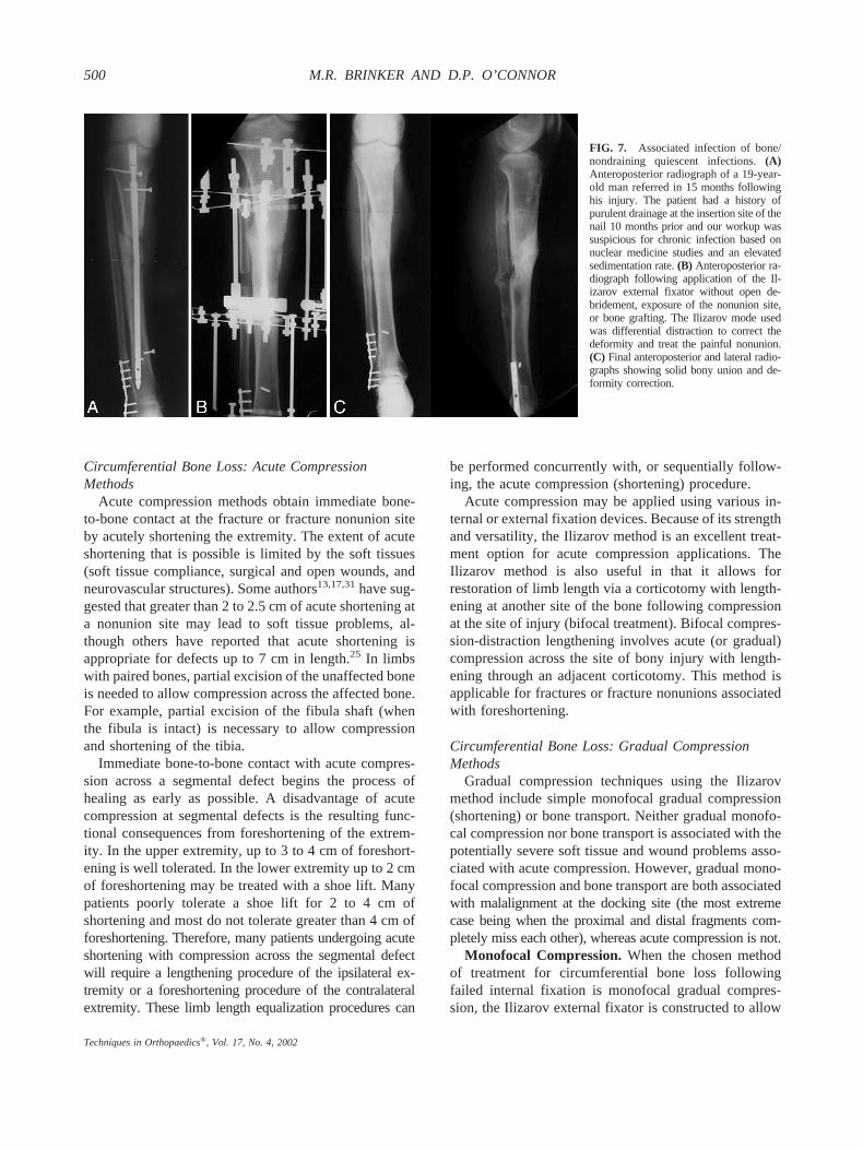
FIG. 7. Associated infection of bone/nondraining quiescent infections. (A)Anteroposterior radiograph of a 19-year-old man referred in 15 months followinghis injury. The patient had a history ofpurulent drainage at the insertion site of thenail 10 months prior and our workup wassuspicious for chronic infection based onnuclear medicine studies and an elevatedsedimentation rate. (B) Anteroposterior ra-diograph following application of the Il-izarov external fixator without open de-bridement, exposure of the nonunion site,or bone grafting. The Ilizarov mode usedwas differential distraction to correct thedeformity and treat the painful nonunion.(C) Final anteroposterior and lateral radio-graphs showing solid bony union and de-formity correction.
500 M.R. BRINKER AND D.P. O’CONNOR
Techniques in Orthopaedics®, Vol. 17, No. 4, 2002
for compression in increments of 0.25 mm. Slow com-pression at a rate of 0.25 mm to 1.0 mm per day isapplied in one or four increments, respectively. When alarge defect exists, compression is applied at a rate of 1.0mm per day; as the fragments approach bony contact, therate is slowed to 0.25 mm to 0.5 mm per day. Asdiscussed above, compression in limbs with paired bonesnecessitates partial excision of the unaffected bone.
Bone Transport. When the chosen method of treat-
ment for circumferential bone loss following failed in-ternal fixation is bone transport, the Ilizarov externalfixator is constructed to allow for bone transport at a rateranging from 0.25 mm every other day to 1.5 mm per day(Fig. 8). The transport is typically started at the rate of0.5 mm to 0.75 mm per day in two or three increments,respectively. The rate is often adjusted (increased ordecreased) based on the quality of the bony regenerate asviewed on serial plain radiographs.
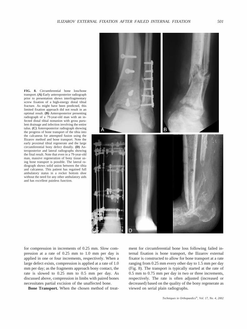
FIG. 8. Circumferential bone loss/bonetransport. (A) Early anteroposterior radiographprior to presentation shows interfragmentaryscrew fixation of a high-energy distal tibialfracture. As might have been predicted, thislimited fixation approach did not result in anoptimal result. (B) Anteroposterior presentingradiograph of a 79-year-old man with an in-fected distal tibial nonunion with gross puru-lent drainage and infection involving the entiretalus. (C) Anteroposterior radiograph showingthe progress of bone transport of the tibia intothe calcaneus for attempted fusion using theIlizarov method and bone transport. Note theearly proximal tibial regenerate and the largecircumferential bony defect distally. (D) An-teroposterior and lateral radiographs showingthe final result. Note that even in a 79-year-oldman, massive regeneration of bony tissue us-ing bone transport is possible. The lateral ra-diograph shows solid union between the tibiaand calcaneus. This patient has regained fullambulatory status in a rocker bottom shoewithout the need for any other ambulatory aidsand has excellent painless function.
501ILIZAROV EXTERNAL FIXATION AFTER FAILED INTERNAL FIXATION
Techniques in Orthopaedics®, Vol. 17, No. 4, 2002
Bone transport (bifocal distraction-compression trans-port) involves the creation of a corticotomy (usuallymetaphyseal) at a site distant from the nonunion. Thebone segment produced by the corticotomy is then trans-ported toward the nonunion site (filling the bony defect) ata gradual rate. The compression produced by the trans-ported segment arriving at the docking site is successful inobtaining bony union in many cases. Occasionally bonegrafting with marrow or open bone graft is required.
The bone formed at the corticotomy site in bonetransport is formed under gradual distraction through theprocess of distraction osteogenesis.2,3,12,15,22 The mech-anism of bone formation in distraction osteogenesis is aresult of increased vascularity and cellular proliferation.In a study of dogs undergoing distraction osteogenesis,Aronson1 reported that blood flow at the distraction siteincreased nearly ten-fold relative to the control limb,peaking about 2 weeks after surgery. The distal tibia,remote from the site of distraction, also showed a similarpattern of increased blood flow. The mechanical tension-stress effect of distraction is known to cause neovascu-larity and cellular proliferation in bone and other tissues.
The success of distraction osteogenesis depends on avariety of mechanical and biologic requirements. First,the corticotomy or osteotomy must be performed using alow-energy technique. Second, corticotomy or osteot-omy in the metaphyseal or metadiaphyseal region ispreferred over diaphyseal sites because of the superiorpotential for regenerate formation. Third, a very stableexternal fixator construct, such as that available in theIlizarov method, is required to promote good bony re-
generate. Fourth, a latency period prior to beginningdistraction of 7 to 14 days is recommended, dependingon various patient characteristics. Fifth, the distractionphase is classically performed at a rate of 1.0 mm per dayin a rhythm of 0.25 mm of distraction performed 4 timesper day. Since some patients make bony regenerate moreslowly, the rate and rhythm of distraction should becarefully controlled by the treating physician, who canmonitor the progression of the regenerate on plain radio-graphs. Sixth, following distraction, maturation and hy-pertrophy of the bony regenerate must be allowed tooccur during the consolidation phase. The consolidationphase is generally two to three times as long as thenumber of days of the distraction phase, but this varieswidely among patients.
For both gradual compression and bone transport,favorable surface characteristics at the docking site (siteof injury) greatly improve the chances of rapid healing.When poor surface characteristics are present, open trim-ming is recommended. When open trimming is per-formed at the time of the initial procedure, the dockingsite can be bone grafted if the anticipated time to dockingis approximately 2 months or less (such as a 6 cm defecttreated with gradual shortening or bone transport at a rateof 1.0 mm per day). If the time to docking will besignificantly greater than 2 months (such as for largerdefects), two options exist. First, gradual compression ortransport can be continued even after bony touchdown atthe docking site is seen on plain radiographs. Continuedcompression at a rate ranging from 0.25 mm per week to0.25 mm per day at the docking site is seen clinically and
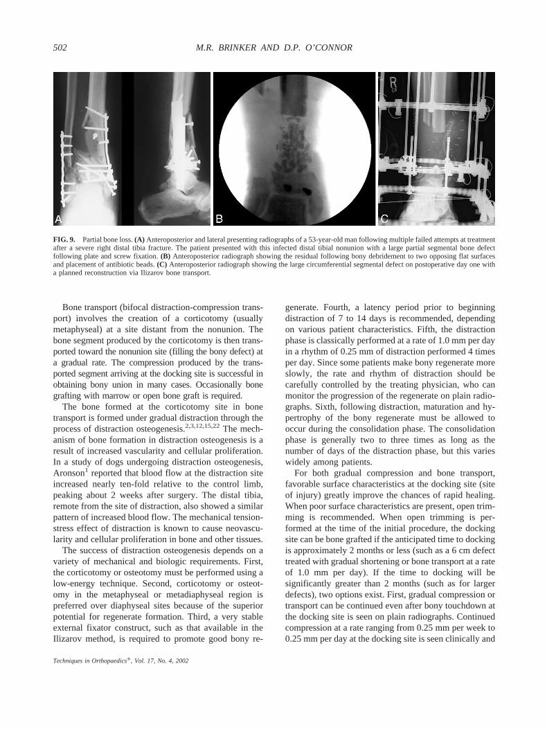
FIG. 9. Partial bone loss. (A) Anteroposterior and lateral presenting radiographs of a 53-year-old man following multiple failed attempts at treatmentafter a severe right distal tibia fracture. The patient presented with this infected distal tibial nonunion with a large partial segmental bone defectfollowing plate and screw fixation. (B) Anteroposterior radiograph showing the residual following bony debridement to two opposing flat surfacesand placement of antibiotic beads. (C) Anteroposterior radiograph showing the large circumferential segmental defect on postoperative day one witha planned reconstruction via Ilizarov bone transport.
502 M.R. BRINKER AND D.P. O’CONNOR
Techniques in Orthopaedics®, Vol. 17, No. 4, 2002
radiographically as bending of the fixation wires, indi-cating that the rings are moving more than the proximaland distal bone fragments. Second, the docking site canbe opened prior to bone contact (usually when the defectis approximately 1 to 2 cm), the proximal and distalsurfaces can be freshened up, and the defect can be bonegrafted. Gradual compression or transport then proceedsinto the graft material.
The literature is not helpful in clarifying whether bonegrafting the docking site significantly decreases the time
to healing. A useful alternative to open bone grafting ispercutaneous marrow injection at the docking site. Thistechnique is minimally invasive and quite effective. I usepercutaneous marrow injection for patients at risk forpersistent nonunion. I reserve open bone grafting of thedocking site for: 1) those patients who fail to demon-strate radiographic evidence of progression to healingdespite 4 months of continued compression after bonytouchdown; 2) those patients at greatly increased risk ofpersistent nonunion at the docking site (those patients
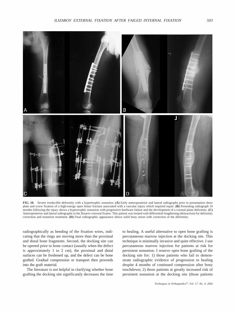
FIG. 10. Severe irreducible deformity with a hypertrophic nonunion. (A) Early anteroposterior and lateral radiographs prior to presentation showplate and screw fixation of a high-energy open femur fracture associated with a vascular injury which required repair. (B) Presenting radiograph 10months following the injury shows a hypertrophic nonunion with progressive hardware failure and the development of a coronal plane deformity. (C)Anteroposterior and lateral radiographs in the Ilizarov external fixator. This patient was treated with differential lengthening (distraction) for deformitycorrection and nonunion treatment. (D) Final radiographic appearance shows solid bony union with correction of the deformity.
503ILIZAROV EXTERNAL FIXATION AFTER FAILED INTERNAL FIXATION
Techniques in Orthopaedics®, Vol. 17, No. 4, 2002
who have several contributing factors for nonunion;5 and3) those patients with poor surface contact at the dockingsite (these patients require trimming of the bone ends toimprove the surface characteristics).
Partial Bone LossBy virtue of their architecture (point to point contact),
nonunions with partial segmental defects are not readilyamenable to many of the treatment strategies that havebeen discussed. These types of defects are most com-monly treated with a static method, such as autologouscancellous bone grafting with internal or external fixa-tion. As the segment of partial bone loss increases inlength, the chances for successful bony union usingconventional bone grafting techniques decreases (Fig. 9).In cases with a large (� 6 cm) segment of partial(incomplete) bone loss, the treatment options are: 1)“splinter (sliver) bone transport,” 2) surgical trimming ofthe bone ends to enhance surface characteristics followedby an acute or gradual compression method, or 3) strutcortical allogenic bone grafting.
Osteopenic StatesThe thin wires used in the Ilizarov method provide
remarkably good purchase in osteopenic bone. The useof 1.8 mm tensioned wires at high crossing angles (up to90°) provides very good stability for the site of bonyinjury even in very weak bone. The stability of theIlizarov external fixator can be improved for use inosteopenic bone by the use of olive wires, which dis-courage translational moments at the wire-bone inter-face. The use of a washer at the olive wire-bone interfacealso helps to distribute the load and prevent erosion ofthe olive into the bone.
Severe Irreducible Deformity with a HypertrophicNonunion
A severe irreducible deformity with a hypertrophic(stiff) nonunion following failed internal fixation is besttreated by gradual deformity correction (Fig. 10). Theadvantage of the Ilizarov method in these cases is thatosseous integration can be achieved simultaneously dur-ing the gradual deformity correction. Furthermore, theIlizarov method allows not only for simple compressionand distraction, but also for differential compression anddistraction to allow for correction of complex deformi-ties. In addition, the Ilizarov method does not requirelarge soft tissue dissection as would be required withdeformity correction using internal fixation techniques.
Distraction of the abundant fibrocartilaginous tissue inhypertrophic nonunions stimulates new bone forma-tion.7,16,24,30 Distraction using the Ilizarov method re-
sults in bony healing in a high percentage of suchcases,7,30 although the exact biologic mechanism re-mains obscure.
CONCLUSION
Treatment with internal fixation fails for a variety ofreasons, and revision internal fixation using the same ora different internal fixation technique may be successful.In certain cases, however, the Ilizarov method may bethe preferred treatment strategy following failed internalfixation. The Ilizarov method offers many advantages fortreatment of fracture or fracture nonunion followingfailed internal fixation. Several modes of treatment areavailable with the Ilizarov method, including monofocal,acute, or gradual compression and bone transport (bifo-cal treatment). The Ilizarov method provides excellentmechanical stability, biologic stimulation at the site ofbony injury, and the ability to generate new bone tissuethrough distraction osteogenesis. Cases of fracture orfracture nonunion that have failed internal fixation thatrespond well to the Ilizarov method include those: 1)with multiple previous attempts using internal fixation;2) with small or numerous bony fragments; 3) with boneinfection; 4) with a bony defect; 5) with osteopenicstates; and 6) with a stiff (hypertrophic) nonunion asso-ciated with a severe irreducible deformity.
ACKNOWLEDGMENT
The authors acknowledge the work of Rodney K.Baker for his assistance with the figures.
REFERENCES
1. Aronson J. Temporal and spatial increases in blood flow duringdistraction osteogenesis. Clin Orthop 1994;301:124–131.
2. Aronson J, Good B, Stewart C, Harrison B, Harp J. Preliminarystudies of mineralization during distraction osteogenesis. ClinOrthop 1990;250:43–49.
3. Aronson J, Harrison B, Boyd CM, Cannon DJ, Lubansky HJ,Stewart C. Mechanical induction of osteogenesis. Preliminarystudies. Ann Clin Lab Sci 1988;18:195–203.
4. Brinker M, Cook S, Dunlap J, Christakis P, Elliott M. Earlychanges in nutrient artery blood flow following tibial nailing withand without reaming: A preliminary study. J Orthop Trauma1999;13:129–133.
5. Brinker MR. Nonunions: evaluation and treatment. In: BrownerBD, Levine AM, Jupiter JB, Trafton PG, eds. Skeletal Trauma:Basic Science, Management, and Reconstruction. Philadelphia:WB Saunders; 2003:507–604.
6. Brinker MR, Bailey DE. Fracture healing in tibia fractures with anassociated vascular injury. J Trauma 1997;42:11–19.
7. Catagni MA, Guerreschi F, Holman JA, Cattaneo R. Distractionosteogenesis in the treatment of stiff hypertrophic nonunions usingthe Ilizarov apparatus. Clin Orthop 1994;301:159–163.
8. Chatziyiannakis AA, Verettas DA, Raptis VK, Charpantitis ST.Nonunion of tibial fractures treated with external fixation. Con-
504 M.R. BRINKER AND D.P. O’CONNOR
Techniques in Orthopaedics®, Vol. 17, No. 4, 2002
tributing factors studied in 71 fractures. Acta Orthop Scand Suppl1997;275:77–79.
9. Christensen NO. Kuntscher intramedullary reaming and nail fixa-tion for non-union of fracture of the femur and the tibia. J BoneJoint Surg Br 1973;55:312–318.
10. Court-Brown CM, Keating JF, Christie J, McQueen MM. Ex-change intramedullary nailing. Its use in aseptic tibial nonunion.J Bone Joint Surg Br 1995;77:407–411.
11. Danckwardt-Lilliestrom G. Reaming of the medullary cavity andits effect on diaphyseal bone. A fluorochromic, microangiographicand histologic study on the rabbit tibia and dog femur. Acta OrthopScand Suppl 1969;128:1–153.
12. Delloye C, Delefortrie G, Coutelier L, Vincent A. Bone regenerateformation in cortical bone during distraction lengthening: an ex-perimental study. Clin Orthop 1990;250:34–42.
13. Green SA. The Ilizarov method. In: Browner BD, Levine AM,Jupiter JB, eds. Skeletal Trauma: Fractures, Dislocations, Liga-mentous Injuries. Vol 1. 2nd ed. Philadelphia: WB Saunders;1998:661–701.
14. Hak DJ, Lee SS, Goulet JA. Success of exchange reamed in-tramedullary nailing for femoral shaft nonunion or delayed union.J Orthop Trauma 2000;14:178–182.
15. Ilizarov GA. Clinical application of the tension-stress effect forlimb lengthening. Clin Orthop 1990;250:8–26.
16. Ilizarov GA. Transosseous Osteosynthesis. Theoretical and Clini-cal Aspects of the Regeneration and Growth of Tissue. Berlin:Springer-Verlag; 1992.
17. Ilizarov GA, Kaplunov AG, Grachova VI, Shpaer LI. Close Com-pression-Distraction Osteosynthesis of the Tibial Pseudoarthroseswith Ilizarov Method (Metodicheskoe Posobie). Kurgan, USSR:Kniiekot Institute; 1971.
18. Indrekvam K, Lekven J, Engesaeter LB, Langeland N. Effects ofintramedullary reaming and nailing on blood flow in rat femora.Acta Orthop Scand 1992;63:61–65.
19. Koval KJ, Seligson D, Rosen H, Fee K. Distal femoral nonunion:treatment with a retrograde inserted locked intramedullary nail.J Orthop Trauma 1995;9:285–291.
20. McKee MD, Miranda MA, Riemer BL, et al. Management ofhumeral nonunion after the failure of locking intramedullary nails.J Orthop Trauma 1996;10:492–499.
21. Megas P, Panagiotopoulos E, Skriviliotakis S, Lambiris E. In-tramedullary nailing in the treatment of aseptic tibial nonunion.Injury 2001;32:233–239.
22. Murray JH, Fitch RD. Distraction histogenesis: principles andindications. J Am Acad Orthop Surg 1996;4:317–327.
23. Oh I, Nahigian SH, Rascher JJ, Farrall JP. Closed intramedullarynailing for ununited femoral shaft fractures. Clin Orthop 1975;106:206–215.
24. Paley D. Treatment of tibial nonunion and bone loss with theIlizarov technique. Instr Course Lect 1990;39:185–197.
25. Paley D, Sen C, Tetsworth K, Herzenberg JE. Acute shorteningwith subsequent relengthening versus bone transport in the treat-ment of tibial bone defects (Paper #7). Paper presented at: 11thAnnual Scientific Meeting, Association for the Study and Appli-cation of the Methods of Ilizarov-North America: The LimbLengthening and Reconstruction Society, 2001; Berkeley, Califor-nia.
26. Papineau LJ, Alfageme A, Dalcourt JP, Pilon L. Osteomyelitechronique: excision et greffe de spongieux à l’air libre après misesà plat extensives. Int Orthop 1979;3:165–176.
27. Pers M, Medgyesi S. Pedicle muscle flaps and their applications inthe surgery repair. Br J Plast Surg 1977;26:313–321.
28. Reichert ILH, McCarthy ID, Hughes SPF. The acute vascularresponse to intramedullary reaming. Microsphere estimation ofblood flow in the intact ovine tibia. J Bone Joint Surg Br 1995;77:490–493.
29. Rosen H. Nonunion and malunion. In: Browner BD, Levine AM,Jupiter JB, eds. Skeletal Trauma: Fractures, Dislocations, Liga-mentous Injuries. Vol 1. 2nd ed. Philadelphia: WB Saunders;1998:501–541.
30. Saleh M, Royston S. Management of nonunion of fractures bydistraction with correction of angulation and shortening. J BoneJoint Surg Br 1996;78:105–109.
31. Schwartsman V, Choi SH, Schwartsman R. Tibial nonunions.Treatment tactics with the Ilizarov method. Orthop Clin North Am1990;21:639–653.
32. Sledge SL, Johnson KD, Henley MB, Watson JT. Intramedullarynailing with reaming to treat non-union of the tibia. J Bone JointSurg Am 1989;71:1004–1019.
33. Templeman D, Thomas M, Varecka T, Kyle R. Exchange reamedintramedullary nailing for delayed union and nonunion of the tibia.Clin Orthop 1995;315:169–175.
34. Warren SB, Brooker AF, Jr. Intramedullary nailing of tibial non-unions. Clin Orthop 1992;285:236–243.
35. Weiland AJ, Daniel RK. Microvascular anastomoses for bonegrafts in the treatment of massive defects in bone. J Bone JointSurg Am 1979;61:98–104.
36. Weiland AJ, Moore JR, Daniel RK. Vascularized bone autografts.Experience with 41 cases. Clin Orthop 1983;174:87–95.
37. Weresh MJ, Hakanson R, Stover MD, Sims SH, Kellam JF, BosseMJ. Failure of exchange reamed intramedullary nails for ununitedfemoral shaft fractures. J Orthop Trauma 2000;14:335–338.
38. Wu CC, Shih CH, Chen WJ, Tai CL. High success rate withexchange nailing to treat a tibial shaft aseptic nonunion. J OrthopTrauma 1999;13:33–38.
505ILIZAROV EXTERNAL FIXATION AFTER FAILED INTERNAL FIXATION
Techniques in Orthopaedics®, Vol. 17, No. 4, 2002