the video revolution: a new view of laryngoscopyintubationvl.com/the video revolution - a new view...
TRANSCRIPT

The Video Revolution: A New View of Laryngoscopy
William E Hurford MD
IntroductionMany Products, Much Confusion
StyletsGuide ChannelsVideo Modifications of Traditional LaryngoscopesGlideScope
Which One Is Best?Summary
The development of less expensive, smaller, and more reliable video cameras has revolutionized thedesign of laryngoscopes and the process of endotracheal intubation. The term video laryngoscopydefines a broad range of devices, distinct from fiberoptic bronchoscopes, in which a video camerais used in place of line-of-sight visualization to accomplish endotracheal intubation. Over a dozenlaryngoscopes are marketed currently. Each model of video laryngoscope has its own uniquestrengths, weaknesses, and best applications. For the purposes of this review, video laryngoscopesare grouped into 3 different designs: stylets, guide channels, and video modifications of the tradi-tional (usually Macintosh) laryngoscope blades. Key words: equipment and supplies; diagnostic equip-ment; endoscopes; laryngoscopes, therapeutics; respiratory therapy; respiratory, artificial; positive-pressure respiration, therapeutics; emergency treatment; resuscitation; respiration, artificial. [RespirCare 2010;55(8):1036–1045. © 2010 Daedalus Enterprises]
Introduction
Despite many advances in patient safety, endotrachealintubation remains a specialized learned skill, and a diffi-cult endotracheal intubation remains an important adverseevent.1 The development of algorithms for airway man-
agement has been helpful, and certain technological ad-vances, such as the laryngeal mask airway, have helpedmake airway management safer.2 Over the past severalyears, enhanced visualization of the airway has been ac-complished with the adaptation of fiberoptic bronchoscopesfor this purpose. These instruments, however, largely re-main in the operating room environment and are awkwardto use in emergency situations. The skill of fiberoptic in-tubation is difficult to learn, and the scopes are expensiveto maintain. Hence, the goals of making endotracheal in-tubation an easier skill to learn and maintain and a highlyreliable procedure remain unattained.
Recently, the development of less expensive, smaller,and more reliable video cameras has revolutionized thedesign of laryngoscopes and the process of endotrachealintubation. The term video laryngoscopy defines a broadrange of devices, distinct from fiberoptic bronchoscopes,in which a video camera is used in place of line-of-sightvisualization to accomplish endotracheal intubation. Over
William E Hurford MD is affiliated with the Department of Anesthesi-ology, University of Cincinnati, Cincinnati Ohio.
William E Hurford MD presented a version of this paper at the 25th NewHorizons Symposium, “Airway Management: Current Practice and Fu-ture Directions,” at the 55th International Respiratory Congress of theAmerican Association for Respiratory Care, held December 5–8, 2009, inSan Antonio, Texas.
The author has disclosed no conflicts of interest.
Correspondence: William E Hurford MD, Department of Anesthesiol-ogy, PO Box 670531, 231 Albert Sabin Way, Cincinnati OH 45267-0531. E-mail: [email protected].
1036 RESPIRATORY CARE • AUGUST 2010 VOL 55 NO 8

a dozen laryngoscopes are marketed currently, and thefield continues to develop and grow. Most use high inten-sity light-emitting diode (LED) or fiberoptic light sources,and charge couple device (CCD) or CMOS (complemen-tary metal oxide semiconductor) video chips for visualiza-tion. This review intends to provide a snapshot of thisgrowing field.
Three advantages of video laryngoscopy are obviouswhen compared to intubation techniques that use the line-of-sight of a single operator (Table 1). First, traditionaldirect laryngoscopy depends on an unobstructed line-of-sight. Certain patient abnormalities, such as limited mouthopening, limited neck mobility, obesity, and craniofacialor chest-wall abnormalities, can prevent visualization. Insuch situations, fiberoptic laryngoscopy has been the usualrecommended technique, but is not quickly available out-side the operating room or available to paramedical per-sonnel. Sometimes the patient’s position within a tractiondevice, within radiologic equipment, or entrapped within avehicle or rubble prevents adequate visualization. In thesesituations, endotracheal intubation can be impossible.
Teaching line-of-site techniques is difficult because theteacher cannot share the view with the learner. Video la-ryngoscopy provides a shared view for the teacher andstudent. In the case of video modifications of traditionallaryngoscope blades such as the Storz DCI or C-MAC, thelearner can practice a traditional direct laryngoscopic tech-nique, while the teacher can view what the student sees.3-5
Indeed, the teacher need not even be physically present, ifthe video output can be streamed through a video confer-encing or Internet link.6 This makes distance learning andconsultation possible.7 The better laryngoscopic view gen-erally provided by video techniques may improve the like-lihood of success for inexperienced operators.8,9
Third, and perhaps most importantly, video techniquespermit the easy sharing of critical information among themedical team. Especially in emergency situations, com-munication among providers can be limited or even mis-leading. By using a video technique, especially with alarge independent view screen, information concerning thepatient’s anatomy, ease and success of endotracheal intu-bation, and any difficulties that the operator is experienc-ing is easily evident. An assistant may have an easier timehelping the operator because the assistant can immediatelysee the effect of maneuvers such as applying cricoid pres-sure or manipulating the position of the thyroid cartilage.For these reasons, video laryngoscopic techniques are be-coming more popular for difficult intubations. In my in-stitution, for example, video techniques are now used innearly half of anticipated difficult intubations in the oper-ating rooms, while optical fiberoptic techniques were usedin only 20%.
Despite the attractiveness of video laryngoscopy, thetechnique is not without its disadvantages. Depending on
the device, the learning curve can be steep. Because of theacute angle of some video blades, insertion of the endo-tracheal tube (ETT) into the trachea may be difficult, de-spite a clear view. An acutely angled stylet often is nec-essary. Depth perception is lost with a 2-dimensional videoimage, and sometimes operators may become fixated onthe video screen and do not directly observe where thelaryngoscope blade or ETT is being placed, which canresult in patient injury.10-14 Fogging or the presence ofsecretions can obscure the video picture as well. In gen-eral, the devices are more complicated and more expensivethan a traditional laryngoscope.15 Some devices also re-quire special attention during reprocessing and steriliza-tion, which can add to costs.
Many Products, Much Confusion
Over a dozen products, some modeled on the familiarMacintosh blades, others quite unique, are currently beingmarketed. Each has particular advantages and disadvan-tages. It is probably impractical for any but the most mo-tivated individual to buy and gain expertise in all of them.What are the characteristics that might be sought in se-lecting a video laryngoscope?
1. Ideally, the device should be intuitive to an operatorskilled in traditional direct laryngoscopy. Thick instructionmanuals are usually not found on an intubation cart. Mostof us prefer that a new skill be easy to learn (and teach),especially if it is a critical one.
2. The device should be adaptable for different types ofendotracheal intubations. Some devices, characterized asguide channels in this paper, are designed only for oralintubation. Ideally, a single device should be equally use-ful for oral or nasal intubation, in both adults and children,and permit the use of special-purpose tubes such as dou-ble-lumen endobronchial tubes.
Table 1. Advantages and Disadvantages of Video Laryngoscopy
AdvantagesEye and airway need not line upBetter view when mouth opening or neck mobility is limitedOthers can see and helpPermits sharing of medical information among the teamGenerally higher success rate, especially in difficult situations
DisadvantagesVariable learning curves; may take longer to intubatePassage of tube may be difficult despite great view; stylet often
necessaryFogging and secretions may obscure viewLoss of depth perceptionMore complicatedExpensiveGreater processing time and expense
THE VIDEO REVOLUTION: A NEW VIEW OF LARYNGOSCOPY
RESPIRATORY CARE • AUGUST 2010 VOL 55 NO 8 1037
3. For teaching purposes and for sharing of informationamong the medical team, a large separate view screen isuseful. Many of the advantages of a video technique arelost if only the operator can see the view screen.
4. The laryngoscope should be inexpensive. Cost ofacquisition can be high for both reusable and disposabledevices. Reusable devices can have costs far in excess ofinitial acquisition costs, since these must be reprocessedand sterilized, and the purchase of additional backup de-vices may be necessary if the turnaround time of process-ing is prolonged. Ideally, the less processing required, thebetter.
5. The laryngoscope should be lightweight, low profile,and easy to maneuver. Video laryngoscopy promises suc-cess in unusual locations in the field, during transport, andon hospital floors.16-18 Some devices are large, difficult totransport, or require a lot of space around the patient’shead for maneuvering. Others are highly portable, andhence may have greater utility.
6. Anti-fog capability is surprisingly important. Thegreatest camera may be rendered useless if condensation ispresent on the lens. Some of the devices have heatingelements to reduce this effect.
7. A long-lasting rechargeable battery with an alterna-tive alternating-current (AC) power source is important.An electrical plug is not always available, and these de-vices are nothing but paperweights if the battery is deadand AC power is unavailable.
8. Image storage for review, quality assurance, andteaching is available on some devices, and may be ofvalue for users in teaching settings or for quality assur-ance.
For the purposes of this review I will group video la-ryngoscopes into 3 very different designs: stylets, guidechannels, and video modifications of the traditional (usu-ally Macintosh) laryngoscope blades (Tables 2 and 3).
Stylets
Stylets are rigid or semi-rigid rods that carry both lightand video bundles. These are typified by the Bonfils stylet(Karl Storz, Tuttlingen, Germany, http://www.karlstorz.com/cps/rde/xchg/SID-642F366A-A24088B8/karlstorz-en/hs.xsl/49.htm) and by the Video RIFL (Rigid and FlexingLaryngoscope, AI Medical Devices, Williamston, Michigan,http://www.aimedicaldevices.com/videorifl.html). The Bon-fils intubation endoscope was invented as an optical de-vice over 25 years ago to permit retromolar intubation inpatients with limited mouth opening (Fig. 1).19 The inser-tion rod is available in 2, 3.5, and 5 mm outside diameters,and has a 1.2 mm working channel for continuous oxygenflow. The insertion rod is bent at a 40° angle at the distalend, and the optics provide a 110° view angle that in-cludes a view of the distal tip of the ETT, which ismounted over the insertion rod and then slid off the rodand through the glottis. A video camera (35,000 pixels)can be attached to the eyepiece and improves both ergo-nomics and visualization for the operator. The light sourcecan be powered by battery or AC; the camera attachmentdoes not have battery power. The device may be helpfulin situations when difficult intubation is expected or in-itial direct laryngoscopy has failed.20,21 The time requiredfor intubation appears to be a bit longer than other videotechniques.
The Video RIFL has an insertion rod that is flexible andcan articulate to 135° by squeezing a lever on the device(Fig. 2). The device is limited to standard ETTs of at least6.5 mm inner diameter. It is battery powered. The LEDlight source is integrated at the tip, and the video image isdisplayed on a small integrated LCD screen. The RIFLimage can also be displayed on an external monitor. Afteruse, the stylet portion of the device can be detached forsterile reprocessing.
Video stylet devices may be advantageous for oral en-dotracheal intubation when mouth opening is limited. Theyrequire a substantial learning curve and are not particularlyintuitive. Both the Bonfils and RIFL have various viewingoptions. Both devices are rather large, and the optics maybe subject to secretions and fogging. These devices appearmost suited to special situations and use by operators will-ing to practice and attain sufficient skill in their use.
Guide Channels
Guide channel scopes have designs similar to theoptical Bullard (Gyrus ACMI, Southborough, Massachu-setts) and Wu Scopes (Achi, San Jose, California), andare used in a similar manner. Representative laryngoscopesinclude the Airtraq (Airtraq, Bonita Springs, Florida,http://www.airtraq.com), Pentax AWS (AMBU, Glen Burnie,Maryland, http://www.ambu.com/com/airway_management/
Table 2. Types of Video Laryngoscopes
StyletsBonfilsRIFL (rigid and flexing laryngoscope)
Guide ChannelsAirTraqPentax AWSRes-Q-Scope II
Traditional ModificationsCoopdech VLP-100Storz DCIStorz C-MacMcGrathGlideScope
THE VIDEO REVOLUTION: A NEW VIEW OF LARYNGOSCOPY
1038 RESPIRATORY CARE • AUGUST 2010 VOL 55 NO 8

airway_management.aspx), and the Res-Q-Scope II (Res-Q-Tech, Humble, Texas, http://www.res-q-tech-na.com). Thesedevices hold an ETT within a guide channel. Image and lightbundles run alongside the channel. The laryngoscopes can beinserted in patients with reduced mouth opening. Also, the
angle of force applied with the laryngoscope is more perpen-dicular to the cervical spine, which may reduce cervical spinedisplacement during laryngoscopy. All are designed for oralendotracheal intubation using a standard ETT. The viewscreens are generally small and attached directly to the de-vice, making the laryngoscopes highly portable.
The Airtraq is a single-use optical device with a 60 minbattery life (Fig. 3). Different sizes are available for var-ious ETT sizes, pediatric, nasotracheal, and double-lumenendobronchial intubations. A video camera can attach tothe optical lens to permit viewing on an external monitoror recording. A 30–45-second warm-up time is necessaryto reduce fogging. Turkstra and colleagues compared theuse of the Airtraq to standard Macintosh laryngoscopes in24 patients with normal anatomy.22 They found that intu-bation could be accomplished with significantly less cer-vical spine motion with the Airtraq, suggesting that thedevice might be useful in patients with unstable cervicalspines or reduced mobility of the cervical spine. Similar find-ings have been reported by Hirabayashi and co-workers.23
The Pentax AWS is a similar device that is powered by2 AA batteries (Fig. 4). The 2.4-inch color LCD viewscreen, CCD camera, and light bundle are reusable. Intu-bation is accomplished with a clear plastic disposable guidechannel blade that snaps onto the device. The tip of the
Table 3. Characteristics of Video Laryngoscopes
Characteristic StyletsGuide
ChannelsCoopdech McGrath
Storz DCI,C-Mac
GlideScope
Intuitive No Acceptable Excellent Good Excellent ExcellentAdaptable No No Excellent Excellent Excellent ExcellentLarge separate view screen No No No No Yes YesSterile processing Required No Required No Required NoSize and maneuverability Acceptable Good Good Good Excellent ExcellentAnti-fog capability Acceptable Good Good Good Acceptable ExcellentBattery and power source Acceptable Acceptable Acceptable Acceptable Excellent Excellent
Fig. 1. The Bonfils intubating stylet with video camera attachment.
Fig. 2. The Video RIFL (rigid and flexing laryngoscope). (Courtesyof AI Medical Devices.)
Fig. 3. The Airtraq. A small video camera (not shown) can attach tothe optical lens.
THE VIDEO REVOLUTION: A NEW VIEW OF LARYNGOSCOPY
RESPIRATORY CARE • AUGUST 2010 VOL 55 NO 8 1039

blade is angled at 135°. A target symbol on the monitorshows the intended path of the ETT. The device is suit-able for use with standard ETTs of at least 6.5 mm innerdiameter. A video-out port permits recording or sepa-rate display. Hirabayashi and Seo reported 405 success-ful intubations with the device.24 The time required forintubation ranged from 13 to 192 seconds. Asai andcolleagues examined the use of the Pentax AWS in 270patients in whom direct laryngoscopy with a Macintoshblade had been difficult.25 Successful intubations wereaccomplished in 268 patients. Similar findings have beenpresented by other authors.26,27 Compared with tradi-tional direct laryngoscopy, tracheal intubation with theAWS appears to result in less movement of the uppercervical spine.28
The Res-Q-Scope II is a similar 2-piece device (Fig. 5).The main unit contains a color LCD view screen with avideo-out port and rechargeable battery (a backup battery
pack that uses 4 AA batteries is available). A plastic dis-posable guide channel containing additional suction andoxygen ports, the video imaging system, and an LED lightsource attaches to the main unit. Clinical studies of theRes-Q-Scope are limited, but one would expect it to per-form similarly to the other guide channel devices.
Video Modifications of Traditional Laryngoscopes
Multiple devices have incorporated video systems intolaryngoscopes of a traditional design, most using va-rious modifications of a typical Macintosh laryngoscopeblade. These devices have the strong advantage of appear-ing familiar to operators experienced in traditional laryn-goscopy. The view screen and enhanced view may makereduce the learning curve for traditional intubation bynovice operators.29
The Coopdech C-scope (Camera Screen Laryngoscope,Brooklyn, New York, distributed by Daiken Medical,Osaka, Japan, http://www.intubate123.com) resembles atraditional battery-powered laryngoscope with an attached3.5-inch 320 � 234 RGB (red, green, blue) view screen onits handle (Fig. 6). The view screen has an external video-out port. A flexible bundle containing the LED light sourceand CCD camera unit attaches to a reusable blade. Theangle of view is 39° � 52°. Sizes 2, 3, and 4 Macintoshand sizes 0 and 1 Miller blades are available. The life ofthe rechargeable lithium-ion battery is approximately onehour. The unit can also be run on AC power via a dedi-cated charger. Experience with this laryngoscope in theUnited States is limited.
Storz produces 2 reusable video laryngoscopes that usean external power source and video system (http://www.karlstorz.com/cps/rde/xchg/sid-642f366a-a24088b8/karlstorz-en/hs.xsl/49.htm). The Berci-Kaplan DCI (V-MAC)video laryngoscope is compatible with other Storz endo-
Fig. 4. The Pentax AWS. (Courtesy of AMBU.)
Fig. 5. The Res-Q-Scope II. (Photo courtesy of AllMed www.allmed.net.)
Fig. 6. The Coopdech C-scope. (Courtesy of Camera ScreenLaryngoscope.)
THE VIDEO REVOLUTION: A NEW VIEW OF LARYNGOSCOPY
1040 RESPIRATORY CARE • AUGUST 2010 VOL 55 NO 8
scopic video imaging systems (Fig. 7).3 Miller and Mac-intosh blades in pediatric and adult sizes are available.Sterile processing of the blade is necessary after use.The camera’s electronics are incorporated into the handleof the laryngoscope. The angle of view is 60°. A high-resolution (15,000 pixel) image is viewed on an externalmonitor, which simplifies shared viewing and teaching.Kaplan and co-workers examined 865 intubations usingtraditional laryngoscopy compared with the Storz videolaryngoscope. While both techniques provided good con-ditions in 85% of patients, they reported that the videosystem provided a better view in 12%. Direct laryngos-copy provided a better view in only 1% of cases.30 Jung-bauer and colleagues randomized 200 patients with Mal-lampati scores of 3 or 4 to receive traditional laryngoscopyor video laryngoscopy with the DCI. The video techniqueproduced better visualization of the glottis, a higher suc-cess rate, and shorter intubation time, compared to directlaryngoscopy.31 In pediatric patients, Vlatten and col-leagues compared the use of the DCI laryngoscope to stan-
dard laryngoscopy using a Miller 1 or Macintosh 2 bladein children � 4 years old. Video laryngoscopy provided abetter view of the glottis, but intubation time was longer(median time 27 s with video, versus 21 s with directlaryngoscopy).32 Macnair and colleagues published simi-lar results.33
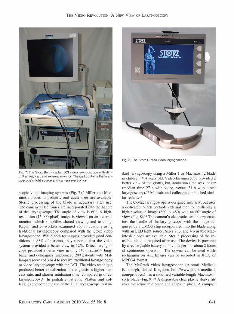
The C-Mac laryngoscope is designed similarly, but usesa dedicated 7-inch portable external monitor to display ahigh-resolution image (800 � 480) with an 80° angle ofview (Fig. 8).34 The camera’s electronics are incorporatedinto the handle of the laryngoscope, with the image ac-quired by a CMOS chip incorporated into the blade alongwith an LED light source. Sizes 2, 3, and 4 reusable Mac-intosh blades are available. Sterile processing of the re-usable blade is required after use. The device is poweredby a rechargeable battery supply that permits about 2 hoursof continuous operation. The system can be used whilerecharging on AC. Images can be recorded in JPEG orMPEG4 format.
The McGrath video laryngoscope (Aircraft Medical,Edinburgh, United Kingdom, http://www.aircraftmedical.com/products) has a modified variable-length Macintosh-style blade (Fig. 9).35 A disposable clear plastic sleeve fitsover the adjustable blade and snaps in place. A compact
Fig. 7. The Storz Berci-Kaplan DCI video laryngoscope with diffi-cult airway cart and external monitor. The cart contains the laryn-goscope’s light source and camera electronics.
Fig. 8. The Storz C-Mac video laryngoscope.
THE VIDEO REVOLUTION: A NEW VIEW OF LARYNGOSCOPY
RESPIRATORY CARE • AUGUST 2010 VOL 55 NO 8 1041

1.7-inch LCD VGA (video graphics array) view screen ismounted on the handle. The screen angle is adjustable.The device is powered by a single AA battery, whichprovides about an hour of use, and uses a high-intensityLED light source. Shippey and McKeown, describing theirinitial experience with the McGrath laryngoscope in 150normal patients, reported a 98% success rate.36 O’Learyand co-workers reported successful endotracheal intuba-tion with the McGrath in 30 instances in which traditionallaryngoscopy had failed.37 In inexperienced hands, how-ever, Walker and colleagues concluded that the McGrathoffered no advantage to traditional laryngoscopy, and theyfound the intubation time longer than with direct laryn-goscopy (median 47 s vs 30 s).38
GlideScope
The GlideScope (Verathon, Bothell, Washington, http://www.verathon.com/glidescope_index.htm) uses a plasticblade curved to a 60° angle to visualize the glottis.39,40
Several different models are available: the GVL and Rangerhave reusable blades; the Cobalt, Cobalt AVL, and RangerSingle-Use have disposable clear plastic blades that fitover a flexible video bundle. All use a CMOS camera chipwith an anti-fogging mechanism and an LED light source.The image is displayed on a separate view screen. TheGVL and Cobalt (Fig. 10) use a 7-inch 320 � 240 mon-itor, which also has a video-out port. The Cobalt AVL hasa higher-resolution camera, a 6.4-inch VGA (640 � 480)monitor, and is capable of recording images in MPEG4
format (Fig. 11). The monitor also has video-out and USBports. A separate digital video recorder is available. TheRanger devices, which have a 3.5-inch, 320 � 240 pixelmonitor, are highly portable and ruggedly designed foremergency or pre-hospital use. The Ranger is powered bya rechargeable battery that permits 90 min of continuoususe. The GVL and AVL can use either battery or ACpower. Various blades sizes are available. Intubation withthe GlideScope is generally facilitated with a curved rigidstylet.
In general, the GlideScope appears to increase the like-lihood of a good laryngoscopic view, compared with tra-ditional techniques.41,42 Stroumpoulis and colleagues per-formed direct laryngoscopy followed by laryngoscopy withthe GlideScope in 112 patients with predicted difficultintubation.43 The percentage of Comack grade 1 and 2views increased from 63% to 90% with the GlideScope,and intubation was successful in 98% of cases. In pediatricpatients, Kim and colleagues reported that the laryngo-
Fig. 9. The McGrath video laryngoscope. (Courtesy of Aircraft Med-ical Limited.)
Fig. 10. The GlideScope Cobalt GVL video laryngoscope. The dis-posable plastic blade snaps onto the reusable video bundle. The“hockey stick” stylet generally facilitates passage of the endotra-cheal tube.
THE VIDEO REVOLUTION: A NEW VIEW OF LARYNGOSCOPY
1042 RESPIRATORY CARE • AUGUST 2010 VOL 55 NO 8

scopic view with GlideScope was equal to or better thanthat with direct laryngoscopy.44 The average time requiredfor tracheal intubation, however, was longer with theGlideScope (36 s vs 24 s). Serocki and co-workers com-pared direct and video laryngoscopic (DCI and GlideScope)views of 120 patients with at least one predictor for dif-ficult intubation.45 They reported a Comack view � 3 in30% with direct laryngoscopy. The incidence was reducedto 11% with either the DCI or the GlideScope. The laryn-goscopic view was improved with the DCI in 64% ofcases, and in 94% with the GlideScope. Intubation wasunsuccessful in 10% with direct laryngoscopy, but only in2.5% of cases with the DCI or GlideScope.
Learning how to use the GlideScope appears to be quiteintuitive, and the learning curve for the device is steep.46
Nouruzi-Sedeh and colleagues studied 20 untrained vol-unteers who attempted 5 intubations with either direct la-ryngoscopy or the GlideScope in patients scheduled forelective surgery.47 The likelihood of successful intubationwas higher (93% vs 51%, P � .01) when the GlideScopewas used, and the time required for intubation was shorter(63 � 30 s vs 89 � 35 s, P � .01). Not all studies,however, have been uniformly positive, perhaps reflectinga certain learning curve for the technique. In a nonrandomobservational study of intubations by residents in an emer-gency department, Platts-Mills and colleagues reported nodifference in success on first intubation attempt for the
GlideScope (81%), compared with direct laryngoscopy(84%), and intubation with the GlideScope took more timeto complete (42 s vs 30 s).48
The separate view screen can be helpful when access tothe patient’s head is limited. Nakstad and Sandberg exam-ined the use of the GlideScope Ranger during intubationsof a simulated entrapped patient.49 In this study, 8 anes-thesiologists intubated a manikin with access only to thecaudal end of the head. While only half could successfullyaccomplish the intubation with a Macintosh laryngoscope,all could secure the airway within 60 seconds using theGlideScope. The utility of the GlideScope Ranger in thehands of paramedics performing out-of-hospital intuba-tions has been studied in a sequential study of 300 patientsundergoing direct laryngoscopy, compared with 315 pa-tients intubated with the GlideScope.15 Intubation with theGlideScope was quicker (average 21 s vs 42 s) and re-quired fewer attempts (1.2 vs 2.3 attempts). The overallsuccess rates were similar (97% vs 95%).
Which One Is Best?
Is one model of video laryngoscope “the best”? Proba-bly not. Van Zundert and co-workers compared successrates of the GlideScope Ranger, Storz DCI, and McGrathwhen used for endotracheal intubation without a stylet in450 patients with normal airways who were to undergoelective surgery. All patients were successfully intubatedwith the assigned laryngoscopic technique, and all videotechniques provided an equal or better view of the glottiscompared with traditional direct laryngoscopy with aMacintosh blade. Intubation with the DCI laryngoscopewas faster (average 18 s) compared with the GlideScope(34 s) or the McGrath (38 s). A stylet had to be used inonly 7% of cases with the DCI, compared with 43% withthe GlideScope and 59% with the McGrath.50
In a follow-up study, this same group specificallyrandomized obese patients to laryngoscopy with theGlideScope Ranger, Storz DCI, or McGrath, comparedwith traditional direct laryngoscopy.51 All the video tech-niques offered a view that was equal to or better thandirect laryngoscopy. The time required for intubation was33 s with the GlideScope, 17 s with the C-Mac, and 41 swith the McGrath, but a stylet was used only if the intu-bation was not successful after 2 attempts. This probablyput the GlideScope, with its 60° angulated blade, at arelative disadvantage.
Each model of video laryngoscope has its own uniquestrengths, weaknesses, and best applications. No one modelappears uniformly superior to another, and none is 100%successful. That said, those instruments that appear fa-miliar and intuitive to the experienced user, such as theC-Mac, Coopdech, GlideScope, and McGrath, may be moreeasily accepted into clinical practice. The use of guide
Fig. 11. The GlideScope Cobalt AVL video laryngoscope.
THE VIDEO REVOLUTION: A NEW VIEW OF LARYNGOSCOPY
RESPIRATORY CARE • AUGUST 2010 VOL 55 NO 8 1043

channel devices, however, appears easy to learn in multi-ple studies and may be advantageous when cervical spinemobility or mouth opening is limited. Devices capable ofdisplaying the image on a separate view screen may bebetter when access to the patient’s head is poor, whenteaching, or during difficult or emergency intubation whenthe assistance of additional individuals is needed. The la-ryngoscopic view and the difficulty or success of intuba-tion is easily communicated when all medical personnel inattendance can share the view.
Summary
Video techniques are quickly replacing direct laryngos-copy in many practices, especially when teaching novicesor when intubation is anticipated to be difficult.52 It islikely that video techniques will continue to evolve, andthat video capability will become available routinely as afirst alternative to direct laryngoscopy. In my own insti-tution, video laryngoscopy already has become our tech-nique of choice, next to fiberoptic techniques, for antici-pated difficult intubations.
REFERENCES
1. Peterson GN, Domino KB, Caplan RA, Posner KL, Lee LA, CheneyFW. Management of the difficult airway: a closed claims analysis.Anesthesiology 2005;103(1):33-39.
2. Practice guidelines for management of the difficult airway: an up-dated report by the American Society of Anesthesiologists TaskForce on Management of the Difficult Airway. Anesthesiology 2003;98(5):1269-1277.
3. Kaplan MB, Ward DS, Berci G. A new video laryngoscope: an aidto intubation and teaching. J Clin Anesth 2002;14(8):620-626.
4. Kaplan MB, Ward D, Hagberg CA, Berci G, Hagiike M. Seeing isbelieving: the importance of video laryngoscopy in teaching and inmanaging the difficult airway. Surg Endosc 2006;20(Suppl 2):S479-S483.
5. You JS, Park S, Chung SP, Park YS, Park JW. The usefulness of theGlideScope video laryngoscope in the education of conventionaltracheal intubation for the novice. Emerg Med J 2009;26(2):109-111.
6. Sibert K, Ricci MA, Caputo M, Callas PW, Rogers FB, Charash W,et al. The feasibility of using ultrasound and video laryngoscopy ina mobile telemedicine consult. Telemed J E Health 2008;14(3):266-272.
7. Boedeker BH, Berg BW, Bernhagen M, Murray WB. Direct versusindirect laryngoscopic visualization in human endotracheal intuba-tion: a tool for virtual anesthesia practice and teleanesthesiology.Stud Health Technol Inform 2008;132:31-36.
8. Low D, Healy D, Rasburn N. The use of the BERCI DCI videolaryngoscope for teaching novices direct laryngoscopy and trachealintubation. Anaesthesia 2008;63(2):195-201.
9. Howard-Quijano KJ, Huang YM, Matevosian R, Kaplan MB, Stead-man RH. Video-assisted instruction improves the success rate fortracheal intubation by novices. Br J Anaesth 2008;101(4):568-572.
10. Cooper RM. Complications associated with the use of the GlideScopevideolaryngoscope. Can J Anaesth 2007;54(1):54-57.
11. Malik AM, Frogel JK. Anterior tonsillar pillar perforation duringGlideScope video laryngoscopy. Anesth Analg 2007;104(6):1610-1611, discussion 1611.
12. Cross P, Cytryn J, Cheng KK. Perforation of the soft palate using theGlideScope videolaryngoscope. Can J Anaesth 2007;54(7):588-589.
13. Vincent RD Jr, Wimberly MP, Brockwell RC, Magnuson JS. Softpalate perforation during orotracheal intubation facilitated by theGlideScope videolaryngoscope. J Clin Anesth 2007;19(8):619-621.
14. Williams D, Ball DR. Palatal perforation associated with McGrathvideolaryngoscope. Anaesthesia 2009;64(10):1144-1145.
15. Wayne MA, McDonnell M. Comparison of traditional versus videolaryngoscopy in out-of-hospital tracheal intubation. Prehosp EmergCare 2010;14(2):278-282.
16. Bjoernsen LP, Parquette BT, Lindsay MB. Prehospital use of videolaryngoscope by an air medical crew. Air Med J 2008;27(5):242-244.
17. Berg B, Walker RA, Murray WB, Boedeker BH. Airway intubationin a helicopter cabin: video vs. direct laryngoscopy in manikins.Aviat Space Environ Med 2009;80(9):820-823.
18. Bjoernsen LP, Lindsay B. Video laryngoscopy in the prehospitalsetting. Prehosp Disaster Med 2009;24(3):265-270.
19. Bonfils P. [Difficult intubation in Pierre-Robin children, a new meth-od: the retromolar route]. Anaesthesist 1983;32(7):363-367. Articlein German.
20. Bein B, Worthmann F, Scholz J, Brinkmann F, Tonner PH, SteinfathM, et al. A comparison of the intubating laryngeal mask airway andthe Bonfils intubation fibrescope in patients with predicted difficultairways. Anaesthesia 2004;59(7):668-674.
21. Bein B, Yan M, Tonner PH, Scholz J, Steinfath M, Dorges V. Tra-cheal intubation using the Bonfils intubation fibrescope after faileddirect laryngoscopy. Anaesthesia 2004;59(12):1207-1209.
22. Turkstra TP, Pelz DM, Jones PM. Cervical spine motion: a fluoro-scopic comparison of the AirTraq laryngoscope versus the Mac-intosh laryngoscope. Anesthesiology 2009;111(1):97-101.
23. Hirabayashi Y, Fujita A, Seo N, Sugimoto H. A comparison ofcervical spine movement during laryngoscopy using the Airtraq orMacintosh laryngoscopes. Anaesthesia 2008;63(6):635-640.
24. Hirabayashi Y, Seo N. Airway Scope: early clinical experience in405 patients. J Anesth 2008;22(1):81-85.
25. Asai T, Liu EH, Matsumoto S, Hirabayashi Y, Seo N, Suzuki A,et al. Use of the Pentax-AWS in 293 patients with difficult airways.Anesthesiology 2009;110(4):898-904.
26. Suzuki A, Toyama Y, Katsumi N, Kunisawa T, Sasaki R, Hirota K,et al. The Pentax-AWS rigid indirect video laryngoscope: clinicalassessment of performance in 320 cases. Anaesthesia 2008;63(6):641-647.
27. Enomoto Y, Asai T, Arai T, Kamishima K, Okuda Y. Pentax-AWS,a new videolaryngoscope, is more effective than the Macintosh la-ryngoscope for tracheal intubation in patients with restricted neckmovements: a randomized comparative study. Br J Anaesth 2008;100(4):544-548.
28. Maruyama K, Yamada T, Kawakami R, Hara K. Randomizedcross-over comparison of cervical-spine motion with the AirWayScope or Macintosh laryngoscope with in-line stabilization: a video-fluoroscopic study. Br J Anaesth 2008;101(4):563-567.
29. Aziz M, Dillman D, Kirsch JR, Brambrink A. Video laryngoscopywith the macintosh video laryngoscope in simulated prehospital sce-narios by paramedic students. Prehosp Emerg Care 2009;13(2):251-255.
30. Kaplan MB, Hagberg CA, Ward DS, Brambrink A, Chhibber AK,Heidegger T, et al. Comparison of direct and video-assisted views ofthe larynx during routine intubation. J Clin Anesth 2006;18(5):357-362.
31. Jungbauer A, Schumann M, Brunkhorst V, Borgers A, Groeben H.Expected difficult tracheal intubation: a prospective comparison ofdirect laryngoscopy and video laryngoscopy in 200 patients. Br JAnaesth 2009;102(4):546-550.
THE VIDEO REVOLUTION: A NEW VIEW OF LARYNGOSCOPY
1044 RESPIRATORY CARE • AUGUST 2010 VOL 55 NO 8

32. Vlatten A, Aucoin S, Litz S, Macmanus B, Soder C. A comparisonof the STORZ video laryngoscope and standard direct laryngoscopyfor intubation in the pediatric airway: a randomized clinical trial.Paediatr Anaesth 2009;19(11):1102-1107.
33. Macnair D, Baraclough D, Wilson G, Bloch M, Engelhardt T. Pe-diatric airway management: comparing the Berci-Kaplan video la-ryngoscope with direct laryngoscopy. Paediatr Anaesth 2009;19(6):577-580.
34. Cavus E, Kieckhaefer J, Doerges V, Moeller T, Thee C, Wagner K.The C-MAC videolaryngoscope: first experiences with a new devicefor videolaryngoscopy-guided intubation. Anesth Analg 2010;110(2):473-477.
35. Shippey B, Ray D, McKeown D. Use of the McGrath videolaryn-goscope in the management of difficult and failed tracheal intuba-tion. Br J Anaesth 2008;100(1):116-119.
36. Shippey B, Ray D, McKeown D. Case series: the McGrath video-laryngoscope: an initial clinical evaluation. Can J Anaesth 2007;54(4):307-313.
37. O’Leary AM, Sandison MR, Myneni N, Cirilla DJ, Roberts KW,Deane GD. Preliminary evaluation of a novel videolaryngoscope, theMcGrath series 5, in the management of difficult and challengingendotracheal intubation. J Clin Anesth 2008;20(4):320-321.
38. Walker L, Brampton W, Halai M, Hoy C, Lee E, Scott I, et al.Randomized controlled trial of intubation with the McGrath Series 5videolaryngoscope by inexperienced anaesthetists. Br J Anaesth2009;103(3):440-445.
39. Cooper RM. Use of a new videolaryngoscope (GlideScope) in the man-agement of a difficult airway. Can J Anaesth 2003;50(6):611-613.
40. Cooper RM. The GlideScope videolaryngoscope. Anaesthesia 2005;60(10):1042.
41. Cooper RM, Pacey JA, Bishop MJ, McCluskey SA. Early clinicalexperience with a new videolaryngoscope (GlideScope) in 728 pa-tients. Can J Anaesth 2005;52(2):191-198.
42. Sun DA, Warriner CB, Parsons DG, Klein R, Umedaly HS, MoultM. The GlideScope video laryngoscope: randomized clinical trial in200 patients. Br J Anaesth 2005;94(3):381-384.
43. Stroumpoulis K, Pagoulatou A, Violari M, Ikonomou I, Kalantzi N,Kastrinaki K, et al. Videolaryngoscopy in the management of the
difficult airway: a comparison with the Macintosh blade. Eur J An-aesthesiol 2009;26(3):218-222.
44. Kim JT, Na HS, Bae JY, Kim DW, Kim HS, Kim CS, et al.GlideScope video laryngoscope: a randomized clinical trial in 203paediatric patients. Br J Anaesth 2008;101(4):531-534.
45. Serocki G, Bein B, Scholz J, Dorges V. Management of the predicteddifficult airway: a comparison of conventional blade laryngoscopywith video-assisted blade laryngoscopy and the GlideScope. Eur JAnaesthesiol 2010;27(1):24-30.
46. Siu LW, Mathieson E, Naik VN, Chandra D, Joo HS. Patient- andoperator-related factors associated with successful Glidescope intu-bations: a prospective observational study in 742 patients. AnaesthIntensive Care;38(1):70-75.
47. Nouruzi-Sedeh P, Schumann M, Groeben H. Laryngoscopy viaMacintosh blade versus GlideScope: success rate and time for en-dotracheal intubation in untrained medical personnel. Anesthesiol-ogy 2009;110(1):32-37.
48. Platts-Mills TF, Campagne D, Chinnock B, Snowden B, GlickmanLT, Hendey GW. A comparison of GlideScope video laryngoscopyversus direct laryngoscopy intubation in the emergency department.Acad Emerg Med 2009;16(9):866-871.
49. Nakstad AR, Sandberg M. The GlideScope Ranger video laryngo-scope can be useful in airway management of entrapped patients.Acta Anaesthesiol Scand 2009;53(10):1257-1261.
50. van Zundert A, Maassen R, Lee R, Willems R, Timmerman M,Siemonsma M, et al. A Macintosh laryngoscope blade for video-laryngoscopy reduces stylet use in patients with normal airways.Anesth Analg 2009;109(3):825-831.
51. Maassen R, Lee R, Hermans B, Marcus M, van Zundert A. Acomparison of three videolaryngoscopes: the Macintosh laryngo-scope blade reduces, but does not replace, routine stylet use forintubation in morbidly obese patients. Anesth Analg 2009;109(5):1560-1565.
52. Saxena S. The ASA difficult airway algorithm: is it time to includevideo laryngoscopy and discourage blind and multiple intubationattempts in the nonemergency pathway? Anesth Analg 2009;108(3):1052.
THE VIDEO REVOLUTION: A NEW VIEW OF LARYNGOSCOPY
RESPIRATORY CARE • AUGUST 2010 VOL 55 NO 8 1045