thigh, hip, & low back evaluation
TRANSCRIPT

Thigh, Hip, & Low Thigh, Hip, & Low Back EvaluationBack Evaluation
www.fisiokinesiterapia.biz
Thigh InjuriesThigh InjuriesQuad contusions - Myositis Ossificans
Trochanteric bursitis - “snapping hip”
Ischial bursitis - “bench-warmer’s bursitis”
Strains Fractures - femur, stress, apophysitisIT band friction syndrome – may predispose an athlete - leg length difference
Hip InjuriesHip InjuriesFractures – pelvis, ischial tuberosity (hamstring attachment)
Iliac crest contusion - hip pointerSprains – hipHip dislocation
Posterior dislocation – lower leg position – adduction & internal rotation
Piriformis syndrome –Proximal insertion: sacrum Distal insertion: medial aspect of greater trochanter
*can compress sciatic nerve between ischium & greater trochanter if tight
LowbackLowback InjuriesInjuriesContusions, lacerations, subluxations, sprains, strains Disc & Neurologic PathologyFractures - stress, apophysitisSpondylosis- arthritis or osteoarthritis of the vertebrae; results in pressures being placed on the vertebral nerve rootsSpondylolysis- degeneration of a vertebral structure secondary to repetitive stress
most commonly affecting pars interarticularis but with no displacement of the vertebral body (Scotty dog)
Spondylolisthesis - anterior slippage of vertebrae superior to pathological site; causes increased back pain upon extension
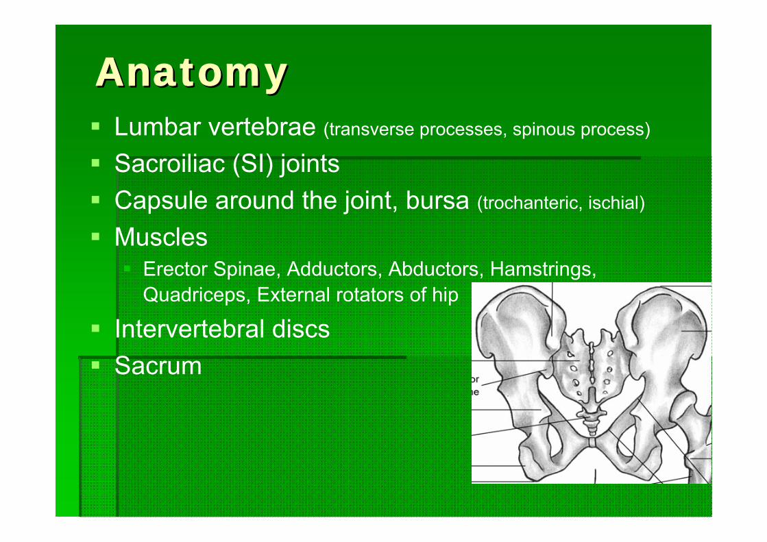
AnatomyAnatomyLumbar vertebrae (transverse processes, spinous process)
Sacroiliac (SI) joints Capsule around the joint, bursa (trochanteric, ischial)
MusclesErector Spinae, Adductors, Abductors, Hamstrings, Quadriceps, External rotators of hip
Intervertebral discsSacrum
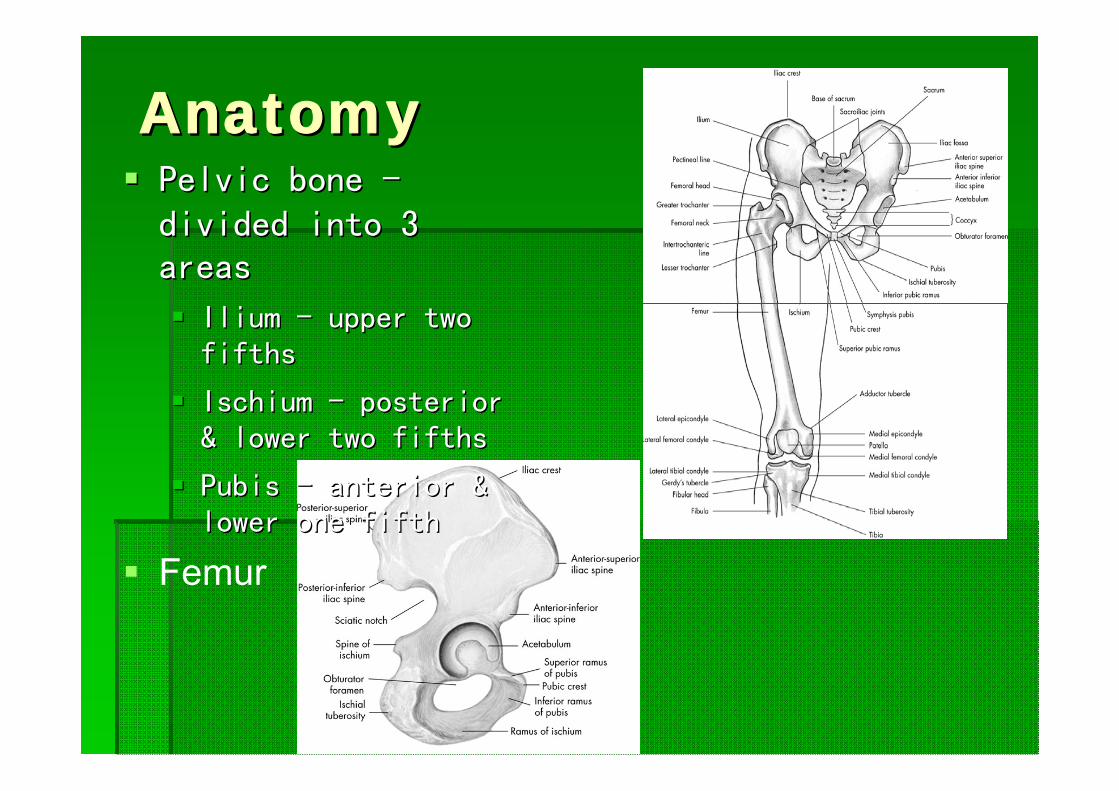
Anatomy Anatomy Pelvic bone Pelvic bone --divided into 3 divided into 3 areasareas
Ilium Ilium -- upper two upper two fifthsfifths
IschiumIschium -- posterior posterior & lower two fifths& lower two fifths
Pubis Pubis -- anterior & anterior & lower one fifth lower one fifth
Femur
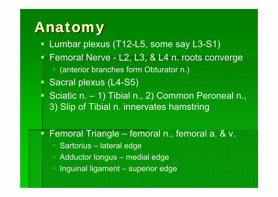
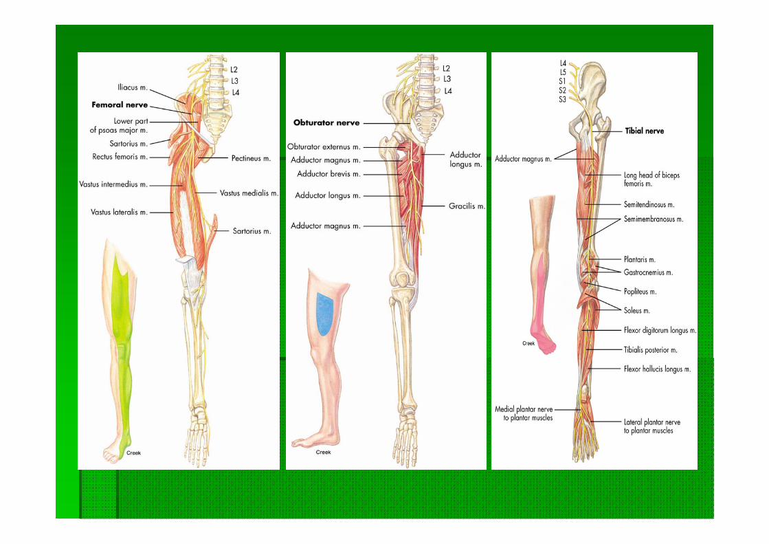
AnatomyAnatomyLumbar plexus (T12-L5, some say L3-S1)Femoral Nerve - L2, L3, & L4 n. roots converge
(anterior branches form Obturator n.)Sacral plexus (L4-S5)Sciatic n. – 1) Tibial n., 2) Common Peroneal n., 3) Slip of Tibial n. innervates hamstring
Femoral Triangle – femoral n., femoral a. & v.Sartorius – lateral edge Adductor longus – medial edge Inguinal ligament – superior edge
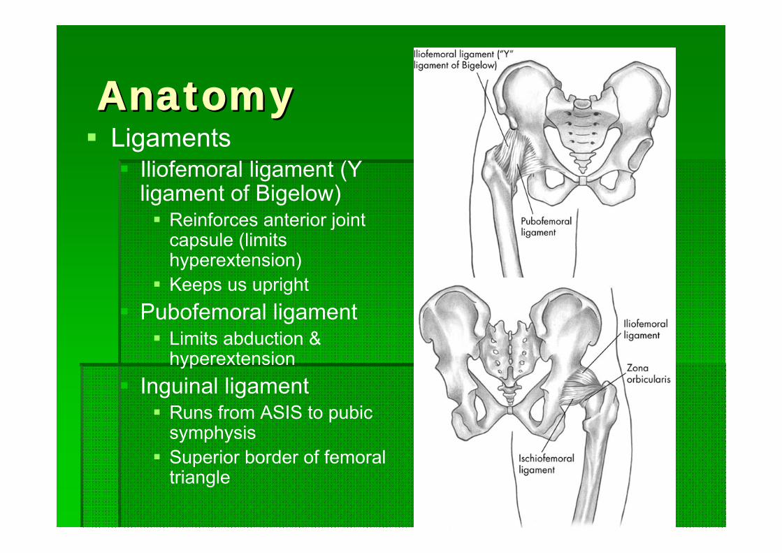
AnatomyAnatomyLigaments
Iliofemoral ligament (Y ligament of Bigelow)
Reinforces anterior joint capsule (limits hyperextension)Keeps us upright
Pubofemoral ligamentLimits abduction & hyperextension
Inguinal ligamentRuns from ASIS to pubic symphysisSuperior border of femoral triangle
Evaluation Evaluation -- HistoryHistoryMOI – direct blow vs. overuse
Pain Pain scale, Type, Location, Time, Consistency (constant/intermittent)Radiating, numbness/tingling, burning, aching, throbbing
Onset Acute vs. Chronic
Training TechniquesChanges in intensity, frequency, & duration of training, surface(terrain, hills), shoes
Prior history Legg-Calve-Perthes disease – avascular necrosis of prox. femoral epiphysis
Evaluation Evaluation -- ObservationObservationDeformities MusculatureBony –
Nelaton’s line - ASIS to ischial tuberosityHip angulations
Angle of Inclination – angular relationship between femur & tibiaFrontal plane Coxa valga, coxa vara, patellar positionNormally 125º (♀ slightly ↓)
Angle of Torsion – relationship between femoral head & shaftTransverse planeNormally 15º
Anteversion: internal femoral rotation, toed-in gait (pigeon-toed), squinting patellaeRetroversion: external femoral rotation, toed-out gait (duck-footed), frog-eyed patellae
Evaluation Evaluation -- ObservationObservationLeg Length Discrepancy
True Leg Length – ASIS to medial malleolusBONY
Apparent Leg Length – Umbilicus to medial malleolusSOFT TISSUE
Greater than ¼” difference is considered a discrepancy
Level of iliac crest when standingGait –
Level of iliac crestROMLimp
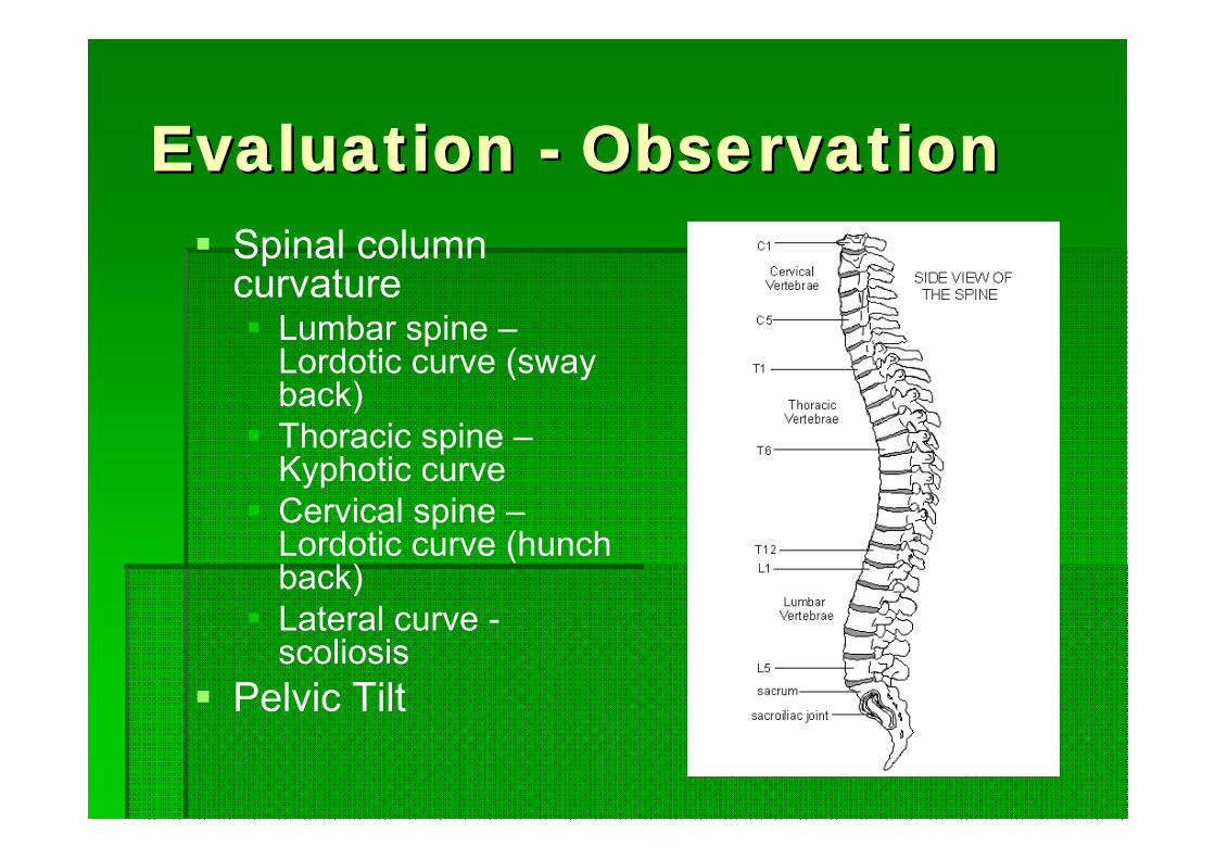
Evaluation Evaluation -- ObservationObservationSpinal column curvature
Lumbar spine –Lordotic curve (sway back)Thoracic spine –Kyphotic curveCervical spine –Lordotic curve (hunch back)Lateral curve -scoliosis
Pelvic Tilt
Evaluation Evaluation -- PalpationPalpation
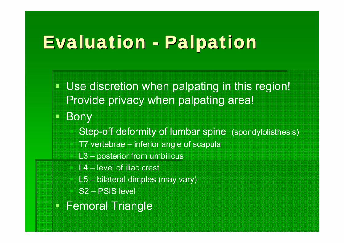
Use discretion when palpating in this region! Provide privacy when palpating area!Bony
Step-off deformity of lumbar spine (spondylolisthesis)T7 vertebrae – inferior angle of scapulaL3 – posterior from umbilicusL4 – level of iliac crestL5 – bilateral dimples (may vary)S2 – PSIS level
Femoral Triangle
Evaluation Evaluation –– Palpation Palpation MusclesMuscles
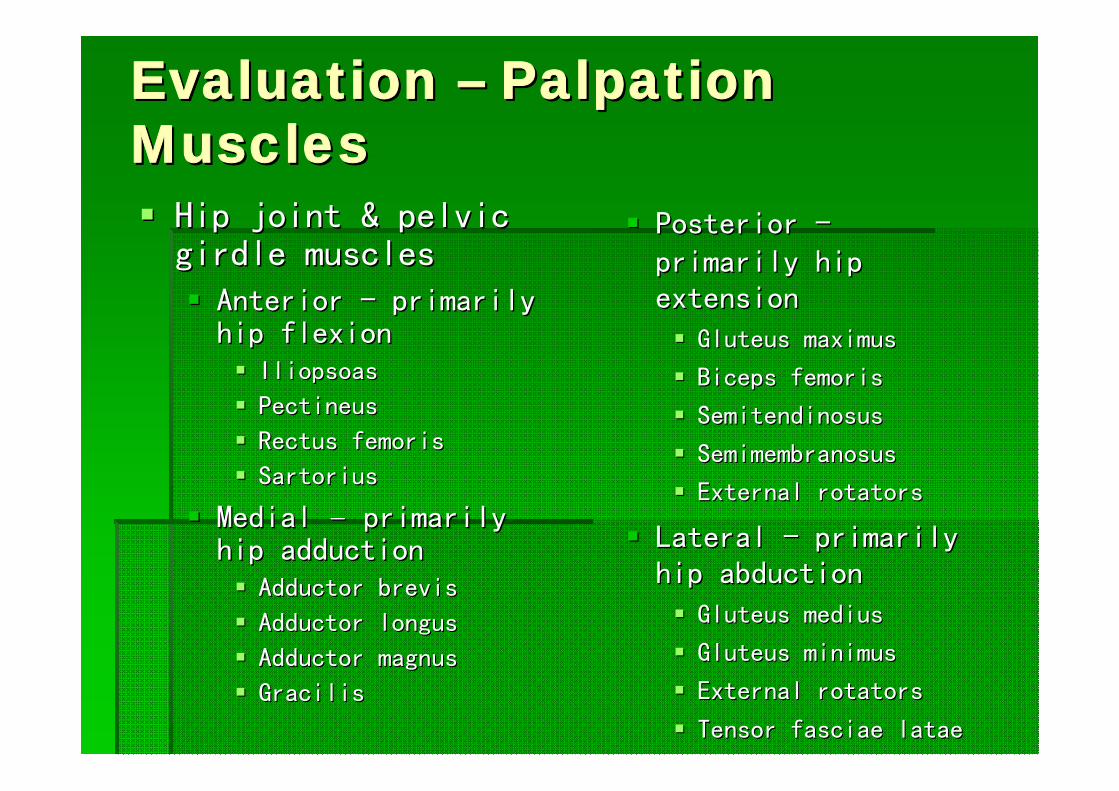
Hip joint & pelvic Hip joint & pelvic girdle musclesgirdle muscles
Anterior Anterior -- primarily primarily hip flexionhip flexion
IliopsoasIliopsoas
PectineusPectineus
Rectus femorisRectus femoris
SartoriusSartorius
Medial Medial –– primarily primarily hip adductionhip adduction
Adductor brevisAdductor brevis
Adductor longusAdductor longus
Adductor magnusAdductor magnus
GracilisGracilis
Posterior Posterior --primarily hip primarily hip extensionextension
Gluteus maximusGluteus maximus
Biceps femorisBiceps femoris
SemitendinosusSemitendinosus
SemimembranosusSemimembranosus
External rotators External rotators
Lateral Lateral -- primarily primarily hip abductionhip abduction
GluteusGluteus mediusmedius
GluteusGluteus minimusminimus
External rotatorsExternal rotators
Tensor fasciae Tensor fasciae lataelatae
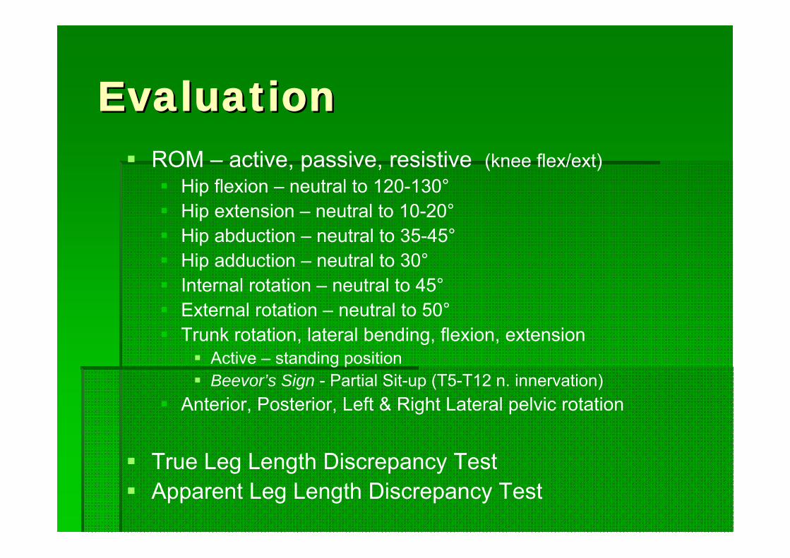
Evaluation Evaluation ROM – active, passive, resistive (knee flex/ext)
Hip flexion – neutral to 120-130°Hip extension – neutral to 10-20°Hip abduction – neutral to 35-45°Hip adduction – neutral to 30°Internal rotation – neutral to 45°External rotation – neutral to 50°Trunk rotation, lateral bending, flexion, extension
Active – standing positionBeevor’s Sign - Partial Sit-up (T5-T12 n. innervation)
Anterior, Posterior, Left & Right Lateral pelvic rotation
True Leg Length Discrepancy TestApparent Leg Length Discrepancy Test
Evaluation Evaluation –– Stress TestsStress Tests
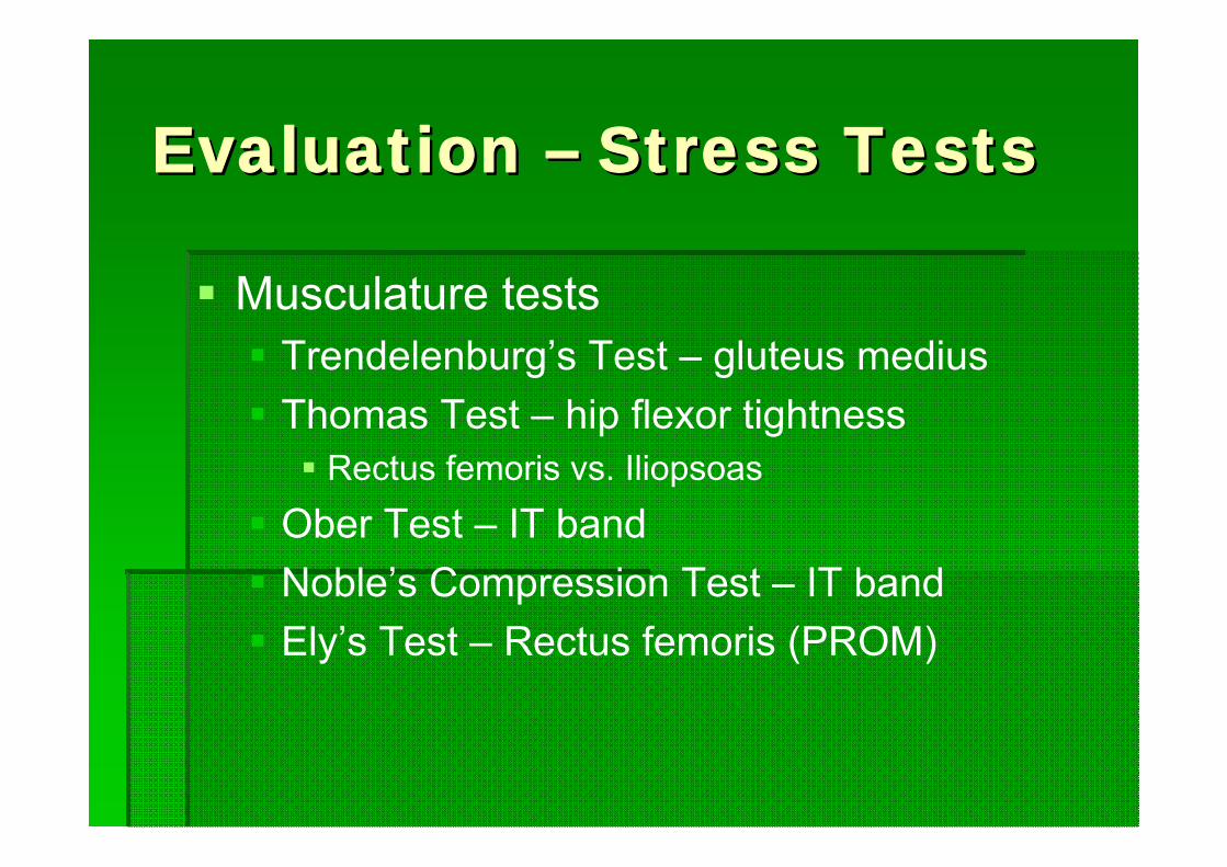
Musculature testsTrendelenburg’s Test – gluteus mediusThomas Test – hip flexor tightness
Rectus femoris vs. IliopsoasOber Test – IT bandNoble’s Compression Test – IT bandEly’s Test – Rectus femoris (PROM)

Evaluation Evaluation –– Stress TestsStress TestsLigamentous testing – no specific testsNeurologic testing –
Beevor’s Sign – thoracic n. inhibitionPiriformis Test
Piriformis Syndrome – impingement of sciatic n. from spasm of piriformis
Resisted hip abd. while seated can duplicate pain caused by this syndromeStraight Leg Raise (SLR) test – sciatic n. irritation or disc (Passive)Well SLR test – disc (opposite side)Increased Intrathecal Pressure
Valsalva test (maneuver) – herniated discMilgram test – disc (active double SLR)Kernig’s test or Kernig/Brudzinski test – disc (active SLR w/ knee extended)
90-90 SLRSlump test – sciatic or other neurologicQuadrant test – nerve vs. facet
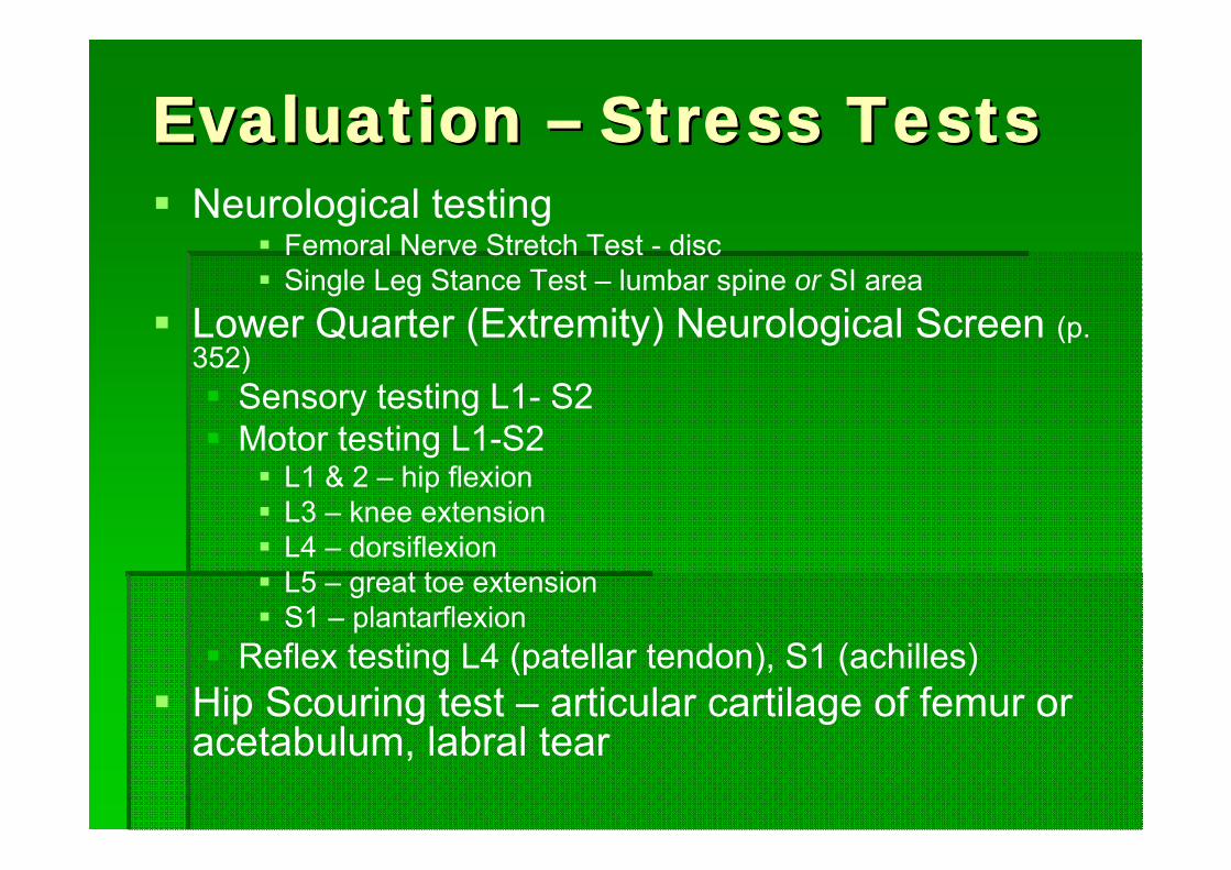
Evaluation Evaluation –– Stress TestsStress TestsNeurological testing
Femoral Nerve Stretch Test - discSingle Leg Stance Test – lumbar spine or SI area
Lower Quarter (Extremity) Neurological Screen (p. 352)
Sensory testing L1- S2Motor testing L1-S2
L1 & 2 – hip flexionL3 – knee extensionL4 – dorsiflexionL5 – great toe extensionS1 – plantarflexion
Reflex testing L4 (patellar tendon), S1 (achilles)Hip Scouring test – articular cartilage of femur or acetabulum, labral tear
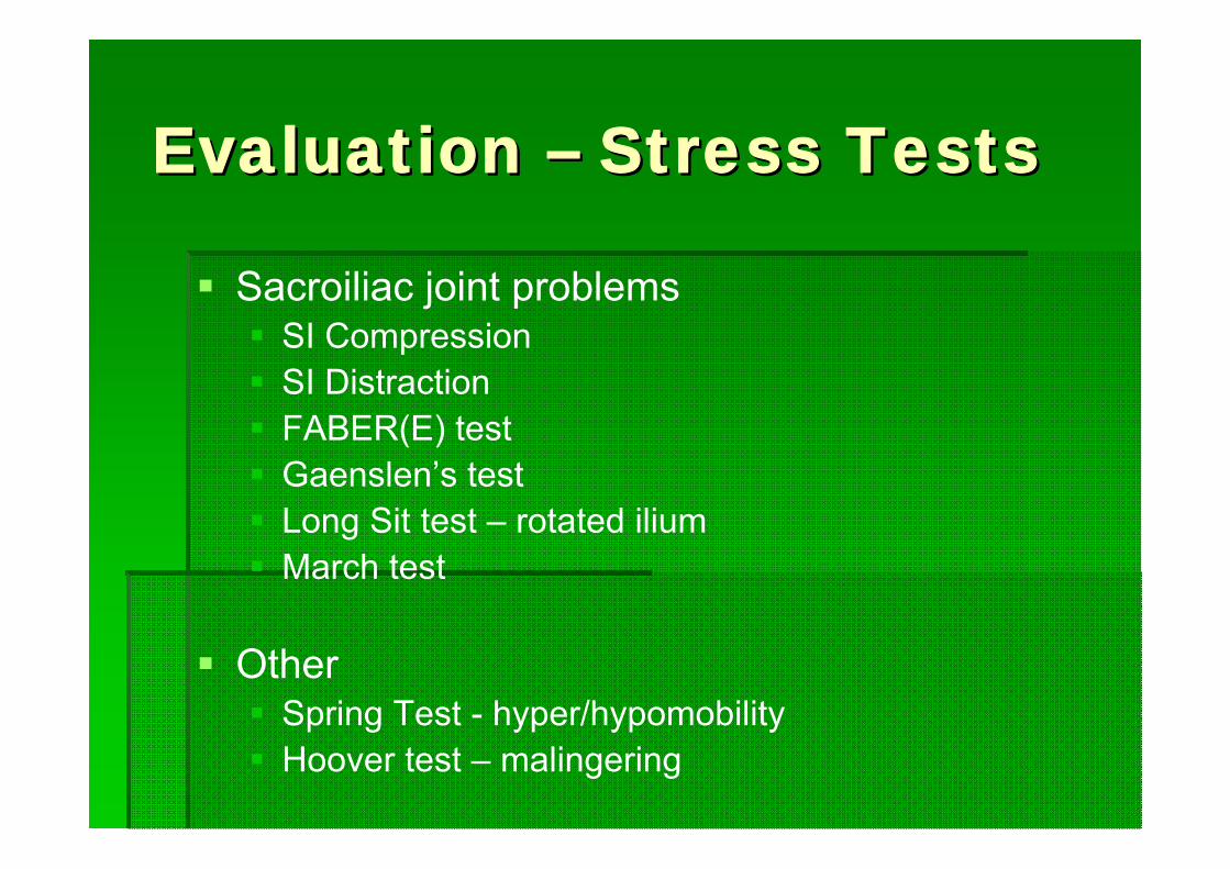
Evaluation Evaluation –– Stress TestsStress Tests
Sacroiliac joint problemsSI Compression SI DistractionFABER(E) test Gaenslen’s testLong Sit test – rotated iliumMarch test
OtherSpring Test - hyper/hypomobilityHoover test – malingering
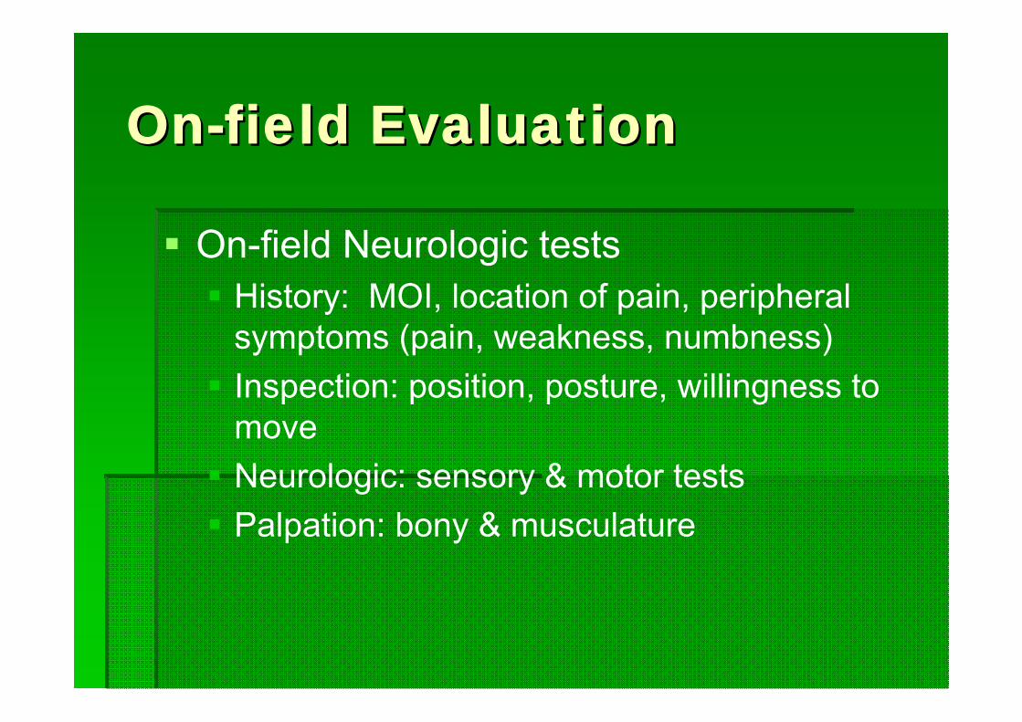
OnOn--field Evaluationfield Evaluation
On-field Neurologic testsHistory: MOI, location of pain, peripheral symptoms (pain, weakness, numbness)Inspection: position, posture, willingness to moveNeurologic: sensory & motor testsPalpation: bony & musculature