three dimensional musculoskeletal modelling of the
TRANSCRIPT

1
Three dimensional musculoskeletal modelling of the abdominal
crunch resistance training exercise
Kim Nolte1, Pieter E. Krüger1, P. Schalk Els2 & Heinrich W. Nolte3
1Department of Biokinetics, Sport and Leisure Sciences, University of Pretoria, Pretoria, South Africa,
2Department of Mechanical and Aeronautical Engineering, University of Pretoria, Pretoria, South
Africa, 3Ergonomics Technologies, Pretoria, South Africa
Abstract
The aim of this study was to evaluate the benefits and limitations of using three
dimensional (3D) musculoskeletal modelling (LifeModelerTM) in assessing the safety
and efficacy of exercising on an abdominal crunch resistance training machine.
Three anthropometric cases were studied, representing a 5th percentile female, 50th
percentile and 95th percentile male. Results indicated that the LifeModelerTM default
model was capable of solving the forward dynamics simulations without adjustments.
The modelling was able to indicate high risk for back injury when performing the
abdominal crunch exercise as a result of the unacceptable intervertebral joint loading
that occurs during the exercise. Individuals with small anthropometric dimensions
such as some females and children cannot be accommodated suitably on the
abdominal crunch machine which negatively impacts exercise posture and
technique. Hip flexor muscle contribution in the execution of the exercise for the 5th
percentile female was substantial thus reducing the efficacy of the exercise in
isolating the abdominal muscles.
Keywords: Resistance training equipment, abdominal crunch, LifemodelerTM,
inverse dynamics, forward dynamics

2
Introduction
The increased popularity of, and participation in resistance training worldwide is
indicative of the level of interest in benefits derivable from this type of training
(Vaughn, 1989). Ironically, participation in any type of physical activity places the
exerciser in situations in which injury is likely to occur. Improvement in exercise
equipment design could reduce the risk of injury (Dabnichki, 1998) as well as
possibly increase the efficacy of the exercise.
Conceptual, physical and mathematical models have all proved useful in
biomechanics (Alexander, 2003). Mathematical and computer modelling is suitable
for a wide variety of applications such as the design, production and alteration of
medical equipment as well as sports and exercise equipment (Alexander, 2003;
Kazlauskiené, 2006). Capable of simulating musculoskeletal human models
interacting with mechanical systems, three dimensional (3D) musculoskeletal
modelling may be able to answer many questions concerning the effects of the
resistance training equipment on the body. Thus we have previously shown that this
method can successfully be used to evaluate a seated biceps curl resistance training
machine (Nolte, Krüger, & Els, 2011).
This study presents the musculoskeletal modelling of three anthropometric cases
while exercising on a commercially available seated abdominal crunch resistance
training machine. The abdominal muscles are the major supporting muscles for the
stomach area. They not only support and protect internal organs, but they aid the
muscles of the lower back to properly align and support the spine for proper posture
as well as in lifting activities (Beachle & Groves, 1992). There are several exercises

3
for the abdominal muscles, such as bent-knee sit-ups, crunches, isometric
contractions as well as exercises using specialized equipment and resistance
training machines (McGill, 1995; Nieman, 2007). Two common types of abdominal
crunch resistance training machines available include, machines that have the
resistance at the back of the upper body and the exerciser has to grasp two handle
bars in front of the chest as opposed to machines that have the resistance in the
front of the chest in the form of a cushion or pad and the exerciser places his or her
arms over the pad. In this study, the latter abdominal resistance training machine
was utilized. Controversy remains as to which exercise method best activates the
muscles of the abdomen and minimizes potentially harmful or excessive joint tissue
loading (McGill, 1995). It is generally believed that a variety of selected abdominal
exercises are required to sufficiently challenge the abdominal muscles and that
these exercises will differ to best meet the different training objectives of the
individual (Axler & McGill, 1997).
Evaluation methods are required to ensure equipment efficacy as well as the safety
of the end-user. Thus, the primary aim of this study was to evaluate the benefits and
limitations of using 3D musculoskeletal modelling in evaluating the abdominal crunch
resistance training machine. We hypothesized that, 1) the LifemodelerTM default
model would be capable of solving the forward dynamics simulations without
adjustments, 2) not all individuals (varying anthropometric dimensions) would be
suitably accommodated by the abdominal crunch machine, 3) the abdominal crunch
resistance training exercise places the exerciser at risk for back injury due to the fact
regular abdominal exercises without resistance have been associated with large

4
loads on the spine (McGill, 1995) and, 4) unsuitable accommodation on the
abdominal crunch machine would negatively impact exercise safety and efficacy.
It should be noted that although the results from this study may be generalised to
other computer modelling software, there may be certain aspects that are unique to
the modelling software used in this study.
Methods
Equipment
Three sex specific 3D musculoskeletal full body models were created using
LifeModeler™ software and incorporated into a multibody dynamics model of the
abdominal crunch machine modelled in MSC ADAMS (Figure 1). The LifeModeler™
(San Clemente, USA) software runs as a plug-in on the MSC ADAMS software.
LifeModelerTM software has previously been used in studies in the fields of sport,
exercise and medicine (Agnesina et al., 2006; De Jongh, 2007; Hofmann, Danhard,
Betzler, Witte & Edelmann, 2006; Olesen, Andersen, Rathleff, de Zee & Rasmussen,
2009; Nolte et al., 2011; Rietdyk & Patla., 1999; Schillings, Van Wezel & Duysens,
1996). Three default models, as generated through the software, were evaluated.
These models consisted of 19 segments including a base set of joints for each body
region. The spine does not consist of individual vertebrae but rather of various
segments that represent different regions of the vertebral column with joints between
these segments. The default models had a full body set of 118 muscle elements
attached to the bones at anatomical landmarks, which includes most of the major
muscle groups in the body (Biomechanics research group, 2006).

5
Figure 1: 3D musculoskeletal modelling of the abdominal crunch resistance training machineand 95th percentile male musculoskeletal model using LifeModelerTM and MSCADAMS software.
Musculoskeletal full body human and the abdominal crunch computer aided design
(CAD) models
Models for the three anthropometric cases were created. The human models were
created using the GeBOD anthropometry database (default LifeModeler™ database)
but were based on body mass index (BMI) data obtained from RSA-MIL-STD-127
Vol 1 (2004)(Table I). This standard is a representative of the South African National
Defence Force (SANDF) which is kept current by a yearly sampling plan and can be
considered an accurate representation of the broader South Africa population. A
process described by Bredenkamp (2007) was followed to characterize the body
forms of SANDF males and females found in RSA-MIL-STD-127 Vol 1. This process
identified variances in body form as identified by principal component analysis. Two

6
principal components (PCs) for the SANDF males and females were included in the
modelling process and presented the positive boundary case (being tall and thin)
and the negative boundary case (being short and heavy). Positive and negative
boundary cases represent the boundary conditions to be accommodated in design
(Gordon & Brantley, 1997). A “small” female, an “average” male, and a “large” male
were the three anthropometric cases chosen for this study. They are traditionally
known as a 5th percentile female, 50th percentile male and a 95th percentile male
based on the BMI. Thus, for the purpose of building these biomechanical models, a
correlation between BMI and functional body strength was assumed. Similar
assumptions have previously been made in biomechanics full body model
simulations (Rasmussen et al., 2005). A study by Annegarn, Rasmussen, Savelberg,
Verdijk & Meijer (2007) also verified scaled modelling strengths against actual
functional body strengths and correlations ranged from 0.64 to 0.99.
This approach was followed in order to test whether the exercise machine could
accommodate the full spectrum of the South African end-user population. A CAD
model of the abdominal crunch resistance training machine was obtained from a
South African exercise equipment manufacturing company (Figure 2). The model in
a Parasolid file format was imported into the ADAMS simulation software.
The Adams software was used to create two design variables in order to adjust the
external resistance (as selected by the amount of weights when using a selectorised
resistance training machine) and to specify the radius of the cam over which the
cable of an actual exercise machine would run in order to lift the selected resistance.
This was possible since this machine employed a circular cam system. A special

7
contact force (solid to solid) was created between the weights being lifted and the
remainder of the weight stack during the simulation. A coupler joint was created
linking the revolute joint (driver joint) of the lever arm attached to the abdominal
crunch machine pad/cushion with the translational joint of the weight stack. The
design variable created for the radius of the cam was then referenced as part of the
function of the coupler joint in calculating the external resistance, taking into account
the resistance selected as well as the radius of the cam on the machine. The design
variable created for the mass of the weights was then adjusted according to the pre-
determined resistance for each anthropometric case, explained in the next section.
The external resistance applied in the models was based on data obtained from
Isokinetic testing results from trunk flexion (Perrin, 1993). Trunk flexion was selected
as it most closely resembles the abdominal crunch movement. Torque (Nm) values
obtained were converted to force values in Kilograms by adjusting for estimated
isokinetic testing device lever arm length for each anthropometric case. Fifty percent
of the functional strength one repetition maximum (1RM) for each anthropometric
case was used, this can be considered a manageable resistance to perform an
exercise with appropriate form and technique for four repetitions (Beachle & Groves,
1992) (Table I).
Table I. Anthropometric and user population strength data forpopulation groups studied.
User population group Body mass(kg)
Stature (mm) User population group exerciseresistance (50% 1RM) kg
5th percentile female 49.5 1500 5
50th percentile male 65.0 1720 14
95th percentile male 85.0 1840 24

8
Simulation
Extreme care was taken with the positioning of the musculoskeletal model onto the
abdominal crunch machine to ensure proper technique, posture and positioning
according to best exercise principles (Table II). The engineered adjustability of the
exercise machine was used in order to ensure correct positioning for each of the
anthropometric cases. A bushing element was applied between the lower torso and
the seat of the abdominal crunch machine as well as the two humeral bones and the
abdominal crunch machine pad/cushion. Bushing elements were preferred to fixed
joint elements because they allow for limited translational and rotational motion.
Also, the amount of motion can be controlled by changing stiffness and damping
characteristics in all three orthogonal directions. While compensatory movements
should be limited during resistance training to ensure proper technique they do occur
in most instances. Thus we applied and gradually increased the stiffness and
dampening until we achieved visually acceptable kinematics in terms of such
compensatory movements.
The inverse dynamics – forward dynamics method was applied during the
simulations. Inverse dynamics simulations are performed on models which are being
manipulated by the use of motion agents or motion splines. During the inverse
dynamics simulation, a rotational motion was applied to the revolute joint of the lever
arm attached to the pad/cushion of the abdominal crunch machine in order to
generate the required movement of the resistance training machine. This movement
replicated the pulling (concentric) and resisting (eccentric) phase of the exercise.
The time for the concentric phase was set at 1.33 seconds and the eccentric phase
slightly longer at 2.66 seconds to mimic conventional resistance training technique in

9
which the eccentric phase is more deliberate to prohibit the use of momentum
(Schilling et al., 2008). The 1.33 second concentric phase included a STEP function
(ramp-up period) approximation over 0.5 seconds to ensure a gradual start to the
movement. The muscles of the model were “trained” during the inverse dynamics
simulation in order to calculate the changes in muscle lengths to result in the
required machine movement. The movement replicated four repetitions of the
exercise separated by a slight pause between repetitions.
After the inverse dynamics simulation was performed, the rotational motion was
removed from the rotational joint of the lever arm of the abdominal crunch machine.
The recorded muscle length changes and resulting joint movements were then used
to drive the model during the forward dynamics simulation in the manner as
developed through the inverse dynamics simulation. During the forward dynamics
simulation the model is guided by the internal forces (muscle length changes
resulting in joint angulations and torques) and influenced by external forces (gravity,
contact and determined exercise resistance).
The muscle elements used during the modelling in this study are referred to as
closed loop simple muscles. Closed loop muscles contain proportional-integral-
differential (PID) controllers. The PID controller algorithm uses a target length-time
curve to generate the muscle activation and the muscles follow this curve. The
closed loop algorithm is governed by the following formula:,
F – Pgain (Perror) + Igain (Ierror) + dgain (derror) where:
· Perror is the target value – current value / range of motion
· Derror is the first derivative of Perror

10
· Ierror is the time integral of Perror (Biomechanics Research Group, 2006).
All results presented are derived from the forward dynamics simulations.
Figure 2: A side view from the right (top left), side view from the left (top right), front view(bottom right), top view (bottom right). Descriptions for the labelled parts are as follows: A =adjustable seat, B = abdominal crunch pad/ cushion, C = foot rest, D = circular cam.
Table II. Exercise starting posture for the 3 anthropometric cases on the abdominal crunchmachine. Results are presented for the sagittal, transverse and frontal planes(degrees). Note that F = flexion, E = extension, and AB = abduction.
Joint 5th percentile female 50th percentile male 95th percentile maleScapula 0.0; 0.0; .0.0 0.0; 0.0; 0.0 0.0; 0.0; 0.0Shoulder 82.0(F); 0.0; 0.0 78.0(F); 0.0; 0.0 78.0(F); 0.0; 0.0
Elbow 90.0(F); 0.0; 90.0(F) 90.0(F); 0.0; 90.0(F) 90.0(F); 0.0; 90.0(F)Wrist 0.0; 0.0; 0.0 0.0; 0.0; 0.0 0.0; 0.0; 0.0Hip 40.0(F); 0.0; 7.0(AB) 63.0(F); 0.0; 7.0(AB) 77.0(F); 0.0; 7.0(AB)
Knee 20.0(F); 0.0; 0.0 55.0(F); 0.0; 0.0 70.0(F); 0.0; 0.0Ankle 8.0(E); 0.0; 0.0 8.0(E); 0.0; 0.0 8.0(E); 0.0; 0.0
Upper neck 0.0; 0.0; .0.0 0.0; 0.0; 0.0 0.0; 0.0; 0.0Lower neck 0.0; 0.0; .0.0 0.0; 0.0; 0.0 0.0; 0.0; 0.0
Thoracic 0.0; 0.0; .0.0 0.0; 0.0; 0.0 0.0; 0.0; 0.0Lumbar 0.0; 0.0; .0.0 0.0; 0.0; 0.0 0.0; 0.0; 0.0

11
Data analysis
Firstly, it was determined if the forward dynamics simulations could be adequately
solved by the LifemodelerTM default model. Kinematic data obtained from the inverse
dynamics simulations was visually compared to that of the forward dynamics
simulations in order to determine if the data was plausible.
Secondly, the anthropometric dimensions and exercise postures of the
musculoskeletal human models were visually assessed in relation to the dimensions
and adjustability of the resistance training equipment in order to determine if all three
anthropometric cases representative of the South African end-user population could
comfortably be accommodated on the abdominal crunch resistance training
machine. Key aspects included start and end exercise posture as well as maintaining
correct technique throughout the exercise during the simulations. Start and end
exercise posture evaluation entailed positioning of the axilla and upper arms
(humerus) on the top of the abdominal crunch pad touching the chest at the sternum.
The trunk positioned at approximately 90º to the horizontal. The feet should be
positioned on the provided supports with the hips flexed in order to protect the lower
lumbar area from excessive strain during the exercise. Correct technique was
assessed in terms of limited compensatory movements and performing the
abdominal crunch through the full range of motion as determined by the inverse
dynamics.
Lastly, in order to determine exercise safety and efficacy peak muscular force
production and joint forces were evaluated. Specifically, peak muscular force
production of the prime movers was analysed to determine exercise efficacy of the

12
seated abdominal crunch. For the purpose of this study, efficacy of the equipment
was assessed by evaluating whether the equipment exercised the muscles it was
designed for, e.g. does the seated abdominal crunch machine exercise the prime
flexors of the trunk / abdominal muscles? Furthermore, the risk of injury to the
musculoskeletal system of the exerciser was ascertained by comparison of
measured forces with safe loading limits for joints of the lumbar and thoracic spine.
Risk to both these structures are real especially during exercises that require spinal
flexion and extension (with and without resistance) and or during execution of
exercise with poor postures. Different joint loading criteria were derived using
biomechanical research taking into consideration the posture and anthropometry
(Cooper & Ghassemieh, 2007). However, criteria for determining whether a
particular task or exercise is “safe” based on tissue-level stresses or joint loading are
available for only a small number of tissues and loading regimes (e.g. lower back
motion segments in compression)(Wagner, Rasmussen & Reed, 2007). Therefore
for this study anterior/posterior (A/P) shear forces and joint compression forces were
used as safety criteria.
The basic descriptive statistical analysis of the results were completed using the
STATISTICA© software package (Statsoft).
Results
Muscle force production (N) and muscle length (mm) for the right side are reported
on. Theoretically, the results of the left and right side should be similar.

13
Force production (N) of the Erector spinae (ER), Rectus abdominis (RA), Oblique (O)
as well as the hip flexor [Psoas major (PM) and Iliacus (I)] muscles are presented in
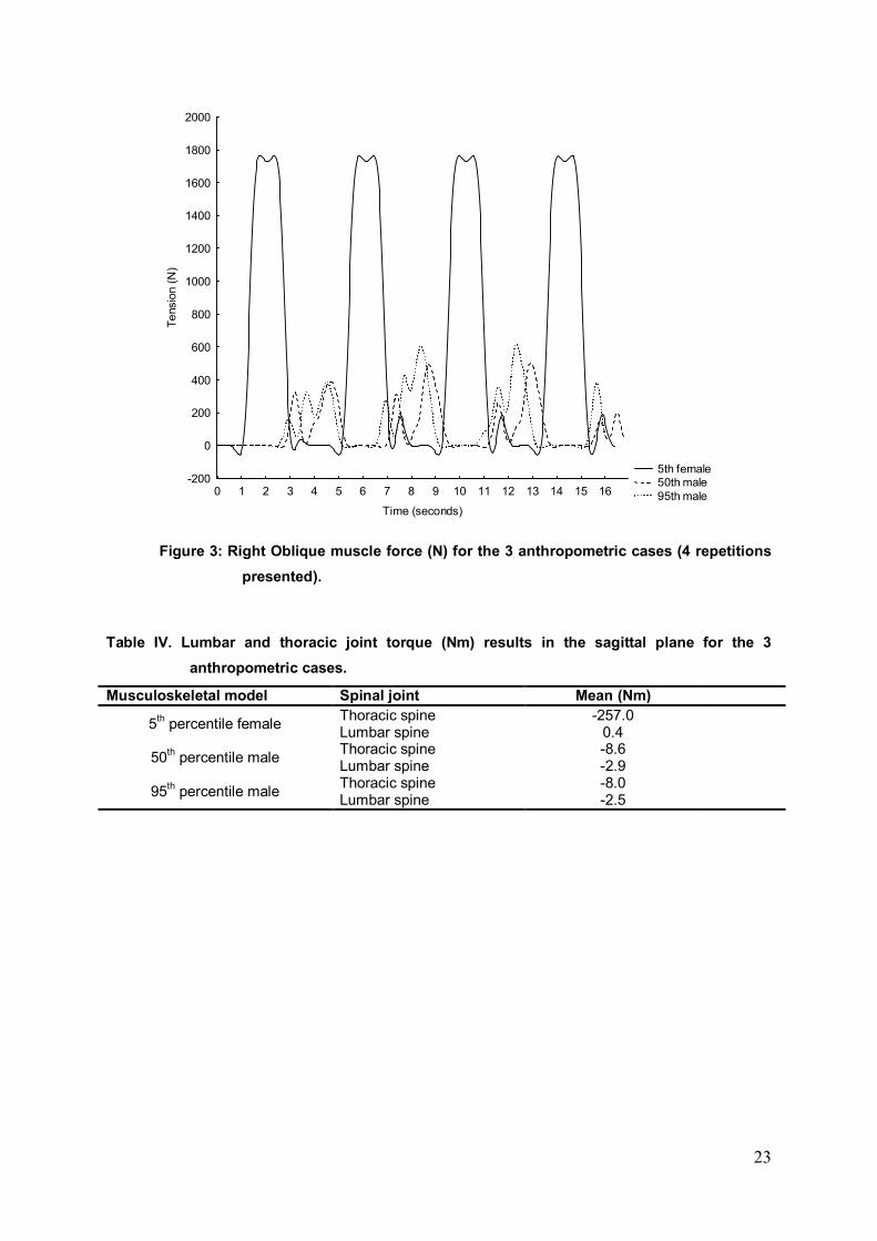
Table III. Maximum force production was greatest for the O muscle in comparison to
the RA muscle for all anthropometric cases (Figure 3). The 5th percentile female
exerted the most force for all muscles analysed and the 50th percentile male the
least, with the exception of the ES muscle which was slightly higher for the 50th
percentile male in comparison with the 95th percentile male. The hip flexor muscles
were only used by the 5th percentile female, specifically the PM muscle.
Muscle length results are presented in Table III. The mean muscle length for the ES,
RA and O is greatest for the 95th percentile male and smallest for the 5th percentile
female. The reverse is true for the PM and I muscles as the 5th percentile female
measured the greatest mean muscle lengths. The mean muscle length is highest for
the RA muscle in comparison with the O muscle and a similar trend was found with
the PM muscle in comparison with the I muscle for the three anthropometric cases.
Due to the involvement of the spinal column in the abdominal crunch exercise,
torque (Nm) for the T12/L1 intervertebral joint (thoracic) and the L5/S1
intertervertebral joint (lumbar) in the sagittal plane are presented in Table IV. For all
anthropometric cases peak thoracic torque was greater than peak lumbar torque.
The 5th percentile female’s peak thoracic torque was greater than that of the other
two anthropometric cases as shown in Figure 4.
Results for the thoracic (T12/L1 intervertebral joint) and lumbar (L5/S1 intervertebral
joint) spine compression and anterior/posterior (A/P) shear forces are presented in

14
Table V. The peak thoracic and lumbar spine joint compression forces are greatest
for the 5th percentile female and least for the 50th percentile male. Peak thoracic
spine joint compression forces are greater than the peak lumbar spine joint
compression forces for all the anthropometric cases with the exception of the 5th
percentile female whose peak lumbar spine joint compression forces exceed her
peak thoracic spine joint compression forces.
Peak thoracic spine joint A/P shear forces are greater than peak lumbar spine joint
A/P shear forces for all anthropometric cases (Table V). The 5th percentile female
has the highest peak thoracic and lumbar spine joint A/P shear forces in comparison
with the 50th and 95th percentile males.
Table III. Right Erector spinae, Rectus abdominis, Internal and External oblique, Psoas majorand Iliacus (hip flexors) muscles force production (N) and muscle length (mm)results for the 3 anthropometric cases.
Musculoskeletal model Muscles Max muscle forceproduction (N)
Mean musclelength (mm)
5th percentile female
Erector spinae (ES) 225.0 240.0Rectus abdominis (RA) 667.0 280.0Oblique (O) 1764.0 140.0Psoas major (PM) 1627.0 220.0Iliacus (I) 0.4 120.0
50th percentile male
Erector spinae (ES) 342.0 260.0Rectus abdominis (RA) 186.0 320.0Oblique (O) 503.0 190.0Psoas major (PM) 0.5 190.0Iliacus (I) 0.4 110.0
95th percentile male
Erector spinae (ES) 340.0 280.0Rectus abdominis (RA) 241.0 350.0Oblique (O) 618.0 200.0Psoas major (PM) 0.4 190.0Iliacus (I) 0.4 100.0

15
Discussion
The first relevant finding of this study was that based on a visual comparison of the
kinematics the LifeModeler™ default models were adequate to solve the forward
dynamics simulations for all the anthropometric cases. This was not the case for a
previous study in which the seated biceps curl resistance training exercise was
modelled (Nolte et al., 2011). Three adjustments had to be made to the
musculoskeletal models on the seated biceps curl machine before the forward
dynamics simulations could be solved namely; 1) increase the physiological cross-
sectional area (pCSA) of the three default elbow flexor muscles, 2) manipulate the
muscle origins and insertions and 3) decrease the joint stiffness in the forward
dynamics simulations (Nolte et al., 2011). The reason for the adjustments not being
necessary in this study could possibly be due to the fact that the trunk musculature
of the default model is more comprehensive than that of the elbow and shoulder
joints. The only relevant muscle that is omitted from the LifeModelerTM default model
is the Transversus abdominis. Caution is advised when implementing such a visual
assessment approach as in the case of the 5th percentile female one could argue
that the resulting forward dynamics simulation was visually correct despite the kinetic
results indicating implausible muscle and joint reaction forces.
The second relevant finding was that the software was able to sufficiently indicate
anthropometric differences with regards to the abdominal crunch machine’s
engineered or manufactured adjustability. The anthropometric dimensions of the
musculoskeletal models could be accommodated comfortably in relation to the
dimensions and adjustability of the abdominal crunch machine except for the 5th
percentile female (Figure 5). The small female’s feet could barely reach the foot rest

16
and the abdominal crunch pad/cushion was positioned too high and therefore could
not be accommodated adequately under her axilla. Furthermore, her lumbar (L5/S1)
spine joint could not be aligned properly with the axis of rotation of the machine. As a
result her movement on the abdominal crunch machine was negatively impacted as
her thoracic spine movement appeared to be exaggerated during the execution of
the exercise to the point where it resulted in highly improbable joint loads, possibly
an artefact of the modelling process.
The movement on the abdominal crunch machine could be compared to a bent knee
sit-up movement, in a study conducted by McGill (1995) the analysis of a bent knee
sit-up showed that most of the flexion rotation movement takes place about the hips
and not the spine. Rather the spine remains close to the isometric flexed posture
throughout the dynamic sit-up cycle. Thus, a sit-up exercise may be considered an
isometric flexion exercise as far as the trunk musculature is concerned. The 50th and
95th percentile males appeared to have produced trunk flexion at the lumbar sacral
region rather than the unnatural flexion of the thoracic region as demonstrated by the
female model. Figure 5 illustrates that the mismatch between the female model
anthropometry and machine adjustability resulted in excessive thoracic spine
movement so that the thoracic joint reached its range of motion limits. While the
results suggests that the female is at increased risk for injury due to poor
accommodation by the machine it is possible that the values obtained for muscle
tensions and joint loads are exacerbated by an artefact in the modelling process
most probably caused by the thoracic joint movement exceeding the default range of
motion. Furthermore, the large muscle lengths recorded specifically in the O muscle
could also be an indication that there was exaggerated movement of the trunk rather

17
than that of an isometric contraction in the small female although the other
anthropometric cases recorded similar muscle lengths.
Thirdly, the following relevant findings were made regarding the biomechanical
evaluation in terms of exercise efficacy and injury risk. The O muscles in comparison
with the RA muscles exerted more force during the exercise for all anthropometric
cases. This result was not entirely expected as the O muscles are traditionally
exercised using trunk rotation or twisting to the left and right which bring the oblique
muscles into more active contraction (Floyd, 2009). The O muscles however, also
aid in lumbar flexion and posterior pelvic rotation and thus could explain its
significant contribution to the execution of the movement of the abdominal crunch
exercise. In addition in a study conducted by McGill (1995) it was found that the RA
muscles activity to be slightly lower in bent knee sit-ups as opposed to the straight
leg variety, while the O muscles were activated to a greater level presumably to
make up the moment deficit. Similar results were obtained in this study in
comparison with McGill (1995) with regards to abdominal RA and O muscle force
production measured by means of electromyography (EMG) during the straight leg
sit-up such as 206N and 236N respectively. However, the muscle force production
results for both muscle groups in this study were higher for all anthropometric cases,
specifically the 5th percentile female. It must be noted that LifemodelerTM default
model only consists of 1 pair of oblique muscles, the orientation of the muscles
appear to resemble that of the External obliques, this could have also contributed to
the high recorded force production of the O muscles.

18
The ES muscle recruitment can be explained by means of its antagonistic role in
relation to the RA and O muscles. In a study conducted on sit-ups it was found that
the antagonist extensor moments are produced particularly by the thoracic extensors
(Iliocostalis lumborum and Longissimus thoracis). Most of the extensor force was
due to neural activation as well as due to passive elastic stretching (McGill, 1995).
Higher levels of coactivity have a significant impact on the spinal loads since
increased antagonistic muscle activity must be offset by the agonist forces. Thus, the
muscle activity from the antagonistic muscles produces more loading in the spinal
(compressive forces) structures without contributing to the ability to offset the
external moment imposed by the spine (Davis & Marras, 2000). One could also
argue that co-contraction of the antagonistic muscles serve a protective role by
reducing shear forces, since the vertebra and discs can withstand compressive loads
but are not well suited to large shear forces (Granata & Marras, 1999).
Usually when abdominal exercises are performed the exerciser tries to reduce the
contribution of the hip flexors with regards to the execution of the movement. The
most commonly recommended manner of reducing the contribution of these muscles
is to bend or flex the hips as this shortens the iliopsoas muscle and other hip flexors
thereby reducing their ability to produce force (Floyd, 2009). In addition, this action of
the hips is supposed to reduce lumbar joint compression. However, Axler and McGill
(1997) found this not to be the case as there were no differences observed in lumbar
spine joint compression or the utilization of the hip flexor muscles in sit-ups
performed with the legs bent versus with the legs straight. The positioning of the
musculoskeletal model on the abdominal crunch resistance training machine in this
study is such that the hips and knees are in a flexed position and results indicate that

19
the Iliopsoas muscles did not significantly contribute to the movement with the
exception of the 5th percentile female. The high recorded PM muscle force
production in the small female appear unrealistic and could be due to a combination
of an artefact as well as poor accommodation of the model. There was much less hip
flexion for the 5th percentile female in comparison with that of the other two
anthropometric cases. Therefore one could postulate that the exercise was not
successful in isolating the abdominal muscles of the small female.
The 5th percentile females force production for all studied muscles was the greatest
in comparison with the other anthropometric cases. This result is not unexpected as
anatomical differences could be the reason for the greater force production in the
small female such as a smaller lever arm as well as unfavourable positioning on the
abdominal crunch machine, even although the resistance used for all three cases
was proportionally calculated to correlate the anthropometric dimensions. In addition,
it is possible that there may have been errors in the forward dynamics simulations
kinetic data.
Joint torque values obtained for the thoracic and lumbar spine in the 50th and 95th
percentile males as well as lumbar spine torque values of the 5th percentile female
appear to be plausible when comparing the results to peak values obtained by
means of isokinetic testing. Langrana and Lee (1984) report trunk flexion/extension
values of 60 Nm and 95 Nm respectively in non-disabled female subjects and 136
Nm and 212 Nm respectively in non-disabled male subjects assessed in a seated
position at 30 degrees per second. Bearing in mind that the values obtained in this
study were not from maximal testing they were still substantially lower than the

20
isokinetic values of Langara and Lee with the exception of the 5th percentile female’s
thoracic spine torque values which were considerably higher. This once again could
have resulted due to her poor positioning, on the abdominal crunch resistance
training machine.
Abdominal exercises are prescribed for both the prevention and treatment of low
back injury. However, these exercises sometimes appear to have hazardous effects
on the spine. A study conducted by Axler and McGill (1997) with the purpose of
identifying abdominal exercises that optimize the challenge to the abdominal
muscles but impose minimal load penalty to the lumbar spine found that no single
exercise optimally trained all of the abdominal muscles while at the same time
incurring minimal intervertebral joint loads. Accurate assessment of the risk of spinal
injuries during occupational, athletic/exercise and daily activities as well as
subsequent design of effective prevention and treatment programmes depend
amongst others, on an accurate estimation of trunk muscle forces and internal spinal
loads (i.e., intervertebral disc compression and shear forces)(Arjmand, Gagnon,
Plamondon, Sharazi-Adl & Lariviére, 2009). Thus, an important aspect of this study
involved assessing the intervertebral joint loads. The intervertebral discs work as a
visco-elastic system that absorb and distribute forces acting on the spine. When
submitted to compressive forces the collagen fibres of the annulus fibrosus are
deformed radially expelling fluid from the nucleus pulposus of the discs (Adams &
Hutton, 1985). It is important to bear in mind when making this analysis and applying
the information that the spine of the default model does not consist of all the
individual vertebrae but rather of various segments that represent the different

21
regions of the vertebral column with joints between these segments. Individualised
vertebra and corresponding joints might produce different results.
Previous research from the American National Institute for Occupational Safety and
Health (NIOSH) recommends that spinal compression forces should not exceed 3.4
kN to avoid injury. However there is a very real threat of musculoskeletal injury
before this failure limit value has been reached (Snook & Ciriello, 1991; Cooper &
Ghassemieh, 2007, Knapik & Marras, 2009). British standards (BS EN 1005-3,
2002) recommend 600N as the cut-off point for carrying masses, no further
recommendations other than “time of exposure needs to be minimised” and “a
preferred system requires optimal ergonomic position with reduced back bending
posture” are made. Therefore, the 5th percentile female’s lumbar and thoracic spine
joint compression forces were far above the recommended failure limit of 3.4 kN and
therefore she would be at possible risk for a back injury bearing in mind that the high
recorded values could have been an artefact of the modelling process. A possible
cause of this artefact could be the improbably high muscle forces recorded for the O
and PM muscles of this model. Future studies to validate the model is required in
order provide clear design guidelines for the likely risk for females of small stature
exercising on the equipment. However, the 50th and 95th percentile males’ thoracic
and lumbar joint spine compression forces were also high and therefore the results
suggest that the exercise may pose a risk for back injury.
The thoracic spine joint A/P shear forces appear to be higher than the lumbar spine
joint A/P for the three anthropometric cases. Both thoracic and lumbar spine joint A/P
shear forces for all anthropometric cases are above the most commonly cited spine

22
tolerance of 1000 N for shear force as stipulated by McGill (1996), with the exception
of the 50th and 95th percentile males’ lumbar spine joint A/P shear forces. Thus, this
exercise appears to place all anthropometric cases at risk of injury. It is important to
note that the modelling does not take conditioning differences between individuals of
similar anthropometric dimensions into account which can protect the individual
against spinal loading. Furthermore, increased strength of trunk flexors and
extensors muscles are thought to raise intra-abdominal pressure and to decrease
spinal loading (Aspden, 1988).
The results regarding the spine reaction forces are not surprising. Predictions of
compressive load on the low back were found to be substantial during both
isometrically held sit-ups and dynamic sit-ups with minimal acceleration components
by Axler and McGill (1997). Therefore, forces on the back during a resistance
exercise such as this can be expected to put substantial strain on the back especially
if positioning is not adequate as with the 5th percentile female.
Lastly, it should be noted when evaluating an exercise in terms of efficacy and injury
risk it is sometimes useful to compare various exercise techniques, different
exercises for the same muscle groups as well as different manufacturer’s equipment
for the same exercise.
23
5th female 50th male 95th male0 1 2 3 4 5 6 7 8 9 10 11 12 13 14 15 16
Time (seconds)
-200
0
200
400
600
800
1000
1200
1400
1600
1800
2000
Tens
ion
(N)
Figure 3: Right Oblique muscle force (N) for the 3 anthropometric cases (4 repetitionspresented).
Table IV. Lumbar and thoracic joint torque (Nm) results in the sagittal plane for the 3anthropometric cases.
Musculoskeletal model Spinal joint Mean (Nm)
5th percentile female Thoracic spine -257.0Lumbar spine 0.4
50th percentile male Thoracic spine -8.6Lumbar spine -2.9
95th percentile male Thoracic spine -8.0Lumbar spine -2.5

24
Table V. Thoracic and lumbar spine joint compression and anterior/posterior shear forces (N)for the 3 anthropometric cases. Note: for the compression forces, positive valuesindicate forces in a superior direction and negative values indicate forces in aninferior direction and for the anterior/posterior shear forces, positive valuesindicate forces in a posterior direction and negative values indicate forces in ananterior direction.
Musculoskeletal model Spinal joint Max compressionforces (N)
Max anterior/posteriorshear forces (N)
5th percentile female Thoracic spine 11043.0 5827.9Lumbar spine 12580.2 5122.3
50th percentile male Thoracic spine 4206.4 3201.3Lumbar spine 3388.6 559.9
95th percentile male Thoracic spine 4673.9 3067.0Lumbar spine 3664.2 436.8
5th female 50th male 95th male0 1 2 3 4 5 6 7 8 9 10 11 12 13 14 15 16
Time (seconds)
-800
-700
-600
-500
-400
-300
-200
-100
0
100
Torq
ue (N
m)
Figure 4: Thoracic spine joint torque (Nm) in the sagittal plane for the 3 anthropometric cases(4 repetitions presented). Note: negative joint angle indicates trunk flexion.

25
Figure 5. 5th percentile female’s positioning on the abdominal crunch resistance trainingmachine
Conclusion
It can be concluded that the default model of the LifemodelerTM software was
successful in evaluating the abdominal crunch resistance training exercise. No
adjustments had to be made to the default model in order to solve the forwards
dynamics simulations. The most significant value of the abdominal crunch resistance
training machine 3D musculoskeletal modelling was in demonstrating the
unacceptable thoracic and lumbar spine joint compression and A/P forces which
could place the exerciser at high risk for a back injury. Therefore, caution should be
used when prescribing the exercise for the training of the abdominal muscles
especially if the individual has a predisposing back problem or injury. In addition,
individuals of small anthropometric dimensions such as some females and children
cannot be accommodated suitably on the machine which unfavourably influences

26
exercise posture and technique which can further place the exerciser at increased
risk for injury and decrease the efficacy of the exercise. Therefore, design
adjustments to the abdominal crunch resistance training machine such as adapting
the foot rest and abdominal crunch pad/ cushion length should be considered by the
manufacturer. The models in this study were not validated and therefore this can be
considered a limitation of the study, it is recommended that motion capture data
should be used to determine individual movement patterns of each anthropometric
case during such a validation.
References
Aspden, R.M. (1988). The spine as an arch. A new mathematical model. Spine, 13,
266–274.
Adams, M.A., & Hutton, W.C. (1985). Gradual disc prolapse. Spine, 10, 524–531.
Agnesina, G., Taiar, R., Havel, N., Guelton, K., Hellard, P., and Toshev, Y. (June
2006). BRG.LifeMODTM modeling and simulation of swimmers impulse during a grab
start. Paper presented at the 9th symposium on 3D analysis of human movement.
Valenciennes, France.
Alexander, R.McN. (2003). Modelling approaches in biomechanics. Philosophical
Transactions of the Royal Society, 358, 1429-1435.

27
Annegarn, J., Rasmussen, J., Savelberg, H.H.C.M., Verdijk, L.B., & Meijer, K.
(2007, May). Scaling strength in human simulation models. Paper presented at the
European Workshop on Movement Sciences, Amsterdam, Netherlands.
Arjmand, N., Gagnon, D., Plamondon, A., Shirazi-Adl, A., & Lariviére, C. (2009).
Comparison of trunk muscle forces and spinal loads estimated by 2 biomechanical
models. Clinical Biomechanics, 24, 533–541.
Axler, C.T., & McGill, S.M. (1997). Low back loads over a variety of abdominal
exercises: searching for the safest challenge. Medicine and Science in Sports and
Exercise, 29(6), 804–811.
Beachle, T.R., & Groves, B.R. (1992). Weight training: steps to success. Champaign,
IL: Human Kinetics.
Biomechanics Research Group, Inc. (2006). LifeMOD biomechanics modeler
manual. San Clemente, USA: LifeModeler.
Bredenkamp, K. (2007). The characterisation of the male and female body forms of
the SANDF. ERGOTECH Document P0683/2007/01. Centurion: ERGOnomics
TECHnologies. South Africa.
BS EN 1005 – 3: (2002). Safety of machinery – Human physical performance – Part
3: Recommended force limits for machinery operations. London: British Standards
Institute.

28
Cooper, G., & Ghassemieh, E. (2007). Risk assessment of patient handling with
ambulance stretcher systems (ramp / winch), easy-loader, tail-lift using
biomechanical failure criteria. Medical Engineering & Physics, 29, 775–787.
Dabnichki, P. (1998). Biomechanical testing and sport equipment design. Sports
Engineering, 1, 93 –105.
Davis, K.G. & Marras, W.S. (2000). The effects of motion on trunk biomechanics.
Clinical Biomechanics, 15, 703 – 717.
De Jongh, C. (2007). Critical evaluation of predictive modelling of a cervical disc
design, Unpublished Masters thesis, University of Stellenbosch, South Africa
Floyd, R.T. (2009). Manual of structural kinesiology (17th Ed). New York: McGraw-
Hill.
Gordon, C.C., & Brantley, J.D. (1997, December). Statistical modelling of
population variation in the head and face. Paper presented at the design and
integration of helmet systems International Symposium. Massachusetts, USA.
Granata, K.P., & Marras, W.S. (1999). Cost-benefit of muscle cocontraction in
protecting against spinal instability. Spine, 25(11): 1398 – 1404.

29
Hofmann, M., Danhard, M., Betzler, N., Witte, K., & Edelmann, J. (2006). Modelling
with BRG.lifeMODTM in sport science. International Journal of Computer Science in
Sport, 5, 68-71.
Knapik, G.G., & Marras, W.S. (2009). Spine loading at different lumbar levels during
pushing and pulling. Ergonomics, 52(1): 60-70.
Kazlauskiené, K. (2006). Design and research of biomechanical models of human
with joint replacements. Unpublished doctoral thesis, Kaunas University of
Technology, Lithuania.
Langara, N.A., & Lee, C.K. (1984). Isokinetic evaluation of trunk muscles. Spine, 9,
171–175.
McGill, S.M. (1995). The mechanics of torso flexion: sit-ups and standing dynamic
flexion manoeuvres. Clinical Biomechanics, 10(4), 184-192.
McGill, S.M. (1996, June). Searching for the safe biomechanical envelope for
maintaining healthy tissue, Paper presented at the pre-Meeting workshop,
International Society for the Study of the Lumbar Spine: The Contribution of
Biomechanics to the prevention and treatment of low back pain, University of
Vermont.
Nieman, D.C. (2007). Exercise testing and prescription: health-related approach (6th
Ed.). New York: McGraw-Hill.

30
National Institute for Occupational Safety and Health. (1997). Musculoskeletal
disorders and workplace factors: a critical review of epidemiologic evidence for work-
related musculoskeletal disorders of the neck, upper extremity, and low back. US
Department of Health and Human Services (DHHS) Public Health Service, Centres
for Disease Control. Cincinnati: National Institute for Occupational Safety and Health
Division of Biomedical of Behavioural Science.
Nolte, K., Krüger, P.E., & Els, P.S. (2011). Three dimensional modelling of the
seated biceps curl resistance training exercise. Sports Biomechanics, 10(2): 146-
160.
Olesen, C.G., Andersen, M.S., Rathleff, M.S., de Zee, M., & Rasmussen, J. (2009,
June). Understanding the biomechanics of medial tibial stress syndrome – A
simulation study using a musculoskeletal model. Paper presented at the 2009
International Society of Biomechanics. Cape Town, South Africa.
Perrin, D.H. (1993). Isokinetic exercise and assessment. Champaign, IL: Human
Kinetics.
Rasmussen, J., de Zee, M., Damsgaard, M., Christensen, S.T., Marek, C. &
Siebertz, K. (2005, July). A general method for scaling musculo-skeletal models.
Paper presented at the International Symposium on Computer Simulation in
Biomechanics, Cleveland, Ohio.

31
Rietdyk, S., & Patla, A.E. (1999). Context-dependent reflex control: Some insights
into the Role of Balance. Experimental Brain Research. 119, 251–259.
RSA-MIL-STD-127. (2004). Ergonomic design: Anthropometry and environment.
RMSS Document, 1. 1–196. Pretoria, RSA:RMSS.
Schilling, B.K., Falvo, M.J. & Chiu, L.Z.F. (2008). Force—velocity, impulse-
momentum relationships: Implications for efficacy of purposefully slow resistance
training. Journal of Sports Sciences and Medicine, 7, 299-304.
Schillings, A.M., Van Wezel, B.M., & Duysens, J. (1996). Mechanically induced
stumbling during human treadmill walking. Journal of Neuroscience Methods, 67,
11–17.
Snook, S.H., & Ciriello, V.M. (1991). The design of manual handling tasks: revised
tables of maximum acceptable weights and forces. Ergonomics, 34: 1197-1213.
Vaughn, C.L. (1989). Biomechanics of sport. Florida: CRC Press.
Wagner, D., Rasmussen, J., & Reed, M. (2007, June). Assessing the importance of
motion dynamics for ergonomic analysis of manual material handling tasks using the
AnyBody modelling system. Paper presented at the meeting of the Digital Human
Modelling for Design and Engineering Conference, Seattle, Washington.