thursday 21st of january - École polytechnique
TRANSCRIPT

2
Thursday 21st of January
EMIRUM Opening 9:00 Michèle Raynaud (LSI - France) Opening
9:05 Nathalie Moncoffre (IP2I - France) Introduction EMIR&A
9:30 Robin Schaeublin (ETH Zurich - Switzerland) Presentation of the scientific committee
9:45 Presentation from CNRS 10:05 Break
Platform session 10:15 Serge Della Negra (IJCLab - Orsay) – ANDROMEDE .................................................... 6 10:40 Jimmy Rangama (CIMAP - Caen) - CIRIL@GANIL ........................................................ 6 11:05 Thierry Sauvage (CEMHTI - Orléans) - Cyclotron and Pelletron ..................................... 6/7 11:30 Sergei Denisov (ICP - Orsay) – ELYSE.......................................................................... 7 11:55 Olivier Tissot (CEA - Saclay) – HVEM ............................................................................ 7 12:20 Lunch break 13:20 1 Slide / 1 Question Daniele Torsello (Politecnico di Torino - Italy) Glass-ceramics irradiation with triple beam for applications in nuclear fusion reactors .............................................. 9
Quentin Tence (CEA - France) Phase separation under irradiation in face-centered cubic Fe-Ni alloy ..................................................................... 10
Karim Medjoubi (LSI - France) Investigation of the emerging III-V//Si tandem solar cell behavior under irradiations for space applications ............. 11
Sabrina Gouasmia (Institut Ruđer Bošković - Croatia) Crater formation from metallic surfaces under impact of metallic and argon clusters ................................................ 12
Nao Harada (INSP - France) Rare-Earth Oxides Thin Films: A New Platform For Quantum Technologies ........................................................... 13
Jingjun Gao (CEA - France) Ion-irradiation induced dislocation loops formation in high purity Co-free High Entropy Alloys ................................. 14
Session n°1 : Irradiation tuning of properties Invited speaker: 14:00 Katharina Lorenz (IST - Portugal) Ion implantation and radiation effects in gallium nitride
Contributed talks: 14:30 Nargisse Khiara (CEA - France) Creep behavior of Cu and He-implanted Cu under heavy ion irradiation ................................................................... 16
14:50 Frédéric Foucher (Centre de Biophysique Moléculaire - France) In situ Raman spectroscopy monitoring of material changes during proton irradiation: application to astrobiology ... 17
15:10 Jihane Jdaini (LSI - France) Damage effects in brushite (CaHPO4.2H2O) following 2.5 MeV electron irradiation, studied by X-ray diffraction and Raman spectroscopy ......................................................................................................................................... 18
15:30 Kan Ma (CEA - France) Drastic impact of micro-alloying on austenitic model alloy microstructure under irradiation ....................................... 19
15:50 Zhiwei Hu (CEMHTI - France) Combination of PAS and TEM to characterize defects in self-irradiated tungsten for fusion application.................... 20
16:10 Break
Session n°2: Liquids and interfaces 16:20 Philippe Martinet (IP2I - France) Effects of proton-induced radiolysis at the interfaces: Application to the study of a stainless steel in a Na2SO4 medium ...................................................................................................................................................................... 22
16 :40 Sophie Le Caër (CEA - France) Reaction Mechanisms of Fluoroethylene Carbonate Degradation, an Additive of Lithium-Ion Batteries, Unraveled by Radiation Chemistry ............................................................................................................................. 23
17:00 Serge Della Negra (ICJLab - France) Surface analysis by impact of energetic nanoparticles ............................................................................................... 24

3
Friday 22nd of January Platform session 9:00 Aurelie Gentils (IJCLab - Orsay) - JANNuS-Orsay ........................................................... 26 9:25 Céline Cabet (CEA - Saclay) - JANNuS-Saclay ............................................................... 26 9:50 Emerick Briand (INSP - Paris) – SAFIR ............................................................................ 27 10 :15 Antonino Alessi (LSI - Palaiseau) – SIRIUS .................................................................. 27 10:40 Break
Session n°3: Nanostructuration Invited speaker: 10 :50 André Vantomme (KU Leuven - Belgium) Ion beam tuning of the Ni-Si solid state reaction : substrate damage vs impurity effects
Contributed talks: 11:20 Natalia Potrzebowska (LSI - France) Flexible Nano-structured Piezo-Generator for Energy Harvesting ............................................................................. 29
11:40 Nathan Meyer (IEM - France) Nanopore Technology: From polymer film irradiation to the detection and study of (bio)macromolecules ................. 30
12:00 Lukas Madauß (University of Duisburg-Essen - Germany) Swift Heavy Ion Modification of Molybdenum Disulfide .............................................................................................. 31
12:20 Jun Lin (ICSM - France) Structural behavior of ordered mesoporous silica under electrons and swift heavy ions irradiation ........................... 32 12:40 Lunch break 13:30 Platforms round-table
Session n°4: Fundamental studies of defects Invited speaker: 14 :10 Anne-Magali Seydoux (UCBL - France) Double-beam irradiations and in situ TEM of monazite and xenotime: focus on alpha annealing
Contributed talks: 14:40 Ruslan Prozorov (Ames Laboratory - USA) Using Controlled Disorder to Probe Iron-based Superconductors ............................................................................ 35
15:00 Marcin Konczykowski (LSI - France) Tuning of electronic and magnetic properties of topological insulators by low temperature electron irradiation ........ 36
15:20 Prudence Ada Bibang (CIMAP - France) Irradiation of complex organic molecules in solid phase ............................................................................................ 37
15:40 Jacob Ruf (Max Planck Institute for Chemical Physics of Solids - Germany) Controllable suppression of the unconventional superconductivity in Sr2RuO4 epitaxial thin films and micro-structured bulk single crystals via high-energy electron irradiation .................................................................. 38
16:00 Elina Zhakina (Max Planck Institute for Chemical Physics of Solids - Germany) Investigation of the universal scattering rate in PdCrO2 by high energy electron irradiation ...................................... 39
16:20 Closing

4
Liens de connexion Titre session 1 : EMIRUM 2021 : Thursday Morning
Ouverture de la diffusion : le 21/01/2021 à 9h00
Durée : 3h40
Lien : https://ecolepolytechnique.zoom.us/j/81763478354
Titre session 2 : EMIRUM 2021 : Thursday Afternoon
Ouverture de la diffusion : le 21/01/2021 à 13h10
Durée : 4h40
Lien: https://ecolepolytechnique.zoom.us/j/82252762828
Titre session 3 : EMIRUM 2021 : Friday Morning
Ouverture de la diffusion : le 22/01/2021 à 9h00
Durée : 3h50
Lien: https://ecolepolytechnique.zoom.us/j/84274355527
Titre session 4 : EMIRUM 2021 : Friday Afternoon
Ouverture de la diffusion : le 22/01/2021 à 13h20
Durée : 3h40
Lien : https://ecolepolytechnique.zoom.us/j/84138159496
Liste des panelistes :
Michèle Raynaud : [email protected]
Nathalie Moncoffre : [email protected]
Cédric Baumier : [email protected]
Romain Grasset : [email protected]
Elodie Dubois : [email protected]

5
Chair : Ian Vickridge
Serge Della Negra (IJCLab - Orsay) – ANDROMEDE .............................................................. 6
Jimmy Rangama (CIMAP - Caen) - CIRIL@GANIL .................................................................. 6
Thierry Sauvage (CEMHTI - Orléans) - Cyclotron and Pelletron ............................................... 6/7
Sergei Denisov (ICP - Orsay) – ELYSE ................................................................................... 7
Olivier Tissot (CEA - Saclay) – HVEM ...................................................................................... 7

6
Serge Della Negra (IJCLab - Orsay) – ANDROMEDE
ANDROMEDE facility: The IN2P3/Université Paris-Saclay Andromède platform1 installed at the
IJCLab laboratory in Orsay was initiated by the Equipex Consortium in 2011 and became
operational in 2017. It uses a NecPelletron 4MV electrostatic accelerator equipped with two ion
sources: a Liquid Metal Ion Source (LMIS/OrsayPhysics) and an Electronic Cyclotron Resonance
Source (ECR Microgan /Pantechnik). The ion beams can be selected and injected into 2 dedicated
experimental lines at 90° and 1°29 with an analyzing magnet. The 90° line offers a wide range of
multi-charged atomic and molecular ion beams of light masses (up to m/q 70) with an intensity of
several microamps ( 2 mm). The analysis chamber EVE installed on the line at 1°29 is dedicated
to surface analysis under ultra-high vacuum conditions. Surface analysis at the submicrometric scale
is possible for molecules with mass up to 1500 Da. Today, this facility can be used for surface
analysis of organic and inorganic samples that are essential for research in biology, astrobiology
and accelerator science. In parallel, fundamental studies on secondary emission processes induced
by the impact of energetic nano-particles are being conducted by the NIM/IJCLab team. In this
framework, a multi-technical approach (XPS, AFM, confocal 3D, …) has been set up around
Andromeda for complementary target characterizations. This facility is available for the platform
users.
***
Jimmy Rangama (CIMAP - Caen) - CIRIL@GANIL
The CIRIL: a platform to welcome interdisciplinary researches at GANIL Since the creation of the GANIL, for more than 35 years, the CIRIL platform welcomes the external
users, develops equipments adapted to the proposed experiments (beam lines, irradiation chambers,
on-line measurements…) manages and coordinates the scientific, administrative and technical
activities of 3 experimental areas (8 beamlines) for interdisciplinary research carried out with
GANIL ion beams. The importance of this activity has regularly increased with the growing number
of possibilities offered to the users: the high energy line at the creation in 1983, the medium energy
line in 1989, the laboratory for the radiobiology research (LARIA) in 2003, the IRRSUD line in
2004, the LIMBE facility in 2000 and its extension ARIBE in 2005. This platform contributes to
the spread of scientific excellence in fields related to ion beams such as atomic physics, solid state
physics, materials, chemistry, radiobiology and astrophysics. These researches are mainly focused
on the effects of the electronic excitations produced by multiply charged ions whatever it is at high-
or low-velocity. During this presentation, I will give an overview of the available beamlines in the
EMIR&A network as well as some setups recently built by the CIMAP for the scientific community.
***
Thierry Sauvage (CEMHTI - Orléans) - Cyclotron and Pelletron
Cyclotron and Pelletron, CEMHTI Facility: The "Particles beam" platform of CEMHTI offers
state-of-the-art scientific equipment for irradiation and material characterization by Ion Beam
Analysis to the EMIR community. This platform includes two ion accelerators: CGR MeV
cyclotron and 3MV Pelletron electrostatic accelerator. The roadmap for technical developments
around CEMHTI accelerator facilities focuses on (i) the investigation of materials physical
properties evolution under irradiation and (ii) the in situ material characterization at temperature
under vacuum or controlled atmosphere. We will give an overview of the experimental setups
installed at CEMHTI accelerators beamlines, with an emphasis on devices for thin films and bulk
materials characterization by ion beam techniques. The originality of the DIADDHEM facility
resides in the coupling of IBA techniques (RBS or NRA) with sample heating and cooling systems.
DIADDHEM provides for example in situ measurements of helium or deuterium desorption yield
7
as a function of temperature and depth profiles after annealing. Diffusion coefficients and activation
energy are extracted from depth profile evolution. Its last upgrade allows to investigate “in real
time” (integrated time of 30 seconds) the surface reactivity of thin films at solid/gas interface as a
function of temperature. The potentialities of the Real Time NRA technique are illustrated through
recent sorption mechanisms study of getter materials.
***
Sergei Denisov (ICP - Orsay) – ELYSE
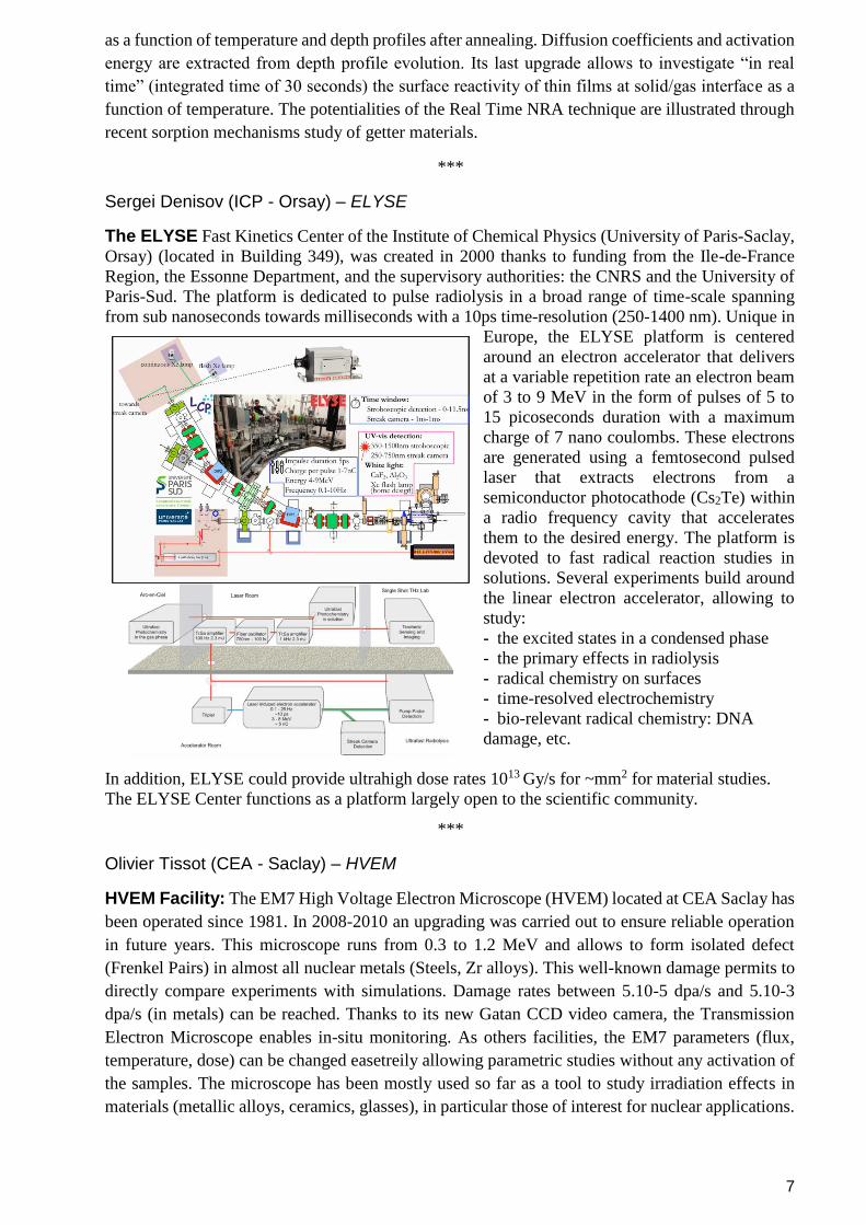
The ELYSE Fast Kinetics Center of the Institute of Chemical Physics (University of Paris-Saclay,
Orsay) (located in Building 349), was created in 2000 thanks to funding from the Ile-de-France
Region, the Essonne Department, and the supervisory authorities: the CNRS and the University of
Paris-Sud. The platform is dedicated to pulse radiolysis in a broad range of time-scale spanning
from sub nanoseconds towards milliseconds with a 10ps time-resolution (250-1400 nm). Unique in
Europe, the ELYSE platform is centered
around an electron accelerator that delivers
at a variable repetition rate an electron beam
of 3 to 9 MeV in the form of pulses of 5 to
15 picoseconds duration with a maximum
charge of 7 nano coulombs. These electrons
are generated using a femtosecond pulsed
laser that extracts electrons from a
semiconductor photocathode (Cs2Te) within
a radio frequency cavity that accelerates
them to the desired energy. The platform is
devoted to fast radical reaction studies in
solutions. Several experiments build around
the linear electron accelerator, allowing to
study:
- the excited states in a condensed phase
- the primary effects in radiolysis
- radical chemistry on surfaces
- time-resolved electrochemistry
- bio-relevant radical chemistry: DNA
damage, etc.
In addition, ELYSE could provide ultrahigh dose rates 1013 Gy/s for ~mm2 for material studies.
The ELYSE Center functions as a platform largely open to the scientific community.
***
Olivier Tissot (CEA - Saclay) – HVEM
HVEM Facility: The EM7 High Voltage Electron Microscope (HVEM) located at CEA Saclay has
been operated since 1981. In 2008-2010 an upgrading was carried out to ensure reliable operation
in future years. This microscope runs from 0.3 to 1.2 MeV and allows to form isolated defect
(Frenkel Pairs) in almost all nuclear metals (Steels, Zr alloys). This well-known damage permits to
directly compare experiments with simulations. Damage rates between 5.10-5 dpa/s and 5.10-3
dpa/s (in metals) can be reached. Thanks to its new Gatan CCD video camera, the Transmission
Electron Microscope enables in-situ monitoring. As others facilities, the EM7 parameters (flux,
temperature, dose) can be changed easetreily allowing parametric studies without any activation of
the samples. The microscope has been mostly used so far as a tool to study irradiation effects in
materials (metallic alloys, ceramics, glasses), in particular those of interest for nuclear applications.

8
Chair : Romain Grasset
Daniele Torsello (Politecnico di Torino - Italy) Glass-ceramics irradiation with triple beam for applications in nuclear fusion reactors .............................................. 9
Quentin Tence (CEA - France) Phase separation under irradiation in face-centered cubic Fe-Ni alloy ...................................................................... 10
Karim Medjoubi (LSI - France) Investigation of the emerging III-V//Si tandem solar cell behavior under irradiations for space applications.............. 11
Sabrina Gouasmia (Institut Ruđer Bošković - Croatia) Crater formation from metallic surfaces under impact of metallic and argon clusters ................................................ 12
Nao Harada (INSP - France) Rare-Earth Oxides Thin Films: A New Platform For Quantum Technologies ............................................................. 13
Jingjun Gao (CEA - France) Ion-irradiation induced dislocation loops formation in high purity Co-free High Entropy Alloys .................................. 14

9
EMIRUM 2021
Glass-ceramics irradiation with triple beam for applications in nuclear fusion reactors
Daniele Torsello1,2
, Valentina Casalegno1, Fabiana D’Isanto
1, Monica Ferraris
1, Roberto
Gerbaldo1,2
, Gianluca Ghigo1,2
, May Ching Lai3, Giorgio Divitini
3, Caterina Ducati
3, Celine Cabet
4,
Gaëlle Gutierrez4, Laurent Roux
4 and Laura Gozzelino1,2
1 Politecnico di Torino, Department of Applied Science and Technology, 10129 Torino, Italy
2 INFN Sez. Torino, 10125 Torino, Italy
3Department of Materials Science & Metallurgy, University of Cambridge, CB30FS, Cambridge, UK
4Université Paris-Saclay, CEA, Service de Recherches de Métallurgie Physique, 91191, Gif-sur- Yvette, France
Presenter’s e-mail address: [email protected]
Abstract
Glass-ceramics based on CaO-Al2O3 (CA) showed potential as SiC-based indirect joining material for
applications in nuclear fusion plants, but the knowledge of the effects of radiation damage associated with
energetic neutrons, needed for their employment in such harsh environment, is still lacking. To fill this gap, CA
pellets were irradiated under triple ions beam (Si + He + H) at 400 °C, 615 °C and 710 °C at JANNuS-Saclay
accelerators. Ion fluences were chosen to reproduce the neutron radiation damage level and typology expected
in working locations of a DEMO reactor model in terms of displacement damage (Si) and transmutation
products (He + H), as calculated by means of the simulation code PHITS. TEM analysis evidences the
amorphization of the irradiated area, with no significant change in relation to transmutation product abundance.
Samples irradiated at 615 °C and 710 °C shows a non-homogene- ous distribution of cavities/bubbles whose
population increases with irradiation temperature. No cracks were detected. The evaluation of the swelling
percent level is ongoing.
Left: TEM image of the transverse cross-section of the CA pellet irradiated at 400 °C. The yellow line highlights the
sharp interface between the irradiated (amorphized – right) and the non-irradiated (crystalline – left) areas. Right: a
cluster of cavities in the CA pellet irradiated at 710 °C
References
M. Ferraris et al. J. Nucl. Mater. 417 (2011) 379.
V. Casalegno et al. J. Nucl. Mater. 501 (2018) 172.
A. Gentils, C. Cabet Nucl. Instrum. Meth. B 447 (2019) 107.
T. Sato et al., J Nucl. Sci. Technol. 50 (2013) 913.

10
EMIRUM 2021
Phase separation under irradiation in face-centered cubic Fe-Ni alloy
Q. Tence1, E.Meslin1, B. Decamps2, M. Nastar1,I. Mouton3
1 Université Paris-Saclay, CEA, Service de Recherches de Métallurgie Physique, Gif-sur-Yvette, France 2 Laboratoire
de Physique des 2 infinis Irène Joliot-Curie (IJCLab), Université Paris-Saclay, Orsay, France
3 Université Paris-Saclay, CEA, Service de Recherches de Métallurgie Appliquée, Gif-sur-Yvette, France
Abstract
Lattice point defects induced by irradiation are recognized to have a significant effect on the stability of phases in metallic
alloys. We investigate their kinetic and thermodynamic effects on the well-known Fe-Ni model alloy. Even if there have
been experimental evidences of a spinodal decomposition of the Ni-rich austenite upon irradiation[3], there are fewer
ones about ordering process[2][4], the L10 and L12 phase stability[1]. Therefore, we conduct ion irradiations on a face-
centered cubic (FCC) Fe-Ni alloy with Ni concentration of from 30 to 40 wt.%. The samples are irradiated at JANNUS
Saclay facility with 5 MeV Fe5+ ions at 442±2°C with a damage rate of 9,0 10-4 dpa/s and up to a damage of 40 dpa in the
area of interest. STEM/EDS analysis on irradiated Fe30wt.%Ni and Fe40wt.%Ni samples both re- vealed a spinodal-like
decomposition into 30% Ni rich and 80% Ni rich zones.
Simultaneously, thermodynamic calculations of the Fe-Ni binary system have been done within the framework of the
CALPHAD approach. We add to this method a non-equilibrium term depending on the relative density of phases and the
energy of point defects, in order to predict the effect of an irradi- ation on the stability of phases in the Fe-Ni system. We
show how the effect of an excess of point defects produced by irradiation may lead to the coexistence of a Ni-rich and Fe-
rich austenites. Both the com- puted boundary and spinodal limits are consistent with the experimental observations.
References [1] Ohnuma, Ikuo, Shota Shimenouchi, Toshihiro Omori, Kiyohito Ishida, et Ryosuke Kainuma. « Experimental Determina- tion and
Thermodynamic Evaluation of Low-Temperature Phase Equilibria in the Fe–Ni Binary System ». Calphad 67 (1 décembre 2019): 101677.
[2] Paulevé, J., D. Dautreppe, J. Laugier, et L. Néel. « Une nouvelle transition ordre-désordre dans Fe-Ni (50-50 ) ». Journal de Physique et le
Radium 23, no 10 (1 octobre 1962): 841‑43.
[3] Reuter, K. B., D. B. Williams, et J. I. Goldstein. « Ordering in the Fe−Ni System under Electron Irradiation ». Metallur- gical Transactions A
20, no 4 (avril 1989): 711‑18.
[4] Tashiro, Takayuki, Masaki Mizuguchi, Takayuki Kojima, Tomoyuki Koganezawa, Masato Kotsugi, Takumi Ohtsuki, Kazuhisa Sato,
Toyohiko Konno, et Koki Takanashi. « Fabrication of L10-FeNi Phase by Sputtering with Rapid Thermal Annealing ». Journal of Alloys and
Compounds 750 (25 juin 2018): 164‑70.

11
EMIRUM 2021
III-V on Si Solar Cells Behavior at NIRT and LILT conditions for Space Applications
Karim Medjoubi1,2, Romain Cariou1, Jérémie Lefèvre2, Laura Vauche1, Elias Veinberg-Vidal1, Bruno
Boizot2
1 Univ. Grenoble Alpes, CEA, LETI, LITEN, INES, Grenoble 38000, France 2 Ecole Ploytechnique, LSI,
CEA/DRF/IRAMIS, CNRS Palaiseau, France
Abstract
Silicon solar cells were widely used and developed for space application since their first use on Vanguard 1 in 1958.
However in 1990s GaAs/Ge followed by GaInP/GaAs/Ge multi-junction solar cells replace the silicon for their high
efficiency and high resistance to space radiation (electrons and protons) [1]. Recent research showed that the degradation
of this solar cells is due largely to germanium bottom cell for the specific Jupiter conditions [2]. Thus, research on
alternative bottom cell materials is needed to find an appropriate power source in low temperature and low intensity
(LILT) conditions.
The study of the behavior of III-V/Si solar cells in deep space conditions seems promising, considering that the
efficiency of III-V/Si is competitive to that of conventional III-V/Ge solar cells [3] and the low cost and low density of
silicon compared to that of germanium.
In order to investigate the effect of the top or top/mid sub-cells on silicon degradation, we used a simple, double and
triple junction (TJ) in which the bottom sub-cell has the same characteristics (p-type- p, FZ and 1-5 Ohm.cm). The
architecture of TJ cells used in this study is schematized in Figure 1, where the top and middle sub-cells were grown by
Fraunhofer ISE, while the silicon bottom sub-cell p-type is developed at CEA INES. The bonding of III-V cells on Si was
carried out at LETI by the surface active bonding process « SAB ».
These cells were irradiated at the LSI « Laboratoire des solides irradiés » with 1-MeV electrons, at both room and low
temperature at three different fluences (1x1014 cm-², 3x1014 cm-² and 1x1015 cm-²) as shown in Fig. 2.
Figure 1: Architecture of TJ solar Figure 2: Irradiations and characterizations
cells III-V/Si used for the first electron irradiation sequences: room temperature normal irradiation
irradiation (NIRT) and low temperature (120K) low irradiation (LILT)
References
[1] P. A. Iles, « Evolution of space solar cells », Sol. Energy Mater. Sol. Cells, vol. 68, no 1, p. 1–13, 2001.
[2] S. Park, J. C. Bourgoin, O. Cavani, V. Khorenko, C. Baur, et B. Boizot, « Origin of the degradation of triple junction solar cells at low
temperature », in E3S Web of Conferences, 2017, vol. 16, p. 04004.
[3] R. Cariou et al., « Monolithic Two-Terminal III–V//Si Triple-Junction Solar Cells With 30.2% Efficiency Under 1-Sun AM1.5g », IEEE J.
Photovolt., vol. 7, no 1, p. 367‑373, janv. 2017.

12
EMIRUM 2021
Crater formation from metallic surfaces under impact of metallic and argon clusters
Sabrina GOUASMIA1, Milko Jakcic1, Fares BOUSSAHOUL1
1 Institut Ruđer Bošković, Bijenička cesta 54, 10000 Zagreb, Croatia.
Abstract
Our work is focused on the study of the physical mechanism responsible to the crater formation and sputtering under
clusters impact [1]. Indeed, we developed a models to explain and predict the volume, shape, depth of the crater formation
and sputtering from solid surfaces on the basis of shock wave generation [2-4]. The predictions of the model for self
bombardement of copper, gold, Argon agree well, with the corresponding data simulated by molecular dynamics. The aim
of next work that I’ll do using EMIR is to study the crater formation experimentally and compare the results obtained with
that obtained using our models.
0 50000 100000 150000 200000 250000
0
30000
60000
90000
120000
150000
180000
Au201---->Au
Au135---->Au
Au87 ---->Au
Volu
me (
ato
ms)
Crater volume against the scaled energy for self-bombardment of gold with cluster sizes n=87, 135 and 201
References
[1] : S. Gouasmia, M. Benguerba, Nucl. Instrum. Methods B 447 (2019) 43–49.
[2]: Y. Kitazoe, N. Hiraoka, Y. Yamamura, Surf. Sci. 111 (1981) 381–394.
[3] I.S. Bitensky, A.M. Goldenberg, E.S. Parillis, J. Phys. Colloque C2, supplement au n2, Tome 50 (1989).
[4] J.F. Mahoney, J. Perel, T.D. Lee, P.A. Martino, P. Williams, J. Am. Soc. Mass Spectrom. 3 (1992) 311.

13
EMIRUM 2021
Rare-Earth Oxides Thin Films: A New Platform For Quantum Technologies
Nao Harada1, Alban Ferrier1,2, Diana Serrano1, Emrick Briand3, Jean-Jacques Ganem3, Ian
Vickridge3, Antoine Seyeux1, Philippe Marcus1, Philippe Goldner1 and Alexandre Tallaire1
1 IRCP, Chimie ParisTech, PSL University, CNRS, , 75005 Paris, France
2 Sorbonne Universités, Faculté des Sciences et Ingénierie, UFR 933, 75005 Paris, France
3 INSP, Sorbonne Universités, UPMC Université Paris 6, CNRS, UMR 7588, Paris, France
Abstract
Harnessing rare-earth (RE) ions outstanding optical coherent properties for quantum information technologies has attracted
a lot of attention recently, in the race for exploiting emerging solid-state quantum-grade systems. Indeed, REs offer a wide
tunability of their ultra-narrow optical transitions, including the useful telecom-band wavelength. While macroscopic bulk
oxide crystals (such as Y2SiO5) are usually the preferred host material for RE ions, the development of a silicon-compatible
thin film platform would greatly facilitate post-processing, up-scalability as well as interfacing with other systems in a
hybrid design. In this work, we focus on the synthesis of nanoscale Eu-doped Y2O3 thin films on silicon wafers using a
modified version of Chemical Vapour Deposition (CVD) based on direct liquid injection (DLI-CVD) of the precursors.
The evaluation of the films’ composition using Rutherford Backscattering Spectroscopy (RBS) at the SAFIR platform of
INSP allows us to reach a precise control of the europium doping within a large range of concentration. Furthermore, RBS
profiles enable an estimation of the inter-diffusion between species that occurs during post-annealing steps. Based on this,
we are able to optimize the optical properties of the films and assess their relevance as a quantum information processing
platform.
Figure 1:
a) RBS Spectra of Eu:Y2O3 100 nm-thin films with different Eu dopings. Inset : SEM picture of the surface of the film b) Optical linewidths for 3 different Eu dopings
References
N.Harada and al. J. Appl. Phys. 128, 055304 (2020)
Ferrier and al. J. Phys. Chem. C 2020, 124, 36, 19725–19735
Cano, D., Ferrier, et al. Nat Commun 11, 4094 (2020)
T.Zhong, P.Goldner. Nanophotonics, Walter de Gruyter, 2019, 8 (11), pp.2003-2015.

14
EMIRUM 2021
Ion-irradiation induced dislocation loops formation in high purity Co-free High Entropy Alloys
J. Gao1, E. Meslin
1, B. Decamps
2, S. Jublot-Leclerc
2, A. Fraczkiewicz
3
1Université Paris-Saclay, CEA, Service de Recherches de Métallurgie Physique, Gif-sur-Yvette, France
2Université Paris-Saclay, Laboratoire de Physique des 2 infinis Irène Joliot-Curie (IJCLab), Orsay, France
3Ecole Nationale Supérieure des Mines de Saint Etienne, Centre Sciences des Matériaux et des Structures (SMS), St
Etienne, France
Abstract
High entropy alloys (HEA) belong to a new class of metallic materials in which exceptional properties are expected due to
the crystallographic structure containing several chemical elements in high propor- tions [George et al., 2019]. These alloys
can probably be used, in the near future, in demanding industrial fields such as the nuclear industry. The most widely studied
HEA with face centered cubic (FCC) struc- ture is Cantor's alloy (CoCrFeMnNi) [Yang et al., 2003], but the presence of
cobalt excludes nuclear applications (activation) [Kumar et al., 2016]. Recently, 2 original HEA-type materials without
cobalt were developed by MINES St-Etienne and Aperam (Y3) and by MINES St-Etienne and EDF (ES1). Both HEAs
exhibit a high mechanical strength and have a greater impact resistance than the 304 stain- less steel alloy. In this study,
thin foils of the two novel Co-free HEAs, Y3 and ES1, were irradiated on the JANNuS-Orsay platform in IJCLab with 2
MeV Fe+ ions at 550°C to study ion-irradiation induced dislocation loop formation mechanisms. The ion flux and fluence
were respectively 4.2 ± 1.1 ×10¹⁰ ions· cm-2· s-1 and 6.3± 1.6 ×10¹³ ions· cm-2, corresponding to 0.05 dpa and 3.8×10⁻⁵ dpa/s
in the area analyzed by Transmission Electron Microscopy (TEM). The radiation-induced both Frank (b=±a/3<111>) and
perfect loops (b=a/2<110>) in both alloys (Fig.1) were analyzed by TEM after irra- diation. Finally, the determination of
Burger vector of dislocation loops, loops nature has been investi- gated on the two HEAs.
Fig.1: TEM images taken from the same zone axis, Z=[101], and two-beam condition g=[0-20] for irra- diated (a) Y3 and
(b) ES1 HEAs.
References
E.P. George, D. Raabe, R.O. Ritchie, Nature Reviews Materials, 2019, 4(8): 515-534.
T. Yang et al., Scripta Mater. 158, 1493 (2003).
N.A.P.K. Kumar, C. Li, K.J. Leonard, et al., Acta Materialia, 2016, 113: 230-244.

15
Chair : Robin Schäublin
Invited speaker:
Katharina Lorenz (IST - Portugal)
Ion implantation and radiation effects in gallium nitride
Contributed talks:
Nargisse Khiara (CEA - France) Creep behavior of Cu and He-implanted Cu under heavy ion irradiation ................................................................... 16
Frédéric Foucher (Centre de Biophysique Moléculaire - France) In situ Raman spectroscopy monitoring of material changes during proton irradiation: application to astrobiology ... 17
Jihane Jdaini (LSI - France) Damage effects in brushite (CaHPO4.2H2O) following 2.5 MeV electron irradiation, studied by X-ray diffraction and Raman spectroscopy .......................................................................................................................................... 18
Kan Ma (CEA - France) Drastic impact of micro-alloying on austenitic model alloy microstructure under irradiation ....................................... 19
Zhiwei Hu (CEMHTI - France) Combination of PAS and TEM to characterize defects in self-irradiated tungsten for fusion application.................... 20

16
EMIRUM 2021
Creep behavior of Cu and He-implanted Cu under heavy ion irradiation
Nargisse Khiara1, Fabien Onimus
1, Michaël Coulombier
2, Stéphanie Jublot-Leclerc
3, Thomas
Jourdan1, Thomas Pardoen
2, Jean-Pierre Raskin
4, Yves Bréchet
5
1Université Paris-Saclay, CEA, Département des Matériaux pour le Nucléaire, 91191, Gif-sur-Yvette, France
2Institute of Mechanics, Materials and Civil Engineering, Université catholique de Louvain, Place Sainte Barbe 2 L5.02.02,
1348, Louvain-la-Neuve, Belgium
3Université Paris-Saclay, CNRS/IN2P3, IJCLab, 91405 Orsay, France
4Institute of Information and Communication Technologies, Electronics and Applied Mathematics, Uni- versité catholique de
Louvain, Place du Levant 2 L5.04.04, 1348, Louvain-la-Neuve, Belgium
5Science et Ingénierie des Matériaux et Procédés (SIMAP), Université Grenoble Alpes, 1130 rue de la Piscine BP 75,
38402, Saint Martin d’Hères, France
Abstract
In nuclear applications, it is of the utmost importance to understand the microstructural and mechanical changes occurring
under irradiation for the selection, development and long-term assessment of struc- tural materials. Irradiation effects are
complex, involving the generation and segregation of point de- fects, but also of byproducts of neutron irradiation such as He
through (n,α) reactions which is insoluble in most metals, leading to the formation of pressurized He bubbles. These irradiation
defects have been shown to enhance or induce physical phenomena such as embrittlement, swelling or irradiation creep.
Among these phenomena, irradiation creep, a viscoplastic deformation mechanism occurring under con- stant load and long
term irradiation, is particularly difficult to characterize. Usually irradiation creep experiments are conducted on bulk material
samples within test reactors under combined fast neutron flux and applied mechanical load [1, 2]. However, such experiments
suffer from major drawbacks such as long irradiation periods, activation of the test material, and no option for in situ
characterization of the elementary physical mechanisms at the origin of the macroscopic behavior.
A novel method has recently been described in Ref. [3,4] to study the irradiation creep with heavy ions on pure copper in the
Jannus-Orsay facility. This method relies on a MEMS inspired technology to apply mechanical stress on a thin metallic film
of submicron thickness that can be fully irradiated with heavy ions. This method requires short experimental times and does
not induce the activation of the matter. In our work, we have used this technology on pure copper and 1%He-implanted copper
in order to evaluate the impact of the induced He bubbles on the creep behavior. We have observed that the He-implantation
induces a reduction of the stress exponent in the power creep law from 4-5 in the case of pure Cu to 3 for He-implanted Cu.
In the case of pure Cu, from the stress exponent obtained and the post mortem TEM micrographs showing evidence of jogged
dislocations, a climb-assisted glide of dislocations mech- anism was proposed [3].
However, this method does not give a direct access to the active mechanisms. To have access to these mechanisms, we have
performed in situ straining experiments on pure Cu inside a TEM. We have ob- served an irradiation induced glide of
dislocations. Based on experimental measurements and theoretical considerations, we attribute this phenomenon to a cascade
assisted glide mechanism rather than a climb assisted glide mechanism.
References
[1] Aitkhozhin, E. S., & Chumakov, E. V. (1996). Journal of nuclear materials, 233, 537-541.
[2] Garnier, J., Bréchet, Y., Delnondedieu, M., Pokor, C., Dubuisson, P., Renault, A., ... & Massoud, J. P. (2011). Journal of nuclear mate- rials,
413(2), 63-69. [3] Lapouge, P., Onimus, F., Coulombier, M., Raskin, J. P., Pardoen, T., & Bréchet, Y. (2017). Acta Materialia, 131, 77-87. [4] Lapouge, P., Onimus, F., Vayrette, R., Raskin, J. P., Pardoen, T., & Bréchet, Y. (2016). Journal of Nuclear Materials, 476, 20-29.

17
EMIRUM 2021
In situ Raman spectroscopy monitoring of material changes during proton irradi- ation:
application to astrobiology
F. Foucher1, M. Baqué2, A. Canizarès3, T. Sauvage3, J.-P. P. de Vera2,
O. Wendling3, A. Bellamy3, P. Sigot3, P. Simon3 and F. Westall1
1CNRS, Centre de Biophysique Moléculaire, Orléans, France
2German Aerospace Center (DLR), Institute of Planetary Research, Department of Planetary Labora- tories, Astrobiological
Laboratories, Berlin, Germany
3CNRS, Conditions Extrêmes et Matériaux : Haute Température et Irradiation, Orléans, France
Presenter’s e-mail address: [email protected]
Abstract
We present proton irradiation experiments carried out at CEMHTI Pelletron, CNRS, Orléans, in the framework of the CNES-
funded APPIMIL project (Astrobiology Project of Photonic and Ionic Martian Irradiation in the Laboratory). This project
aims to study the change and degradation of specific organic molecules of interest for astrobiology (i.e. bio-molecules and
their precursors), as well as microfossils, under particle irradiation. These experiments are relevant in preparation of the
future missions to Mars dedicated to the search for traces of life (NASA Mars 2020 and ESA/ROSCOSMOS ExoMars 2022
missions) and in preparation of the future exposure experiment in space (BioSigN) to be conducted onboard the ISS in 2025.
The high originality of the APPIMIL project comes from the interfacing of a Raman probe with the irradiation chamber,
allowing us to follow the evolution of the molecular system in situ during irradiation (Fig. 1).
Figure 1: Raman spectroscopy probe interface with the Orléans CEMHTI Pelletron chamber.
The system shown in Figure 1 permitted us to demonstrate the relevance of this coupling and, with a wealth of this experiment,
a new micro-Raman microprobe was designed. Presently under construction, this new configuration will measurably improve
the resolution and the signal to noise ratio. This system will permit us to study changes in molecules mixed in Mars analogue
mineral matrices and to study the fate of Precambrian microfossils, considered as good analogue of putative Martian traces
of life.

18
EMIRUM 2021
Damage effects in brushite (CaHPO4.2H2O) following 2.5 MeV electron irradiation, studied by
X-ray diffraction and Raman spectroscopy
1,2Jihane Jdaini,
1Marie-Noëlle de Noirfontaine,
3Enrique Garcia-Caurel,
3Daniel Funes-Hernando,
1,4Mireille Courtial,
5Sandrine Tusseau-Nenez,
1Olivier Cavani,
2Céline Cau-Dit-Coumes,
1 Frédéric
Dunstetter, 1Dominique Gorse-Pomonti
1 Laboratoire des Solides Irradiés, CNRS, Ecole polytechnique, CEA/DRF/IRAMIS, Institut Polytechnique de Paris, F-
91128 Palaiseau, France
2 CEA, DES, ISEC, DE2D, SEAD, LCBC, Univ Montpellier, Marcoule, France
3 Laboratoire de Physique des Interfaces et des Couches Minces, CNRS, Ecole polytechnique, Institut Polytechnique de
Paris, F-91128 Palaiseau, France
4 Université d’Artois, 1230 rue de l’Université, CS 20819, 62408 Béthune, France 5 Laboratoire de Physique de la Matière
Condensée, CNRS, Ecole polytechnique, Institut Polytechnique de Paris, F-91128, Palaiseau, France
Presenter’s e-mail address: [email protected]
Abstract
This work is part of the research effort under way in LSI in order to determine the resistance to electron irradiation of a number
of hydrous minerals with layered structures for a large number of applications, from biomedical to nuclear.
Fig. 1. Powder X-ray diffraction patterns of virgin and irradiated brushite, CaHPO4.2H2O, up to high doses. Flux 8.5 x 10¹³ e- cm-2 s-1. Dose rate 25 kGy/s, T 40°C.
High risk of radiation for human health makes continuous interest in finding effective methods for radi- onuclides
immobilization. Currently, sorption technologies are developed very intensively for the de- contamination of liquid
radioactive waste containing strontium radionuclides. Brushite cement may of- fer interesting prospects for the desired
application since strontium may be incorporated into the brushite crystal (CaHPO4.2H2O) by ionic substitution of calcium [1].
For such application, it is important to assess the behavior of this mineral phase under irradiation. The idea is here to
investigate the structural stability of brushite under electron irradiation using the accelerator NEC Pelletron of the SIRIUS
plat- form at 2.5 MeV. Powder X-ray diffraction and Raman spectroscopy are associated to evaluate the damages. Progressive
amorphization of brushite is observed with increasing dose, and found almost complete at 5.5 GGy (Fig. 1.) [2]. For
intermediate doses, brushite coexists with an amorphous com- pound which was identified as an amorphous calcium
pyrophosphate based on the analysis of the Raman spectra. The mechanisms of transformation of brushite into amorphous
calcium pyrophosphate are dis- cussed.
References
[1] Alkhraisat et al, Journal of Functional Biomaterials, (2011).
[2] De Noirfontaine et al, Journal of Nuclear Materials, accepted for publication.

19
EMIRUM 2021
Drastic impact of micro-alloying on austenitic model alloy microstructure under irradiation
K. Ma*,1,3, B. Décamps2, T. Jourdan1, F. Prima3, M. Loyer-Prost**1
1DEN-Service de Recherches de Métallurgie Physique, CEA, Université Paris-Saclay, F-91191 Gif-sur-Yvette, France
2 Centre de Sciences Nucléaires et de Sciences de la Matière (CSNSM), CNRS-IN2P3- Université Paris-Sud, 101 Domaine
de l'Université de Paris Sud, 91400 Orsay, France
3 Institut de Recherche de Chimie Paris, CNRS UMR 8247– Chimie ParisTech, 11 rue Pierre et Marie Curie, Paris, 75005,
* Presenter’s e-mail address: [email protected]
** Corresponding author: [email protected]
Abstract
Austenitic Stainless Steels (ASSs) are foreseen as cladding material for next generation reactors even though their
swelling under irradiation will limit the fuel burnup. Micro-additions of solute elements, such as chromium (Cr) and titanium
(Ti), are known to efficiently reduce this swelling [Garner2012, Benkaddour1994] but the mechanism is still unexplained.
To increase the swelling threshold, it is utmost important to get a better understanding of the mechanisms involved.
Here we focus on the effect micro-additions (0,4% wt.) of an element (for example Ti and Cr) on the microstructure
evolution of nickel at small irradiation dose. Nickel is considered as a model alloy for ASSs (same crystallographic structure).
Ni and its alloys were irradiated at high temperatures (450-510°C) by 2 MeV Ni2+ ions with a flux of 4 ± 0.8 x10¹¹ ions/cm2/s
in a TEM using the JANNuS-Orsay facility. The fluence was up to 9 ± 1.8 x10¹³ ions/cm2 (0.06 dpa by SRIM-2013 and
IRADINA). The microstructure evolution of samples was recorded and analyzed. The drastic impact of solute elements on
loop growth, morphology and nature will be detailed. The mechanisms involved will be discussed.
References
[Garner2012] F.A.Garner, Radiation Damage in Austenitic Steels , volume 4, p. 33 (2012).
[Benkaddour1994] A. Benkaddour et al. J Nucl. Mater. 217 (118-26). (1994).

20
EMIRUM 2021
Combination of PAS and TEM to characterize defects in self–irradiated tungsten for fusion
application
Z. Hu1, M-F. Barthe1, P. Desgardin1, C. Genevois1, J. Joseph1, B. Decamps2, R. Schaublin3
1CNRS, CEMHTI UPR3079, Univ. Orléans, F-45071 Orléans, France
2 IJCLab/CNRS, Orsay University, France
3Laboratory of Metal Physics and Technology, Dept. of Materials, ETH Zurich, Switzerland
Abstract
Due to emerging challenges in energy production, thermonuclear fusion could be one of the key solu- tions in answering the
requirements of the greenhouse effect limitations and the world increasing de- mand. Hence, after the ITER reactor building,
it is necessary to prepare the next step in the development of this technology, the Demonstration power plant (DEMO).
Tungsten has been chosen to cover the divertor in ITER and is envisaged for the first walls in DEMO. Such components
must be able to suffer high heat flux and irradiations. It is already known that under such severe operational conditions
material properties could be degraded. To dimension the reactor components, it is essential to predict precisely their impact.
Among the critical open questions, better knowledge is required on the actions of radiation-induced defects and their
interactions with impurities. Theoretical studies show their potential impact on the evolution of the microstructure under
irradiation and we propose to carry out experimental investigations.
We attempt to combine Transmission electron microscopy (TEM) and Positron annihilation spectros- copy (PAS), especially
adapted to characterize small voids (from single vacancy to vacancy clusters), and to study the effect of W purity on their
formation and evolution under irradiation and post-annealing. We irradiated various W samples with different purities from
99.95% to 99.9999%. PAS and TEM analysis exhibited the effect of purity for irradiation in different conditions.

21
Chair : Krzysztof Bobrowski
Contributed talks:
Philippe Martinet (IP2I - France) Effects of proton-induced radiolysis at the interfaces: Application to the study of a stainless steel in a Na2SO4 medium ...................................................................................................................................................................... 22
Sophie Le Caër (CEA - France) Reaction Mechanisms of Fluoroethylene Carbonate Degradation, an Additive of Lithium-Ion Batteries, Unraveled by Radiation Chemistry ............................................................................................................................. 23
Serge Della Negra (ICJLab - France) Surface analysis by impact of energetic nanoparticles ............................................................................................... 24

22
EMIRUM 2021
Effects of proton-induced radiolyis at the interfaces : Application to the study of a stainless
steel in a Na2SO4 medium
P. Martinet (1, 2)
, N. Bérerd(1, 3)
, N. Moncoffre(1)
, B. Normand(2)
, S. Marcelin(2)
, D. Baux(4)
,
T. Sauvage(4)
1 Université de Lyon, Université Lyon 1, CNRS/IN2P3, IPNL, UMR5822, Villeurbanne
2 Université de Lyon, INSA-Lyon, MATEIS UMR CNRS 5510, Villeurbanne
3 Université de Lyon, UCBL IUT Lyon 1, département chimie, Villeurbanne
(4) CNRS, UPR 3079 CEMHTI, Orléans
Abstract
In nuclear reactors, some stainless steel components are subject to multifunctional wear phenomena. In reactor, the steel’s
surface is subjected to both water radiolysis caused by irradiation, and periodical friction, damaging its protective oxide
layer. Wear phenomena in reactors are well controlled, but a better understanding of synergetic effects between those
processes remain important. To this end, these processes need to be decorrelated in a controlled manner and within a
simplified system.
In order to characterise the behaviour of a 316L stainless steel under the combined effects of corro- sion, friction and
radiolysis at the interface, an experimental cell was developed. This device imple- mented on MeV accelerators allowed
generating radiolysis at the interface between a 316L thin foil and a Na2SO4 solution while being able to wear the surface
at the same time using a motorized and instrumented Al2O3 pin. An electrochemical setup composed by three electrodes
was used to directly monitor the evolution of the electrochemical behaviour in situ. The irradiation experiments were done
using a few MeV protons either at the CEMHTI cyclotron (CNRS Orléans, France) or at the 4MV Van de Graff accelerator
at IP2I Lyon (France). Both accelerators allow using different energy and irradiation parameters.
By modulating the energy of the protons, it was shown that the value of the corrosion potential of the sample during
irradiation was independent on the distance between the Bragg peak and the interface. Moreover, Electrochemical
Impedance Spectroscopy (EIS) measurements show modifications of the migration kinetics through the passive layer
under irradiation. This set of results lead to the hypothesis that the radicals produced closest to the interface are the one
controlling the electrochemical state of the interface. It is proposed that water molecules present inside hydrated layers of
the oxide (hydroxides) are subject to radiolysis, thus greatly reducing the migration distance of species in the oxide layer.
Pre- liminary measurements of repassivation kinetics under irradiation after friction were also obtained and shall be
presented.

23
EMIRUM 2021
Reaction Mechanisms of Fluoroethylene Carbonate Degradation, an Addi- tive of Lithium-Ion
Batteries, Unraveled by Radiation Chemistry
Marin Puget1, Viacheslav Shcherbakov
2, Sergey Denisov
2, Philippe Moreau
3, Jean- Pierre Dognon
1,
Mehran Mostafavi2 and Sophie Le Caër
1
1 NIMBE, UMR 3685 CEA, CNRS, Université Paris-Saclay, CEA Saclay F-91191 Gif-sur-Yvette Cedex, France.
2 Institut de Chimie-Physique/ELYSE, UMR 8000 CNRS/Université Paris Saclay, F-91405 Orsay Ce- dex, France.
3 Institut des Matériaux Jean Rouxel, IMN, Université de Nantes, CNRS, 2 rue de la Houssinière, BP 32229 F-44322 Nantes
Cedex 3, France.
Abstract
Numerous additives are used in electrolytes of lithium-ion batteries, especially for the formation of ef- ficient solid electrolyte
interphase at the surface of the electrodes. It is, therefore, necessary to elucidate the degradation processes of these compounds
since it directly affects the lifetime of the battery. These mechanisms can be obtained through radiolysis. In this work, we
investigated the degradation mecha- nisms induced by irradiation in fluoroethylene carbonate (FEC), a cyclic carbonate, which
is an additive commonly used in lithium-ion batteries. The first reaction steps were studied by pulse radiolysis. At long
timescales, the radiolytic yields of produced gases (H2, CO, and CO2) were quantified. Pulse radi- olysis experiments
evidenced the formation of the FEC●- radical anion, characterized by an absorption band centered ca. 430 nm. The radical
anion is not detected when FEC is solubilized in other solvents: ethanol, diethylcarbonate, etc. This radical is indeed stabilized
in neat FEC, whereas the ring opens to form more stable radical anions when FEC is a solute in other solvents, as confirmed
by calculations. A multi-species deconvolution of the spectrum measured in pure FEC revealed a small absorption band
centered around 560 nm, attributed to the solvated electron, decaying in ca. 100 ps. In neat FEC, excess electrons primarily
undergo attachment compared to solvation. Together with gas chromatography cou- pled to mass spectrometry measurements,
all these observations have allowed us to propose a reaction scheme for both the oxidizing and reducing pathways at stake in
irradiated FEC. This work gives clues for the reaction mechanisms undergone by FEC present in electrolytes of lithium-ion
batteries and evi- dences that the nature of the primary species formed in FEC depends on the amount of FEC in the solution.

24
EMIRUM 2021
ANDROMEDE, Surface analysis by impact of energetic nanoparticles.
T.L. Lai1, I. Ribaud1, D. Jacquet1, M. J. Eller2, D. Verkhoturov 3, E.A. Schweikert3,
L. H. G. Tizei4, F. Shao4, S. Della Negra1
1IJCLab - Laboratoire de Physique des 2 Infinis Irène Joliot-Curie, CNRS / PARIS-SACLAY, 15 rue Georges Clemenceau,
91405 ORSAY CEDEX
2 Department of Chemistry and Biochemistry, California State University, Northridge, 18111 Nordhoff Street, Northridge,
CA 91330
3Department of Chemistry, Texas A&M University, College Station, TX 77843-3255, USA
4 Université Paris-Saclay, CNRS, Laboratoire de Physique du Solide, UMR8502, 91405 Orsay Cedex, France
Abstract
One of the Andromeda platform's major research axis is the surface analysis by time-of-flight mass spectrometry and ion
imaging using beams of clusters and energetic nanoparticles. Following the description of the mass spectrometry device,
experimental works to characterize secondary emission processes will be presented. Finally, the surface analysis capability
will be illustrated by applications on materials such as graphene or ultrafine polymers.
The nanoparticle beams of Au4004+ accelerated at 12 MeV are selected and deflected by the magnet onto the line at 1°29 to
analyze various types of samples. The samples can be characterized over areas of several mm² by XYZ nanometric
displacement of the sample holder with an analysis field for each position of 500x500µm. The multianode secondary ion
detector (64 independent anodes) allows coincidence measurements of the emitted ions, impact by impact.
The characteristics of the secondary ion emission induced by the impact of nanoparticles in the MeV domain depend on the
surface and the volume of emission. Their values were estimated by measuring the diameter of the traces produced by a
single impact in very thin graphene foils (a few monolayers) and the average volume ejected by impact on thick polymer
materials (120nm). The study on graphene targets has established that a single impact induces a trace diameter of 100 nm,
which is much greater than the 10 nm 1 maximum value obtained with the same nanoparticle ions accelerated to a few tens
or hundreds of keV. The volume of the ejected molecular matter was estimated to be about 106 nm3 per impact on the thick
polymer materials. Nevertheless, during these measurements, only very slight degradation of the underneath molecular layers
was observed after the bombardment, which allowed to measure the depth profile of the molecules over about a hundred
nanometers. The last essential point for surface analysis is the ions emission yields, which were estimated to be significantly
higher than that of commercial probes.
We will illustrate the analytical capacity of high-energy nanoparticles for the characterization of biological surfaces by the
detection of a conjugated antibody labeled with different halogens (either fluorine, bromine, or iodine). The objective is to
detect several differently labeled antibodies with a single impact and to co-locate them in a mixture. Finally, additional
information on the homogeneity of the deposit based on the distribution of the number of secondary ions emitted of a given
mass for each NP impact will also be presented.
References 1Michael J. Eller, Chao-Kai Liang, Serge Della-Negra, Aaron B. Clubb, Hansoo Kim, Amanda E. Young, and Emile A. Schweikert, J. Chem. Phys.
142, 044308 (2015)
25
Chair : Romain Grasset
Aurelie Gentils (IJCLab - Orsay) - JANNuS-Orsay ................................................................... 26
Céline Cabet (CEA - Saclay) - JANNuS-Saclay ....................................................................... 26
Emerick Briand (INSP - Paris) – SAFIR .................................................................................... 27
Antonino Alessi (LSI - Palaiseau) – SIRIUS ............................................................................. 27

26
Aurelie Gentils (IJCLab - Orsay) - JANNuS-Orsay
The JANNuS-Orsay platform at IJCLab: in situ characterization of materials under ion beams
The JANNuS-SCALP platform at IJCLab in Orsay combines various machines into a unique facility
mainly used for ion beam modification of materials (implantation/irradiation), ion beam analysis of
materials, and high purity isotope targets production. It is composed of JANNuS-Orsay (ARAMIS
2 MV ion accelerator, IRMA 190 kV ion implanter, 200 kV transmission electron microscope), and
the high-resolution isotope separator, SIDONIE. The facility benefits of many years of technical
and scientific expertise and operates various machines and dedicated end-stations, providing ion
beams of most of the stable elements in a wide energy range from 50 eV to 11 MeV, in a temperature
range from -170°C to 1000°C on the target. The particularity of the platform is the in situ techniques
available for materials structure and chemical characterization (i.e. in situ Rutherford
Backscattering Spectrometry in Channelling geometry (RBS-C), and in situ Transmission Electron
Microscopy (TEM) with single/dual ion beam irradiation) that are unique in the world thanks to the
wide diversity of elements and energies available, and open to users through the EMIR&A call for
proposal. The scientific themes that take advantage of the platform are diverse, including nuclear
materials, materials for microelectronics, nuclear astrophysics, physics for health, and geology. The
JANNuS-SCALP platform has been offering its facilities and services to users from academic
research and industry for more than 30 years, and was labelled as an IN2P3 platform in 2018. Since
2005, JANNuS-Orsay is closely linked to the triple ion beam JANNuS-Saclay facility at CEA,
through the Scientific Interest Group JANNuS (http://jannus.in2p3.fr), Joint Accelerators for Nano-
science and Nuclear Simulation.
Contact: Cyril Bachelet ([email protected]), Aurélie Gentils ([email protected])
***
Céline Cabet (CEA - Saclay) - JANNuS-Saclay
Ion beams are employed to understand neutron-induced effects in nuclear materials for decades as
they can produce nuclear recoil damage and implant a large variety of elements mimicking helium
and hydrogen from nuclear reactions, transmutation products, fission products and gasses. At CEA
Paris-Saclay, the triple beam irradiation facility JANNuS-Saclay has been installed for more than
a decade for simultaneous ballistic damage, gas implantation and/or electronic excitation. Samples
can be irradiated in the wide temperature range from liquid nitrogen to 800°C. Evolution in the ion-
irradiated material microstructure and changes in the service properties (mechanical, thermal…) are
then characterized by on line Raman spectrometry or post mortem. Simulation can greatly help in
validating the transposition of material laws derived from ion irradiations –formation and evolution
of defect loops and of cavities, segregation, amorphization– to in-reactor conditions.
JANNuS-Saclay forms with JANNuS-Orsay at IJCLab the multi-ion beam irradiation platform
JANNuS for Joint Accelerators for Nanosciences and Nuclear Simulation (GIS JANNuS) since
2005 and is a founding member of EMIR, which later became EMIR&A.
The latest development at JANNuS-Saclay is the commissioning of a new beam line and irradiation
chamber in 2019.
***

27
Emerick Briand (INSP - Paris) – SAFIR
SAFIR Facility: The SAFIR IBA platform is based on a 2.5 MV Van de Graaff accelerator,
installed on thje Jussieu campus in central Paris in 1968. This accelerator, designed initially for
nuclear physics research, was the first in the world to be specifically installed in a condensed matter
physics laboratory so as to exploit particle-matter interactions for compositional and structural
analysis of materials. Under the leadership of Georges Amsel, SAFIR contributed to the
development of a number of IBA techniques including RBS, ion channeling, and Nuclear Reaction
Analysis. This development ethos continues to day with the development of new techniques such
as Kossel diffraction, and PIXE with soft X-rays for light element analysis. From the very beginning
SAFIR has been open to a large palette of users from many disciplines, through structures such as
the RCP and GDR of the CNRS, through European infrastructures, and most recently as a Sorbonne
University Platform. This openness to the outside continues today with the integration of SAFIR
into EMIR+A, offering expertise and equipment for RBS, NRA, ERDA, PIXE NRP and MEIS.
***
Antonino Alessi (LSI - Palaiseau) – SIRIUS
SIRIUS, a polyvalent electron accelerator
Sirius is an electron accelerator operating with electron beam energies between 150 keV and 2.5
MeV. In these range of energies the stopping power of the different materials can be constant (at
high energies) or features some variation (lower energies), while the efficiency of the desplace
damage change significantly. The accelerator current can be changed in a large range from few nA
to about 20 µA allowing a huge variation of the flux. Nowadays, Sirius is equipped with different
irradiation cells that are designed for specific irradiation allowing the control the temperature of
irradiation or to irradiate samples with large area. In particular, depending on the different cells it
is possible to perform irradiations in the range 300-600 K, 300-100 K and 20 K. Thanks to its
versatility, Sirius is used by a large community of researchers working in basic physics topics
(polymers, defects in glasses, superconductors and etc…) or in more applicative fields (solar cells
in space enviroment, effects of irradiation on materials for radioactive environments and etc…).

28
Chair: Serge Bouffard
Invited speaker:
André Vantomme (KU Leuven - Belgium)
Ion beam tuning of the Ni-Si solid state reaction: substrate damage vs impurity
effects
Contributed talks: Natalia Potrzebowska (LSI - France) Flexible Nano-structured Piezo-Generator for Energy Harvesting ............................................................................. 29
Nathan Meyer (IEM - France) Nanopore Technology: From polymer film irradiation to the detection and study of (bio)macromolecules ................. 30
Lukas Madauß (University of Duisburg-Essen - Germany) Swift Heavy Ion Modification of Molybdenum Disulfide .............................................................................................. 31
Jun Lin (ICSM - France) Structural behavior of ordered mesoporous silica under electrons and swift heavy ions irradiation ........................... 32

29
EMIRUM 2021
FLEXIBLE NANO-STRUCTURED PIEZO-GENERATOR FOR ENERGY HARVESTING
N. Potrzebowska1, O. Cavani1, G. Melilli1, O. Doaré2, O. Oral1, J-E Wegrowe1, M-C. Clochard1
1LSI, CEA-Ecole Polytechnique-CNRS UMR7642, Institut Polytechnique de Paris, F-91128 PALAISEAU, France
2ENSTA, IMSIA, Institut Polytechnique de Paris, Boulevard des Maréchaux, F-91128 PALAISEAU, France
Presenter’s e-mail address: [email protected]
Abstract
Technological development and ecological needs have now become a challenge in all fields of science. The
possibility of obtaining free energy is the object of interest of many research groups. Energy nanogenerators employing the
piezoelectric effect, i.e. the one in which energy is obtained through de- formation, is the answer to these requirements. A
properly prepared poly (vinyl difluoride) polymer film, i.e. through irradiation and track-etching, exhibits outstanding
piezoelectric properties.
Previously, Melilli et. al. have studied two different sources of irradiation: e-beam and Swift Heavy Ion (SHI)
irradiation for dose range of 0.1 to 100kGy. That resulted in a remarkable resistance of the piezoelectric response of polarized
irradiated PVDF. In the present work, we have pursued the work of Melilli et al. by exploring higher dose range (up to 2.5
MGy). Despite induced irradiation de- fects, we confirm the exceptional conservation of the piezoelectricity of polarized
beta-PVDF films.
To go beyond, the idea is to mix both irradiation sources: SHI, to nanostructure the polarized PVDF thin films and
insert metallic inclusions to increase the dielectric permittivity, and e-beam, to play on elastic modulus of PVDF component.
Thanks to electrodeposition, metallic nanowires NWs, such as nickel, cobalt or cobalt-terbium, are grown in the ion-track-
etched piezoPVDF membranes. Final composites are thus composed of piezoPVDF membranes filled with metallic NWs
sandwiched with two layers of gold. A FESEM equipped with EDX was used to observe and determine the atomic com-
position of the NWs. Additionally, a wide spectrum of analytical techniques is used to track changes in membrane preparing
parameters and correlate them with physical properties. The degree of crystallinity and alpha to beta crystalline phase ratios
were determined through DSC and FT-IR, while both amor- phous and crystalline phases were studied by solid NMR. The
energy harvesting properties are herein only studied for piezo-PVDF/Ni NWs composites. The best results are obtained for
irradiated compo- sites at 1 MGy, providing 4.1 V. cm-2 (corresponding to approximately 25 µW.cm-2).
Once single-contacted, our magnetic NWs exhibit an anisotropic magnetoresistance (AMR) re- sponse. The
magnetostrictive properties of studied metals and alloys composing our NWs result in a specific signature of the AMR signal
when submitted to an external pressure. The final objective of the project would be to exploit this property to accurately
understand the electromechanical constant of the piezoPVDF matrix at the nanoscale.
References
G. Melilli, D. Lairez, D. Gorse, E. Garcia-Caurel, A. Peinado, O. Cavani, B. Boizot, M.-C. Clochard, Radiat. Phys. Chem.
142, (2018), 54–59

30
EMIRUM 2021
Nanopore Technology: From polymer film irradiation to the detection and study of
(bio)macromolecules
Nathan Meyer1-2, Tianji Ma1, Nicoletta Giamblanco1, Jean-Marc Janot1, Joan Torrent2, Sebastien
Balme1
1Institut Européen des membranes, UMR5635, Univ Montpellier, ENSCM, CNRS, 34095 Montpellier
France
2Institut des Neurosciences de Montpellier, UMR_S 1051, Univ Montpellier, INSERM, 34091
Montpellier France [email protected]
Abstract
Single nanopore technology is a versatile tool allowing the sensing, study and discrimination of (bio)macromolecules. The
principle of the detection consists to measure transient changes in the ionic current inside the pore caused by the passage
of an analyte. In this area, the biological nanopores made of a pore-forming protein, such as α-hemolysin, MspA or α-
aerolysin, are the most sensitive.
Their use for the DNA sequencing by Oxford Nanopore Technology is a major breakthrough in nanotechnology.
However, due to their small diameter (2 nm), large objects cannot be detected with biological pores.
To overcome this drawback, one way was to drill synthetic nanopores on polymer film by track- etching method. The
process consists to irradiate a polymer film with one heavy swift ion to create a latent track. This latter is then revealed by
an etching solution allowing the formation of a nanopore[1]. Such technique allows greater flexibility in the pore
dimensions and a tunable geometry (conical, cylindrical, semi-cigar, etc.). Moreover, their surfaces can be easily
functionalized to tune their properties. They allow detection of (bio)macromolecules as well as larger protein aggregates
such amyloid fibrils with a length of several hundreds of nanometers.
In this talk, we will report our work relative to the Emir proposals 18-3582 and 20-5870. First, we will remind the heavy
ion drilling process of a polymer film allowing the formation of a single pore. Then, we will show the influence of the pore
surface properties on the current behavior[2]. We will discuss the several applications of this technology used in our lab for
the biomacromolecules detection[3 4].
Finally, we will focus on our main achievement about the amyloid detection to solve protein aggregation
processes[5 6 7].
References: [1] : Track‐Etched Nanopore/Membrane: From Fundamental to Applications. Ma et al. 2020.
Small Methods 4 (9), 2000366
[2] : Single conical track-etched nanopore for a free-label detection of OSCS contaminants in heparin. Ma et al. 2019. Biosensors and
Bioelectronics 137, 207-212
[3] : Machine Learning to Improve the Sensing of Biomolecules by Conical Track-Etched Nanopore. Meyer et al. 2020Biosensors 10 (10), 140
[4] : Dynamics of long hyaluronic acid chains through conical nanochannels for characterizing enzyme reactions in confined spaces. Ma et al. 2020.
Nanoscale 12 (13), 7231-7239
[5] : Mechanisms of Heparin-Induced Tau Aggregation Revealed by a Single Nanopore. Giamblanco et al. 2020.
ACS sensors 5 (4), 1158-1167
[6] : Characterization of Food Amyloid Protein Digestion by Conical Nanopore. Giamblanco et al. 2020. Small Methods, 1900703
[7] : Amyloid Growth, Inhibition, and Real-Time Enzymatic Degradation Revealed with Single Conical Nanopore. Giamblanco et al.
2018. Anal. Chem. 2018, 90, 21, 12900–12908

31
EMIRUM 2021
Swift Heavy Ion Modification of Molybdenum Disulfide
Lukas Madauß1, Henning Lebius2, Abdenacer Benyagoub2, Marika Schleberger1
1University of Duisburg-Essen, Lotharstraße 1, 47057 Duisburg, Germany
2Normandie Univ, ENSICAEN, UNICAEN, CEA, CNRS, CIMAP, 14000 Caen, France
Abstract
Swift heavy ion (SHI) irradiation represents a precise and often applied technique for bulk material modification.1 When
dealing with extremely thin samples, the underlying substrate plays a crucial role during the defect creation mechanism.2
Here, molybdenum disulfide (arguably the most famous repre- sentative of transition metal dichalcogenides (TMDCs)) is
irradiated with SHI both in bulk and two- dimensional form under grazing incidence with respect to the surface. Both substrate
supported and fully suspended molybdenum disulfide is analyzed showing different features dependent on the irradia- tion
conditions. Thickness dependent features are also observable with direct applicability in fields of membrane science3 and
catalysis.4
Schematic of a modified MoS2 single-layer after SHI irradiation under grazing incidence.
References
1. O. Ochdowski et al. Nature Communications, 5 (2014), 3913. 2. L. Madauß et al. 2D Materials, 4 (2017), 015034.
3. R. Kozubek et al. J. Phys. Chem. Lett., 10 (2019), 904–910
4. L. Madauß et al. Nanoscale, 10 (2018) 22908
32
EMIRUM 2021
Structural behavior of ordered mesoporous silica under electrons and swift heavy ions irradiation
J. LIN1, X. DESCHANELS
1, C. GRYGIEL
2, S. DOURDAIN
1, and G. TOQUER
1
1ICSM, CEA, CNRS, ENSCM, Univ Montpellier, Marcoule, France
2CIMAP, CEA-CNRS-ENSICAEN-UNICAEN Bd. Henri Becquerel, BP 5133, F-14070 Caen, France
Abstract
Ordered mesoporous silica still attracts considerable attention because of a myriad of industrial potential applications,
such as catalysis, gas adsorption, electronics, optics, drug delivery and nuclear waste stor- age. More precisely concerning
the latter example, alternative processes for the treatment of radioactive effluents containing actinides and fission
products are developing. In this way, a precise knowledge of the damage induced in those materials, due to the radiation,
is then required.
Several studies have revealed that surface and bulk behave differently under irradiation. [1][2] The high internal surface
area of mesoporous materials may induce special irradiated behavior. Other studies [3] have highlighted the collapse of
the mesoporous network of silica-based materials induced by the bal- listic radiation damage for an energy deposited
close to a few 1021 keV/cm3.
In this work, mesoporous silicas have been irradiated to evaluate pore structure and silica network changes in electronic
regime. Thin films (~100 nm) of mesoporous silica deposited by dip-coating onto silicon wafer were irradiated at
IRRSUD (GANIL) using ions with a stopping power between 1 and 10 keV / nm. Furthermore, pellets obtained by
compaction of silica powders were irradiated with electrons of 0.6 to 2.4 MeV (LSI) and protons of 15 MeV (CEMHTI).
These samples were analyzed before and after irradiation by X-ray reflectometry (thin films), nitrogen
adsorption/desorption isotherm and small angle X-ray scattering measurements (pellets), electron microscopy (Scanning,
Transmission) to inves- tigate the evolution of the porous structure and also by spectroscopy (nuclear magnetic resonance,
infra- red) to evaluate the silica network changes.
References
[1] Bringa, E. M., et al. (2012). "Are nanoporous materials radiation resistant?" Nano Letters 12(7): 3351- 3355. [2] Mir, A. H., et al. (2016). "Surface and bulk electron irradiation effects in simple and complex glasses." Journal of Non-Crystalline Solids 453: 141-149. [3] Lou, Y., et al. (2017). "Structure evolution of mesoporous silica under heavy ion irradiations of interme- diate energies." Microporous and Mesoporous Materials 251: 146-154.

33
Chair : Antonino Alessi

34
Chair: Serge Bouffard
Invited speaker:
Anne-Magali Seydoux (UCBL - France)
Double-beam irradiations and in situ TEM of monazite and xenotime: focus on
alpha annealing
Contributed talks: Ruslan Prozorov (Ames Laboratory - USA) Using Controlled Disorder to Probe Iron-based Superconductors.............................................................................. 35
Marcin Konczykowski (LSI - France) Tuning of electronic and magnetic properties of topological insulators by low temperature electron irradiation ........ 36
Prudence Ada Bibang (CIMAP - France) Irradiation of complex organic molecules in solid phase ............................................................................................ 37
Jacob Ruf (Max Planck Institute for Chemical Physics of Solids - Germany) Controllable suppression of the unconventional superconductivity in Sr2RuO4 epitaxial thin films and micro-structured bulk single crystals via high-energy electron irradiation .................................................................. 38
Elina Zhakina (Max Planck Institute for Chemical Physics of Solids - Germany) Investigation of the universal scattering rate in PdCrO2 by high energy electron irradiation ...................................... 39

35
EMIRUM 2021
Using Controlled Disorder to Probe Iron-based Superconductors
R. Prozorov1,2, M. Kończykowski3, Kyuil Cho1, M. A. Tanatar1,2
1Ames Laboratory, Ames, Iowa 50011, USA
2Department of Physics and Astronomy, Iowa State University, Ames, Iowa 50011, USA 3 Laboratoirė des Solides Irradiės,
CEA/DRF/lRAMIS, Ecole Polytechnique, CNRS, Institut Polytechnique de Paris, F-91128 Palaiseau, France
Abstract
After more than ten years of intense research, it is commonly accepted that iron-based superconductors have a sign-changing
order parameter that is usually nodeless but can also be nodal [1-3]. Details of the superconducting gap structure and relative
strengths of inter-band and intra-band potentials of the pair- ing matrix can be probed experimentally by studying the response
to controlled non-magnetic disorder [1,3-5]. For this purpose, in our experiments, the point-like disorder is produced by MeV-
range electron irradiation in liquid hydrogen to avoid fast diffusion of the created defects and their clustering. The irradiation
is performed at the “SIRIUS” pelletron linear accelerator, located at Ecole Polytechnique in Palaiseau, France, and it is a part
of EMIR&A French national network of accelerators. To probe the complex behavior of superconductors, the measurements
of the superconducting transition alone are insufficient, and other quantities are needed to arrive at objective conclusions. We
use anisotropic resis- tivity to examine nematic response as well as Matthiessen’s rule in the normal state [4,5] and precision
measurements of London penetration depth in the superconducting state [1-3]. Knowing the response to a controlled disorder,
we can also analyze other properties in materials where natural “as-grown” disor- der often determines the thermodynamic
behavior and explains the differences between clean stoichio- metric compounds, such as CaK1144 and charge-doped 122
pnictides [2-5]. I will conclude by stating that artificial disorder is a unique control parameter, just as important as temperature,
magnetic field, stoichiometry, and pressure, allowing the exploration of the complex phase space of unconventional
superconductors.
References
[1] R. Prozorov and V. G. G. Kogan, Rep. Prog. Phys. 74, 124505 (2011). [2] S. Teknowijoyo et al., Phys. Rev. B 97, 140508 (2018). [3] K. Cho et al., Sci.
Adv. 2, 1600807 (2016).
[4] R. Prozorov et al., Phys. Rev. X 4, 41032 (2014).
[5] R. Prozorov et al., npj Quan. Mater. 4, 34 (2019).

36
EMIRUM 2021
Tuning of electronic and magnetic properties of topological insulators by low temperature
electron irradiation
Marcin Konczykowski
Laboratoire des Solides Irradiés, CEA/DRF/lRAMIS, Ecole Polytechnique, CNRS, Institut Polytechnique de Paris,
91128 Palaiseau, France
Abstract
Irradiation of crystalline solids with range relativistic electrons in 0.5-2.5 MeV range, available on SIRIUS platform,
produces vacancy-interstitial, Frenkel pairs. This process realized at low tempera- tures, below migration threshold of
both species, generates uniform spread of out-of-equilibrium de- fects modifying electronic and magnetic properties
of crystals. We use this controlled point disorder as a touchstone for the theoretical models and as the way to produce
materials with unique properties, unachievable by other means. Several collaborative research projects, focused on
different types of materials and topical issues, have formed around SIRIUS platform. In my talk, I will present activity
on irradiation induced doping of topological insulators (TI). The key interest of TI’s is electronic transport via the
fully spin-polarized topologically protected surface states. In practice, observation of this surface conduction channel
is hindered by bulk contribution arising from ill controlled charged defects. We demonstrated that low temperature
electron irradiation of Bi2Te3 and Bi2Se3 combined with annealing can drive bulk of TI to charge neutrality point
(CNP) and reveal surface conduction [1,2]. In the next step, we applied the same strategy to magnetic TI’s, obtained
by doping by Mn or Cr ions to the level sufficient to form ferromagnetic state. We found that the uniform incorporation
of Mn produces p-type doping and pinning of Fermi level (Ef) within valence band. Prohibitive long irradia- tions of
several C/cm2 are required to unpin Ef and drive bulk to CNP. Recently, we found that in spe- cific growth procedure
of (BiMn)2Te3 crystals, Mn ions precipitate between van der Walls force cou- pled layers forming spontaneously
superlattice. On this material we were able to reproduce the proce- dure of ref. [1] and observe elusive Quantum
Anomalous Hall Effect [3]. At the same time, we dis- covered that magnetic properties of (BiMn)2Te3 are also strongly
modified by irradiation, precisely by the type and concentration of charge carriers. This is a proof of importance of
the RKKY interaction, indirect coupling of magnetic moments of Mn ions via free charge carriers. Evolution of surface
and bulk properties is explored by electronic transport properties, magnetic resonance techniques, static and time
resolved ARPES by international consortium formed around SIRIUS platform and composed by teams lead by: (1)
Lia Krusin-Elbaum, CUNY, New York, USA, (2) Agnieszka Wołoś, Warsaw University, Poland, (3) Luca Perfetti,
LSI, (4) Łukasz Pluciński, KFA Julich, Germany
References
[1] “Stable topological insulators achieved using high energy electron beams“, Lukas Zhao, Marcin Konczykowski, Haiming Deng, Inna
Korzhovska, Milan Begliarbekov, Zhiyi Chen, Evangelos Papalzrou, Marino Marsi, Luca Perfetti,
Andrzej Hruban, Agnieszka Wolos, Lia Krusin-Elbaum, Nature Comm. 7, 10957 (2016).
[2] “Bulk defects and surface state dynamics in topological insulators: The effects of electron beam irradiation on the ultra- fast relaxation of
Dirac fermions in Bi2Te3“, L. Khalil, E. Papalazarou, M. Caputo, N. Nilforoushan, L. Perfetti, A. Taleb- Ibrahimi, M. Konczykowski, A.
Hruban, A. Wołoś, L. Krusin-Elbaum, and M. Marsi
J. Appl. Phys. 125, 025103 (2019); https://doi.org/10.1063/1.5057754
[3] “High-temperature quantum anomalous Hall regime in a MnBi2Te4/Bi2Te3 superlattice“
Haiming Deng, Zhiyi Chen, Agnieszka Wołoś, Marcin Konczykowski, Kamil Sobczak , Joanna Sitnicka, Irina V. Fedorchen- ko, Jolanta
Borysiuk, Tristan Heider, Łukasz Pluciński, Kyungwha Park, Alexandru B. Georgescu, Jennifer Cano, and Lia Krusin -Elbaum,
Nat. Phys. (2020). https://doi.org/10.1038/s41567-020-0998-2

37
EMIRUM 2021
Irradiation of complex organic molecules in solid phase
Prudence Ada Bibang1,*, Phillipe Boduch1, Alicja Domaracka1, Zuzana Kanuchova2, Hermann Rothard1
1Centre de Recherche sur les Ions, les Matériaux et la Photonique, Normandie Univ, ENSICAEN, UNICAEN,
CEA, CNRS, CIMAP, 14000 Caen, France
2 Astronomical Institute of the Slovak Academy of Science, 059 60 Tatranska Lomnica, Slovak Republic
Abstract
In the cold regions in space, ices are omnipresent on the surface of several objects. Examples include planets and their
satellites, transneptunian objects, dust grains and comets. Although such ices are mainly formed from small molecules (e.g.
water, carbon oxides, nitrogen, methane) larger complex or- ganic molecules (COMs, among them glycine and several
aromatic molecules) may be present. These ices are exposed to ionizing radiation including keV, MeV and GeV ions from
solar wind or cosmic rays. Organic molecules have been and are constatly being delivered from space to Earth by e.g. micro-
meteorites and thus may have contributed to the emergence of life. In order to determine the survival time of complex
molecules in space, it is necessary to study their radiation resistance.[1]
At CIMAP (Caen, France) using the different beam lines of the GANIL facility, the interaction oh swift highly charged heavy
ions with astrophysical ices has been investigated in a wide projectile energy range from keV to GeV with the CASIMIR
experimental set-up. We study the radiation resistance to radiolysis effects by in situ Fourier transform infrared absorption
spectroscopy. We focus on the evolution of the chemical composition with projectile fluence in order to deduce the
destruction cross section of COMs. We present results on pyridine in mixtures with water. We discuss the question if the
presence of water (omnipresent in space environments) has a protective effect or on the contrary enhances radiosensitivity, a
question also of importance in radiation biology). Futhermore, we studied the temperature dependence of nucleobases
(cytosine, adenine).
Irradiation of COMs at 12K with heavy ions Zn26+ (650 MeV) and O6+ (90 keV).
Reference
[1] Ada Bibang et al, Low Temp. Phys. 45, 692 (2019)

38
EMIRUM 2021
Controllable suppression of the unconventional superconductivity in Sr2RuO4 epitaxial thin
films and micro-structured bulk single crystals via high-energy electron irradiation
Jacob Ruf1,2, Hilary Noad1, Elina Zhakina1, Romain Grasset3, Marcin Konczykowski3, Kyle Shen2, Andrew Mackenzie1
1Max Planck Institute for Chemical Physics of Solids, Dresden, Germany 2Laboratory for Atomic and Solid-State Physics,
Cornell University, Ithaca, NY, USA 3Laboratoire des Solides Irradies, Ecole Polytechnique, Palaiseau, France
Abstract
The past few years have witnessed a resurgence of interest in the enigmatic superconductivity of Sr2RuO4, following a
revision of the Knight shift results that had long formed the bedrock of the case for a spin-triplet order parameter [1].
Concurrently with these developments in the study of single crystals, the recently successful synthesis of epitaxial thin films
of Sr2RuO4 that are electronically clean enough to exhibit superconductivity with sharp transitions is a major step forward for
the field [2]. The availability of superconducting Sr2RuO4 thin films promises to enable many future junction-based electrical
transport experiments, scanning-probe measurements of (dia)magnetism, and optical conductivity studies, all of which
promise to grant new insight into (re)open(ed) questions about the order parameter as well as the mechanism of the
unconventional superconductivity in this material. A careful interpretation of results obtained on Sr2RuO4 thin films, however,
will ultimately rely on methods for precisely characterizing and controllably modifying the quasiparticle scattering rates,
since these scattering rates are well known to dramatically affect the susceptibilities towards forming exotic ordered states in
the ruthenates [3].
In this work, we demonstrate that high-energy electron irradiation is an effective tool for this job of controllably introducing
additional point-like defects into Sr2RuO4, and thereby altering the superconducting properties. We present electrical
resistivity measurements of the superconducting transition temperatures and upper critical magnetic fields for as-grown and
irradiated samples of Sr2RuO4. By comparing results obtained for epitaxially strained thin films and for micro-structured bulk
single crystals, we show how electron irradiation may offer a unique angle by which to disentangle the interplay between
strain-induced and disorder-induced modulations of the characteristic binding energies and length scales of the Cooper pairs,
which is an important outstanding goal in the study of unconventional superconductors more broadly.
References
[1] Pustogow, A. et al. Constraints on the superconducting order parameter in Sr2RuO4 from oxygen-17 nuclear magnetic resonance. Nature 574,
72 (2019). [2] Nair, H. P. et al. Demystifying the growth of superconducting Sr2RuO4 thin films. APL Materials 6, 101108 (2018).
[3] Mackenzie, A. P. et al. Extremely Strong Dependence of Superconductivity on Disorder in Sr2RuO4. Phys. Rev. Lett. 80, 161 (1998).

39
EMIRUM 2021
Investigation of the universal scattering rate in PdCrO2 by high energy electron irradiation
Elina Zhakina1,Philippa McGuiness1, Seunghyun Khim1, Veronika Sunko1, Markus König1, Andrew Mackenzie1, Marcin
Konczykowski2, Cavani Olivier2, Romain Grasset2
1Max Planck Institute for chemical physics of solids, Nöthnitzer Str. 40, 01187 Dresden
2LSI Laboratoire des Solides Irradiés, École Polytechnique, Route de Saclay, 91120 Palaiseau, France
Presenter’s e-mail address: [email protected]
Abstract
PdCrO2 is an extremely pure magnetic delafossite metal. The CrO2 layer is Mott insulating with an anti-ferromagnetic
transition at a temperature of about 38K. PdCrO2 combines opposite ends of possibilities for electrons' motion in a solid:
nearly free electron metal and Mott insulating state [1].
The resistivity of PdCrO2 exhibits a T-linear dependence between 150 K and 300 K. In the region of the T-linear resistivity,
the scattering rate per kelvin is well approximated by the ratio of kB /h. Several other materials reveal similar results in the
T-linear region, despite large differences in the microscopic origins of the scattering [2].
To investigate the universal behaviour of scattering rate by changing the number of pointlike defects in a controlled way,
we have performed an electron irradiation experiment in the PdCrO2 microstructures. By comparison with the in-situ
irradiation data of PdCoO2, we can confirm that the resistivity increase is affected by introducing pointlike defects in the
conductive Pd layer. The resistivity was increased extensively according to Matthiessen's rule in a T-linear region.
References
[1] Mackenzie, A.P., 2017. Rep. Prog. Phys. 80, 32501
[2] H. Takatsu, et.al. Journal of Physics: Conference Series 145 (2009) 012046
40
Liste des participants
ADA BIBANG Prudence [email protected]
ADHIKARY Amitava [email protected]
ALESSI Antonino [email protected]
BACHELET Cyril [email protected]
BALDACCHINO Gérard [email protected]
BARTHE Marie-France [email protected]
BAUMIER Cédric [email protected]
BEAUFORT Marie-France [email protected]
BECHADE Jean-Luc [email protected]
BELLAMY Aurélien [email protected]
BERERD Nicolas [email protected]
BOBROWSKI Krzystof [email protected]
BOIZOT Bruno [email protected]
BOUFFARD Serge [email protected]
BOUSSEKSOU Mohand-Ouyahia [email protected]
BRENET Florian [email protected]
BRIAND Emrick [email protected]
CABET Celine [email protected]
CASSIMI Amine [email protected]
CAVANI Olivier [email protected]
CERICO Dee Jay [email protected]
CHO Kyuil [email protected]
CLOCHARD Marie-Claude [email protected]
COURCELLE Arnaud [email protected]
COURPRON Audrey [email protected]
COURTIAL Mireille [email protected]
DAUBISSE François [email protected]
DE NOIRFONTAINE Marie-Noëlle [email protected]
DEBELLE Aurélien [email protected]
DECAMPS Brigitte [email protected]
DELLA NEGRA Serge [email protected]
DENISOV Sergey [email protected]
DESCHANELS Xavier [email protected]
DUBOIS (organisation) Elodie [email protected]
FARGET Fanny [email protected]
FOUCHER Frédéric [email protected]
FOURCHES Nicolas [email protected]
GAILLARD Clotilde [email protected]
GAO Jingjun [email protected]
GARDES Emmanuel [email protected]
GARRIDO Frederico [email protected]
GENTILS Aurelie [email protected]
GORSE - POMONTI Dominique [email protected]
GOUASMIA BOUSSAHOUL Sabrina [email protected]
GOZZELINO Laura [email protected]
GRASSET Romain [email protected]
GREGOIRE (WEBMASTER) Sébastien [email protected]
GRYGIEL Clara [email protected]
GUTIERREZ Gaelle [email protected]
HARADA Nao [email protected]
HU Zhiwei [email protected]
JACQUET Dominique [email protected]

41
JALABERT Denis [email protected]
JDAINI Jihane [email protected]
JUBLOT-LECLERC Séphanie [email protected]
KHIARA Nargisse [email protected]
KHODJA Hicham [email protected]
KONCZYKOWSKI Marcin [email protected]
LARBRE Jean-Philippe [email protected]
LE CAER Sophie [email protected]
LE DANTEC Florian [email protected]
LEBIUS Henning [email protected]
LIN Jun [email protected]
LOMONACO Julien [email protected]
LORENZ Katharina [email protected]
LOYER-PROST Marie [email protected]
LUNEVILLE Laurence [email protected]
MA Kan [email protected]
MADAUß Lukas [email protected]
MARTINET Philippe [email protected]
MEDJOUBI Karim [email protected]
MESLIN Estelle [email protected]
MEYER Nathan [email protected]
MIZUKAMI Yuta [email protected]
MONCOFFRE Nathalie [email protected]
MONNET Isabelle [email protected]
OLLIER Nadège [email protected]
ONIMUS Fabien [email protected]
ORAL Ozlem [email protected]
ORIOL Christine [email protected]
PAREIGE Cristelle [email protected]
POTRZEBOWSKA Natalia [email protected]
PROZOROV Ruslan [email protected]
RAHALI Radia [email protected]
RAVY Sylvain [email protected]
RANGAMA Jimmy [email protected]
RAYNAUD Michèle [email protected]
RIBAUD Isabelle [email protected]
ROTHARD Hermann [email protected]
ROUX Laurent [email protected]
RUF Jacob [email protected]
SALEH AFIF Marie-José [email protected]
SALL Mamour [email protected]
SAUVAGE Thierry [email protected]
SCEPANOVIC Masa [email protected]
SCHAEUBLIN Robin [email protected]
SEYDOUX-GUILLAUME Anne-Magali [email protected]
SHARMA Prashant [email protected]
SIGOT Paul [email protected]
SIMEONE David [email protected]
SIMORRE Jean-Pierre [email protected]
TANATAR Makariy [email protected]
TENCE Quentin [email protected]
TISSOT Olivier [email protected]
TORSELLO Danièle [email protected]
VANTOMME André [email protected]

42
VÉNIARD Valérie [email protected]
VICKRIDGE Ian [email protected]
WENDLING Olivier [email protected]
WISHART James [email protected]
WISS Thierry [email protected]
ZHAKINA Elina [email protected]

43