titel der dissertation modulation of cardiac sodium...
TRANSCRIPT

DISSERTATION
Titel der Dissertation
Modulation of cardiac sodium channel variants by
Fyn tyrosine kinase
Verfasser
Gowri Shankar Bagavananthem Andavan, B.Tech, M.S.
angestrebter akademischer Grad
Doktor der Naturwissenschaften (Dr.rer.nat.)
Wien, 2011
Studienkennzahl lt. Studienblatt:
A 091 490
Dissertationsgebiet lt. Studienblatt:
Molekulare Biologie
Betreuerin / Betreuer: Ao.Univ.Prof. Mag. Dr. Rosa Lemmens-Gruber

Acknowledgements
Many thanks to my supervisor, ao.Univ.Prof. Dr. Rosa Lemmens-Gruber, for her
tremendous job guiding me during this thesis, for spending so many hours, giving the
right advice at the right time and for her outstanding contribution to my studies.
I would also thank the head of the Department and co-ordinator of the postgraduate
program “Molecular Drug Targets” Univ.-Prof. Dr. Steffen Hering. I would thank my
Masters supervisor Dr. Michael Charleston from University of Sydney, Sydney,
Australia and my undergraduate supervisors Dr K. Sekar from Indian Institute of
Science, Bangalore, India and Prof. P. Gautham from Anna University, Chennai, India
for their continuous guidance and support.
I would also thank my fellow researchers in the Department of Pharmacology and
Toxicology for their continuous support and fun.
Finally, I would like to thank my family who supported me so much during my whole
period of studies.

Contents
Introduction -------------------------------------------------------------------------------------- 1 1.1 Background --------------------------------------------------------------------------------1
1.1.1 Cardiac action potential ------------------------------------------------------------2 1.2 Voltage-gated ion channels --------------------------------------------------------------3 1.3 Voltage-gated sodium channels ---------------------------------------------------------4
1.3.1 Structure of voltage-gated sodium channels ------------------------------------4
1.3.2 Sodium channel gating -------------------------------------------------------------6 1.3.3 Voltage-gated sodium channel subtypes ----------------------------------------6
1.4 Channelopathies ---------------------------------------------------------------------------7 1.4.1 Cardiac sodium (SCN5A) channelopathies -------------------------------------9
1.5 Cardiac sodium channel splice variants ------------------------------------------------9 1.5.1 Functional NaV1.5 splice variants ---------------------------------------------- 11 1.5.2 Non-functional NaV1.5 splice variants ----------------------------------------- 12
1.6 Src tyrosine kinase ---------------------------------------------------------------------- 12 1.6.1 Members of Src tyrosine kinase and its cellular location ------------------- 13 1.6.2 Functional regions of Src tyrosine kinase ------------------------------------- 13
1.6.3 Structure of Src PTK and activation of tyrosine kinase --------------------- 15
1.6.4 Mechanism of Src activation ---------------------------------------------------- 17 1.7 Src kinase and ion channels ------------------------------------------------------------ 18
1.7.1 Src tyrosine kinase and voltage-gated sodium channels -------------------- 19
1.7.2 Physiological relevance of cardiac sodium channels and Src family
tyrosine kinase modulation ------------------------------------------------------ 20
1.7.3 Modulation of sodium channel variants by Fyn tyrosine kinase ----------- 20 1.8 Nomenclature ---------------------------------------------------------------------------- 21
Aims ---------------------------------------------------------------------------------------------- 22
Methods and Materials ----------------------------------------------------------------------- 23 3.1 Molecular Biology ---------------------------------------------------------------------- 23
3.1.1 DNA clones ------------------------------------------------------------------------ 23
3.1.2 DNA amplification and isolation ----------------------------------------------- 23 3.1.3 Site-directed mutagenesis -------------------------------------------------------- 24
3.2 Cell Culture ------------------------------------------------------------------------------ 26
3.2.1 Cells --------------------------------------------------------------------------------- 26 3.2.2 Cell culture media ---------------------------------------------------------------- 26 3.2.3 Sub-culturing cells ---------------------------------------------------------------- 27 3.2.4 Transfection ------------------------------------------------------------------------ 28
3.3 Electrophysiology ----------------------------------------------------------------------- 28
3.3.1 Patch pipettes ---------------------------------------------------------------------- 28 3.3.2 Solutions --------------------------------------------------------------------------- 29 3.3.3 Experimental set-up -------------------------------------------------------------- 29
3.3.4 Pulse protocol --------------------------------------------------------------------- 30 3.4 Bioinformatics Analysis ---------------------------------------------------------------- 31

II
Results ------------------------------------------------------------------------------------------- 32 4.1 Voltage-dependent kinetic parameters for Q1077 and ΔQ1077 cardiac
sodium channel variants ---------------------------------------------------------------- 32 4.2 Inactivation properties of ΔQ1077 and Q1077 with Fyn
CA, Fyn
KD and
tyrosine kinase inhibitor PP2 ---------------------------------------------------------- 36
4.3 Activation properties of ΔQ1077 and Q1077 with FynCA
------------------------- 41 4.4 Sequence alignment of sodium channel subunits of the intracellular
loop LDII-DIII ------------------------------------------------------------------------------ 43 4.5 Kinetics of Q1077 cardiac sodium channel mutants ------------------------------- 45 4.6 Inactivation kinetics of Q1077 mutants with Fyn
CA and Fyn
KD ------------------ 47
4.6.1 Modulation of inactivation kinetics of Q1077K mutant with FynCA
and FynKD
-------------------------------------------------------------------------- 47
4.6.2 Modulation of inactivation kinetics of Q1077A mutant with FynCA
and FynKD
-------------------------------------------------------------------------- 48 4.6.3 Modulation of inactivation kinetics of Q1077P mutant with Fyn
CA
and FynKD
-------------------------------------------------------------------------- 48 4.6.4 Modulation of inactivation kinetics of Q1077Y mutant with Fyn
CA
and FynKD
-------------------------------------------------------------------------- 48 4.6.5 Correlation between the mutant amino acid properties and the shift of
inactivation curves. ------------------------------------------------------------------- 51
Discussion --------------------------------------------------------------------------------------- 54 5.1 Splice variant specificity of Fyn modulation of cardiac sodium channels ------ 54 5.2 Mutational effects at position 1077 --------------------------------------------------- 56
5.3 Uniqueness of cardiac sodium channel intracellular loop LDII-DIII --------------- 57
5.4 Possible binding sites for the SH2 domain in the cardiac sodium channel ----- 58 5.5 Putative SH3 domain binding sites in sodium channels --------------------------- 59 5.6 Importance of the tyrosine residues in the sodium channel for Fyn
tyrosine kinase --------------------------------------------------------------------------- 62
Conclusion -------------------------------------------------------------------------------------- 64
Abbreviations ---------------------------------------------------------------------------------- 65
References --------------------------------------------------------------------------------------- 66
Appendix ---------------------------------------------------------------------------------------- 78 9.1 Abstract ----------------------------------------------------------------------------------- 78 9.2 Zusammenfassung----------------------------------------------------------------------- 78 9.3 Alignment -------------------------------------------------------------------------------- 82 9.4 Curriculum vitae ------------------------------------------------------------------------- 89

III
List of Figures
Figure 1: Representation of an action potential from a ventricular myocardial cell
showing all the possible currents responsible for different phases. ................ 2
Figure 2: A schematic representation of the voltage-gated sodium channel
showing all four domains (DI-DIV) of the α-subunit and two β-subunits. ..... 5
Figure 3: Structure of NaV1.5 splice variants. ............................................................. 10
Figure 4: Organization of Src tyrosine kinase domain structure ................................. 13
Figure 5: Ribbon diagram illustrating the structure of human Src. ............................. 16
Figure 6: The activation mechanism of Src tyrosine kinase. ....................................... 18
Figure 7: Original traces illustrating steady-state activation and steady-state
inactivation of sodium channel currents of both (1) Q1077Present
and (2) ΔQ1077. ........................................................................................... 33
Figure 8: Boltzmann fitted steady-state inactivation and activation curves of (A)
Q1077Present and (B) ΔQ1077 stably transfected cells. .............................. 35
Figure 9: Expression of catalytic active FynCA alters stably expressed
Q1077Present and ΔQ1077 cells in the steady state inactivation curves. ... 39
Figure 10: Steady-state activation of stably expressed Q1077 and ΔQ1077
cardiac sodium channels. ............................................................................ 42
Figure 11: Alignment of the intracellular loop of sodium channel subtypes
connecting domain II and III (LDII-DIII). ...................................................... 44
Figure 12: Original traces illustrating steady state inactivation of all the
transiently transfected mutants at position Q1077 in tsA-201 cells: ......... 45
Figure 13: Expression of catalytic active FynCA
alters steady state inactivation
kinetics of Q1077 mutants. ........................................................................ 50
Figure 14: Plot showing the linear relationship (R2 = 0.99) between the
hydrophobicity of an amino acid and the difference in shift of the
inactivation curves ..................................................................................... 51
Figure 15: Plot showing the linear relationship between different properties
of amino acids and the difference in shift of the inactivation curves ........ 52
Figure 16: Amino acid sequence of the Q1077 (Accession Number AC137587)
cardiac sodium channel. ............................................................................. 61
Figure 17: Amino acid sequence of the NaV1.2 (Accession Number
NP_001035232) neuronal sodium channel. ............................................... 62

IV
List of Tables
Table 1: Major currents during the cardiac ventricular action potential. ....................... 3
Table 2: Different sodium channel isoforms and their associated channelopathies. ..... 8
Table 3: Different cardiac sodium channel variants .................................................... 21
Table 4: Forward and reverse primers for Q1077 mutations. ...................................... 25
Table 5: PCR cycling parameters Quick Change Site-Directed Mutagenesis. ............ 25
Table 6: Medium composition for culturing stably transfected cells. ......................... 27
Table 7: Voltage-dependent kinetic parameters for Q1077 and ΔQ1077 cardiac
sodium channel variants. ................................................................................ 34
Table 8: Kinetic parameters for Q1077 and ΔQ1077 cardiac sodium channel
variants with and without FynCA
. ................................................................... 37
Table 9: Half-maximal inactivation of both Q1077 and ΔQ1077 with FynCA
, FynKD
and with FynCA
+ PP2. ................................................................................... 38
Table 10: Half-maximal activation and slope of both Q1077 and ΔQ1077
with FynCA
.................................................................................................... 41
Table 11: Kinetic parameters for all four mutants of the Q1077 cardiac sodium
channel. ........................................................................................................ 46
Table 12: Half-maximal inactivation of all four Q1077 mutants with FynCA
and
FynKD
. .......................................................................................................... 49

Introduction
1.1 Background
Ion channels are intrinsic membrane proteins that control the flow of specific
ions across the cellular membrane. All ion channels are large transmembrane proteins.
In living cells there are 300 types of ion channels (Gabashvili et al., 2007). Channels
are ion selective and can differentiate between size and charge of the ions. Ion
channels are classified based on the physical and chemical modulator of their gating
properties. Some of the important groups of ion channels are listed below.
- Ligand-gated channels
- Voltage-gated channels
- Second messenger-gated channels
- Mechanosensitive channels
- Gap junctions
Voltage-gated ion channels in the cell membrane possess three different states: closed,
open and inactivated. Ion channels in the cell membrane open or close depending on
the cell membrane potential. Specific ions flow down their electrochemical gradient,
when a channel is in an open state. The ion channel protein adopts different
conformational states depending on the membrane potential. Ionic current is measured
from the ion flow through an open channel. The best method to study this process is
the patch clamp technique.

Introduction
2
1.1.1 Cardiac action potential
The cardiac action potential is necessary for the electrical conduction system
of the heart. Action potential of a single cardiac myocyte and the electrophysiological
and mechanical function of the heart are interdependent. Various ion channels are
responsible for the cardiac action potential.
The action potential is divided into five distinct phases corresponding to clearly
recognizable landmarks in its contour.
Phase 0: Fast depolarization or upstroke
Phase 1: Fast initial repolarization
Phase 2: Plateau
Phase 3: Fast terminal repolarization
Phase 4: Electric diastole
Figure 1: Representation of an action potential from a ventricular myocardial
cell showing all the possible currents responsible for different phases.
An action potential is generated when the membrane potential is partially depolarized
from the resting potential to the threshold potential.

Introduction
3
Table 1: Major currents during the cardiac ventricular action potential.
1.2 Voltage-gated ion channels
Hodgkin and Huxley performed experiments on the ionic events
responsible for the action potential, and with their classical equations they described
the conductance and currents quantitatively (Hodgkin and Huxley, 1952b). The rising
phase generation in the action potential was analyzed and explained by a conductance
to sodium ions.
The eel electroplax, was the source where the first voltage-dependent ion channel was
isolated and purified (Agnew et al., 1978). Several years later the sequence for the eel
sodium channel was deduced from its mRNA (Noda et al., 1984). The voltage-
dependent potassium channel (KvAP) from Aeropyrum pernix is the first X-ray
crystallographic structure solved at a resolution of 3.2 Å (Jiang et al., 2003). Till now
this forms the template for modelling other voltage-gated sodium, calcium and
potassium channels. This research was awarded with the Nobel Prize in 2003. A basic
pattern emerged from all these sequences: the functional channels are made up of four
subunits (K+ channels) or one protein with four homologous domains (Na
+ and Ca
2+
channels).
In this thesis I will focus on the voltage-gated sodium channel.
Ions Current Protein Gene Phase/Role
Na+ INa NaV1.5 SCN5A 0
Ca2+
ICa(L) CaV1.2 CACNA1C 0-2
K+ Ito1 KV4.2/4.3 KCND2/KCND3 1, notch
K+ IKs KV7.1 KCNQ1 2,3
K+ IKr hERG (KV11.1) KCNH2 3
K+ IK1 KIR2.1/2.2/2.3 KCNJ2/ KCNJ12/KCNJ4 3,4

Introduction
4
1.3 Voltage-gated sodium channels
Voltage-gated sodium channels (NaV) belong to a family of membrane
proteins that selectively conduct sodium due to changes in membrane potential.
Sodium channels have various functional and pharmacological properties in different
tissues and species (Mandel, 1992). Some sodium channels are not voltage-gated,
such as the epithelial sodium channel (ENaC), which is responsible for sodium
transport, and it is unrelated to other NaV channels (Catterall, 2000; Yu and Catterall,
2003).
1.3.1 Structure of voltage-gated sodium channels
NaV channels have a large, complex multimeric structure that is comprised of
a core α-subunit and auxiliary function modifying β-subunits (Catterall, 2000) (Figure
2). These auxiliary subunits are involved in cellular signalling, channel trafficking,
cell adhesion, stability of the membrane, gating modulation, and they may be targets
for proteases involved in disease (Fahmi et al., 2001; Isom, 2001; Isom et al., 1994;
Isom et al., 1992; Isom et al., 1995; Morgan et al., 2000; Zhou et al., 2000). Although
NaV channels have auxiliary subunits, channel function, such as channel opening, ion
selection and inactivation, is controlled by the core α-subunit. The central core is
formed by a four domain-folding (DI-DIV) pattern, which determines the ion
selectivity and conductance. Each domain is composed of six transmembrane helices
(S1-S6). The S4 of each domain is the voltage sensor; it contains a distinct amino acid
sequence, which has a positively charged residue followed by two hydrophobic
residues. The re-entrant P-loop connecting S5-S6 forms a narrow and selective ion
pore (Kass, 2006). Two negatively charged amino acids are present in an analogous
position in all four domains, which form the receptor site and selectivity filter. The
mammalian sodium channel resembles a subunit from the potassium channel of
primitive bacteria (Ren et al., 2001). Substitution of a lysine at position 1,422 in
repeat III and/or an alanine at position 1,714 in repeat IV of rat sodium channel II
alters ion selectivity such that the channel takes on properties of a calcium channel.
These data suggest that these residues constitute a portion of the selectivity filter
(Heinemann et al., 1992).

Introduction
5
Figure 2: A schematic representation of the voltage-gated sodium channel
showing all four domains (DI-DIV) of the α-subunit and two β-
subunits.
Each domain has 6 transmembrane helices with a voltage senor S4 (in yellow) and
two pore lining segments S5 and S6 (in green). Blue circles in the intracellular loops
of domains III and IV indicate the inactivation gate IFM motif and its receptor (h,
inactivation gate); P, phosphorylation sites (in red circles: sites for protein kinase A;
in red diamonds: sites for protein kinase C); ψ, probable N-linked glycosylation sites.
Adapted from Yu and Catterall (2003).

Introduction
6
1.3.2 Sodium channel gating
The sodium channel gates between three functional states: resting, active and
inactivated – in a manner controlled by the membrane potential (Hodgkin and Huxley,
1952a). Channel opening (activation) is caused by the outward movement of the
voltage sensors (S4) in each of the four domains in response to depolarization of the
membrane potential. The movement of the voltage sensors is somehow coupled to
opening of the channel, allowing entry of Na+ ions. The short duration of channel
activation is due to rapid inactivation. Inactivation is controlled by amino acids within
the cytoplasmic loop between domains III and IV (George, 2005). Sodium channels
undergo a slow inactivation if the membrane is depolarized for a long duration (Vilin
and Ruben, 2001). This slower inactivation affects the amount of channels in the
activation state. The detailed molecular mechanism of voltage-gated sodium channel
gating is documented (Catterall, 1991, 2000; Denac et al., 2000; French and Horn,
1983; Hodgkin and Huxley, 1952a; Kuhn and Greeff, 1999; Marban et al., 1998;
Mitrovic et al., 1995; Morgan et al., 2000; Romine et al., 1974; Tomaselli et al., 1995;
Yang et al., 1996).
1.3.3 Voltage-gated sodium channel subtypes
There are nine sodium channel subtypes that are categorised by amino acid
sequence and channel function (Table 1). These nine subtypes are broadly classified
into three groups. The first group of channels is located in the central nervous system
(CNS) and is comprised of the NaV1.1, NaV1.2, NaV1.3 and NaV1.6 sodium channels.
The NaV1.3 channel is highly expressed in the dorsal root ganglion (DRG). All four of
the sodium channel subtypes are located on chromosome 2 (Catterall et al., 2005;
Malo et al., 1994), and they are sensitive to tetrodotoxin (TTX) with a nanomolar IC50
value (Goldin, 2001). The next group of sodium channel subtypes is more diverse and
is present on chromosome 3. This group is comprised of the cardiac NaV1.5 channel
subtype and the nociceptive neuron channel subtypes NaV1.8 and NaV1.9. These latter
subtypes are TTX-resistive with micromolar IC50 values (Goldin, 2001). Single amino
acid change from aromatic tyrosine or phenylalanine in chromosome 2 located
subtypes to hydrophilic cystine, as present in NaV1.5, reduces the TTX sensitivity 200
fold (Satin et al., 1992). On the other hand, change from phenylalanine to serine,

Introduction
7
which is present in NaV1.8 and NaV1.9, results in even higher resistance to TTX
(Sivilotti et al., 1997). TTX-resistant subtypes differ from TTX-sensitive subtypes in
that they have slow inactivation kinetics and a single amino acid substitution in the
pore-lining region of domain I (Catterall et al., 2005; Rogers et al., 2006). The third
group of sodium channel subtypes is the intermediate group, which is composed of
the TTX-sensitive skeletal muscle channel NaV1.4 and the DRG and sympathetic
ganglion sodium channel NaV1.7 (Catterall et al., 2005; Rogers et al., 2006).
Sodium channels remain important targets for the development of novel drugs to treat
many neurological, muscular and cardiac disorders. To date, numerous isoforms have
been identified. Yet, it is unclear whether there are additional sodium channel
isoforms. Advances in molecular biology have made it possible to discover channel
mutations that occur in clinical disorders. However, additional analysis of the
signalling pathways and molecules that modulate sodium channels is necessary.
Proteins that are associated with sodium channels and the site of interaction should be
identified in order to have a better understanding of channel function. Phenotype-
based research should be performed to elucidate the role of sodium channels in
physiology and disease. Lastly, since there is no crystal structure of mammalian
sodium channels, understanding the structure-function relationship of sodium
channels is hindered. Recent studies have shown that the sodium channel is far more
complex than anticipated in terms of its function and association with other signalling
molecules.
1.4 Channelopathies
Research of the molecular properties of sodium channels has elucidated
mutations that cause multiple inherited hyper-excitability diseases in humans –
considerably unexpected since a priori mutations might be expected to produce
primarily hypo-excitability. These disorders are termed channelopathies. The first
channelopathy involving sodium channels was found in skeletal muscle. Other
channelopathies have been reported in cardiac and neuronal subtypes. A brief
overview of review articles and original papers dealing with the various
channelopathies is given in Table 2, followed by reviewing very recent publications in
the subsequent section.

Introduction
8
Table 2: Different sodium channel isoforms and their associated channelopathies.
Isoform Gene Location Channelopathies References
NaV1.1 SCN1A CNS and
DRG
Generalized epilepsy with
febrile seizure
Dravet syndrome
(Heron et al., 2007; Lossin, 2009)
NaV1.2 SCN2A CNS and
DRG
Generalized epilepsy with
febrile seizure
Dravet syndrome
Benign familial neonatal-
infantile seizure
(Herlenius et al., 2007; Heron et al.,
2007; Misra et al., 2008; Sugawara
et al., 2001)
NaV1.3 SCN3A embryos,
DRG and
CNS
(Chen et al., 2000; Cummins and
Waxman, 1997)
NaV1.4 SCN4A skeletal
muscle
Potassium-aggravated
myotonia
Paramyotonia congenita
Hyperkalemic periodic
paralysis
Hypokalemic periodic
paralysis 2
(Heine et al., 1993; Orrell et al.,
1998)
(Koch et al., 1995; Ptacek et al.,
1992; Wu et al., 2001)
(Bendahhou et al., 1999; Ptacek et
al., 1993)
(Bendahhou et al., 2000; Bulman et
al., 1999; Davies et al., 2001)
NaV1.5 SCN5A heart,
embryos
and DRG
Long QT syndrome
Brugada syndrome
Conduction dysfunction
Sinus node dysfunction
SIDS
Atrial fibrillation
(Bennett et al., 1995; Goldenberg
and Moss, 2008; Heron et al., 2009)
(Benito et al., 2009; Brugada et al.,
2009; Chen et al., 1998)
(Schott et al., 1999)
(Benson et al., 2003; Lei et al.,
2008)
(Makielski, 2006; Skinner et al.,
2005; Wedekind et al., 2006)
(Ellinor et al., 2008; McNair et al.,
2004; Olson et al., 2005)
NaV1.6 SCN8A DRG and
CNS
In jolting mice- inherited
cerebellar ataxias
(Kohrman et al., 1996)
NaV1.7 SCN9A DRG and
sympa-
thetic
ganglion
Primary erythermalgia
Paroxysmal extreme pain
disorder
Insensitivity to pain
(Drenth et al., 2005; Han et al.,
2006; Harty et al., 2006; Yang et
al., 2004)
(Fertleman et al., 2006)
(Cox et al., 2006)
NaV1.8 SCN10A DRG Neuropathic injury of DRG
Trigeminal ganglia model of
neuropathic pain
In human cases radicular
pain
(Boucher et al., 2000; Coward et
al., 2000; Dib-Hajj et al., 1998)
(Eriksson et al., 2005)
(Abe et al., 2002; Coward et al.,
2000)
NaV1.9 SCN11A DRG and
CNS
Neuropathic injury of DRG
Trigeminal ganglia model of
neuropathic pain
In human cases radicular
pain
(Boucher et al., 2000; Coward et
al., 2000; Dib-Hajj et al., 1998)
(Eriksson et al., 2005)
(Abe et al., 2002; Coward et al.,
2000)

Introduction
9
1.4.1 Cardiac sodium (SCN5A) channelopathies
Impulse conduction in the atria, His-Purkinje system and the ventricle is
sustained by the transient increase in sodium permeability (Hoffman and Cranefield,
1960). Cardiac sodium channels are the target of antiarrhythymic drugs (Grant et al.,
1984; Hondeghem and Katzung, 1977). Disturbances in conduction and life-
threatening arrhythmias are caused by decreased sodium channel function (Cascio,
2001; Tomaselli and Zipes, 2004).
NaV1.5 is a 2016 amino acid protein. Mutations in SCN5A, which encodes the
primary sodium channel in cardiac tissues, cause sodium channel dysfunction and are
associated with a number of unrelated arrhythmic syndromes, such as long QT
syndrome (LQTS) (Bennett et al., 1995; Goldenberg and Moss, 2008), Brugada
syndrome (BrS) (Benito et al., 2009; Brugada et al., 2009; Chen et al., 1998),
conduction dysfunction (Schott et al., 1999), sinus node dysfunction (Benson et al.,
2003; Lei et al., 2008), sudden infant death syndrome (SIDS) (Makielski, 2006;
Skinner et al., 2005; Wedekind et al., 2006) and atrial fibrillation (Ellinor et al., 2008;
McNair et al., 2004; Olson et al., 2005).
Sodium channel mutation causes cardiac arrhythmia by one of the two mechanisms:
a) Loss-of-function (LoF) mutations: The consequences of these mutations are non-
functional channels or rapidly inactivating channels. This results in a decrease of
available sodium current during the fast depolarization phase.
b) Gain-of-function (GoF) mutations: These mutations cause an increase in
inactivation reversibility in the late component of the sodium current. In addition,
these mutations prolong action potential duration and the QT interval.
1.5 Cardiac sodium channel splice variants
Exons coding for the cardiac sodium channel subtype undergo alternative
splicing which eventually can produce biochemically, pharmacologically and
functionally distinct sodium channels (Choi et al., 2010; Gazina et al., 2010; Kerr et
al., 2008; Makielski et al., 2003; Schirmeyer et al., 2010; Schroeter et al., 2010; Tan et
al., 2005). Figure 3 shows different splice variants of the cardiac sodium channel.

Introduction
10
Figure 3: Structure of NaV1.5 splice variants.
(A) Proposed membrane topology of NaV1.5. (B) Alternative splicing of NaV1.5 results in
exon skipping (NaV1.5a, NaV1.5b, NaV1.5f), in alternative usage of the exon 18 splice
acceptor site and the extension of this exon by a CAG trinucleotide coding for Q1077
(NaV1.5c), in partial deletion of exon 17 (NaV1.5d), in the alternative usage of one of two
exon 6 variants (NaV1.5e), in abnormal exon 27/exon 28 splicing (variants E28B, E28C) or in
premature transcript termination (E28D; last amino acid is G1642). The original numbering
system of hH1 with 2016 amino acids (Gellens et al., 1992) is used in the sequence
numbering. Reproduced from Schroeter et al. (2010).

Introduction
11
1.5.1 Functional NaV1.5 splice variants
NaV1.5a was the first functional splice variant with the deletion of exon 18,
which encodes for 53 amino acids of the DII-III linker (Zimmer et al., 2002). This
variant is a species–specific expression in the mammalian heart. It is not expressed in
human, whereas it is expressed in pig, rat and mouse (Blechschmidt et al., 2008).
Electrophysiological studies show similar kinetics to mouse NaV1.5 when expressed
in HEK293 cells (Zimmer et al., 2002).
NaV1.5c is the most abundant splice variant in the human heart. A glutamine at
position 1077 by a 5′-extension of exon 18 (trinucleotide CAG), makes the splice
variant NaV1.5c (Makielski et al., 2003). Electrophysiological properties of Q1077
and ∆Q1077 were alike (Makielski et al., 2003). Recent studies show that this splice
variant plays an important role in SCN5A channelopathies like LQTS and BrS
(Cocquet et al., 2006; Wang et al., 2007). The predicted phosphorylation site for CK2
(„Casein kinase 2‟, regulated through protein-protein interactions and changes in its
concentration (Rodriguez et al., 2008)) is destroyed when Q1077 is included (Kerr et
al., 2004). ∆Q1077 is the most abundant variant present in human heart. It is present
in 45% of the human population (Makielski et al., 2003) (Table 3). Physiology of this
splice variant is uncertain.
NaV1.5d is a splice variant, with alternative exon 17 splicing as shown in Figure 3.
Electrophysiological measurement shows that NaV1.5d significantly altered the
kinetics, with depolarized steady-state activation and inactivation and 20% reduction
in peak INa (Camacho et al., 2006; Schroeter et al. 2010).
Functional splice variant NaV1.5e is present is human brain (Ou et al., 2005) and in
human breast cancer cells (Brackenbury et al., 2007; Brackenbury et al., 2008; Fraser
et al., 2005). NaV1.5e variant is alternative usage of „neonatal‟ exon 6a. Shift of the
steady-state activation to more depolarized potentials, slower recovery from
inactivation and reduced availability are the electrophysiological differences between
NaV1.5e and NaV1.5 (Onkal et al., 2008).

Introduction
12
1.5.2 Non-functional NaV1.5 splice variants
Non-functional splice variant NaV1.5b is generated by deletion of exon 17 and
18 which codes for the C-terminal end of DIIS6 that forms the large portion of the
intracellular loop LDII-DIII (Zimmer et al., 2002). It is expressed in mouse heart but it is
not expressed in rat, pig and human heart (Blechschmidt et al., 2008).
NaV1.5f is a non-functional splice variant present in various rat tissues and in human
brain. NaV1.5f is characterized by deletion of exon 24 and thus removal of 18 amino
acids in the DIII pore region (Schroeter et al., 2010; Wang et al., 2009; Wang et al.,
2008).
C-terminal truncated splice variants (E28B, E28C and E28D) of NaV1.5 were the
splice variant which showed first evidence in the pathophysiology of the human heart
(Shang et al., 2008; Shang et al., 2007). The mechanism of splicing is uncertain. All
three novel C-terminal truncated splice variants were transcribed in human
lymphoblasts, and one of them was expressed in human skeletal muscle (E28D). None
of the truncated variants were observed in rats and mice (Schroeter et al., 2010; Shang
et al., 2008; Shang et al., 2007).
1.6 Src tyrosine kinase
Tyrosine phosphorylation plays a vital role in the regulation of a variety of
biological responses, which includes cell proliferation, migration, differentiation and
survival (Thomas and Brugge, 1997). The protein tyrosine kinase (PTK) encompasses
a diverse spectrum of proteins which mediate the above responses, as well as the
receptors which activate them (Thomas and Brugge, 1997). There are many different
distinct families of tyrosine kinases which execute the responses that lead to complex
extensive cross talk between different receptor pathways. One such large family of
cytoplasmic tyrosine kinases which is capable of communicating with a large number
of different receptors is the Src protein tyrosine kinase (Src PTK) (Brugge and
Erikson, 1977). The prototype of Src was first identified as the transforming protein
(v-Src) of the oncogenic retrovirus, Rous sarcoma virus (Brugge and Erikson, 1977).
V-Src is the mutant variant of a cellular protein ubiquitously expressed and highly

Introduction
13
conserved over evolution. In 1978, two groups of researchers found that Src proteins
possess protein tyrosine kinase activity, which led to deep investigation of Src on cell
proliferation and its kinase activity (Collett and Erikson, 1978; Levinson et al., 1978).
1.6.1 Members of Src tyrosine kinase and its cellular location
Ten proteins were identified in this large Src PTK family which have structural
features and significant amino acid sequence homology to Src. The ten members are
Fyn, Yes, Yrk, Blk, Fgr, Hck, Lyn and the Frk subfamily proteins Frk/Rak and
Iyk/Bsk (Brown and Cooper, 1996; Cance et al., 1994; Oberg-Welsh and Welsh,
1995; Thuveson et al., 1995). The Src PTK is further divided into three major groups
based on their expression. Src, Fyn and Yes are expressed in almost all tissues (Brown
and Cooper, 1996). The second group consists of hematopoietic cell expressed Src
PTK Blk, Fgr, Hck, Lck and Lyn (Bolen and Brugge, 1997). The third subgroup,
Frk/Rak and Iyk/Bsk Src PTK, is expressed in epithelial-derived cells (Cance et al.,
1994; Lee et al., 1994; Oberg-Welsh and Welsh, 1995; Thuveson et al., 1995). Thus,
Src-PTKs can function in many distinct cells and in distinct sub-cellular locations.
1.6.2 Functional regions of Src tyrosine kinase
Src PTKs are 52-62 kDa proteins composed of six distinct functional regions (Figure
4) (Brown and Cooper, 1996)
Figure 4: Organization of Src tyrosine kinase domain structure
The Figure shows all 6 different functional regions as explained in the text.
Reproduced from Boggon and Eck (2004).

Introduction
14
I. Src Homology (SH) 4 domain
This domain is a 15-amino acid sequence which contains signals for lipid
modification of Src PTKs. Glycine at position 2 is responsible for the addition
of myristic acid moiety, which is involved in targeting Src PTKs to cellular
membranes (Resh, 1993). The cysteine residues in this domain are subjected to
palmitylation (Resh, 1993; Thomas and Brugge, 1997).
II. Unique region
The name suggests its properties; this domain is unique and distinct for each
member. It is proposed that this domain is involved in mediating interactions
with receptors or proteins that are specific for each family member (Morgan et
al., 1989; Winkler et al., 1993). It is also speculated that this region is involved
in modulating proteins: protein interactions or regulation of catalytic activity
(Thomas and Brugge, 1997).
III. SH3 domain
SH3 domains of Src PTK are composed of 50 amino acids. It is very important
for intra- and intermolecular interactions that regulate Src catalytic activity,
Src localization and recruitment of substrates. All SH3 domains target a
proline-rich core consensus motif P-X-X-P (Feng et al., 1994; Rickles et al.,
1995; Yu et al., 1994). Amino acids surrounding the prolines play a vital role
in additional affinity and specificity for individual SH3 domains. Binding
affinities of SH3 domains are in the micro-molar range. These affinities are
strengthened in vivo by additional contacts with the target protein and other
domains of Src (Cohen et al., 1995; Thomas and Brugge, 1997).
IV. SH2 domain
The SH2 domain is also an important domain involved in the regulation of the
catalytic activity of Src PTKs, as well as the localization of Src and its binding
proteins (Cohen et al., 1995; Pawson, 1995). All SH2 domains bind to short
contiguous amino acid sequences containing phosphotyrosine. The specificity
of individual SH2 domains lies in the 3-5 residues following the
phosphotyrosine (Pawson, 1995). Structural analysis of SH2 revealed that the
ligand-binding surface of SH2 domains is composed of two pockets. One
pocket contacts the phosphotyrosine , the other pocket contacts the +3 amino
acid residue following the phosphotyrosine (Songyang et al., 1993).

Introduction
15
V. Kinase domain (SH1)
This domain possesses tyrosine Y-416, specific for protein kinase activity. The
autophosphorylation site is very important for the regulation of kinase activity
(Hubbard et al., 1994; Johnson et al., 1996; Knighton et al., 1991a; Knighton
et al., 1991b). Phosphorylation of Tyr-416 stimulates complete activation of
Src and provides a binding site for SH2 domains of other cellular proteins. The
SH3 domain interacts with sequences in the kinase domain, as well as with
sequences in the linker region that lies between the SH2 and kinase domain
(Sicheri et al., 1997; Xu et al., 1997).
VI. Short negative regulatory region
This segment plays an important role in the activation of src kinase. It has the
critical tyrosine residue Tyr 527. The SH2 domain interacts with pTyr 527
(Src) and adjacent residues in the negative regulatory tail (Brown and Cooper,
1996). Y527 in c-Src, and the corresponding tyrosine in other Src PTKs, are
the primary sites of tyrosine phosphorylation in vivo. Kinase activity is
reduced when pTyr 527 is phosphorylated and bound to the SH2 domain
(Brown and Cooper, 1996).
Apart from all these domains the very important SH2 and SH3 domains have four
principal functions in the regulation of Src kinase (Brown and Cooper, 1996):
1. They constrain the activity of the enzyme via intramolecular contacts.
2. Proteins that contain SH2 or SH3 ligands can bind to the SH2 or SH3 domains
of Src and attract them to specific cellular locations.
3. Displacement of the intramolecular SH2 or SH3 domains results in activation
of Src kinase activity.
4. Proteins containing SH2 or SH3 ligands can enhance their ability to function
as substrates for Src protein–tyrosine kinase.
1.6.3 Structure of Src PTK and activation of tyrosine kinase
The Src kinase domain consists of the characteristic bilobed protein kinase
architecture as illustrated in Figure 5 (Glass et al., 1997; Xu et al., 1997). The human

Introduction
16
Src gene encodes a protein of 536 amino acids, and the chicken Src gene encodes a
533-residue protein. The small amino-terminal lobe of the kinase consists of residues
267–337 and is involved in anchoring and orienting ATP. This smaller lobe has
mainly antiparallel β-sheet structure (Roskoski, 2004). Residues 341–520 form the
large carboxyl-terminal lobe which is responsible for binding the protein substrate,
and part of the ATP-binding site occurs in this lobe. It has predominantly α-helical
structure (Roskoski, 2004). The cleft between the two lobes is the catalytic site for Src
kinase. The two lobes move relative to each other and can open or close the cleft.
Active site residues are from both the small and large lobes of the kinase; hence
changes in the orientation of the two lobes can promote or restrain activity (Roskoski,
2004).
Figure 5: Ribbon diagram illustrating the structure of human Src.
This figure is reproduced from Roskoski (2004) and Xu et al. (1999).
The network controlling Src is a three step mechanism described by Harisson: the
latch, the clamp, and the switch (Harrison, 2003).
A latch is formed when SH2 domain binds to phosphotyrosine 527 in the C-terminal
tail. Tyr-527 is phoshrylated by csk kinase (Nada et al., 1991). This latch in turn
stabilizes the attachment of the SH2 domain to the large lobe. Then the SH3 domain
Introduction
17
contacts the small lobe. Prolines in the linker between the SH2 and kinase domains
function as a motif that binds the SH3 domain and attaches the SH3 domain to the
small kinase lobe. The assembly of the SH2 and SH3 domains behind the kinase
domain is the clamp. This prevents the opening and closing of the cleft between the
small and large lobes. The switch is the kinase-domain activation loop; the activation
loop can switch between active and inactive conformations.
1.6.4 Mechanism of Src activation
Src tyrosine kinase is activated by unlatching, unclamping and switching.
In the inactive state, Tyr 416, which is present in the activation loop, is segregated and
is not a substrate for phosphorylation by another kinase (Roskoski, 2004). The protein
is unlatched when phosphotyrosine 527 dissociates or is displaced from the SH2-
binding pocket. When the protein is unlatched the clamp no longer locks the catalytic
domain in an inactive conformation (Harrison, 2003; Xu et al., 1999). This
dissociation allows dephosphorylation by enzymes such as protein tyrosine
phosphatase-α (PTPα) (Brown and Cooper, 1996). This in turn allows the activation
loop to assume their active conformations. Tyr416 can then undergo
autophosphorylation by another Src kinase molecule. Following autophosphorylation,
the enzyme is stabilized in its active state (Roskoski, 2004).
Studies show that substitution of Tyr-527 by another amino acid residue leads to
activation of c-Src (Kmiecik and Shalloway, 1987). The inhibition of the csk gene
activity also stimulates activity of PTK of the Src family (Imamoto and Soriano,
1993). Phosphorylation at Tyr-416 in Src (or homologous amino acid residues in other
tyrosine kinases) is necessary for complete activation of most kinases studied so far.
In the absence of phosphorylation, the activating loop acquires different
conformations, which often inhibit protein-protein interactions. In this active
conformation the loop forms a part of the site recognized by the substrates (Xu et al.,
1997).

Introduction
18
Figure 6: The activation mechanism of Src tyrosine kinase.
Reproduced from Roskoski (2004) and Xu et al. (1999).
1.7 Src kinase and ion channels
Both Src family and receptor tyrosine kinases are known to be potent
modulators of ion channels (Levitan, 1994; Siegelbaum, 1994). Modulation by Fyn
and other Src-family tyrosine kinases on potassium channels, calcium channels,
sodium channels and glutamate receptors has been reported earlier. Studies show that
potassium channel KV2.1 is inhibited by Fyn tyrosine phosphorylation (Sobko et al.,
1998; Tsai et al., 1999). Tyrosine phorsphorolated KV1.5 interacts with the SH3
domain of Src kinase (Holmes et al., 1996a; Nitabach et al., 2001). KV1.2 currents are
inhibited by tyrosine phosphorylation (Peretz et al., 2000; Wischmeyer et al., 1998).
In 2003, Hou et al. showed that Src/Fyn binds and modulates voltage-gated calcium
channels (Hou et al., 2003). Cellular plasticity, a form of plasticity that modifies the
input–output relationships of the entire neuron, is mediated by modulation of voltage-
gated sodium, calcium, and potassium channels by tyrosine posphorylation/
dephosphorylation (Cantrell and Catterall, 2001).

Introduction
19
1.7.1 Src tyrosine kinase and voltage-gated sodium channels
Physiological and biochemical studies show that sodium channels are
regulated by tyrosine kinase. Sodium channel studies have shown that the kinase
activity enhances the intrinsic slow inactivation gating process, and thereby reduces
the availability of channels (Cantrell and Catterall, 2001; Carr et al., 2003; Chen et al.,
2006). Tyrosine phosphorylation and dephosphorylation have also been implicated in
rapid sodium channel modulation (Hilborn et al., 1998; Ratcliffe et al., 2000).
Tyrosine phosphorylation of these sodium channels is associated with a
hyperpolarizing shift in steady-state inactivation, resulting in fewer available channels
for generating an action potential. Recent studies done with sodium channels show
that Fyn kinase binds to the rat brain sodium channel NaV1.2, enhances fast
inactivation, and mediates inhibition of sodium currents by brain-derived neurotrophic
factor (BDNF), acting through the neurotrophin receptor tyrosine receptor kinase B
(TrkB) (Ahn et al., 2007). The molecular mechanism underlying the fast inactivation
on NaV1.2 with Fyn tyrosine kinase is also reported. Fyn kinase binds to a Src
homology 3 (SH3)-binding motif in the second half of the intracellular loop
connecting domains I and II (LDI–DII) of NaV1.2. Mutation of that SH3-binding motif
prevents Fyn binding and Fyn enhancement of fast inactivation of sodium currents.
Fast inactivation of the closely related NaV1.1 channel is not modulated by Fyn. These
channels do not contain a SH3-binding motif in LDI–DII.
In cardiac sodium channels, very few studies show that they behave oppositely from
their neuronal counterparts. Hyperpolarizing shift is observed in the inactivation-
voltage relationship, when tyrosine kinase inhibitors are added to cardiac myocytes
(Wang et al., 2003). This suggests that the phosphorylated form of the cardiac channel
displays enhanced excitability. In 2005, Richard Horn‟s group (Ahern et al., 2005)
expressed SCN5A clones (hH1) in HEK cells. They found that Fyn shifts the
inactivation-voltage relationship towards more hyperpolarizing potentials and the
kinase activity dead mutant of Fyn reversed the effect.

Introduction
20
1.7.2 Physiological relevance of cardiac sodium channels and Src family
tyrosine kinase modulation
Cardiac sodium channel NaV1.5 (Maier et al., 2002) and Src tyrosine kinase
(Holmes et al., 1996b; Toyofuku et al., 2001) are accumulated at the adherens
junctions. Adherens junctions are made up of protein complexes linking the cell
membranes and cytoskeletal elements within and between cells. Adherens junctions
are responsible for the electrical coupling between cardiac myocytes (Ahern et al.,
2005). Gap junctions in cardiac myocytes are reduced because of the c-Src-mediated
tyrosine phosphorylation of Connexin-43. It also decreases the stability of Connexin-
43 at the cell surface as well as in the whole cell (Toyofuku et al., 2001). Non-
pathological stimulation of adrenergic ligands (Ma and Huang, 2002), angiotensin II
(Sadoshima et al., 1995), epidermal growth factor (Wu et al., 2000) or insulin (Zhang
and Hancox, 2003) are induced by tyrosine kinase activity in the heart.
1.7.3 Modulation of sodium channel variants by Fyn tyrosine kinase
In cardiac sodium channel variants, there is evidence which shows that sodium
currents behave oppositely to their neuronal counterparts. In 2005, Ahern et al.
showed that Fyn tyrosine kinase shifts the inactivation kinetics to more depolarizing
potentials in HEK cells expressing NaV1.5 (hH1 clone). Later in 2007, Beacham et al.
argued that the cardiac sodium channel does not have equivalent SH3 domain binding
residues compared to the neuronal counterpart, so that the fast inactivation is shifted
to more depolarizing potentials. Y1495 in the loop LDIII-DIV is required for
phosphorylation, in turn leading to the shift in inactivation. Equivalent Y1495 in the
neuronal sodium channel is the site for phosphorylation.

Introduction
21
1.8 Nomenclature
Table 3: Different cardiac sodium channel variants
International Human Genome Sequencing Consortium (IHGSC) sequence and
reference sequence; hH1, hH1b and hH1c are previous cDNA clones of SCN5A;
Accession No., GenBank nucleotide accession numbers, amino acid frequency (AAF),
and variant name, name relative to SCN5A (defined herein as identical to IHGSC),
AA No. indicates amino acid position number in the protein, using the full-length
numbering consistent with the IHGSC databases. Population frequency indicates
estimated percentage of channels in the study population.
Common Name IHGSC hH1 hH1b hH1c AAF
Accession No. AC137587 M77235 AF482988 AY148488
AA. No. 558 H H R H 70% H
559 T T T T 100% T
618 L L I L 100% L
1027 R Q R R 100% R
1077 Q Q Δ Δ 65% Δ
Variant name SCN5A
[Q1077]
R1027Q [H558R;
L618I;Q1077del]
Q1077del
Population
Frequency
25% 0% 0% 45%

Aims
The main aim of this project is to investigate the role of the Glutamate1077
residue in the intracellular loop (LDII-III) connecting Domain II and III of the voltage-
gated cardiac sodium channel in modulation by Fyn tyrosine kinase. In particular I
address the following questions:
1. The role of Q1077 in LDII-III in the modulation of the sodium channel by Fyn
tyrosine kinase?
2. Can Q1077 mutation affect the modulation by Fyn?
3. What is the potential SH3 binding motif in NaV1.5 which differs from NaV1.2
in modulation by Fyn kinase?
4. What are the potential residues involved in modulation of different sodium
channel isoforms by Fyn kinase?
5. What are the unique amino acid sequences in the loop LDII-III of cardiac
sodium channel, which makes it different from other sodium channel
subtypes?
Methods and Materials
3.1 Molecular Biology
3.1.1 DNA clones
Q1077Pre and Q1077Del clones in pcDNA3-N were kindly provided by Prof.
Jonathan C Makielski, University of Wisconsin, Madison. Fyn tyrosine kinase active
(∆Tyr 527) clone pCS2-c-FynCA
and tyrosine kinase dead (K299M) clone pCS2-c-
FynKD
were gifts from Dr Richard Horn, Jefferson Medical College, USA. πH3-CD8
was a gift from Dr. B. Seed, Harvard Medical School, Boston, MA, USA
3.1.2 DNA amplification and isolation
Subcloning Efficiency™ DH5α™ (Invitrogen®) competent E. coli cells were
used to transform the gifted cDNAs. Transformation was performed according to the
manufacturer‟s protocol. The transformed cells were plated on LB Ampicilline plates.
Then the transformed clones were picked from the LB plates and grown in SOC
medium overnight. The overnight culture was used to isolate cDNA. Isolation of
cDNA from the overnight culture was done using QIAGEN® mini and midiprep kits
according to the manufacturer‟s protocol. Using Nanodrop spectrophotometer the
concentration of the purified DNA from QIAGEN® mini and midiprep was obtained.
cDNA were verified using restriction enzyme digestion and run on an agarose gel
electrophoresis.

Methods and Materials
24
The components of the LB Ampicillin plates were:
10 g Tryptone Yeast Extract
5 g Yeast Extract
10 g NaCl
20 g Agar
1 l double distilled H2O and autoclaved
10 ml of Ampicillin (concentration 100 mg/ml) was added and 7-10 ml were
poured in each plate.
The components of the SOC medium were:
5 g Yeast Extract
20 g Tryptone
0.5 g NaCl
1 l double distilled H2O and autoclaved
10 ml filter-sterilized (0.2 µm filter) solution of 1M MgCl2, 1M MgSO4 and 2M
glucose was added.
3.1.3 Site-directed mutagenesis
Site-directed mutagenesis was performed using Stratagene Quick Change Site-
Directed Mutagenesis Kit according to the manufacturer‟s protocol. Q1077Present
(GenBank AC137587) NaV1.5 cDNA was used as the template for the mutation
process. Both forward and reverse primers were designed using Stratagene primer
design program. The forward primer and reverse primers for all the mutants are given
in Table 4. Primers were ordered from Sigma® Life Sciences.

Methods and Materials
25
Table 4: Forward and reverse primers for Q1077 mutations.
Oligo Name Sequence 5' to 3'
Q1077A Forward CCAGCAAGCAGGCGGAATCCCAGC
Q1077A Reverse GCTGGGATTCCGCCTGCTTGCTGGAC
Q1077K Forward GAGTCCAGCAAGCAGAAGGAATCCCAGCC
Q1077K Reverse CAGGCTGGGATTCCTTCTGCTTGCTGGACTC
Q1077P Forward GTCCAGCAAGCAGCCGGAATCCCAGCCTG
Q1077P Reverse CAGGCTGGGATTCCGGCTGCTTGCTGGAC
Q1077Y Forward GGAGTCCAGCAAGCAGTATGAATCCCAGCCTGTGT
Q1077Y Reverse ACACAGGCTGGGATTCATACTGCTTGCTGGACTCC
The reagents were added according to the manufacturer‟s instruction manual in 200 µl
Polymerase chain reaction (PCR) tubes. Then the tubes were placed in the PCR
machine, with programmed cycling parameters given below in Table 5.
Table 5: PCR cycling parameters Quick Change Site-Directed Mutagenesis.
Segment Cycle Temperature Time
1 1 95ºC 2 minutes
2 18 95ºC 20 seconds
95ºC 10 seconds
95ºC 6.1 minute
3 1 95ºC 5 minutes
The PCR product was then transformed into the XL10-Gold ultra-competent cells
according to the transformation protocol provided by the manufacturer. The
transformed cells were plated on LB Ampicillin plates for more than 16 hours. Then

Methods and Materials
26
the transformed clones were picked from the LB plates and grown in SOC medium
overnight. The overnight culture was used to isolate cDNA. Isolation of cDNA from
the overnight culture was done using QIAGEN®
mini and midiprep kits according to
the manufacturer‟s protocol. Using Nanodrop spectrophotometer from Thermo
Scientific, the concentration of the purified DNA from QIAGEN® mini and midiprep
was obtained. The sequence of the DNA was verified using restriction enzyme
digestion and sequencing.
3.2 Cell Culture
3.2.1 Cells
Human embryonic kidney tsA-201 cells with a passage number less than 25
were used for the experiments.
HEK293 stable cells encoding for Q1077Present (GenBank AC137587) and Q1077
Deleted (GenBank AY148488) were obtained from Prof. Jonathan C. Makielski,
University of Wisconsin, Madison.
3.2.2 Cell culture media
DMEM-F-12 (Dulbecco‟s modified Eagle‟s medium/nutrient mixture F12
Ham) was purchased from Sigma-Aldrich GmbH for tsA-201cells. MEM (Modified
Eagle‟s medium), MEM Sodium Pyruvate, L-Glutamine, Non-essential Amino Acids,
and Geneticin were purchased from Sigma-Aldrich GmbH for stably transfected cells.
Fetal calf serum (FCS) and Fetal bovine serum (FBS) was purchased from Sigma-
Aldrich GmbH. PBS without Ca2+
/Mg2+
10X: 2.00 g KCl, 2.0 g KH2PO4, 80.0 g
NaCl, 27.07 g Na2PO4.2H2O, 1000 ml distilled H2O. Mixed to dissolve, this solution
was diluted 1:10 with distilled H2O, sterile filtrated before use and stored at 4 °C.
Trypsin/EDTA 10X: 0.25 g trypsin (1:250), 0.20 g EDTA, 10 ml PBS without
Ca2+
/Mg2+
1X. Mix to dissolve. This solution was sterile filtrated and stored at –20
°C, and diluted 1:10 with sterile PBS without Ca2+
/Mg2+
1X before use.

Methods and Materials
27
3.2.3 Sub-culturing cells
3.2.3.1 Stably transfected cells
Stably transfected cells were grown on tissue culture flasks until 70%
confluent cells. Medium was aspirated from plates, then cells were washed with 2 ml
1x PBS, twice. 2 ml trypsin (0.25% trypsin, 0.02% EDTA) was added and incubated
for 2 min at 37°C. 3 ml of medium was added to quench and transferred to a 15 ml
tube. The tube was centrifuged for 2 min at 1,000 RPM. After the medium was
aspirated off the plates, 5 ml medium was added to resuspend the cells. 1/10 of the
cell/media mixture was added to a new plate with fresh medium. The flask was placed
in a 37°C incubator and checked every 2-3 days for growth. Cells for transfection
were plated on Petri dishes (Falcon) at 30–50% confluence ∼16 h before transfection.
For cryostorage the resuspended cells were added to a medium (without antibiotics)
containing 10% FBS and 5% DMSO.
Table 6: Medium composition for culturing stably transfected cells.
Reagent Volume
MEM 500 ml bottle
MEM Sodium Pyruvate 5 ml
L-Glutamine 5 ml
MEM non-essential AA solution 5 ml
FBS (Fetal Bovine Serum) 50 ml
3.2.3.2 tsA Cells
Human embryonic kidney tsA-201 cells were grown at 5% CO2 and 37 °C to
70% confluence in Dulbecco‟s modified Eagle‟s/F-12 medium supplemented with
10% fetal bovine serum (FBS). Cells were split using trypsin/ EDTA and plated on
35-mm Petri dishes (Falcon) at 30–50% confluence ∼16 h before transfection.

Methods and Materials
28
3.2.4 Transfection
3.2.4.1 Transient transfection of stably transfected HEK cells
Before transfection, the medium was replaced with fresh medium, and the
cells were transiently transfected with cDNAs of either 1µg cDNA of FynCA
or FynKD
with πH3-CD8 cDNA using the QIAGEN polyfect transfection reagents according to
the manufacturer‟s protocol. Cells were incubated overnight at 37°C in 5% CO2. After
12–16 h, the medium was replaced, and the cells were allowed to recover for 9–24 h
before experiments. Anti-CD8-coated beads (Dynal, Oslo, Norway) were used to
identify transfected cells (Jurman et al., 1994).
.
3.2.4.2 Transient transfection of mutant clones
Sodium channels were transiently expressed for electrophysiological analysis
by transfecting tsA-201 cells in 35 mm dishes with 1.2 µg of cDNA encoding the
NaV1.5 (Genebank AC13758) α- subunit mutants Q1077A, Q1077P, Q1077Y,
Q1077K and πH3-CD8 cDNAs with or without 0.1 µg of FynCA
or FynKD
cDNA,
using the QIAGEN polyfect transfection reagents according to the manufacturer‟s
protocol. Anti-CD8-coated beads (Dynal, Oslo, Norway) were used to identify
transfected cells (Jurman et al., 1994).
3.3 Electrophysiology
3.3.1 Patch pipettes
Patch pipettes were made in three stages: pipette pulling, heat polishing and
filling. The patch pipettes were pulled from borosilicate glass 1.5mm O.D. X 0.86mm
I.D. (Harvard Apparatus, UK) using a programmed Flaming/Brown micropipette
puller (P-87, Sutter Instrument Company, CA, USA). The pipette tips were heat
polished on a Micro-Forge model MF-79 (Narishige Scientific Instrument Lab.,
Tokyo, Japan). The pipette tip polishing was observed at 16 x 35 magnification using

Methods and Materials
29
a compound microscope with a long distance objective (Leitz Biomed, Wetzlar,
Germany). This procedure further reduced the tip opening to a final resistance of ≤ 5-
8 MΏ. The pipette solution was back filled using hypodermic needles of size 0.60 X
60mm 23GX2-3/8” (Enosa, ROSE GmBh, Germany). The pipette solution must be
filtered before using with a pore size equivalent to 0.2 mm in diameter (Sartorius,
Göttingen, Germany). The pipette resistance was controlled before patch formation.
Pipettes with a resistance lower than 1.5 MΏ were rejected and also pipettes with a
resistance higher than 10 MΏ. Usually, the pipette resistance between 1.5-3.5 MΏ
was used for whole cell patch clamp experiments.
3.3.2 Patch clamp solutions
Macroscopic INa was recorded using the whole cell patch-clamp technique.
3.3.2.1 Stably transfected HEK cells
The bath (extracellular) solution contained (in mM) 20 NaCl, 120 C5H14ClNO,
4 KCl, 1.8 CaCl2, 0.75 MgCl2, and 5 HEPES (pH 7.4 with NaOH). The pipette
solution contained (in mM) 120 CsF, 20 CsCl, 5 EGTA, and 5 HEPES (pH 7.4 set
with CsOH).
3.3.2.2 tsA-201 cells with Q1077 mutants
The bath (extracellular) solution contained (in mM) 140 NaCl, 4 KCl, 1.8
CaCl2, 0.75 MgCl2, and 5 HEPES (pH 7.4 with NaOH). The pipette solution
contained (in mM) 120 CsF, 20 CsCl, 5 EGTA, and 5 HEPES (pH 7.4 set with
CsOH).
3.3.3 Experimental set-up
The patch clamp set-up consisted of the microscope (Axiovert 100, Carl Zeiss,
Germany), amplifier (Axopatch 200B, Axon Instruments, CA, USA), headstage (CV
203BU Axon Instruments, CA, USA), pipette holder (HL-U, Axon Instruments, Inc.,

Methods and Materials
30
Foster City, Ca, USA), micromanipulator (Patchman, Eppendorf, Hamburg,
Germany), computer, metal cage and vibration isolation table (Newport Corporation,
Irvine, CA, USA). The silver wire was chlorinated every week before using as an
electrode.
Membrane currents were recorded with an Axopatch 200B amplifier (Axon
Instruments). Data were acquired using pCLAMP 10.2. Peak INa was obtained after
passive leak subtraction. Data were digitized at 100 kHz and were low pass filtered at
10 kHz. Parameter fits were obtained using Origin 7.0.
3.3.4 Pulse protocol
Pulse protocol for activation curves
Current was elicited by a clamp from a holding potential of -140 mV to
various test pulses (-60, -55, -50, -45, -40, -35, -30, -25, -20, -15, -10, 0, +10, +20,
+30, +40, +50, +60, +70 mV) for 24 ms. 10 s was given between each sweep.
Pulse protocol for inactivation curves
Current recordings were obtained when the cells were clamped from a holding
potential of -140 mV to 100 ms prepulses at -130, -120, -110, -105, -100, -95, -90,
-85, -80, -75, -70, -65, -60, -50, -40, -30, -20, -10, and 0 mV, followed by a 24 ms test
pulse to 0 mV.
3.3.5 Data acquisition and analysis
For the study of peak current-voltage relationships, data were normalized to
the peak INa in each data set. The current-voltage (I-V) curves were fitted according to
the following modified Boltzmann function: GNa = [1 + exp (V1/2-V)/ kact]-1
, where
V1/2 and kact are the mid-point and the slope factor, respectively, and GNa = INa/(V-
Vrev) where Vrev is the reversal potential. For voltage dependence of “steady-state”
inactivation, INa was obtained in response to a test depolarization to 0 mV from a
holding potential of –150 mV, followed by a 1 sec conditioning step to the various
conditioning potentials (Vc). In order to normalize the capacity transients a 0.2 ms
step back to -150 mV was applied before a test depolarization. The voltage dependent

Methods and Materials
31
availability from inactivation was determined by fitting the data to the Boltzmann
function: INa= INa-max [1+ exp (Vc-V1/2)/kinact]-1
, where V1/2 and kinact are the midpoint
and the slope factor, respectively, and V is the membrane potential.
3.4 Bioinformatics Analysis
Sequences of all the sodium channel variants were obtained from NCBI
protein database. Later the sequences were aligned using the CLUSTALX: Multiple
sequence alignment program (Larkin et al., 2007; Thompson et al., 1994) with default
parameter. Then the aligned sequence was corrected manually if there was a
mismatch.

Results
Previous results published on the modulation of the sodium channel by Fyn
show that active Fyn shifts the steady-state inactivation in NaV1.2 (Beacham et al.,
2007) and NaV1.5 (Ahern et al., 2005) without affecting the steady-state activation
kinetics. Therefore, particular attention was payed on studies of the steady-state
inactivation kinetics of cardiac sodium channel variants (Q1077 and ΔQ1077) in
presence and absence of Fyn.
4.1 Voltage-dependent kinetic parameters for Q1077 and ΔQ1077
cardiac sodium channel variants
Modulation of sodium current kinetics by Fyn tyrosine kinases on stably
expressed Q1077 and ΔQ1077 variants in HEK-293 cells was investigated. Figure 7
shows the original current traces obtained with Q1077 and ΔQ1077 variants. From the
Figure it is evident that ΔQ1077 has an increased peak INa compared to the Q1077
variant.

Results
33
1 ms
0.5 nA
1 ms
0.5 nA
a b
1. Q1077Present
1 nA
1 ms
1 nA
1 ms
a b
2. ΔQ1077.
Figure 7: Original traces illustrating steady-state activation and steady-state
inactivation of sodium channel currents of both (1) Q1077Present and
(2) ΔQ1077.
In both variants, (1) and (2), (a) represents the activation current elicited from a
holding potential of -140 mV to various test pulses (-60, -55, -50, -45, -40, -35, -30, -
25, -20, -15, -10, 0, +10, +20, +30, +40, +50, +60, +70) for 24 ms. 10 s was given
between each sweep. (b) In both (1) and (2) current recordings are shown, which were
obtained when the cells where clamped from a holding potential of -140 mV to 100
ms prepulses at -130, -120, -110, -105, -100, -95, -90, -85, -80, -75, -70, -65, -60, -50,
-40, -30, -20, -10, and 0 mV, followed by a 24 ms test pulse to 0 mV.
Table 7 shows the voltage-dependent kinetic parameters for both Q1077 and ΔQ1077
variants. There was no significant difference in the activation kinetics among both the
variants. However, the V1/2 steady-state inactivation of ΔQ1077 was observed at more
depolarized potentials (-79.39 ± 0.38 mV) than that of the Q1077 variant (-88.21 ±
0.31 mV) (p<0.001). The slope (Kinact) of the inactivation curve of ΔQ1077 (5.20 ±
0.27 mV) also differs from that of Q1077 (6.55 ± 0.21 mV) (p<0.001). These results
indicate that the glutamate residue (Q) at position 1077 plays a role in the steady-state
inactivation of the cardiac sodium channel. As shown in Figure 7 and Table 7 the

Results
34
peak INa of ΔQ1077 (3.78 ± 0.11 nA) was larger than that of the Q1077Present variant
(1.92 ± 0.10 nA) (p<0.001). The decay of INa at 0 mV, for the portion of the INa trace
after 90% of peak, was fitted using double exponential decay function. The values
obtained from the fit are also tabulated in Table 7. The results on the decay of INa
indicate that the fast component of the time constant (τf) in ΔQ1077 (1.69 ± 0.14 ms)
is significantly different (p<0.001) from that of the Q1077Present variant (0.79 ± 0.46
ms) (Table 7, Figure 7).
Table 7: Voltage-dependent kinetic parameters for Q1077 and ΔQ1077 cardiac
sodium channel variants.
Data are given as the means ± SEM, obtained from curve fitting to n experiments.
Activation, inactivation, and decay were obtained according to the protocol described
in the Materials and Methods section. For the decay of INa at 0 mV, the portion of the
INa trace after decay to 90% of the peak current was fit to a sum of exponentials:
INa (t) = Af X exp –t/τf + As X exp –t/τs + offset, where t is time, τf and τs represent the
time constants of the fast and slow components, and Af and As are amplitudes of fast
and slow components, respectively.
* Significant difference (ANOVA) for Q1077 and ΔQ1077 stably transfected cells
p<0.001
ΔQ1077 Q1077Present
Inactivation
V1/2, mV -79.39 ± 0.38* -88.21 ± 0.31
Slope, mV 5.20 ± 0.27* 6.55 ± 0.21
n 9 8
Activation
V1/2, mV -48.16 ± 0.20 -47.65 ± 0.29
Slope, mV 3.44 ± 0.10 3.60 ± 0.10
n 5 4
Peak Current
INa, nA 3.78 ± 0.11* 1.92 ± 0.10
n 8 4
Decay ( 0 mV)
Af 0.87 ± 0.11 0.85 ± 0.03
τf, ms 1.69 ± 0.14* 0.79 ± 0.46
τs, ms 3.78 ± 0.15 4.09 ± 0.90
n 7 5
Results
35
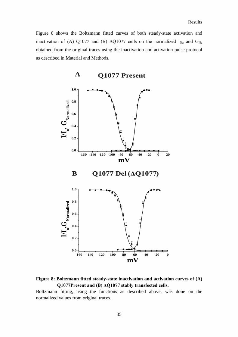
Figure 8 shows the Boltzmann fitted curves of both steady-state activation and
inactivation of (A) Q1077 and (B) ΔQ1077 cells on the normalized INa and GNa
obtained from the original traces using the inactivation and activation pulse protocol
as described in Material and Methods.
-160 -140 -120 -100 -80 -60 -40 -20 0 20
0.0
0.2
0.4
0.6
0.8
1.0
I/I o
,GN
orm
ali
zed
mV
Q1077 Present
-160 -140 -120 -100 -80 -60 -40 -20 0
0.0
0.2
0.4
0.6
0.8
1.0
I/I o
,GN
orm
ali
zed
mV
Q1077 Del ( )ΔQ1077
A
B
Figure 8: Boltzmann fitted steady-state inactivation and activation curves of (A)
Q1077Present and (B) ΔQ1077 stably transfected cells.
Boltzmann fitting, using the functions as described above, was done on the
normalized values from original traces.

Results
36
4.2 Inactivation properties of ΔQ1077 and Q1077 with FynCA
, FynKD
and tyrosine kinase inhibitor PP2
To test whether Fyn tyrosine kinase can differently modulate ΔQ1077 and
Q1077, the stably transfected cells were transiently transfected with FynCA
and FynKD
.
The constitutively active mutant, FynCA
lacks its inhibitory carboxyl terminus (amino
acids upstream of 525 were deleted), whereas the kinase dead mutant FynKD
has a
single point mutation at position K299M (Ahern et al., 2005; Nitabach et al., 2001).
Figure 9 shows the steady-state inactivation curves of the sodium current from stably
expressed cells (ΔQ1077 and Q1077), either alone or transiently transfected with
active FynCA
or dead FynKD
, or FynCA
incubated with the potent Src tyrosine kinase
inhibitor PP2.
Experiments with FynCA
on Q1077Present caused a 16-mV depolarizing shift of the
steady-state inactivation curve (Figure 9 and Table 9). The V½ inactivation of
Q1077Present was -88.21 ± 0.31 mV (n=8) and the V½ inactivation of Q1077Present
with FynCA
was shifted to -72.98 ± 0.06 mV (p<0.001, n=6). This result indicates that
the shift in steady-state inactivation of Q1077Present in presence of FynCA
is similar
to the hH1 cardiac sodium channel variant modulation by FynCA
. In the hH1 cardiac
sodium channel, FynCA
caused a 5-mV shift of the steady-state inactivation curve
from a V½ of -94.9 ± 2.4 mV to -89.4 ± 0.9 mV (Ahern et al., 2005). To prove that
this shift was due to Fyn, Q1077Present cells were transiently transfected with FynKD
.
The data were confirmed as no shift was observed with FynKD
(-87.13±0.29, n=4).
Further evidence was obtained with the kinase inhibitor PP2. PP2 inhibited the Fyn
action and reversed back the depolarizing shift from -72.98 ± 0.06 mV caused by
FynCA
to -88.41 ± 0.09 mV (n=3). These results obtained with Q1077Present in
presence of FynCA
, FynKD
and Src tyrosine inhibitor PP2 show that the depolarizing
shift was caused by Fyn tyrosine kinase.
Our experiments with FynCA
on ΔQ1077 caused a 9-mV hyperpolarizing potential
shift of the steady-state inactivation curve (Figure 9 and Table 9) from an average V½
of -79.39 ± 0.38 mV (n=9) for ΔQ1077 stably expressed cells to -88.51 ± 0.31 mV in
the presence of FynCA
(n=5) (p < 0.001). The hyperpolarizing shift caused by FynCA
was similar to that observed in the NaV1.2 channel, in which a 5-mV shift from -64.2
± 0.42 mV to -70.1 ± 0.32 mV was caused by Fyn (Beacham et al., 2007). When the

Results
37
ΔQ1077 cells were transiently transfected with FynKD
no shift of the steady-state
inactivation curve was observed (n=6). This study confirms that the hyperpolarizing
shift was caused by FynCA
. To substantially confirm that the shift in the inactivation
of ΔQ1077 was due to Fyn, the cells which were transfected with FynCA
were treated
with 1 µmol/L of the Src kinase inhibitor PP2. The kinase inhibitor PP2 inhibited the
Fyn action and reversed back the FynCA
-caused hyperpolarizing shift from -88.51 ±
0.31 mV to -78.52 ± 0.14 mV. This clearly confirms that the hyperpolarizing shift was
caused by Fyn tyrosine kinase.
No significant difference was seen in the peak INa and the decay of current in both
Q1077Present and ΔQ1077 with and without FynCA
(Table 9). However, from
analysis of steady-state inactivation it was evident that Q1077Present and ΔQ1077
cardiac sodium channels behave differently in the modulation by Fyn tyrosine kinase.
Table 8: Kinetic parameters for Q1077 and ΔQ1077 cardiac sodium channel
variants with and without FynCA
.
ΔQ1077 ΔQ1077 + FynCA
Q1077Present Q1077 + FynCA
Peak Current
INa, nA 3.78 ± 0.11 3.97 ± 0.28 1.92 ± 0.10 1.71 ± 0.26
n 8 3 4 4
Decay ( 0 mV)
Af 0.87 ± 0.11 0.84 ± 0.12 0.85 ± 0.03 0.89 ± 0.49
τf, ms 1.69 ± 0.14 1.45 ± 0.21 0.79 ± 0.46 0.96 ± 0.38
τs, ms 3.78 ± 0.15 4.13 ± 0.63 4.09 ± 0.90 4.62 ± 0.16
n 7 3 5 4

Results
38
Table 9: Half-maximal inactivation of both Q1077 and ΔQ1077 with FynCA
,
FynKD
and with FynCA
+ PP2.
Data are derived from fits of Boltzmann function to normalized inactivation curves
determined from individual cells as described in Materials and Methods. * p<0.001.
Variant Half-maximal
inactivation (V1/2)
(mV)
KInact
(mV) n
Q1077Present -88.21 ± 0.31 5.55 ± 0.21 8
Q1077Present + FynCA
-72.98 ± 0.06* 6.73 ± 0.35* 6
Q1077Present + FynKD
-87.13 ± 0.29 5.18 ± 0.24 4
Q1077Present + FynCA
+ PP2 -88.41 ± 0.09 5.91 ± 0.63 3
∆Q1077 -79.13 ± 0.38 5.76 ± 0.27 9
∆Q1077 + FynCA
-88.21 ± 0.31* 7.63 ± 0.18* 5
∆Q1077 + FynKD
-80.61 ± 0.36 5.97 ± 0.28 6
∆Q1077 + FynCA
+ PP2 -78.88 ± 0.14 6.28 ± 0.37 2

Results
39
A Q1077 Present
-160 -140 -120 -100 -80 -60 -40 -20
0.0
0.2
0.4
0.6
0.8
1.0
Q1077 Present
Q1077 Present + FynCA
Q1077 Present + FynKD
Q1077 Present + FynCA
+ PP2
No
rm
ali
zed
I Na
mV
Figure 9A: Expression of catalytic active FynCA
alters steady state inactivation
curves in stably expressed Q1077Present and ΔQ1077 cells.
A. Steady-state inactivation properties of stably expressed Q1077Present cardiac
sodium channel current with FynCA
. Solid line in (A) with filled squares corresponds
to the Boltzmann fit of current recorded from stably expressed Q1077Present cells.
The V1/2 of Q1077Present is -88.21 ± 0.31 mV. The Boltzmann fit curve with open
circles represents data obtained from stably expressed Q1077Present cells with FynCA
.
V1/2 of Q1077Present expressed with FynCA
is -72.98 ± 0.06 mV. When Q1077Present
cells were expressed with FynKD
, represented by filled up-pointing triangles, the V1/2
is -87.13 ± 0.29 mV. To test the Src tyrosine kinase inhibitor action, the
Q1077Present cells tranfected with FynCA
were incubated with the specific tyrosine
kinase inhibitor PP2 (1 µmol/L) for 30 minutes before current recording. The curve
with filled down-pointing triangles denotes the Boltzmann fit of Q1077Present in
presence of FynCA
and the specific tyrosine kinase inhibitor PP2; the V1/2 is
-88.41 ± 0.09 mV.

Results
40
-160 -140 -120 -100 -80 -60 -40 -20
0.0
0.2
0.4
0.6
0.8
1.0
Q1077 Deleted
Q1077 Deleted + FynCA
Q1077 Deleted + FynKD
Q1077 Deleted + FynCA
+ PP2
mV
No
rm
ali
zed
I Na
Q1077 DeletedB
Figure 9B:
B. Similar inactivation curves for ΔQ1077 cells are shown as explained above for
Q1077Present cells. Boltzmann fit of stably expressed ΔQ1077 cells is denoted with
filled squares. The V1/2 inactivation of ΔQ1077 is -79.39 ± 0.38 mV. The inactivation
curve of stably expressed ΔQ1077 with FynCA
is represented by open circles. V1/2 for
ΔQ1077 expressed with FynCA
is -80.76 ± 0.36 mV. The inactivation curve for
ΔQ1077 cells expressed with FynKD
is represented by filled up-pointing triangles; the
V1/2 is -80.76 ± 0.36 mV. The Boltzmann fitted inactivation curve for the ΔQ1077
cells in presence of FynCA
and PP2 is depicted with filled down-pointing triangles.
V1/2 inactivation, when treated with PP2, is -78.52 ± 0.14 mV.

Results
41
4.3 Activation Properties of ΔQ1077 and Q1077 with FynCA
To determine whether FynCA
can modulate the steady-state activation of
ΔQ1077 and Q1077Present, the cells were transfected with FynCA
, and the normalized
conductance of ΔQ1077 and Q1077 with or without FynCA
was analysed. The steady-
state activation of ΔQ1077 and Q1077Present in presence or absence of FynCA
is
shown in Figure 10 and Table 10.
Table 10: Half-maximal activation and slope of both Q1077 and ΔQ1077 with
FynCA
.
Data derived from fits of a Boltzmann function to normalized activation curves,
determined from individual cells as described in Materials and Methods.
Variant Half-maximal
activation (V1/2)
(mV)
KAct
(mV) n
Q1077 -48.91 ± 0.22 3.87 ± 0.13 5
Q1077 + FynCA
-49.17 ± 0.23 4.05 ± 0.23 3
∆Q1077 -47.65 ± 0.29 3.69 ± 0.10 4
∆Q1077 + FynCA
-48.56 ± 0.63 4.07 ± 0.59 3

Results
42
A Q1077 Present
B Q1077 Deleted
-100 -90 -80 -70 -60 -50 -40 -30 -20 -10
0.0
0.2
0.4
0.6
0.8
1.0
mV
No
rm
ali
zed
GN
a
Q1077 Deleted
Q1077 Deleted + FynCA
-100 -90 -80 -70 -60 -50 -40 -30 -20 -10
0.0
0.2
0.4
0.6
0.8
1.0Q1077 Present
Q1077 Present + FynCA
mV
Norm
ali
zed
GN
a
Figure 10: Steady-state activation curves of stably expressed Q1077 and ΔQ1077
cardiac sodium channels.
Boltzmann functions fit to normalized conductance were determined from individual
cells as described in Materials and Methods.

Results
43
Figure 10 shows the activation curves of Q1077Present in absence and presence of
FynCA
. FynCA
did not cause any shift of the activation curve. Similarly, in ΔQ1077
FynCA
also did not alter the activation curve (Table 10).
These data clearly show that activation kinetics of the two variants, Q1077Present and
ΔQ1077, is not modulated by Fyn. These results were consistent with earlier
published results on Fyn modulation of steady state activation of NaV1.2 and NaV1.5
(hH1) sodium channels (Ahern et al., 2005; Beacham et al., 2007).
4.4 Sequence alignment of sodium channel subunits of the
intracellular loop LDII-DIII
To ascertain the uniqueness of cardiac sodium channel sequence, the amino
acid sequences of all the nine sodium channel isoforms were aligned using the
multiple sequence alignment CLUSTAL X program. The Pfam database is a large
collection of protein families, each represented by multiple sequence alignments and
hidden Markov models (Finn et al., 2010). According to pfam06512 database
(Marchler-Bauer et al., 2011), amino acids of the cardiac sodium channel between
positions 953-1214 form one of the sodium ion-transport associated regions of the
channel. In the predicted cardiac sodium channel structure (George et al., 1992),
amino acids from position 953 to 1214 also form the intracellular loop connecting the
DII and DIII region (LDII-DIII) of the cardiac sodium channel. Since Q1077 is present
in the intracellular loop connecting domains II and III, amino acid sequences of this
loop of all the sodium channel isoforms were aligned and analysed (Figure 11).
From Figure 11 it is evident that the Q1077 residue is located in the amino acid
sequence 1075-KQQESQP-1081, which is unique in the cardiac sodium channel. In
LDII-DIII the amino acid sequences 1047-GEQPGQGTPGDPEPVCVPIAVAE-1053
and 1116-QQWKAEPQAPGCGETP-1132 are also unique among different sodium
channel isoforms (Figure 11).

Results
44
Figure 11: Alignment of the intracellular loop of sodium channel subtypes
connecting Domain II and III (LDII-DIII).
Amino acid numbering of cardiac sodium channel and the proposed structure (George
et al., 1992) forms the base of the alignment. Corresponding LDII-DIII residues of the
other sodium channel subtypes were used for the alignment.
Asterisks denote consensus amino acids, colons denote high similarity of amino acids,
and dots denote less similarity among the aligned amino acids. No symbol denotes
lack of similarity among amino acids.

Results
45
4.5 Kinetics of Q1077 cardiac sodium channel mutants
To analyse the importance of Q1077 in the cardiac sodium channel, the amino
acid glutamate was replaced with different amino acids using the Stratagene Quick
Change Site-Directed Mutagenesis Kit with the Q1077Present cDNA as the template.
The Q1077 was mutated to lysine (Q1077K), alanine (Q1077A), proline (Q1077P),
and tyrosine (Q1077Y). The steady-state inactivation kinetics was obtained as
described in the Materials and Methods section.
Q1077A Q1077Y
Q1077K Q1077P(a) (b)
(c) (d)
1 nA
0.5 ms
1 nA
0.5 ms
1 nA
0.5 ms
0.5 nA
0.5 ms
Figure 12: Original traces illustrating steady-state inactivation of the transiently
transfected mutants at position Q1077 in tsA-201 cells:
(a) Q1077K, (b) Q1077P (c) Q1077A (d) Q1077Y. Current recordings are shown,
which were obtained when the cells were clamped from a holding potential of -140
mV to 100 ms prepulses at -150, - 140, -130, -120, -110, -105, -100, -95, -90, -85, -80,
-75,-70,-65, -60, -50, -40, -30, -20, -10, and 0 mV, followed by a 24 ms test pulse to 0
mV.

Results
46
From Figure 12 it is evident that the peak INa of Q1077P was smaller than that of the
other counterparts. To obtain detailed inactivation kinetics, the original traces were
fitted with Boltzmann functions as done for Q1077Present and ΔQ1077 (Table 11).
Table 11: Kinetic parameters for all four mutants of the Q1077 cardiac sodium
channel.
§ (p<0.001) Significant difference (ANOVA) of the mutants relative to Q1077Present.
* (p<0.001) Significant difference (ANOVA) among all 4 different Q1077 mutants.
As illustrated in Table 11 the steady-state inactivation of mutants Q1077A and
Q1077K was shifted to more hyperpolarizing potentials. The V1/2 inactivation of
Q1077K was -97.47 ± 0.23 mV (n=3) and for Q1077A it was -101.61 ± 0.30 mV
(n=6), which was statistically different (p<0.001) from the Q1077Present variant
(-88.21 ± 0.31 mV, n=8). In contrast, there was no statistically significant difference
in the steady-state inactivation kinetics of Q1077P (-88.21 ± 0.47 mV, n=4) and
Q1077Y (-90.43 ± 0.35 mV, n=4) compared to the Q1077Present variant (-88.21 ±
0.3 mV, n=8). This indicates that the mutation at position 1077 with alanine and
lysine (Q1077A and Q1077K) altered the V1/2 inactivation. The peak INa of Q1077P
Q1077K Q1077P Q1077A Q1077Y
Inactivation
V1/2, mV -97.47 ± 0.23*§ -88.21 ± 0.47 -101.61 ± 0.30*
§ -90.43 ± 0.35
Slope, mV 6.13 ± 0.22 7.16 ± 0.32 6.39 ± 0.25 6.49 ± 0.32
n 3 4 6 4
Peak Current
INa, nA -3.59 ± 0.92 0.81 ± 0.09* -4.48 ± 0.87 -3.24 ± 0.51
n 4 3 3 3
Decay ( 0 mV)
Af 0.94 ± 0.27 0.64 ± 0.26 0.69 ± 0.25 0.86 ± 0.43
τf, ms 0.53 ± 0.76 0.76 ± 0.11 0.65 ± 0.24 0.78 ± 0.63
τs, ms 2.84 ± 0.33 2.44 ± 0.44 5.45 ± 0.70* 2.13 ± 0.14
n 3 4 3 3

Results
47
(0.81 ± 0.09 nA, n=3) was statistically different (p<0.001) from that of the other
mutants Q1077K (-3.59 ± 0.92 nA, n=4), Q1077A (-4.48 ± 0.87 nA, n=3) and
Q1077Y (-3.24 ± 0.51 nA, n=3). Thus, the mutation from glutamine at position 1077
to proline significantly reduced the peak INa. For the decay of INa at 0 mV, the portion
of the INa trace measured after 90% of peak was fit to a sum of exponentials and the
parameters are listed in Table 11. The decay time constant of the slow component (τs)
for Q1077A was significantly (p<0.001) different from that of the other 1077 mutants
(Q1077K, Q1077P and Q1077Y).
4.6 Inactivation kinetics of Q1077 mutants with FynCA
and FynKD
To test the modulation of Q1077 mutants by FynCA
and FynKD
, the mutant
clones were co-transfected with FynCA
and FynKD
in tsA-201 cells. Original traces
were obtained using the inactivation protocol as described in the Materials and
Method section. The obtained traces were then analysed and fitted using Boltzmann
function. The results were plotted (Figure 13) and tabulated (Table 12). The
difference in the modulation by FynCA
among different Q1077 mutants is presented in
the following sections.
4.6.1 Modulation of inactivation kinetics of Q1077K mutant with FynCA
and FynKD
When the neutral amino acid glutamine at position Q1077 was replaced with
the basic residue lysine (Q1077K), the inactivation was shifted to more
hyperpolarizing potentials (-97.47 ± 0.23 mV, n=6). Cells co-transfected with FynCA
produced a statistically significant (p<0.001) hyperpolarizing shift from -97.47 ± 0.23
mV to -102.95 ± 0.43 mV (n=3). This result was similar to that of the NaV1.2 sodium
channel modulation by Fyn and ∆Q1077 stably tranfected cell modulation by Fyn.
Q1077K with FynKD
(V1/2: -97.63 ± 0.98 mV, n=3) did not produced any shift of the
inactivation curve. This strongly emphasises that the modulation in inactivation was
due to Fyn.

Results
48
4.6.2 Modulation of inactivation kinetics of Q1077A mutant with FynCA
and FynKD
The polar glutamine residue at position 1077 was replaced with the unpolar
residue alanine (Q1077A). Q1077A also produced a hyperpolarizing shift, which was
even larger (13.4 mV) compared to the other three mutants (Table 12). Cells co-
transfected with FynCA
produced a statistically significant hyperpolarizing shift from
-101.61 ± 0.30 mV (n=6) to -118.02 ± 0.36 mV (n=3). When the cells were co-
transfected with FynKD
no statistically significant shift in the inactivation (V1/2:
-105.33 ± 0.51 mV) was observed. The results obtained for inactivation of INa of
Q1077A were in line with the Q1077K mutant, but the shift caused due to Q1077A
mutant was larger (Table 12).
4.6.3 Modulation of inactivation kinetics of Q1077P mutant with FynCA
and FynKD
The aliphatic glutamine residue at position 1077 in the cardiac sodium channel
was replaced with the heterocyclic amino acid residue proline (Q1077P). The steady-
state inactivation of the Q1077P mutant (-88.21 ± 0.47 mV, n=6) was not shifted.
When the Q1077P mutant was co-transfected with FynCA
, a statistically significant
(p<0.001) hyperpolarizing shift from -88.21 ± 0.47 mV to -97.71 ± 0.38 mV (n=4)
was produced. The hyperpolarizing shift was not observed in the Q1077P mutant
transfected with FynKD
(-88.85 ± 0.89 mV, n=4). This hyperpolarizing shift caused by
the mutant Q1077P with FynCA
was less compared to the Q1077A mutant, but more
pronounced than with the Q1077K mutant.
4.6.4 Modulation of inactivation kinetics of Q1077Y mutant with FynCA
and FynKD
The aromatic amino acid residue tyrosine was substituted instead of the
aliphatic glutamine residue at position 1077 (Q1077Y). Similar to mutant Q1077P,
the steady-state inactivation of Q1077Y was also not shifted (-90.43 ± 0.35 mV, n=4).
When the Q1077Y mutant was co-transfected with FynCA
, a statistically significant

Results
49
(p<0.001) hyperpolarizing shift from -90.43 ± 0.35 mV to -99.79 ± 0.18 mV (n=3)
was observed. Such a hyperpolarizing shift was not seen when the cells were co-
transfected with FynKD
(-90.45 ± 0.32 mV, n=3).
Table 12: Half-maximal inactivation of all four Q1077 mutants with FynCA
and
FynKD
.
Data are derived from fits of Boltzmann function to normalized inactivation curves.
Mutant Half-maximal
inactivation (V1/2)
(mV)
KInact
(mV)
n
Q1077Y -90.43 ± 0.35 6.49 ± 0.32 4
Q1077Y + FynCA
-99.79 ± 0.18* 7.98 ± 0.13* 3
Q1077Y + FynKD
-90.45 ± 0.32 6.62 ± 0.59 3
Q1077P -88.21 ± 0.47 7.16 ± 0.32 6
Q1077P + FynCA
-97.71 ± 0.38* 7.41 ± 0.14 4
Q1077P + FynKD
-88.85 ± 0.89 6.65 ± 0.53 4
Q1077K -97.47 ± 0.23 6.13 ± 0.22 6
Q1077K + FynCA
-102.95 ± 0.43* 8.96 ± 0.24* 3
Q1077K + FynKD
-97.63 ± 0.98 6.82 ± 0.63 3
Q1077A -101.61 ± 0.30 6.39 ± 0.25 6
Q1077A + FynCA
-118.02 ± 0.36* 6.84 ± 0.17 3
Q1077A + FynKD
-105.33 ± 0.51 5.73 ± 0.28 5

Results
50
Q1077K
Q1077P
Q1077A
-160 -140 -120 -100 -80 -60
0.0
0.2
0.4
0.6
0.8
1.0Q1077K
Q1077K + FynCA
Q1077K + FynKD
No
rmali
zed
I Na
mV-160 -140 -120 -100 -80 -60
0.0
0.2
0.4
0.6
0.8
1.0 Q1077A
Q1077A + FynCA
Q1077A + FynKD
No
rmali
zed
I Na
mV
Q1077Y
-160 -140 -120 -100 -80 -60 -40
0.0
0.2
0.4
0.6
0.8
1.0
Norm
ali
zed
I Na
mV
Q1077P
Q1077P + FynCA
Q1077P + FynKD
BA
C D
-160 -140 -120 -100 -80 -60 -40
0.0
0.2
0.4
0.6
0.8
1.0 Q1077Y
Q1077Y + FynCA
Q1077Y + FynKD
Norm
ali
zed
I Na
mV
Figure 13: Expression of catalytic active FynCA
alters steady-state inactivation
kinetics of Q1077 mutants.
Boltzmann function fitted steady-state inactivation curves of four Q1077 mutants -
(A) Q1077K, (B) Q1077A, (C) Q1077P, (D) Q1077Y with FynCA
and FynKD
are
shown. Solid lines with filled squares correspond to Boltzmann fits of the respective
transiently transfected mutant. Cells expressing both, Q1077 mutants and FynCA
, are
represented by open circles. Q1077 mutants expressed with FynKD
are represented by
open triangles. Number of experiments is given in Table 12.
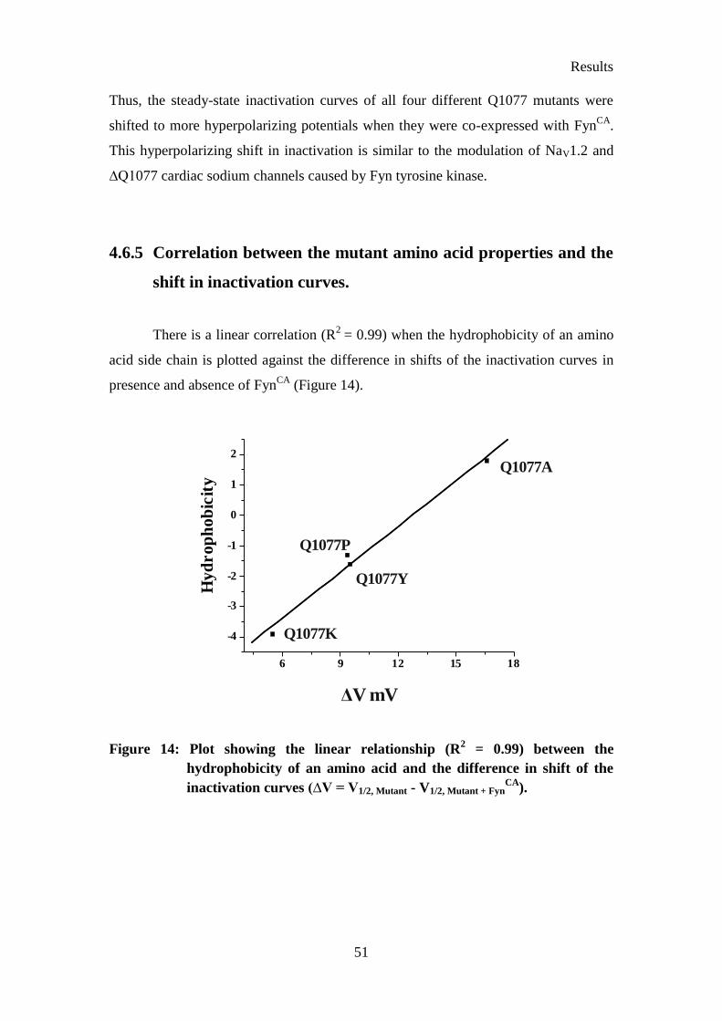
Results
51
Thus, the steady-state inactivation curves of all four different Q1077 mutants were
shifted to more hyperpolarizing potentials when they were co-expressed with FynCA
.
This hyperpolarizing shift in inactivation is similar to the modulation of NaV1.2 and
∆Q1077 cardiac sodium channels caused by Fyn tyrosine kinase.
4.6.5 Correlation between the mutant amino acid properties and the
shift in inactivation curves.
There is a linear correlation (R2
= 0.99) when the hydrophobicity of an amino
acid side chain is plotted against the difference in shifts of the inactivation curves in
presence and absence of FynCA
(Figure 14).
6 9 12 15 18
-4
-3
-2
-1
0
1
2
Hyd
ro
ph
ob
icit
y
ΔV mV
Q1077A
Q1077K
Q1077Y
Q1077P
Figure 14: Plot showing the linear relationship (R2 = 0.99) between the
hydrophobicity of an amino acid and the difference in shift of the
inactivation curves (∆V = V1/2, Mutant - V1/2, Mutant + FynCA
).

Results
52
4 6 8 10 12 14 16 185
6
7
8
9
10
Iso
elctr
icp
oin
t(p
I)
4 6 8 10 12 14 16 1880
100
120
140
160
180
Avera
ge
Ma
ss(D
a)
∆V mV
Q1077Y
Q1077A
Q1077P
Q1077K
∆V mV
Q1077K
Q1077A
Q1077P
Q1077Y
4 6 8 10 12 14 16 1860
70
80
90
100
110
120
130
140
150
Van
der
Waa
lsvo
lum
e(V
w)
∆V mV
Q1077A
Q1077K
Q1077Y
Q1077P
4 6 8 10 12 14 16 180
20
40
60
80
100
120
140
Mola
rM
ass
(g/m
ol)
∆V mV
Q1077AQ1077P
Q1077YQ1077
Q1077K
4 6 8 10 12 14 16 181.9
2.0
2.1
2.2
2.3
2.4
Dis
socia
tio
nC
on
sta
nt
(-C
OO
H,p
K1)
4 6 8 10 12 14 16 18
9.2
9.6
10.0
10.4
10.8
Dis
soci
ati
on
Co
nst
an
t(-
NH
3,p
K2)
Q1077A
Q1077A
Q1077YQ1077P
Q1077YQ1077K
Q1077K
Q1077P
∆V mV ∆V mV
(a) (b)
(c)(d)
(e) (f)
Figure 15: Plot showing the linear relationship between different properties of
amino acids and the difference in shift of the inactivation curves
(a) average mass (R2
= 0.45), (b) isoelectric point (R2
= 0.45), (c) Van der Waals
volume (R2
= 0.67), (d) molar mass (R2
= 0.76), (e) dissociation constant (α-COOH)
pK1 (R2
= 0.33) and (f) dissociation constant (α-NH3) pK2 (R2
= 0.16) of an amino
acid and the difference in shift of the inactivation curves (∆V = V1/2, Mutant - V1/2, Mutant +
FynCA
) is shown.
Results
53
Linear correlation graphs of different properties of the amino acids plotted against the
difference in shift of the inactivation curves (∆V mV) show weak or no correlation
(Figure 15). Only molar mass (R2
= 0.76) and the Van der Waals volume (Vw) (R2
=
0.67) were weakly correlated, whereas the other parameters like average mass (R2
=
0.45), isoelectric point (R2
= 0.45), dissociation constant pK1 (R2
= 0.33) and pK2 (R2
= 0.16) were poorly correlated.
Discussion
In this study, it is shown that the voltage-gated sodium channel variants are
modulated differently by Fyn, which is a member of the large Src tyrosine kinase
family. The effect of Fyn tyrosine kinase has been evaluated by shifts observed in the
steady-state inactivation curves of the sodium channel variants. The evidence of this
difference in the modulation by Fyn tyrosine kinase is further supported by mutating
the glutamine residue (Q1077) in the intracellular loop (LDII-DIII) to four different
residues (A, P, K and Y). The amino acid sequence of the intracellular loop (LDII-DIII)
of all the sodium channel isoforms was analyzed and the uniqueness of the cardiac
sodium channel to other sodium channel isoforms is documented. Lastly, a search for
putative Src tyrosine kinase SH3 and SH2 binding motifs in the amino acid sequence
of the cardiac sodium channel was performed.
5.1 Splice variant specificity of Fyn modulation of cardiac sodium
channels
Our results indicate that Fyn regulates inactivation of different cardiac sodium
channel variants differently. The Q1077Present variant shifted the inactivation curve
to more depolarizing potentials (15 mV), while the ∆Q1077 variant shifted the
inactivation curve to more hyperpolarizing potentials (9 mV). Studies on the hH1
polymorphic cardiac sodium channel variant shifted the inactivation curve with Fyn to
more depolarizing potentials (5 mV) (Ahern et al., 2005). The hH1 sodium channel
variant is composed of 2016 amino acids, and it has a glutamine residue at position

Discussion
55
1027 compared to Q1077Present, which has an arginine residue at this position.
∆Q1077 (Q1077del) is a splice variant of the Q1077Present variant where an
additional glutamine is inserted at position 1077 by a 5′-extension of exon 18 by the
trinucleotide CAG (Makielski et al., 2003). The peak INa of ∆Q1077 is larger than in
hH1, and V1/2 inactivation appeared at more depolarized potentials (+5 mV) than in
the hH1 variant. The activation curve is not shifted in both the ∆Q1077 and hH1
variant (Makielski et al., 2003). This suggests that Q1077 and surrounding residues
may play a role in the steady-state inactivation kinetics of the cardiac sodium channel.
There is also evidence that the ∆Q1077 variant behaves differently in the background
of cardiac mutations causing channelopathies. The loss of function mutation G1406R
in the cardiac sodium channel, which causes Brugada syndrome, shows difference in
the partial expression defect with ∆Q1077 and Q1077 in the background. These data
suggest that there might be a partial trafficking defect, expression defect or gating
abnormalities due to loss of function of the G1406R mutation (Tan et al., 2006).
Therefore, intracellular trafficking might play an important role in the difference of
modulation.
The putative site for CK2 (casein kinase 2) phosphorylation is reported to be
(S/T)XX(E/D) (Meggio and Pinna, 2003). This sequence -SKQE- is present in loop
LDII-DIII in the ∆Q1077 variant of the NaV1.5 cardiac sodium channel at position 1074
to 1077. Inserting a glutamine residue (Q1077Present) abolishes the putative
phosphoacceptor site for CK2 (Kerr et al., 2004).
There is also a study which reports that ∆Q1077 is present in 45% of the NaV1.5
transcript in human hearts, and the Q1077Present variant transcript is present in 25%
of the human hearts (Makielski et al., 2003). The hH1 cardiac sodium channel
transcript (Accession No. M77235), on which modulation by Fyn was studied (Ahern
et al., 2005), is not present (0%) in the human population (Makielski, 2006). Thus,
based on these previously published data and our own results on the modulation of
inactivation of ∆Q1077 and Q1077Present by Fyn, we speculate that the glutamine
residue at position 1077 and the neighbouring residues in loop LDII-DIII could play a
role in the difference of modulation of the cardiac sodium channel by Fyn.

Discussion
56
5.2 Mutational effects at position 1077
Our results indicate that substitution of aliphatic glutamine at position 1077 to
aromatic tyrosine or heterocyclic proline does not have any effect on inactivation
kinetics, as it was similar to the Q1077Present variant. Whereas, when aliphatic and
polar glutamine was mutated to aliphatic and hydrophobic alanine or to the polar and
basic amino acid lysine there was a shift in V1/2 inactivation observed compared to
Q1077Present. In the case of Q1077A, alanine is a hydrophobic residue and it is
ambivalent; that means it can be inside or outside the protein accessible area. This
could lead to different hydrophobic interactions between other nearby residues, in turn
hindering the inactivation process. In Q1077K, lysine substitution shifted the V1/2
inactivation to more hyperpolarizing potentials compared to the Q1077Present
variant. The lysine residue has the ability to interact with the solvent readily, which
might have played a role in shifting the inactivation curve to more hyperpolarizing
potentials. Q1077P and Q1077Y, which are bulky residues, could hinder the folding
of loop LDII-DIII which is the ion transport associated region.
Our results on mutation at position 1077 revealed that all the amino acid mutations
from glutamine to alanine, lysine, tyrosine and proline caused a shift of the respective
inactivation curves to more hyperpolarizing potentials with Fyn as observed in
∆Q1077 (Figure 9) and NaV1.2 (Beacham et al., 2007). However, there is different
extent of the hyperpolarizing shift among different mutations. The mutation with
alanine caused a very large hyperpolarizing shift by Fyn, and the least hyperpolarizing
shift was produced by the positively charged residue lysine. There was also a strong
linear correlation (R2=0.99) between the hydrophobicity of the mutated amino acids
and the extent of the Fyn-caused hyperpolarizing shift. Highly hydrophobic residues
are present in the SH2 domain of the Src tyrosine kinase, and in addition also the
peptide binding site of SH3 is composed of a hydrophobic groove. When Q is present
at position 1077 there might be hindrance to the hydrophobic interaction between the
SH domains and the cardiac sodium channel. There is weak linear correlation
observed with molar mass (R2=0.76) or the Van-der-Waals volume (Vw) (volume of
the union of overlapping spheres) (R2=0.67), respectively, and the difference in the
hyperpolarizing shifts caused by Fyn. Vw plays an important role in the packing and
folding of a protein (Richards, 1977). Thus, from our mutational analysis we can

Discussion
57
conclude that hydrophobicity, and to a lesser extent, the Van-der-Waals volume (Vw)
of the amino acids is vital in the different modulation of the sodium channel by Fyn.
5.3 Uniqueness of cardiac sodium channel intracellular loop LDII-DIII
From the performed sequence analysis it is obvious that the glutamine residue
at position 1077 is unique among different sodium channel isoforms (Figure 11).
There are three short unique amino acid sequences present in the cardiac sodium
channel compared to other sodium channel isoforms. The glutamine at position 1077
is present in one of these unique sequences of the cardiac sodium channel as shown in
the Figure 11. Glutamine at position 1076 and 1077 is surrounded by the charged
amino acids lysine at position 1075 and glutamic acid at position 1078. In turn, both
of these charged amino acids are surrounded by the polar residue serine. Thus, the two
glutamine residues are hidden in between the two polar serine residues, which might
hinder the flexibility of the sodium channel. This serine residue has the ability to form
a hydrogen bond with other polar residues. It has the property of either accepting or
donating a hydrogen bond. So, an additional glutamine at position 1077 might hinder
the flexibility of the loop LDII-DIII. This might play an anchoring role in facilitating the
SH domains of the Src tyrosine kinase to bind to the cardiac sodium channel.
Recent studies on MOG1 (multicopy suppressor of gsp1), which is present in the
nucleus and the cytosol of cardiac cells (Marfatia et al., 2001), demonstrate that it
plays an important role in the regulation of nuclear protein trafficking (Wu et al.,
2008). It showed that MOG1 is an important cofactor for optimal expression of
NaV1.5 at the cell membrane. When MOG1 and NaV1.5 were co-expressed in
HEK293 cells, there was only an increase in the peak INa with no change in the
biophysical characteristics. The interaction site for MOG1 in NaV1.5 is the loop
connecting domain II and domain III (LDII-DIII) (Abriel and Kass, 2005; Wu et al.,
2008). Ankyrins are intracellular proteins which target and stabilize the membrane
proteins including ion channels, transporters, exchangers and cell adhesion molecules
in various tissues (Cunha and Mohler, 2006). Ankyrin-G binds to the -VPIAXXSD-
motif in loop LDII-DIII of the cardiac sodium channel (Abriel and Kass, 2005; Mohler et
al., 2004). SCN5A mutation (pE1053K) in Brugada syndrome patients leads to

Discussion
58
disruption of the interaction between NaV1.5 and ankyrin-G (Mohler et al., 2004).
Thus, these studies on different intracellular proteins, which modulate the sodium
channel, reveal that LDII-DIII is an essential target. From our results on cardiac sodium
channel variants, modulation by Fyn suggests that the protein-protein interaction site
for the sodium channel and Fyn should be present in LDII-DIII.
5.4 Possible binding sites for the SH2 domain in the cardiac sodium
channel
Our results on the possible SH3 domain binding, poly-proline motif show that
there are five such motifs in loop LDII-DIII. As shown in Figure 15, the NaV1.2 does not
contain any proline-rich motif in LDII-DIII. Thus, this loop LDII-DIII could play an
important role in SH3 binding and modulation of the cardiac sodium channel by Fyn.
In NaV1.2 the consensus sequence for the potential binding site of the Fyn SH2
domain, after phosphorylation, is present in the N- and C-terminal (Beacham et al.,
2007). The sequence for the potential SH2 binding site in NaV1.2 is reported as 66-
YGDI-69 in the N-terminal, and 1893-YEPI-1896 in the C-terminal. In the cardiac
sodium channel corresponding sites were analysed using the sequence alignment
program. There, the C-terminal -YGDI- is replaced by 68-YGNP-, whereas the N-
terminal has a corresponding 1988-YEPI-1992 sequence (Beacham et al., 2007).
Similarly, in the NaV1.2 channel there is a Y730 which is located in LDI-DII near the
SH3 binding motif (-RVLPILP-), which is required for optimal regulation by Fyn
(Beacham et al., 2007). This Y730 residue is conserved in all other sodium channel
isoforms except the cardiac sodium channel. In the cardiac sodium channel the
corresponding Y730 is replaced by N688 (asparagine). In NaV1.2, it was speculated
that the SH2 motifs in the N- and C-terminal and the SH3 motif in LDI-DII play a role
in maintaining a high local concentration of Fyn. But in the cardiac sodium channel
there is only one SH2 binding motif in the C-terminal and several putative SH3
binding motifs in LDII-DIII. Earlier studies show that the Fyn SH2 domain binds to the
-pYQQI- motif of the target protein (Songyang et al., 1993). However, there are some
other SH2 domains which bind to different patterns. The growth factor receptor-
bound protein 2 (Grb2) SH2 domain binds to the -pYXNX- motif of the target protein
Discussion
59
(Rahuel et al., 1996), whereas the SH2 domain of the growth factor phospholipase C-
γ1 (PLC-γ1) binds to hydrophobic residues that follow pTyr (Ji et al., 1999). Thus,
Fyn could also bind to different SH2 motifs in the cardiac sodium channel other than
the putative SH2 binding motif in NaV1.2. Putative SH2 binding motifs are situated in
the C-terminal of the cardiac sodium channel, because there are motifs (1811-YDSV-
1814 and 1977-YSVL-1980) apart from the conserved -YEPI-, which have a
hydrophobic residue at the pY+3 residue. The pY+3 residue is tightly bound to the
hydrophobic pocket, and this pocket is critical for the peptide specificity (Brändén and
Tooze, 1999). Thus, valine in the 1811-YDSV-1814 motif and leucine in the 1977-
YSVL-1980 motif might form a tight hydrophobic interaction with the hydrophobic
pocket of the SH2 domain. Mutational studies on these motifs will give an insight
view on the SH2 binding pockets of the cardiac sodium channel.
5.5 Putative SH3 domain binding sites in sodium channels
The SH3 domain consists of a five-stranded up-and-down antiparallel β sheet.
The peptide binding site is made up of a hydrophobic groove (Brändén and Tooze,
1999). The SH3 domain of the Src tyrosine kinase binds to the target peptides which
have proline-rich motifs (Lim, 1996; Yu et al., 1994). When the peptide binds to the
SH3 domain, proline-rich segments adopt a left-handed poly-proline type II helix with
three amino acids per turn. There are three ridges in the poly-proline helix of which
two contact the SH3 domain of the protein tyrosine kinase. The SH3 domain of the
Src tyrosine kinase contains a group of acidic amino acids and less hydrophobic
amino acid residues (Brändén and Tooze, 1999). All known SH3 target proteins carry
a consensus sequence -PXXP-. When v-src is expressed with the human voltage gated
potassium channel subtype (hKV1.5) (Holmes et al., 1996b), it is phosphorylated. It
was found that hKV1.5 contains two SH3 binding motifs (RPLPPLP) between amino
acid residues 65 and 82 of the channel protein (Holmes et al., 1996b; Tamkun et al.,
1991).
In NaV1.2 the SH3 binding motif for Fyn is present in LDI-DII (Figure 17) (Beacham et
al., 2007). Insertion of a glycine residue in this motif (-PTLP- to -PTGLP-) nullifies
the modulation of NaV1.2 by Fyn (Beacham et al., 2007). In Figure 17, the other

Discussion
60
putative SH3 binding motifs are also shown; there is no proline-rich motif present in
LDII-DIII, but two putative SH3 binding proline-rich motifs are present in the N-
terminal region. In the cardiac sodium channel there are five -PXXP- motifs in
LDII-DIII. The cardiac sodium channel has one -PXXP- motif in the loop LDI-DII, which
is different from that of NaV1.2. Two of the -PXXP- motifs in LDII-DIII of the cardiac
sodium channel are present in the unique sequence found when aligned with other
sodium channel isoforms (Figure 16). This suggests that the putative SH3 domain
binding motif of the cardiac sodium channel is present in loop LDII-DIII. These studies
reveal that the SH3 domain binding domain of the NaV1.2 and NaV1.5 is different.
Thus, this could contribute to the difference observed in the modulation by Fyn
tyrosine kinase.
Cardiac sodium channel sequence analysis reveals five putative SH3 binding domains
in loop LDII-DIII (Figure 16), but there is no proline-rich motif in LDII-DIII of the NaV1.2
sodium channel (Figure 17). Whereas, for the NaV1.2 channel it is proven that there is
one SH3 binding motif present in the LDI-DII. However, the same motif is not present
in NaV1.5. Inserting a residue in the SH3 motif of NaV1.2 alters the modulation of
inactivation kinetics by Fyn. According to suggested proline-rich motifs binding the
SH3 domain of Src tyrosine kinases (Yu et al., 1994), in the cardiac sodium channel
out of five putative motifs, the sequences 986-RQRPQKP-992, 1120-KAEPQAP-
1126 and 1008-PYSPPPP-1014 in LDII-DIII are most likely to form the SH3 binding
site for Fyn kinase (Figure 16). The first two putative motifs have a charged amino
acid residue (R and K) two residues (any amino acid) before the first proline of the
proline-rich motif which is responsible for the SH3 domain binding groove (Brändén
and Tooze, 1999; Yu et al., 1994). The third, above mentioned, proline rich motif
contains four continuous proline residues making it an ideal candidate for SH3
domain binding. Apart from the previously mentioned proline-rich motifs there are
two other SH3 binding -PXXP- motifs present in LDII-DIII. The two proline-rich motifs
in LDII-DIII are 1039-PGDPEP-1044 and 1086-PEAPP-1090; these can also form the
SH3 domain binding motif, but it lacks the charged R/K residue, two residues before
the first proline of the -PXXP- motif. But the 1086-PEAPP-1090 is in very close
proximity to the glutamine residue at position1077. Hence, the consideration of this
motif for SH3 binding should not be left out. Apart from LDII-DIII, there is one weak
SH3 binding, proline-rich motif (PLAP) in loop LDI-DII (Figure 16). Insertion mutation

Discussion
61
or removing the entire motifs will give an insight view on the SH3 binding site of the
cardiac sodium channel.
N-Terminal
Domain I
Domain II
Domain III
Domain IV
C-Terminal
LDII-DIII
LDIII-DIV
LDI-DII
1
2016
MANFLLPRGT SSFRRFTRES LAAIEKRMAE KQARGSTTLQ ESREGLPEEE
APRPQLDLQA SKKLPDLYGN PPQELIGEPL EDLDPFYSTQ KTFIVLNKGK
TIFRFSATNA LYVLSPFHPI RRAAVKILVH SLFNMLIMCT ILTNCVFMAQ
HDPPPWTKYV EYTFTAIYTF ESLVKILARG FCLHAFTFLR DPWNWLDFSV
IIMAYTTEFV DLGNVSALRT FRVLRALKTI SVISGLKTIV GALIQSVKKL
ADVMVLTVFC LSVFALIGLQ LFMGNLRHKC VRNFTALNGT NGSVEADGLV
WESLDLYLSD PENYLLKNGT SDVLLCGNSS DAGTCPEGYR CLKAGENPDH
GYTSFDSFAW AFLALFRLMT QDCWERLYQQ TLRSAGKIYM IFFMLVIFLG
SFYLVNLILA VVAMAYEEQN QATIAETEEK EKRFQEAMEM LKKEHEALTI
RGVDTVSRSS LEMSPLAPVN SHERRSKRRK RMSSGTEECG EDRLPKSDSE
DGPRAMNHLS LTRGLSRTSM KPRSSRGSIF TFRRRDLGSE ADFADDENST
AGESESHHTS LLVPWPLRRT SAQGQPSPGT SAPGHALHGK KNSTVDCNGV
VSLLGAGDPE ATSPGSHLLR PVMLEHPPDT TTPSEEPGGP QMLTSQAPCV
DGFEEPGARQ RALSAVSVLT SALEELEESR HKCPPCWNRL AQRYLIWECC
PLWMSIKQGV KLVVMDPFTD LTITMCIVLN TLFMALEHYN MTSEFEEMLQ
VGNLVFTGIF TAEMTFKIIA LDPYYYFQQG WNIFDSIIVI LSLMELGLSR
MSNLSVLRSF RLLRVFKLAK SWPTLNTLIK IIGNSVGALG NLTLVLAIIV
FIFAVVGMQL FGKNYSELRD SDSGLLPRWH MMDFFHAFLI IFRILCGEWI
ETMWDCMEVS GQSLCLLVFL LVMVIGNLVV LNLFLALLLS SFSADNLTAP
DEDREMNNLQ LALARIQRGL RFVKRTTWDF CCGLLRQRPQ KPAALAAQGQ
LPSCIATPYS PPPPETEKVP PTRKETRFEE GEQPGQGTPG DPEPVCVPIA
VAESDTDDQE EDEENSLGTE EESSKQQESQ PVSGGPEAPP DSRTWSQVSA
TASSEAEASA SQADWRQQWK AEPQAPGCGE TPEDSCSEGS TADMTNTAEL
LEQIPDLGQD VKDPEDCFTE GCVRRCPCCA VDTTQAPGKV WWRLRKTCYH
IVEHSWFETF IIFMILLSSG ALAFEDIYLE ERKTIKVLLE YADKMFTYVF
VLEMLLKWVA YGFKKYFTNA WCWLDFLIVD VSLVSLVANT LGFAEMGPIK
SLRTLRALRP LRALSRFEGM RVVVNALVGA IPSIMNVLLV CLIFWLIFSI
MGVNLFAGKF GRCINQTEGD LPLNYTIVNN KSQCESLNLT GELYWTKVKV
NFDNVGAGYL ALLQVATFKG WMDIMYAAVD SRGYEEQPQW EYNLYMYIYF
VIFIIFGSFF TLNLFIGVII DNFNQQKKKL GGQDIFMTEE QKKYYNAMKK
LGSKKPQKPI PRPLNKYQGF IFDIVTKQAF DVTIMFLICL NMVTMMVETD
DQSPEKINIL AKINLLFVAI FTGECIVKLA ALRHYYFTNS WNIFDFVVVI
LSIVGTVLSD IIQKYFFSPT LFRVIRLARI GRILRLIRGA KGIRTLLFAL
MMSLPALFNI GLLLFLVMFI YSIFGMANFA YVKWEAGIDD MFNFQTFANS
MLCLFQITTS AGWDGLLSPI LNTGPPYCDP TLPNSNGSRG DCGSPAVGIL
FFTTYIIISF LIVVNMYIAI ILENFSVATE ESTEPLSEDD FDMFYEIWEK
FDPEATQFIE YSVLSDFADA LSEPLRIAKP NQISLINMDL PMVSGDRIHC
MDILFAFTKR VLGESGEMDA LKIQMEEKFM AANPSKISYE PITTTLRRKH
EEVSAMVIQR AFRRHLLQRS LKHASFLFRQ QAGSGLSEED APEREGLIAY
VMSENFSRPL GPPSSSSISS TSFPPSYDSV TRATSDNLQV RGSDYSHSED
LADFPPSPDR DRESIV
Figure 16: Amino acid sequence of the Q1077 (Accession Number AC137587)
cardiac sodium channel.
The cardiac sodium channel numbering and the proposed structure (George et al., 1992)
forms the base of different structural parts. The membrane embedded parts and the
extracellular loops of domains I-IV are not highlighted. The intracellular parts of the sodium
channel are highlighted in grey. The unique sequences in LDII-DIII are highlighted in red. The
possible SH3 domain binding motifs (proline-rich) are highlighted in yellow. The intracellular
tyrosine residues are marked in green.

Discussion
62
N-Terminal
Domain I
Domain II
Domain III
Domain IV
C-Terminal
LDII-DIII
LDIII-DIV
LDI-DII
1
2005
MAQSVLVPPGPDSFRFFTRESLAAIEQRIAEEKAKRPKQERKDEDDENGPKPNSDLEAGKSLPFIYGDIP
PEMVSVPLEDLDPYYINKKTFIVLNKGKAISRFSATPALYILTPFNPIRKLAIKILVHSLFNMLIMCTIL
TNCVFMTMSNPPDWTKNVEYTFTGIYTFESLIKILARGFCLEDFTFLRDPWNWLDFTVITFAYVTEFVDL
GNVSALRTFRVLRALKTISVIPGLKTIVGALIQSVKKLSDVMILTVFCLSVFALIGLQLFMGNLRNKCLQ
WPPDNSSFEINITSFFNNSLDGNGTTFNRTVSIFNWDEYIEDKSHFYFLEGQNDALLCGNSSDAGQCPEG
YICVKAGRNPNYGYTSFDTFSWAFLSLFRLMTQDFWENLYQLTLRAAGKTYMIFFVLVIFLGSFYLINLI
LAVVAMAYEEQNQATLEEAEQKEAEFQQMLEQLKKQQEEAQAAAAAASAESRDFSGAGGIGVFSESSSVA
SKLSSKSEKELKNRRKKKKQKEQSGEEEKNDRVRKSESEDSIRRKGFRFSLEGSRLTYEKRFSSPHQSLL
SIRGSLFSPRRNSRASLFSFRGRAKDIGSENDFADDEHSTFEDNDSRRDSLFVPHRHGERRHSNVSQASR
ASRVLPILPMNGKMHSAVDCNGVVSLVGGPSTLTSAGQLLPEGTTTETEIRKRRSSSYHVSMDLLEDPTS
RQRAMSIASILTNTMEELEESRQKCPPCWYKFANMCLIWDCCKPWLKVKHLVNLVVMDPFVDLAITICIV
LNTLFMAMEHYPMTEQFSSVLSVGNLVFTGIFTAEMFLKIIAMDPYYYFQEGWNIFDGFIVSLSLMELGL
ANVEGLSVLRSFRLLRVFKLAKSWPTLNMLIKIIGNSVGALGNLTLVLAIIVFIFAVVGMQLFGKSYKEC
VCKISNDCELPRWHMHDFFHSFLIVFRVLCGEWIETMWDCMEVAGQTMCLTVFMMVMVIGNLVVLNLFLA
LLLSSFSSDNLAATDDDNEMNNLQIAVGRMQKGIDFVKRKIREFIQKAFVRKQKALDEIKPLEDLNNKKD
SCISNHTTIEIGKDLNYLKDGNGTTSGIGSSVEKYVVDESDYMSFINNPSLTVTVPIAVGESDFENLNTE
EFSSESDMEESKEKLNATSSSEGSTVDIGAPAEGEQPEVEPEESLEPEACFTEDCVRKFKCCQISIEEGK
GKLWWNLRKTCYKIVEHNWFETFIVFMILLSSGALAFEDIYIEQRKTIKTMLEYADKVFTYIFILEMLLK
WVAYGFQVYFTNAWCWLDFLIVDVSLVSLTANALGYSELGAIKSLRTLRALRPLRALSRFEGMRVVVNAL
LGAIPSIMNVLLVCLIFWLIFSIMGVNLFAGKFYHCINYTTGEMFDVSVVNNYSECKALIESNQTARWKN
VKVNFDNVGLGYLSLLQVATFKGWMDIMYAAVDSRNVELQPKYEDNLYMYLYFVIFIIFGSFFTLNLFIG
VIIDNFNQQKKKFGGQDIFMTEEQKKYYNAMKKLGSKKPQKPIPRPANKFQGMVFDFVTKQVFDISIMIL
ICLNMVTMMVETDDQSQEMTNILYWINLVFIVLFTGECVLKLISLRYYYFTIGWNIFDFVVVILSIVGMF
LAELIEKYFVSPTLFRVIRLARIGRILRLIKGAKGIRTLLFALMMSLPALFNIGLLLFLVMFIYAIFGMS
NFAYVKREVGIDDMFNFETFGNSMICLFQITTSAGWDGLLAPILNSGPPDCDPDKDHPGSSVKGDCGNPS
VGIFFFVSYIIISFLVVVNMYIAVILENFSVATEESAEPLSEDDFEMFYEVWEKFDPDATQFIEFAKLSD
FADALDPPLLIAKPNKVQLIAMDLPMVSGDRIHCLDILFAFTKRVLGESGEMDALRIQMEERFMASNPSK
VSYEPITTTLKRKQEEVSAIIIQRAYRRYLLKQKVKKVSSIYKKDKGKECDGTPIKEDTLIDKLNENSTP
EKTDMTPSTTSPPSYDSVTKPEKEKFEKDKSEKEDKGKDIRESKK
Figure 17: Amino acid sequence of the NaV1.2 (Accession Number
NP_001035232) neuronal sodium channel.
The membrane embedded parts and the extracellular loops of domains I-IV are not
highlighted. The intracellular parts of the sodium channel are highlighted in grey. The
SH3 binding site in NaV1.2 loop LDI-DII is highlighted in red. The other possible SH3
domain binding motifs (proline-rich) are highlighted in yellow. The tyrosine residues
are marked in green.
5.6 Importance of the tyrosine residues in sodium channel for Fyn
tyrosine kinase
In both NaV1.5 and NaV1.2, the tyrosine residue in the inactivation loop is
essential for the modulation of the sodium channel by Fyn (Ahern et al., 2005;
Beacham et al., 2007). In the neuronal sodium channel isoform NaV1.2, tyrosine
residues Y1497 and Y1498 in the inactivation loop (LDIII–DIV) play an important role
in the regulation by Fyn, whereas in the cardiac sodium channel NaV1.5, tyrosine at
position Y1495 in LDIII-DIV is the residue responsible for the modulation. When Y1495
is substituted to phenylalanine (Y1495F), the shift caused by Fyn is abolished in
cardiac sodium channel (hH1). Mutation in NaV1.2 tyrosine residue Y1498F annuls

Discussion
63
the effect of Fyn. Our analysis on LDIII-DIV of the sodium channel isoforms reveal that
95% of the amino acids were conserved in LDIII-DIV. The tyrosine residue, which is
present in LDIII-DIV, is conserved in all sodium channel isoforms (Fig. 14 and 15,
Appendix alignment). It plays an important role in the modulation of sodium channels
by Fyn. Although the tyrosine residue is conserved in the inactivation loop, Fyn
modulates the sodium channel isoforms differently.
NaV1.1 is not modulated by Fyn (Beacham et al., 2007), whereas in NaV1.2 Fyn shifts
the inactivation curve to more hyperpolarizing potentials (Beacham et al., 2007). In
hH1 (Ahern et al., 2005) and Q1077Present (Figure 9 and Table 9) cardiac sodium
channel variants, Fyn shifts the inactivation curves to more depolarizing potentials. In
the ∆Q1077 (Figure 9 and Table 9) cardiac sodium channel variant and the Q1077A,
Q1077Y, Q1077P and Q1077K mutants, the inactivation curves are shifted to more
hyperpolarizing potentials. Thus, although all sodium channels have a conserved
tyrosine residue in LDIII-DIV, which is responsible for the modulation, different sodium
channels are modulated differently by Fyn.
In loop LDII-DIII, there are two tyrosine residues, one at the distal end and one
embedded in the proline-rich motif -PYSPPPP-. In NaV1.2, there is Y730 in LDI-DII,
which is in close proximity to the SH3 binding domain that is involved in the
regulation of Fyn (Beacham et al., 2007). In the cardiac sodium channel five putative
SH3 binding sites are present in LDII-DIII. The tyrosine residues at position 1008 and
1118 in LDII-DIII could contribute to the regulation of Fyn tyrosine kinase. Y1008 is
unique in NaV1.5 compared to other sodium channel isoforms. The distal end tyrosine
residue Y1118 in LDII-DIII is conserved in NaV1.2, NaV1.7, NaV1.3 and cardiac sodium
channel NaV1.5. Hence, these two tyrosine residues in LDII-DIII of the cardiac sodium
channel might also be involved in the regulation of the sodium channel by Fyn.

Conclusion
Results from the thesis proclaim that Q1077Present and ∆Q1077 encoding for
cardiac sodium channel (NaV1.5) variants are differently regulated by Fyn tyrosine
kinase. In Q1077Present, Fyn shifted the inactivation curve to more depolarizing
potentials, whereas in ∆Q1077 it was shifted to more hyperpolarizing potentials.
Mutational analysis at position 1077 revealed the role of glutamine at this position,
which is important for the modulation by Fyn. Fyn shifted the inactivation curves of
all mutants (Q1077A, Q1077K, Q1077P and Q1077Y) to more hyperpolarizing
potentials. Hydrophobicity of the substituted amino acid is one of the prominent
factors in the extent of the shift. Sequence alignment showed that the region around
LDII-DIII is unique and the glutamine residue at position 1077 is present in one of the
unique segments of LDII-DIII. Results from this thesis speculate that the putative SH3
binding site should be situated in LDII-DIII, and the SH2 binding site in the C-terminal.
These data will be pertinent in understanding the role of Q1077 and its surrounding
amino acids, which are present in the transport associated region (LDII-DIII). Results
show that LDII-DIII of the cardiac sodium channel is pivotal in Fyn binding and
regulation of function.

Abbreviations
AF Atrial Fibrillation
BrS Brugada syndrome
Cav Voltage-gated calcium channel
CK2 Casein Kinase 2
CNS Central Nervous System
DI-DIV Voltage-gated sodium channel domain (I-IV)
DRG Dorsal Root Ganglion
GoF Gain-of-function
hNaV Human voltage gated sodium channel
INa Sodium Current
KV Voltage gated potassium channel
LB Lysogeny broth
LQT Long QT syndrome
LoF Loss-of-function
mRNA Messenger ribonucleic acid
Na+ Sodium ion
NaV Voltage gated sodium channel
NaV1.5X cardiac sodium channel splice variants
PTK Protein tyrosine kinase
rNaV Rat voltage gated sodium channel
S1-S6 Trans-membrane helices (1-6)
SA Sinoatrial
SCNXA Voltage gated sodium channel (X- Subtype)
SH2 Src Homology 2
SH3 Src Homology 3
SIDS Sudden infant death syndrome
SOC Super optimal broth (modified)
Src tyrosine kinase Sarcoma tyrosine kinase
SSS Sick Sinus Syndrome
TTX Tetrodotoxin
WT Wild type

References
Abe, M., Kurihara, T., Han, W., Shinomiya, K., and Tanabe, T., 2002, Changes in
expression of voltage-dependent ion channel subunits in dorsal root ganglia of
rats with radicular injury and pain, Spine (Phila Pa 1976) 27(14):1517-24;
discussion 1525.
Abriel, H., and Kass, R. S., 2005, Regulation of the voltage-gated cardiac sodium
channel Nav1.5 by interacting proteins, Trends Cardiovasc Med 15(1):35-40.
Agnew, W. S., Levinson, S. R., Brabson, J. S., and Raftery, M. A., 1978, Purification
of the tetrodotoxin-binding component associated with the voltage-sensitive
sodium channel from Electrophorus electricus electroplax membranes, Proc
Natl Acad Sci U S A 75(6):2606-10.
Ahern, C. A., Zhang, J. F., Wookalis, M. J., and Horn, R., 2005, Modulation of the
cardiac sodium channel NaV1.5 by Fyn, a Src family tyrosine kinase, Circ Res
96(9):991-8.
Ahn, M., Beacham, D., Westenbroek, R. E., Scheuer, T., and Catterall, W. A., 2007,
Regulation of Na(v)1.2 channels by brain-derived neurotrophic factor, TrkB,
and associated Fyn kinase, J Neurosci 27(43):11533-42.
Beacham, D., Ahn, M., Catterall, W. A., and Scheuer, T., 2007, Sites and molecular
mechanisms of modulation of Na(v)1.2 channels by Fyn tyrosine kinase, J
Neurosci 27(43):11543-51.
Bendahhou, S., Cummins, T. R., Hahn, A. F., Langlois, S., Waxman, S. G., and
Ptacek, L. J., 2000, A double mutation in families with periodic paralysis
defines new aspects of sodium channel slow inactivation, J Clin Invest
106(3):431-8.
Bendahhou, S., Cummins, T. R., Tawil, R., Waxman, S. G., and Ptacek, L. J., 1999,
Activation and inactivation of the voltage-gated sodium channel: role of
segment S5 revealed by a novel hyperkalaemic periodic paralysis mutation, J
Neurosci 19(12):4762-71.
Benito, B., Brugada, J., Brugada, R., and Brugada, P., 2009, Brugada syndrome, Rev
Esp Cardiol 62(11):1297-315.
Bennett, P. B., Yazawa, K., Makita, N., and George, A. L., Jr., 1995, Molecular
mechanism for an inherited cardiac arrhythmia, Nature 376(6542):683-5.
Benson, D. W., Wang, D. W., Dyment, M., Knilans, T. K., Fish, F. A., Strieper, M. J.,
Rhodes, T. H., and George, A. L., Jr., 2003, Congenital sick sinus syndrome
caused by recessive mutations in the cardiac sodium channel gene (SCN5A), J
Clin Invest 112(7):1019-28.
Blechschmidt, S., Haufe, V., Benndorf, K., and Zimmer, T., 2008, Voltage-gated Na+
channel transcript patterns in the mammalian heart are species-dependent,
Prog Biophys Mol Biol 98(2-3):309-18.
Boggon, T. J., and Eck, M. J., 2004, Structure and regulation of Src family kinases,
Oncogene 23(48):7918-27.
Bolen, J. B., and Brugge, J. S., 1997, Leukocyte protein tyrosine kinases: potential
targets for drug discovery, Annu Rev Immunol 15:371-404.

References
67
Boucher, T. J., Okuse, K., Bennett, D. L., Munson, J. B., Wood, J. N., and McMahon,
S. B., 2000, Potent analgesic effects of GDNF in neuropathic pain states,
Science 290(5489):124-7.
Brackenbury, W. J., Chioni, A. M., Diss, J. K., and Djamgoz, M. B., 2007, The
neonatal splice variant of Nav1.5 potentiates in vitro invasive behaviour of
MDA-MB-231 human breast cancer cells, Breast Cancer Res Treat
101(2):149-60.
Brackenbury, W. J., Djamgoz, M. B., and Isom, L. L., 2008, An emerging role for
voltage-gated Na+ channels in cellular migration: regulation of central
nervous system development and potentiation of invasive cancers,
Neuroscientist 14(6):571-83.
Brändén, C.-I., and Tooze, J., 1999, Introduction to protein structure, Garland Pub.,
New York, pp. xiv, 410 p.
Brown, M. T., and Cooper, J. A., 1996, Regulation, substrates and functions of src,
Biochim Biophys Acta 1287(2-3):121-49.
Brugada, P., Benito, B., Brugada, R., and Brugada, J., 2009, Brugada syndrome:
update 2009, Hellenic J Cardiol 50(5):352-72.
Brugge, J. S., and Erikson, R. L., 1977, Identification of a transformation-specific
antigen induced by an avian sarcoma virus, Nature 269(5626):346-8.
Bulman, D. E., Scoggan, K. A., van Oene, M. D., Nicolle, M. W., Hahn, A. F., Tollar,
L. L., and Ebers, G. C., 1999, A novel sodium channel mutation in a family
with hypokalemic periodic paralysis, Neurology 53(9):1932-6.
Camacho, J. A., Hensellek, S., Rougier, J. S., Blechschmidt, S., Abriel, H., Benndorf,
K., and Zimmer, T., 2006, Modulation of Nav1.5 channel function by an
alternatively spliced sequence in the DII/DIII linker region, J Biol Chem
281(14):9498-506.
Cance, W. G., Craven, R. J., Bergman, M., Xu, L., Alitalo, K., and Liu, E. T., 1994,
Rak, a novel nuclear tyrosine kinase expressed in epithelial cells, Cell Growth
Differ 5(12):1347-55.
Cantrell, A. R., and Catterall, W. A., 2001, Neuromodulation of Na+ channels: an
unexpected form of cellular plasticity, Nat Rev Neurosci 2(6):397-407.
Carr, D. B., Day, M., Cantrell, A. R., Held, J., Scheuer, T., Catterall, W. A., and
Surmeier, D. J., 2003, Transmitter modulation of slow, activity-dependent
alterations in sodium channel availability endows neurons with a novel form
of cellular plasticity, Neuron 39(5):793-806.
Cascio, W. E., 2001, Myocardial ischemia: what factors determine
arrhythmogenesis?, J Cardiovasc Electrophysiol 12(6):726-9.
Catterall, W. A., 1991, Structure and function of voltage-gated sodium and calcium
channels, Curr Opin Neurobiol 1(1):5-13.
Catterall, W. A., 2000, From ionic currents to molecular mechanisms: the structure
and function of voltage-gated sodium channels, Neuron 26(1):13-25.
Catterall, W. A., Goldin, A. L., and Waxman, S. G., 2005, International Union of
Pharmacology. XLVII. Nomenclature and structure-function relationships of
voltage-gated sodium channels, Pharmacol Rev 57(4):397-409.
Chen, Q., Kirsch, G. E., Zhang, D., Brugada, R., Brugada, J., Brugada, P., Potenza,
D., Moya, A., Borggrefe, M., Breithardt, G., Ortiz-Lopez, R., Wang, Z.,
Antzelevitch, C., O'Brien, R. E., Schulze-Bahr, E., Keating, M. T., Towbin, J.
A., and Wang, Q., 1998, Genetic basis and molecular mechanism for
idiopathic ventricular fibrillation, Nature 392(6673):293-6.

References
68
Chen, Y., Yu, F. H., Surmeier, D. J., Scheuer, T., and Catterall, W. A., 2006,
Neuromodulation of Na+ channel slow inactivation via cAMP-dependent
protein kinase and protein kinase C, Neuron 49(3):409-20.
Chen, Y. H., Dale, T. J., Romanos, M. A., Whitaker, W. R., Xie, X. M., and Clare, J.
J., 2000, Cloning, distribution and functional analysis of the type III sodium
channel from human brain, Eur J Neurosci 12(12):4281-9.
Choi, J. S., Cheng, X., Foster, E., Leffler, A., Tyrrell, L., Te Morsche, R. H., Eastman,
E. M., Jansen, H. J., Huehne, K., Nau, C., Dib-Hajj, S. D., Drenth, J. P., and
Waxman, S. G., 2010, Alternative splicing may contribute to time-dependent
manifestation of inherited erythromelalgia, Brain 133(Pt 6):1823-35.
Cocquet, J., Chong, A., Zhang, G., and Veitia, R. A., 2006, Reverse transcriptase
template switching and false alternative transcripts, Genomics 88(1):127-31.
Cohen, G. B., Ren, R., and Baltimore, D., 1995, Modular binding domains in signal
transduction proteins, Cell 80(2):237-48.
Collett, M. S., and Erikson, R. L., 1978, Protein kinase activity associated with the
avian sarcoma virus src gene product, Proc Natl Acad Sci U S A 75(4):2021-4.
Coward, K., Plumpton, C., Facer, P., Birch, R., Carlstedt, T., Tate, S., Bountra, C.,
and Anand, P., 2000, Immunolocalization of SNS/PN3 and NaN/SNS2 sodium
channels in human pain states, Pain 85(1-2):41-50.
Cox, J. J., Reimann, F., Nicholas, A. K., Thornton, G., Roberts, E., Springell, K.,
Karbani, G., Jafri, H., Mannan, J., Raashid, Y., Al-Gazali, L., Hamamy, H.,
Valente, E. M., Gorman, S., Williams, R., McHale, D. P., Wood, J. N.,
Gribble, F. M., and Woods, C. G., 2006, An SCN9A channelopathy causes
congenital inability to experience pain, Nature 444(7121):894-8.
Cummins, T. R., and Waxman, S. G., 1997, Downregulation of tetrodotoxin-resistant
sodium currents and upregulation of a rapidly repriming tetrodotoxin-sensitive
sodium current in small spinal sensory neurons after nerve injury, J Neurosci
17(10):3503-14.
Cunha, S. R., and Mohler, P. J., 2006, Cardiac ankyrins: Essential components for
development and maintenance of excitable membrane domains in heart,
Cardiovasc Res 71(1):22-9.
Davies, N. P., Eunson, L. H., Samuel, M., and Hanna, M. G., 2001, Sodium channel
gene mutations in hypokalemic periodic paralysis: an uncommon cause in the
UK, Neurology 57(7):1323-5.
Denac, H., Mevissen, M., and Scholtysik, G., 2000, Structure, function and
pharmacology of voltage-gated sodium channels, Naunyn Schmiedebergs Arch
Pharmacol 362(6):453-79.
Dib-Hajj, S. D., Tyrrell, L., Black, J. A., and Waxman, S. G., 1998, NaN, a novel
voltage-gated Na channel, is expressed preferentially in peripheral sensory
neurons and down-regulated after axotomy, Proc Natl Acad Sci U S A
95(15):8963-8.
Drenth, J. P., te Morsche, R. H., Guillet, G., Taieb, A., Kirby, R. L., and Jansen, J. B.,
2005, SCN9A mutations define primary erythermalgia as a neuropathic
disorder of voltage gated sodium channels, J Invest Dermatol 124(6):1333-8.
Ellinor, P. T., Nam, E. G., Shea, M. A., Milan, D. J., Ruskin, J. N., and MacRae, C.
A., 2008, Cardiac sodium channel mutation in atrial fibrillation, Heart Rhythm
5(1):99-105.
Eriksson, J., Jablonski, A., Persson, A. K., Hao, J. X., Kouya, P. F., Wiesenfeld-
Hallin, Z., Xu, X. J., and Fried, K., 2005, Behavioral changes and trigeminal

References
69
ganglion sodium channel regulation in an orofacial neuropathic pain model,
Pain 119(1-3):82-94.
Fahmi, A. I., Patel, M., Stevens, E. B., Fowden, A. L., John, J. E., 3rd, Lee, K.,
Pinnock, R., Morgan, K., Jackson, A. P., and Vandenberg, J. I., 2001, The
sodium channel beta-subunit SCN3b modulates the kinetics of SCN5a and is
expressed heterogeneously in sheep heart, J Physiol 537(Pt 3):693-700.
Feng, S., Chen, J. K., Yu, H., Simon, J. A., and Schreiber, S. L., 1994, Two binding
orientations for peptides to the Src SH3 domain: development of a general
model for SH3-ligand interactions, Science 266(5188):1241-7.
Fertleman, C. R., Baker, M. D., Parker, K. A., Moffatt, S., Elmslie, F. V.,
Abrahamsen, B., Ostman, J., Klugbauer, N., Wood, J. N., Gardiner, R. M., and
Rees, M., 2006, SCN9A mutations in paroxysmal extreme pain disorder:
allelic variants underlie distinct channel defects and phenotypes, Neuron
52(5):767-74.
Finn, R. D., Mistry, J., Tate, J., Coggill, P., Heger, A., Pollington, J. E., Gavin, O. L.,
Gunasekaran, P., Ceric, G., Forslund, K., Holm, L., Sonnhammer, E. L., Eddy,
S. R., and Bateman, A., 2010, The Pfam protein families database, Nucleic
Acids Res 38(Database issue):D211-22.
Fraser, S. P., Diss, J. K., Chioni, A. M., Mycielska, M. E., Pan, H., Yamaci, R. F.,
Pani, F., Siwy, Z., Krasowska, M., Grzywna, Z., Brackenbury, W. J.,
Theodorou, D., Koyuturk, M., Kaya, H., Battaloglu, E., De Bella, M. T.,
Slade, M. J., Tolhurst, R., Palmieri, C., Jiang, J., Latchman, D. S., Coombes,
R. C., and Djamgoz, M. B., 2005, Voltage-gated sodium channel expression
and potentiation of human breast cancer metastasis, Clin Cancer Res
11(15):5381-9.
French, R. J., and Horn, R., 1983, Sodium channel gating: models, mimics, and
modifiers, Annu Rev Biophys Bioeng 12:319-56.
Gabashvili, I. S., Sokolowski, B. H., Morton, C. C., and Giersch, A. B., 2007, Ion
channel gene expression in the inner ear, J Assoc Res Otolaryngol 8(3):305-
28.
Gazina, E. V., Richards, K. L., Mokhtar, M. B., Thomas, E. A., Reid, C. A., and
Petrou, S., 2010, Differential expression of exon 5 splice variants of sodium
channel alpha subunit mRNAs in the developing mouse brain, Neuroscience
166(1):195-200.
Gellens, M. E., George, A. L., Jr., Chen, L. Q., Chahine, M., Horn, R., Barchi, R. L.,
and Kallen, R. G., 1992, Primary structure and functional expression of the
human cardiac tetrodotoxin-insensitive voltage-dependent sodium channel,
Proc Natl Acad Sci U S A 89(2):554-8.
George, A. L., Jr., 2005, Inherited disorders of voltage-gated sodium channels, J Clin
Invest 115(8):1990-9.
George, A. L., Jr., Knittle, T. J., and Tamkun, M. M., 1992, Molecular cloning of an
atypical voltage-gated sodium channel expressed in human heart and uterus:
evidence for a distinct gene family, Proc Natl Acad Sci U S A 89(11):4893-7.
Glass, C. K., Rosenfeld, M. G., Rose, D. W., Kurokawa, R., Kamei, Y., Xu, L.,
Torchia, J., Ogliastro, M. H., and Westin, S., 1997, Mechanisms of
transcriptional activation by retinoic acid receptors, Biochem Soc Trans
25(2):602-5.
Goldenberg, I., and Moss, A. J., 2008, Long QT syndrome, J Am Coll Cardiol
51(24):2291-300.

References
70
Goldin, A. L., 2001, Resurgence of sodium channel research, Annu Rev Physiol
63:871-94.
Grant, A. O., Starmer, C. F., and Strauss, H. C., 1984, Antiarrhythmic drug action.
Blockade of the inward sodium current, Circ Res 55(4):427-39.
Han, C., Rush, A. M., Dib-Hajj, S. D., Li, S., Xu, Z., Wang, Y., Tyrrell, L., Wang, X.,
Yang, Y., and Waxman, S. G., 2006, Sporadic onset of erythermalgia: a gain-
of-function mutation in Nav1.7, Ann Neurol 59(3):553-8.
Harrison, S. C., 2003, Variation on an Src-like theme, Cell 112(6):737-40.
Harty, T. P., Dib-Hajj, S. D., Tyrrell, L., Blackman, R., Hisama, F. M., Rose, J. B.,
and Waxman, S. G., 2006, Na(V)1.7 mutant A863P in erythromelalgia: effects
of altered activation and steady-state inactivation on excitability of nociceptive
dorsal root ganglion neurons, J Neurosci 26(48):12566-75.
Heine, R., Pika, U., and Lehmann-Horn, F., 1993, A novel SCN4A mutation causing
myotonia aggravated by cold and potassium, Hum Mol Genet 2(9):1349-53.
Heinemann, S. H., Terlau, H., Stuhmer, W., Imoto, K., and Numa, S., 1992, Calcium
channel characteristics conferred on the sodium channel by single mutations,
Nature 356(6368):441-3.
Herlenius, E., Heron, S. E., Grinton, B. E., Keay, D., Scheffer, I. E., Mulley, J. C., and
Berkovic, S. F., 2007, SCN2A mutations and benign familial neonatal-
infantile seizures: the phenotypic spectrum, Epilepsia 48(6):1138-42.
Heron, S. E., Hernandez, M., Edwards, C., Edkins, E., Jansen, F. E., Scheffer, I. E.,
Berkovic, S. F., and Mulley, J. C., 2009, Neonatal seizures and Long QT
Syndrome: A cardiocerebral channelopathy?, Epilepsia.
Heron, S. E., Scheffer, I. E., Berkovic, S. F., Dibbens, L. M., and Mulley, J. C., 2007,
Channelopathies in idiopathic epilepsy, Neurotherapeutics 4(2):295-304.
Hilborn, M. D., Vaillancourt, R. R., and Rane, S. G., 1998, Growth factor receptor
tyrosine kinases acutely regulate neuronal sodium channels through the src
signaling pathway, J Neurosci 18(2):590-600.
Hodgkin, A. L., and Huxley, A. F., 1952a, Currents carried by sodium and potassium
ions through the membrane of the giant axon of Loligo, J Physiol 116(4):449-
72.
Hodgkin, A. L., and Huxley, A. F., 1952b, A quantitative description of membrane
current and its application to conduction and excitation in nerve, J Physiol
117(4):500-44.
Hoffman, B. F., and Cranefield, P. F., 1960, Electrophysiology of the heart [by] Brian
F. Hoffman [and] Paul F. Cranefield. With a foreword by Franklin D.
Johnston, McGraw-Hill, New York, pp. 323p.
Holmes, T. C., Fadool, D. A., and Levitan, I. B., 1996a, Tyrosine phosphorylation of
the Kv1.3 potassium channel, J Neurosci 16(5):1581-90.
Holmes, T. C., Fadool, D. A., Ren, R., and Levitan, I. B., 1996b, Association of Src
tyrosine kinase with a human potassium channel mediated by SH3 domain,
Science 274(5295):2089-91.
Hondeghem, L. M., and Katzung, B. G., 1977, Time- and voltage-dependent
interactions of antiarrhythmic drugs with cardiac sodium channels, Biochim
Biophys Acta 472(3-4):373-98.
Hou, X. Y., Zhang, G. Y., Yan, J. Z., and Liu, Y., 2003, Increased tyrosine
phosphorylation of alpha(1C) subunits of L-type voltage-gated calcium
channels and interactions among Src/Fyn, PSD-95 and alpha(1C) in rat
hippocampus after transient brain ischemia, Brain Res 979(1-2):43-50.
References
71
Hubbard, S. R., Wei, L., Ellis, L., and Hendrickson, W. A., 1994, Crystal structure of
the tyrosine kinase domain of the human insulin receptor, Nature
372(6508):746-54.
Imamoto, A., and Soriano, P., 1993, Disruption of the csk gene, encoding a negative
regulator of Src family tyrosine kinases, leads to neural tube defects and
embryonic lethality in mice, Cell 73(6):1117-24.
Isom, L. L., 2001, Sodium channel beta subunits: anything but auxiliary,
Neuroscientist 7(1):42-54.
Isom, L. L., De Jongh, K. S., and Catterall, W. A., 1994, Auxiliary subunits of
voltage-gated ion channels, Neuron 12(6):1183-94.
Isom, L. L., De Jongh, K. S., Patton, D. E., Reber, B. F., Offord, J., Charbonneau, H.,
Walsh, K., Goldin, A. L., and Catterall, W. A., 1992, Primary structure and
functional expression of the beta 1 subunit of the rat brain sodium channel,
Science 256(5058):839-42.
Isom, L. L., Ragsdale, D. S., De Jongh, K. S., Westenbroek, R. E., Reber, B. F.,
Scheuer, T., and Catterall, W. A., 1995, Structure and function of the beta 2
subunit of brain sodium channels, a transmembrane glycoprotein with a CAM
motif, Cell 83(3):433-42.
Ji, Q. S., Chattopadhyay, A., Vecchi, M., and Carpenter, G., 1999, Physiological
requirement for both SH2 domains for phospholipase C-gamma1 function and
interaction with platelet-derived growth factor receptors, Mol Cell Biol
19(7):4961-70.
Jiang, Y., Lee, A., Chen, J., Ruta, V., Cadene, M., Chait, B. T., and MacKinnon, R.,
2003, X-ray structure of a voltage-dependent K+ channel, Nature
423(6935):33-41.
Johnson, J. A., Gray, M. O., Chen, C. H., and Mochly-Rosen, D., 1996, A protein
kinase C translocation inhibitor as an isozyme-selective antagonist of cardiac
function, J Biol Chem 271(40):24962-6.
Jurman, M. E., Boland, L. M., Liu, Y., and Yellen, G., 1994, Visual identification of
individual transfected cells for electrophysiology using antibody-coated beads,
Biotechniques 17(5):876-81.
Kass, R. S., 2006, Sodium channel inactivation in heart: a novel role of the carboxy-
terminal domain, J Cardiovasc Electrophysiol 17 Suppl 1:S21-S25.
Kerr, N. C., Holmes, F. E., and Wynick, D., 2004, Novel isoforms of the sodium
channels Nav1.8 and Nav1.5 are produced by a conserved mechanism in
mouse and rat, J Biol Chem 279(23):24826-33.
Kerr, N. C., Holmes, F. E., and Wynick, D., 2008, Novel mRNA isoforms of the
sodium channels Na(v)1.2, Na(v)1.3 and Na(v)1.7 encode predicted two-
domain, truncated proteins, Neuroscience 155(3):797-808.
Kmiecik, T. E., and Shalloway, D., 1987, Activation and suppression of pp60c-src
transforming ability by mutation of its primary sites of tyrosine
phosphorylation, Cell 49(1):65-73.
Knighton, D. R., Zheng, J. H., Ten Eyck, L. F., Ashford, V. A., Xuong, N. H., Taylor,
S. S., and Sowadski, J. M., 1991a, Crystal structure of the catalytic subunit of
cyclic adenosine monophosphate-dependent protein kinase, Science
253(5018):407-14.
Knighton, D. R., Zheng, J. H., Ten Eyck, L. F., Xuong, N. H., Taylor, S. S., and
Sowadski, J. M., 1991b, Structure of a peptide inhibitor bound to the catalytic
subunit of cyclic adenosine monophosphate-dependent protein kinase, Science
253(5018):414-20.

References
72
Koch, M. C., Baumbach, K., George, A. L., and Ricker, K., 1995, Paramyotonia
congenita without paralysis on exposure to cold: a novel mutation in the
SCN4A gene (Val1293Ile), Neuroreport 6(15):2001-4.
Kohrman, D. C., Smith, M. R., Goldin, A. L., Harris, J., and Meisler, M. H., 1996, A
missense mutation in the sodium channel Scn8a is responsible for cerebellar
ataxia in the mouse mutant jolting, J Neurosci 16(19):5993-9.
Kuhn, F. J., and Greeff, N. G., 1999, Movement of voltage sensor S4 in domain 4 is
tightly coupled to sodium channel fast inactivation and gating charge
immobilization, J Gen Physiol 114(2):167-83.
Larkin, M. A., Blackshields, G., Brown, N. P., Chenna, R., McGettigan, P. A.,
McWilliam, H., Valentin, F., Wallace, I. M., Wilm, A., Lopez, R., Thompson,
J. D., Gibson, T. J., and Higgins, D. G., 2007, Clustal W and Clustal X version
2.0, Bioinformatics 23(21):2947-8.
Lee, J., Wang, Z., Luoh, S. M., Wood, W. I., and Scadden, D. T., 1994, Cloning of
FRK, a novel human intracellular SRC-like tyrosine kinase-encoding gene,
Gene 138(1-2):247-51.
Lei, M., Huang, C. L., and Zhang, Y., 2008, Genetic Na+ channelopathies and sinus
node dysfunction, Prog Biophys Mol Biol 98(2-3):171-8.
Levinson, A. D., Oppermann, H., Levintow, L., Varmus, H. E., and Bishop, J. M.,
1978, Evidence that the transforming gene of avian sarcoma virus encodes a
protein kinase associated with a phosphoprotein, Cell 15(2):561-72.
Levitan, I. B., 1994, Modulation of ion channels by protein phosphorylation and
dephosphorylation, Annu Rev Physiol 56:193-212.
Lim, W. A., 1996, Reading between the lines: SH3 recognition of an intact protein,
Structure 4(6):657-9.
Lossin, C., 2009, A catalog of SCN1A variants, Brain Dev 31(2):114-30.
Ma, Y. C., and Huang, X. Y., 2002, Novel signaling pathway through the beta-
adrenergic receptor, Trends Cardiovasc Med 12(1):46-9.
Maier, S. K., Westenbroek, R. E., Schenkman, K. A., Feigl, E. O., Scheuer, T., and
Catterall, W. A., 2002, An unexpected role for brain-type sodium channels in
coupling of cell surface depolarization to contraction in the heart, Proc Natl
Acad Sci U S A 99(6):4073-8.
Makielski, J. C., 2006, SIDS: genetic and environmental influences may cause
arrhythmia in this silent killer, J Clin Invest 116(2):297-9.
Makielski, J. C., Ye, B., Valdivia, C. R., Pagel, M. D., Pu, J., Tester, D. J., and
Ackerman, M. J., 2003, A ubiquitous splice variant and a common
polymorphism affect heterologous expression of recombinant human SCN5A
heart sodium channels, Circ Res 93(9):821-8.
Malo, M. S., Blanchard, B. J., Andresen, J. M., Srivastava, K., Chen, X. N., Li, X.,
Jabs, E. W., Korenberg, J. R., and Ingram, V. M., 1994, Localization of a
putative human brain sodium channel gene (SCN1A) to chromosome band
2q24, Cytogenet Cell Genet 67(3):178-86.
Mandel, G., 1992, Tissue-specific expression of the voltage-sensitive sodium channel,
J Membr Biol 125(3):193-205.
Marban, E., Yamagishi, T., and Tomaselli, G. F., 1998, Structure and function of
voltage-gated sodium channels, J Physiol 508 ( Pt 3):647-57.
Marchler-Bauer, A., Lu, S., Anderson, J. B., Chitsaz, F., Derbyshire, M. K.,
DeWeese-Scott, C., Fong, J. H., Geer, L. Y., Geer, R. C., Gonzales, N. R.,
Gwadz, M., Hurwitz, D. I., Jackson, J. D., Ke, Z., Lanczycki, C. J., Lu, F.,
Marchler, G. H., Mullokandov, M., Omelchenko, M. V., Robertson, C. L.,

References
73
Song, J. S., Thanki, N., Yamashita, R. A., Zhang, D., Zhang, N., Zheng, C.,
and Bryant, S. H., 2011, CDD: a Conserved Domain Database for the
functional annotation of proteins, Nucleic Acids Res 39(Database issue):D225-
9.
Marfatia, K. A., Harreman, M. T., Fanara, P., Vertino, P. M., and Corbett, A. H.,
2001, Identification and characterization of the human MOG1 gene, Gene
266(1-2):45-56.
McNair, W. P., Ku, L., Taylor, M. R., Fain, P. R., Dao, D., Wolfel, E., and Mestroni,
L., 2004, SCN5A mutation associated with dilated cardiomyopathy,
conduction disorder, and arrhythmia, Circulation 110(15):2163-7.
Meggio, F., and Pinna, L. A., 2003, One-thousand-and-one substrates of protein
kinase CK2?, FASEB J 17(3):349-68.
Misra, S. N., Kahlig, K. M., and George, A. L., Jr., 2008, Impaired NaV1.2 function
and reduced cell surface expression in benign familial neonatal-infantile
seizures, Epilepsia 49(9):1535-45.
Mitrovic, N., George, A. L., Jr., Lerche, H., Wagner, S., Fahlke, C., and Lehmann-
Horn, F., 1995, Different effects on gating of three myotonia-causing
mutations in the inactivation gate of the human muscle sodium channel, J
Physiol 487 ( Pt 1):107-14.
Mohler, P. J., Rivolta, I., Napolitano, C., LeMaillet, G., Lambert, S., Priori, S. G., and
Bennett, V., 2004, Nav1.5 E1053K mutation causing Brugada syndrome
blocks binding to ankyrin-G and expression of Nav1.5 on the surface of
cardiomyocytes, Proc Natl Acad Sci U S A 101(50):17533-8.
Morgan, D. O., Kaplan, J. M., Bishop, J. M., and Varmus, H. E., 1989, Mitosis-
specific phosphorylation of p60c-src by p34cdc2-associated protein kinase,
Cell 57(5):775-86.
Morgan, K., Stevens, E. B., Shah, B., Cox, P. J., Dixon, A. K., Lee, K., Pinnock, R.
D., Hughes, J., Richardson, P. J., Mizuguchi, K., and Jackson, A. P., 2000,
beta 3: an additional auxiliary subunit of the voltage-sensitive sodium channel
that modulates channel gating with distinct kinetics, Proc Natl Acad Sci U S A
97(5):2308-13.
Nada, S., Okada, M., MacAuley, A., Cooper, J. A., and Nakagawa, H., 1991, Cloning
of a complementary DNA for a protein-tyrosine kinase that specifically
phosphorylates a negative regulatory site of p60c-src, Nature 351(6321):69-
72.
Nitabach, M. N., Llamas, D. A., Araneda, R. C., Intile, J. L., Thompson, I. J., Zhou,
Y. I., and Holmes, T. C., 2001, A mechanism for combinatorial regulation of
electrical activity: Potassium channel subunits capable of functioning as Src
homology 3-dependent adaptors, Proc Natl Acad Sci U S A 98(2):705-10.
Noda, M., Shimizu, S., Tanabe, T., Takai, T., Kayano, T., Ikeda, T., Takahashi, H.,
Nakayama, H., Kanaoka, Y., Minamino, N., and et al., 1984, Primary structure
of Electrophorus electricus sodium channel deduced from cDNA sequence,
Nature 312(5990):121-7.
Oberg-Welsh, C., and Welsh, M., 1995, Cloning of BSK, a murine FRK homologue
with a specific pattern of tissue distribution, Gene 152(2):239-42.
Olson, T. M., Michels, V. V., Ballew, J. D., Reyna, S. P., Karst, M. L., Herron, K. J.,
Horton, S. C., Rodeheffer, R. J., and Anderson, J. L., 2005, Sodium channel
mutations and susceptibility to heart failure and atrial fibrillation, JAMA
293(4):447-54.

References
74
Onkal, R., Mattis, J. H., Fraser, S. P., Diss, J. K., Shao, D., Okuse, K., and Djamgoz,
M. B., 2008, Alternative splicing of Nav1.5: an electrophysiological
comparison of 'neonatal' and 'adult' isoforms and critical involvement of a
lysine residue, J Cell Physiol 216(3):716-26.
Orrell, R. W., Jurkat-Rott, K., Lehmann-Horn, F., and Lane, R. J., 1998, Familial
cramp due to potassium-aggravated myotonia, J Neurol Neurosurg Psychiatry
65(4):569-72.
Ou, S. W., Kameyama, A., Hao, L. Y., Horiuchi, M., Minobe, E., Wang, W. Y.,
Makita, N., and Kameyama, M., 2005, Tetrodotoxin-resistant Na+ channels in
human neuroblastoma cells are encoded by new variants of Nav1.5/SCN5A,
Eur J Neurosci 22(4):793-801.
Pawson, T., 1995, Protein modules and signalling networks, Nature 373(6515):573-
80.
Peretz, A., Gil-Henn, H., Sobko, A., Shinder, V., Attali, B., and Elson, A., 2000,
Hypomyelination and increased activity of voltage-gated K(+) channels in
mice lacking protein tyrosine phosphatase epsilon, EMBO J 19(15):4036-45.
Ptacek, L. J., George, A. L., Jr., Barchi, R. L., Griggs, R. C., Riggs, J. E., Robertson,
M., and Leppert, M. F., 1992, Mutations in an S4 segment of the adult skeletal
muscle sodium channel cause paramyotonia congenita, Neuron 8(5):891-7.
Ptacek, L. J., Gouw, L., Kwiecinski, H., McManis, P., Mendell, J. R., Barohn, R. J.,
George, A. L., Jr., Barchi, R. L., Robertson, M., and Leppert, M. F., 1993,
Sodium channel mutations in paramyotonia congenita and hyperkalemic
periodic paralysis, Ann Neurol 33(3):300-7.
Rahuel, J., Gay, B., Erdmann, D., Strauss, A., Garcia-Echeverria, C., Furet, P.,
Caravatti, G., Fretz, H., Schoepfer, J., and Grutter, M. G., 1996, Structural
basis for specificity of Grb2-SH2 revealed by a novel ligand binding mode,
Nat Struct Biol 3(7):586-9.
Ratcliffe, C. F., Qu, Y., McCormick, K. A., Tibbs, V. C., Dixon, J. E., Scheuer, T.,
and Catterall, W. A., 2000, A sodium channel signaling complex: modulation
by associated receptor protein tyrosine phosphatase beta, Nat Neurosci
3(5):437-44.
Ren, D., Navarro, B., Xu, H., Yue, L., Shi, Q., and Clapham, D. E., 2001, A
prokaryotic voltage-gated sodium channel, Science 294(5550):2372-5.
Resh, M. D., 1993, Interaction of tyrosine kinase oncoproteins with cellular
membranes, Biochim Biophys Acta 1155(3):307-22.
Richards, F. M., 1977, Areas, volumes, packing and protein structure, Annu Rev
Biophys Bioeng 6:151-76.
Rickles, R. J., Botfield, M. C., Zhou, X. M., Henry, P. A., Brugge, J. S., and Zoller,
M. J., 1995, Phage display selection of ligand residues important for Src
homology 3 domain binding specificity, Proc Natl Acad Sci U S A
92(24):10909-13.
Rodriguez, F. A., Contreras, C., Bolanos-Garcia, V., and Allende, J. E., 2008, Protein
kinase CK2 as an ectokinase: the role of the regulatory CK2beta subunit, Proc
Natl Acad Sci U S A 105(15):5693-8.
Rogers, M., Tang, L., Madge, D. J., and Stevens, E. B., 2006, The role of sodium
channels in neuropathic pain, Semin Cell Dev Biol 17(5):571-81.
Romine, W. O., Jr., Schoepfle, G. M., Smythies, J. R., Al-Zahid, G., and Bradley, R.
J., 1974, Pharmacological evidence related to the existence of two sodium
channel gating mechanisms, Nature 248(5451):797-9.

References
75
Roskoski, R., Jr., 2004, Src protein-tyrosine kinase structure and regulation, Biochem
Biophys Res Commun 324(4):1155-64.
Sadoshima, J., Qiu, Z., Morgan, J. P., and Izumo, S., 1995, Angiotensin II and other
hypertrophic stimuli mediated by G protein-coupled receptors activate tyrosine
kinase, mitogen-activated protein kinase, and 90-kD S6 kinase in cardiac
myocytes. The critical role of Ca(2+)-dependent signaling, Circ Res 76(1):1-
15.
Satin, J., Kyle, J. W., Chen, M., Bell, P., Cribbs, L. L., Fozzard, H. A., and Rogart, R.
B., 1992, A mutant of TTX-resistant cardiac sodium channels with TTX-
sensitive properties, Science 256(5060):1202-5.
Schirmeyer, J., Szafranski, K., Leipold, E., Mawrin, C., Platzer, M., and Heinemann,
S. H., 2010, A subtle alternative splicing event of the Na(V)1.8 voltage-gated
sodium channel is conserved in human, rat, and mouse, J Mol Neurosci
41(2):310-4.
Schott, J. J., Alshinawi, C., Kyndt, F., Probst, V., Hoorntje, T. M., Hulsbeek, M.,
Wilde, A. A., Escande, D., Mannens, M. M., and Le Marec, H., 1999, Cardiac
conduction defects associate with mutations in SCN5A, Nat Genet 23(1):20-1.
Schroeter, A., Walzik, S., Blechschmidt, S., Haufe, V., Benndorf, K., and Zimmer, T.,
2010, Structure and function of splice variants of the cardiac voltage-gated
sodium channel Na(v)1.5, J Mol Cell Cardiol 49(1):16-24.
Shang, L. L., Gao, G., and Dudley, S. C., Jr., 2008, The tail of the cardiac sodium
channel, Channels (Austin) 2(3):161-2.
Shang, L. L., Pfahnl, A. E., Sanyal, S., Jiao, Z., Allen, J., Banach, K., Fahrenbach, J.,
Weiss, D., Taylor, W. R., Zafari, A. M., and Dudley, S. C., Jr., 2007, Human
heart failure is associated with abnormal C-terminal splicing variants in the
cardiac sodium channel, Circ Res 101(11):1146-54.
Sicheri, F., Moarefi, I., and Kuriyan, J., 1997, Crystal structure of the Src family
tyrosine kinase Hck, Nature 385(6617):602-9.
Siegelbaum, S. A., 1994, Channel regulation. Ion channel control by tyrosine
phosphorylation, Curr Biol 4(3):242-5.
Sivilotti, L., Okuse, K., Akopian, A. N., Moss, S., and Wood, J. N., 1997, A single
serine residue confers tetrodotoxin insensitivity on the rat sensory-neuron-
specific sodium channel SNS, FEBS Lett 409(1):49-52.
Skinner, J. R., Chung, S. K., Montgomery, D., McCulley, C. H., Crawford, J., French,
J., and Rees, M. I., 2005, Near-miss SIDS due to Brugada syndrome, Arch Dis
Child 90(5):528-9.
Sobko, A., Peretz, A., and Attali, B., 1998, Constitutive activation of delayed-rectifier
potassium channels by a src family tyrosine kinase in Schwann cells, EMBO J
17(16):4723-34.
Songyang, Z., Shoelson, S. E., Chaudhuri, M., Gish, G., Pawson, T., Haser, W. G.,
King, F., Roberts, T., Ratnofsky, S., Lechleider, R. J., and et al., 1993, SH2
domains recognize specific phosphopeptide sequences, Cell 72(5):767-78.
Sugawara, T., Tsurubuchi, Y., Agarwala, K. L., Ito, M., Fukuma, G., Mazaki-
Miyazaki, E., Nagafuji, H., Noda, M., Imoto, K., Wada, K., Mitsudome, A.,
Kaneko, S., Montal, M., Nagata, K., Hirose, S., and Yamakawa, K., 2001, A
missense mutation of the Na+ channel alpha II subunit gene Na(v)1.2 in a
patient with febrile and afebrile seizures causes channel dysfunction, Proc
Natl Acad Sci U S A 98(11):6384-9.

References
76
Tamkun, M. M., Knoth, K. M., Walbridge, J. A., Kroemer, H., Roden, D. M., and
Glover, D. M., 1991, Molecular cloning and characterization of two voltage-
gated K+ channel cDNAs from human ventricle, FASEB J 5(3):331-7.
Tan, B. H., Valdivia, C. R., Rok, B. A., Ye, B., Ruwaldt, K. M., Tester, D. J.,
Ackerman, M. J., and Makielski, J. C., 2005, Common human SCN5A
polymorphisms have altered electrophysiology when expressed in Q1077
splice variants, Heart Rhythm 2(7):741-7.
Tan, B. H., Valdivia, C. R., Song, C., and Makielski, J. C., 2006, Partial expression
defect for the SCN5A missense mutation G1406R depends on splice variant
background Q1077 and rescue by mexiletine, Am J Physiol Heart Circ Physiol
291(4):H1822-8.
Thomas, S. M., and Brugge, J. S., 1997, Cellular functions regulated by Src family
kinases, Annu Rev Cell Dev Biol 13:513-609.
Thompson, J. D., Higgins, D. G., and Gibson, T. J., 1994, CLUSTAL W: improving
the sensitivity of progressive multiple sequence alignment through sequence
weighting, position-specific gap penalties and weight matrix choice, Nucleic
Acids Res 22(22):4673-80.
Thuveson, M., Albrecht, D., Zurcher, G., Andres, A. C., and Ziemiecki, A., 1995, iyk,
a novel intracellular protein tyrosine kinase differentially expressed in the
mouse mammary gland and intestine, Biochem Biophys Res Commun
209(2):582-9.
Tomaselli, G. F., Chiamvimonvat, N., Nuss, H. B., Balser, J. R., Perez-Garcia, M. T.,
Xu, R. H., Orias, D. W., Backx, P. H., and Marban, E., 1995, A mutation in
the pore of the sodium channel alters gating, Biophys J 68(5):1814-27.
Tomaselli, G. F., and Zipes, D. P., 2004, What causes sudden death in heart failure?,
Circ Res 95(8):754-63.
Toyofuku, T., Akamatsu, Y., Zhang, H., Kuzuya, T., Tada, M., and Hori, M., 2001, c-
Src regulates the interaction between connexin-43 and ZO-1 in cardiac
myocytes, J Biol Chem 276(3):1780-8.
Tsai, W., Morielli, A. D., Cachero, T. G., and Peralta, E. G., 1999, Receptor protein
tyrosine phosphatase alpha participates in the m1 muscarinic acetylcholine
receptor-dependent regulation of Kv1.2 channel activity, EMBO J 18(1):109-
18.
Vilin, Y. Y., and Ruben, P. C., 2001, Slow inactivation in voltage-gated sodium
channels: molecular substrates and contributions to channelopathies, Cell
Biochem Biophys 35(2):171-90.
Wang, D. W., Desai, R. R., Crotti, L., Arnestad, M., Insolia, R., Pedrazzini, M.,
Ferrandi, C., Vege, A., Rognum, T., Schwartz, P. J., and George, A. L., Jr.,
2007, Cardiac sodium channel dysfunction in sudden infant death syndrome,
Circulation 115(3):368-76.
Wang, J., Ou, S. W., Wang, Y. J., Kameyama, M., Kameyama, A., and Zong, Z. H.,
2009, Analysis of four novel variants of Nav1.5/SCN5A cloned from the
brain, Neurosci Res 64(4):339-47.
Wang, J., Ou, S. W., Wang, Y. J., Zong, Z. H., Lin, L., Kameyama, M., and
Kameyama, A., 2008, New variants of Nav1.5/SCN5A encode Na+ channels
in the brain, J Neurogenet 22(1):57-75.
Wang, Y., Wagner, M. B., Kumar, R., Cheng, J., and Joyner, R. W., 2003, Inhibition
of fast sodium current in rabbit ventricular myocytes by protein tyrosine
kinase inhibitors, Pflugers Arch 446(4):485-91.

References
77
Wedekind, H., Bajanowski, T., Friederich, P., Breithardt, G., Wulfing, T., Siebrands,
C., Engeland, B., Monnig, G., Haverkamp, W., Brinkmann, B., and Schulze-
Bahr, E., 2006, Sudden infant death syndrome and long QT syndrome: an
epidemiological and genetic study, Int J Legal Med 120(3):129-37.
Winkler, D. G., Park, I., Kim, T., Payne, N. S., Walsh, C. T., Strominger, J. L., and
Shin, J., 1993, Phosphorylation of Ser-42 and Ser-59 in the N-terminal region
of the tyrosine kinase p56lck, Proc Natl Acad Sci U S A 90(11):5176-80.
Wischmeyer, E., Doring, F., and Karschin, A., 1998, Acute suppression of inwardly
rectifying Kir2.1 channels by direct tyrosine kinase phosphorylation, J Biol
Chem 273(51):34063-8.
Wu, F. F., Takahashi, M. P., Pegoraro, E., Angelini, C., Colleselli, P., Cannon, S. C.,
and Hoffman, E. P., 2001, A new mutation in a family with cold-aggravated
myotonia disrupts Na(+) channel inactivation, Neurology 56(7):878-84.
Wu, J. Y., Yu, H., and Cohen, I. S., 2000, Epidermal growth factor increases i(f) in
rabbit SA node cells by activating a tyrosine kinase, Biochim Biophys Acta
1463(1):15-9.
Wu, L., Yong, S. L., Fan, C., Ni, Y., Yoo, S., Zhang, T., Zhang, X., Obejero-Paz, C.
A., Rho, H. J., Ke, T., Szafranski, P., Jones, S. W., Chen, Q., and Wang, Q.
K., 2008, Identification of a new co-factor, MOG1, required for the full
function of cardiac sodium channel Nav 1.5, J Biol Chem 283(11):6968-78.
Xu, W., Doshi, A., Lei, M., Eck, M. J., and Harrison, S. C., 1999, Crystal structures of
c-Src reveal features of its autoinhibitory mechanism, Mol Cell 3(5):629-38.
Xu, W., Harrison, S. C., and Eck, M. J., 1997, Three-dimensional structure of the
tyrosine kinase c-Src, Nature 385(6617):595-602.
Yang, N., George, A. L., Jr., and Horn, R., 1996, Molecular basis of charge movement
in voltage-gated sodium channels, Neuron 16(1):113-22.
Yang, Y., Wang, Y., Li, S., Xu, Z., Li, H., Ma, L., Fan, J., Bu, D., Liu, B., Fan, Z.,
Wu, G., Jin, J., Ding, B., Zhu, X., and Shen, Y., 2004, Mutations in SCN9A,
encoding a sodium channel alpha subunit, in patients with primary
erythermalgia, J Med Genet 41(3):171-4.
Yu, F. H., and Catterall, W. A., 2003, Overview of the voltage-gated sodium channel
family, Genome Biol 4(3):207.
Yu, H., Chen, J. K., Feng, S., Dalgarno, D. C., Brauer, A. W., and Schreiber, S. L.,
1994, Structural basis for the binding of proline-rich peptides to SH3 domains,
Cell 76(5):933-45.
Zhang, Y. H., and Hancox, J. C., 2003, A novel, voltage-dependent nonselective
cation current activated by insulin in guinea pig isolated ventricular myocytes,
Circ Res 92(7):765-8.
Zhou, J., Yi, J., Hu, N., George, A. L., Jr., and Murray, K. T., 2000, Activation of
protein kinase A modulates trafficking of the human cardiac sodium channel
in Xenopus oocytes, Circ Res 87(1):33-8.
Zimmer, T., Bollensdorff, C., Haufe, V., Birch-Hirschfeld, E., and Benndorf, K.,
2002, Mouse heart Na+ channels: primary structure and function of two
isoforms and alternatively spliced variants, Am J Physiol Heart Circ Physiol
282(3):H1007-17.

Appendix
9.1 Abstract
Voltage-gated sodium channels are very large membrane proteins. They are
encoded by ten genes in mammals. Sodium channels are a crucial component of
excitable tissues; hence, they are a target for various drugs and toxins. NaV1.5 is the
cardiac voltage-gated sodium channel involved in the rapid depolarizing phase of the
cardiac action potential. This isoform is also involved in the propagation of electrical
impulses in the heart. Out of nine Nav1.5 splice variants, four of them were functional.
The splice variant ∆Q1077 and Q1077Present variant of NaV1.5 are present in 45%
and 25 % of human population, respectively. Cardiac sodium channels are modulated
by different intracellular proteins. Tyrosine kinases are one of the potent intracellular
modulators of ion channels. Fyn, a member of the Src tyrosine kinase family,
modulates sodium channel isoforms distinctly.
Results on the Q1077Present and the ΔQ1077 variants show different steady-state
inactivation in presence of catalytic active FynCA
. While the Q1077Present variant
shifted the inactivation curve to more depolarizing potentials, the ΔQ1077 variant
shifted the inactivation curve to more hyperpolarizing potentials, similar to neuronal
sodium channel NaV1.2. The Fyn-induced shift in inactivation of Q1077Present was
similar to the modulation observed in the hH1 polymorphic cardiac sodium channel
variant with Fyn. The Src tyrosine kinase inhibitor PP2 reversed back the shifts
caused by FynCA
in both Q1077Present and ΔQ1077 cardiac sodium channel variants.
Moreover, the kinase dead mutant FynKD
did not shift both the ΔQ1077 and
Q1077Present inactivation curves. The activation curves of both Q1077Present and
ΔQ1077 were not affected by active Fyn. Sequence analysis of all the sodium channel
isoforms reveal that the Q1077glutamine is present in one the three unique sequences
in the intracellular loop connecting the domain DII and DIII, which forms the „sodium
ion transport associated‟ region. Point mutation at position 1077 from glutamine to
alanine, lysine, tyrosine and proline shifted the inactivation curves to more
hyperpolarizing potentials with FynCA
. Linear correlation was observed between the
Appendix
79
hyperpolarizing shift caused by FynCA
and the hydrophobicity of the amino acids‟
side chains. Putative SH2 and SH3 binding sites for Fyn in the cardiac sodium
channel were also determined. Five unique proline-rich motifs were identified, which
are potential SH3 binding sites for Fyn in the cardiac sodium channel. Putative SH2
binding motifs for Fyn were found in the C-terminal of Nav1.5.
These data will be pertinent in understanding the role of Q1077 and its surrounding
amino acids. The intracellular loop LDII-DIII, which is distinct in cardiac sodium
channels from other sodium channel isoforms, forms one of the „sodium ion transport
associated‟ regions. Thus, LDII-DIII plays a pivotal role in regulating cardiac sodium
channel binding to various intracellular signaling molecules.
Appendix
80
9.2 Zusammenfassung
Spannungabhängige Natriumkanäle sind sehr große Membranproteine. In
Säugetieren werden sie von zehn Genen kodiert. Natriumkanäle spielen eine wichtige
Rolle in erregbaren Zellen und sie sind deshalb ein wichtiger Angriffspunkt für
Arzneistoffe und Toxine. NaV1.5 ist der spannungsabhängige Natriumkanal im
Herzen, wo er für die rasche Depolarisationsphase im Aktionspotential verantwortlich
ist und auch an der Reizweiterleitung beteiligt ist. Von den neun Spleißvarianten von
NaV1.5 sind vier funktionell. Die Spleißvariante ΔQ1077 und die Q1077Present
Variante des kardialen Natriumkanals sind in 45% bzw. 25% der Bevölkerung
anzutreffen. Kardiale Natriumkanäle werden durch unterschiedliche intrazelluläre
Proteine moduliert. Tyrosinkinasen sind eine der potenten Modulatoren verschiedener
Ionenkanäle. Fyn, das zur Src Tyrosinkinase-Familie gehört, moduliert verschiedene
Natriumkanal-Isoformen auf unterschiedliche Weise. Die mit den Q1077Present und
ΔQ1077 Varianten erhaltenen Ergebnisse zeigten unterschiedliche Inaktivierung in
Gegenwart katalytisch aktiver FynCA
. Während die Q1077Present Variante die
Inaktivierungskurve in die depolarisierende Richtung verschob, trat die halbmaximale
Inaktivierung bei ΔQ1077 bei negativeren Potentialen auf, ähnlich wie beim
neuronalen Natriumkanal NaV1.2. Die durch FynCA
bedingte Verschiebung der
Inaktivierungskurve in der Q1077Present Variante war vergleichbar mit der durch
FynCA
hervorgerufenen Modulation in der hH1 polymorphen kardialen Natriumkanal-
Variante. Mit der Kinase-toten Mutante FynKD
kam es weder bei ΔQ1077 noch bei
Q1077Present zu einer Verschiebung der Inaktivierungskurven. Weiters konnte in
beiden Varianten die durch FynCA
bedingte Verschiebung der Inaktivierungskurven
durch den Src Tyrosinkinase-Hemmer PP2 aufgehoben werden. Die
Aktivierungkurven beider Varianten, Q1077Present und ΔQ1077, wurden durch
FynCA
nicht beeinflusst. Eine Sequenzanalyse aller Natriumkanal-Isoformen zeigte,
dass Glutamin in Position 1077 in einer der drei für NaV1.5 einzigartigen Sequenzen
in der intrazellulären Schleife liegt, die die Domänen DII und DIII verbindet und die
"Natriumionentransport-assoziierte" Region bildet. Eine Punktmutation in Position
1077 von Glutamin zu Alanin, Lysin, Tyrosin oder Prolin verschob die
Inaktivierungskurven in Gegenwart von FynCA
zu negativeren Potentialen. Es konnte

Appendix
81
eine lineare Korrelation zwischen dem Ausmaß der Verschiebung der
Inaktivierungskurven durch FynCA
und der hydrophoben Eigenschaft der
Aminosäuren-Seitenketten beobachtet werden. Mögliche SH2 und SH3
Bindungsstellen für Fyn im kardialen Natriumkanal wurden ebenfalls identifiziert.
Fünf spezifische, prolinreiche Motive, die als SH3 Bindungsstelle für Fyn im
kardialen Natriumkanal fungieren könnten, wurden aufgefunden. Mögliche SH2
Bindungsmotive für Fyn wurden im C-Terminus von NaV1.5 identifiziert.
Diese Daten tragen zum Verständnis der Rolle des Q1077 und den angrenzenden
Aminosäuren bei. Die intrazelluläre Schleife LDII-DIII des kardialen Natriumkanals, die
sich wesentlich von der anderer Natriumkanal-Isoformen unterscheidet, bildet eine
der "Natriumionentransport-assoziierten" Regionen. Deshalb spielt LDII-DIII eine
wichtige Rolle bei der Bindung intrazellulärer Signalmoleküle an den Natriumkanal.
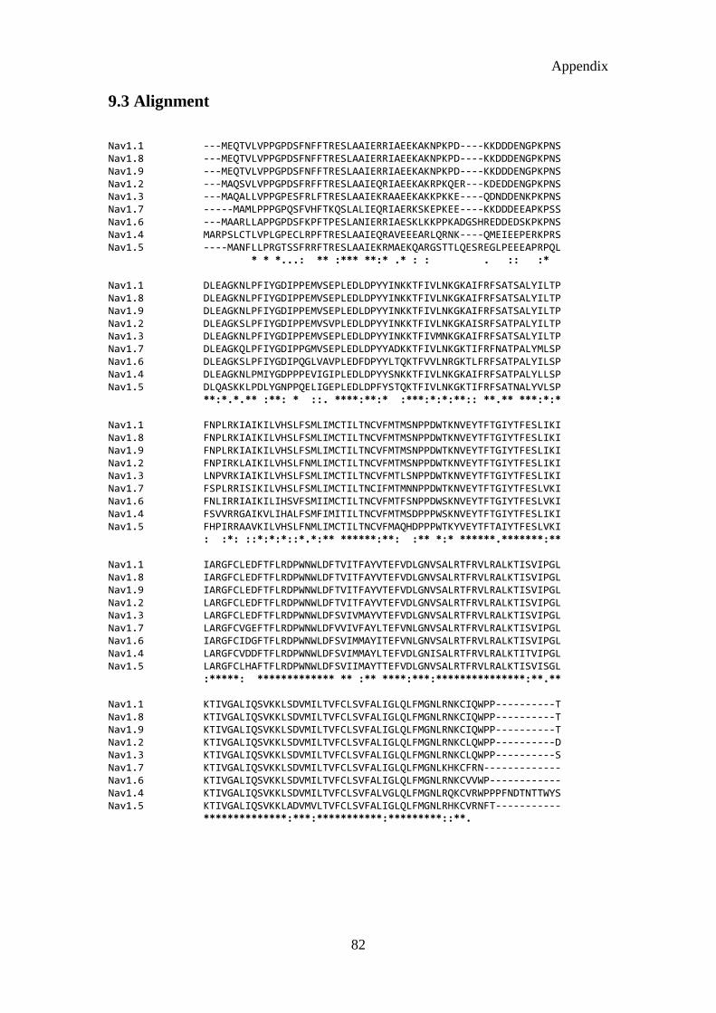
Appendix
82
9.3 Alignment Nav1.1 ---MEQTVLVPPGPDSFNFFTRESLAAIERRIAEEKAKNPKPD----KKDDDENGPKPNS Nav1.8 ---MEQTVLVPPGPDSFNFFTRESLAAIERRIAEEKAKNPKPD----KKDDDENGPKPNS Nav1.9 ---MEQTVLVPPGPDSFNFFTRESLAAIERRIAEEKAKNPKPD----KKDDDENGPKPNS Nav1.2 ---MAQSVLVPPGPDSFRFFTRESLAAIEQRIAEEKAKRPKQER---KDEDDENGPKPNS Nav1.3 ---MAQALLVPPGPESFRLFTRESLAAIEKRAAEEKAKKPKKE----QDNDDENKPKPNS Nav1.7 -----MAMLPPPGPQSFVHFTKQSLALIEQRIAERKSKEPKEE----KKDDDEEAPKPSS Nav1.6 ---MAARLLAPPGPDSFKPFTPESLANIERRIAESKLKKPPKADGSHREDDEDSKPKPNS Nav1.4 MARPSLCTLVPLGPECLRPFTRESLAAIEQRAVEEEARLQRNK----QMEIEEPERKPRS Nav1.5 ----MANFLLPRGTSSFRRFTRESLAAIEKRMAEKQARGSTTLQESREGLPEEEAPRPQL * * *...: ** :*** **:* .* : : . :: :* Nav1.1 DLEAGKNLPFIYGDIPPEMVSEPLEDLDPYYINKKTFIVLNKGKAIFRFSATSALYILTP Nav1.8 DLEAGKNLPFIYGDIPPEMVSEPLEDLDPYYINKKTFIVLNKGKAIFRFSATSALYILTP Nav1.9 DLEAGKNLPFIYGDIPPEMVSEPLEDLDPYYINKKTFIVLNKGKAIFRFSATSALYILTP Nav1.2 DLEAGKSLPFIYGDIPPEMVSVPLEDLDPYYINKKTFIVLNKGKAISRFSATPALYILTP Nav1.3 DLEAGKNLPFIYGDIPPEMVSEPLEDLDPYYINKKTFIVMNKGKAIFRFSATSALYILTP Nav1.7 DLEAGKQLPFIYGDIPPGMVSEPLEDLDPYYADKKTFIVLNKGKTIFRFNATPALYMLSP Nav1.6 DLEAGKSLPFIYGDIPQGLVAVPLEDFDPYYLTQKTFVVLNRGKTLFRFSATPALYILSP Nav1.4 DLEAGKNLPMIYGDPPPEVIGIPLEDLDPYYSNKKTFIVLNKGKAIFRFSATPALYLLSP Nav1.5 DLQASKKLPDLYGNPPQELIGEPLEDLDPFYSTQKTFIVLNKGKTIFRFSATNALYVLSP **:*.*.** :**: * ::. ****:**:* :***:*:*:**:: **.** ***:*:* Nav1.1 FNPLRKIAIKILVHSLFSMLIMCTILTNCVFMTMSNPPDWTKNVEYTFTGIYTFESLIKI Nav1.8 FNPLRKIAIKILVHSLFSMLIMCTILTNCVFMTMSNPPDWTKNVEYTFTGIYTFESLIKI Nav1.9 FNPLRKIAIKILVHSLFSMLIMCTILTNCVFMTMSNPPDWTKNVEYTFTGIYTFESLIKI Nav1.2 FNPIRKLAIKILVHSLFNMLIMCTILTNCVFMTMSNPPDWTKNVEYTFTGIYTFESLIKI Nav1.3 LNPVRKIAIKILVHSLFSMLIMCTILTNCVFMTLSNPPDWTKNVEYTFTGIYTFESLIKI Nav1.7 FSPLRRISIKILVHSLFSMLIMCTILTNCIFMTMNNPPDWTKNVEYTFTGIYTFESLVKI Nav1.6 FNLIRRIAIKILIHSVFSMIIMCTILTNCVFMTFSNPPDWSKNVEYTFTGIYTFESLVKI Nav1.4 FSVVRRGAIKVLIHALFSMFIMITILTNCVFMTMSDPPPWSKNVEYTFTGIYTFESLIKI Nav1.5 FHPIRRAAVKILVHSLFNMLIMCTILTNCVFMAQHDPPPWTKYVEYTFTAIYTFESLVKI : :*: ::*:*:*::*.*:** ******:**: :** *:* ******.*******:** Nav1.1 IARGFCLEDFTFLRDPWNWLDFTVITFAYVTEFVDLGNVSALRTFRVLRALKTISVIPGL Nav1.8 IARGFCLEDFTFLRDPWNWLDFTVITFAYVTEFVDLGNVSALRTFRVLRALKTISVIPGL Nav1.9 IARGFCLEDFTFLRDPWNWLDFTVITFAYVTEFVDLGNVSALRTFRVLRALKTISVIPGL Nav1.2 LARGFCLEDFTFLRDPWNWLDFTVITFAYVTEFVDLGNVSALRTFRVLRALKTISVIPGL Nav1.3 LARGFCLEDFTFLRDPWNWLDFSVIVMAYVTEFVDLGNVSALRTFRVLRALKTISVIPGL Nav1.7 LARGFCVGEFTFLRDPWNWLDFVVIVFAYLTEFVNLGNVSALRTFRVLRALKTISVIPGL Nav1.6 IARGFCIDGFTFLRDPWNWLDFSVIMMAYITEFVNLGNVSALRTFRVLRALKTISVIPGL Nav1.4 LARGFCVDDFTFLRDPWNWLDFSVIMMAYLTEFVDLGNISALRTFRVLRALKTITVIPGL Nav1.5 LARGFCLHAFTFLRDPWNWLDFSVIIMAYTTEFVDLGNVSALRTFRVLRALKTISVISGL :*****: ************* ** :** ****:***:***************:**.** Nav1.1 KTIVGALIQSVKKLSDVMILTVFCLSVFALIGLQLFMGNLRNKCIQWPP----------T Nav1.8 KTIVGALIQSVKKLSDVMILTVFCLSVFALIGLQLFMGNLRNKCIQWPP----------T Nav1.9 KTIVGALIQSVKKLSDVMILTVFCLSVFALIGLQLFMGNLRNKCIQWPP----------T Nav1.2 KTIVGALIQSVKKLSDVMILTVFCLSVFALIGLQLFMGNLRNKCLQWPP----------D Nav1.3 KTIVGALIQSVKKLSDVMILTVFCLSVFALIGLQLFMGNLRNKCLQWPP----------S Nav1.7 KTIVGALIQSVKKLSDVMILTVFCLSVFALIGLQLFMGNLKHKCFRN------------- Nav1.6 KTIVGALIQSVKKLSDVMILTVFCLSVFALIGLQLFMGNLRNKCVVWP------------ Nav1.4 KTIVGALIQSVKKLSDVMILTVFCLSVFALVGLQLFMGNLRQKCVRWPPPFNDTNTTWYS Nav1.5 KTIVGALIQSVKKLADVMVLTVFCLSVFALIGLQLFMGNLRHKCVRNFT----------- **************:***:***********:*********::**.

Appendix
83
Nav1.1 NASLEEHSIEKN-ITVNYNGTLINETVF----------EFDWKSYIQDSRYHYFLEGFLD Nav1.8 NASLEEHSIEKN-ITVNYNGTLINETVF----------EFDWKSYIQDSRYHYFLEGFLD Nav1.9 NASLEEHSIEKN-ITVNYNGTLINETVF----------EFDWKSYIQDSRYHYFLEGFLD Nav1.2 NSSFEINITSFFNNSLDGNGTTFNRTVS----------IFNWDEYIEDKSHFYFLEGQND Nav1.3 DSAFETNTTSYFNGTMDSNGTFVNVTMS----------TFNWKDYIGDDSHFYVLDGQKD Nav1.7 --SLENNETLES---------IMNTLES----------EEDFR------KYFYYLEGSKD Nav1.6 ---------------INFNESYLENGTK----------GFDWEEYINNKTNFYTVPGMLE Nav1.4 NDTWYGNDTWYGNEMWYGNDSWYANDTWNSHASWATNDTFDWDAYISDEGNFYFLEGSND Nav1.5 -------------ALNGTNGSVEADGLV----------WESLDLYLSDPENYLLKNGTSD . . * : Nav1.1 ALLCGNSSDAGQCPEGYMCVKAGRNPNYGYTSFDTFSWAFLSLFRLMTQDFWENLYQLTL Nav1.8 ALLCGNSSDAGQCPEGYMCVKAGRNPNYGYTSFDTFSWAFLSLFRLMTQDFWENLYQLTL Nav1.9 ALLCGNSSDAGQCPEGYMCVKAGRNPNYGYTSFDTFSWAFLSLFRLMTQDFWENLYQLTL Nav1.2 ALLCGNSSDAGQCPEGYICVKAGRNPNYGYTSFDTFSWAFLSLFRLMTQDFWENLYQLTL Nav1.3 PLLCGNGSDAGQCPEGYICVKAGRNPNYGYTSFDTFSWAFLSLFRLMTQDYWENLYQLTL Nav1.7 ALLCGFSTDSGQCPEGYTCVKIGRNPDYGYTSFDTFSWAFLALFRLMTQDYWENLYQQTL Nav1.6 PLLCGNSSDAGQCPEGYQCMKAGRNPNYGYTSFDTFSWAFLALFRLMTQDYWENLYQLTL Nav1.4 ALLCGNSSDAGHCPEGYECIKTGRNPNYGYTSYDTFSWAFLALFRLMTQDYWENLFQLTL Nav1.5 VLLCGNSSDAGTCPEGYRCLKAGENPDHGYTSFDSFAWAFLALFRLMTQDCWERLYQQTL **** .:*:* ***** *:* *.**::****:*:*:****:******** **.*:* ** Nav1.1 RAAGKTYMIFFVLVIFLGSFYLINLILAVVAMAYEEQNQATLEEAEQKEAEFQQMIEQLK Nav1.8 RAAGKTYMIFFVLVIFLGSFYLINLILAVVAMAYEEQNQATLEEAEQKEAEFQQMIEQLK Nav1.9 RAAGKTYMIFFVLVIFLGSFYLINLILAVVAMAYEEQNQATLEEAEQKEAEFQQMIEQLK Nav1.2 RAAGKTYMIFFVLVIFLGSFYLINLILAVVAMAYEEQNQATLEEAEQKEAEFQQMLEQLK Nav1.3 RAAGKTYMIFFVLVIFLGSFYLVNLILAVVAMAYEEQNQATLEEAEQKEAEFQQMLEQLK Nav1.7 RAAGKTYMIFFVVVIFLGSFYLINLILAVVAMAYEEQNQANIEEAKQKELEFQQMLDRLK Nav1.6 RAAGKTYMIFFVLVIFVGSFYLVNLILAVVAMAYEEQNQATLEEAEQKEAEFKAMLEQLK Nav1.4 RAAGKTYMIFFVVIIFLGSFYLINLILAVVAMAYAEQNEATLAEDKEKEEEFQQMLEKFK Nav1.5 RSAGKIYMIFFMLVIFLGSFYLVNLILAVVAMAYEEQNQATIAETEEKEKRFQEAMEMLK *:*** *****:::**:*****:*********** ***:*.: * ::** .*: :: :* Nav1.1 KQQEAAQQAATAT----ASEHSREPSA--AGRLSDSSSEASKLSSKSAKERRNRRKKRKQ Nav1.8 KQQEAAQQAATAT----ASEHSREPSA--AGRLSDSSSEASKLSSKSAKERRNRRKKRKQ Nav1.9 KQQEAAQQAATAT----ASEHSREPSA--AGRLSDSSSEASKLSSKSAKERRNRRKKRKQ Nav1.2 KQQEEAQAAAAAASA--ESRDFSGAGG--IGVFSESSSVASKLSSKSEKELKNRRKKKKQ Nav1.3 KQQEEAQAVAAAS-A--ASRDFSGIGG--LGELLESSSEASKLSSKSAKEWRNRRKKRRQ Nav1.7 KEQEEAEAIAAAA----AEYTSIRRSR--IMGLSESSSETSKLSSKSAKERRNRRKKKNQ Nav1.6 KQQEEAQAAAMATSAGTVSEDAIEEEGEEGGGSPRSSSEISKLSSKSAKERRNRRKKRKQ Nav1.4 KHQEELEKAKAAQ----------------------------------------------- Nav1.5 KEHEALTIRGVDT-------------------VSRSSLEMSPLAPVNSHERRSKRRKR-- *.:* Nav1.1 KEQSGGEEKDEDE-FQKSESEDSIRRKGFRFSIEGNRLTYEKRYSSPHQSLLSIRGSLFS Nav1.8 KEQSGGEEKDEDE-FQKSESEDSIRRKGFRFSIEGNRLTYEKRYSSPHQSLLSIRGSLFS Nav1.9 KEQSGGEEKDEDE-FQKSESEDSIRRKGFRFSIEGNRLTYEKRYSSPHQSLLSIRGSLFS Nav1.2 KEQSGEEEKNDR--VRKSESEDSIRRKGFRFSLEGSRLTYEKRFSSPHQSLLSIRGSLFS Nav1.3 REHLEGNNKGERDSFPKSESEDSVKRSSFLFSMDGNRLTSDKKFCSPHQSLLSIRGSLFS Nav1.7 KKLSSGEEKGDAEKLSKSESEDSIRRKSFHLGVEGHRRAHEKRLSTPNQSPLSIRGSLFS Nav1.6 KELSEGEEKGDPEKVFKSESEDGMRRKAFRLPD--NRIG--RKFSIMNQSLLSIPGSPFL Nav1.4 -ALEGGEADGDP-----------------------------------------------A Nav1.5 --MSSGTEECGEDRLPKSDSEDGPR-------------------AMNHLSLTRGLSRTSM . Nav1.1 PRRNSRTSLFSFRG--RAKDVGSENDFADDEHSTFEDNESRRDSLFVPRRHGERRNSNL- Nav1.8 PRRNSRTSLFSFRG--RAKDVGSENDFADDEHSTFEDNESRRDSLFVPRRHGERRNSNL- Nav1.9 PRRNSRTSLFSFRG--RAKDVGSENDFADDEHSTFEDNESRRDSLFVPRRHGERRNSNL- Nav1.2 PRRNSRASLFSFRG--RAKDIGSENDFADDEHSTFEDNDSRRDSLFVPHRHGERRHSNV- Nav1.3 PRRNSKTSIFSFRG--RAKDVGSENDFADDEHSTFEDSESRRDSLFVPHRHGERRNS--- Nav1.7 ARRSSRTSLFSFKG--RGRDIGSETEFADDEHSIFGDNESRRGSLFVPHRPQERRSSNI- Nav1.6 SRHNSKSSIFSFRGPGRFRDPGSENEFADDEHSTVEESEGRRDSLFIPIRARERRSSYSG Nav1.4 HGKDCNGSLDTSQG---------------------------------------------- Nav1.5 KPRSSRGSIFTFRR----RDLGSEADFADDENSTAGESESHHTSLLVPWPLRRTSAQGQ- :... *: : :

Appendix
84
Nav1.1 ----SQTSRSSRMLAVFPANGKMHSTVDCNGVVSLVGGPSVPTSPVGQLLP--------- Nav1.8 ----SQTSRSSRMLAVFPANGKMHSTVDCNGVVSLVGGPSVPTSPVGQLLP--------- Nav1.9 ----SQTSRSSRMLAVFPANGKMHSTVDCNGVVSLVGGPSVPTSPVGQLLP--------- Nav1.2 ----SQASRASRVLPILPMNGKMHSAVDCNGVVSLVGGPSTLTS-AGQLLP--------- Nav1.3 ------------------------------------------------------------ Nav1.7 ----SQASRSP---PMLPVNGKMHSAVDCNGVVSLVDGRSALMLPNGQLLP--------- Nav1.6 YSGYSQGSRSSRIFPSLRRSVKRNSTVDCNGVVSLIGGPGSHIG--GRLLP--------- Nav1.4 ------------------------------------------------------------ Nav1.5 -------PSPGTSAPGHALHGKKNSTVDCNGVVSLLGAGDPEATSPGSHLLRPVMLEHPP Nav1.1 -EGTTTETEMRKRRSSSFHVSMDFLEDPSQRQRAMSIASILTNT-VEELEESRQKCPPCW Nav1.8 -EGTTTETEMRKRRSSSFHVSMDFLEDPSQRQRAMSIASILTNT-VEELEESRQKCPPCW Nav1.9 -EGTTTETEMRKRRSSSFHVSMDFLEDPSQRQRAMSIASILTNT-VEELEESRQKCPPCW Nav1.2 -EGTTTETEIRKRRSSSYHVSMDLLEDPTSRQRAMSIASILTNT-MEELEESRQKCPPCW Nav1.3 -NGTTTETEVRKRRLSSYQISMEMLEDSSGRQRAVSIASILTNT-MEELEESRQKCPPCW Nav1.7 -EGTTNQIHK-KRRCSSYLLSEDMLNDPNLRQRAMSRASILTNT-VEELEESRQKCPPWW Nav1.6 -EATT-EVEIKKKGPGSLLVSMDQLASYGRKDRINSIMSVVTNTLVEELEESQRKCPPCW Nav1.4 --------------------------EKGAPRQSSSGDSGISDA-MEELEEAHQKCPPWW Nav1.5 DTTTPSEEPGGPQMLTSQAPCVDGFEEPGARQRALSAVSVLTSA-LEELEESRHKCPPCW . : * * ::.: :*****:::**** * Nav1.1 YKFSNIFLIWDCSPYWLKVKHVVNLVVMDPFVDLAITICIVLNTLFMAMEHYPMTDHFNN Nav1.8 YKFSNIFLIWDCSPYWLKVKHVVNLVVMDPFVDLAITICIVLNTLFMAMEHYPMTDHFNN Nav1.9 YKFSNIFLIWDCSPYWLKVKHVVNLVVMDPFVDLAITICIVLNTLFMAMEHYPMTDHFNN Nav1.2 YKFANMCLIWDCCKPWLKVKHLVNLVVMDPFVDLAITICIVLNTLFMAMEHYPMTEQFSS Nav1.3 YRFANVFLIWDCCDAWLKVKHLVNLIVMDPFVDLAITICIVLNTLFMAMEHYPMTEQFSS Nav1.7 YRFAHKFLIWNCSPYWIKFKKCIYFIVMDPFVDLAITICIVLNTLFMAMEHHPMTEEFKN Nav1.6 YKFANTFLIWECHPYWIKLKEIVNLIVMDPFVDLAITICIVLNTLFMAMEHHPMTPQFEH Nav1.4 YKCAHKVLIWNCCAPWLKFKNIIHLIVMDPFVDLGITICIVLNTLFMAMEHYPMTEHFDN Nav1.5 NRLAQRYLIWECCPLWMSIKQGVKLVVMDPFTDLTITMCIVLNTLFMALEHYNMTSEFEE : :: ***:* *:..*. : ::*****.** **:**********:**: ** .*. Nav1.1 VLTVGNLVFTGIFTAEMFLKIIAMDPYYYFQEGWNIFDGFIVTLSLVELGLANVEGLSVL Nav1.8 VLTVGNLVFTGIFTAEMFLKIIAMDPYYYFQEGWNIFDGFIVTLSLVELGLANVEGLSVL Nav1.9 VLTVGNLVFTGIFTAEMFLKIIAMDPYYYFQEGWNIFDGFIVTLSLVELGLANVEGLSVL Nav1.2 VLSVGNLVFTGIFTAEMFLKIIAMDPYYYFQEGWNIFDGFIVSLSLMELGLANVEGLSVL Nav1.3 VLTVGNLVFTGIFTAEMVLKIIAMDPYYYFQEGWNIFDGIIVSLSLMELGLSNVEGLSVL Nav1.7 VLAIGNLVFTGIFAAEMVLKLIAMDPYEYFQVGWNIFDSLIVTLSLVELFLADVEGLSVL Nav1.6 VLAVGNLVFTGIFTAEMFLKLIAMDPYYYFQEGWNIFDGFIVSLSLMELSLADVEGLSVL Nav1.4 VLTVGNLVFTGIFTAEMVLKLIAMDPYEYFQQGWNIFDSIIVTLSLVELGLANVQGLSVL Nav1.5 MLQVGNLVFTGIFTAEMTFKIIALDPYYYFQQGWNIFDSIIVILSLMELGLSRMSNLSVL :* :*********:*** :*:**:*** *** ******.:** ***:** *: :..**** Nav1.1 RSFRLLRVFKLAKSWPTLNMLIKIIGNSVGALGNLTLVLAIIVFIFAVVGMQLFGKSYKD Nav1.8 RSFRLLRVFKLAKSWPTLNMLIKIIGNSVGALGNLTLVLAIIVFIFAVVGMQLFGKSYKD Nav1.9 RSFRLLRVFKLAKSWPTLNMLIKIIGNSVGALGNLTLVLAIIVFIFAVVGMQLFGKSYKD Nav1.2 RSFRLLRVFKLAKSWPTLNMLIKIIGNSVGALGNLTLVLAIIVFIFAVVGMQLFGKSYKE Nav1.3 RSFRLLRVFKLAKSWPTLNMLIKIIGNSVGALGNLTLVLAIIVFIFAVVGMQLFGKSYKE Nav1.7 RSFRLLRVFKLAKSWPTLNMLIKIIGNSVGALGNLTLVLAIIVFIFAVVGMQLFGKSYKE Nav1.6 RSFRLLRVFKLAKSWPTLNMLIKIIGNSVGALGNLTLVLAIIVFIFAVVGMQLFGKSYKE Nav1.4 RSFRLLRVFKLAKSWPTLNMLIKIIGNSVGALGNLTLVLAIIVFIFAVVGMQLFGKSYKE Nav1.5 RSFRLLRVFKLAKSWPTLNTLIKIIGNSVGALGNLTLVLAIIVFIFAVVGMQLFGKNYSE ******************* ************************************.*.: Nav1.1 CVCKIASDCQLPRWHMNDFFHSFLIVFRVLCGEWIETMWDCMEVAGQAMCLTVFMMVMVI Nav1.8 CVCKIASDCQLPRWHMNDFFHSFLIVFRVLCGEWIETMWDCMEVAGQAMCLTVFMMVMVI Nav1.9 CVCKIASDCQLPRWHMNDFFHSFLIVFRVLCGEWIETMWDCMEVAGQAMCLTVFMMVMVI Nav1.2 CVCKISNDCELPRWHMHDFFHSFLIVFRVLCGEWIETMWDCMEVAGQTMCLTVFMMVMVI Nav1.3 CVCKINDDCTLPRWHMNDFFHSFLIVFRVLCGEWIETMWDCMEVAGQTMCLIVFMLVMVI Nav1.7 CVCKINDDCTLPRWHMNDFFHSFLIVFRVLCGEWIETMWDCMEVAGQAMCLIVYMMVMVI Nav1.6 CVCKINQDCELPRWHMHDFFHSFLIVFRVLCGEWIETMWDCMEVAGQAMCLIVFMMVMVI Nav1.4 CVCKIALDCNLPRWHMHDFFHSFLIVFRILCGEWIETMWDCMEVAGQAMCLTVFLMVMVI Nav1.5 LRD--SDSGLLPRWHMMDFFHAFLIIFRILCGEWIETMWDCMEVSGQSLCLLVFLLVMVI . ****** ****:***:**:***************:**::** *:::****

Appendix
85
Nav1.1 GNLVVLNLFLALLLSSFSADNLAATDDDNEMNNLQIAVDRMHKGVAYVKRKIYEFIQQSF Nav1.8 GNLVVLNLFLALLLSSFSADNLAATDDDNEMNNLQIAVDRMHKGVAYVKRKIYEFIQQSF Nav1.9 GNLVVLNLFLALLLSSFSADNLAATDDDNEMNNLQIAVDRMHKGVAYVKRKIYEFIQQSF Nav1.2 GNLVVLNLFLALLLSSFSSDNLAATDDDNEMNNLQIAVGRMQKGIDFVKRKIREFIQKAF Nav1.3 GNLVVLNLFLALLLSSFSSDNLAATDDDNEMNNLQIAVGRMQKGIDYVKNKMRECFQKAF Nav1.7 GNLVVLNLFLALLLSSFSSDNLTAIEEDPDANNLQIAVTRIKKGINYVKQTLREFILKAF Nav1.6 GNLVVLNLFLALLLSSFSADNLAATDDDGEMNNLQISVIRIKKGVAWTKLKVHAFMQAHF Nav1.4 GNLVVLNLFLALLLSSFSADSLAASDEDGEMNNLQIAIGRIKLGIGFAKAFLLGLLHGKI Nav1.5 GNLVVLNLFLALLLSSFSADNLTAPDEDREMNNLQLALARIQRGLRFVKRTTWDFCCGLL ******************:*.*:* ::* : ****::: *:: *: :.* : Nav1.1 IRKQKILDEIKPLDDLNNKKDSCMS--NHT-AEIGKDLDYLKDVNGTTSGIGT------- Nav1.8 IRKQKILDEIKPLDDLNNKKDSCMS--NHT-AEIGKDLDYLKDVNGTTSGIGT------- Nav1.9 IRKQKILDEIKPLDDLNNKKDSCMS--NHT-AEIGKDLDYLKDVNGTTSGIGT------- Nav1.2 VRKQKALDEIKPLEDLNNKKDSCIS--NHTTIEIGKDLNYLKDGNGTTSGIG-------- Nav1.3 FRKPKVIEIHE-----GNKIDSCMS--NNTGIEISKELNYLRDGNGTTSGVGT------- Nav1.7 SKKPKISREIRQAEDLNTKKENYIS--NHTLAEMSKGHNFLKE-KDKISGFG-------- Nav1.6 KQRE--ADEVKPLDELYEKKANCIA--NHTGADIHRNGDFQKNGNGTTSGIG-------- Nav1.4 LSPKDIMLSLGEADGAGEAGEAGET--APEDEKKEPPEEDLKKDNHILNHMG-------- Nav1.5 RQRPQKPAALAAQGQLPSCIATPYSPPPPETEKVPPTRKETRFEEGEQPGQGTPGDPEPV : . . : : * Nav1.1 -------GSSVEKYIIDESDYMSFINNPS------LTVTVPIAVGESDFENLNTEDFSSE Nav1.8 -------GSSVEKYIIDESDYMSFINNPS------LTVTVPIAVGESDFENLNTEDFSSE Nav1.9 -------GSSVEKYIIDESDYMSFINNPS------LTVTVPIAVGESDFENLNTEDFSSE Nav1.2 --------SSVEKYVVDESDYMSFINNPS------LTVTVPIAVGESDFENLNTEEFSSE Nav1.3 -------GSSVEKYVIDENDYMSFINNPS------LTVTVPIAVGESDFENLNTEEFSSE Nav1.7 --------SSVDKHLMEDSDGQSFIHNPS------LTVTVPIAPGESDLENMNAEELSSD Nav1.6 --------SSVEKYIIDE-DHMSFINNPN------LTVRVPIAVGESDFENLNTEDVSSE Nav1.4 --------LADGPPSSLELDHLNFINNPY------LTIQVPIASEESDLEMPTEEETDTF Nav1.5 CVPIAVAESDTDDQEEDEENSLGTEEESSKQQESQPVSGGPEAPPDSRTWSQVSATASSE : : . .:. . * * :* .: Nav1.1 SDLEESKE--KLNE-------------SSSSSEGSTVDIGAPVE--EQPVVEPEETLEPE Nav1.8 SDLEESKE--KLNE-------------SSSSSEGSTVDIGAPVE--EQPVVEPEETLEPE Nav1.9 SDLEESKE--KLNE-------------SSSSSEGSTVDIGAPVE--EQPVVEPEETLEPE Nav1.2 SDMEESKE--KLN--------------ATSSSEGSTVDIGAPAEG-EQPEVEPEESLEPE Nav1.3 SELEESKE--KLN--------------ATSSSEGSTVDVVLPREG-EQAETEPEEDLKPE Nav1.7 SDSEYSKV--RLN--------------RSSSSECSTVDNPLPGEG-EEAEAEPMNSDEPE Nav1.6 SDPEGSKD--KLD--------------DTSSSEGSTIDIKPEVE--EVPVEQPEEYLDPD Nav1.4 SEPEDSKKPPQPLY-------------DGNSSVCSTADYKPPEEDPEEQAEENPEGEQPE Nav1.5 AEASASQADWRQQWKAEPQAPGCGETPEDSCSEGSTADMTNTAELLEQIPDLGQDVKDPE :: . *: : ..* ** * * * : .*: Nav1.1 ACFTEGCVQRFKCCQINVEEGRGKQWWNLRRTCFRIVEHNWFETFIVFMILLSSGALAFE Nav1.8 ACFTEGCVQRFKCCQINVEEGRGKQWWNLRRTCFRIVEHNWFETFIVFMILLSSGALAFE Nav1.9 ACFTEGCVQRFKCCQINVEEGRGKQWWNLRRTCFRIVEHNWFETFIVFMILLSSGALAFE Nav1.2 ACFTEDCVRKFKCCQISIEEGKGKLWWNLRKTCYKIVEHNWFETFIVFMILLSSGALAFE Nav1.3 ACFTEGCIKKFPFCQVSTEEGKGKIWWNLRKTCYSIVEHNWFETFIVFMILLSSGALAFE Nav1.7 ACFTDGCVRRFSCCQVNIESGKGKIWWNIRKTCYKIVEHSWFESFIVLMILLSSGALAFE Nav1.6 ACFTEGCVQRFKCCQVNIEEGLGKSWWILRKTCFLIVEHNWFETFIIFMILLSSGALAFE Nav1.4 ECFTEACVQRWPCLYVDISQGRGKKWWTLRRACFKIVEHNWFETFIVFMILLSSGALAFE Nav1.5 DCFTEGCVRRCPCCAVDTTQAPGKVWWRLRKTCYHIVEHSWFETFIIFMILLSSGALAFE ***: *::: :. .. ** ** :*::*: ****.***:**::************ Nav1.1 DIYIDQRKTIKTMLEYADKVFTYIFILEMLLKWVAYGYQTYFTNAWCWLDFLIVDVSLVS Nav1.8 DIYIDQRKTIKTMLEYADKVFTYIFILEMLLKWVAYGYQTYFTNAWCWLDFLIVDVSLVS Nav1.9 DIYIDQRKTIKTMLEYADKVFTYIFILEMLLKWVAYGYQTYFTNAWCWLDFLIVDVSLVS Nav1.2 DIYIEQRKTIKTMLEYADKVFTYIFILEMLLKWVAYGFQVYFTNAWCWLDFLIVDVSLVS Nav1.3 DIYIEQRKTIKTMLEYADKVFTYIFILEMLLKWVAYGFQTYFTNAWCWLDFLIVDVSLVS Nav1.7 DIYIERKKTIKIILEYADKIFTYIFILEMLLKWIAYGYKTYFTNAWCWLDFLIVDVSLVT Nav1.6 DIYIEQRKTIRTILEYADKVFTYIFILEMLLKWTAYGFVKFFTNAWCWLDFLIVAVSLVS Nav1.4 DIYIEQRRVIRTILEYADKVFTYIFIMEMLLKWVAYGFKVYFTNAWCWLDFLIVDVSIIS Nav1.5 DIYLEERKTIKVLLEYADKMFTYVFVLEMLLKWVAYGFKKYFTNAWCWLDFLIVDVSLVS ***::.::.*: :******:***:*::****** ***: :************* **:::

Appendix
86
Nav1.1 LTANALGYSELGAIKSLRTLRALRPLRALSRFEGMRVVVNALLGAIPSIMNVLLVCLIFW Nav1.8 LTANALGYSELGAIKSLRTLRALRPLRALSRFEGMRVVVNALLGAIPSIMNVLLVCLIFW Nav1.9 LTANALGYSELGAIKSLRTLRALRPLRALSRFEGMRVVVNALLGAIPSIMNVLLVCLIFW Nav1.2 LTANALGYSELGAIKSLRTLRALRPLRALSRFEGMRVVVNALLGAIPSIMNVLLVCLIFW Nav1.3 LVANALGYSELGAIKSLRTLRALRPLRALSRFEGMRVVVNALVGAIPSIMNVLLVCLIFW Nav1.7 LVANTLGYSDLGPIKSLRTLRALRPLRALSRFEGMRVVVNALIGAIPSIMNVLLVCLIFW Nav1.6 LIANALGYSELGAIKSLRTLRALRPLRALSRFEGMRVVVNALVGAIPSIMNVLLVCLIFW Nav1.4 LVANWLGYSELGPIKSLRTLRALRPLRALSRFEGMRVVVNALLGAIPSIMNVLLVCLIFW Nav1.5 LVANTLGFAEMGPIKSLRTLRALRPLRALSRFEGMRVVVNALVGAIPSIMNVLLVCLIFW * ** **::::*.*****************************:***************** Nav1.1 LIFSIMGVNLFAGKFYHCINTTTGDR-FDIEDVNNHTDCLKLIE-RNETARWKNVKVNFD Nav1.8 LIFSIMGVNLFAGKFYHCINTTTGDR-FDIEDVNNHTDCLKLIE-RNETARWKNVKVNFD Nav1.9 LIFSIMGVNLFAGKFYHCINTTTGDR-FDIEDVNNHTDCLKLIE-RNETARWKNVKVNFD Nav1.2 LIFSIMGVNLFAGKFYHCINYTTGEM-FDVSVVNNYSECKALIE-SNQTARWKNVKVNFD Nav1.3 LIFSIMGVNLFAGKFYHCVNMTTGNM-FDISDVNNLSDCQALG----KQARWKNVKVNFD Nav1.7 LIFSIMGVNLFAGKFYECINTTDGSR-FPASQVPNRSECFALMN-VSQNVRWKNLKVNFD Nav1.6 LIFSIMGVNLFAGKYHYCFNETSEIR-FEIEDVNNKTECEKLMEGNNTEIRWKNVKINFD Nav1.4 LIFSIMGVNLFAGKFYYCINTTTSER-FDISEVNNKSECESLMH--TGQVRWLNVKVNYD Nav1.5 LIFSIMGVNLFAGKFGRCINQTEGDLPLNYTIVNNKSQCESLNL--TGELYWTKVKVNFD **************: *.* * : * * ::* * * ::*:*:* Nav1.1 NVGFGYLSLLQVATFKGWMDIMYAAVDSRNVELQPKYEESLYMYLYFVIFIIFGSFFTLN Nav1.8 NVGFGYLSLLQVATFKGWMDIMYAAVDSRNVELQPKYEESLYMYLYFVIFIIFGSFFTLN Nav1.9 NVGFGYLSLLQVATFKGWMDIMYAAVDSRNVELQPKYEESLYMYLYFVIFIIFGSFFTLN Nav1.2 NVGLGYLSLLQVATFKGWMDIMYAAVDSRNVELQPKYEDNLYMYLYFVIFIIFGSFFTLN Nav1.3 NVGAGYLALLQVATFKGWMDIMYAAVDSRDVKLQPVYEENLYMYLYFVIFIIFGSFFTLN Nav1.7 NVGLGYLSLLQVATFKGWTIIMYAAVDSVNVDKQPKYEYSLYMYIYFVVFIIFGSFFTLN Nav1.6 NVGAGYLALLQVATFKGWMDIMYAAVDSRKPDEQPKYEDNIYMYIYFVIFIIFGSFFTLN Nav1.4 NVGLGYLSLLQVATFKGWMDIMYAAVDSREKEEQPQYEVNLYMYLYFVIFIIFGSFFTLN Nav1.5 NVGAGYLALLQVATFKGWMDIMYAAVDSRGYEEQPQWEYNLYMYIYFVIFIIFGSFFTLN *** ***:********** ******** . ** :* .:***:***:*********** Nav1.1 LFIGVIIDNFNQQKKKFGGQDIFMTEEQKKYYNAMKKLGSKKPQKPIPRPGNKFQGMVFD Nav1.8 LFIGVIIDNFNQQKKKFGGQDIFMTEEQKKYYNAMKKLGSKKPQKPIPRPGNKFQGMVFD Nav1.9 LFIGVIIDNFNQQKKKFGGQDIFMTEEQKKYYNAMKKLGSKKPQKPIPRPGNKFQGMVFD Nav1.2 LFIGVIIDNFNQQKKKFGGQDIFMTEEQKKYYNAMKKLGSKKPQKPIPRPANKFQGMVFD Nav1.3 LFIGVIIDNFNQQKKKFGGQDIFMTEEQKKYYNAMKKLGSKKPQKPIPRPANKFQGMVFD Nav1.7 LFIGVIIDNFNQQKKKLGGQDIFMTEEQKKYYNAMKKLGSKKPQKPIPRPGNKIQGCIFD Nav1.6 LFIGVIIDNFNQQKKKFGGQDIFMTEEQKKYYNAMKKLGSKKPQKPIPRPLNKIQGIVFD Nav1.4 LFIGVIIDNFNQQKKKLGGKDIFMTEEQKKYYNAMKKLGSKKPQKPIPRPQNKIQGMVYD Nav1.5 LFIGVIIDNFNQQKKKLGGQDIFMTEEQKKYYNAMKKLGSKKPQKPIPRPLNKYQGFIFD ****************:**:****************************** ** ** ::* Nav1.1 FVTRQVFDISIMILICLNMVTMMVETDDQSEYVTTILSRINLVFIVLFTGECVLKLISLR Nav1.8 FVTRQVFDISIMILICLNMVTMMVETDDQSEYVTTILSRINLVFIVLFTGECVLKLISLR Nav1.9 FVTRQVFDISIMILICLNMVTMMVETDDQSEYVTTILSRINLVFIVLFTGECVLKLISLR Nav1.2 FVTKQVFDISIMILICLNMVTMMVETDDQSQEMTNILYWINLVFIVLFTGECVLKLISLR Nav1.3 FVTRQVFDISIMILICLNMVTMMVETDDQGKYMTLVLSRINLVFIVLFTGEFVLKLVSLR Nav1.7 LVTNQAFDISIMVLICLNMVTMMVEKEGQSQHMTEVLYWINVVFIILFTGECVLKLISLR Nav1.6 FVTQQAFDIVIMMLICLNMVTMMVETDTQSKQMENILYWINLVFVIFFTCECVLKMFALR Nav1.4 LVTKQAFDITIMILICLNMVTMMVETDNQSQLKVDILYNINMIFIIIFTGECVLKMLALR Nav1.5 IVTKQAFDVTIMFLICLNMVTMMVETDDQSPEKINILAKINLLFVAIFTGECIVKLAALR :**.*.**: **.************.: *. :* **::*: :** * ::*: :** Nav1.1 HYYFTIGWNIFDFVVVILSIVGMFLAELIEKYFVSPTLFRVIRLARIGRILRLIKGAKGI Nav1.8 HYYFTIGWNIFDFVVVILSIVGMFLAELIEKYFVSPTLFRVIRLARIGRILRLIKGAKGI Nav1.9 HYYFTIGWNIFDFVVVILSIVGMFLAELIEKYFVSPTLFRVIRLARIGRILRLIKGAKGI Nav1.2 YYYFTIGWNIFDFVVVILSIVGMFLAELIEKYFVSPTLFRVIRLARIGRILRLIKGAKGI Nav1.3 HYYFTIGWNIFDFVVVILSIVGMFLAEMIEKYFVSPTLFRVIRLARIGRILRLIKGAKGI Nav1.7 HYYFTVGWNIFDFVVVIISIVGMFLADLIETYFVSPTLFRVIRLARIGRILRLVKGAKGI Nav1.6 HYYFTIGWNIFDFVVVILSIVGMFLADIIEKYFVSPTLFRVIRLARIGRILRLIKGAKGI Nav1.4 QYYFTVGWNIFDFVVVILSIVGLALSDLIQKYFVSPTLFRVIRLARIGRVLRLIRGAKGI Nav1.5 HYYFTNSWNIFDFVVVILSIVGTVLSDIIQKYFFSPTLFRVIRLARIGRILRLIRGAKGI **** .**********:**** *:::*:.**.***************:***::*****

Appendix
87
Nav1.1 RTLLFALMMSLPALFNIGLLLFLVMFIYAIFGMSNFAYVKREVGIDDMFNFETFGNSMIC Nav1.8 RTLLFALMMSLPALFNIGLLLFLVMFIYAIFGMSNFAYVKREVGIDDMFNFETFGNSMIC Nav1.9 RTLLFALMMSLPALFNIGLLLFLVMFIYAIFGMSNFAYVKREVGIDDMFNFETFGNSMIC Nav1.2 RTLLFALMMSLPALFNIGLLLFLVMFIYAIFGMSNFAYVKREVGIDDMFNFETFGNSMIC Nav1.3 RTLLFALMMSLPALFNIGLLLFLVMFIYAIFGMSNFAYVKKEAGIDDMFNFETFGNSMIC Nav1.7 RTLLFALMMSLPALFNIGLLLFLVMFIYAIFGMSNFAYVKKEDGINDMFNFETFGNSMIC Nav1.6 RTLLFALMMSLPALFNIGLLLFLVMFIFSIFGMSNFAYVKHEAGIDDMFNFETFGNSMIC Nav1.4 RTLLFALMMSLPALFNIGLLLFLVMFIYSIFGMSNFAYVKKESGIDDMFNFETFGNSIIC Nav1.5 RTLLFALMMSLPALFNIGLLLFLVMFIYSIFGMANFAYVKWEAGIDDMFNFQTFANSMLC ***************************::****:****** * **:*****:**.**::* Nav1.1 LFQITTSAGWDGLLAPILNSKPPDCDPNKVNPGSSVKGDCGNPSVGIFFFVSYIIISFLV Nav1.8 LFQITTSAGWDGLLAPILNSKPPDCDPNKVNPGSSVKGDCGNPSVGIFFFVSYIIISFLV Nav1.9 LFQITTSAGWDGLLAPILNSKPPDCDPNKVNPGSSVKGDCGNPSVGIFFFVSYIIISFLV Nav1.2 LFQITTSAGWDGLLAPILNSGPPDCDPDKDHPGSSVKGDCGNPSVGIFFFVSYIIISFLV Nav1.3 LFQITTSAGWDGLLAPILNSAPPDCDPDTIHPGSSVKGDCGNPSVGIFFFVSYIIISFLV Nav1.7 LFQITTSAGWDGLLAPILNSKPPDCDPKKVHPGSSVEGDCGNPSVGIFYFVSYIIISFLV Nav1.6 LFQITTSAGWDGLLLPILN-RPPDCSLDKEHPGSGFKGDCGNPSVGIFFFVSYIIISFLI Nav1.4 LFEITTSAGWDGLLNPILNSGPPDCDPNLENPGTSVKGDCGNPSIGICFFCSYIIISFLI Nav1.5 LFQITTSAGWDGLLSPILNTGPPYCDPTLPNS-NGSRGDCGSPAVGILFFTTYIIISFLI **:*********** **** ** *. :. .. .****.*::** :* :*******: Nav1.1 VVNMYIAVILENFSVATEESAEPLSEDDFEMFYEVWEKFDPDATQFMEFEKLSQFAAALE Nav1.8 VVNMYIAVILENFSVATEESAEPLSEDDFEMFYEVWEKFDPDATQFMEFEKLSQFAAALE Nav1.9 VVNMYIAVILENFSVATEESAEPLSEDDFEMFYEVWEKFDPDATQFMEFEKLSQFAAALE Nav1.2 VVNMYIAVILENFSVATEESAEPLSEDDFEMFYEVWEKFDPDATQFIEFAKLSDFADALD Nav1.3 VVNMYIAVILENFSVATEESAEPLSEDDFEMFYEVWEKFDPDATQFIEFSKLSDFAAALD Nav1.7 VVNMYIAVILENFSVATEESTEPLSEDDFEMFYEVWEKFDPDATQFIEFSKLSDFAAALD Nav1.6 VVNMYIAIILENFSVATEESADPLSEDDFETFYEIWEKFDPDATQFIEYCKLADFADALE Nav1.4 VVNMYIAIILENFNVATEESSEPLGEDDFEMFYETWEKFDPDATQFIAYSRLSDFVDTLQ Nav1.5 VVNMYIAIILENFSVATEESTEPLSEDDFDMFYEIWEKFDPEATQFIEYSVLSDFADALS *******:*****.******::**.****: *** ******:****: : *::*. :*. Nav1.1 PPLNLPQPNKLQLIAMDLPMVSGDRIHCLDILFAFTKRVLGESGEMDALRIQMEERFMAS Nav1.8 PPLNLPQPNKLQLIAMDLPMVSGDRIHCLDILFAFTKRVLGESGEMDALRIQMEERFMAS Nav1.9 PPLNLPQPNKLQLIAMDLPMVSGDRIHCLDILFAFTKRVLGESGEMDALRIQMEERFMAS Nav1.2 PPLLIAKPNKVQLIAMDLPMVSGDRIHCLDILFAFTKRVLGESGEMDALRIQMEERFMAS Nav1.3 PPLLIAKPNKVQLIAMDLPMVSGDRIHCLDILFAFTKRVLGESGEMDALRIQMEDRFMAS Nav1.7 PPLLIAKPNKVQLIAMDLPMVSGDRIHCLDILFAFTKRVLGESGEMDSLRSQMEERFMSA Nav1.6 HPLRVPKPNTIELIAMDLPMVSGDRIHCLDILFAFTKRVLGDSGELDILRQQMEERFVAS Nav1.4 EPLRIAKPNKIKLITLDLPMVPGDKIHCLDILFALTKEVLGDSGEMDALKQTMEEKFMAA Nav1.5 EPLRIAKPNQISLINMDLPMVSGDRIHCMDILFAFTKRVLGESGEMDALKIQMEEKFMAA ** :.:** :.** :*****.**:***:*****:**.***:***:* *: **::*::: Nav1.1 NPSKVSYQPITTTLKRKQEEVSAVIIQRAYRRHLLKRTVKQASFTYNKNKIK--GGANLL Nav1.8 NPSKVSYQPITTTLKRKQEEVSAVIIQRAYRRHLLKRTVKQASFTYNKNKIK--GGANLL Nav1.9 NPSKVSYQPITTTLKRKQEEVSAVIIQRAYRRHLLKRTVKQASFTYNKNKIK--GGANLL Nav1.2 NPSKVSYEPITTTLKRKQEEVSAIIIQRAYRRYLLKQKVKKVSSIYKKDKGK--ECDGTP Nav1.3 NPSKVSYEPITTTLKRKQEEVSAAIIQRNFRCYLLKQRLKNISSNYNKEAIK--GRIDLP Nav1.7 NPSKVSYEPITTTLKRKQEDVSATVIQRAYRRYRLRQNVKNISSIYIKDGDR--D-DDLL Nav1.6 NPSKVSYEPITTTLRRKQEEVSAVVLQRAYRGHLARR--------------------GFI Nav1.4 NPSKVSYEPITTTLKRKHEEVCAIKIQRAYRRHLLQRSMKQASYMYRHSHDG--SGDDAP Nav1.5 NPSKISYEPITTTLRRKHEEVSAMVIQRAFRRHLLQRSLKHASFLFRQQAGSGLSEEDAP ****:**:******:**:*:*.* :** :* : :: . Nav1.1 IKEDMIIDRI-------NENSITEKTDLTMSTAACPPSYDRVTKPIVEK---------HE Nav1.8 IKEDMIIDRI-------NENSITEKTDLTMSTAACPPSYDRVTKPIVEK---------HE Nav1.9 IKEDMIIDRI-------NENSITEKTDLTMSTAACPPSYDRVTKPIVEK---------HE Nav1.2 IKEDTLIDKL-------NENSTPEKTDMTPSTTS-PPSYDSVTKPEKEK---------FE Nav1.3 IKQDMIIDKL-------NGNSTPEKTDGSSSTTS-PPSYDSVTKPDKEK---------FE Nav1.7 NKKDMAFDNV-------NENSSPEKTDATSSTTS-PPSYDSVTKPDKEK---------YE Nav1.6 CKKTTSNKLE-------NGGTHREKKESTPSTAS-LPSYDSVTKPEKEKQQR------AE Nav1.4 EKEGLLANTMSKMYGHENGNSSSPSPEEKGEAGDAGPTMGLMPISPSDT--A------WP Nav1.5 EREGLIAYVMSEN----FSRPLGPPSSSSISSTSFPPSYDSVTRATSDNLQVRGSDYSHS :: . . . .: *: . :. . :.

Appendix
88
Nav1.1 QEGKDEKAKGK------- Nav1.8 QEGKDEKAKGK------- Nav1.9 QEGKDEKAKGK------- Nav1.2 KDKSEKEDKGKDIRESKK Nav1.3 KDKPEKESKGKEVRENQK Nav1.7 QDRTEKEDKGKDSKESKK Nav1.6 EGRRERAKRQKEVRESKC Nav1.4 PAPPPGQTVRPGVKESLV Nav1.5 EDLADFPPSPDRDRESIV

Appendix
89
9.4 Curriculum Vitae
PERSONAL DETAILS
First Name Gowri Shankar
Last Name Bagavananthem Andavan
Date of Birth 25 November 1983
Place of Birth Chennai, India
Nationality Indian
Email [email protected]
1. First Contact Initiative Grant from European Society of Cardiology
(ESC).Feb 2011-March 2011.
1. Voltage Gated Sodium Channels: mutations, channelopathies and subtype
specific targets
Gowri Shankar Bagavananthem Andavan and Rosa Lemmens-Gruber
Current Medicinal Chemistry, 2011, 18(3):377-97.
ACADEMIC PROFILE
INSTITUTE YEAR Primary and Secondary
Schooling
Sindhi Model Senior Secondary School,
Chennai India.
June 1988 - April 1999
Higher school Certificate Jaigopal Garodia Higher Secondary School
Chennai, India
June 1999 - April 2001
Bachelor of Technology Centre for Biotechnology (CBT),
Anna University, Chennai, India.
June 2001 - May 2005
Master of Science
(Research)
University of Sydney,
Sydney, Australia.
March 2006 - March 2007
GRANT
SCIENTIFIC PAPERS

Appendix
90
2. Essential structural features of TNF-α lectin-like domain derived peptides
for activation of amiloride-sensitive sodium current in A549 cells.
Parastoo Hazemi, Susan J. Tzotzos, Bernhard Fischer, Gowri Shankar
Bagavananthem Andavan, Hendrick Fischer, Helmut Pietschmann, Rudolf Lucas,
Rosa Lemmens-Gruber.
Journal of Medicinal Chemistry, 2010, 53(22):8021-9.
3. Cyclodepsipeptides from marine sponges: natural agents for drug
research.
Gowri Shankar Bagavananthem Andavan and Rosa Lemmens-Gruber
Marine Drugs, 2010, 8(3), 810-834.
4. A Structural and Phylogenetic Analysis of Cytochrome P450
B.A.Gowri Shankar, Michael Charleston, Michael Murray and David Hibbs
International Proceedings of the 15th International Conference on Cytochrome P450:
Biochemistry, Biophysics, Functional Genomics, 2007, 75-80.
5. Ion-pairs in non-redundant protein structures
B.A.Gowri Shankar, R.Sarani, Daliah Michael, P.Mridula, C.Vasuki Ranjani,
G.Sowmiya, B.Vasundhar, P.Sudha, J.Jeyakanthan, D.Velmurugan and K.Sekar. J.
Biosci., 2007, 32(4), 693-704.
1. ESC Congress 2010, 28 August - 1 September, Stockholm, Sweden. Title:
Modulation of cardiac voltage gated sodium channel splice variants with its auxiliary
subunit by Fyn tyrosine kinase.
2. Frontiers in Cardiovascular Biology, Berlin, Germany, 16-19 July 2010,
Title: Cardiac sodium channel variants and fyn tyrosine kinase modulation.
3. XXth
World Congress of the International Society of Heart Research 2010,
May 13-16 2010, Kyoto, Japan. Title: Fyn tyrosine kinase modulation on
cardiac voltage gated sodium channel splice variants.
4. 54th
Biophysical Society’s Annual Meeting at The Moscone Convention
Center 20-24 February 2010, San Francisco, California. Title: Modulation of
Nav1.5 varients by Src tyrosine kinase.
POSTER PRESENTATIONS

Appendix
91
5. 15th
International Conference on Intelligent System for Molecular Biology
(ISMB) Jointly with European Conference on Computational Biology
(ECCB) on 21-25 July, 2007, Vienna, Austria.
6. 11th
Annual International Conference on Research in Computational
Molecular Biology (RECOMB 2007), 21-25 April, 2007, San Francisco Bay,
California.
7. Fifth Asia-Pacific Bioinformatics Conference, 15-17 January, 2007,
Hong Kong.
8. 4th Annual Rocky Mountain Bioinformatics Conference, 1-6 December,
2006, Aspen/Snowmass, Colorado.
9. Bioinformatics Australia 2006, Sydney, 21-22 November, 2006.
1. Travel grant from the ESC Council on Basic Cardiovascular Science to
attend ESC Congress 2010, Stockholm, Sweden.
2. International ISHR Travel Grant to attend XXth
World Congress of the
International Society of heart Research 2010, Kyoto, Japan.
3. International Travel Award from Biophysical Society to attend and present
my data in 54th
Annual Meeting at San Francisco, California.
4. Travel scholarship from RECOMB 2007 committee, to attend RECOMB
2007 in San Francisco Bay, California, USA.
5. BioinfoSummer 2006, ICE-EM Scholarship to attend summer symposium
in Bioinformatics, ANU, Canberra; Australia.
6. Travel scholarship to attend PhD Winter School in Data Mining &
Bioinformatics, Lorne, Victoria, Australia
7. Travel scholarship to attend APAC Summer School in Computational
Science 2006, Brisbane, Australia.
8. Chief Minister Meritorious Award to students to continue their
undergraduate studies, Chennai, India.
SCHOLARSHIPS

Appendix
92
Demonstrating Medicinal Chemistry PHAR2632 in Faculty of Pharmacy,
University of Sydney. (March 2007 – June 2007)
Demonstrating Medicinal Chemistry PHAR2618 in Faculty of Pharmacy,
University of Sydney. (July 2006 – November 2006)
TEACHING