title study on oxidase/peroxidase-based biosensors with
TRANSCRIPT

Title Study on Oxidase/Peroxidase-based Biosensors withPentacyanoferrate-bound Polymer( Dissertation_全文 )
Author(s) Nieh, Chi-Hua
Citation 京都大学
Issue Date 2013-09-24
URL https://doi.org/10.14989/doctor.k17895
Right
Type Thesis or Dissertation
Textversion ETD
Kyoto University
Study on Oxidase/Peroxidase-based Biosensors
with Pentacyanoferrate-bound Polymer
Chi-Hua Nieh
2013

Table of Contents
General Introduction ....................................................................................................... 1
Chapter 1 Electrostatic and steric interaction between redox polymers and some
flavoenzymes in mediated bioelectrocatalysis ....................................................... 7
Introduction .................................................................................................................... 7
Experimental ................................................................................................................ 11
Results and Discussion ................................................................................................ 14
Reference ..................................................................................................................... 23
Chapter 2 Four enzyme-based biosensor mediated by PVI[Fe(CN)5] for creatinine
determination ......................................................................................................... 25
Introduction .................................................................................................................. 25
Experimental ................................................................................................................ 28
Results and Discussion ................................................................................................ 30
Reference ..................................................................................................................... 40
Chapter 3 Sensitive D-amino acid bienzyme biosensor mediated by PVI[Fe(CN)5]
................................................................................................................................. 41
Introduction .................................................................................................................. 41
Experimental ................................................................................................................ 43
Results and Discussion ................................................................................................ 45
Reference ..................................................................................................................... 56
Conclusions .................................................................................................................... 57
Appendix ........................................................................................................................ 59
Acknowledgement .......................................................................................................... 63
List of publications ........................................................................................................ 64


1
General Introduction H2O2-producing oxidase and peroxidase (POD)-coupled system has been widely
utilized for analyte detection by the spectrophotometric method, since POD reacts with
several kinds of reduced reagents that can be oxidized into fluorescent or colored
product for detection, thanks to poor substrate specificity of POD [1-3]. However due to
the property, the POD system is labile to cause negative interference by oxidizing
electroactive compounds such as ascorbic acid (AA) or uric acid in physiological fluids,
especially in solution reactions (Eq. 1) [4]. The reaction of the interference with H2O2
catalyzed by POD decreases the H2O2 concentration to cause the underestimation of the
H2O2 concentration.
interferences + H2O2 POD oxidized interferences + 2H2O (1)
To solve the problem, electrochemical biosensors with immobilized
H2O2-producing oxidases covered with membrane have been developed with the
advantages of low cost, rapid and simple operation, since Clark and Lyons proposed an
amperometric enzyme-immobilized electrode for glucose detection in 1962 [5]. In
enzyme-based amperometric biosensors, the concentration of analyte can be directly
determined based on the detection of oxygen consumption (Eqs. 2 and 3) or generated
H2O2 (Eq. 4). The detection system is so-called the first generation biosensor [6, 7].
substrate + O2 oxidase producing-OH 22 product + H2O2 (2)
O2 + 4H+ + 4e− |AgCl vs.AgV 0.6- 2H2O (3)
H2O2 |AgCl vs.AgV 0.7 - 0.15 O2 + 2H+ + 2e− (4)
In addition, the usage of membrane on the top of immobilized enzyme layer can
eliminate the interference effect based on perm-selectivity [8]. Interferences can be

2
blocked based on size exclusion or electrostatic repulsion. The interference effect due to
the poor specificity of POD which exists in the aforementioned spectrophotometric
method can be overcome.
However, in the detection of oxygen consumption based on the reduction of
oxygen, the signal response is seriously influenced by the concentration of dissolved
oxygen in samples and the diffusion rate of oxygen from the bulk solution to the surface
of working electrode. On the other hand, the direct electrooxidation of H2O2 requires
high operating potential (+0.7 V vs. Ag|AgCl); although membrane can exclude
interferences, for the purpose of high sensitivity, sometimes membrane is insufficient
since the high operating potentials lead to oxidation of the small amount of electroactive
compounds in samples which may pass through the membrane to cause serious
interference problem.
In order to overcome this problem, the second generation biosensors have been
evolved by using mediators to regenerate oxidases (Eqs.5 and 6) [5, 9-11],
substrate + H2O2-producing oxidase (FAD/FMN)
→ product + H2O2-producing oxidase (FADH2/FMNH2) (5)
H2O2-producing oxidase (FADH2/FMNH2) + mediatorox
→ H2O2-producing oxidase (FAD/FMN) + mediatorred (6)
where FAD is flavin adenine dinucleotide and FMN is flavin mononucleotide. This type
of biosensor has been evolved by using redox substances (mediators) such as DCPIP
and benzoquinone to shuttle electrons between the redox center of enzyme and electrode,
which provides higher signal response and allows lower operating potential.
Nevertheless, it is still hard for the second generation biosensors to satisfy the need
of high sensitivity since the operation at positive potentials (0 − 0.5 V vs. Ag|AgCl)
leads to an increase in oxidative interferences in physiological samples (e.g., the
oxidation potential of AA is ca. 0 V vs. Ag|AgCl); furthermore, dissolved oxygen needs
to be removed to avoid the competition with the mediator, which is difficult in practical
analysis.

3
To circumvent the problems, H2O2-producing oxidase/POD-based biosensors have
been proposed, in which H2O2 generated from an oxidase reaction is reductively
detected at low operating potentials around 0 V vs. Ag|AgCl with the aid of POD in the
mediated-electron-transfer system [12-14]. Since the low potential operation and the
usage of membrane decrease the background current and noise levels, furthermore
eliminate the undesirable oxidation of electroactive interference, bienzyme biosensors
show high sensitivity and stability.
Nevertheless, the other thing which needs to be concerned is that the mediators
may react with both of oxidase and POD. The mediator oxidized by POD may be
reduced not only at electrode but by oxidase, because most of oxidases show
dehydrogenase activity to transfer electrons to artificial mediators (Scheme 1) [15, 16].
Such cross reaction diminishes the cathodic current to cause a decrease in the
electrochemical response of mediator reduction.
In consideration of 1) the possibility of cross reaction of mediators with POD and
oxidases, 2) the interference effect of O2 reduction around −0.2 V (vs. Ag|AgCl), and 3)
the occurrence of interference oxidation at positive potentials, it becomes an important
issue for mediated bienzyme biosensors to select an appropriate mediator with high
selectivity for POD alone and a suitable operating potential in the narrow range from
−0.2 to 0 V.
The author in this research attempted to find a suitable mediator with selective
reactivity against POD alone for such oxidase/POD-based biosensors, and constructed
two kinds of biosensors for practical purpose.
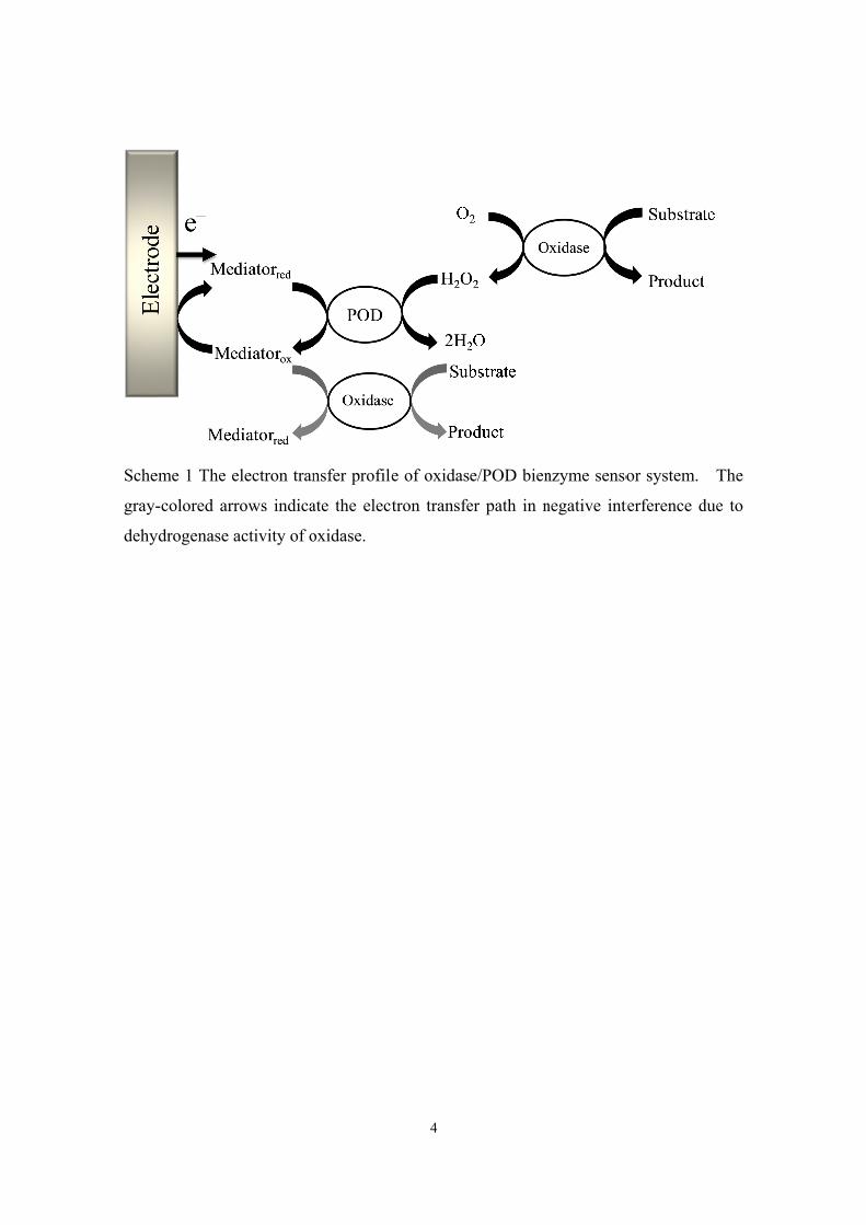
Sche
gray-
dehy
eme 1 The e
-colored arr
ydrogenase a
electron tran
rows indica
activity of o
nsfer profile
ate the elect
oxidase.
4
e of oxidas
tron transfe
e/POD bien
er path in n
nzyme senso
negative int
or system.
terference d
The
due to

5
Reference
[1] B. Porstmann, T. Porstmann, E. Nugel, U. Evers, J. Immunol. Methods, 79 (1985) 27.
[2] R.H. Yolken, Rev. Infect. Dis., 4 (1982) 35. [3] Q.Y. Liu, Y.Y. Liang, A.H. Liang, Z.L. Jiang, Spectrosc. Spectr. Anal., 29 (2009)
2535. [4] R. Maidan, A. Heller, Anal. Chem., 64 (1992) 2889. [5] A. Chaubey, B.D. Malhotra, Biosens. Bioelectron., 17 (2002) 441. [6] T. Tsuchida, K. Yoda, Clin. Chem., 29 (1983) 51. [7] V.K. Nguyen, C.M. Wolff, J.L. Seris, J.P. Schwing, Anal. Chem., 63 (1991) 611. [8] M.G. Garguilo, A.C. Michael, Anal. Chem., 66 (1994) 2621. [9] A.E.G. Cass, G. Davis, M.J. Green, H.A.O. Hill, J. Electroanal. Chem., 190 (1985)
117. [10] S.V. Dzyadevych, V.N. Arkhypova, A.P. Soldatkin, A.V. El'skaya, C. Martelet, N.
Jaffrezic-Renault, Irbm, 29 (2008) 171. [11] M. Mehrvar, M. Abdi, Anal. Sci., 20 (2004) 1113. [12] M. Gu, J.W. Wang, Y.F. Tu, J.W. Di, Sens. Actuator B-Chem., 148 (2010) 486. [13] Y.L. Yao, K.K. Shiu, Electroanalysis, 20 (2008) 2090. [14] A.R. Vijayakumar, E. Csoregi, A. Heller, L. Gorton, Anal. Chim. Acta, 327 (1996)
223. [15] T.J. Ohara, M.S. Vreeke, F. Battaglini, A. Heller, Electroanalysis, 5 (1993) 825. [16] R. Matsumoto, M. Mochizuki, K. Kano, T. Ikeda, Anal. Chem., 74 (2002) 3297.

6
7
Chapter 1 Electrostatic and steric interaction between redox polymers
and some flavoenzymes in mediated bioelectrocatalysis
H2O2-producing oxidase/peroxidase (POD)-based mediated biosensors are very
useful to minimize interference, but require suitable mediators which work well only for
POD but not against the oxidase. Pentacyanoferrate-bound poly(1-vinylimidazole)
(PVI[Fe(CN)5]), PVI[Os(dcbbpy)2Cl] (dcbbpy = 4,4'-dicarboxy-2,2'-bipyridine) and
PVI[Os(dmebpy)2Cl] (dmebpy = 4,4'-dimethyl-2,2'-bipyridine) have been utilized to
investigate the interaction with four kinds of H2O2-producing oxidases: glucose oxidase,
sarcosine oxidase, choline oxidase (ChOD) and lactate oxidase. The mediated
bioelectrocatalytic activities of the redox polymers for the enzymes have been
determined by cyclic voltammetry in the presence of the substrates. The highly
negatively charged PVI[Fe(CN)5] shows practically no mediating activity against the
four flavoenzymes, but strong one to POD. On the other hand, PVI[Os(dmebpy)2Cl]
with neutral ligands shows a high activity for the oxidases except ChOD. The mediating
activity of PVI[Os(dcbbpy)2Cl] with negatively charged ligands is much smaller than
that of PVI[Os(dmebpy)2Cl]. These results reveal that electrostatic repulsion and steric
hindrance are enhanced by using negatively charged polymers to realize minimum
activity against the oxidases.
Introduction
Various types of transition metal redox polymers such as Os and Ru complexes and
ferrocene derivatives have been developed for co-immobilization of enzymes to
construct mediated electron transfer (MET) bioelectrocatalytic systems over the past
two decades [1-3]. These electroconductive polymers are conspicuous due to their
unique properties over diffusional mediators: they can be covalently bound to enzymes
by crosslinkers on electrode surface; they provide three-dimensional electrocatalytic
systems which are not leachable but swollen in water to form redox hydrogels for MET

8
between the redox center of enzymes and electrodes; they have so high density of the
redox groups immobilized on electrode surface that they are more efficient than low
molecular weight mediators in the electron transfer; moreover, transition metal redox
polymers are more stable than quinone-containing polymers since reactive semiquinone
radicals formed in the one-electron reduction of quinones easily react with thiols,
amines, phenols and other functions [4-6]. These unique properties of metal redox
polymers are very useful for MET-type biosensor and biofuel cell application [5, 7, 8].
In MET glucose biosensors, Os-containing polymers have frequently been utilized to
shuttle electrons from the redox center of glucose oxidase (GOD) or flavin adenine
dinucleotide (FAD)-dependent glucose dehydrogenase (GDH) to electrode, and show
higher stability and sensitivity than low molecular weight mediators [1, 9]. In MET
bioelectrocatalysis, we have to consider the linear free energy relationships (LFER) of
the electron transfer rate constant (k) between enzyme and mediator to the formal
potential of mediator (E°') [10-12]. The relation is given by Eq. (1.1) for the oxidation
of the substrate,
( )iji
j EERT
nFkk
''303.2
log °−°= β (1.1)
where β is a proportional constant (0 < β < 1), n is the number of electrons, F is the
Faraday constant, R is the gas constant, T is absolute temperature, and the subscripts i, j
indicate given mediators as a series of redox compounds with similar structure. In order
to increase the current density, it is essential to use a mediator with a large k value and
then with a more positive E°' value, which leads to increase the oxidative interferences
such as ascorbic acid and uric acid in physiological samples. Furthermore, as mentioned
in General Introduction, O2 must sometimes be removed to avoid the competition with
mediators in the case of oxidase, which would be difficult and not practical in real
sample measurements.
In order to solve the problems, H2O2-producing oxidase/peroxidase (POD)
bienzyme biosensors mediated by Os-containing polymers were developed [13].
Oxidase-based mediated biosensors coupled with POD allow the determination of H2O2

9
generated from the oxidase such as GOD at low operating potentials around 0 V vs.
Ag|AgCl, with high sensitivity and stability, and the elimination of the undesirable
oxidation of interferences [14, 15]. However, as mentioned in General Introduction,
mediators may react with both of oxidase and POD. The oxidized mediators may also
act as electron acceptors of H2O2-producing oxidases based on the dehydrogenase
activity of the oxidase to cause a decrease in the electrochemical response of mediator
reduction. Therefore, it is necessary to select an appropriate mediator with highly
selective reactivity for POD but practically no reactivity for oxidase.
The k value in MET bioelectrocatalysis also depends on the structure and charge of
the mediator. In the case of PQQ-dependent GDH, the k values of Os-complexes are
much lower than those of quinone compounds at a given E°' [12]. The main factor to
cause the difference seems to be the steric effect, since the size of the Os-complex is
much larger than that of the active site of the enzyme. In the case of GOD, the k values
of negatively charged inorganic and organic mediators are much lower than those of
neutral quinone mediators with almost identical E°' [16]. The electrostatic repulsion
between negatively charged mediators and the active site of GOD is expected.
The author expects in this work that such steric hindrance and electrostatic
repulsion will be enhanced by using redox polymers with large molecular weight and
high density of negative charge. On the other hand, such negative effects seem to be
minimized for POD, because the catalytic center of POD is located on the surface of the
enzyme and the vicinity of the catalytic center is positively charged [17].
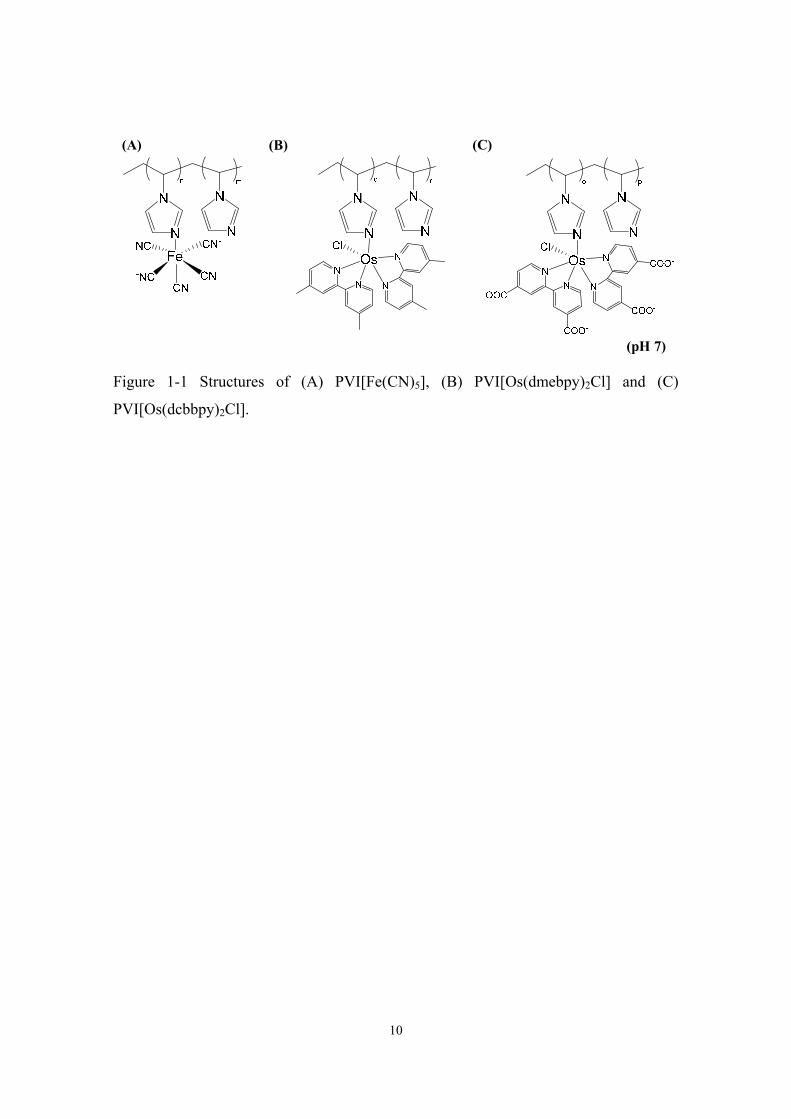
In order to verify the hypothesis, the author focuses on pentacyanoferrate-bound
poly(1-vinylimidazole) (PVI[Fe(CN)5]) (Fig. 1-1A) as a redox polymer mediator for
oxidase/POD bienzyme biosensors. The mediator has been synthesized for high MET
activity with bilirubin oxidase (BOD) [18]. Os-complex-bound PVIs,
PVI[Os(dmebpy)2Cl] (dmebpy = 4,4'-dimethyl-2,2'-bipyridine) with neutral ligands (Fig.
1-1B) and PVI[Os(dcbbpy)2Cl] (dcbbpy = 4,4'-dicarboxy-2,2'-bipyridine) with
negatively charged ligands (Fig. 1-1C) are also used as references.
10
Figure 1-1 Structures of (A) PVI[Fe(CN)5], (B) PVI[Os(dmebpy)2Cl] and (C)
PVI[Os(dcbbpy)2Cl].
(pH 7)
(A) (B) (C)

11
Experimental
Reagents
(NH4)2[OsCl6], 4,4'-dimethyl-2,2'-bipyridine, and 1-vinylimidazole were purchased
from Sigma-Aldrich Co. (USA). 2,2'-Bipyridine-4,4'-dicarboxylic acid, ethylene glycol,
sodium hydrosulfite (Na2S2O4), 2,2'-azobisisobutyronitrile (AIBN), diethyl ether,
sodium pentacyanonitrosylferrate(III) dihydrate (Na2[Fe(CN)5(NO)]·2H2O), glucose,
sarcosine, choline chloride and L-lactate were obtained from Wako Pure Chemical
Industries (Osaka, Japan). POD from horseradish (POD, 257 U mg−1), GOD from
Aspergillus sp. (100 U mg−1), sarcosine oxidase from microorganism (SOD, 16.6 U
mg−1), choline oxidase from Alcaligenes sp. (ChOD, 16.9 U mg−1) and lactate oxidase
from microorganism (LOD, 101 U mg−1) were from Toyobo Co. (Osaka, Japan).
Substrate solutions and other enzyme solutions were prepared with a phosphate buffer
solution (100 mM, pH 7.0). 1 M Na2S2O4 was prepared with distilled water. Other
chemicals were of analytical grade and used as received.
Synthesis of mediator-containing polymers
Os(dmebpy)2Cl2 and Os(dcbbpy)2Cl2 were synthesized as reported [19]. In brief,
(NH4)2[OsCl6] (0.57 mmol) and 2 equivalents of 4,4'-dimethyl-2,2'-bipyridine or
2,2'-bipyridine-4,4'-dicarboxylic acid were dissolved in 9 mL of ethylene glycol, heated
under reflux and stirring for 2 h in Ar. After cooling to room temperature, the solution
was treated with 15 mL of 1 M Na2S2O4 to reduce [Os(dmebpy)2Cl2]+ or
[Os(dcbbpy)2Cl2]+ which might be formed during the synthesis, and then cooled in an
ice bath for 30 min. The dark-violet precipitate was obtained after being washed with
distilled water and diethyl ether.
Poly(1-vinylimidazole) (PVI) was prepared according to the literature [1]. Briefly,
6 mL of 1-vinylimidazole mixed with 0.5 g of AIBN which works as an initiator was
heated at 70 °C for 2 h under Ar with stirring. After cooling, the yellow precipitate was
observed and re-dissolved with methanol, followed by adding the methanol solution

12
dropwise to acetone under strong stirring. White PVI powder was obtained after
filtering and drying.
PVI[Os(dmebpy)2Cl] and PVI[Os(dcbbpy)2Cl] were also prepared according to the
literature [1]. The powder of synthesized Os(dmebpy)2Cl2 (66 mg, ca. 0.105 mmol) or
Os(dcbbpy)2Cl2 (85 mg, ca. 0.105 mmol) and PVI (100 mg, ca. 1.05 mmol) were
dissolved in 100 mL of absolute ethanol to avoid the replacement of second chloride ion
in Os complex in the presence of H2O during the following step of heating. The mixture
was heated at reflux and stirred for 3 days. The precipitate was obtained by adding the
solution to diethyl ether under stirring. After filtering and drying, the precipitate was
dissolved in 10 mL of the phosphate buffer solution (10 mM, pH 7.0) and stored at 4 °C.
The final concentration of the two polymers was about 18 mg mL−1. The ratio of the
imidazole unit in PVI to the Os(dmebpy)2Cl complex may be approximately 10
according to the literature [1].
PVI[Fe(CN)5] was synthesized similar to the method in the literature [18]. In brief,
200 mg of Na2[Fe(CN)5(NO)]·2H2O and 188 mg of PVI were dissolved in 50 mL of 0.6
M NaOH and were refluxed at 65 °C for 24 h. During the process, NO+ was released
through the reaction with OH−, and the pentacyanoferrate ion was linked to N in
imidazole. The mixture was afterward dialyzed against distilled water for 24 h to
remove unreacted compounds. After centrifuged at 5000 g for 20 min 2 times to remove
red precipitate, the suspension was vacuum freeze-dried at −40 °C for 24 h to get
PVI[Fe(CN)5] powder. The ratio of the imidazole unit in PVI to the Fe(CN)5 complex
was 4.3 as measured by elemental analysis. The stock solution of PVI[Fe(CN)5] was
prepared by dissolving in 10 mM phosphate buffer at pH 7.0.
Electrochemical measurements
Electrochemical measurements were performed with glassy carbon electrodes (3
mm, BAS) and carried out in the phosphate buffer solution (100 mM pH 7.0) at 25 °C
with an electrochemical analyzer (BAS CV 50 W, BAS Inc., Japan). All potentials are
referred to Ag|AgCl|sat. KCl reference electrode in this work. Considering the fact that
the carboxyl group of PVI[Os(dcbbpy)2Cl] is also crosslinked with poly(ethylene glycol)
diglycidyl ether, the redox polymers were used in soluble state without immobilization
on electrode surface. The final concentration of the Os-containing polymers in the

13
sample solution was one tenth of the Os-containing polymer stock solutions, while the
final concentration of PVI[Fe(CN)5] was 0.3 mg mL−1 in the sample solution. The peak
anodic currents of the diluted PVI[Os(dmebpy)2Cl] and PVI[Os(dcbbpy)2Cl] solutions
were, respectively, 260 and 350 nA at a scan rate of 20 mV s−1 in cyclic voltammetry.
Supposing that the diffusion coefficients of the two Os-containing polymers are close to
each other, the concentrations of Os2+/3+ in the sample solutions were in the same level.
The activity of the metal redox polymer against enzyme was also evaluated by cyclic
voltammetry.
Measurements of enzyme’s concentration
The concentrations of GOD, SOD and LOD were determined
spectrophotometrically. The molar extinction coefficients of GOD, SOD and LOD were
chosen as 13.0 mM−1 cm−1 at 450 nm [20], 12.2 mM−1 cm−1 at 454 nm [21] and 12.5
mM−1 cm−1 at 450 nm [22], respectively.

14
Results and Discussion
The formal potentials (E°') of PVI[Os(dmebpy)2Cl], PVI[Os(dcbbpy)2Cl] and
PVI[Fe(CN)5] were determined, respectively, as 0.156 V, 0.204 V and 0.213 V by cyclic
voltammetry. Their MET activities against GOD are shown in Fig. 1-2. The clear
catalytic oxidation current of glucose was observed with PVI[Os(dmebpy)2Cl] after
addition of GOD (Fig. 1-2A), while the catalytic current with PVI[Os(dcbbpy)2Cl] was
much smaller than that with PVI[Os(dmebpy)2Cl] (Fig. 1-2B). The MET activity can be
quantitatively expressed by the bi-molecular reaction rate constant between enzyme and
mediator, kcat/KM, where kcat is the catalytic constant of the enzyme and KM is the
Michaelis constant for the mediator. The kcat/KM can be easily evaluated from the slope
of the linear relation between the limited catalytic current (Ic) and the total concentration
of mediator ([M]) at [M] < KM [23],
McatMMSMc /]E[)/(]M[ KkDnnFAnI = (1.2)
where nM and nS are the number of electron of mediator and substrate, respectively, A is
the electrode surface area, DM is the diffusion coefficient of mediator, and [E] is the
concentration of enzyme. In this work, the ]M[MD values for the redox polymers
were evaluated from the peak current (IP) of cyclic voltammetry of the polymer solution
in the absence of enzyme by assuming the reversible response of the mediator at a given
scan rate (v) [24].
C) 25(at /]M[4463.0 MMMP °−= RTFvDnFAnI (1.3)
The kcat/KM value was calculated from the (Ic/Ip)2 at a given concentration of mediator,
2
pcMcat )]E[59.3/(/ IIKk = (1.4)
15
at nM =1, nS = 2, and v = 0.02 V s–1 (for the experimental conditions). The result is
summarized in Table 1-1, which indicates that the MET activity of PVI[Os(dmebpy)2Cl]
is approximately one order larger than that of PVI[Os(dcbbpy)2Cl]. Since the E°' and
the size of the two Os-containing polymers are almost identical with each other, the
large difference in the MET activity cannot be explained in terms of LFER, but some
electrostatic effect is expected.
The active site of GOD, FAD, is in hydrophobic surroundings and is buried in the
molecule [25]. Moreover, GOD has essentially negative electrostatic surface potential at
pH 7 and the surface electrostatic potential of the channel to FAD is negative (Fig. 1-3)
[26, 27]. Therefore, PVI[Os(dcbbpy)2Cl] with the negatively charged ligand (–COO–) is
more difficult to reach the FAD center in GOD than PVI[Os(dmebpy)2Cl] with the
neutral ligand due to the repulsive electrostatic interaction. The results are consistent
with the previous research that the rate constant of the electron transfer between GOD
and Os-complexes is strongly related to the charge of the Os complexes:
Os(dmebpy)2(pyNH3+)(imNH3
+) (global charge (the net charge of Os3+ and ligands) =
+5, py = pyridine, im = imidazole) > Os(dmebpy)2(py)(imNH3+) (global charge = +4) >
Os(dmebpy)2Cl(pyNH3+) (global charge = +3) [28], indicating the attractive
electrostatic interaction between positively charged Os-complex and GOD [29, 30].

16
Figure 1-2 Cyclic voltammograms of 500 mM glucose solution containing (A)
PVI[Os(dmebpy)2Cl], (B) PVI[Os(dcbbpy)2Cl], and (C) PVI[Fe(CN)5] in the absence
(dashed line) and presence (solid line) of GOD (39.2 U mL−1) at a scan rate of 20 mV
s−1.
Figure 1-3 Surface electrostatic potential of GOD dimer. The circle indicates one of the
positions of FAD. Red: negative potential, Blue: positive potential. (PDB: 1GPE)
-2.0
0.0
2.0
4.0
6.0
-0.1 0 0.1 0.2 0.3 0.4
I/ μ
A
E / V
-2.0
-1.0
0.0
1.0
2.0
-0.1 0 0.1 0.2 0.3 0.4 0.5
I/ μ
A
E / V
(A) (B)
(C)
-2.0
-1.0
0.0
1.0
2.0
3.0
-0.1 0 0.1 0.2 0.3 0.4
I/ μ
A
E / V

17
When PVI[Fe(CN)5] was used as a mediator, no clear catalytic current was
observed in the GOD system (Fig. 1-2C), in spite of the fact that the size of
PVI[Fe(CN)5] (7.2 × 6.2 Å2) is approximately three times smaller than that of
PVI[Os(dcbbpy)2Cl] (11.2 × 13.8 Å2) (Fig. 1-4) and the E' is almost identical with that
of PVI[Os(dcbbpy)2Cl] (Table 1-1). The extremely low MET activity of PVI[Fe(CN)5]
against GOD can be interpreted by the increased repulsive electrostatic interaction
between PVI[Fe(CN)5] and GOD, since the negative charge density of PVI[Fe(CN)5] is
much higher than that of PVI[Os(dcbbpy)2Cl].
It is well known that hexacyanoferrate ion is frequently utilized as a mediator of
GOD-based MET-type glucose biosensor [31-33], although the MET activity is low [16].
The fact and the present result indicate that the polymerization of pentacyanoferrate
increases repulsive electrostatic effect. The polymerization also seems to introduce the
steric hindrance effect. As a result, the author has successfully found a redox polymer
(PVI[Fe(CN)5]) with practically no MET activity against GOD.
Figure 1-4 Molecule model of imidazole-Fe[(CN)5] (left) and
imidazole-Os[(dcbbpy)2Cl] (right) drawn on Spartan molecular modeling software
(Wavefunction, Inc., USA). The sizes of Fe[(CN)5] and Os[(dcbbpy)2Cl] were estimated
as 7.2 × 6.2 Å2 and 11.2 × 13.8 Å2 (height × width), respectively.

18
SOD is a kind of flavoenzymes; the global charge of SOD (pI = 4.9, Toyobo Co.)
at pH 7 is negative but the surface electrostatic potential near the redox center is slightly
positive (Fig. 1-5). Therefore, some MET activity against SOD might be expected even
for negatively charged metal complex-containing polymers. However, no catalytic
current was observed for both PVI[Os(dcbbpy)2Cl] and PVI[Fe(CN)5]. In addition, the
MET activity of PVI[Os(dmebpy)2Cl] was extremely low compared to that in the GOD
reaction (Table 1-1). The poor MET activity of these redox polymers seems to be
ascribed to the steric hindrance in the reaction with SOD, since the FAD of SOD is
deeply buried in the molecule; moreover, the path of the channel to the FAD with
positive surface potential outside comprises hydrophobic residues [34]. The steric effect
and electrostatic repulsion with the tunnel are enhanced by the polymerization. Anyway,
PVI[Fe(CN)5] as well as PVI[Os(dcbbpy)2Cl] may be utilized as a mediator of
SOD/POD-based bienzyme biosensors.
All the redox polymers used did not give any catalytic current for ChOD. The
disappearance of the MET activity by the polymerization seems to be ascribed to the
steric hindrance as in the case of SOD. The FAD is buried near the center of the subunit
of the homodimer and only 2.1% of the FAD surface area is exposed to the solvent [35],
therefore even PVI[Os(dmebpy)2Cl] is hard to react with the FAD of ChOD.
LOD with flavin mononucleotide (FMN) as the redox center was also examined.
The surface electrostatic potential of the tunnel to the FMN of LOD is positive although
the entrance is slightly negatively charged (Fig. 1-6). In addition, the FMN located on
the bottom of the tunnel is somewhat accessible to solvent compared with the FAD in
ChOD and SOD. Therefore, the steric hindrance does not seem to be so strong as SOD
and ChOD, and the negatively charged electron acceptor may also react with LOD.
Actually, the rate constant of LOD with ferricyanide (5.7×10³ M−1s−1 in pH 7.5, 0.1 M
PBS at 25 °C) is approximately one order larger than that of GOD (3.2×10² M−1s−1 in
pH 7.0, 0.1 M phosphate/citrate at 25 °C) [16, 36].
As expected from the structural information, PVI[Os(dcbbpy)2Cl] gave clear
catalytic current in the presence of LOD and lactate (Fig. 1-7A) and even PVI[Fe(CN)5]
gave catalytic current although it was very small (Fig. 1-7B). However, the larger
catalytic current was observed with PVI[Os(dmebpy)2Cl]. The MET activity of these
redox polymers is summarized in Table 1-1.

19
Figure 1-5 Surface electrostatic potential of SOD. The molecule in the blue circle
represents FAD. (PDB: 3AD9)
Figure 1-6 Overall structure of LOD (tetramer). Left: ribbon diagram of LOD. Pink
ribbon and yellow ribbon represent alpha helix and beta sheet, respectively. The four
molecules are the redox center, FMN. Blue arrow indicates the possible entrance for
solvent to one of FMN. Right: surface electrostatic potential of LOD. The circle
indicates the position of FMN in one of the subunits. (PDB: 2DU2)

20
Figure 1-7 Cyclic voltammograms of 11.8 mM lactate solution containing (A)
PVI[Os(dcbbpy)2Cl], (B) PVI[Fe(CN)5] in the absence (dashed line) and presence (solid
line) of LOD (40.4 U mL−1) at a scan rate of 20 mV s−1.
Table 1-1 Values of log(kcat/KM) for the substrate oxidation catalyzed by flavoenzymes
with mediator-containing polymers
E°' (V)
log(kcat/KM)
GOD SOD ChOD LOD
PVI[Os(dmebpy)2Cl] 0.156 7.1 4.3 N/D 6.2
PVI[Os(dcbbpy)2Cl] 0.204 6.2 N/D N/D 5.7
PVI[Fe(CN)5] 0.213 N/D N/D N/D 4.7
(A) (B)
-0.5
0.0
0.5
1.0
-0.1 0 0.1 0.2 0.3 0.4
I/ μ
A
E / V
-2.0
-1.0
0.0
1.0
2.0
-0.1 0 0.1 0.2 0.3 0.4 0.5
I/ μ
A
E / V

21
On the other hand, it was found that PVI[Fe(CN)5] works as a good mediator for
POD to reduce H2O2 as shown in Fig. 1-8. The protoheme, the redox center of POD,
locates near the enzyme surface; the surface electrostatic potential near the protoheme is
positively charged and the entrance channel into the protoheme for solvent is widely
open (Fig. 1-9). Therefore, the high MET activity of PVI[Fe(CN)5] to POD is
reasonably understood. Similar situation is observed for PVI[Fe(CN)5] to BOD [18], of
which the type 1 redox center locates near the enzyme surface.

22
Figure 1-8 Cyclic voltammograms of a solution containing POD (205.6 U mL−1) and
PVI[Fe(CN)5] in the absence (dashed line) and presence (solid line) of 2 mM H2O2 at a
scan rate of 20 mV s−1.
Figure 1-9 Overall structure of POD. Left: ribbon diagram of POD. The space filled
molecule is the redox center, protoheme. Right: surface electrostatic potential of POD.
Blue arrows indicate the possible entrance for solvent to the protoheme. (PDB: 1W4W)
-2.0
-1.0
0.0
1.0
2.0
-0.2 -0.1 0 0.1 0.2 0.3 0.4 0.5
I/ μ
A
E / V

23
Reference [1] T.J. Ohara, R. Rajagopalan, A. Heller, Anal. Chem., 65 (1993) 3512. [2] E.J. Calvo, R. Etchenique, C. Danilowicz, L. Diaz, Anal. Chem., 68 (1996) 4186. [3] A. Heller, J. Phys. Chem., 96 (1992) 3579. [4] A. Heller, Curr. Opin. Chem. Biol., 10 (2006) 664. [5] A. Heller, B. Feldman, Accounts Chem. Res., 43 (2010) 963. [6] N.K. Cenas, A.K. Pocius, J.J. Kulys, Bioelectrochem. Bioenerg., 11 (1983) 61. [7] F. Barriere, P. Kavanagh, D. Leech, Electrochim. Acta, 51 (2006) 5187. [8] S.C. Barton, J. Gallaway, P. Atanassov, Chem. Rev., 104 (2004) 4867. [9] M.N. Zafar, N. Beden, D. Leech, C. Sygmund, R. Ludwig, L. Gorton, Anal.
Bioanal. Chem., 402 (2012) 2069. [10] K. Takagi, K. Kano, T. Ikeda, J. Electroanal. Chem., 445 (1998) 211. [11] J. Kulys, T. Buchrasmussen, K. Bechgaard, V. Razumas, J. Kazlauskaite, J.
Marcinkeviciene, J.B. Christensen, H.E. Hansen, J. Mol. Catal., 91 (1994) 407. [12] N. Okumura, T. Abo, S. Tsujimura, K. Kano, Electrochem., 74 (2006) 639. [13] T.J. Ohara, M.S. Vreeke, F. Battaglini, A. Heller, Electroanalysis, 5 (1993) 825. [14] H. Sakai, R. Baba, K. Hashimoto, A. Fujishima, A. Heller, J. Phys. Chem., 99
(1995) 11896. [15] H. Kinoshita, M. Torimura, K. Kano, T. Ikeda, Electroanalysis, 9 (1997) 1234. [16] J.J. Kulys, N.K. Cenas, Biochim. Biophys. Acta, 744 (1983) 57. [17] N.C. Veitch, Phytochem., 65 (2004) 249. [18] K. Ishibashi, S. Tsujimura, K. Kano, Electrochem., 76 (2008) 594. [19] S.R. Johnson, T.D. Westmoreland, J.V. Caspar, K.R. Barqawi, T.J. Meyer, Inorg.
Chem., 27 (1988) 3195. [20] F.R. Duke, M. Weibel, D.S. Page, V.G. Bulgrin, J. Luthy, J. Am. Chem. Soc., 91
(1969) 3904. [21] P.A. Sullivan, C.Y. Soon, W.J. Schreurs, J.F. Cutfield, M.G. Shepherd, J. Biochem, 165
(1977) 375. [22] M.A. Wagner, P. Khanna, M.S. Jorns, Biochem., 38 (1999) 5588. [23] R. Matsumoto, K. Kano, T. Ikeda, J. Electroanal. Chem., 535 (2002) 37. [24] J.A. Bard, L.R. Faulkner, Electrochemical Methods: Fundamentals And
Applications, 2 ed., Wiley, New York 2001. [25] G. Wohlfahrt, S. Witt, J. Hendle, D. Schomburg, H.M. Kalisz, H.J. Hecht, Acta
Crystallogr., Sect D: Biol. Crystallogr., 55 (1999) 969. [26] A. Heller, Accounts Chem. Res., 23 (1990) 128.

24
[27] H.J. Hecht, H.M. Kalisz, J. Hendle, R.D. Schmid, D. Schomburg, J. Mol. Biol., 229 (1993) 153.
[28] M. Campàs, Functional oligonucleotide recognition nanomodules for electrochemical DNA biosensors in: Departament d enginyeria química, vol. Master, Universitat Rovira i Virgili, Spain, 2002, pp. 113-137.
[29] D.M. Fraser, S.M. Zakeeruddin, M. Gratzel, J. Electroanal. Chem., 359 (1993) 125.
[30] Y. Nakabayashi, K. Nakamura, M. Kawachi, T. Motoyama, O. Yamauchi, J. Biol. Inorg. Chem., 8 (2003) 45.
[31] P. Schlapfe, W. Mindt, P. Racine, Clin. Chim. Acta, 57 (1974) 283. [32] S. Ikeda, T. Yoshioka, S. Nankai, Denki Kagaku, 63 (1995) 1145. [33] S. Nankai, T. Yoshioka, K.I. Adachi, T. Ishihara, H. Tsutsumi, S. Nishimoto,
Denki Kagaku, 63 (1995) 592. [34] T. Moriguchi, K. Ida, T. Hikima, G. Ueno, M. Yamamoto, H. Suzuki, J. Biochem.,
148 (2010) 491. [35] O. Quaye, G.T. Lountos, F. Fan, A.M. Orville, G. Gadda, Biochem., 47 (2008)
243. [36] K. Hirano, H. Yamato, K. Kunimoto, M. Ohwa, Biosens. Bioelectron., 17 (2002)
315.
Web reference
Toyobo Co., http://www.toyobo.co.jp/seihin/xr/enzyme/j_top.html, retrieved August 30, 2012.
25
Chapter 2 Four enzyme-based biosensor mediated by PVI[Fe(CN)5]
for creatinine determination
PVI[Fe(CN)5] (PVI = poly(1-vinylimidazole)) was selected as a mediator for
amperometric creatinine determination based on the reductive H2O2 detection.
Creatinine amidohydrolase (CNH), creatine amidohydrolase (CRH), sarcosine oxidase
(SOD), peroxidase (POD), and PVI[Fe(CN)5] were crosslinked with poly(ethylene
glycol) diglycidyl ether (PEGDGE) on a glassy carbon electrode for a creatinine
biosensor fabrication. Reduction current was monitored at −0.1 V in the presence of
creatinine and O2. It is revealed that PVI[Fe(CN)5] is suitable as a mediator for a
bioelectrocatalytic reaction of POD, since PVI[Fe(CN)5] neither reacts with reactants
nor works as an electron acceptor of SOD. The amounts of PVI[Fe(CN)5], PEGDGE,
and enzymes were optimized towards creatinine detection. Nafion as a protecting film
successfully prevented the enzyme layer from interferences. The detection limit and
linear range in creatinine determination were 12 μM and 12 − 500 μM (R2 = 0.993),
respectively, and the sensitivity was 11 mA cm−2 M−1, which is applicable for urine
creatinine tests. The results of the creatinine determination for four urine samples
measured with this proposed method were compared with Jaffe method, and a good
correlation was obtained between the results.
Introduction
Creatinine is the final product of creatine metabolism in muscle of mammals and is
mainly filtered out of blood in kidneys. The creatinine levels are related to the state of
renal function, thyroid malfunction, and muscular disorders. The physiologically normal
concentration ranges of creatinine in serum and urine are 40 − 150 μM and 2.5 − 23 mM,
respectively; high creatinine level may result from renal impairment, while the low
creatinine level indicates decreased muscle mass [1, 2]. The determination of urine
creatinine is also important in other disease measurements since it is widely used as a
26
calibration index for evaluating disease markers based on the constant excretion rate
every day [3]. The current clinical determination of creatinine is based on colorimetric
Jaffe reaction, which involves the formation of red products with picric acid in alkaline
solution [4]. However, Jaffe method shows poor selectivity since it is affected by
numerous metabolites containing carbonyl group found in biological samples, such as
glucose, bilirubin, and ascorbic acid (AA) [5, 6]. To increase specificity, creatinine
deiminase (CD) has been utilized to generate ammonia for amperometric detection
though it is interfered from endogenous ammonia [7, 8].
Rather than CD, creatinine amidohydrolase (CNH), creatine amidohydrolase
(CRH), and sarcosine oxidase (SOD) have more widely been utilized for creatinine
determination in amperometric method based on the detection of oxygen consumption
or generated H2O2 [9, 10]. However, as mentioned in General Introduction, the signal
response is seriously influenced by the concentration of dissolved oxygen and the high
operating potential oxidizes other electroactive metabolites.
The second generation biosensors also have been evolved by using mediators to
regenerate oxidized SOD [11]. Various kinds of redox mediators such as DCPIP, PMS,
ferricyanide, and benzoquinone were utilized for the SOD reaction [12, 13].
Nevertheless, besides the requirement of oxygen removal, the mediating capabilities of
DCPIP, PMS and ferricyanide for SOD reaction are not good, and in our knowledge,
most of quinones react with sarcosine to generate colored products (see more details in
Appendix).
On the contrary, mediated biosensors coupled with POD have been mentioned
before that they allow the H2O2 determination at low operating potentials around 0 V to
avoid the interference oxidation with high sensitivity and high stability [14-16]. In this
chapter, PVI[Fe(CN)5] (PVI = poly(1-vinylimidazole)) was selected as a mediator for
creatinine determination considering its poor mediating capability against SOD. The
principle of the creatinine detection is shown in Scheme 2-1. The three enzymes, POD,
and PVI[Fe(CN)5] were crosslinked with PEGDGE on a glassy carbon (GC) electrode
and the reactions are shown in Fig. 2-1. Creatinine was hydrolyzed and oxidized to
generate H2O2, then the reduction current of PVI[Fe(CN)5] was observed at −0.1 V. The
catalytic effect of PVI[Fe(CN)5] on SOD and POD, electrode optimization, interference
effect, and the comparison with Jaffe method will be described.

27
Scheme 2-1 The detection mechanism of creatinine biosensor. The hollow arrows and
the cross symbol indicate the extremely low reactivity of PVI[Fe(CN)5] against SOD.
Figure 2-1 The chemical structure of PEGDGE and the reactions of an epoxide with an
amine and an imidazole. E: enzyme.
O
R2
R1 NH2
R1 NH
R2
OH
N
N
R1
O
R2
N
N+
R1
R2
OH
+
+
Crosslinking reaction
E E
OO
OO
n
PEGDGE
n=9
28
Experimental
Reagents
The sources of some chemicals were mentioned in Chapter 1. Creatine, creatinine,
and saturated picric acid solution were obtained from Wako Chem. Co. (Osaka, Japan).
CRH from microorganism (13 U mg−1) and CNH from microorganism (258 U mg−1)
were purchased from Toyobo Co. (Osaka, Japan). Nafion (5 wt% in mixture of lower
aliphatic alcohols and water, contains 45% water), acetaminophen, and dopamine were
from Sigma-Aldrich (USA). Uric acid (UA) solution was prepared by dissolving in 10
mM NaOH, and the enzymes, substrates, several interferences and PEGDGE solutions
were prepared using 100 mM phosphate buffer (pH 7.0). Other chemicals were of
analytical grade and used as received. Urine samples were donated from healthy
volunteers. The synthesis of PVI[Fe(CN)5] was described in Chapter 1.
Fabrication of enzymes and PVI[Fe(CN)5]-modified electrode
The surface of a GC electrode (3 mm diameter, BAS) was polished with alumina
powder, washed with distilled water and dried before use. Two μL of PVI[Fe(CN)5], 1
μL of PEGDGE, and 2 μL of enzyme solution were successively cast onto the surface of
GC electrode and well mixed with a syringe needle. The electrode was dried at 4 °C for
24 h. Before measurements, the proposed electrode was immersed into 100 mM
phosphate buffer (pH 7.0) for at least 30 min. For interference tests, 5 μL of 1% Nafion
in ethanol was cast onto the surface of the proposed electrode and air-dried before
immersing the electrode into buffer for pre-conditioning.
Electrochemical measurements
All electrochemical investigations were carried out in 100 mM phosphate buffer
(pH 7.0) under moderate stirring at 25 °C with an electrochemical analyzer (BAS CV 50
W, BAS Inc., Japan). A platinum wire electrode and an Ag|AgCl|sat. KCl electrode were
used as the counter and reference electrodes, respectively.
29
Creatinine determination by Jaffe method
This electro-enzymatic method was compared with spectrophotometric Jaffe
method [17]. One hundred μL of urine sample or creatinine standard solution (0.5 − 2.5
mg mL−1) prepared in a 10 mM HCl solution was added into a reagent solution
containing 2 mL of saturated picric acid solution and 150 μL of 10 wt% NaOH. After
10-min incubation at room temperature, 7.75 mL of distilled water was added into the
test solution. After 5.0 min, the absorbance at λ = 520 nm was measured with a
spectrophotometer (MultiSpec-1500, Shimadzu Co., Japan).
30
Results and Discussion
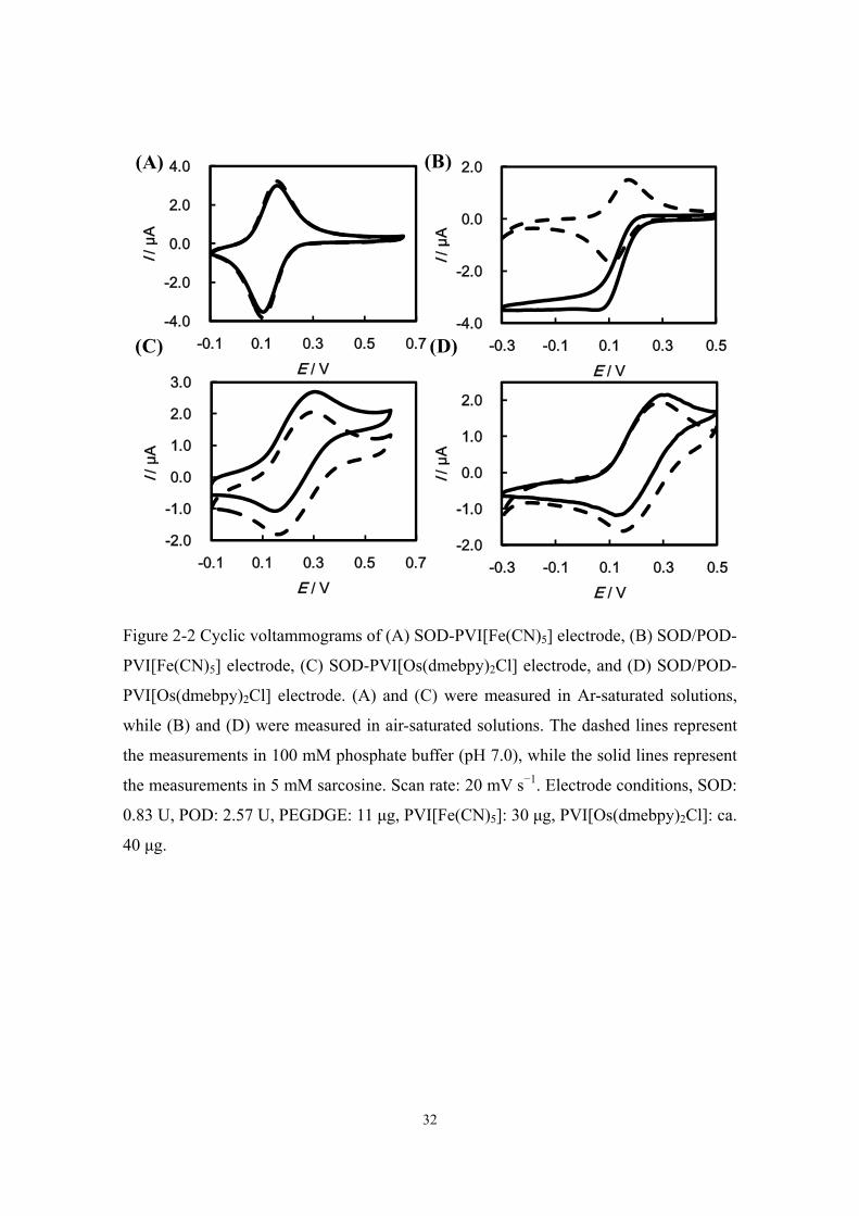
Catalytic effect of PVI[Fe(CN)5]on SOD and POD
In oxidase/POD bienzyme system, the mediator oxidized in the POD reaction may
also be reduced by receiving the electron from the reduced oxidase generated in the
substrate oxidation, which interferes with the detection of mediator reduction on the
electrode. To evaluate the mediating effect for mediator selection, PVI[Fe(CN)5] and
PVI[Os(dmebpy)2Cl] (dmebpy = 4,4'-dimethyl-2,2'-bipyridine) were used to investigate
the interactions with SOD and POD. The cyclic voltammetric responses of
SOD/POD-PVI[Fe(CN)5] electrode and SOD-PVI[Fe(CN)5] electrode are shown in Fig.
2-2. In Fig. 2-2A, PVI[Fe(CN)5] did not mediate the SOD reaction, while the catalytic
reduction current from the POD reaction was clearly observed (Fig. 2-2B). On the other
hand, PVI[Os(dmebpy)2Cl] reacted with SOD; the catalytic oxidation current of
creatinine was obtained (Fig. 2-2C). Therefore, in the cyclic voltammogram of
SOD/POD-PVI[Os(dmebpy)2Cl] electrode, the catalytic reduction current from POD
reaction was hardly observed as shown in Fig. 2-2D.
The reason which causes the difference in the reactivity between PVI[Fe(CN)5]
and PVI[Os(dmebpy)2Cl] was explained as in Chapter 1. Briefly, the meditating
capability of hexacyanoferrate ion on the SOD reaction is originally very low because
of the steric hindrance effect and the electrostatic repulsion between negatively charged
hexacyanoferrate and the deeply buried FAD of SOD [18]. After binding
pentacyanoferrate with PVI, it may become more difficult to enter into the active site of
SOD due to the increased charge density and fixation. For this reason, there is no
mediating effect of PVI[Fe(CN)5] on the SOD reaction. On the other hand,
Os(dmebpy)2Cl is more hydrophobic than pentacyanoferrate, which decreases the
difficulty in entering the active site of the oxidase. In POD reaction, both of the
polymers can transfer electrons to the protoheme, the redox center of POD, because the
location of protoheme is near the surface of POD, and the size of POD is smaller than
that of SOD, which shortens the distance between the mediator and the redox center
[19]. Therefore, it is easier for PVI[Fe(CN)5] to react with POD than with SOD. Thus,

31
the data in Fig. 2-2 clearly confirm the concept that PVI[Fe(CN)5] has extremely low
reactivity against SOD, while it has high reactivity against POD; the linear range of
H2O2 measured by the POD-PVI[Fe(CN)5] sensor was from 3 to 65 μM and the
sensitivity was approximately 410 mA cm−2 M−1(data not shown). Based on the results,
PVI[Fe(CN)5] is suitable as a mediator for the SOD/POD bienzyme system, and the
signal intensity was practically independent of the oxygen tension at least in the range
from 0.2 to 1 atm (data not shown).
32
Figure 2-2 Cyclic voltammograms of (A) SOD-PVI[Fe(CN)5] electrode, (B) SOD/POD-
PVI[Fe(CN)5] electrode, (C) SOD-PVI[Os(dmebpy)2Cl] electrode, and (D) SOD/POD-
PVI[Os(dmebpy)2Cl] electrode. (A) and (C) were measured in Ar-saturated solutions,
while (B) and (D) were measured in air-saturated solutions. The dashed lines represent
the measurements in 100 mM phosphate buffer (pH 7.0), while the solid lines represent
the measurements in 5 mM sarcosine. Scan rate: 20 mV s−1. Electrode conditions, SOD:
0.83 U, POD: 2.57 U, PEGDGE: 11 μg, PVI[Fe(CN)5]: 30 μg, PVI[Os(dmebpy)2Cl]: ca.
40 μg.
-4.0
-2.0
0.0
2.0
-0.3 -0.1 0.1 0.3 0.5
I / μ
A
E / V
-2.0
-1.0
0.0
1.0
2.0
-0.3 -0.1 0.1 0.3 0.5
I/ μ
A
E / V
-4.0
-2.0
0.0
2.0
4.0
-0.1 0.1 0.3 0.5 0.7
I/ μ
A
E / V
(A) (B)
-2.0
-1.0
0.0
1.0
2.0
3.0
-0.1 0.1 0.3 0.5 0.7
I/ μ
A
E / V
(C) (D)
33
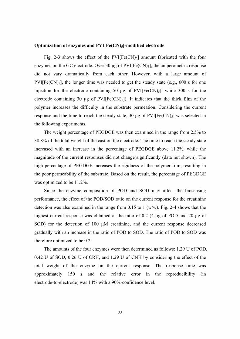
Optimization of enzymes and PVI[Fe(CN)5]-modified electrode
Fig. 2-3 shows the effect of the PVI[Fe(CN)5] amount fabricated with the four
enzymes on the GC electrode. Over 30 μg of PVI[Fe(CN)5], the amperometric response
did not vary dramatically from each other. However, with a large amount of
PVI[Fe(CN)5], the longer time was needed to get the steady state (e.g., 600 s for one
injection for the electrode containing 50 μg of PVI[Fe(CN)5], while 300 s for the
electrode containing 30 μg of PVI[Fe(CN)5]). It indicates that the thick film of the
polymer increases the difficulty in the substrate permeation. Considering the current
response and the time to reach the steady state, 30 μg of PVI[Fe(CN)5] was selected in
the following experiments.
The weight percentage of PEGDGE was then examined in the range from 2.5% to
38.8% of the total weight of the cast on the electrode. The time to reach the steady state
increased with an increase in the percentage of PEGDGE above 11.2%, while the
magnitude of the current responses did not change significantly (data not shown). The
high percentage of PEGDGE increases the rigidness of the polymer film, resulting in
the poor permeability of the substrate. Based on the result, the percentage of PEGDGE
was optimized to be 11.2%.
Since the enzyme composition of POD and SOD may affect the biosensing
performance, the effect of the POD/SOD ratio on the current response for the creatinine
detection was also examined in the range from 0.15 to 1 (w/w). Fig. 2-4 shows that the
highest current response was obtained at the ratio of 0.2 (4 μg of POD and 20 μg of
SOD) for the detection of 100 μM creatinine, and the current response decreased
gradually with an increase in the ratio of POD to SOD. The ratio of POD to SOD was
therefore optimized to be 0.2.
The amounts of the four enzymes were then determined as follows: 1.29 U of POD,
0.42 U of SOD, 0.26 U of CRH, and 1.29 U of CNH by considering the effect of the
total weight of the enzyme on the current response. The response time was
approximately 150 s and the relative error in the reproducibility (in
electrode-to-electrode) was 14% with a 90%-confidence level.

34
Figure 2-3 Dependence of the current response on the PVI[Fe(CN)5] amount for the
detection of 100 μM creatinine at –0.1 V. Electrode conditions, CNH: 1.29 U, CRH:
0.26 U, SOD: 0.33 U, POD: 1.03 U, PEGDGE: 20 μg. The error bars were evaluated by
the Student t-distribution with a 90%-confidence level.
Figure 2-4 Dependence of the amperometric response on the weight ratio of POD to
SOD for the detection of 100 μM creatinine at –0.1 V. Electrode conditions, CNH: 1.29
U, CRH: 0.26 U, SOD: 0.33 U (20 μg), PEGDGE: 11.2%, PVI[Fe(CN)5]: 30 μg. The
error bars were evaluated by the Student t-distribution with a 90%-confidence level.
0
20
40
60
80
100
120
0 20 40 60 80Rel
etiv
e cu
rrent
resp
onse
/ %
Weight of PVI[Fe(CN)5] / μg
0
20
40
60
80
100
120
0 0.3 0.6 0.9 1.2Rel
ativ
e cu
rrent
resp
onse
/ %
POD / SOD (w/w)

35
Interference effect
The creatinine biosensor based on the reductive H2O2 detection at a low operating
potential (−0.1 V vs. Ag|AgCl) minimizes the undesirable oxidation of electroactive
interference in physiological fluids.
In order to eliminate the interference effect on POD which is due to the poor
substrate specificity mentioned in General Introduction, negatively charged Nafion was
utilized as a protecting film on the top of the enzymes-PVI[Fe(CN)5]-modified electrode
to exclude anionic species such as AA and UA. The interference effect on the
amperometric response measured with the proposed electrode covered with and without
Nafion film is shown in Fig. 2-5. For the detection of 150 μM creatinine, the
amperometric response with the Nafion-coated electrode was smaller than that with the
electrode without Nafion film because of the inhibition of the mass transfer. However,
the interference effect was eliminated by the protection of Nafion film, while the current
responses of 150 μM UA and 10 μM AA were observed at the electrode without Nafion
film. In the case of urine, the normal concentrations of creatinine and UA are in the
same level [20], and the concentration of creatinine is about thirty times higher than that
of AA [21]. This means that Nafion used as a protecting film satisfies the creatinine
determination in real urine samples. Acetaminophen and dopamine were also tested for
the interference test. Small interference effects were observed in the solutions
containing 150 μM creatinine and 10 μM acetaminophen (recovery = 108 ± 7%) or
dopamine (recovery = 96 ± 10%). Considering the fact that the concentrations of these
interferences are much smaller than that of creatinine, these interferences do not
seriously affect the creatinine detection in this biosensing system.
Internal creatine in urine might also interfere with creatinine determination since it
reacts with CRH immobilized on the enzymes-PVI[Fe(CN)5]-modified electrode to
result in the overestimation of creatinine. In the case of urine, the excretion rate of
creatinine is about twenty times higher than that of creatine [22]. In our experiment, as
the concentration ratio of creatine to creatinine is 6.7% (10 μM creatine/150 μM
creatinine), the signal ratio of creatine to creatinine is only about 2% (data not shown)
though H2O2 generation from creatine requires fewer enzymatic steps than that from
creatinine. It may be described that in neutral pH, creatinine is positively charged while

36
creatine is a zwitterion, therefore creatinine is easier to penetrate the negatively charged
Nafion film into the enzyme layer to get a larger amperometric response [23]. As a
result, the internal creatine in urine does not significantly affect the creatinine
determination in our method.

37
Figure 2-5 Amperometric responses of the proposed electrodes without Nafion (solid
line) and with 5 μL of 1% Nafion (dashed line). CTN: 150 μM creatinine, UA: 150 μM,
AA: 10 μM. The arrows indicate the injection time of the respective solutions. Electrode
conditions, CNH: 1.29 U, CRH: 0.26 U, SOD: 0.42 U, POD: 1.29 U, PEGDGE: 11.2%,
PVI[Fe(CN)5]: 30 μg. Operating potential: −0.1 V.
CTN
AA
CTN
UA
100 s
20 nA
AA CTN
UA

38
Comparison with Jaffe method
The amperometric response for the optimized biosensing electrode at −0.1 V is
presented in Fig. 2-6. The detection limit of creatinine is 12 μM (S/N > 3) and the linear
range is from 12 to 500 μM (R2 = 0.993), and the sensitivity is 11 mA cm−2 M−1, which
is sufficient for urine sample test. This method was applied to the creatinine
determination of urine from four volunteers and was compared with Jaffe method which
is widely used in clinical diagnosis. In the electro-enzymatic method, 10 μL of urine
sample was injected into 1 mL of 100 mM phosphate buffer (pH 7.0) for measurements.
Table 2-1 shows the creatinine concentrations evaluated from the absorbance at λ = 520
nm based on Jaffe method and from the current response at −0.1 V measured by this
proposed method, respectively. Numbers 1 and 2 are the urine samples which were
donated by male, while No. 3 and 4 were donated by female. The data show that the
concentration of urine creatinine in male is higher than that in female, which is
consistent with the typical human reference ranges, and a good correlation is obtained
between the two methods. The values measured by the electro-enzymatic method are
lower than Jaffe method, most probably because other compounds in urine (UA or AA)
caused positive interference in Jaffe method. The results also show that the creatinine
concentrations of No. 1 and 2 measured by Jaffe method are similar with each other,
while the value of No. 2 is smaller than that of No. 1 measured by this method. This
seems to be resulted from a high concentration of interference in No. 2, which reacted
with picric acid to overestimate the creatinine concentration in Jaffe method.

39
Figure 2-6 Dependence of the amperometric response on the creatinine concentration at
–0.1 V. Electrode conditions were the same as those in Fig. 2-5 with Nafion film. The
broken line is a linear regression curve; current density (μA cm−2) = 11 (mA cm−2 M−1)
×[creatinine] (μM) − 120 (nA cm−2), R2=0.993. The inset shows the amperometric
response.
Table 2-1 Determination of creatinine from urine samples
Number Creatinine / mM
Jaffe method This method
1 17.0 ± 0.5 13.3 ± 0.82 17.1 ± 0.5 11.9 ± 0.23 5.7 ± 0.2 5.2 ± 0.34 11.6 ± 0.5 9.1 ± 0.7
The errors were evaluated by the Student t-distribution with a 90%-confidence level.
0
2
4
6
8
10
12
0 500 1000 1500 2000
j / μ
A cm
-2
Creatinine / μM
-1000
-800
-600
-400
-200
0
300 800 1300 1800 2300 2800I/
nA
t / s
40
Reference [1] T. Arndt, Forensic Sci.Int., 186 (2009) 48. [2] U. Lad, S. Khokhar, G.M. Kale, Anal. Chem., 80 (2008) 7910. [3] S. Narayanan, H.D. Appleton, Clin. Chem., 26 (1980) 1119. [4] R.W. Bonsnes, H.H. Taussky, J. Biol. Chem., 158 (1945) 581. [5] J.A. Weber, A.P. Vanzanten, Clin. Chem., 37 (1991) 695. [6] M.H. Kroll, N.A. Roach, B. Poe, R.J. Elin, Clin. Chem., 33 (1987) 1129. [7] M. Gutierrez, S. Alegret, M. del Valle, Biosens. Bioelectron., 23 (2008) 795. [8] C.S. Rui, Y. Kato, K. Sonomoto, Biosens. Bioelectron., 9 (1994) 429. [9] T. Tsuchida, K. Yoda, Clin. Chem., 29 (1983) 51. [10] V.K. Nguyen, C.M. Wolff, J.L. Seris, J.P. Schwing, Anal. Chem., 63 (1991) 611. [11] A.E.G. Cass, G. Davis, M.J. Green, H.A.O. Hill, J. Electroanal. Chem., 190 (1985)
117. [12] A. Ramanavicius, Anal. Bioanal. Chem., 387 (2007) 1899. [13] I. Taniguchi, S. Miyamoto, S. Tomimura, F.M. Hawkridge, J. Electroanal. Chem.,
240 (1988) 333. [14] H. Sakai, R. Baba, K. Hashimoto, A. Fujishima, A. Heller, J. Phys. Chem., 99
(1995) 11896. [15] H. Kinoshita, M. Torimura, K. Kano, T. Ikeda, Electroanal., 9 (1997) 1234. [16] T. Ikeda, T. Shiraishi, M. Senda, Agric. Biol. Chem., 52 (1988) 3187. [17] M. Bader, R. Wrbitzky, M. Blaszkewicz, C. van Thriel, Arch. Toxicol., 81 (2007)
335. [18] J.J. Kulys, N.K. Cenas, Biochim. Biophys. Acta, 744 (1983) 57. [19] N.C. Veitch, Phytochem., 65 (2004) 249. [20] Y.G. Zuo, Y. Yang, Z. Zhu, W.S. He, Z. Aydin, Talanta, 83 (2011) 1707. [21] M.P. Westerman, Y. Zhang, J.P. McConnell, P.A. Chezick, R. Neelam, S. Freels,
L.S. Feldman, S. Allen, R. Baridi, L.E. Feldman, L.W.M. Fung, Am. J. Hematol., 65 (2000) 174.
[22] C. Beyer, Clin. Chem., 39 (1993) 1613. [23] J.H. Shin, Y.S. Choi, H.J. Lee, S.H. Choi, J. Ha, I.J. Yoon, H. Nam, G.S. Cha,
Anal. Chem., 73 (2001) 5965.

41
Chapter 3 Sensitive D-amino acid bienzyme biosensor mediated by
PVI[Fe(CN)5]
A sensitive D-amino acid oxidase (DAAO)/peroxidase (POD) bienzyme biosensor
is constructed, in which PVI[Fe(CN)5] (PVI = poly(1-vinylimidazole)) is selected as a
mediator. Reduction current of PVI[Fe(CN)5] related to the H2O2 concentration
generated in the DAAO reaction was measured at −0.1 V vs. Ag|AgCl with a
DAAO/POD-PVI[Fe(CN)5] electrode. The result revealed that PVI[Fe(CN)5] is suitable
as a mediator for this bienzyme system because of its appropriate formal potential and
its extremely low reactivity against DAAO. The stability of DAAO was improved by
adding free flavin adenine dinucleotide and the electrode composition was optimized for
the detection of D-alanine. Nafion and ascorbate oxidase-immobilized films worked
successfully to prevent severe interference from uric acid and ascorbic acid. The low
detection limits of D-alanine (2 μM) and D-serine (2 μM) imply its possibility for the
determination of extremely low concentration of D-amino acids in physiological fluids.
The proposed bienzyme biosensor is proved to be capable of detecting D-amino acids in
urine.
Introduction
The role of D-amino acids (DAAs) has become of great importance in life science,
since several DAAs are found to have physiological functions in mammals. With regard
to the central nervous system in brain, D-serine acts as a co-agonist of N-methyl-
D-aspartate receptor, which is associated with learning and memory [1], and D-alanine in
the anterior pituitary gland, pancreas, and plasma might have a physiological function to
the insulin regulation [2]. The concentration of DAAs also shows a strong correlation to
some diseases. For example, the ratio of D-serine to the total (D- + L-) serine
concentration in the serum of Alzheimer’s disease patients was reported to be lower
than that of normal subjects [3]. As the dynamic monitoring of the DAAs concentration

42
is of acute interest, a rapid, simple and highly sensitive method for the DAA detection is
essential for further study. Separation-based analytical methods such as high
performance liquid chromatography (HPLC) and capillary electrophoresis are
conventionally used for DAA determination with high sensitivity [4, 5]. D-Amino acid
oxidase (DAAO) oxidizes DAAs to the corresponding imino acids, which have been
further utilized to react with o-phenylenediamine and 2-mercaptoethanol to form the
corresponding fluorescent quinoxalinol derivatives. The fluorescent derivatives can be
separated by HPLC, and this method eliminates the interference from extremely high
concentrations of L-amino acids [6, 7]. Considering the convenience for in vivo
detections and the undesirability of cumbersome instruments, enzyme-based
amperometric biosensors are favored for DAA detection. DAAO is a flavoenzyme and
is widely used in enzymatic biosensors for DAA determination, either based on the
direct detection of generated H2O2 [8-10] or the detection of artificial mediators used as
electron acceptors of the substrate-reduced DAAO [11, 12]. However, as mentioned in
General Introduction, the oxidation of both H2O2 and mediators requires high operation
potentials, which always accompanies the co-oxidation of other electroactive
metabolites in physiological fluids. In addition, the exclusion of O2 in the case of
mediated biosensors seems to be impractical for in vivo analysis.
On the other hand, peroxidase (POD) in combination with H2O2–producing
oxidases including DAAO has been studied intensively in recent years for the substrate
detection in the way of direct or mediated electron transfer [13, 14]. Since the low
potential operation decreases the background current and noise levels, and eliminates
the undesirable oxidation of electroactive interferences, bienzyme biosensors show high
sensitivity and stability. Nevertheless, the oxidized mediators may also act as electron
acceptors of H2O2-producing oxidases based on the dehydrogenase activity of the
oxidase just mentioned in General Introduction.
Here, the author focuses the attention on PVI[Fe(CN)5] (PVI =
poly(1-vinylimidazole)) as a mediator for the DAAO/POD bienzyme biosensor
considering its suitable operating potential and the poor mediating capability for other
flavoenzymes. The interactions of PVI[Fe(CN)5] with POD and DAAO will be studied,
and the electrode optimization and the interference effect will also be investigated.
43
Experimental
Reagents
The sources of some chemicals were mentioned in Chapter 1 and 2. D-Serine,
glutaraldehyde (GA, 20%), and flavin adenine dinucleotide (FAD) disodium salt were
obtained from Wako Chem. Co. (Osaka, Japan). DAAO from porcine kidney (8.2 U
mg−1) and D-alanine were from Sigma-Aldrich (USA). Ascorbate oxidase from cucumis
sp. (AO, 333 U mg−1) was purchased from Toyobo Co. (Osaka, Japan). Uric acid (UA)
solution was prepared with 10 mM NaOH; FAD was dissolved in distilled water, and
the enzymes, substrates, ascorbic acid (AA) and poly(ethylene glycol) diglycidyl ether
(PEGDGE) solutions were prepared with 30 mM potassium phosphate buffer (pH 7.0).
Other chemicals were of analytical grade and used as received. The urine sample was
donated from a healthy volunteer.
The synthesis of PVI[Fe(CN)5] was described in Chapter 1 except that the stock
solution of PVI[Fe(CN)5] was prepared in distilled water.
Co-immobilization of POD, DAAO and PVI[Fe(CN)5]
A solution containing PVI[Fe(CN)5], PEGDGE, POD and DAAO was cast onto the
surface of a glassy carbon electrode (3 mm diameter, BAS) and well mixed with a
syringe needle. The electrode was dried at 4 °C for 24 h. Before measurements, the
proposed electrode was immersed into a 30 mM potassium phosphate buffer (pH 7.0)
for 20 min.
Fabrication of interference-free bienzyme electrode
To improve the stability of DAAO, 1 μL of 400 μM FAD solution (4×10⁻¹⁰ mol,
ca. 0.3 μg) was cast onto the aforementioned electrode (see Results and Discussion for
details). For interference tests, 3 μL of Nafion solution diluted by distilled water was
then added onto the electrode after air-drying. Unbounded polymer and enzymes were
removed by dipping the electrode into distilled water and 3μL of diluted Nafion solution
44
was cast onto the electrode again after air-drying. Finally, AO was crosslinked with GA
on the electrode at 4 °C for 2 h.
Electrochemical measurements
Electrochemical experiments were carried out in a potassium phosphate buffer (pH
8.0, 30 mM) under stirring at 37 °C with an electrochemical analyzer (611B or 1000,
CH Instrument, USA). The reference and counter electrodes were an Ag|AgCl|sat. KCl
and a Pt wire, respectively. Cyclic voltammetry was performed at a scan rate of 20 mV
s−1.
Oxygen depletion due to the DAAO reaction was measured at 37 °C and −0.6 V
with a Clark-type oxygen electrode (Optoscience, Japan). The pH 8.0, 30 mM
potassium phosphate buffer solution containing 3 μg mL−1 DAAO and 4 mM D-alanine
was used for experiments.

45
Results and Discussion
Reactivities of PVI[Fe(CN)5] against DAAO and POD
DAAO/POD bienzyme systems may suffer from interference because of the
dehydrogenase activity of DAAO. In this sensing system, PVI[Fe(CN)5] was selected as
a mediator. Fig. 3-1A shows that PVI[Fe(CN)5] has extremely low reactivity against
DAAO, while in the POD reaction the catalytic current is clearly observed with
PVI[Fe(CN)5] (Fig. 3-1B). The result indicates the poor interaction between DAAO and
PVI[Fe(CN)5]. The quite different reactivities of PVI[Fe(CN)5] against DAAO and POD
are interpreted as follows: The redox center (FAD) of DAAO locates in hydrophobic
surroundings and is deeply buried in the interior to induce the steric hindrance [15];
PVI[Fe(CN)5] with negatively charged ligands would be difficult to enter into the
FAD-catalytic center. Therefore, no obvious mediating effect of PVI[Fe(CN)5] was
observed for the DAAO reaction. On the other hand, as mentioned in Chapter 1, the
redox center of POD locates near the enzyme exterior with widely open entrance;
furthermore, the electrostatic potential around the redox center is positively charged,
which makes it easier for PVI[Fe(CN)5] to shuttle electrons between POD and electrode.
Thus, the author can conclude that PVI[Fe(CN)5] is very suitable as the mediator for the
DAAO/POD bienzyme biosensor with high specificity.

46
Figure 3-1 Cyclic voltammograms of (A) DAAO-PVI[Fe(CN)5] electrode in an
Ar-saturated solution and (B) DAAO/POD-PVI[Fe(CN)5] electrode in an air-saturated
solution. The dashed lines are the detections in a buffer solution and the solid lines are
the detections in 4 mM D-alanine (pH 8.0). Electrode composition, DAAO: 0.33 U,
POD: 2.14 U, PEGDGE: 10 μg, PVI[Fe(CN)5]: 20 μg. Scan rate is 20 mV s−1.
-4
-2
0
2
4
-0.1 0.1 0.3 0.5
I / μ
A
E / V
(A)
-8
-6
-4
-2
0
2
4
-0.3 -0.1 0.1 0.3 0.5
I/ μ
A
E / V
(B)

47
Stability of DAAO/POD-PVI[Fe(CN)5] electrode
DAAO/POD-PVI[Fe(CN)5] electrode has been successfully constructed herein for
the DAA detection. However, as shown in Fig. 3-2A, the current response of D-alanine
decreased with successive operations (open square); the current value in the third
detection is only two third of that in the first detection. It was considered that the poor
repeatability of the detection is mainly due to the non-covalent binding property of FAD
with DAAO [11]. Since the binding between FAD and apo-DAAO is weak, FAD
gradually releases from holo-DAAO to the bulk solution to lead the decrease in the
DAAO activity. Considering keeping the activity of DAAO, sufficient amount of FAD
was added into the experimental solutions to avoid the leakage of FAD from
holo-DAAO. The effect of the FAD addition was checked by detecting the oxygen
depletion in the DAAO reaction measured with the Clark-type oxygen electrode (Fig.
3-2B). At a low concentration of DAAO without FAD addition (dashed line), the
activity of DAAO gradually decreased since FAD was easily released from holo-DAAO
to the bulk solution. On the other hand, the oxygen depletion rate of the DAAO reaction
increased and DAAO became more stable in the presence of 40 μM FAD (solid line).
The high oxygen depletion rate in the FAD-containing solution indicates that the
addition of FAD keeps the DAAO activity. Good repeatability of the proposed
biosensing system was then proven by addition of FAD in the test solution, as evidenced
in Fig. 3-2A (closed rhombus); no drastic decrease in the current response was observed
in the successive experiments. Therefore, the experimental solution containing 40 μM
FAD was selected in the following experiments for other optimization.
Considering the fact that the biosensor sensitivity is related to the deposited
polymer thickness, the composition of POD and DAAO, and the total enzyme capacity,
the dependence of the current response on the amount of PVI[Fe(CN)5], the ratio of
POD to DAAO, and the weight ratio of the enzymes to the total weight were examined.
Fig. 3-3A shows the effect of the PVI[Fe(CN)5] amount fabricated with 0.16 U DAAO
and 2.1 U POD. The amperometric response did not vary drastically from each other
over 20 μg of PVI[Fe(CN)5], therefore, 20 μg of PVI[Fe(CN)5] was selected in the
following experiments. The effect of the ratio of POD to DAAO is shown in Fig. 3-3B
with fixing the weight of DAAO at 20 μg (0.16 U). The amperometric response of 5 μM

48
D-alanine reached approximately 60 nA when the ratio was 0.1 and no significant
variation was observed over the ratio 0.1. Fig. 3-3C indicated that the amount of the
enzyme is sufficient for the reaction when the weight ratio of the enzymes to the total
weight reached to 0.6.
The optimized values were then determined as follows: 20 μg of PVI[Fe(CN)5], 0.1
(the ratio of POD to DAAO), and 0.6 (the weight ratio of the enzymes to the total
weight). The final composition of proposed electrode contains 20 μg of PVI[Fe(CN)5],
10 μg of PEGDGE ,1.07 U of POD and 0.33 U of DAAO.

49
Figure 3-2 (A) Dependence of the current response at –0.1 V on the successive
measurements in the solution containing 40 μM FAD and 5 μM D-alanine (closed
rhombus) or 5 μM D-alanine only (open square). Electrode composition, DAAO: 0.25 U,
POD: 2.67 U, PEGDGE: 10 μg, PVI[Fe(CN)5]: 30 μg. (B) Time dependence of the
dissolved oxygen concentration during the DAAO reaction in the solution containing 40
μM FAD and 4 mM D-alanine (solid line) or 4 mM D-alanine only (dashed line).
Figure 3-3 Dependence of the amperometric response on the (A) amount of
PVI[Fe(CN)5], (B) the weight ratio of POD to DAAO and (C) the weight ratio of the
enzymes to the total weight in the detection of 5 μM D-alanine at –0.1 V. All
experimental solutions contain 40 μM FAD. The error bars were evaluated by the
Student t-distribution with a 90%-confidence level.
40
60
80
100
120
0 1 2 3 4
ΔI/ n
A
Successive measurement
(A) (B)
0
0.05
0.1
0.15
0.2
0 1000 2000 3000 4000
[O2]
/ mM
t / s
DAAO
0
20
40
60
80
0 0.1 0.2 0.3
ΔI/ n
A
POD / DAAO (w/w)
0
30
60
90
120
0 20 40 60
ΔI/ n
A
PVI[Fe(CN)5] / μg
0
50
100
150
0.2 0.4 0.6 0.8
ΔI/ n
A
Enzyme / total weight (w/w)
(A) (B)
(C)

50
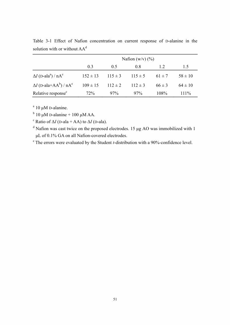
Interference effect
The DAA bienzyme biosensor at low operating potentials minimizes the
undesirable oxidation of electroactive interferences in physiological samples.
Considering the poor specificity of POD mentioned in General Introduction, negatively
charged Nafion was used as a protecting film on the top of the bienzyme electrode to
exclude anionic interferences such as AA and UA based on electrostatic repulsion. In
addition, 0.3 μg of FAD was cast between the bienzyme layer and the Nafion film in
place of the FAD addition in the experimental bulk solution. Furthermore, regarding the
low oxidation potential of AA, 15 μg of AO was immobilized with GA on the top of the
Nafion film to minimize the interference effect of AA for the purpose of getting high
sensitivity. In separated experiments, it has been proved that PVI[Fe(CN)5] works
neither as a substrate (reduced form) for AO nor as an electron acceptor of AO in the
catalytic oxidation of AA (data not shown). Since PVI[Fe(CN)5] shows little reactivity
against AO, AO utilization does not affect the DAA detection. The interference effect on
the amperometric response with different concentrations of Nafion is shown in Table
3-1; the current response of 10 μM D-alanine decreased with an increase of the Nafion
amount because of the thick film which decreases the mass transfer of the substrate.
Comparing the response of D-alanine with that in the AA-containing solution, the
relative response increased with an increase of the Nafion amount; thick Nafion film
shows a better protecting effect. The relative responses over than 100% with very thick
Nafion films may be due to the partial desorption of the Nafion film in succeeding
measurements for the DAA+AA mixture, which increases the penetration of D-alanine.
Taking the current response and the protecting effect into account, 0.8% Nafion was
selected for the biosensor fabrication, and no significant interference was observed in
the 10 μM D-alanine solution containing 500 μM UA and 100 μM AA (data not shown).
The physiologically normal concentration of UA in urine is about thirty times higher
than DAAs, and the normal concentration of AA is in the same level with DAAs [16-18].
Therefore, this bienzyme biosensor protected by Nafion and AO seems to satisfy the
DAA determination in urine.
51
Table 3-1 Effect of Nafion concentration on current response of D-alanine in the
solution with or without AAd
Nafion (w/v) (%)
0.3 0.5 0.8 1.2 1.5
ΔI (D-alaa) / nAe 152 ± 13 115 ± 3 115 ± 5 61 ± 7 58 ± 10
ΔI (D-ala+AAb) / nAe 109 ± 15 112 ± 2 112 ± 3 66 ± 3 64 ± 10
Relative responsec 72% 97% 97% 108% 111%
a 10 μM D-alanine. b 10 μM D-alanine + 100 μM AA. c Ratio of ΔI (D-ala + AA) to ΔI (D-ala). d Nafion was cast twice on the proposed electrodes. 15 μg AO was immobilized with 1 μL of 0.1% GA on all Nafion-covered electrodes.
e The errors were evaluated by the Student t-distribution with a 90%-confidence level.

52
Sensors for DAAs
The proposed bienzyme biosensor protected by Nafion and AO has successfully
worked for the DAA determination in the presence of high concentrations of
interference. However, in the preliminary test of urine sample, the sensitivity decreased
gradually with successive measurements (data not shown). Some molecules in the urine
sample may nonspecifically adsorb on the AO layer of the electrode, which fouled the
electrode to affect the sensitivity. To eliminate the nonspecific adsorption, 6 μL of 0.8%
Nafion was cast on the top of the AO layer for protection. The illustrated structure of the
DAA biosensor is shown in Scheme 3-1. Most of the interferences are inhibited from
entering into the sensing part by the outer Nafion film, and the residual UA and AA are
blocked by the inner Nafion film and oxidized by AO, respectively. Dopamine and
acetaminophen were also tested and only small interference effects were observed in the
solutions containing 10 μM D-alanine and 2 μM each of interferences. Considering the
fact that the concentrations of these interferences are sufficiently smaller than that of
DAA, these interferences would not practically affect the detection in the proposed
method. Thus, DAAs can successfully be detected without practical interference effect.
Amperometric responses of two major DAAs in human body, D-serine and
D-alanine, are presented in Fig. 3-4, in which the current was measured with the
optimized biosensing electrode at −0.1 V. The detection limits of D-serine and D-alanine
are both 2 μM (S/N > 3); the linear ranges are from 10 to 350 μM and 5 to 150 μM, and
the slopes are 5.4 and 9.7 nA μM−1, respectively. It shows that the biosensor has very
low detection limit and high sensitivity, which is suitable for the detection of extremely
low concentrations of DAAs in physiological samples. The different reactivities of
different DAAs indicate that it is difficult for DAAO-based biosensors to determine the
total concentration of DAA. Nevertheless, it is still benefit to easily determine the total
performance of DAAs with selecting one of DAAs, such as D-alanine, as a standard.
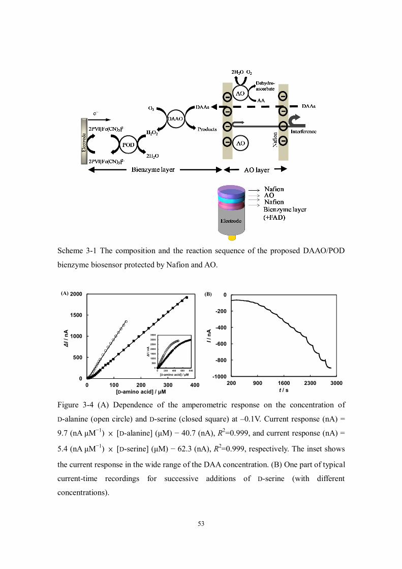
53
Scheme 3-1 The composition and the reaction sequence of the proposed DAAO/POD
bienzyme biosensor protected by Nafion and AO.
Figure 3-4 (A) Dependence of the amperometric response on the concentration of
D-alanine (open circle) and D-serine (closed square) at –0.1V. Current response (nA) =
9.7 (nA μM−1) × [D-alanine] (μM) − 40.7 (nA), R2=0.999, and current response (nA) =
5.4 (nA μM−1) × [D-serine] (μM) − 62.3 (nA), R2=0.999, respectively. The inset shows
the current response in the wide range of the DAA concentration. (B) One part of typical
current-time recordings for successive additions of D-serine (with different
concentrations).
0
500
1000
1500
2000
0 100 200 300 400
ΔI/ n
A
[D-amino acid] / μM
0
500
1000
1500
2000
2500
3000
3500
0 200 400 600 800
ΔI/ n
A
[D-amino acid] / μM
(A)
-1000
-800
-600
-400
-200
0
200 900 1600 2300 3000
I / n
A
t / s
(B)

54
The standard addition method was then used for a urine sample test. D-Alanine was
successively injected into the urine solution which was 100 times diluted with 30 mM
potassium phosphate buffer (pH 8.0) and the slope is similar to that measured in the
standard D-alanine solution (Fig. 3-5). The concentration of DAAs in the urine was
evaluated to be 150 ± 20 μM, when D-alanine was selected as a standard, where the
error was evaluated by the Student t-distribution with a 90%-confidence level. The
sensitivity of biosensor is not affected by the interferences in urine.
The proposed method was compared with a method of oxygen consumption
measured with a Clark oxygen electrode. The experimental condition of the method of
oxygen consumption was described briefly as follows: 150 μL of urine was injected into
the 850 μL of buffer solution (pH 8) containing 200 μg DAAO (1.64 U), and the rate of
current difference within 50 s was measured at −0.6 V and 37 °C. The concentration
was evaluated as 120 ± 50 μM, the error being evaluated by the Student t-distribution
with a 90%-confidence level. The difference between the two estimated values may
result from the different responses to the different types of DAA in the immobilized
enzyme and the free enzyme in the solution. Besides, considering the low concentration
of DAA in urine, a large amount of sample (~7 times diluted) is needed for the detection
of the oxygen consumption, while only 100 times diluted sample is sufficient for the
proposed method. Therefore, the other substances in urine may affect the free DAAO’s
activity in the solution and inhibit the reaction.
The proposed electrode worked properly for at least 24 times experiments (for
approximately 1.5 h) without change of the sensitivity. Therefore, it can be concluded
that the possible leakage of FAD and AO from the modified electrode is minimized.
Considering the application of disposable-type sensing trip, the proposed method may
well be utilized in the rather short-term usage. As a result, the DAAO/POD bienzyme
biosensor is applicable for clinical diagnosis.

55
Figure 3-5 Standard addition measurements of a urine sample, which was 100 times
diluted with 30 mM potassium phosphate buffer (pH 8.0). Current response (nA) = 5.8
± 0.2 (nA μM−1) × [D-alanine] (μM) + 8.8 ± 1.2 (nA), R2=0.998. The average values
were obtained from triplicate measurements, and the error bars were evaluated by the
Student t-distribution with a 90%-confidence level. The slope of 5.7 nA μM−1 means the
sensitivity of the bienzyme biosensor for the D-alanine detection in the solution without
urine.
0
20
40
60
80
100
-2 3 8 13
ΔI/
nA
[D-alanine] / μM
5.7
56
Reference
[1] J.S. Bains, S.H.R. Oliet, Trends Neurosci., 30 (2007) 417. [2] K. Hamase, A. Morikawa, S. Etoh, Y. Tojo, Y. Miyoshi, K. Zaitsu, Anal. Sci., 25
(2009) 961. [3] K. Hashimoto, T. Fukushima, E. Shimizu, S.I. Okada, N. Komatsu, N. Okamura, K.
Koike, H. Koizumi, C. Kumakiri, K. Imai, M. Iyo, Prog. Neuro-Psychopharmacol. Biol. Psychiatry, 28 (2004) 385.
[4] A. Hashimoto, T. Nishikawa, T. Oka, K. Takahashi, T. Hayashi, J. Chromatogr.-Biomed. Appl., 582 (1992) 41.
[5] Z. Quan, Y. Song, Y.Z. Feng, M.H. LeBlanc, Y.M. Liu, Anal. Chim. Acta, 528 (2005) 101.
[6] S. Oguri, M. Nomura, Y. Fujita, J. Chromatogr. A, 1078 (2005) 51. [7] S. Kato, Y. Kito, H. Hemmi, T. Yoshimura, J. Chromatogr. B, 879 (2010) 3190. [8] S. Sacchi, L. Pollegioni, M.S. Pilone, C. Rossetti, Biotechnol. Tech., 12 (1998) 149. [9] P. Pernot, J.-P. Mothet, O. Schuvailo, A. Soldatkin, L. Pollegioni, M. Pilone, M.-T.
Adeline, R. Cespuglio, S. Marinesco, Anal. Chem., 80 (2008) 1589. [10] Z.M. Zain, R.D. O'Neill, J.P. Lowry, K.W. Pierce, M. Tricklebank, A. Dewa, S. Ab
Ghani, Biosens. Bioelectron., 25 (2010) 1454. [11] G. Arai, T. Noma, M. Hayashi, I. Yasumori, J. Electroanal. Chem., 452 (1998) 43. [12] A.S.N. Murthy, J. Sharma, Electroanalysis, 11 (1999) 188. [13] V. Kacaniklic, K. Johansson, G. Markovarga, L. Gorton, G. Jonssonpettersson, E.
Csoregi, Electroanalysis, 6 (1994) 381. [14] R. Dominguez, B. Serra, A.J. Reviejo, J.M. Pingarron, Anal. Biochem., 298 (2001)
275. [15] H. Mizutani, I. Miyahara, K. Hirotsu, Y. Nishina, K. Shiga, C. Setoyama, R.
Miura, J. Biochem., 120 (1996) 14. [16] H. Bruckner, A. Schieber, Biomed. Chromatogr., 15 (2001) 166. [17] Y.G. Zuo, Y. Yang, Z. Zhu, W.S. He, Z. Aydin, Talanta, 83 (2011) 1707. [18] M.P. Westerman, Y. Zhang, J.P. McConnell, P.A. Chezick, R. Neelam, S. Freels,
L.S. Feldman, S. Allen, R. Baridi, L.E. Feldman, L.W.M. Fung, Am. J. Hematol., 65 (2000) 174.

57
Conclusions
Mediated electron transfer (MET)-based biosensors have been developed and
utilized for various kinds of clinical diagnosis, and the interference effect due to the
oxidation of electroactive compounds in samples is always the problem to be conquered
even for electrodes covered with a protecting membrane. Though recently the
H2O2-producing oxidase/peroxidase (POD) bienzyme biosensors mediated by redox
polymers based on the reductive H2O2 detection have been proposed to eliminate the
interference oxidation, the cross reaction of mediator with H2O2-producing oxidase
drastically interferes with the detection to cause the underestimation. Therefore, in
bienzyme biosensors, it becomes of great importance to find a suitable mediator which
has the high specificity for POD reaction only.
To find the suitable mediator, in Chapter 1, the MET activity of the three kinds of
metal complex-containing polymers: PVI[Os(dmebpy)2Cl] with neutral ligands,
PVI[Os(dcbbpy)2Cl] and PVI[Fe(CN)5] with negatively charged ligands (PVI =
poly(1-vinylimidazole); dmebpy = 4,4'-dimethyl-2,2'-bipyridine; dcbbpy =
4,4'-dicarboxy-2,2'-bipyridine), has been estimated for four H2O2-producing
flavoenzymes. The results reveal that the MET reactivity of the negatively charged
polymers is very low, which is strongly related to the electrostatic repulsive interaction
between the local surface charge of the flavoenzymes and the polymer’s charge as well
as the steric hindrance. Especially, in PVI[Fe(CN)5], the polymerization of Fe(CN)5 via
PVI increases the negative charge density and then enhances the electrostatic repulsive
effect. However, PVI[Fe(CN)5] works as a good mediator for the POD reaction to
reduce H2O2. This specific catalytic property of PVI[Fe(CN)5] is very convenient to
construct H2O2-producing oxidase/POD-based bienzyme biosensors.
For the application, in Chapter 2, PVI[Fe(CN)5] has been utilized for constructing
the creatinine biosensor which is based on the reductive H2O2 detection. PVI[Fe(CN)5]
is appropriate for the enzyme immobilization in this biosensing system and it shows the
superiority of being a mediator, since it only mediates the POD reaction but not against
the sarcosine oxidase reaction. The interference effect due to the poor specificity of

58
POD has been successfully eliminated by the Nafion film and the proposed method is
applicable for clinical diagnosis.
To construct highly sensitive biosensors mediated by PVI[Fe(CN)5], in Chapter 3,
D-amino acid oxidase (DAAO)/POD bienzyme biosensor mediated by PVI[Fe(CN)5]
has successfully been constructed for the DAA determination. There is no cross reaction
between PVI[Fe(CN)5] and DAAO, and the severe interference effect resulted from
high concentrations of interference has been eliminated by the Nafion films and AO.
The stability of DAAO has been improved by adding free flavin adenine dinucleotide
(FAD) and the Nafion film prevents the considerable leakage of FAD. The biosensor has
low detection limit and high sensitivity, which indicates its applicability for clinical
diagnosis. Since the main species of DAAs in human brain is D-serine, the proposed
biosensor seems to have a potential for D-serine determination in brain without the
problem of the DAA specificity of DAAO.
In summary, the author provides the information about the interaction between
several enzymes and polymers in this research, and has successfully used the specific
catalytic property of PVI[Fe(CN)5] to overcome the problem of cross reaction in two
kinds of bienzyme biosensors. The strategy in this research greatly improves the
sensitivity and specificity of bienzyme biosensor, and the author hopes the achievement
pushes the development of biosensor.

59
Appendix
The reactions of several quinones/phenols with sarcosine
Several quinones/phenols were investigated to check the reaction with sarcosine.
Some have been found to react with sarcosine directly: 1,4-benzoquinone (1,4-BQ),
2,5-dichloro-1,4-BQ, 2,6-dichloro-1,4-BQ, 2,6-dimethyl-1,4-BQ (all generate purple
product); 9,10-phenanthrenequinone and 2,3,5,6-tetramethyl-1,4-BQ (both generate
yellow-white product); 2,3,5,6-tetrachloro-1,4-BQ (it generates brown-green product).
CRH/SOD electrode was used for investigating the reaction between sarcosine and
mediator, and between sarcosine and SOD. The cyclic voltammograms of CRH/SOD
electrode with 2,6-dichloro-1,4-BQ, 1,4-BQ and 2,6-dimethyl-1,4-BQ addition in the
creatine solution are shown in Fig. A1. All quinones work as mediators for SOD. Since
the colored product was generated on the electrode surface, the catalytic oxidation
current decreased with time. The change of cyclic voltammetric profile in the presence
of 2,6-dimethyl-1,4-BQ (Fig. A1C) is hard to be observed since the generation rate of
colored product is slow. The comparison of generation rate is shown as follows:
2,6-dichloro-1,4-BQ > 1,4-BQ > 2,6-dimethyl-1,4-BQ.
2,4-diaminophenol and p-aminophenol were also tested for SOD reaction by
CRH/SOD electrode and the results show both react with SOD (Fig. A2). However,
2,4-diaminophenol is unstable in pH 8 buffer; the cyclic voltammetric profile of
2,4-diaminophenol shifted and the peak decreased with time (Fig. A2B), while
p-aminophenol may react with water to produce 1,4-BQ (Eq. A1), which reacts with
sarcosine.
Some show no mediating capability against SOD: 3,5-di-tert-butyl-1,2-BQ,
2,6-di-tert-butyl-1,4-BQ and 9,10-nathrenequinone.
(A1)
NH2
OH
NH
O
+ 2H+ + 2e
-
NH
O
+ H2O
O
O
+ NH3

60
Figure A1 Cyclic voltammograms of CRH/SOD electrode in the pH 8, 50 mM creatine
solution containing 250 μM (A) 2,6-dichloro-1,4-BQ, (B) 1,4-BQ and (C)
2,6-dimethyl-1,4-BQ. The arrows represent the successive scan. The CV in (B) is one
part of the inset in (B). Enzymes were immobilized with glutaraldehyde. Scan rate: 20
mV s−1.
-1.5
0.5
2.5
4.5
6.5
-0.1 0 0.1 0.2 0.3 0.4
I/ μ
A
E / V
(A)
12
12.2
12.4
12.6
12.8
0.3 0.35 0.4 0.45 0.5 0.55 0.6
I/ μ
A
E / V
0
5
10
0 0.2 0.4 0.6
(B)
(C)
-1
0
1
2
3
4
5
-0.1 0 0.1 0.2 0.3 0.4 0.5
I/ μ
A
E / V

61
Figure A2 Cyclic voltammograms of (A) CRH/SOD electrode in pH 8, 50 mM creatine
solution with successive addition of 10 μM 2,4-diaminophenol (arrow direction); (B)
0.5 mM 2,4-diaminophenol (pH 8). The arrow represents the successive scan; (C)
CRH/SOD electrode in pH 8, 50 mM creatine solution with successive addition of 10
μM p-aminophenol (arrow direction). Scan rate: 20 mV s−1.
(A)
-2
-1
0
1
2
3
4
5
6
7
-0.4 -0.3 -0.2 -0.1 0 0.1 0.2 0.3 0.4 0.5
I/ μ
A
E / V
(B)
-4
-2
0
2
4
6
8
-0.4 -0.3 -0.2 -0.1 0 0.1 0.2 0.3 0.4 0.5
I/ μ
A
E / V
(C)
0
2
4
6
8
10
12
0 0.1 0.2 0.3 0.4 0.5
I/ μ
A
E / V

62

63
Acknowledgement
I would like to express the appreciation to the advisor: Prof. Kenji Kano for his
continuous support of the research, for his patience, guidance and encouragement
during the three-year doctoral life. He has always made himself available to clarify my
doubts and instruct me despite his busy schedules. I would also like to give the sincere
thanks to Associate Prof. Osamu Shirai for his advice and insightful comments on the
study. I gratefully thank Assistant Prof. Yuki Kitazumi for his helpful suggestions and
technical support.
I am grateful to Mr. Katsumi Hamamoto, the researcher in Kyoto University, for
his valuable insights and the skilled technical support of microelectrode fabrication, and
also grateful to Associate Prof. Seiya Tsujimura in Tsukuba University for his critical
advice and research support in my first year of doctoral course. I thank Dr. Tatsuo Noda
for his valuable discussion and academic suggestions. I am indebted to the members I
have met in the lab who provide me useful advice, teach me Japanese and give me the
happiness and encouragement during three years. Also thank Ms. Toshie Koyama, the
secretary in the lab, for her kindness and support. I really gained a lot from them
through their personal and scholarly interaction. I thank Yoshida Scholarship
Foundation for their financial support during three years, which let me concentrate on
the research without worries about life.
Finally, I would like to thank my family in Taiwan and relatives in Japan whose
love, encouragement and unconditional support allow me to finish this journey.
Thank all mentioned above and all friends in Taiwan and Japan again, who support
me in every possible way, help me complete the thesis, the research, and enrich my life
in Japan.

64
List of publications
1) Chi-Hua Nieh, Yuki Kitazumi, Osamu Shirai and Kenji Kano
Sensitive D-amino acid biosensor based on oxidase/peroxidase system mediated by
pentacyanoferrate-bound polymer.
Biosens. Bioelectron. 47 (2013) 350-355. (Chapter 3)
2) Chi-Hua Nieh, Seiya Tsujimura, Osamu Shirai and Kenji Kano
Amperometric biosensor based on reductive H2O2 detection using
pentacyanoferrate-bound polymer for creatinine determination.
Anal. Chim. Acta 767 (2013) 128-133. (Chapter 2)
3) Chi-Hua Nieh, Seiya Tsujimura, Osamu Shirai and Kenji Kano
Electrostatic and steric interaction between redox polymers and some
flavoenzymes in mediated bioelectrocatalysis.
J. Electroanal. Chem. 689 (2013) 26-30. (Chapter 1)