t.j. williams (ph.d) dept. animal physiology, unaab
TRANSCRIPT

T.J. Williams (Ph.D) Dept. Animal Physiology, UNAAB
1
UNIVERSITY OF AGRICULTURE, ABEOKUTA
DEPARTMENT OF ANIMAL PHYSIOLOGY
PHYSIOLOGY OF GROWTH IN FARM ANIMALS (ANP 505) DR. T.J. WILLIAMS Course content
1. Introduction - Definitions and concept - Basic concepts of animal structure
2. Body systems - Digestive, respiratory, reproductive, circulatory, urinary, endocrine etc,
- intergument 3. Bones, connective tissues & fats
Bones Connective tissues (muscle)
Muscle identification & development - Early development of muscles - Post-natal growth of muscles
4. Growth of the whole animal - Pre-natal growth - Measurement of growth - Growth curves - Allometry
5. Hormonal regulation of animal growth
1. INTRODUCTION What Is Growth? It is difficult to give a perfect definition of animal growth because many of the changes involved are reversible. If an animal increases its live weight by drinking water, do the resulting increments to live weight really constitute growth? Yet, meat often contains 80% water, and it must come from somewhere. If an animal increases its body weight by accumulating fat between and within its muscles, most people might accept that these are true growth increments. Yet, these fat depots readily might be lost if the animal is placed on reduced feed. Likewise, even the myofibrillar proteins of lean meat may be used as an energy reserve in fasting animals, although the growth of the vital organs and nervous system is usually considered to be practically irreversible. Growth can therefore be described as an increase in body height, length, girth and weight that occurs when a healthy young animal is given adequate food, water and shelter. Live weight is the most important and commonly measured of these features and, if recorded at regular intervals, it may be used to plot a simple growth curve. Basic concepts of animal structure A number of anatomical terms are needed to describe the relative positions of structures within the body of an animal.
Anterior = towards the head Posterior = towards the tail Dorsal = towards the upper part or back of the standing animal Ventral = towards the lower part or belly of the standing animal

T.J. Williams (Ph.D) Dept. Animal Physiology, UNAAB
2
Medial = towards the midline plane that separates right and left sides of the body Lateral = towards the sides of a standing animal Proximal = towards the body in a limb of the animal Distal = away from the body in a limb of the animal
Fig. 1. Structural terms of a pig
Species is both singular and plural. One species - two species. The singular, specie, has no connection to farm animals or science at all. For cattle, sheep, pigs and poultry, the sire or father is called a bull, a ram, a boar or a cock (tom in turkeys), respectively, while the dam or mother is called a cow, a ewe, a sow or a hen, respectively. A heifer is an immature female bovine, and a gilt is an immature female pig. A hogget is a yearling sheep. The neonates or new-born of cattle, sheep and pigs are called calves, lambs or piglets, respectively. For pigs, the process of birth or parturition is called farrowing. Newly hatched chickens, turkeys, ducks and geese are called chicks, poults, ducklings or goslings, respectively.For cattle, sheep, pigs and poultry, a castrated male is called a steer, a wether, a barrow or a capon, respectively.
Fig. 2. Points of a pig

T.J. Williams (Ph.D) Dept. Animal Physiology, UNAAB
3
Fig. 3. Points of a beef cattle
Fig. 4. points of lamb
2. BODY SYSTEMS Digestive system The alimentary tract of the digestive system is composed of the mouth, pharynx, esophagus, stomach, small and large intestines, rectum and anus. Associated with the alimentary tract are the following accessory organs; teeth, tongue, salivary glands, liver and pancreas. mouth, pharynx, esophagus
After the mouth, the alimentary tract leads to the pharynx. The larynx tops the windpipe from the lungs. The esophagus is a long muscular tube that moves food which has been swallowed. It is located dorsally
to the trachea so that it appears behind the trachea when the throat is opened ventrally in the abattoir. In poultry, just before the esophagus enters the thoracic cavity, there is a large sack-like expansion on the
right side known as the crop. The crop is a temporary storage area for feed.
Stomach The stomach differs in structure between pigs, ruminants, and poultry. Pigs have a relatively simple, single-chambered stomach (monogastric). Cattle and sheep have three additional chambers before the true stomach. Poultry have a second chamber after the true stomach. The stomach has complex glands in its wall.

T.J. Williams (Ph.D) Dept. Animal Physiology, UNAAB
4
Fig.5. Stomach of a pig on the floor of an abattoir, with some of the small intestines showing in the top right corner. In cattle and sheep, instead of opening directly into a glandular stomach where digestion begins , the esophagus leads to a series of three extra compartments, the rumen, the reticulum and the omasum. These compartments are lined with stratified squamous epithelium. In young lambs and calves that are still suckling, the rumen and reticulum may be by-passed. The presence of the milk is detected by sensory nerve endings in the mouth and pharynx. Reflex activity brings heavy muscular folds in the walls of the rumen and reticulum together forming an esophageal groove that leads directly from the cardia to the omasum. The rumen or paunch is a very large muscular bag on the left side of the body, extending from the diaphragm back to the pelvis. The smooth muscle of the rumen wall consists of two layers; a superficial layer from anterior to posterior, and an inner layer running transversely to form muscular pillars. The reticulum is lined by thin, wall-like ridges arranged in a honeycomb pattern. The reticulum is posterior to the heart and diaphragm. The rumen and reticulum contain countless microorganisms whose metabolic activity greatly enhances the nutritive value of typical ruminant feed.
The omasum is almost spherical in shape and is filled with muscular plates hanging from the dorsal roof. These plates or laminae are studded with short, blunt papillae whose function is to grind roughage. The trade name of the omasum is the manyplies or book-bag. The true glandular stomach or abomasum is located ventrally to the omasum. The epithelium of the abomasum is glandular with many mucus cells. In a typical lean beef steer, the emptied weight of the rumen, reticulum, omasum and abomasum comprises about 2.5% of the live weight. The growth of the rumen and reticulum in calves is very rapid but the abomasum grows more slowly. The gut fill is extremely variable, but is often around 15% of the live weight.
A number of different types of animals have the ability to digest cellulose with the help of symbiotic bacteria and ciliates in modified parts of the alimentary canal, but the ruminant system has a number of superior features that account for its great efficiency. Chewing the cud (the repeated regurgition and mastication of feed) would not be possible unless the main fermentation chamber, the rumen, was situated before the true stomach or abomasum. Thus, ruminants are able to achieve a very high efficiency of feed grinding, unlike the horse in whose faeces may be seen quite large particles of intact plant material. Another advantage of the ruminant system is that a long length of intestine is available for absorption after the point at which fermentation occurs. Rumen microorganisms themselves are very effective - they synthesize protein from low-grade nitrogenous materials such as ammonium salts and urea added to the feed. Equally important, they utilize sulfate to produce the essential amino acids cysteine and methionine, and they also synthesize B group vitamins, particularly vitamin B12. Rumen microorganisms obtain their own energy anaerobically with only a relatively low energy yield. Thus, the ruminant is able to take the residual energy from products of fermentation such as acetic, propionic, and butyric acids. The acidity of these substances is buffered by sodium bicarbonate from the saliva.

T.J. Williams (Ph.D) Dept. Animal Physiology, UNAAB
5
Fig.6. Proventriculus & gizzard (in chicken)
In poultry, the stomach is divided into two chambers. As is often the case, there is a fair amount of fat or adipose tissue covering both chambers. The first chamber, the proventriculus or glandular stomach secretes pepsin and hydrochloric acid. The second chamber, the gizzard seen on the right in the color frame, is thick and muscular with a horny internal epithelium and a high collagen content. In the figure above, the collagen can be seen as a blue sheen, radiating across the gizzard. The gizzard grinds the feed and mixes it with the enzyme mixture from the proventriculus. This is the reverse of the sequence found in the omasum and abomasum of ruminants where grinding of the feed takes place before it is exposed to the enzymes from the true stomach.
Small intestine
Fig. 7. Small intestine of a pig
In beef animals, slightly over 2% of the live weight is from the emptied weight of the intestines. The small intestine is composed of three regions, the duodenum, the jejunum and the ileum. The duodenum receives the hepatic and pancreatic ducts and has a complex glandular structure.
Large intestine The mammalian large intestine consists of the caecum and the colon. A caecum is a sac that opens into the alimentary tract.

T.J. Williams (Ph.D) Dept. Animal Physiology, UNAAB
6
Fig. 8. Caecum of a pig.
The colon is divided into ascending, transverse and descending parts, and it terminates at the rectum and anus. Poultry have two caeca just before the rectum. In poultry, but not in cattle, sheep or pigs, the inner surface area of the large intestine is expanded by villi. In poultry, the equivalent aperture to the anus is part of a compound structure called the cloaca. The cloaca is divided into three regions but these are difficult to distinguish. The rectum enters the cloaca at the coprodeum, the urinary and genital ducts enter at the urodeum and the opening to the exterior is called the proctodeum. Dorsal to the proctodeum is a region of lymphoidal tissue called the bursa of Fabricius. Byproducts - Some parts of the alimentary canal have a considerable commercial value as natural casings. After extensive cleaning and preparation, they are used as natural casings to contain different types of sausages and processed meat products. Teeth The teeth of meat animals have a complex structure. Briefly, dental formulae may be used to describe the patterns of teeth in meat animals. The four types of teeth are indicated by a letter notation;
I - for incisors or biting teeth C - for canine or tearing teeth P - for premolars or anterior grinding teeth M - for molars or posterior grinding teeth
The numerator and denominator of a fraction are used to indicate upper and lower numbers of teeth, respectively. Left and right sides of the jaws are not written separately but are indicated by the initial factor, x2. A prefix, D, is used to denote deciduous teeth that are present in the young animal but replaced in the older animal. Calf and lamb = 2 x (DI 0/4, DC 0/0, DP 3/3) = 20
Mature cattle and sheep = 2 x (I 0/4, C 0/0, P 3/3, M 3/3) = 32
Young pig = 2 x (DI 3/3, DC 1/1, DP 4/4) = 32
Mature pig = 2 x (I 3/3, C 1/1, P 4/4, M 3/3) = 44 The transition from deciduous to permanent dentition follows a rather complex pattern between 1.5 to 4 years in cattle, 0.25 to 4 years in sheep, and 8 to 20 months in pigs. Most commercially reared meat animals will, therefore, be at an intermediate stage between deciduous and permanent dentition when slaughtered. Salivary glands Salivary glands have a yellowish color and occur at three major locations.
The sublingual glands are located under the tongue and between the lower jaw bones, and they have a multiple duct system that drains saliva into the mouth.
The submaxillary glands are located at the angle of the lower jaw and have large ducts that open onto the floor of the mouth, beneath the tip of the tongue.
Beneath each ear, is a parotid salivary gland with a duct that opens into the mouth, near to the molar teeth.

T.J. Williams (Ph.D) Dept. Animal Physiology, UNAAB
7
The salivary glands of ruminants are extremely productive since they must produce much of the fluid with which the feed is mixed to form a slurry. The salivary glands of a steer, for example, might secrete well over 100 liters of saliva per day. Liver The liver is a large organ, about 1.5% of the live weight in beef cattle. In mammals, it is located in the anterior part of the visceral cavity, just posterior to the diaphragm.
Fig. 9. A pig liver The pig has four equally large lobes plus a small caudate lobe on the right side. Apart from a small caudate lobe on the right side, the bovine liver is not subdivided into lobes. The liver in sheep is similar to that in the bovine, but there is something of a fissure in the main lobe. The function of the liver is to store carbohydrate, process nutrient-rich incoming blood from the gut, and produce bile. Livers are condemned if they are infected by trematode flukes such as Fasciola hepatica in ruminants, or by nematodes such as Ascaris suum in pigs.
Fig. 10. Liver of a hen Pancreas The pancreas is a pale yellow gland located between the stomach and the small intestine in mammals, and in a loop of the duodenum in poultry. It has one or two ducts that convey pancreatic juice to the duodenum. The external secretions of the pancreas are controlled by the nervous sytem (vagus nerve) and the endocrine system (the hormone secretin from the duodenum). The three major constituents of the pancreatic juice are trypsin (for the hydrolysis of proteins when in conjunction with enterokinase present in the small intestine), amylase (for initial digestion of starch), and lipase (for the digestion of fats). Respiratory system The nasal cavity opens into the pharynx (shared with the alimentary canal), and then opens into the larynx. The larynx has a cartilagenous skeleton with muscles that support and stretch the vocal cords. In poultry, however,

T.J. Williams (Ph.D) Dept. Animal Physiology, UNAAB
8
sound is produced by a separate organ, the syrinx, which is located farther down the respiratory system. The epiglottis is a spout-shaped cartilage that protects the entrance to the larynx. The larynx leads to the trachea or windpipe. Trachea The trachea is a flexible tube held open by rings of cartilage. If it was not held open, it would collapse when the animal tried to breath in. The continuity of each ring of cartilage is broken by a small dorsal gap. The trachea divides into two bronchi at a "Y" fork. The bronchi connect with the right and left lungs, where they branch into progressively smaller ducts called bronchioles. Epithelium The trachea, bronchi and bronchioles are lined with ciliated epithelium and mucous glands, as seen in the image above which is a transverse section through the wall of the trachea. The cilia seen in a row across the top of the cells in tha image above (facing into the lumen of the trachea) are extremely fine whip-like hairs on the lumenal surfaces of cells. A complex system of mobile protein strands along the length of each cilium provides the motive power for movements that appear whip-like. Millions of cilia beat in a coordinated manner so that they can propel a continuous stream of mucus from the lungs to the nasal cavity. Thus, any small particles that have entered the lungs, despite the protective filtering of incoming air by the turbinate bones, can be removed.
Fig. 11. A beef cattle Lung Gaseous exchange
This occurs between inhaled air and the blood in the lungs, and takes place across the moist surfaces of alveoli or alveolar sacs. In mammals, the alveoli are the final blind-ending branches of the air duct system. Beneath the moist epithelium which lines each alveolus is an extensive meshwork of lung capillaries. Oxygen is taken up by the blood in a loose combination with the hemoglobin of red blood cells or erythrocytes. There are three ways in which carbon dioxide may be carried in the blood; (1) in solution, (2) combined with blood proteins, or (3) as bicarbonate. Carbon dioxide is more soluble and diffuses faster than oxygen. The ratio of bicarbonate to carbonic acid determines the pH or acidity of the blood. This ratio is regulated by the rate of escape of carbon dioxide from the blood in the lungs: loss of carbon dioxide increases pH (decreases acidity). Gaseous exchange does not occur across the walls of the major air ducts that lead into the lungs. Thus, the last fraction of air that is inhaled becomes the first fraction to be exhaled, and the oxygen it contains is not utilized. Typical resting rates of respiration are 12 to 18 breaths per minute in cattle, 12 to 20 in sheep and 10 to 18 in pigs.The rate of respiration is controlled by the medulla oblongata in the posterior part of the brain. The medulla responds primarily to the pH and the carbon dioxide content of the blood; it increases the rate of respiration if the blood becomes acidic with a high level of carbon dioxide.

T.J. Williams (Ph.D) Dept. Animal Physiology, UNAAB
9
Fig. 12. A pork lung Pleural membranes - when the lungs are removed from the body, slippery pleural membranes may be seen covering both the inner surface of the thoracic cavity and the lung surface. Pleural membranes prevent friction between the lungs and the body wall. Inspiration and expiration, caused by movements of the intercostal muscles, the ribs, the diaphragm and, sometimes, the abdominal muscles. Diaphragm - The diaphragm resembles a strong drumskin that divides the thoracic and abdominal cavities, but it is thickened by muscle where it joins the body wall. In a dressed carcass, the muscular part of the diaphragm remains as a flap of muscle running diagonally across the inside of the ribcage. By-Products - The proteins of the lungs (as well as those of the rumen and spleen), may be recovered by alkaline extraction followed by reacidification. Protein may be isolated as a powder or texturized to form fibers. Lungs also may be processed to isolate heparin, an anticoagulant for medical use. Poultry - The respiratory system in poultry is quite different from that found in mammals. The lung is the red tissue in the image above. There is no diaphragm separating thoracic from abdominal cavities. Instead of being drawn into the lungs and then exhaled, air is drawn through the lungs and into air sacs outside the lungs. On exhalation, the air passes back through the lungs to the exterior. In poultry, therefore, the gaseous exchange between air and blood takes place as the air is moving through the lungs. The lungs of poultry are much smaller than those of mammals (relative to body size). Instead of occupying almost the whole of the thoracic cavity, they are located under the vertebral column where they are shaped to fit between the deep arches of the ribs where they meet the vertebral column. The lungs of poultry are usually removed with a suction tube during commercial slaughter procedures whereas, in meat animals, the lungs are removed together with the trachea, bronchi and heart, as plucks. The extensive system of air sacs in poultry extends between many of the viscera and even into certain bones. The interclavicular air sac is a single structure in the midline but the other air sacs are paired (right and left). The cervical extends towards the neck. The axillary is within the body at the junction with the wing. The anterior thoracic, posterior thoracic and abdominal sacs are in the body cavity. The humeral is located within the humerus as a branch of the axillary sac. Air sacs have extremely thin walls and, when poultry are dissected, they should be identified while the viscera are in a relatively undisturbed condition. REPRODUCTIVE SYSTEM Males The paired testes of male farm mammals are located in a muscular bag called the scrotum where they can be Each testis can be raised by the cremaster externus muscle.{Evidence of cremaster muscles may appear in carcasses of sows and gilts and so cannot be used to identify male carcasses!}. The connective tissue round the testis is called the tunica albugenia: it is white with a good blood supply. maintained several degrees below body temperature for the efficient production of spermatozoa or sperm. Spermatozoa are produced in seminiferous tubules tightly packed into the oval shape of the testis.

T.J. Williams (Ph.D) Dept. Animal Physiology, UNAAB
10
Fig. 13. Two boar testes, the one on the left sliced open.
Fig. 14. One tubule under the microscope.
The higher power image below shows part of the tubule (lumen downwards) with meiotic divisions leading to the formation of spermatozoa, whose tails can be seen faintly at the bottom of the image. The many seminiferous tubules in each testis open into a labyrinth of tubes called the rete testis. Immature spermatozoa from the rete testis pass in a number of efferent ducts to a further tubular system, the epididymis, located on the surface of the testis as shown in the image below.
Spermatozoa mature during storage in the epididymis and are carried to the urethra during mating by peristalsis of the vas deferens. The urethra is located ventrally in the penis. Seminal fluid to carry the spermatozoa is produced
Fig. 15. Surface of a boar testis

T.J. Williams (Ph.D) Dept. Animal Physiology, UNAAB
11
by the paired seminal vesicles, by the prostate gland, and by the paired bulbo-urethral glands (= Cowper's glands). The glands are located along the urethra, near to the bladder.
Fig. 16. seminal vesicles of a boar. The penis has a sigmoid flexure or S-shaped bend along its length. The sigmoid flexure is straightened out when the penis is extended for mating. This occurs when a pair of muscles, the ischio-cavernosus muscles, compress the veins which drain the blood from the penis. Arterial blood pressure then expands the volume of vascular tissue in the penis. The ischio-cavernosus muscles are attached to the ischium and the trimmed stump of the muscle may be seen on dressed sides of beef as a pizzle eye. The pizzle eye is poorly developed in steer carcasses and is larger and darker in bull carcasses. During the embryonic development of both mammals and birds, the testes are formed from tissue located near to the kidneys. In male mammals, the testes normally move to the scrotum outside the body cavity, and they pass through the body wall in the inguinal canal. The testes are attached to the inside of the scrotum by the gubernaculum which is contractile in fetal animals and is responsible for pulling the testis through the inguinal canal. The layers of connective tissue that cover each testis are the (1) tunica vaginalis communis and (2) the inner layer formed by the tunica vaginalis propria. Both layers are derived from modified layers of peritoneum gathered by the testes during their migration. The inner layer supports the blood vessels and nerves to the testis.
In cryptorchid pigs or ridgelings, movement of the testes along the inguinal canal is incomplete and they do not reach the scrotum. This abnormality causes infertility, but an older cryptorchid pig may still develop boar taint like a normal boar. The cause of the condition is not fully known but the normal mechanism of testicular movement appears to involve the action of testosterone on the genito-femoral nerve which then produces a peptide that activates the gubernaculum.
In male poultry, the testes remain in their original position near the kidneys, and a highly coiled vas deferens links each testis separately to the urodeum of the cloaca. The testes of cockerels may be removed through an incision made posterior to the ribs and anterior to the pelvis. Capons produced in this way are less aggressive in their behavior and they tend to deposit fat more readily than entire males. Before genetic advances in poultry growth rates made capon production uncompetitive, capons also used to be produced by administering the female hormone, estrogen. In poultry, some of the male hormone testosterone is converted to estrogen in the central nervous system and this estrogen is used to control testosterone production. Thus, increased estrogen levels thereby lower testosterone levels to produce a capon with tender meat and a high fat content. Except for ducks, male poultry have no functional penis.
Females Female mammals have a pair of ovaries located posteriorly and dorsally in the abdominal cavity. The mage below shows one ovary of a cow.

T.J. Williams (Ph.D) Dept. Animal Physiology, UNAAB
12
Fig. 17. One ovary of a cow
Fig. 19. histological image of ova Ova develop in the cortex (outer layer) of the ovary. Each ripe ovum is enclosed in a fluid-filled follicle, as shown in the histological image below. At oestrus, ova are released into a ciliated funnel or infundibulum at the end of each oviduct (fallopian tube). The oviduct on each side leads into a horn of the uterus where embryonic development takes place.
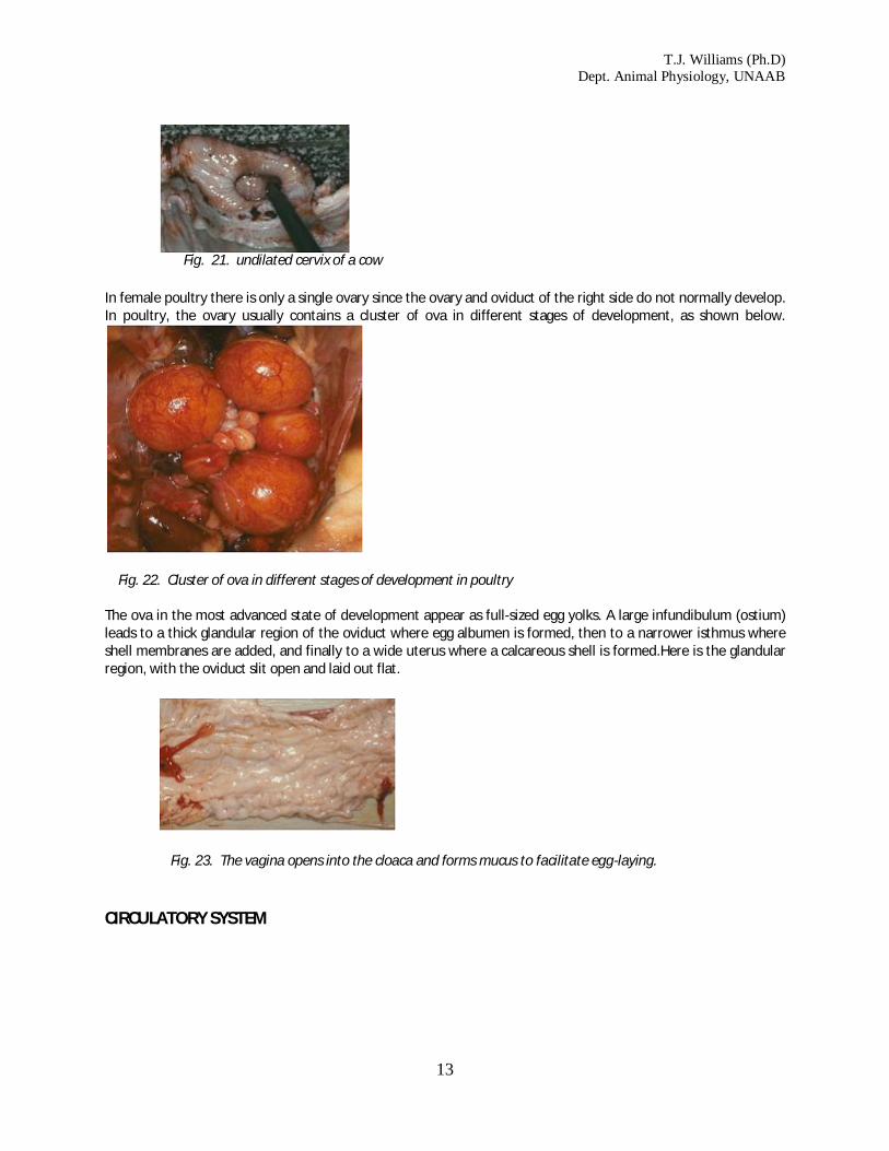
Fig. 20. one horn of the uterus of a cow. At birth, the offspring emerge through the dilated cervix and vagina.The image below shows an undilated cervix of a cow. In female poultry there is only a single ovary since the ovary and oviduct of the right side do not normally develop
T.J. Williams (Ph.D) Dept. Animal Physiology, UNAAB
13
Fig. 21. undilated cervix of a cow
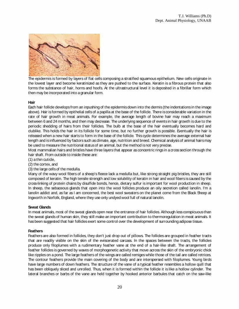
In female poultry there is only a single ovary since the ovary and oviduct of the right side do not normally develop. In poultry, the ovary usually contains a cluster of ova in different stages of development, as shown below.
Fig. 22. Cluster of ova in different stages of development in poultry The ova in the most advanced state of development appear as full-sized egg yolks. A large infundibulum (ostium) leads to a thick glandular region of the oviduct where egg albumen is formed, then to a narrower isthmus where shell membranes are added, and finally to a wide uterus where a calcareous shell is formed.Here is the glandular region, with the oviduct slit open and laid out flat.
Fig. 23. The vagina opens into the cloaca and forms mucus to facilitate egg-laying.
CIRCULATORY SYSTEM

T.J. Williams (Ph.D) Dept. Animal Physiology, UNAAB
14
Heart and Blood Vessels The right ventricle pumps blood through the semilunar valves and into the pulmonary arteries and then to the lungs. Here are the bovine semilunar valves. Oxygenated blood returns to the left atrium in the pulmonary veins, through the bicuspid valve. Here is the bovine pulmonary vein. Here is the bovine bicuspid valve. The atrium fills the left ventricle, and oxygenated blood is then pumped through the aorta to the body tissues. Here is the bovine aorta. The aorta branches to form the major arteries. These branch again many times and eventually give rise to arterioles and, finally, to capillaries. Blood is collected from the body tissues by the venous system, and eventually returns to the right atrium via the anterior vena cava and the posterior vena cava for another cycle through the lungs. Thus, relative to other arteries, the pulmonary artery is unusual because it contains de-oxygenated blood. And, relative to other veins, the pulmonary vein is unusual because it contains oxygenated blood. In the arterial system of meat animals, the aorta bends to the LEFT side of the body and then runs posteriorly in the midline, ventral to the vertebral column. It supplies arterial blood to all the body except the lungs. In the arterial system of poultry, the aorta swings to the RIGHT side of the body after leaving the heart . If poultry develop heavy muscling, the thin-walled right ventricle may be unable to cope with the increased cardiovascular demands and the ventricular wall may thicken as it adapts to the situation. But this may prevent the atrioventricular valve from functioning properly and the back pressure to the liver may cause ascites, an accumulation of fluid in the abdomen that may kill the bird. The venous system of meat animals is dominated by the anterior and posterior vena cava. The venous system of poultry is distinguished by a loop in the neck created by the jugular anastomosis and a loop around the kidneys. Cardiovascular function There are three different types of muscle tissue in the body -smooth, cardiac and skeletal. Smooth muscle occurs in the digestive and reproductive tracts, cardiac muscle is only found in the heart, while skeletal muscle forms all the meat of the commercial carcass. Most cardiac muscle cells are mononucleate. They are arranged in rows to form branching fibers, but individual cells are separated by intercalated discs. Cardiac muscles have a striated appearance due to the precise alignment of sliding filaments in their contractile fibrils, but skeletal muscles also are striated.
Fig. 25. Tansverse section of a group of cardiac muscle fibers, showing how their nuclei are located centrally in the axis of each fiber.
Fig. 24. A pig's heart.

T.J. Williams (Ph.D) Dept. Animal Physiology, UNAAB
15
Cardiac muscle cells are continuously pumping out sodium ions through their membranes. This causes the inside of the cell to have an electrical charge of approximately -90 mV with respect to the outside of the cell. This is called a resting potential. Extrinsic factors such as electrical activity (ionic movements) in adjacent cells may decrease the resting potential towards zero. When it reaches a value of approximately -65 mV, the threshold potential, the decrease in electrical potential accelerates, and it shoots past the zero value so that for a brief instant (about one tenth of a second) the membrane potential is positive. This sudden reversal of electrical charges is called an action potential. Action potentials are propogated into the interior of cardiac muscle cells by transverse tubules. In each cell, the transverse tubular system is an extensive series of finger-like indentations of the surface membrane. The arrival of an action potential in the interior of the cardiac muscle cell causes the release of calcium ions from the sarcoplasmic reticulum. The sarcoplasmic reticulum is a series of membrane-bounded vesicles in the interior of the cell. Unlike the transverse tubular system, the sarcoplasmic reticulum does not open to the surface of the muscle cell. Units of the sarcoplasmic reticulum surround the contractile fibrils in the interior of cardiac muscle cells. The sarcoplasmic reticulum sequesters and stores calcium ions, but it releases them again when prompted to do so by the transverse tubular system. Calcium ions activate the system of sliding protein filaments which is responsible for muscle contraction. The intrinsic rhythm of heart contraction originates from a group of cells at the sinu-atrial node. The membranes of these cells behave as if they had a sodium ion leak. Thus, at regular intervals their resting potentials drop to their threshold values, and they initiate action potentials. Action potentials then spread in a coordinated wave through right and left atria. The atria then contract and pump blood into the ventricles. However, the ventricles also are capable of filling themselves as they expand after pumping out their previous fill of blood. Under normal conditions, atrial contraction contributes to the overall cardiovascular efficiency, but its contribution may become vital when the heart is weakened by disease. In the medial wall of the heart, at the junction between the atria and ventricles, is a sensitive group of cells forming the atrioventricular node. This node is connected to a conduction system called the bundle of His that runs down the medial wall separating left and right ventricles. The atrioventricular node is activated by contraction of the atrial cells, and the bundle of His conducts the action potential wave to the base of the ventricles. From this point, a wave of contraction spreads upwards through the ventricles so that the the blood that has just filled the ventricles is now pumped out of the heart. The intrinsic heart rate is determined by the rate at which the sinu-atrial cells "leak" or depolarize, by the value of their threshold potentials, and by their resting potential. The flow of blood through the heart is directed by the heart valves. Mitral and tricuspid valves make a "lub" sound and the semilunar valves make a "dup" sound. Coordinated electrical activity of cardiac muscle cells generates an electrical signal that may be detected on the surface of the fore flank as an ELECTROCARDIOGRAM
The P wave is due to atrial excitation, PQ is the delay as the action potential passes down the bundle of His, QRS is due to ventricular contraction or systole, and T is caused by repolarization of the ventricles.
Activity of the heart is greatly influenced by its ionic environment. Isotonic sodium chloride plus calcium ions tend to stop the heart in systole (contracted) while isotonic sodium chloride plus potassium ions tend to stop the heart in diastole (relaxed). The nervous system also has an effect on heart rate. The thoracic nerve of the sympathetic nervous system releases catecholamines that increase the heart rate (tachycardia) while the vagus nerve of the parasympathetic nervous system releases acetylcholine that slows the heart (bradycardia). The neural regulation of cardiac activity is a reflex response to inputs from blood pressure receptors or baroreceptors, and from chemoreceptors that monitor the concentration of carbon dioxide in the blood. When the heart contracts, it works against the resistance to blood flow created by the peripheral blood vessels in the body tissues. Thus, if the peripheral blood vessels decrease their diameter (vasoconstriction), the blood pressure tends to rise. Conversely, if peripheral blood vessels are dilated (vasodilation), the blood pressure tends to drop. URINARY SYSTEM The urinary system has two major functions: It removes waste products from the blood stream It regulates the amount of water present in the body

T.J. Williams (Ph.D) Dept. Animal Physiology, UNAAB
16
In mammals, the kidneys are ventral to the vertebral column in the anterior lumbar region. The kidneys of pigs and sheep are oval in shape while the kidneys of cattle are each divided into approximately 20 lobules, as shown below.
Pork kidneys are not lobulated, are flat, and are usually pale (not as dark as the image shown below with its scale in good old-fashioned inches). In healthy well-fed animals, the kidneys are usually surrounded by perirenal fat, which is called leaf fat in the pork carcass
.
As shown in the image above, where a pork kidney has been sliced longitudinally, each kidney has a depression or hilum where the renal artery enters the kidney, and where the renal vein and ureter leave the kidne. The ureter from each kidney carries urine to the bladder. In the image below, the large pale bladder on the left of the screen was taken at slaughter from a boar. Although sometimes difficult to see, when a kidney is cut open, a pale inner medulla may be seen surrounded by a dark red corte. The wide entrance to the ureter is called the pelvis of the kidney. Running through the medulla, towards the pelvis of the kidney, are many small collecting tubules. Each of these terminates at a small conical mound called the pyramid, so that the pyramids project into the pelvis of the kidney. Urine is produced from the blood by a functional unit of the kidney called a nephron. There are large numbers of nephrons in each kidney. Urine leaves the bladder in a single tube, the urethra, that runs to the penis or to the vagina. In poultry, the kidneys are pressed closely against the ventral surface of the vertebral column, posterior to the lungs. Urine from each kidney leaves in a ureter but passes directly to the cloaca. Here, the urine rapidly loses water and the main component of nitrogenous excretion for poultry, uric acid, is precipitated as a sludge.
Fig. 26. Cattle kidney
Fig. 27. Pork kidney
Fig. 28. Pork kidney sliced longitudinally

T.J. Williams (Ph.D) Dept. Animal Physiology, UNAAB
17
ENDOCRINE SYSTEM Communication between cells and organs within the body is essential for the efficient control of body metabolism. Nerve impulses and hormones are the two best known types of communication. Neurons communicate rapidly by the transmission of action potentials, but they rely on chemical transmitters for the final step of the journey to their destination. Whereas, endocrine glands have made this last step their whole journey: they release chemical transmitters or hormones directly into the blood stream to act on cells at remote destinations. Unlike exocrine glands that release their secretions onto the skin or into the alimentary canal, the endocrine glands do not need a duct for the removal of their secretions. Abbreviations ACTH = adrenocorticotropic hormone ADH = anitdiuretic hormone = vasopressin CRH = corticotropin releasing hormone FSH = follicle stimulating hormone GnRH = gonadotropin releasing hormone ICSH = interstitial-cell stimulating hormone LH = luteinizing hormone LTH = luteotropic hormone MSH = melanocyte stimulating hormone PTH = parathyroid hormone STH = somatotropic hormone TRH = thyrotropin releasing hormone TSH = thyroid-stimulating hormone Pituitary The pituitary gland or hypophysis is a small round gland located ventrally to the brain. Embryologically, the pituitary is formed from the conjunction of an outgrowth from the floor of the brain (neurohypophysis or posterior pituitary) and a detached upgrowth from the roof of the mouth (adenohypophysis or anterior pituitary). The hypothalamus releases CRH which releases ACTH, and GnRH which releases LTH, FSH and LH, and TRH with TSH. The neurohypophysis releases ADH which causes water retention by the kidney, and OXYTOCIN which causes uterine contraction during parturition, then milk release. The adenohypophysis produces STH which stimulates body growth, LTH which stimulates mammary glands, ACTH which stimulates the adrenal cortex, TSH which activates the thyroid glands and adipose tissue lipase, FSH which activates the testes or prepares the ovarian follicles, and LH which completes spermatogenesis and stimulates androgen secretion OR (depending on the sex of the animal) which stimulates ovarian follicle growth, estrogen secretion, ovulation, formation of the corpus luteum, and progesterone secretion. An odd bit of the gland, the pars intermedia, produces MSH which stimulates pigment cells. Pineal The pineal gland is a neurosecretory gland whose evolutionary origin may be traced back to the third eye found in the skull roof of certain fossil fishes. The pineal is innervated by sympathetic nerves and is located deep in the brain, anterior to the cerebellum. It releases the hormone melatonin which acts on the ovaries to inhibit the estrus cycle. Melatonin also has wider effects on other neuroendocrine control systems. Melatonin synthesis is inhibited by nerve impulses to the pineal gland; the frequency of impulses is inversely related to the amount of visible light reaching the retinas of the eyes. In poultry, the pineal gland is probably responsible for circadian rhythms (24-hour cycles) in physiological activity. Thyroid The thyroid is located around the trachea, near to the larynx in mammals. Left and right thyroid glands are joined ventrally in pigs; in cattle and sheep the junction is restricted to a narrow connecting isthmus. In poultry, left and

T.J. Williams (Ph.D) Dept. Animal Physiology, UNAAB
18
right thyroid glands are deep red in color instead of pale brown, and they are completely separated at the base of the neck. The thyroid glands receive an abundant supply of blood from which they are able to capture iodine. Iodine is used for the synthesis of hormones which contain three or four iodine atoms, triiodothyronine and thyroxine. Thyroid hormones regulate oxidative metabolism and heat production in the body. Some cells in the thyroid also produce the hormone calcitonin. Parathyroids In mammals, two pairs of very small parathyroid glands are located in or near the thyroid glands. Their position is variable and they are difficult to identify in the abattoir. In poultry, there is a small parathyroid gland at the posterior end of each thyroid. The parathyroid hormone produced by the parathyroid glands forms one circuit of a double feedback system that regulates calcium levels in blood and bone. The other circuit is mediated by the hormone calcitonin from the thyroid gland. Parathyroid hormone causes the mobilization of calcium from bone. Calcitonin causes the inhibition of calcium release from bone. Thymus The thymus is a large gland, particularly in young animals, and is located anteriorly to the heart with lateral extensions into the neck. Thymus glands are sold for human consumption as sweetbreads. The thymus gland is composed of lymphoidal tissue and has a vital immunological function in young animals. It produces hormones which act on other cellular elements of the immune system. The animal's immune system, with which it defends itself against invading microorganisms, exhibits two types of responses (humoral and cell-mediated).Humoral antibody responses include: (1) the production of circulating antibodies, (2) the binding of antibodies to antigens, (3) the facilitation of phagocytic ingestion, and (4) the activation of certain blood proteins (the complement) to aid in the destruction of antigens. Cell mediated responses occur when specialized types of cells directly attack, or encourage macrophages to attack diseased cells bearing the target antibodies. The body contains two types of lymphocytes - the B cells responsible for humoral responses, and the T cells responsible for cell-mediated responses. Both types of lymphocytes originate from hemopoietic (blood-forming) stem cells of the fetal liver or adult bone marrow. Early in their development, immature lymphocytes migrate from their source into the circulation. In both mammals and poultry, immature T cells collect in the thymus where they undergo further development before they migrate out to peripheral lymphoid tissues (such as the spleen, the lymph nodes or Peyer's patches in the intestinal wall) to become mature T cells. In poultry, the B cells migrate to the Bursa of Fabricius for a period of development before they are released. In mammals, however, there is no Bursa or equivalent structure, and B cells are retained in the bone marrow for this period of their development. Adrenals Left and right adrenal glands are located anteriorly to the kidneys; in cattle they are roughly triangular, in sheep and poultry they are oval, and in pigs they are elongated. Each adrenal gland is composed of two distinct endocrine glands. In mammals, the adrenal cortex seen below, is wrapped around the adrenal medulla, although the two glands are mingled in poultry. Part of the cortex, the multiformis, responding to Na and K ions in the blood, produces mineralocorticoids (deoxycorticosterone and aldosterone) which regulate homeostasis of extracellular electrolytes (the fluids between the cells of the body). Other parts of the cortex (fasciculata and reticulata), controlled by ACTH and CRH produces glucocorticoids (cortisone, hydrocortisone, and corticosterone) which faciliatate gluconeogenesis (building up new sugars), proteolysis (breaking down proteins), and release of fatty acids from adipose tissue depots (as in slimming, although most philosophers agree it is better to be fat and happy than thin and miserable). The medulla of the adrenal gland under neural control produces catecholamines (epinephrine and norepinephrine) that enable animals to respond to stress (this is the gland responsible for exam fever). Pancreas

T.J. Williams (Ph.D) Dept. Animal Physiology, UNAAB
19
The islets of Langerhans seen below are microscopic areas of the pancreas with an important endocrine
function.
The islets contain alpha cells that produce glucagon, and beta cells that produce insulin. Insulin facilitates
the uptake and utilization of blood glucose by body cells. Thus, insulin deficiency causes the elevated blood sugar levels that occur in diabetes.
Pancreas glands may be collected in abattoirs for the commercial isolation of insulin. Insulin concentration is highest in the tail end of the pancreas, and the tissue must be kept dry before being frozen since insulin is water soluble. The action of glucagon is the opposite to that of insulin.
The testes produce testosterone; the ovaries produce estrogen, progesterone and relaxin; and during gestation, the uterus and placenta secrete chorionic gonadotropin. The stomach wall also secretes gastrin while the kidney produces the hormone renin. The liver produces somatomedins.
INTEGUMENT Skin Animal skin is composed of three basic layers. From outside to inside these layers may be called the (1) epidermis, (2) the dermis, and (3) the hypodermis.
T.J. Williams (Ph.D) Dept. Animal Physiology, UNAAB
20
The epidermis is formed by layers of flat cells composing a stratified squamous epithelium. New cells originate in the lowest layer and become keratinized as they are pushed to the surface. Keratin is a fibrous protein that also forms the substance of hair, horns and hoofs. At the ultrastructural level it is deposited in a fibrillar form which then may be incorporated into a granular form. Hair Each hair follicle develops from an inpushing of the epidermis down into the dermis (the indentations in the image above). Hair is formed by epithelial cells of a papilla at the base of the follicle. There is considerable variation in the rate of hair growth in meat animals. For example, the average length of bovine hair may reach a maximum between 6 and 24 months, and then may decrease. The underlying sequence of events in hair growth is due to the periodic shedding of hairs from their follicles. The bulb at the base of the hair eventually becomes hard and clublike. This holds the hair in its follicle for some time, but no further growth is possible. Eventually the hair is released when a new hair starts to form in the base of the follicle. This cycle determines the average external hair length and is influenced by factors such as climate, age, nutrition and breed. Chemical analysis of animal hairs may be used to measure the nutritional status of an animal, but the method is not very precise. Most mammalian hairs and bristles have three layers that appear as concentric rings in a cross section through the hair shaft. From outside to inside these are: (1) a thin cuticle, (2) the cortex, and (3) the large cells of the medulla. Many of the wavy wool fibers of a sheep's fleece lack a medulla but, like strong straight pig bristles, they are still composed of keratin. The high tensile strength and low solubility of keratin in hair and wool fibers is caused by the cross-linking of protein chains by disulfide bonds, hence, dietary sulfur is important for wool production in sheep. In sheep, the sebaceous glands that open into the wool follicles produce an oily secretion called lanolin. I'm a lanolin addict and, as far as I am concerned, the best wool sweaters on the planet come from the Black Sheep at Ingworth in Norfolk, England, where they use only undyed wool full of natural lanolin. Sweat Glands In meat animals, most of the sweat glands open near the entrance of hair follicles. Although less conspicuous than the sweat glands of human skin, they still make an important contribution to thermoregulation in meat animals. It has been suggested that hair follicles exert some control over the development of surrounding adipose tissue. Feathers Feathers are also formed in follicles, they don't just drop out of pillows. The follicles are grouped in feather tracts that are readily visible on the skin of the eviscerated carcass. In the spaces between the tracts, the follicles produce only filoplumes with a rudimentary feather vane at the end of a hair-like shaft. The arrangement of feather follicles is governed by waves of morphogenetic activity that move across the skin of the embryonic chick like ripples on a pond. The large feathers of the wings are called remiges while those of the tail are called retrices. The contour feathers provide the main covering of the body and are interspersed with filoplumes. Young birds have large numbers of down feathers. The structure of the vane of a typical feather resembles a hollow quill that has been obliquely sliced and unrolled. Thus, when it is formed within the follicle it is like a hollow cylinder. The lateral branches or barbs of the vane are held together by hooked anterior barbules that catch on the saw-like

T.J. Williams (Ph.D) Dept. Animal Physiology, UNAAB
21
edges of adjacent posterior barbules. The skin of poultry is dry and does not produce its own oil. In poultry, there is an oil gland located dorsally to the stumpy tail of the bird. The oil is distributed when the feathers are kept in order as the bird preens itself. Pigmentation Pigment cells or melanocytes are located in the deepest layers of the epidermis or in the underlying dermis. Melanin is a pigment formed in organelles called melanosomes. Melanin is passed from melanocytes to skin cells by cytocrine secretion. Melanin is formed from the oxidation of tyrosine by tyrosinase. Absence of this enzyme results in an albino animal. Variation in the color of farm animals is caused by variations in the amount and distribution of melanin. Melanin may be extracted with an aqueous solution of sodium hydroxide and then recovered by acidification. Melanin is a polymer based on indole monomers, but there is also a protein component involved that makes precise determination of its structure difficult. The distribution of melanin over the animals' skin is determined prenatally by an interaction between the migration patterns of melanocytes and the diffusion patterns of the messenger substances that either activate or suppress the synthesis of melanin. A single dominant gene determines the belt pattern marking that runs over the shoulders and forelimbs of some breeds of pigs. Leather The epidermis is supported on the ridged surface of the underlying dermis. The upper region of the dermis, often called the papillary layer of the dermis, is a tightly woven network of collagen fibers with some elastin fibers. After the tanning of a hide to make leather, the papillary layer becomes the top surface of the leather. With a hand lens, the openings where the hair follicles once penetrated the dermis are easily visible. When the leather is turned over, the much looser coarse fibrous weave of the lower dermis is evident. In pigskin, the follicles of the strong bristles are rooted at the lowest level of the dermis so that many of the follicles almost perforate the leather.
When the hide is removed from the carcass, the separation is made through the deepest layer of the integument - the hypodermis. Fat is often deposited in the hypodermis and, particularly in sheep, may even infiltrate the dermis. Numerous blood vessels run through the hypodermis to reach the extensive vascular bed (for heat dissipation) in the dermis. The hide weight of a typical lean steer is about 7% of the live weight, but there is considerable seasonal variation with colder climates inducing heavier hides and there are also differences between types of cattle.
Beef hides are graded on their cleanliness and degree of damage due to branding or warble fly larvae. If beef hides have been processed with a high standard of hygiene, the collagen of the inner layer of the hide may be used for processed food products such as sausage casings. Green hides (those from recently slaughtered animals) are treated with sodium chloride prior to tanning. The hides are trimmed, split into left and right sides, and soaked for several days in water. Then the hides are dehaired in a calcium hydroxide solution that contains sodium or calcium hydrosulfide. The conversion of a hide to leather occurs when it is TANNED, originally with a tree bark extract but now usually with sodium dichromate. Hair remnants are physically forced from the hair follicles (scudding) prior to deliming in sulfuric acid. Elastin fibers are removed enzymatically before the hides are pickled in sodium chloride acidified with sulfuric acid.
3. BONES, CONNECTIVE TISSUES & FATS Tissue is many cells together that serve a function. The carcass of an animal is comprised of three types of tissue: bone, muscle (connective tissue), and fat. Bone tissue is 50% organic matter, and 50% minerals. Bones grow at their ends which are made of cartilage. When the cartilage has hardened, the bones can not grow longer. The bone can still grow wider and repairs them. Muscle cells are made of one cell that divides many times, and then fuses itself together. Bone is important to livestock production because it is the frame that muscle is built on.

T.J. Williams (Ph.D) Dept. Animal Physiology, UNAAB
22
BONE Bones as a type of connective tissue A meat animal's body is supported by bone, is held together by fibrous connective tissue, and is protected against starvation and cold by adipose tissue. These three types of tissue, although they differ radically in appearance and properties, are all classified as types of connective tissue. All three types contain cells located in a matrix with fibres. In bones, both the matrix and the fibres make an important contribution to mechanical strength. The hardness of bone originates from a calcified matrix, and strength comes from embedded collagen fibres. The cells of bone, osteocytes, are trapped in small caves called lacunae. The gristle of the carcass is formed from tendons (by which muscles pull on bones), from ligaments (which hold bones together at the joints of the skeleton), from aponeuroses (which cover some muscles) and from fasciae (which form strong sheets between muscles). The dominant protein in gristle is collagen. Since connective tissues permeate nearly all parts of the body at the microscopic level, collagen is the most abundant protein in the animal body. The collagen fibres in meat are converted from strong fibres to jelly (gelatin) by the action of moist heat during cooking, but the collagen in bones may be removed by mild hydrolysis to produce gelatin for use in other food products or for other uses such as photographic emulsions. Skeletal variation In the section on skeletal anatomy, the number of vertebrae in pork carcasses was seen to vary between breeds. The heritability of the number of vertebrae is about 0.74 (which is quite high). Each extra vertebra adds about 15 mm to the length of the carcass at slaughter weight, so variation in vertebral numbers enables breeders to change carcass length. Breeds with a large size when mature and with heavy bone development tend to have more thoracic vertebrae than lighter breeds. Sometimes the ribs on extra thoracic vertebrae are only partially formed, but usually they are complete. The minimum number of lumbar vertebrae is generally found in cacasses with the maximum number of thoracic vertebrae. However, the variability of vertebral numbers frequently leads to an increase in the total number of vertebrae, so that the phenomenon is not due simply to the substitution of one type of vertebra for another. But how is this variability created? Embryology tells us that the number of vertebrae in an animal is determined by the number of somites or tissue blocks that develops along the length of the spinal cord. By definition, in mammals, the vertebrae that bear ribs are identified as thoracic vertebrae. In the embryo there are ossification centers on each side of the developing vertebrae. In vertebrae that do not normally develop ribs, these lateral ossification centers contribute their bone tissue to the centra or bodies of adjacent vertebrae. In the thoracic vertebrae, however, thes laterally derived bone tissue remains separate from the centra and forms the ribs. Thus, the numbers of pairs of ribs and the numbers of thoracic vertebrae are determined by the developmental mechanism that controls the fate of the tissue which is derived from the lateral ossification centers. Cartilage
Cartilage cells, called chondrocytes, occupy lacunae in a stiff flexible matrix formed from collagen fibres embedded proteoglycan. Hyaline cartilage, with a white translucent appearance, occurs on the smooth surfaces of joints. In the larynx, trachea and bronchi of the plucks, hyaline cartilage forms the rings and tubes that hold these air ducts

T.J. Williams (Ph.D) Dept. Animal Physiology, UNAAB
23
open during respiration. Flexible parts of the skeleton, such as the dorsal part of the scapula and the linkages between the sternum and the posterior ribs, also are formed from hyaline cartilage. The importance of cartilage to the butcher is in helping to estimate the age of an animal at slaughter. Most of the bones of the carcass are initiated prenatally as cartilagenous models that subsequently become ossified. Complete ossification is a slow process, and the bones of young meat animals are more flexible than those of adults. Thus, the degree of ossification is a useful clue to animal age in the carcass. Degree of ossification or its opposite, survival of cartilage, enables the carcass to be placed into a maturity group. This is done by examining a number of the features of the skeleton. In young cattle (around one year of age), the interiors of the vertebral centra are soft, red and porous in appearance. The medial surfaces of the ribs are rounded and streaked with red. As cattle grow older, the interiors of bones become harder, more white, and less porous. Carcasses from young animals exhibit a lot of relatively soft cartilage, particularly on the tips of the dorsal spines of the thoracic vertebrae. As animals grow older, cartilage in such locations becomes hard and ossified. In young animals, the sacral vertebrae are only loosely fused together, wheras older animals have their sacral vertebrae solidly fused together. Chondrocytes are initially capable of both cell division (mitosis) and matrix formation. So clusters of related cells become pushed apart by their new matrix in a process called interstitial growth. Cartilaginous models of prenatal bones are covered by a membrane known as the perichondrium. Inner perichondrial cells differentiate into chondrocytes so that, in addition to interstitial growth, new cells and matrix may be added superficially in a process known as appositional growth. Cartilage may acquire numerous elastic or collagen fibres to become elastic cartilage or fibrocartilage, respectively. The dominant type of collagen in hyaline cartilage is what biochemists call Type II collagen and it accounts for 50 to 70% of the dry weight or collagen. But cartilage also contains some unusual minor types of collagen, such as Type M collagen, which is much shorter than other collagen molecules, and has a helical molecular structure that is stabilized by disulphide bonds. Under the microscope, there are a number of features of cartilage that indicate an animal's physiological age. (1) With age, the matrix becomes increasingly rigid. (2) Appositional growth is the dominant growth process in older animals. (3) Young chondrocytes are flattened or elliptical with their long axis parallel to the surface of the cartilage. (4) Old chondrocytes are large (up to 0.04 mm diameter) and rounded in shape. (5) In older animals, chondrocytes form nests or isogenous groups within single lacunae. (6) With age, the staining of the cartilage matrix by alkaline stains used for microscopy decreases. (6) Relative to young chondrocytes, older chondrocytes have more stored glycogen. Unfortunately these things are invisible to the butcher, and seldom examined even by scientists, otherwise they would be very useful in judging a carcass. Bones under the microscope
Fig. 33. Section of very young bone, with marrow cavity towards the top, and calcified tissue stained red. Oxygen, nutrients and waste products may travel to and from the chondrocytes in cartilage by diffusion through the surrounding matrix but, when the matrix becomes ossified by the deposition of submicroscopic hydroxyapatite crystals, diffusion is greatly reduced. In bone, osteocytes can only survive if they develop root-like cytoplasmic extensions radiating from the lacunae to regions where exchange by diffusion can take place. These cytoplasmic extensions run through fine tubes or canaliculi in the ossified matrix, but are limited in length. Consequently, large

T.J. Williams (Ph.D) Dept. Animal Physiology, UNAAB
24
numbers of blood vessels permeate the matrix of bone. Most of these blood vessels run longitudinally through the bone in large haversian canals surrounded by concentric rings of osteocytes and bone lamellae. Bones are covered by a connective tissue membrane called the periosteum. The prenatal formation of bone is initiated by either of two basic mechanisms, (1) intramembranous ossification or (2) endochondral ossification. Intramembramous ossification is typical of the bones that form the vault of the skull, and it occurs when sheets of connective tissue produce osteoblasts which then initiate centers of ossification. Endochondral ossification is more common, and is the process by which cartilagenous models become ossified to form the bones of a commercial meat carcass. The internal structure of carcass bones becomes visible when they are split longitudinally on a band saw. The shaft of a bone is called the diaphysis, while the knob at each end is called the epiphysis. Between the diaphysis and each epiphysis is a cartilagenous growth plate called the epiphyseal plate. In a young animal, the chondrocytes of the epiphyseal plate are constantly dividing to form new matrix. However, on each face of the plate, cartilage is being continuously resorbed and replaced by bone so that the thickness of the epiphyseal plate tends to remain constant in growing animals. This process allows a bone to grow longitudinally without disrupting the articular surface on the epiphysis. The rate of the longitudinal growth of bones is the product of two factors; (1) the rate of production of new cells, and (2) the size that cells reach before they degenerate at the point of ossification. The strength and thickness of epiphyseal plates is modified by sex hormones. At puberty, chondrocyte growth slows down and fails to keep pace with ossification on the surface of the epiphyseal plate. Thus, epiphyseal plates are lost in mature animals, and the epiphyses become firmly ossified to their diaphyses. However, the factors that regulate the closure of the epiphyseal plate and, hence, the frame size of the animal, are poorly understood at present. Although regulation is likely to be an interaction between animal age and circulating hormones, there are no obvious hormonal changes when the plate closes. If whethers are implanted with the female horme estradiol, the ossification of growth plates is accelerated. Bone growth in mature animals is restricted to the girth or thickness of the bone, and it occurs by the recruitment of periosteal cells to become osteoblasts. Bone as a calcium store Bones are the main storage site for calcium in the body, and calcium ions are absolutely vital to the normal functioning of body cells. So when calcium is in short supply, because a cow is suddenly producing lots of milk or a hen is laying eggs like crazy, calcium is released from the bones into the blood. Milk fever may occur in a cow if the skeleton cannot keep pace with the demand. Regulation of skeletal growth Carcasses from young animals have a relatively high bone content because the skeleton is well developed at birth. As an animal grows to market weight, its proportion of bone decreases on a relative basis, because of the growth of muscle and fat. The long-term control of bone growth is superimposed on the short-term regulation of bone metabolism that occurs in response to changes in blood calcium levels, or to remodelling in response to local functional demands. A number of hormones exert secondary effects on skeletal development. Thyroxine, insulin, growth hormone and gonadal hormones tend to be anabolic, building up the bone mass. Estrogens may inhibit resorption of bone. Adrenal corticosteroids stimulate resorption of bone and inhibit the formation of new bone. In cattle, castration delays the completion of growth in epiphyseal plates. This is most noticeable in the distal bones of the limbs and enables the continued longitudinal growth of the legs. In the vertebral column, however, castration reduces bone growth. The reduction is centered on the first thoracic vertebra. Removal of the ovaries from heifers also causes an increase in the longitudinal growth of distal bones. Growth factors may mediate or augment the activity of the hormones controlling bone growth. Both rapidly growing and adult bones may contain growth factors such as transforming growth factor beta, beta-2 microglobulin, and insulinlike growth factor I. In adults, these factors may be involved in skeletal remodeling. There is a lot of uncertainty about the local control of bone growth, something that enables bones to develop struts of strong bone where they are most needed. One hypothesis is that loads frequently placed on a region of bone cause the conversion of mechanical energy to electrical energy by a piezoelectric effect (the same effect used in the crystal oscillator of an electric wrist-watch). In a frequently loaded and negatively charged region of bone, growth is stimulated. Experimentally, the growth of both bone and cartilage may be modified by the application of pulsed magnetic fields and, in electrical fields, osteoclasts that erode bone migrate towards the positive electrode while osteoblasts that deposit bone migrate towards the negative electrode.

T.J. Williams (Ph.D) Dept. Animal Physiology, UNAAB
25
In an unloaded and positively charged region, resorption is stimulated. Differences in the arrangement of the hydroxyapatite crystallites of bone, in lacunar structure, and in the transition from spongy to compact bone have been observed between the bones of wild and domesticated sheep. These differences may have accompanied a reduction in exercise during domestication. The gross anatomy of muscles and skeletal units are closely matched, and mutual or interacting control systems probably exist. Because most farm animals are slaughtered in a fairly immature condition, the relationship between muscles and the bony processes that they pull on may not be immediately obvious. But knobs and wrinkles on bone surfaces become more conspicuous with age, and they are readily seen in the carcasses of old bulls. One possible relationship between muscle activity and bone growth may be that isometric contraction (like in a weight lifter), by stopping or slowing the venous blood flow, may stimulate bone growth. Alternatively, by pulling on the periosteum, the effect of muscle activity may be mediated by connective tissue. The importance of local factors is seen in bone transplants, where growth of the transplant almost immediately becomes regulated by the new local conditions. Frame size and bone development Breeds of cattle with a large mature size usually produce lean meat at a faster rate than early maturing traditional beef breeds with a relatively small adult size. Differences in adult size are produced by differences in skeletal growth, and relationships between the quantitative anatomy of individual bones and meat production traits in beef cattle have been identified. Relationships between skeletal and muscular development may involve meat quality because large-framed animals produce leaner meat during their production life span. Large-framed breeds mature late and have a later cessation of linear skeletal growth at their epiphyseal plates. The time of maturation is related to the distribution and amount of adipose tissue in the carcass, particularly marbling fat. Differential bone growth between large and small breeds of cattle is usually established prior to a slaughter weight of 500 kg in males. But the emphasis that is placed on animal height by many beef breeders may be misplaced. Comparing present day cattle with those born 20 years ago, faster growing modern animals may be longer in the body, but not necessarily taller than their predecessors. Pelvic dimensions in cows of different breeds are related to the incidence of difficult calving or dystocia. Dystocia may be particularly serious when a homozygous double-muscled calf is born. Double-muscling in the calf is caused by increase in the number of muscle fibres so that the shape of the calf is very bulky. The dam, which may be either a heterozygous carrier or completely double muscled, may also exhibit some reduced bone development. Although one might expect the proportion of bone in a carcass to affect specific gravity measurements, this may not be evident in practice. Growth promotants usually have little or no effect on skeletal development, but environmental factors do affect bone development, since certain confinement conditions cause lameness involving skeletal joints. In the early 1950s, attempts were made to use measurements of isolated carcass bones such as the cannon bone to predict the muscle to bone ratios of carcasses. Although the method worked satisfactorily when applied to a wide range of dissimilar carcasses, it was of little practical value when applied to more uniform commercial carcasses. Muscle to bone ratios improve as animals grow older or fatter, since longitudinal bone growth slows down in older animals and muscles start to accumulate appreciable amounts of intramuscular fat. Animal age is the dominant factor that determines muscle to bone ratios. However, when adjustments are made for animal age and carcass weight, considerable unexplained variation still is found in muscle to bone ratios. An emphasis on larger, leaner breeds of cattle has obscured the fact that, some years ago, the trend was in the opposite direction. The desire to produce small compact animals with bulging muscles favored the survival of dwarf animals with impaired longitudinal bone growth. Although mildly affected animals looked very muscular, severely affected animals became increasingly common and were poorly suited for beef production. Dwarfism from impaired longitudinal growth of bones is a recessive trait that affects males more strongly than females. Mechanical deboning The meat fragments remaining on bones after they have been boned out may be retrieved by grinding the bones and meat fragments, and then subjecting them to great pressure behind a metal screen or sieve. Residual meat fragments are pushed through the screen and are collected as mechanically deboned meat. Mechanical deboning enables the recovery of meat from bones such as vertebrae that are difficult to clean manually. However, very

T.J. Williams (Ph.D) Dept. Animal Physiology, UNAAB
26
small bone particles and fat also are extruded through the screen, together with the meat particles. With a hole size of 0.46 mm in the screen, bone particles from beef neck muscles ranged in size from 0.08 to 0.11 mm. Bone particles of this size are dissolved in the acidic conditions found in the adult human stomach. Residual calcium content normally is maintained at relatively low levels in mechanically deboned meat. Mechanically deboned meat is generally used as a supplement in processed meat products. Connective tissue (Muscle) Myogenesis Myogenesis is the creation of muscle tissue from stem cells in the embryo. Muscle becomes meat, and we are interested in its early origin. Some animals produce high-quality meat, others produce it very rapidly. Can one animal do both? Bulk meat, such as a steak or roast, is composed of countless microscopic muscle fibres (myofibres). Each myofibre is multinucleate (has numerous nuclei) because the myofibres are very long (usually many centimetres). Thus, one nucleus could not possibly produce enough RNA for protein synthesis in the whole myofibre. How does this multinucleate condition arise? The components involved are as follows. Premyoblasts - cells capable of mitosis but not yet producing muscle proteins. Myoblasts - cells no longer capable of mitosis but now starting to produce muscle proteins. Myotube - a multinuclear myofibre produced by the fusion of myoblasts. Secondary fibre - a multinuclear myofibre produced by the fusion of myoblasts on the surfaces of a myotube. Myofibre - a muscle fibre matured from either a myotube or a secondary fibre. Mitosis Mitosis is cell division. First the nucleus doubles its DNA, then it divides. Then the rest of the cell divides to produce two identical daughter cells. The mesodermal cells of somites and limb buds undergo frequent mitosis, with a variety of factors such as IGF-1 (insulin-like growth factor) and PDGF (platelet-derived growth factor - platelets are cell fragments in the blood stream) being mitogenic (causing mitosis). The peak of mitotic activity in the limb buds of the chick embryo is at about 5 days incubation - we would love to have similar information on cattle, sheep and pigs. Dividing premyoblasts are rounded in shape (but compressed together) and are locked into a mitotic cycle.
G1, (gap one) or rest after the last mitosis (2.0 hr) S, synthesis of new DNA (4.3 hr) G2, (gap two) or rest after DNA synthesis (2.4 hr) M, mitosis (0.8 hr) return to G1 or become a myoblast (5-7 hr)
The times given are approximations for premyoblasts growing in the laboratory. They give us a guess of how long these events might take in farm animals. The escape from this cycle - when a premyoblast becomes a postmitotic myoblast - is thought to be irreversible. The cycle preceding a premyoblasts's escape has been termed the quantal division. The number of times a clone of premyoblasts remains locked into the mitotic cycle might have a profound importance on myoblast numbers. Just one extra cycle by all premyoblasts might double the number of myoblasts and give rise to extra myofibres (hyperplasia). The population of premyoblasts capable of mitosis may not be completely homogeneous since it might contain true stem cells and committed precursors. A committed precursor is a cell giving rise to a cohort of 16 terminally differentiated myoblasts. Obviously, factors regulating premyoblast proliferation, such as triiodothyronine (a hormone produced by the thyroid gland and otherwise associated with heat regulation in the body), are extremely important to the meat industry. Another way of looking at this system of cell proliferation is to consider premyoblasts at the escape point in their mitotic cycle. Both the daughter cells produced by mitosis may stay in the cycle, both may escape to become myoblasts, or one may stay in and one may escape. With a population of cells, the percentage of escaping cells starts at 0% in very young embryos, before the appearance of any myoblasts, and then increases towards, but

T.J. Williams (Ph.D) Dept. Animal Physiology, UNAAB
27
never reaches 100% (some stem cells remain as satellite cells, a source of muscle nuclei during growth and regeneration). Cell populations containing mixtures of premyoblast stem cells, mononucleate myoblasts and fused myoblasts can be sorted with arabinocytidine. This prevents the formation of new myoblasts but does allow cell fusion. In cultures from 11-day chick embryos, about 20% of cells are myoblasts, but the percentage is lower in younger embryos. Another way of sorting cells is to determine what percentage may be cloned to give rise to myoblasts capable of fusion. Chick leg bud mesoderm at 72 hours incubation contains 0%, at 80 hours it contains 10%, and at 6 days it reaches 60%. In human limb buds, comparable values are 14% at 36 days, with a 90% plateau from 100 to 172 days. Another factor controlling cell proliferation might be the duration of the mitotic cycle, possibly by a variation of the duration of G1 . Premyoblasts escaped from the mitotic cycle to become myoblasts eventually fuse together, but the fusion of cells eventually becomes less frequent, as if inhibited. Alternatively, escape from the mitotic cycles may be in late G1. Cells in G1 may respond to PROSTAGLANDIN E1 with a transient increase in intracellular cyclic AMP. This may activate protein kinase and the onset of myoblast fusion. The nervous system exerts some regulation over muscle development, and its control over myoblast proliferation is probably achieved by varying the duration of G1 rather than G2. Because of the importance of G1 in the regulation of cell numbers, it is interesting to note the G1 -S boundary is the point at which the cell synthesizes calmodulin. Calmodulin is a protein able to bind calcium ions, and is thought to be involved together with cyclic AMP in the regulation of many aspects of cell metabolism, growth and division. Myoblasts The morphological features of premyoblasts are similar to those of other types of precursor cells in the embryo. RNA synthesis dominates cell activity and results in a large oval-shaped nucleus, prominent nucleoli (which vary in number between species), diffuse chromatin (nuclear DNA) and many ribosomes (granules in the cytoplasm responsible for protein synthesis). The large amount of RNA (an acid) in the cytoplasm binds to basic (alkaline) dyes, and the cytoplasm is described as basophilic (base-loving). Myoblasts are bipolar, spindle-shaped cells, whereas fibroblasts tend to be triangular in shape. Myoblasts may form tight junctions where they are in contact with each other, usually at the tips of their elongated cytoplasmic extensions. Here we see myoblasts fusing and becoming lined up. The process of lining up is very important. It can only occur if the free end
of the myoblast at one end of the line attaches itself to an appropriate point at one end of the future muscle and if the free end of the myoblast at the other end of the line attaches itself to the other end of the future muscle. This brings the line of myoblasts into line with the long axis of the muscle. The mechanism may be myoblasts following the lines of connective tissue fibres in the developing muscle (contact guidance). If bad connections are

T.J. Williams (Ph.D) Dept. Animal Physiology, UNAAB
28
made (say both ends of the line of myoblasts attach to the same end of the muscle) - then the line of myoblasts degenerates. Thus, a developing muscle contains many degenerating myoblasts which have failed to develop appropriate connections. Only if the line of myoblasts is properly attached at both ends can the myoblasts contract, stretch their membranes, and take up amino acids for further protein synthesis. Fusion is preceeded by a period of cell to cell recognition in which the myoblasts may still be dispersed chemically with EDTA (which removes calcium ions and loosens cell contact). Recognition is followed by a period of adhesion in which trypsin (an ezyme able to attack proteins) must be added experimentally in order to disperse the cells. Finally, after membrane fusion, fused cells cannot be dispersed. Cultured myoblasts fuse when their numbers reach a certain density, perhaps in response to a chemical signal. Within the myoblast, an increase in the level of cyclic AMP initiates the events that lead to fusion. Myoblasts have surface antigens for cell-cell recognition. Myoblast fusion is triggered by calcium ions but is inhibited by magnesium and potassium ions. Myotubes This simple explanation only holds true for muscles with a simple structure - parallel myofibres running from one end of the muscle to the other. Most muscles in meat animals have a complex structure with an angular arrangment of myofibres onto a tendon at one of the muscle. Thus, in most muscles, development occurs in subunits (in bundles of myofibres called fasciculi - the singular is fasciculus). Within each fasciculus, therefore, lines of myoblasts develop running from one end to the other.
Next, the myotubes start to develop myofibrils. Much more will be said about myofibrils later - here we only need to know they are responsible for muscle contraction and they run longitudinally with their striations in line across the future myofibre. Myofibrils are added around the nuclei and the nuclei remain in the long axis. In the early days of microscopy, the nuclei were difficult to see (because they are only easily visible if they are stained, and appropriate stains had not yet been invented). Thus, early microscopists saw only tubular structures (formed by the myofibrils) and named them myotubes. Secondary fibres Getting myotubes properly lined up in the future muscles takes a long time - and the time for parturition (birth) is rapidly approaching. The muscles have only about 20% of their future myofibres formed by these myotubes. What next? A very rapid process of forming secondary fibres from a new generation of myoblasts.

T.J. Williams (Ph.D) Dept. Animal Physiology, UNAAB
29
New myoblasts take advantage of the myotubes being properly lined up in the approriate pattern for the future muscle. The new myoblasts attach themselves to the myotube surface and, when the myotube contracts, it brings the myoblasts into the correct alignment for fusion. The fused myoblasts now become a secondary fibre as they start to produce myofibrils. But the secondary fibre is not yet innervated - it does not contract next time the myotube contracts. So the secondary fibre shears off from the surface of the myotube once it is sufficiently stiffened by its new myofibrils. In this remarkable process of mass production we see 80% of the future myofibres of our meat animals being formed very rapidly just before birth. Implications
80% of our meat comes from secondary fibres. Anything inhibiting myotube contraction will reduce the numbers of future myofibres (for example, loss of
amniotic fluid preventing limb movements by the foetus or environmental toxins affecting neuromuscular excitation).
Although both myotubes and secondary fibres go on to become myofibres, they will often become different types of myofibres. Often the myotubes will become slow-contracting myofibres used for fatigue-resistant contraction while secondary fibres will become fast-contracting myofibres used for bursts of strong muscle contraction (which are easily fatigued). Slow-contracting myofibres give meat much of its taste and succulence. Fast-contracting myofibres account for much of the rapid muscle growth in meat animals. Obviously, there are many complexities to add to this simple but true generalization - these will be explained later.
An example of the importance of muscle innervation in meat production! Commitment, differentation & maturation The overall sequence of events in myogenesis may be separated into commitment, differentiation and maturation.
Commitment occurs when a stem celll has its future restricted to myogenesis. Dfferentiation is marked by the transcription of genes coding for typical features of the myofibre. Maturation or terminal differentiation occurs after innervation.
Myogenin and MyoD are genes in a family activated when commitment to a myogenic lineage occurs. These genes could be very useful in exploring the factors determining muscle size in meat animals. Myogenin and MyoD are sensitive to thyroid hormones, as well as being regulated by muscle electrical activity, possibly via a mechanism dependent on cyclic-AMP. Innervation controls the abundance of myogenic factors such as MyoD1 and myogenin, and denervated muscle reverts to a neonatal state (that is, cut the nerve to a muscle, and the muscle may revert to

T.J. Williams (Ph.D) Dept. Animal Physiology, UNAAB
30
the state it had before it was first innervated). Subject to neural regulation, MyoD is prevalent in fast muscles, and myogenin in slow muscles. Transforming growth factor beta 1 (TGF-ß1) is a small peptide involved the joint develop of myofibres and connective tissues. Following local induction of TGF-ß1, it may produce local gradients enhancing the development of connective tissues by fibroblasts, but inhibiting myogenesis. Thus, a reduction of TGF-ß1 gradients might produce a condition similar to that found in double-muscled cattle. Myofibre arrangement
The major nerve trunks grow into a limb bud by following the connective tissue framework of the bud, but developing muscles with more than about 10 myotubes may be necessary to invoke the formation of side branches of nerves to the muscle.
Muscles may be attached to either the shaft (diaphysis) or the knob (epiphysis) of a bone. But the longitudinal growth of bones occurs at cartilagenous epiphyseal plates, and one of these plates is located between each epiphysis and its diaphysis. Thus, to retain their positions relative to each other during epiphyseal plate growth, some muscle attachments must migrate over the bone surface. Muscle migrations are regulated by the bone and traction by the periosteum (the membrane around the bone) is responsible for the migration of tendon insertions.
Muscle development in the limbs of foetal meat animals may be shaped by a dynamic interaction between linear skeletal growth and the resistance of muscles to stretching.
If muscle stretching shapes muscle growth, the determination of myofibre arrangment might be explained by the contact guidance theory attempting to explain how nerve cells invade developing tissues. Myotubes and myoblasts might be guided by a matrix of very fine connective tissue fibres. Migrating myogenic stem cells in chick embryos branch into filopodia at their leading edges, and stem cells follow the alignment of fine connective tissue fibres. The ends of myotubes actively grow through the tissue of the future muscle and have a well developed cytoskeleton dominated by microtubules.
Molecules of fibronectin have binding sites for a number of the components surrounding cells (such as for collagen and glycosaminoglycans) but also they can bind to the surfaces of cells. Thus, matrices of fibronectin may be involved in the guiding of cell migrations and the determination of muscle architecture.
The initial arrangement of connective tissue fibres is probably determined by fibroblasts stretching the extracellular matrix.
Cultured myoblasts only develop a parallel alignment if they are cultured on a type of collagen able to form distinct collagen fibres.
Myotubes may be pulled into alignment by their already anchored ends to follow the dominant directions of a stretched matrix.
The angular arrangement of myofibres is difficult to explain. Perhaps the tensile forces shaping the connective tissue matrix of a pennate muscle are transmitted by intramuscular tendons. Another possibility is myoblast arrangement may be influenced by the orientation of electrical fields. Cultured myoblasts become arranged with their long axes perpendicular to electric fields of 36 to 170 mV/cm.
Intracellularly, the parallel arrangement of myofibrils is dependent on the proper attachment of the whole myotube or secondary fibre. New filamentous proteins for incorporation into myofibrils appear first at the periphery of cells - thus, the longitudinal orientation of filaments may follow the direction of membrane stretching. Degeneration and survival Many of the early histologists who studied myogenesis were impressed by the widespread evidence of cellular degeneration they found in developing muscles. Lysosomes (membranous bags full of deadly digestive enzymes - often called suicide bags) capable of causing degeneration are well developed even in myoblasts. Experimentally, if myofibres are slowly stretched they will continue to develop. But they degenerate if they are not stretched. The passive stretching of myotubes activates the sodium ion pump of their membranes, and this is followed by increases in amino acid uptake and protein synthesis. The stimulation of amino acid transport and protein synthesis induced by the stretching of myotubes may act through the release of messenger substances such as arachidonic acid, diacylglycerol and prostaglandins.

T.J. Williams (Ph.D) Dept. Animal Physiology, UNAAB
31
This is a very important point concerning muscle growth - not just in meat animals, but in ourselves as well. We all know exercise encourages muscle development while inactivity allows muscles to waste away. The mechanism involves cell membranes. When myotubes or secondary fibres get properly attached at their ends, they can contract. When they can contract, they can stretch their cell membranes. When their cell membranes are stretched, the uptake of amino acids is enhanced. FAT In the overall balance sheet for energy in agriculture, relatively large amounts of feed energy are used when animals deposit fat in their bodies, but much of this fat is removed and wasted after the animal is slaughtered. About 6% of the live weight of a steer may be removed as fat in the abattoir, and an equal amount may be trimmed from the dressed carcass by the butcher.
Fig. 37. Cells of adipose tissue (fat) Adipose tissue, the cells of which are shown above, only serves its proper function when an animal uses the energy and insulation provided by its adipose tissue to survive a period of inadequate feed intake or cold weather. Adipose tissue in meat, however, is not altogether undesirable and wasteful. It is desirable in moderation to give a "finished" appearance to a carcass and, without at least some subcutaneous fat, a carcass is judged to be unattractive by traditional standards. Fat that is deposited within muscles (intramuscular adipose tissue) appears as a delicate pattern of wavy lines in the meat - hence its common name, marbling fat. It is traditionally maintained that marbling fat contributes to the juiciness of cooked meat because it melts away from between bundles of muscle fibres to make the meat more tender and more succulent. However, even though I agree with this intuitively, it is difficult to find much scientific evidence in support of this traditional view, except in poultry where subcutaneous and intermuscular fat baste the meat as it is being roasted. It has been proved that steaks without marbling may have desirable palatability after rapid, high temperature cooking, but only if they have been properly conditioned or aged (21 days). Unscientific as it may be, there is a very sensible reluctance on the part of many butchers to allow completely lean beef onto the market as top quality meat, unless there is some redeeming factor such as extreme youth (heavy baby beef) or excellent conditioning (21 days or more). Much of the characteristic flavour associated with different types of meat originates from carbonyl compounds concentrated in the adipose tissue. But the flavour of meat also may be modified by the animals' diet. Not all the fat in the carcass is macroscopically visible because many muscle fibres, particularly those in postural muscles, contain large numbers of microscopic fat droplets. The storage of fat droplets within muscle fibres is related to the overall metabolism of the animal. For example, the deposition of lipid in the postural muscles of cows reaches a peak at about one week after calving when lipid stores in the liver also are at their highest. The ancestors of present-day farm animals once were used to feed populations of people, many of whom were manual workers who expended large amounts of energy in tasks that we now perform by machine. Fat contains more stored energy than lean, and the high fat content of meat once supplied much of the energy that people expended in their daily work. Now this extra energy is undesirable, and a reduction of the fat content of meat is a major goal in the continued improvement of meat animals. Most of the meat eaten in the 19th century was derived from older animals than are marketed now, and most of these animals had already given long service in the production of milk or wool, or had been used to pull ploughs or wagons. The presence of marbling fat would have greatly improved the palatability of the tough and strong tasting

T.J. Williams (Ph.D) Dept. Animal Physiology, UNAAB
32
meat derived from these mature animals. Also, large families were more common a hundred years ago, and large joints of meat cut from large carcasses were well suited to domestic requirements. To understand how meat may be made leaner, we need to look at the origin, metabolism and proliferation of adipose cells. Adipose cells A mature adipose cell or adipocyte may have a diameter of about 0.1 mm and is filled with triglyceride. Thus, its nucleus and cytoplasm are restricted to a thin layer under the cell membrane. When isolated from their surroundings, adipose cells are rounded in shape but, when packed together in adipose depots, they are compressed with flattened sides. Pockets of very small adipose cells sometimes appear between normal-sized cells and this may bias measurements of mean cell size, depending on how well the small cells are detected. Mature adipose cells have very little cytoplasm, which is why the water content of fat is so low. Think about this for a minute. When an animal makes 20 grams of meat protein, it adds them to 80 grams of water to make meat. But when it makes 20 grams of triglyceride, it can only add 1 gram of water to make fat. So this is why fat is an economic disaster: triglyceride not only has a high energy content but, even worse, it carries no water for free! In many locations in the body, large numbers of adipose cells are grouped together to form adipose depots. Adipose cells are kept in place by a meshwork of fine reticular fibres. Large adipose depots usually are subdivided into layers or lobules by partitions or septa of fibrous connective tissue. In the layered subcutaneous fat of a pork carcass, the septa may follow the body contours and may create a weak boundary layer echo in the ultrasonic estimation of fat depth. Adipose depots are well supplied by blood capillaries, which are readily visible if the animal is not properly bled out at slaughter. Origin of adipose cells Speculation concerning the origin of adipose cells has continued since the end of the 19th century, by which time most of the feasible possibilities had already been proposed. In certain locations in the body of a foetus, where adipose tissue will develop later in life, mesenchyme cells congregate in lobules resembling glandular tissue (in an embryo, the mesenchyme is a diffuse tissue composed of cells that may differentiate to form the connective tissues of the body, as well as blood and lymphatic vessels). The mesenchyme cells in gland-like lobules begin to accumulate small droplets of triglyceride. The droplets coalesce as they are crowded together and, finally, they form a single large mass in the center of each cell, thus we say that a multilocular structure has been replaced by a unilocular one. In other locations where adipose tissue is about to develop, the adipose cell precursors (sometimes called preadipocytes) may resemble fibroblasts (the cells that make connective tissue fibres). In this case, cells are spindle shaped with an oval nucleus. It is difficult to prove that the gland-like cells and the fibroblast-like cells that give rise to adipose cells are distinct types of cells with a rigid pattern of development. However, the possibility that cells are rigidly programmed to develop exclusively into adipose cells is supported by the behaviour of transplanted cells. Cells that have been transplanted from precursor adipose lobules to future low-fat regions will store triglyceride and become adipose cells, even in an inappropriate location. Similarly, precursor adipose cells from certain sites will continue their programmed development into adipose cells, even if they are removed from the body and cultured in the laboratory. However, the trigger to differentiation appears to be hormonal because it is influenced by insulin, thyroid hormones and insulin-like growth factor (which is called IGF-1 and is a hot topic for research right now). The trigger or inducer acts upon a committing gene for the differentiation pathway that has recently been identified. One source of adipose cells may be the recruitment of fibroblasts. Fibroblasts can increase in number by mitosis. When recruited to become adipose cells, they give up the shape and activities of a fibroblast, and pass through a multilocular stage of triglyceride accumulation, finally to become adipose cells resembling those derived from precursor cells. Adipose cells sometimes originate from the endothelial cells of the blood vascular system and there is evidence of this in pigs. Although extra adipose cells may be recruited when animals become obese, the extra cells are retained when an animal returns to its normal level of fatness.

T.J. Williams (Ph.D) Dept. Animal Physiology, UNAAB
33
Adipose tissue distribution Adipose depots range in size from small groups of adipose cells located between muscle fibre bundles, to the vast numbers of adipose cells that are located subcutaneously and viscerally. It is important to distinguish between anatomical sites and systemic locations. Specific muscles or regions of the carcass are anatomical sites. Intermuscular (between muscles), intramuscular (within muscles), visceral (around the guts) and subcutaneous (under the skin) are systemic locations. For example, fat from a specified anatomical site such as the shoulder may be separated into different systemic depots (subcutaneous, intermuscular and intramuscular). The distinction between anatomical sites and systemic locations is important commercially. For example, bovine intramuscular marbling fat sometimes first becomes noticeable in rump and loin muscles, where it adds to their value. In the same systemic location, but at a different anatomical site such as the brisket, marbling fat confers no economic advantage and is wasteful. In cattle, the relative growth of subcutaneous fat is similar in both the forequarter and the hindquarter. However, the relative growth of intermuscular fat is higher in the forequarter than in the hindquarter. There is no guarantee that systemic fat depots are homogeneous, even at a single anatomical site. For example, the backfat seen on pork carcasses is subdivided into three layers that differ in their composition and pattern of growth. The systemic deposition of fat in a carcass influences commercial indices of carcass composition such as the dressing percentage. Intramuscular fat present in meat at the time of cooking is mostly retained within the meat. The total of omental fat, mesenteric fat and kidney fat may constitute about 30% of the total fat in a beef steer. DRESSING % = (CARCASS WEIGHT / LIVE WEIGHT) % Thus, most of the fat deposited around the viscera is removed with the viscera at slaughter, and this reduces the dressing percentage. Fat that is deposited between or within carcass muscles increases the dressing percentage. In cattle, it was traditionally maintained that fat deposition followed three systemic phases.
In the first phase, fat was thought to be deposited around the viscera and kidneys, and within the caul and mesenteries. Caul fat is a thin sheet of adipose tissue contained in a large fold of connective tissue over the stomach and adjacent organs.
In the second phase, fat was thought to be deposited subcutaneously and intermuscularly. The third phase was thought to be the deposition of marbling fat within muscles.
But, with modern cattle, no simple chronological separation of the three phases of fat deposition is detectable, and the relative amounts of fat in the main systemic locations may remain constant. On a high energy ration, cattle may deposit subcutaneous fat at a greater rate than they deposit intermuscular fat. However, this difference does not appear when animals are on a low energy ration. Breeds of cattle differ in the way that they develop their systemic fat depots. For example, Herefords may produce more subcutaneous fat and less perirenal and pelvic fat than Angus, Friesian and Charolais crossbred cattle. However, when adipose growth at different anatomical sites is examined, relative to total fat, only minor differences generally are found between breeds. In pigs, subcutaneous fat grows at the same rate as total body fat, intermuscular fat grows more slowly, and visceral fat grows faster. The separation of phases in adipose deposition is complicated by the fact that, while the experimenter usually works in terms of calendar days and weeks, the experimental animal is following its own physiological calendar. The animal's physiological calendar is based on events that mark the progress through its life cycle. For example, skeletal and reproductive development follow an orderly sequence of events, and different breeds may progress through this sequence at different rates. Measured in calendar weeks and months, early-maturing breeds deposit noticeable amounts of marbling fat before late-maturing breeds. Thus, the introduction of late-maturing breeds with a large adult size may be used to delay fat deposition and to enhance lean growth in cattle populations. Sex hormones may produce large and economically important differences in the overall fatness of beef, mutton and pork carcasses. Provided that comparisons are made at equal fatness, however,
the distribution of fat within beef carcasses may be similar for both sexes, bulls, rams and boars generally produce leaner carcasses than steers, wethers and barrows, respectively, steers and wethers generally produce leaner carcasses than cows and ewes, respectively, in pigs the situation is reversed, and gilts generally produce leaner carcasses than barrows, and the deposition of fat tends to occur at a lighter weight in heifers than in steers, and at a lighter weight in steers
than in bulls.
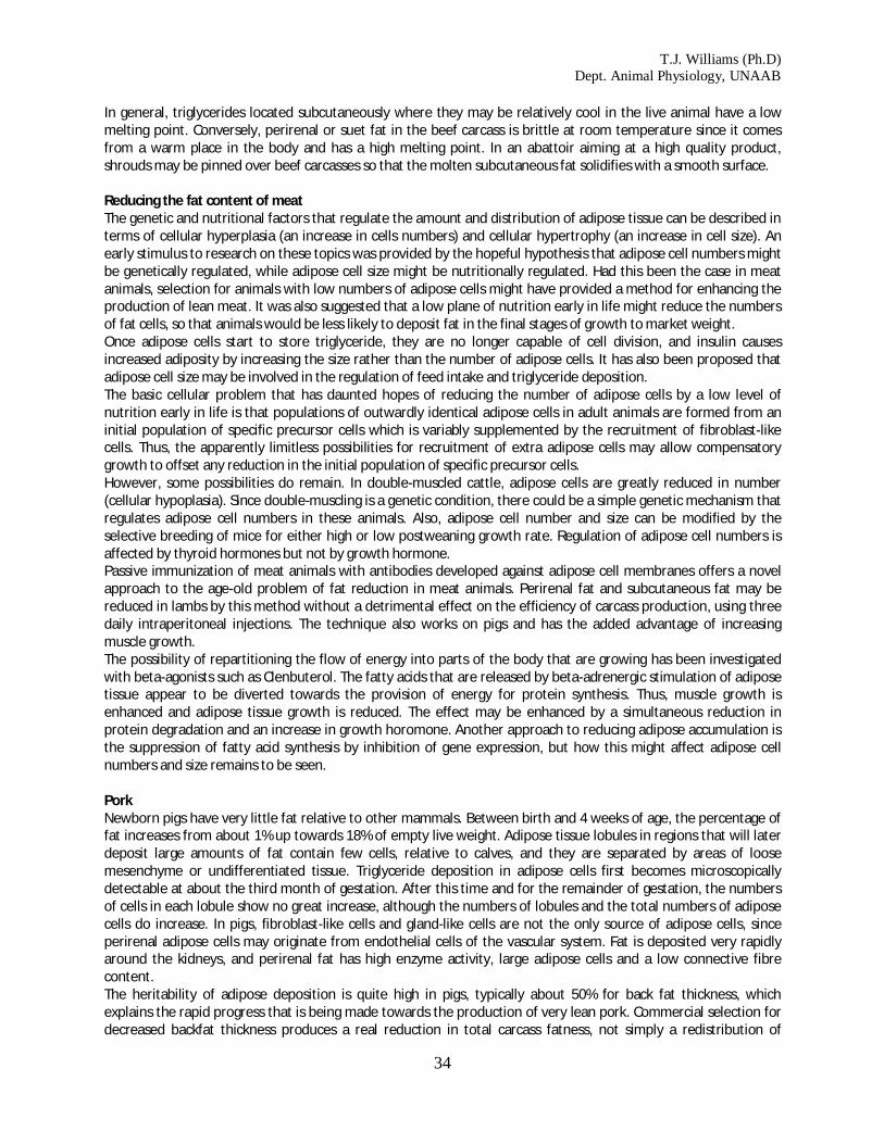
T.J. Williams (Ph.D) Dept. Animal Physiology, UNAAB
34
In general, triglycerides located subcutaneously where they may be relatively cool in the live animal have a low melting point. Conversely, perirenal or suet fat in the beef carcass is brittle at room temperature since it comes from a warm place in the body and has a high melting point. In an abattoir aiming at a high quality product, shrouds may be pinned over beef carcasses so that the molten subcutaneous fat solidifies with a smooth surface. Reducing the fat content of meat The genetic and nutritional factors that regulate the amount and distribution of adipose tissue can be described in terms of cellular hyperplasia (an increase in cells numbers) and cellular hypertrophy (an increase in cell size). An early stimulus to research on these topics was provided by the hopeful hypothesis that adipose cell numbers might be genetically regulated, while adipose cell size might be nutritionally regulated. Had this been the case in meat animals, selection for animals with low numbers of adipose cells might have provided a method for enhancing the production of lean meat. It was also suggested that a low plane of nutrition early in life might reduce the numbers of fat cells, so that animals would be less likely to deposit fat in the final stages of growth to market weight. Once adipose cells start to store triglyceride, they are no longer capable of cell division, and insulin causes increased adiposity by increasing the size rather than the number of adipose cells. It has also been proposed that adipose cell size may be involved in the regulation of feed intake and triglyceride deposition. The basic cellular problem that has daunted hopes of reducing the number of adipose cells by a low level of nutrition early in life is that populations of outwardly identical adipose cells in adult animals are formed from an initial population of specific precursor cells which is variably supplemented by the recruitment of fibroblast-like cells. Thus, the apparently limitless possibilities for recruitment of extra adipose cells may allow compensatory growth to offset any reduction in the initial population of specific precursor cells. However, some possibilities do remain. In double-muscled cattle, adipose cells are greatly reduced in number (cellular hypoplasia). Since double-muscling is a genetic condition, there could be a simple genetic mechanism that regulates adipose cell numbers in these animals. Also, adipose cell number and size can be modified by the selective breeding of mice for either high or low postweaning growth rate. Regulation of adipose cell numbers is affected by thyroid hormones but not by growth hormone. Passive immunization of meat animals with antibodies developed against adipose cell membranes offers a novel approach to the age-old problem of fat reduction in meat animals. Perirenal fat and subcutaneous fat may be reduced in lambs by this method without a detrimental effect on the efficiency of carcass production, using three daily intraperitoneal injections. The technique also works on pigs and has the added advantage of increasing muscle growth. The possibility of repartitioning the flow of energy into parts of the body that are growing has been investigated with beta-agonists such as Clenbuterol. The fatty acids that are released by beta-adrenergic stimulation of adipose tissue appear to be diverted towards the provision of energy for protein synthesis. Thus, muscle growth is enhanced and adipose tissue growth is reduced. The effect may be enhanced by a simultaneous reduction in protein degradation and an increase in growth horomone. Another approach to reducing adipose accumulation is the suppression of fatty acid synthesis by inhibition of gene expression, but how this might affect adipose cell numbers and size remains to be seen. Pork Newborn pigs have very little fat relative to other mammals. Between birth and 4 weeks of age, the percentage of fat increases from about 1% up towards 18% of empty live weight. Adipose tissue lobules in regions that will later deposit large amounts of fat contain few cells, relative to calves, and they are separated by areas of loose mesenchyme or undifferentiated tissue. Triglyceride deposition in adipose cells first becomes microscopically detectable at about the third month of gestation. After this time and for the remainder of gestation, the numbers of cells in each lobule show no great increase, although the numbers of lobules and the total numbers of adipose cells do increase. In pigs, fibroblast-like cells and gland-like cells are not the only source of adipose cells, since perirenal adipose cells may originate from endothelial cells of the vascular system. Fat is deposited very rapidly around the kidneys, and perirenal fat has high enzyme activity, large adipose cells and a low connective fibre content. The heritability of adipose deposition is quite high in pigs, typically about 50% for back fat thickness, which explains the rapid progress that is being made towards the production of very lean pork. Commercial selection for decreased backfat thickness produces a real reduction in total carcass fatness, not simply a redistribution of

T.J. Williams (Ph.D) Dept. Animal Physiology, UNAAB
35
adipose tissue. However, the backfat of pigs is divided into two or three layers (depending on the position of measurement) and the different layers exhibit some degree of independence in their depth changes in response to selection. The deepest layer may be most responsive. Lean breeds of pigs may have a greater ability than fat breeds to mobilize their fat. Beef An increase in adipose cell numbers is mainly responsible for the postnatal accumulation carcass fat in cattle. As in pigs, visceral adipose tissue in cattle develops before intramuscular adipose tissue. Adipose precursor cells make their greatest contribution to cell numbers viscerally, while the recruitment of fibroblast-like cells is more important intramuscularly. The increase in cell numbers (hyperplasia) is nearly complete in subcutaneous and perirenal depots by approximately 8 months. But, within muscles, progressive cellular recruitment, may continue for much longer, which is how the marbling of beef originates. Also it appears that the balance between adipose cell hypertrophy and hyperplasia might be influenced by animal nutrition. Steers fed hay may produce less total adipose tissue with larger cells than steers fed a high-energy ration. The animals on the high-energy ration may produce more total adipose tissue, but with smaller cells. It is likely that the difference created by this nutritional treatment is simply one of a static adipose cell population versus an actively growing population. Mean cell size might be kept low by the formation of new cells in the active population. The subjective assessment of marbling fat is very important in the commercial evaluation of beef. Our basic problem, as butchers, is that our customers want to buy beef that looks very lean - even completely devoid of marbling. But when we feed them steaks in a restaurant, they always prefer the ones that were moderately or highly marbled. In other words we have a real problem, because our customers have a taste preference for beef that they don't like the look of! Lamb Lambs have little prenatal development of subcutaneous fat, although it develops rapidly immediately after birth. Poultry Avian adipose cells differ from those of farm mammals since they have only a limited capacity for lipogenesis - forming new triglyceride within the cell. Thus, they rely mainly on the capture of circulating lipids that have been synthesized in the liver or released by digestion in the gut. There are marked differences in adipose cell numbers and diameters between layer-type and broiler-type fowl, with the lean broiler-type birds having fewer and smaller cells. An early restriction of nutrient intake, although it inhibits adipose cell hypertrophy, has only a slight effect on adipose cell hyperplasia. Thus, adequate cell numbers for fat deposition are present in birds once they are returned to an ad libitum diet. Growth of the abdominal fat pad in chickens is from a combination of adipose cell hyperplasia and hypertrophy, up to about 12 to 14 weeks, then it continues mainly by hypertrophy. Abnormal development of adipose tissue This is a topic that seems to interest many butchers with a lot of cutting experience. After cutting thousands of normal carcasses, it is always a surprise to find one that is radically different in its fat distribution. The accumulation of adipose cells is a common end result of a number of pathological conditions that affect skeletal muscle. In most of these conditions there is usually some evidence of muscle fibre regeneration. In meat animals, however, it is not uncommon to find that muscle fibres have been replaced by adipose cells without any evidence of muscle fibre regeneration and with no decrease in overall muscle volume. The most appropriate name for this condition is muscular steatosis. Muscular steatosis is most frequent in cattle and pigs, but it may also occur in sheep. Sometimes the occurrence of muscular steatosis is indicated before slaughter by an abnormal gait, but usually the condition is not found until a carcass is butchered. The dividing line between excessive marbling fat and muscular steatosis is sometimes difficult to establish, and it is often only the restriction of muscular steatosis to a single muscle or muscle group in an otherwise poorly marbled carcass that makes it conspicuous. Muscular steatosis sometimes occurs in conditions which suggest that it has been caused by muscle damage or denervation. Strenuous muscle exertion may cause extensive muscle damage, particularly in those muscles that are used when an animal rears up on its hind legs. Steatosis can sometimes be a major problem when it affects a high proportion of pigs in a herd.

T.J. Williams (Ph.D) Dept. Animal Physiology, UNAAB
36
4. Growth of the whole animal Prenatal growth and development
Pig Commercial importance If the start of growth is measured from the time of conception rather than from the time of birth or of hatching, we find that a considerable proportion of the time taken to reach market weight is spent in the uterus or in the egg. Beef 33% Lamb 43% Pork 42% Chicken 31% Obviously, these data are approximate, because of variability in the duration of gestation between breeds and the even greater flexibility of animal age at slaughter, but the figures are close to those of many commercial operations. Bearing in mind that breeding stock must be fed, housed, tended, and paid for this period, these figures have a profound impact on the economics of animal production, not forgetting the money that must be spent to get the stock to the age at which they can reproduce, as well as the time taken to achieve conception! The control that can be exercised over the prenatal growth of farm animals is relatively slight, because the environment created for the foetus by the uterus or cleidoic egg is buffered against changes in the mother's environment. Mammals versus Poultry A cleidoic egg is one that, except for temperature and the exchange of gases, is isolated from its environment by membranes and a shell, as in the chicken's egg. This contrast to the situation in mammals, where the developing foetus receives a continuous external supply of nutrients from the mother's uterus. A mammalian egg has virtually no yolk tissue (oligolecithal) whereas an avian egg contains a lot of yolk (telolecithal). Mammals evolved from reptiles with telolecithal eggs, and so they retain features that indicate that their early embryos once had to develop on a large inert yolk mass. The yolk mass of the poultry egg creates problems because it is thick and viscous, and cannot split into subunits as fast as the small cells of the developing embryo. Phenotypes versus Genotypes
Fig. 38 A pig foetus

T.J. Williams (Ph.D) Dept. Animal Physiology, UNAAB
37
The selective breeding of meat animals should be based on the selection of animals bearing phenotypic characters (those seen in the animal) that are related to the economic yield of meat. The biological linkages between genotypes and phenotypes pass through an epigenetic space in which gene products react among themselves and with environmental factors. The major epigenetic interactions involved in the formation of a new animal take place in the prenatal period so that prenatal development may be the most important, but least accessible phase of meat animal development. Embryo versus Foetus While the newly formed animal is developing its various types of tissues it is called an embryo but, after these tissues are acquired and until birth or hatching, it is called a foetus. Stages of Embryology In a telolecithal egg, cell division starts as a small disk and does not spread very far into the viscous yolk mass on which the disk is located. Cleavage is the process by which a zygote or fertilized ovum subdivides into smaller cells called blastomeres. When a new embryo has subdivided into a ball of eight or more blastomeres, but has not yet formed layers of blastomeres, it is called a morula. The morula starts to form a new animal from a clump cells called the inner call mass (A). Then the morula forms itself into a layer of cells called the trophoblast(B) surrounding a fluid filled space - the blastocoele(C) . The trophoblast contributes to the placenta and is lost at birth. The inner cell mass together with the trophoblast form a blasotocyst. The embryo developing from the inner cell mass becomes roofed-over by amniotic folds that later fuse to form a complete layer - the amnion. Endoderm cells (F; which eventually form the gut and its associated organs like the liver) spread from the inner cell mass over the inner surface of the trophoblast and, at this stage, the blastocyst is said to be bilaminar or two- layered. The blastocyst becomes trilaminar or three-layered when mesoderm cells (E; which eventually will form muscles, bones, and fat) migrate from the inner cell mass spreads between the outer trophoblast layer and the inner endoderm layer. The mesoderm layer then splits internally, and a cavity expands within the mesoderm to become the extra-embryonic coelom(G). Expansion of the extra-embryonic coelom cavity allows the yolk sac and allantois to expand into the coelom cavity from the gut of the embryo. In poultry, the rate of growth of the extra-embryonic membranes may be an

T.J. Williams (Ph.D) Dept. Animal Physiology, UNAAB
38
important factor affecting overall growth so that, in genetic strains of poultry with a high growth rate, the growth of the nutrient-obtaining extra-embryonic membranes is faster than normal. Thus, paradoxically, although overall embryonic growth may be slower in the strain with a high growth rate, this allows growth to proceed at a faster rate than normal because nutrients can be absorbed from the yolk at a faster rate.
Fig. 41. Generalized view of how things fit inside each other:A = embryo, B = coelom, C = yolk sac, D = allantois, E = two layers forming the chorion, F = four layers forming the allantochorion, G = two layers of mesoderm, H = endoderm, and I = ectodermal trophoblast. In chicks, the yolk sac contains the yolk whereas in cattle, sheep and pigs the yolk sac is rudimentary (because the developing mammal gets most of its nutrients from the mother's uterus rather than from stored yolk in a cleidoic egg). In chicks, the allantois is used for respiration and storage of waste products whereas in cattle, sheep and pigs, the allantois is developed to provide vascular communication between the developing animal and its mother. The double layer of cells formed from mesodermal cells pushed up against the ectodermal trophoblast is called the chorion. Likewise, the allantoic membrane is pushed against the chorion to form the allantochorion.
The interface between the allantochorion and the mammalian uterus is increased in surface area by the formation of chorionic villi. In pigs, villi are scattered over the chorion to form a diffuse placenta whereas, in cattle and sheep, villi are grouped into about 100 separate mushroom-like structures called cotyledons.
Fig. 42. Membranes around a foetal piglet: A = allantoic cavity, B = amniotic cavity, C = coelom, D = allantois, and E = chorion.

T.J. Williams (Ph.D) Dept. Animal Physiology, UNAAB
39
The space within an avian egg is restricted so that the growth of the fetus and allantois must be balanced against the shrinkage of the yolk sac as its nutrients are used for the developing fetus. The mammalian uterus, however, is capable of considerable expansion, which allows the allantoic cavity to expand enormously. Measurement of Prenatal Growth The most widely used measurement of embryonic and fetal growth is called the crown-rump or CR length. This measurement is a straight line taken from the crown of the head to the base of the tail. The crown is the point midway between the eye orbits. Growth in weight is usually more variable than CR length, so CR length provides is more reliable estimate than weight when attempting to estimate the age of a fetus. Head to Tail Development Embryonic growth and development proceed in an anterior to posterior sequence (from head to tail) so that the head reaches a relatively large size early in development. During fetal development, the remainder of the body catches up to, and overtakes the growth of the head. The head to body ratio, however, is rather variable between animals. The feet and the tail develop last. This pattern of differential growth is called allometric growth (change in body proportions). Compare this 20 to 25 mm pig embryo
with this 45 to 50 mm embryo.
looking at the differences in relatives sizes of the head, body, and limbs.
Fig. 43. Growing pig embryo
Fig. 45. Growing pig embryo (Bigger)

T.J. Williams (Ph.D) Dept. Animal Physiology, UNAAB
40
Now compare this 9-day chick
with this 12 day chick. using the scale to correct for differences in magnification. See any feathers developing? Slow Growth Severly inadequate maternal nutrition or competition between fetuses may inhibit skeletomuscular growth. In such competitive situations, males sustain "less inhibition of growth than females. Differences in body weight between twin and triplet lambs are related to differences in degree of placental development and position in the uterus. In prolific ewes, high fetal losses early in development are associated with the suboptimal growth of surviving fetuses. Sows Have Litters Sows are multiparous (they normally have a litter of ten or more rather than one or two fetuses). Wild pigs have a longer gestation period (130 days) and fewer offspring per litter than modern commercial pigs, so that selection for larger litters may have decreased the length of gestation in pigs. Pig breeds with few piglets per litter have embryos with low embryonic growth rate and fewer cells. Fetal piglets compete with each other for the nutrients made available by the sow's uterus, and the success of a fetus is related to its position within the uterus. Selection for muscle growth in pigs may have resulted in piglets that are heavier but less mature at birth. Longitudinal growth of bones depends more on nutritional status and fetal body weight than on the age of the fetus. Maturation of the epiphyses, however, depends more on age than on nutritional status. The muscles of runt pigs are smaller than those of their more successful littermates. Runt pigs appear to have fewer muscle fibers, particularly white fibers, most of which are formed in the late fetal phase of fiber mass production. The smaller pigs of a litter, if they are able to compete successfully for milk after birth, may be able to catch up in their growth by the time of weaning. Below a birth weight of about 1 kg, however, runt piglets are doomed to produce carcasses with less muscle and extra fat. Splayleg Piglets Sows sometimes sit on their unwary offspring and kill them, and any degree of locomotor impairment in baby pigs increases this risk. Splayleg is a condition in which the hindlimbs and sometimes the forelimbs of a newborn pig temporarily are unable to support the body. If affected pigs are nursed through the period of high risk just after farrowing, they may compensate by enhanced synthesis of muscle proteins and then may grow normally. Slippery floors make the splayleg problem worse. Affected pigs may be helped with a loose coupling between the hindlimbs to prevent the limbs splaying outwards.
Fig.46. 9-day chick embryo
Fig. 47. 12-day chick embryo
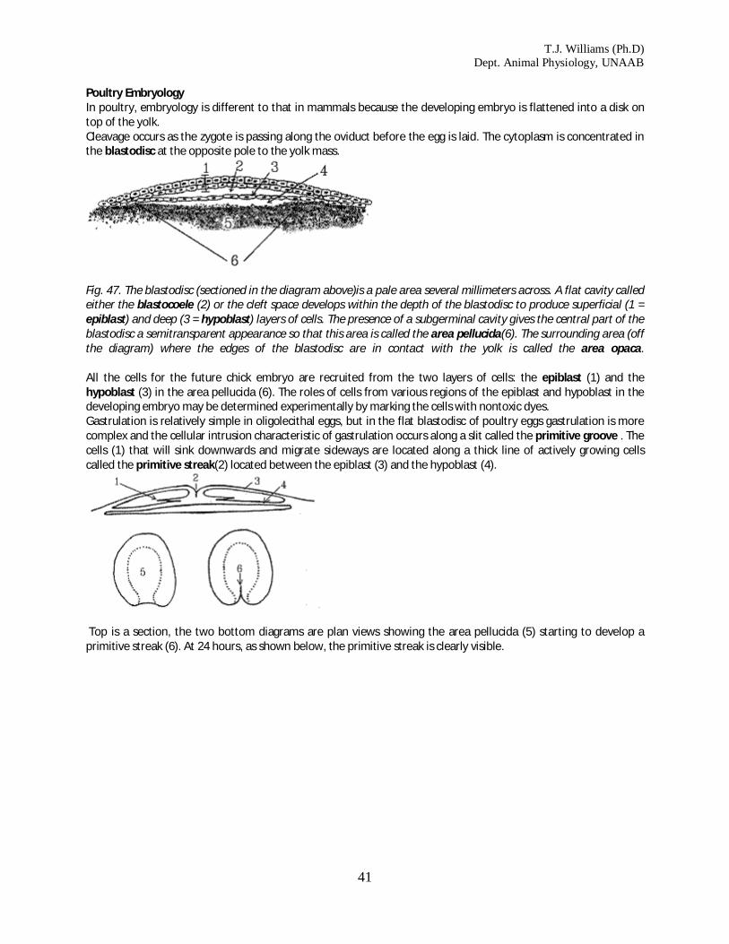
T.J. Williams (Ph.D) Dept. Animal Physiology, UNAAB
41
Poultry Embryology In poultry, embryology is different to that in mammals because the developing embryo is flattened into a disk on top of the yolk. Cleavage occurs as the zygote is passing along the oviduct before the egg is laid. The cytoplasm is concentrated in the blastodisc at the opposite pole to the yolk mass.
Fig. 47. The blastodisc (sectioned in the diagram above)is a pale area several millimeters across. A flat cavity called either the blastocoele (2) or the cleft space develops within the depth of the blastodisc to produce superficial (1 = epiblast) and deep (3 = hypoblast) layers of cells. The presence of a subgerminal cavity gives the central part of the blastodisc a semitransparent appearance so that this area is called the area pellucida(6). The surrounding area (off the diagram) where the edges of the blastodisc are in contact with the yolk is called the area opaca. All the cells for the future chick embryo are recruited from the two layers of cells: the epiblast (1) and the hypoblast (3) in the area pellucida (6). The roles of cells from various regions of the epiblast and hypoblast in the developing embryo may be determined experimentally by marking the cells with nontoxic dyes. Gastrulation is relatively simple in oligolecithal eggs, but in the flat blastodisc of poultry eggs gastrulation is more complex and the cellular intrusion characteristic of gastrulation occurs along a slit called the primitive groove . The cells (1) that will sink downwards and migrate sideways are located along a thick line of actively growing cells called the primitive streak(2) located between the epiblast (3) and the hypoblast (4).
Top is a section, the two bottom diagrams are plan views showing the area pellucida (5) starting to develop a primitive streak (6). At 24 hours, as shown below, the primitive streak is clearly visible.

T.J. Williams (Ph.D) Dept. Animal Physiology, UNAAB
42
The primitive streak develops after the egg is laid and by about 16 hours of incubation is the most conspicuous feature of the developing embryo. The primitive streak indicates the location of the future vertebral axis. The mesoderm develops from a middle layer of cells that accumulates and spreads sideways below the primitive groove.
After the longitudinal axis of the chcik is established, primitive streak activity is concluded. The embryo is lifted off the yolk by the development of infoldings below the future head and tail regions of the embryo. These infoldings unite along the sides of the embryo so that its connection to the yolk is progressively restricted.
Fig. 48. Growing chick embryo

T.J. Williams (Ph.D) Dept. Animal Physiology, UNAAB
43
The amnion grows up and over, all around the embryo, and the amniotic folds then fuse to form a tent-like roof over the embryo.
Fig. 51. Sectioned chick embryos showing how the neural tube rolls up and sinks downwards: 1 = neural tube, 2 = epidermis, 3 = somite, 4 = intermediate mesoderm, 5 = somatic mesoderm, 6 = coelom, 7 = splanchnic mesoderm, and 8 = notochord. The notochord establishes the future axis of the vertebral column and is formed from some of the mesodermal cells in a middle layer between the ectoderm and the endoderm along the primitive streak. Formation of the notochord starts anteriorly and then posterior epiblast cells are recruited as the notochord grows posteriorly. Not all of the mesoderm within the primitive streak is used for notochord formation and along each side of the notochord there remains a long strip of mesoderm that develops into somitic and intermediate mesoderm. Laterally, the remaining mesoderm splits into outer somatic mesoderm and inner splanchnic mesoderm. By this time, the nerve cord has developed from a sunken roll of ectoderm sunken located above the notochord. Embryonic Induction The phenomenon of embryonic induction is still one of the great mysteries of animal development, although very rapid advances are being made in this field as the various genes that control develop are discovered. For example, one recent discovery is the gene that controls asyymetric development between left and right sides of the body (for example, why the heart is normally on the left side of the body while the appendix is on the right side). Most classical scientific names are rather difficult and boring, but in trendy new areas of science like particle physics and genetic engineering things are a little different: the newly discovered gene controlling asymmetric development is called "Sonic Hedgehog". Understanding induction eventually may enable scientists to manipulate muscle development in the meat animals but, so far, attempts to identify the biochemicals responsible for embryonic induction have failed because of the infinitesimally small volumes involved. At present, we should be cautious in assuming that transmissible substances are solely responsible, because the messages that cause induction may be transmitted or faciliated by other means of cellular communication. However, the messengers of embryonic induction are most likely to be ribonucleoproteins. Embryology of Farm Mammals Duration of prenatal life To keep things simple, we are going to call hatching - birth. This will enable us to use prenatal to indicate events occurring before birth in mammals and before hatching in poultry, and postnatal for events after birth or hatching. The duration of the prenatal period is very important. Obviously, the producer needs a good idea of when new animals will be born. A more subtle point is the length of the prenatal period determines the cost of final products. To produce a new calf takes a certain length of time - a time during which all the costs of production must be paid by the producer. Most of the animal's live weight is obtained by postnatal growth. Thus, veal must be more expensive per kilogram than beef - the prenatal period is the same for both, but beef gets a magnifier effect from longer postnatal growth. Lamb must be more expensive per kilogram than mutton.
Stage of development (Days) Beef Lamb Pork Chicken
Forelimb bud 24 20 16-17 2.2

T.J. Williams (Ph.D) Dept. Animal Physiology, UNAAB
44
Hind limb bud 26 21 17-18 2.2
Foetus 45 21 20 5
Birth 285 150 112 22
Slaughter 850 350 270 70
Prenatal duration % 33 43 42 31
The data in the table are days approximately. Gestation varies between breeds and slaughter animals vary in age, depending on market conditions. But the percentages in the bottom line are close to commercial approximations. Our control over prenatal growth is minimal because the environment created for the foetus by the uterus or cleidoic egg is buffered against the farm environment. A cleidoic egg is one that, except for temperature and the exchange of gases, is isolated from its environment by membranes and a shell, as in poultry. Embryology Oligolecithal versus telolecithal eggs. Mammalian eggs are oligolecithal - they contain very little yolk. Poultry eggs are telolecithal - they contain lots of yolk. In a telolecithal egg, cell division in the relatively small amount of protoplasm does not spread very far into the viscous yolk mass. The future chick develops as a flat disk of cells embedded on the surface of undivided yolk. Mammals have very little yolk but have evolved from reptiles with telolecithal eggs and, thus, they retain features to cope with the problem of a large inert yolk mass. Cleavage. Cleavage is the process by which a zygote (fertilized ovum) is subdivided into smaller cells called blastomeres. Morula. When a new embryo has subdivided into a ball of eight or more blastomeres, but has not yet formed layers of blastomeres, it is called a morula.
Inner cell mass versus trophoblast. The morula starts to form a new animal from the inner cell mass in the morula. The morula also forms a fluid-filled sphere of cells called the trophoblast. The fluid-filled cavity is called the blastocoel. The trophoblast contributes to the placenta and is lost at birth. The inner cell mass together with

T.J. Williams (Ph.D) Dept. Animal Physiology, UNAAB
45
the trophoblast is called a blasotocyst. The embryo will develop from the inner cell mass roofed-over by amniotic folds - these later fuse. Endoderm from the inner cell mass spreads over the inner surface of the trophoblast and, at this stage, the blastocyst is said to be bilaminar. The blastocyst becomes trilaminar when mesoderm from the inner cell mass spreads between the outer trophoblast layer and the inner endoderm layer. The mesoderm layer then splits within itself to form a cavity within the mesoderm, the extra-embryonic coelom.
Expansion of the extra-embryonic coelom allows the yolk sac and allantois to expand into the coelom from the gut of the embryo. The rate of growth of the extra-embryonic membranes may be an important factor affecting overall growth. Thus, in animals with a high growth rate, growth of the nutrient-obtaining extra-embryonic membranes is faster than normal. Initially, embryonic growth is slow in animals with a high growth rate, but later it proceeds faster than normal as nutrients are assimilated at a faster rate.
In chicks, the yolk sac contains the yolk whereas in cattle, sheep and pigs the yolk sac is rudimentary. In chicks, the allantois is used for respiration and storage of waste products whereas in cattle, sheep and pigs, the
allantois enables vascular communication between the developing animal and its mother. The double layer of cells formed by the mesoderm and the ectodermal trophoblast is called the chorion. The allantoic membrane is pushed against the chorion to form the allantochorion. The interface between the allantochorion and the mammalian uterus is increased in surface area by the formation
of chorionic villi. In pigs, villi are scattered over the chorion (diffuse placenta) whereas, in cattle and sheep, villi are grouped into
about 100 patches called cotyledons.
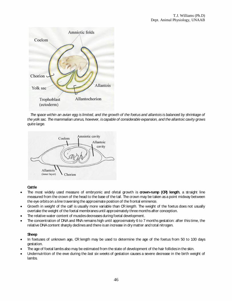
T.J. Williams (Ph.D) Dept. Animal Physiology, UNAAB
46
The space within an avian egg is limited, and the growth of the foetus and allantois is balanced by shrinkage of the yolk sac. The mammalian uterus, however, is capable of considerable expansion, and the allantoic cavity grows quite large.
Cattle
The most widely used measure of embryonic and ofetal growth is crown-rump (CR) length, a straight line measured from the crown of the head to the base of the tail. The crown may be taken as a point midway between the eye orbits on a line traversing the approximate position of the frontal eminence.
Growth in weight of the calf is usually more variable than CR length. The weight of the foetus does not usually overtake the weight of the foetal membranes until approximately three months after conception.
The relative water content of muscles decreases during foetal development. The concentration of DNA and RNA remains high until approximately 6 to 7 months gestation: after this time, the
relative DNA content sharply declines and there is an increase in dry matter and total nitrogen. Sheep
In foetuses of unknown age, CR length may be used to determine the age of the foetus from 50 to 100 days gestation.
The age of foetal lambs also may be estimated from the state of development of the hair follicles in the skin. Undernutrition of the ewe during the last six weeks of gestation causes a severe decrease in the birth weight of
lambs.

T.J. Williams (Ph.D) Dept. Animal Physiology, UNAAB
47
Embryonic growth and development proceed in an anterior to posterior sequence with the head reaching a relatively large size early in development. During foetal development, the remainder of the body catches up to, and overtakes the growth of the head. The head to body ratio, however, is rather variable between animals.
The feet and the tail develop last. Birth weight has a strong effect on the early postnatal growth rate of lambs which slowly diminishes with age. Competition between twin and triplet foetuses may inhibit growth. In competitive situations, males sustain less
inhibition of growth than females. Pigs
Sows are multiparous and usually have ten or more offspring in each litter. Wild pigs have a longer gestation period (130 days) and fewer offspring per litter than modern commercial pigs, so
selection for larger litters may have decreased the length of gestation. Breeds with few piglets per litter have embryos with low embryonic growth rate and fewer cells. Foetuses compete with each other for the nutrients made available by the uterus, and the success of a foetus is
related to its position within the uterus. Selection for muscle growth in pigs may have resulted in piglets being heavier but less mature at birth. Longitudinal growth of bones depends more on nutritional status and foetal body weight than on the age of the
foetus. Runt pigs (the smallest in a litter) have fewer myofibres than normal. If runts are able to compete successfully for milk after birth, may be able to catch up in their growth by the time of
weaning. But, below a birth weight of about 1 kg, runt piglets are doomed to produce carcasses with less muscle and extra
fat than normal. Splayleg is a condition in which newborn piglets cannot bring their hindlimbs together normally. Thus, their
locomotion is impaired and they may be crushed by the sow. Measurement of growth METHODS, PROS & CONS CAT and NMR Medical methods for the non-destructive analysis of body composition, such as computer-assisted tomography (CAT) and nuclear magnetic resonance (NMR) imaging, can be used to measure the growth of meat animals, but their extremely high cost and complexity restrict these methods to research. They would be extremely difficult to apply routinely in commercial agriculture and could not be cost effective. Ultrasonics Ultrasonic imaging is widely used for humans, and it is far, far less expensive than CAT or NMR. Ultrasonic measurements of fat and muscle thickness generally are quite reliable and are used routinely in commercial agriculture.
High frequncy sound waves are generated by a transducer on the animal's back and, since the velocity of the waves though the tissue is known, the time taken for echoes to return from tissue boundary layers may be used to determine their depth.
Strong echoes are returned from the muscle-fat boundary over the longissimus dorsi muscle, but other echoes are returned from connective tissue septa in the subcutaneous adipose tissue and from the skeleton.
Depth measurements may be assembled into a map of the carcass section by moving the
transducer in an arc over the animal's back.

T.J. Williams (Ph.D) Dept. Animal Physiology, UNAAB
48
An alternative ultrasonic method is to measure changes in the velocity of the signal as it passes through the tissue, since the proportion of adipose tissue is correlated with the reciprocal of velocity.
Photogrammetry The relative development of commercially valuable carcass components such as the loin and hindquarter is difficult to assess subjectively in the live animal, but may be done by photogrammetry. This utilises the parallax effect of a stereo photograph compounded from two photographs taken simultaneously by two cameras separated by a known distance. The volume of the animal body or anatomically defined parts of the body may be calculated from the degree of the stereo effect. Although this method may be accurate, its cost and complexity have so far limited its use. Specific gravity The relative development of the carcass also may be studied using specific gravity. Instead of being immersed in water by the classical method of Archimedes, the air displacement technique uses negative air pressures around an animal in an experimental chamber. The accumulation of fat (low density) decreases the overall density of the body so that density then provides an index of fatness. In practice, however, it is difficult to obtain accurate volumetric measurements that allow for the offsetting effects of bones. In cattle, bone density increases with age and varies between sexes and breeds. Water content An alternative method for adipose tissue volume is based on the fact that adipose tissue has a very low water content. Chemicals such as antipyrin or one of its derivates are injected into the vascular system from whence they diffuse into the total volume of aqueous body fluids. Provided that the injected substance is only slowly metabolized or excreted by the body, and provided that it becomes adequately dispersed, the extent to which the injected substance becomes diluted indicates the total diluting volume. In general, fat animals have a proportionately smaller aqueous volume than lean animals. Isotope methods Another indirect method of estimating body composition is based on the fact that nearly all the potassium in the body is intracellular. Since adipose cells contain only a trace of potassium in their scanty amount of cytoplasm, the total amount of potassium in the body is approximately proportional to the lean body mass.
A constant proportion (0.012%) of potassium atoms are radioactive (the isotope 40K), and they emmit gamma rays that may be converted into small flashes of light and counted electronically.
If an animal is shielded from environmental gamma rays, its total gamma emission gives an index of its total muscle mass. A background count of the extrinsic radiation that penetrates the shield is subtracted from the count made during the three minutes when the animal is being measured.
Animals are taken off their feed and water for 24 hours prior to being measured to avoid
complications from potassium in the rumen, but different types of animals require different calibration factors from which to predict their muscle mass. Individual muscles of the carcass also differ in their concentration of potassium.
Radioactivity also may be employed in a similar manner using isotopes of hydrogen (deuterium or tritium) to estimate the water volume and, indirectly, the fat content of a live animal. The advantage of deuterium is that it is not radioactive, while the advantage of tritium is that it may be measured rapidly by liquid scintillation. Electromagnetic methods The volume of electrolytes in the animal body is estimated from the disturbance that their electrical conductivity creates in an externally-applied electromagnetic field. However, this system is adversely affected by any variation of the animal's position and orientation within the electromagnetic field and, consequently, it is not accurate

T.J. Williams (Ph.D) Dept. Animal Physiology, UNAAB
49
enough for live animals. However, it works very well for boxed beef and carcasses that can be neatly lined up on a conveyer to pass through the machine - but that's another story. Serial slaughter experiments After an animal has been slaughtered its composition may be determined very accurately. Thus, a group of animals can be slaughtered one at a time throughout their growth to assemble a pseudo-growth curve. The major problem is the degree of variation between individual animals, the high cost involved, and the fact that all the animals are dead when the experiment is finished. The method can be used with the off-spring of expensive breeding stock (progeny testing), but all this takes a long time and is one step removed from the animal of interest - usually a prize bull or boar.
Growth Curves
Measuring Growth Absolute measurements of body composition can only be made after an animal has been killed and then, of course, further growth is impossible. There are several solutions to this problem, but none of them is entirely satisfactory.
Firstly, the animals may be kept alive and allowed to grow, and the meat yield measured with a non-destructive method of limited accuracy. If nondestructive measurements of growth are made from animals kept alive and remeasured at intervals, the data require careful statistical analysis since each measurement on the same animal is partially related to preceding and succeeding measurements.
As a second solution, a homogeneous group of animals may be individually slaughtered in a sequence that enables the growth attainment of living animals to be visualized. This is the serial slaughter method. The problem here is any differences between the animals are superimposed on the visualized growth sequence. For example, a slow-growing animal slaughtered in sequence after a fast-growing animal may lead to a dip in the reconstituted growth curve.
A third solution is often used in the comparison of breeds or diets: the animals are grown to a set point, slaughtered, and then compared. But then the problem is to identify an equivalent slaughter point at which comparisons may be made. Animals may be slaughtered when they reach a set age, degree of fat deposition, or live weight, but these may differ between breeds or sexes, and different slaughter end points may give different results.
Although studies of carcass composition have an important place in agricultural research, they are not the best means of investigating the process of growth itself. Linear measurements are largely a reflection of skeletal development rather than muscular development, and it is difficult to make accurate skeletal measurements on large animals when they are still alive. With poultry, however, the measurement of linear dimensions may be useful for studying growth and as a basis for genetic selection. Curvilinear measurements made round the girth of the hind-limb of a meat animal are a reasonable index of muscle development, but these are difficult to standardize.

T.J. Williams (Ph.D) Dept. Animal Physiology, UNAAB
50
20.3 Surface to volume ratio Imagine a simple geometrical structure such as a sphere: if it increases its height, its surface area will increase in proportion to the square of height, while its volume will increase in proportion to the cube of height. If isolated animal cells are grown in suspension in a constantly replenished medium, they form balls of cells. These grow up to several millimetres across, until they reach the limit to which radial diffusion and surface to volume ratio may supply nutrients and oxygen, and the limit at which waste products may still be removed from the center of the ball. If these cells were primitive life forms early in geological time, little further evolutionary increase in size would be possible until they had solved these problems. One solution is gastrulation, to increase the surface to volume ratio by becoming cup-shaped (like pushing a finger into a hollow, soft ball). Another solution is to develop a blood vascular system to enhance the transport of nutrients, oxygen and waste materials. Even for a ball of cells growing in a laboratory dish, however, there are often subtle factors limiting or regulating growth. Biophysical studies with microelectrodes have revealed mechanisms by which cells communicate with each other and "agree" on which cells should divide.
As meat animals grow from birth to slaughter weight, they do not maintain a constant shape. In general, however, even a small increase in linear dimensions causes a proportionately greater increase in body weight because body weight is a function of volume. In newborn farm animals, the rate of heat loss from the body may be a serious problem. In small animals, the surface area for heat loss is relatively high, while the volume of muscle capable of generating heat is relatively small. Since animals maintain an approximately constant body temperature during growth, there must be subtle interactions between body size and the mechanisms which regulate body temperature. Basal metabolism (energy use in a resting animal) is proportional to body weight raised to the power of 0.75, rather than to the power of 0.66, which would be expected on simple geometrical grounds.
20.4 Growth and development - how they differ Among meat animals, it is difficult to find a situation in which an animal may grow to any marked extent without also exhibiting development of one type or another. Whereas growth may be considered as an increase in height, length, girth or weight, development may be considered as a change in composition, structure or ability, although neither definition is completely satisfactory.
In functional systems such as the locomotory system, development may occur in direct response to growth in live weight. For example, as an animal grows heavier, it may need to use more of its muscle mass to oppose gravity, and there may be changes in the physiological properties of the diverted muscle mass. Similar functional changes occur in the digestive system, bones and other body systems. After growth has ceased in the adult, developmental changes continue as the animal passes through maturity to senescence. Developmental changes are directed towards the attainment of a mature composition, structure or ability. However, the retrogressive changes that occur later in life and which are associated with a decline in composition, structure and ability, are also developmental changes (senescence). Development and senescence are

T.J. Williams (Ph.D) Dept. Animal Physiology, UNAAB
51
merely the early and late stages of ontogeny, the progress of an individual animal through its life cycle.
Although some aspects of growth such as fat deposition appear to be reversible, this is rarely true of developmental processes. As an animal is growing, a vast number of developmental changes are taking place concurrently. These changes are not usually undone if an animal simply loses weight. For example, a reversible accumulation of the triglyceride stored in fat cells may be accompanied by an increase in the number and size of fat cells, but the loss of a moderate amount of triglyceride may occur by all cells releasing a share of their triglyceride. Thus, after "degrowth" appears to have occurred, the animal has not reverted to the developmental state (adipose cell number) it had before the period of growth started.
20.5 Objective measures of growth CAT and NMR Medical methods for the non-destructive analysis of body composition, such as computer-assisted tomography (CAT) and nuclear magnetic resonance (NMR) imaging, can be used to measure the growth of meat animals, but their extremely high cost and complexity restrict these methods to research. They would be extremely difficult to apply routinely in commercial agriculture and could not be cost effective.
Ultrasonics
Ultrasonic imaging is widely used for humans, and it is far, far less expensive than CAT or NMR. Ultrasonic measurements of fat and muscle thickness generally are quite reliable and are used routinely in commercial agriculture.
High frequency sound waves are generated by a transducer on the animal's back and, since the velocity of the waves though the tissue is known, the time taken for echoes to return from tissue boundary layers may be used to determine their depth.
Strong echoes are returned from the muscle-fat boundary over the longissimus dorsi muscle, but other echoes are returned from connective tissue septa in the subcutaneous adipose tissue and from the skeleton.

T.J. Williams (Ph.D) Dept. Animal Physiology, UNAAB
52
Depth measurements may be assembled into a map of the carcass section by moving the transducer in an arc over the animal's back.
An alternative ultrasonic method is to measure changes in the velocity of the signal as it passes through the tissue, since the proportion of adipose tissue is correlated with the reciprocal of velocity.
Photogrammetry The relative development of commercially valuable carcass components such as the loin and hindquarter is difficult to assess subjectively in the live animal, but may be done by photogrammetry. This utilizes the parallax effect of a stereo photograph compounded from two photographs taken simultaneously by two cameras separated by a known distance. The volume of the animal body or anatomically defined parts of the body may be calculated from the degree of the stereo effect. Although this method may be accurate, its cost and complexity have so far limited its use.
Specific gravity
The relative development of the carcass also may be studied using specific gravity. Instead of being immersed in water by the classical method of Archimedes, the air displacement technique uses negative air pressures around an animal in an experimental chamber. The accumulation of fat (low density) decreases the overall density of the body so that density then provides an index of fatness. In practice, however, it is difficult to obtain accurate volumetric measurements allowing for the offsetting effects of bones. In cattle, bone density increases with age and varies between sexes and breeds.
Water content
An alternative method for adipose tissue volume is based on the fact adipose tissue has a very low water content. Chemicals such as antipyrin or one of its derivatives are injected into the vascular system from whence they diffuse into the total volume of aqueous body fluids. Provided the injected substance is only slowly metabolized or excreted by the body, and provided it becomes adequately dispersed, the extent to which the injected substance becomes diluted indicates the total diluting volume. In general, fat animals have a proportionately smaller aqueous volume than lean animals.
Isotope methods
Another indirect method of estimating body composition is based on the fact nearly all the potassium in the body is intracellular. Since adipose cells contain only a trace of potassium in their scanty amount of cytoplasm, the total amount of potassium in the body is approximately proportional to the lean body mass.
A constant proportion (0.012%) of potassium atoms are radioactive (the isotope 40K), and they emit gamma rays that may be converted into small flashes of light and counted electronically.
If an animal is shielded from environmental gamma rays, its total gamma emission gives an index of its total muscle mass. A background count of the extrinsic radiation that

T.J. Williams (Ph.D) Dept. Animal Physiology, UNAAB
53
penetrates the shield is subtracted from the count made during the three minutes when the animal is being measured.
Animals are taken off their feed and water for 24 hours prior to being measured to avoid complications from potassium in the rumen, but different types of animals require different calibration factors from which to predict their muscle mass. Individual muscles of the carcass also differ in their concentration of potassium.
Radioactivity also may be employed in a similar manner using isotopes of hydrogen (deuterium or tritium) to estimate the water volume and, indirectly, the fat content of a live animal. The advantage of deuterium is it is not radioactive, while the advantage of tritium is it may be measured rapidly by liquid scintillation.
Electromagnetic methods
The volume of electrolytes in the animal body is estimated from the disturbance that their electrical conductivity creates in an externally applied electromagnetic field. However, this system is adversely affected by any variation of the animal's position and orientation within the electromagnetic field and, consequently, it is not accurate enough for live animals. However, it works very well for boxed beef and carcasses that can be neatly lined up on a conveyer to pass through the machine.
20.6 Growth curves
The sigmoid curve
If animal height or weight are measured from conception to senescence, the data usually follow a flattened "S" shape called the sigmoid curve. But the growth curves of meat animals raised under commercial conditions may appear as relatively flat slopes (the middle segment of the flat "S"), and the sigmoid shape may only become apparent if the data include very young animals or animal beyond a typical market weight. In other words, growth velocity is approximately constant during the commercial growing period.
Above is a sigmoid liveweight growth curve for Holsteins. The "S" shape is far from being exactly "S" shaped.

T.J. Williams (Ph.D) Dept. Animal Physiology, UNAAB
54
Acceleration and deceleration At first, the fertilized ovum divides mitotically with little or no growth in mass. However, as soon as it develops the means of assimilating energy, the growth of the embryo accelerates. At birth and weaning there may be a temporary deceleration of growth as an animal switches from one source of nutrients to another. Except for a slight acceleration at puberty, subsequent growth maintains a steady average velocity until the terminal deceleration as animals reach their mature size. However, although the average velocity may be smooth, there may be an underlying circadian (daily) periodicity in growth rate or even erratic spurts of growth.
Above is the velocity curve derived from the sigmoid growth curve for Holsteins.
Anabolism versus catabolism Anabolism is the building up of body components, while catabolism is their destruction.
All live animals show both anabolism and catabolism. If anabolism exceeds catabolism, the animal is growing. If catabolism exceeds anabolism, the animal is wasting away. If anabolism equals catabolism, animal weight is constant.
Protein turnover Protein turnover is the time between anabolism and catabolism of an individual protein. If protein turnover rates are measured in growing meat animals, the half-life for protein
synthesis is shorter than the half-life for protein catabolism. The half-life is used, as in studies of radioactivity, because it may take an impossibly long
time for 100% to turnover, so we only wait for 50%. For meat animals, the half-lives of the important meat proteins are around
several days in length, with muscle proteins being much faster than connective tissue proteins.
In the muscles of young animals, both the rates of protein synthesis and degradation are faster than in older animals. In the muscle tissue of growing animals, insulin is the most important factor that inhibits the degradation of myofibrillar proteins when anabolism exceeds catabolism. Epinephrine and serotonin have similar but minor effects.

T.J. Williams (Ph.D) Dept. Animal Physiology, UNAAB
55
In fasting animals, where catabolism exceeds anabolism, proteolysis is promoted by glucocorticoids.
von Bertalanffy's equation This is based on the concept each increment to body weight (W) represents the triumph of anabolism over catabolism,
dW/dt = a.Wb - c.Wd
body weight increment = anabolic increment - catabolic loss This yields values for parameters b and d between 2/3 and 1, much like the power relationships of body mass or volume to body surface area. Heat loss tends to be proportional to skin area, while gut areas may be rate limiting for nutrient uptake. Both heat generation and anabolic systems tend to be related to body volume. The von Bertalanffy growth equation requires some integration over time since the rates of anabolism and catabolism may have short-term fluctuations.
The curve above shows a typical shape for a von Bertalanffy growth curve. Note: it is sigmoidal, it does reach a flat asymptote, but it is not symmetrical.
The occurrence of muscle growth in a meat animal provides clear evidence the average rate of protein synthesis exceeds the average rate of protein degradation.
It must be remembered, however, the inbalance in favour of anabolism may not be very large or very stable. With in vitro measurements of protein metabolism in excised muscle samples there is a serious risk the muscle may have been released from its normal physiological duties and from endocrine and neural control mechanisms responsible for its long-term metabolic control. For example, isolated fast and slow growing muscles from broiler and layer-type poultry may exhibit similar rates of protein anabolism and catabolism when maintained in vitro, despite the occurrence of extreme differences in muscle growth in vivo.
However, when myoblasts from embryos of broiler and layer-type chickens are compared in vitro, the greater capacity of cells from broilers to accumulate protein may be attributable to a lower rate of protein degradation.
Another subtlety complicating the picture is animals with a rapid rate of protein accumulation also may have an elevated rate of muscle protein degradation and it has been proposed that selection for meat yield in poultry should be based on decreasing the rates of degeneration, rather than attempting to increase the rate of synthesis. However,

T.J. Williams (Ph.D) Dept. Animal Physiology, UNAAB
56
one would not want to jeopardize post mortem proteolysis because it makes an important contribution to taste and tenderness.
Energy content Body weight is partitioned between several commercially important compartments. Less energy is required to form a kilogram of lean meat than to form a kilogram of dissectable fat. Thus, a progressive diversion of feed energy away from protein accumulation and towards fat deposition may contribute to the decelerating curve of body weight growth in meat animals, although in consistently fat animals such as pigs this effect may be only slight. The decrease in feed efficiency that occurs with increased live weight in pigs is primarily caused by increased maintenance cost and not increased fat deposition. The time at which the diversion to fat deposition occurs may depend on the breed of animal, and is late in animals with a large adult size. The relative amount of water in the lean tissues of the body decreases with age. The rate of decrease in water content may be altered at weaning and at the onset of sexual maturity.
Body area versus mass In attempting to give a biological meaning to the terms b and d in von Bertalanffy's equation, one cannot simply assume the proportional growth of the surface areas of the body keeps pace with the growth of the whole body. In pigs, for example, the small intestine grows rapidly after birth but reaches its mature length between 5 and 6 months of age. A similar early growth attainment of final size is found in the alimentary tract of cattle. Similarly, with heat loss from the body surface, the increased insulation provided by subcutaneous fat must be taken into account. Various regions of the body may differ in their depth of subcutaneous adipose insulation, and there are considerable differences between breeds in the contribution of each body region to the total body area.
Robertson's autocatalytic equation
There is a resemblance between sigmoid growth curves and those of autocatalytic or self-accelerating chemical reactions in which the product of a simple reaction acts as a catalyst to accelerate further reaction. In a first order chemical reaction, the reaction rate is proportional to only one concentration. For example, in a decomposition reaction the disappearance of the decomposing substance is given by,
-dc/dt = k.c
disappearance rate = reaction constant x substance concentration The initial concentration of the substance (a) is reduced by the amount lost (x) so that its concentration becomes a-x and the equation soon changes to,

T.J. Williams (Ph.D) Dept. Animal Physiology, UNAAB
57
- d(a-x)/dt = k(a-x)
Since da/dt, the derivative of a constant is zero,
dx/dt = k(a-x) Robertson took the case of an autocatalytic reaction where the reaction constant (k) was multiplied by the mass or concentration of the catalyst that was the reaction product itself (x),
dx/dt = kx(a-x) The curve of this reaction was sigmoid and resembled growth curves for body weight (W),
dW/dt = kW(a-W)
Above is an autocatalytic growth curve. Note: it is sigmoidal, does reach a flat asymptote AND is symmetrical. What does symmetrical mean? Imagine you can stick a pin half way along the curve and flip the curve upside down - it will look identical. Its initial acceleration results from the increase of the catalytic reaction product, and its later deceleration results from a depletion of the raw material or starting substance. Robertson was aware of the fact overall body growth was the sum of a very large number of individual reactions rather than a single reaction, but he forced himself to chose between two alternatives: either the similarity of autocatalytic reaction curves and animal growth curves was a mere superficial resemblance or, alternatively, it was a meaningful indication of an underlying "master-reaction" working on autocatalytic principles. Robertson chose the latter alternative on an intuitive basis. It is usually considered there is little or no evidence in support of Robertson's choice, and his logistic equation survives today only as a means of fitting a curve to growth data. It might be wise, however, to refrain from passing judgment on Robertson's intuition until we actually find out how growth is regulated?
Robertson's initial chemical reaction catalyzed itself: animals initially add new cells that form further new cells, and for a while the system may behave like a population of microorganisms proliferating in an abundant nutrient medium. Even under these conditions, however, mitotic rates usually show some deceleration. For example, in chick embryos at the 17-somite stage, the
T.J. Williams (Ph.D) Dept. Animal Physiology, UNAAB
58
number of cells is doubled in 4 hours, but to double the numbers again then takes about 13 hours. As young farm animals grow larger, they consume more feed and store increasing amounts of fat. After a certain point, however, the capacity for energy storage becomes limited by the increasing demands of cellular maintenance and repair, and by the diversion of energy towards reproduction. These self-accelerating and limiting factors work in the same way as those in von Bertalanffy's equation. If the postnatal body tissues of birds and mammals were composed solely of undifferentiated cells like those of the embryo, self-acceleration by cell proliferation might be taken seriously. In meat animals, however, this is not the case.
In the skeletal muscles, massive amounts of protein are assembled into myofibrils. Myofibrils grow in length by forming new sarcomeres at their ends, and they grow radially before they subdivide by longitudinal splitting. New proteins for the myofibril are formed by the activity of ribosomes. Thus, myofibrillar growth per se is not self-accelerating, except in an indirect manner - stronger animals might get more to eat. The growth of adipose tissue is dominated by triglyceride accumulation. Triglyceride accumulation is not self-accelerating, and it provides an even smaller indirect advantage to the animal's growth. Thus, the accelerating phase of growth in meat animals becomes all the more remarkable when we realize that much of the newly added biological material is not self-replicating.
Age-based equations
In the equations considered so far, the final deceleration necessary to create a sigmoid curve has originated from, or has been proportional to the enlarged body mass itself. Brody championed another way of thinking about the control of deceleration - to envisage it a function of time, so the growth rate decreases in simple inverse proportion to animal age
1/W . dW/dt = k/t
specific growth rate = species constant / age
Growth curves constructed in this manner, with animal age as the controlling factor in deceleration, never reach a point of zero growth in older animals. Although perpetual growth may occur in lower vertebrates, until cut short by accidental death, there is a finite upper limit to the size of mammals and birds. In agriculture, age-based growth curves may provide a reasonable fit to growth data since the commercial growing period is in the first half of the sigmoid curve.

T.J. Williams (Ph.D) Dept. Animal Physiology, UNAAB
59
The curve above shows a typical shape for a time-based growth curve. Note - it is NOT sigmoid, it never reaches a flat asymptote and it is not symmetrical.
Instead of growth rate declining in simple inverse proportion to age, it may decline in logarithmic proportion, as in the equation proposed by Gompertz way back in 1825 to calculate financial returns on monetary investments! Thus, in a Gompertz relationship, the logarithm of specific growth rate plotted against time yields a negative linear slope. The Gompertz curve is asymmetric about its inflection point, which occurs early in the curve while the logistic curve is symmetrical.
Linear growth - the flat part of the "S" During the commercial growth of meat animals to market weight, growth may appear almost linear with a constant velocity. During this time, myofibrils, triglyceride and bone matrix accumulate passively by accretionary growth, and the constraints to growth have no measurable impact that may be isolated from sampling error and experimental error. This biologically distinct period of linear growth may be isolated from its sigmoid context to improve the fit of predicted curves to actual data.
20.7 Chronological versus physiological age In the comparison of breeds differing in their rate of physiological development, it is difficult to justify the use of chronological age rather than physiological age to predict the decline in the logarithm of the specific growth rate. Another problem with time as the regulatory parameter of growth is it makes no allowance for periods when an animal may cease growing because of restricted nutrient intake or stress. This problem does not arise with body weight as the regulatory parameter, and it could also be avoided by using physiological age rather than chronological age. There are, however, no simple units with which to measure physiological age.
The concept of physiological age has been used to create a physiological time scale called metabolic age, based on the relationship between mature size and the chronological time taken to reach it. The chronological time taken to reach mature weight is made proportional to the mature weight raised to the 0.27th power so that the units of metabolic age are those of

T.J. Williams (Ph.D) Dept. Animal Physiology, UNAAB
60
chronological age divided by the 0.27th power of mature weight. Another approach to the problem of measuring physiological age is to evaluate animals on the basis of a set of criteria changing as animals grow older. Some of the criteria suggested are:
physical and chemical tests of the degree of cross linking in collagen, elasticity of the aorta, accumulation of lipofuscin brain cell pigment, and the mitotic potential of cultured cells.
Genetic selection for leanness in pigs may have delayed their physiological maturation. Thus, genetic selection might be more effective if animals are given a high plane of nutrition and suitable environmental conditions for optimal growth. In model systems, however, the responses to selection appear to be more complex, and genetic advances made under optimal conditions may not automatically be expressed if the animals are switched from optimal to suboptimal environmental growth restraints. One aspect of this problem is physiological maturation may be delayed in a population whose growth is environmentally restrained.
20.8 Growth rhythms Intrinsic seasonal rhythms in muscle growth are difficult to identify in farm animals reaching market weight in less than a year since intrinsic rhythms are difficult to isolate from seasonal reproductive cycles and seasonal cycles in the nutrient quality of animal feed. In laboratory animal experiments, intrinsic seasonal changes in rates of growth and cellular regeneration have been found to persist even in animals kept under constant conditions of temperature and day length. Seasonal rhythms occur in milk and egg production. In Scottish free-grazing sheep, the average empty body weight of ewes peaks in November and reaches a minimum at the end of April, with subcutaneous fat providing the primary energy reserve depleted during gestation.
20.9 Effect of light In poultry, light has a profound effect on aspects of animal behavior such as feeding and roosting secondarily affecting growth rate. For example, poultry may be reared at a low intensity of light so they expend less energy on muscular activity, but then they may start to use other clues, such as temperature or sounds of activity to adjust or entrain their circadian rhythm of activity. Chickens exhibit maximum growth if they area fed late in the evening, because then they have all night to work through the feed stored in their crop. But to do this, the chickens must learn how to anticipate the start of the period of darkness, and they may do this more readily if the light intensity is slowly decreased to simulate dusk. The more rapid growth of chickens under these conditions originates from increased feed intake and increased feed efficiency. Chickens also show enhanced growth if kept at a continuously low light intensity, but in this case there is no increase in feed efficiency. The effects of illumination may be found in the levels of thyroxin production in baby chicks. Supplemental illumination also stimulates growth in lambs and cattle, possibly acting via the gonads since stimulation of growth may occur in heifers but not in steers.

T.J. Williams (Ph.D) Dept. Animal Physiology, UNAAB
61
1.6 Range in size & complexity The live animal is slaughtered to produce a carcass composed of edible meat (muscle), fat (adipose tissue) and bone. The viscera (guts) are removed at slaughter. Muscle, fat and bone are tissues - this describes their composition rather than their position. Individual muscles and bones have names, whereas fat is described as subcutaneous (under the skin), visceral (around the guts), intermuscular (between muscles) and intramuscular (within muscles). Study hint: do not confuse the two prefixes 'intra-' and 'extra-'. Intra- means within. Inter- means between. Tissues have distinct functions. Muscle tissue contracts to create movement. Adipose tissue stores energy and provides thermal and mechanical insulation. Bone provides support. All tissues are composed of cells, and many are held together by fibres. Cells and fibres are often surrounded by a back-ground material or matrix. Tissues are spread throughout the body and occur at many different locations. Organs are distinct anatomical structures with one or more functions. The heart pumps blood through the vascular system. The lungs add oxygen and remove carbon dioxide from the blood. Organs usually contain several types of tissue - the heart, for example, is dominated by cardiac muscle tissue but also contains vascular tissue (its own blood supply), nervous tissue (to control heart rate), and connective tissue (forming valves, holding the heart together and isolating the heart in the thoracic cavity). Tissues and organs are mostly visible by eye - without a microscope. Individual cells can only be seen with a microscope. Inside cells are organelles - specific parts of the cell with special functions. For example, inside muscle fibres (the fibre is a giant cell with many nuclei), are organelles called myofibrils responsible for contraction. The cell nucleus contains genetic information coded in DNA (deoxyribonucleic acid). Myofibrils are very important in this course - because meat is very important in animal agriculture. Meat is either the primary product (beef, pork, lamb and poultry meat) or the terminal product (dairy cows, wool sheep and spent laying hens) of animal agriculture. Myofibrils are composed of very large proteins just individually visible at the highest magnifications of electron microscopy.