tn5: a molecular window on transposition
TRANSCRIPT
T
WLM*†
R
vopTibi1silh
thoptgaievmbeDgc
3
o4
Biochemical and Biophysical Research Communications 266, 729–734 (1999)
Article ID bbrc.1999.1891, available online at http://www.idealibrary.com on
n5: A Molecular Window on Transposition
illiam S. Reznikoff,*,1 Archna Bhasin,* Douglas R. Davies,*,† Igor Y. Goryshin,*isa A. Mahnke,* Todd Naumann,* Ivan Rayment,*,†indy Steiniger-White,* and Sally S. Twining*,2
Department of Biochemistry, University of Wisconsin, 433 Babcock Drive, Madison, Wisconsin 53706; andInstitute for Enzyme Research, University of Wisconsin, 1710 University Avenue, Madison, Wisconsin 53705
eceived September 28, 1999
DfcsDDertjmifig
tittmpccctttiaiiG
t1tatp
DNA transposition is an underlying process in-olved in the remodeling of genomes in all types ofrganisms. We analyze the multiple steps in cut-and-aste transposition using the bacterial transposonn5 as a model. This system is particularly illuminat-
ng because of the existence of structural, genetic, andiochemical information regarding the two participat-ng specific macromolecules: the transposase and the9-bp sequences that define the ends of the transpo-on. However, most of the insights should be of generalnterest because of similarities to other transposition-ike systems such as HIV-1 DNA integration into theost genome. © 1999 Academic Press
DNA transposition is an important mechanism forhe remodeling of genomes and for the facilitation oforizontal genetic transfer. Transposition is likely toccur in all organisms. One specific example of thishenomenon is the root cause of a modern medicalragedy; the integration of HIV-1 DNA into target cellenomes. Another important medical problem (the risend dissemination of antibiotic resistant determinantsn bacteria) is due, in large part, to transposition. How-ver, transposition is also the causative process behindarious forms of beauty such as color variegation inaize kernels. In fact it was this phenomenon that was
eing studied by Barbara McClintock when she discov-red transposition (1). Furthermore, developmentalNA rearrangement processes such as immunoglobinene formation are likely to occur by similar pro-esses (2).
1 To whom correspondence should be addressed. Fax: (608) 262-452. E-mail: [email protected].
2 Current address: Department of Biochemistry, Medical Collegef Wisconsin, 8701 Watertown Plank Road, Milwaukee, WI 53226-801.
729
Transposition is a complex process giving rise toNA insertions, inversions, deletions and chromosome
usions. The DNA structure behind this process isalled a transposon or a transposable element. In itsimplest manifestation, a transposable element is aNA sequence that is defined by specific short invertedNA sequences at its ends. The transposable elementncodes a protein called a transposase (or in retrovi-uses an integrase) that catalyzes transposition. Al-hough other proteins are required to repair DNAoints at the end of the transposition process and may
odulate its frequency, the critical macromoleculesn transposition are three: the transposon DNA de-ned by the end sequences, the transposase and tar-et DNA.Our laboratory has been studying the bacterial
ransposon Tn5 as a molecular window for understand-ng transposition. The detailed examination of any par-icular transposon yields valuable insights into allypes of transposition since the underlying chemicalechanisms are very similar and indeed the trans-
osase (and retroviral integrase) proteins have similaratalytic core architectures and active sites (3). Thehoice of Tn5 has been very fortuitous; however, be-ause: the Tn5 transposase (and a related protein, theransposase inhibitor) is soluble and amenable to crys-allographic analysis (3), there exist powerful geneticools that have been used to isolate and characterizemportant mutant versions of the transposase (4–7)nd the transposon end sequences (8, 9), and biochem-cal assays are available for studying many of the stepsn the transposition process (7, 10, 11, and Bhasin,oryshin, York and Reznikoff, unpublished).The natural version of Tn5 is a very complex struc-
ure 5.8 kbp in length encoding 7 proteins (Fig. 1a) (12,3). We can simplify our analysis by focusing on theransposase and the end sequences. The transposase is476 AA polypeptide; the structure of an N-terminal
runcated version of the transposase (the inhibitor) isictured in Fig. 1b (3). The missing N-terminus con-
0006-291X/99 $30.00Copyright © 1999 by Academic PressAll rights of reproduction in any form reserved.

tsttgtrst(cdta(
otsttssarlbi
T
caqcIt
E
itp(hrstaBDqpt
htStopdrCbt
tts4sai
bb((taDf
S
pmtleYtm
ivcutcb
cmaiiim
Vol. 266, No. 3, 1999 BIOCHEMICAL AND BIOPHYSICAL RESEARCH COMMUNICATIONS
ains an amino acid sequence critical for end DNAequence binding (7, 14). Key components of this struc-ure are the catalytic core, which is similar in architec-ure to those of avian sarcoma virus and HIV-1 inte-rases and virus Mu transposase (3, 15, 16, 17), andhe catalytic active site containing critical D, D and Eesidues. There are two different natural 19 bp endequences that the transposase can recognize leadingo transposition (Fig. 1a). These are the outside endOE) and inside end (IE) sequences normally found asomponents of IS50 (a Tn5 substructure) (8, 12, 13). Weiscovered that the OE and IE sequences were subop-imal for transposition when we created and analyzedhyperactive related sequence called the mosaic end
Fig. 1a) (9).The rest of this communication will briefly review
ur knowledge of each of the steps in Tn5 transposi-ion. We will enrich our presentation by relating thistory to information learned about other selectedransposable elements. Transposition is a very disrup-ive process vis-a-vis the genome; therefore, it is noturprising that it is severely down-regulated. As wehall show, the analysis of Tn5 has indicated that therere multiple systems in place to accomplish this down-egulation. Finally, we shall discuss some issues re-ated to host functions and Tn5 transposition, andriefly mention practical laboratory tools that are be-ng developed using the Tn5 system.
HE PROCESS
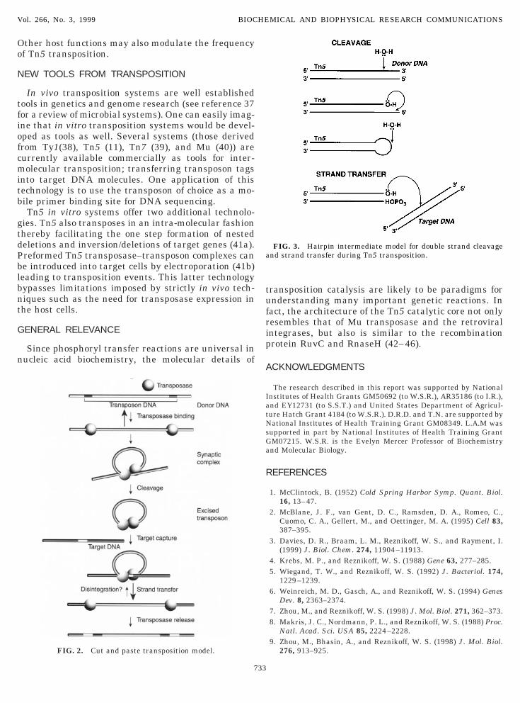
Tn5 transposition is a multi-step cut and paste pro-ess schematically presented in Fig. 2. Other transpos-ble elements appear to proceed through seeminglyuite different transposition paths [for instance repli-ative transposition for Mu (18) or circle formation forS911 (19)], but these likely result from minor varia-ions in the basic chemistry as will be described.
ND SEQUENCE BINDING
Tn5 transposase end sequence binding involves thenteraction of an N-terminal amino acid sequence withhe 19 bp end sequence. This is likely to involve arotein monomer-DNA monomer sequence interactionsee below). Our genetic data suggests that at least aelix turn helix motif is involved in the DNA sequenceecognition (9). This motif was correlated with the endequence binding function through the isolation of mu-ants that enhance the binding affinity or, in one case,lters the binding preference regarding OE versus IE.ut co-crystallographic studies of the Tc3 transposaseNA binding domain bound to its cognate DNA se-uence (20) and recent genetic data on Tn5 trans-osase (Naumann and Reznikoff, unpublished) suggesthat the DNA binding motif is more complex.
730
A critical feature of the binding reaction is that it isighly disfavored probably due to two features. First,he DNA binding domain sequence is suboptimal (7).econdly, the C-terminus of the protein interferes withhe DNA binding (removal of the C-terminal sequencesr occupation of the C-terminal sequences by protein-rotein interaction is required to permit DNA bindingetection) (3, 10, 14, 21). Thus, an unfavorable alloste-ic change in transposase structure removing the-terminal inhibition is likely to occur prior to DNAinding. It is possible that the DNA binding reaction ishe critical rate limiting step in Tn5 transposition.
The transposase C-terminal interference with theransposase DNA binding activity may be related tohe intriguing observation that Tn5 transposase, likeome other transposases, prefers to act in cis (12, 13,7, 48). This explanation would posit that transposasetill in the process of being translated might be betterble to bind to nearby end DNA sequences since thenhibitory C-terminus would not yet exist.
As has been found for other sequence specific DNAinding proteins, the transposase has been found toend end sequence containing DNA (21, 22). This bendat least 35°) occurs at the transposon–donor backboneDBB) boundary and might explain the observationhat missing bases near the end sequence–DBB bound-ry enhance transposase binding (23). Perhaps theNA bending locally destabilizes the helix and thereby
acilitates the subsequent cleavage reactions.
YNAPTIC COMPLEX FORMATION
The next step is highly favored. It is synaptic com-lex formation resulting from the dimerization of theonomeric transposase-end sequence complexes. It is
he stoichiometry of the synaptic complexes formed atow transposase concentrations (two transposase mol-cules with two DNA sequences [Bhasin, Goryshin,ork, and Reznikoff, unpublished]) which suggests
hat the preceding bound complexes involve a mono-er of transposase with a monomer of DNA.Most of the interactions leading to synapsis likely
nvolve protein-protein interactions. Although the in-olved dimerization domains are uncertain (3, 24), re-ent genetic studies (Steiniger-White and Reznikoff,npublished) suggest that dimerization mediated byhe C-terminal alpha helix (3) is critical for this pro-ess. This is surprising since this domain is not sharedy the related Tn10 transposase (24).The observations that DNA cleavage involves trans
atalysis (see below) and that transposase containingutations of lysines 330 and 333 are defective in syn-
psis suggest trans protein–DNA interactions are alsonvolved (Naumann and Reznikoff, unpublished). Thats, nucleotide pairs at the very end of the specific bind-ng sequence and/or at the adjoining DBB sequence
ay interact with opposite subunit lysines (residues

3Tr
pbcl
etFmlby
D
bcgttFnbgiTapae
ctacalaopttwsI
vtleoa
pttatatbc
T
sottmfq
S
gtwat
D
tagb
R
ticTatG
a
D
mbp
Vol. 266, No. 3, 1999 BIOCHEMICAL AND BIOPHYSICAL RESEARCH COMMUNICATIONS
30 and 333, see Fig. 1b) near the catalytic active site.hese are the contacts that possibly were detected byetroviral integrase–DNA cross-linking studies (25a).
Synaptic complex formation is typically studied ex-erimentally by analyzing the formation of a compara-le structure, the paired end complex. The paired endomplex is formed when the two end sequences areocated on separate DNA fragments.
Although our model postulates that transposase–nd sequence binding precedes synapsis, it is possiblehat a more complex mixture of these two steps occurs.or instance, unstable dimerization of transposaseonomers may expose the DNA binding domains al-
owing end sequence binding and then this is followedy protein–protein and protein–DNA rearrangementsielding the final synaptic complexes.
NA CLEAVAGE
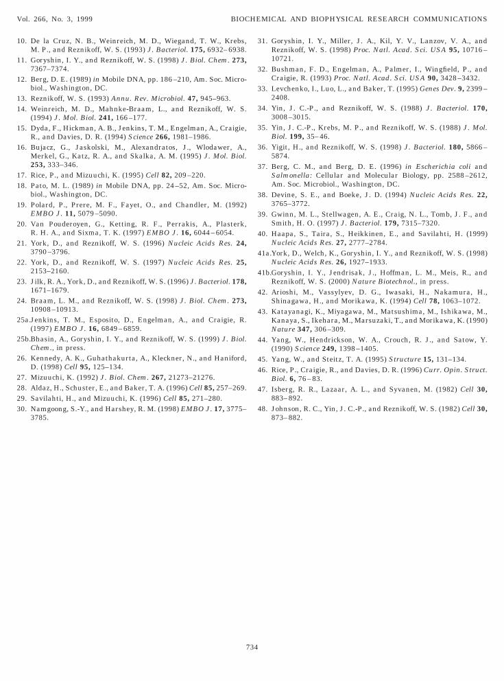
The final product of transposon–DBB cleavage is alunt ended transposon DNA. How can one active siteleave the two DNA strands of opposite polarity? Theeneration of this blunt end cleavage product involveshree phosphoryl transfer reactions in Tn5 transposi-ion (Fig. 3) (25b) as it does for Tn10 transposition (26).irst an OH group recruited from H2O is used as aucleophile to break the 39 strand phosphodiester bondordering the end sequence. Second the released 39 OHroup attacks the 59 strand phosphodiester bond caus-ng release of the DBB and formation of a DNA hairpin.he third step in DNA cleavage is presumed to use andditional water donated OH group to break the hair-in phosphodiester bond. This three step mechanismllows both strands to be cleaved with no major reori-ntation of the active site.The three-step cleavage mechanism distinguishes
ut and paste transposition from replicative transposi-ion. For instance, the active site for Mu transposasend Tn3 transposase must not be able to perform theross strand hairpin attack thus the 59 strand remainsttached to the DBB. Retroviral integrase is similarlyimited to cutting the 39 strand, but in this case thettached 59 strand extension is limited to a few nucle-tides (27). The details of the hairpin attack step alsorobably distinguishes Tn5 (and Tn10) linear typeransposition from IS911 circle formation transposi-ion. For Tn5 the cross strand attack occurs in cisithin each bound DNA in the synaptic complex (Bha-
in, Goryshin and Reznikoff, unpublished) while forS911 the attack is in trans (between bound DNAs) (19).
Finally, all of the phosphoryl transfer reactions in-olved in DNA cleavage are catalyzed in trans withinhe synaptic complex (Naumann and Reznikoff, unpub-ished). That is, the transposase monomer bound to onend sequence catalyzes the strand cleavages at thether end. This was determined by studying the cleav-ge reactions for an OE–IE defined (IS50-like) trans-
731
oson using a mixture of IE specific, catalytically activeransposase and OE specific, catalytically inactiveransposase; cleavage occurred at the OE–DBB bound-ry. The reverse localization of the catalytically inac-ive monomer directs cleavage to the IE–DBB bound-ry. Trans cleavage has been found to occur during Muransposition (28–30). It ensures that cleavages atoth ends only occur within the context of a synapticomplex and not by transposase bound to only one end.
ARGET CAPTURE
Target capture is not well understood. Through aequence analysis of inserts in an extensive collectionf in vivo and in vitro transposition events, we knowhat there are some DNA target sequence biases andhere is intriguing evidence that formation of multi-eric transposase filaments on target sequences can
acilitate this step (31). However, the transposase se-uences involved in target capture are unknown.
TRAND TRANSFER INTO TARGET
Once bound to the target, the transposon 39OHroups attack target strands with the individual at-acks being spaced 9 bp apart thus leading to an insertith 9 base gaps bounding each side. This will lead,fter repair, to the signature target site 9 bp duplica-ion.
ISINTEGRATION
The reversal of the integration reaction is disintegra-ion. This reaction has never been detected for Tn5lthough it has been used as an assay for HIV-1 inte-rase core activity (32). On the basis of intuition, weelieve that disintegration is highly disfavored for Tn5.
EMOVING TRANSPOSASE
At the end of this process transposase is boundightly to the transposon containing products (11). Howt is removed remains a mystery. For Mu, a hosthaperone-protease is involved in removal (33). Forn5 we have suggestive evidence that a proteolyticttack following a lysine residue at position 40 withinhe DNA binding domain may be involved (Twining,oryshin and Reznikoff, unpublished).It is assumed that host functions, DNA polymerase
nd ligase, repair the gaps.
OWN-REGULATING TRANSPOSITION
Tn5 transposition is a very rare process in vivo asight be expected for such a destructive process. We
elieve that there are multiple mechanisms accom-lishing this. These include (i) The wild-type protein is

apm2stab
(a(trqtt
ttssdhm
Vol. 266, No. 3, 1999 BIOCHEMICAL AND BIOPHYSICAL RESEARCH COMMUNICATIONS
lmost a non-functional protein probably because of, inart, a suboptimal DNA binding domain and C-ter-inal interference with the DNA binding (3, 7, 10, 14,
1); (ii) The natural end DNA sequences OE and IE areuboptimal (9); (iii) Tn5 encodes an N-terminal dele-ion variant of the transposase called the inhibitor thatcts as a trans-dominant negative regulator probablyy forming inactive inhibitor-transposase complexes
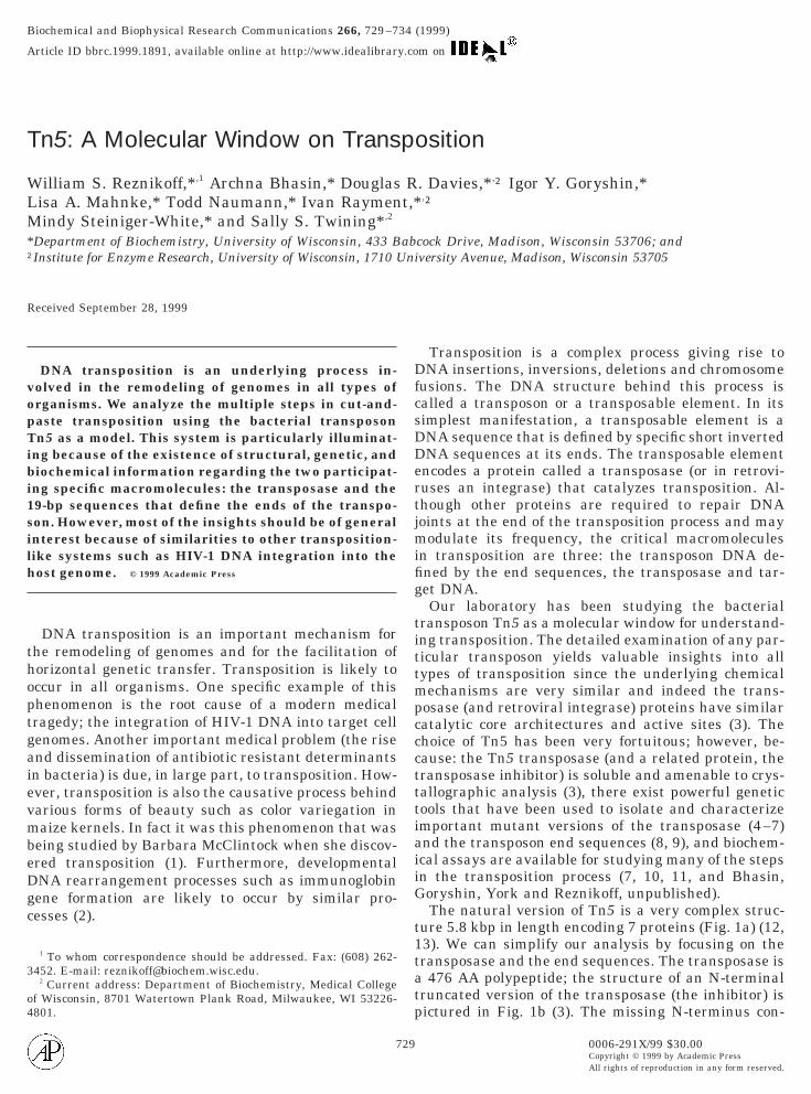
FIG. 1. (a) Tn5 structure and specific 19-bp end sequences. The sransposon in which two IS50 elements bracket three antibiotic resistaransposition inhibitor (Inh). Inh lacks the N-terminal 55 amino acidshown). IS50 is bordered by two 19-bp sequences (OE and IE) to which Ttrand of OE and IE are shown below. One strand of a 19-bp mosaic sequiagram representation of Inh (3). The catalytic core whose architectuighlighted in dark blue. The DDE active site residues (Asp 97, Asp 188otif. Not visible are the 14 N-terminal amino acids of Inh (or the 69 N
732
10, 34). The abundance of inhibitory peptides is likelyugmented by proteolytic attack on the transposase24); and (iv) Host functions modulate the transposi-ion frequency. For instance dam methylase down-egulates transposase synthesis and use of the IE se-uence (35). In addition, Topo-1 positively modulatesransposition possibly by influencing the target cap-ure step (36 and Yigit and Reznikoff, unpublished).
ture of Tn5 has been described in detail (12, 13). Tn5 is a compositegenes. IS50R encodes the transposase (Tnp) and the trans-dominantTnp. IS50L encodes inactive truncated versions of Tnp and Inh (notbinds as the first step in transposition (see Fig. 2). The sequence of onee found to be hyperactive for transposition (9) is also shown. (b) A ribbonresembles that of HIV-1 and ASV integrases and Mu transposase isd Glu 325) are shown as well as residues found in the conserved YRKKrminal amino acids of Tnp).
trucnceofnp
encre
, an-te
Oo
N
tfiofcmitb
gtdPblbnt
G
n
tufrip
A
IatNsGa
R
a
Vol. 266, No. 3, 1999 BIOCHEMICAL AND BIOPHYSICAL RESEARCH COMMUNICATIONS
ther host functions may also modulate the frequencyf Tn5 transposition.
EW TOOLS FROM TRANSPOSITION
In vivo transposition systems are well establishedools in genetics and genome research (see reference 37or a review of microbial systems). One can easily imag-ne that in vitro transposition systems would be devel-ped as tools as well. Several systems (those derivedrom Ty1(38), Tn5 (11), Tn7 (39), and Mu (40)) areurrently available commercially as tools for inter-olecular transposition; transferring transposon tags
nto target DNA molecules. One application of thisechnology is to use the transposon of choice as a mo-ile primer binding site for DNA sequencing.Tn5 in vitro systems offer two additional technolo-
ies. Tn5 also transposes in an intra-molecular fashionhereby facilitating the one step formation of nestedeletions and inversion/deletions of target genes (41a).reformed Tn5 transposase–transposon complexes cane introduced into target cells by electroporation (41b)eading to transposition events. This latter technologyypasses limitations imposed by strictly in vivo tech-iques such as the need for transposase expression inhe host cells.
ENERAL RELEVANCE
Since phosphoryl transfer reactions are universal inucleic acid biochemistry, the molecular details of
FIG. 2. Cut and paste transposition model.
733
ransposition catalysis are likely to be paradigms fornderstanding many important genetic reactions. Inact, the architecture of the Tn5 catalytic core not onlyesembles that of Mu transposase and the retroviralntegrases, but also is similar to the recombinationrotein RuvC and RnaseH (42–46).
CKNOWLEDGMENTS
The research described in this report was supported by Nationalnstitutes of Health Grants GM50692 (to W.S.R.), AR35186 (to I.R.),nd EY12731 (to S.S.T.) and United States Department of Agricul-ure Hatch Grant 4184 (to W.S.R.). D.R.D. and T.N. are supported byational Institutes of Health Training Grant GM08349. L.A.M was
upported in part by National Institutes of Health Training GrantM07215. W.S.R. is the Evelyn Mercer Professor of Biochemistrynd Molecular Biology.
EFERENCES
1. McClintock, B. (1952) Cold Spring Harbor Symp. Quant. Biol.16, 13–47.
2. McBlane, J. F., van Gent, D. C., Ramsden, D. A., Romeo, C.,Cuomo, C. A., Gellert, M., and Oettinger, M. A. (1995) Cell 83,387–395.
3. Davies, D. R., Braam, L. M., Reznikoff, W. S., and Rayment, I.(1999) J. Biol. Chem. 274, 11904–11913.
4. Krebs, M. P., and Reznikoff, W. S. (1988) Gene 63, 277–285.5. Wiegand, T. W., and Reznikoff, W. S. (1992) J. Bacteriol. 174,
1229–1239.6. Weinreich, M. D., Gasch, A., and Reznikoff, W. S. (1994) Genes
Dev. 8, 2363–2374.7. Zhou, M., and Reznikoff, W. S. (1998) J. Mol. Biol. 271, 362–373.8. Makris, J. C., Nordmann, P. L., and Reznikoff, W. S. (1988) Proc.
Natl. Acad. Sci. USA 85, 2224–2228.9. Zhou, M., Bhasin, A., and Reznikoff, W. S. (1998) J. Mol. Biol.
276, 913–925.
FIG. 3. Hairpin intermediate model for double strand cleavagend strand transfer during Tn5 transposition.
10. De la Cruz, N. B., Weinreich, M. D., Wiegand, T. W., Krebs,
1
1
11
1
1
11
1
2
2
2
2
2
2
2
2
2223
31. Goryshin, I. Y., Miller, J. A., Kil, Y. V., Lanzov, V. A., and
3
3
3
3
3
3
3
3
4
4
4
4
4
4
44
4
4
Vol. 266, No. 3, 1999 BIOCHEMICAL AND BIOPHYSICAL RESEARCH COMMUNICATIONS
M. P., and Reznikoff, W. S. (1993) J. Bacteriol. 175, 6932–6938.1. Goryshin, I. Y., and Reznikoff, W. S. (1998) J. Biol. Chem. 273,
7367–7374.2. Berg, D. E. (1989) in Mobile DNA, pp. 186–210, Am. Soc. Micro-
biol., Washington, DC.3. Reznikoff, W. S. (1993) Annu. Rev. Microbiol. 47, 945–963.4. Weinreich, M. D., Mahnke-Braam, L., and Reznikoff, W. S.
(1994) J. Mol. Biol. 241, 166–177.5. Dyda, F., Hickman, A. B., Jenkins, T. M., Engelman, A., Craigie,
R., and Davies, D. R. (1994) Science 266, 1981–1986.6. Bujacz, G., Jaskolski, M., Alexandratos, J., Wlodawer, A.,
Merkel, G., Katz, R. A., and Skalka, A. M. (1995) J. Mol. Biol.253, 333–346.
7. Rice, P., and Mizuuchi, K. (1995) Cell 82, 209–220.8. Pato, M. L. (1989) in Mobile DNA, pp. 24–52, Am. Soc. Micro-
biol., Washington, DC.9. Polard, P., Prere, M. F., Fayet, O., and Chandler, M. (1992)
EMBO J. 11, 5079–5090.0. Van Pouderoyen, G., Ketting, R. F., Perrakis, A., Plasterk,
R. H. A., and Sixma, T. K. (1997) EMBO J. 16, 6044–6054.1. York, D., and Reznikoff, W. S. (1996) Nucleic Acids Res. 24,
3790–3796.2. York, D., and Reznikoff, W. S. (1997) Nucleic Acids Res. 25,
2153–2160.3. Jilk, R. A., York, D., and Reznikoff, W. S. (1996) J. Bacteriol. 178,
1671–1679.4. Braam, L. M., and Reznikoff, W. S. (1998) J. Biol. Chem. 273,
10908–10913.5a.Jenkins, T. M., Esposito, D., Engelman, A., and Craigie, R.
(1997) EMBO J. 16, 6849–6859.5b.Bhasin, A., Goryshin, I. Y., and Reznikoff, W. S. (1999) J. Biol.
Chem., in press.6. Kennedy, A. K., Guhathakurta, A., Kleckner, N., and Haniford,
D. (1998) Cell 95, 125–134.7. Mizuuchi, K. (1992) J. Biol. Chem. 267, 21273–21276.8. Aldaz, H., Schuster, E., and Baker, T. A. (1996) Cell 85, 257–269.9. Savilahti, H., and Mizuuchi, K. (1996) Cell 85, 271–280.0. Namgoong, S.-Y., and Harshey, R. M. (1998) EMBO J. 17, 3775–
3785.
734
Reznikoff, W. S. (1998) Proc. Natl. Acad. Sci. USA 95, 10716–10721.
2. Bushman, F. D., Engelman, A., Palmer, I., Wingfield, P., andCraigie, R. (1993) Proc. Natl. Acad. Sci. USA 90, 3428–3432.
3. Levchenko, I., Luo, L., and Baker, T. (1995) Genes Dev. 9, 2399–2408.
4. Yin, J. C.-P., and Reznikoff, W. S. (1988) J. Bacteriol. 170,3008–3015.
5. Yin, J. C.-P., Krebs, M. P., and Reznikoff, W. S. (1988) J. Mol.Biol. 199, 35–46.
6. Yigit, H., and Reznikoff, W. S. (1998) J. Bacteriol. 180, 5866–5874.
7. Berg, C. M., and Berg, D. E. (1996) in Escherichia coli andSalmonella: Cellular and Molecular Biology, pp. 2588–2612,Am. Soc. Microbiol., Washington, DC.
8. Devine, S. E., and Boeke, J. D. (1994) Nucleic Acids Res. 22,3765–3772.
9. Gwinn, M. L., Stellwagen, A. E., Craig, N. L., Tomb, J. F., andSmith, H. O. (1997) J. Bacteriol. 179, 7315–7320.
0. Haapa, S., Taira, S., Heikkinen, E., and Savilahti, H. (1999)Nucleic Acids Res. 27, 2777–2784.
1a.York, D., Welch, K., Goryshin, I. Y., and Reznikoff, W. S. (1998)Nucleic Acids Res. 26, 1927–1933.
1b.Goryshin, I. Y., Jendrisak, J., Hoffman, L. M., Meis, R., andReznikoff, W. S. (2000) Nature Biotechnol., in press.
2. Arioshi, M., Vassylyev, D. G., Iwasaki, H., Nakamura, H.,Shinagawa, H., and Morikawa, K. (1994) Cell 78, 1063–1072.
3. Katayanagi, K., Miyagawa, M., Matsushima, M., Ishikawa, M.,Kanaya, S., Ikehara, M., Marsuzaki, T., and Morikawa, K. (1990)Nature 347, 306–309.
4. Yang, W., Hendrickson, W. A., Crouch, R. J., and Satow, Y.(1990) Science 249, 1398–1405.
5. Yang, W., and Steitz, T. A. (1995) Structure 15, 131–134.6. Rice, P., Craigie, R., and Davies, D. R. (1996) Curr. Opin. Struct.
Biol. 6, 76–83.7. Isberg, R. R., Lazaar, A. L., and Syvanen, M. (1982) Cell 30,
883–892.8. Johnson, R. C., Yin, J. C.-P., and Reznikoff, W. S. (1982) Cell 30,
873–882.