topical delivery of sirna-based spherical nucleic acid nanoparticle conjugates for gene regulation
TRANSCRIPT
Topical delivery of siRNA-based spherical nucleicacid nanoparticle conjugates for gene regulationDan Zhenga,b, David A. Giljohanna,b, David L. Chenc, Matthew D. Massicha,b, Xiao-Qi Wangc, Hristo Iordanovc,Chad A. Mirkina,b,1, and Amy S. Pallerc,1
aDepartment of Chemistry, Northwestern University, 2145 Sheridan Road, Evanston, IL 60208; bInternational Institute for Nanotechnology, NorthwesternUniversity, 2145 Sheridan Road, Evanston, IL 60208; and cDepartment of Dermatology, Feinberg School of Medicine, Northwestern University, 676 NorthSt. Clair Street, Suite 1600, Chicago, IL 60611
Edited by Rakesh K. Jain, Harvard Medical School and Massachusetts General Hospital, Boston, MA, and approved June 6, 2012 (received for review November22, 2011)
Topical application of nucleic acids offers many potential therapeu-tic advantages for suppressing genes in the skin, and potentiallyfor systemic gene delivery. However, the epidermal barrier typi-cally precludes entry of gene-suppressing therapy unless the bar-rier is disrupted. We now show that spherical nucleic acid nano-particle conjugates (SNA-NCs), gold cores surrounded by a denseshell of highly oriented, covalently immobilized siRNA, freely pene-trate almost 100% of keratinocytes in vitro, mouse skin, and hu-man epidermis within hours after application. Significantly, thesestructures can be delivered in a commercial moisturizer or phos-phate-buffered saline, and do not require barrier disruption ortransfection agents, such as liposomes, peptides, or viruses. SNA-NCs targeting epidermal growth factor receptor (EGFR), an impor-tant gene for epidermal homeostasis, are >100-fold more potentand suppress longer than siRNA delivered with commercial lipidagents in cultured keratinocytes. Topical delivery of 1.5 uM EGFRsiRNA (50 nM SNA-NCs) for 3 wk to hairless mouse skin almost com-pletely abolishes EGFR expression, suppresses downstream ERKphosphorylation, and reduces epidermal thickness by almost 40%.Similarly, EGFRmRNA in human skin equivalents is reduced by 52%after 60 h of treatment with 25 nM EGFR SNA-NCs. Treated skinshows no clinical or histological evidence of toxicity. No cytokineactivation in mouse blood or tissue samples is observed, and after3 wk of topical skin treatment, the SNA structures are virtually un-detectable in internal organs. SNA conjugates may be promisingagents for personalized, topically delivered gene therapy of cuta-neous tumors, skin inflammation, and dominant negative geneticskin disorders.
transdermal delivery ∣ nanocluster ∣ antisense ∣ nanotechnology ∣dermatology
The recent development of small molecule inhibitors and anti-bodies that target components of signaling pathways has re-
volutionized the treatment of cancers, inflammatory diseases, andgenetic disorders, including those that largely manifest in skin (1–3). These protein-based therapeutics, however, are costly, havelimited targeting ability, and, when delivered through traditionalintravenous or gastrointestinal routes, can lead to systemic toxi-city (4, 5). Topical delivery is particularly attractive for the therapyof skin disorders. However, proteins larger than a few hundreddaltons cannot easily enter the skin, and high concentrations ofproteins must be applied for a cutaneous effect (6). An alterna-tive to protein-based pathway inhibition involves the blockingand/or degradation of precursor mRNA before translation intoprotein. Gene silencing leads to down-regulation of protein ex-pression and functions with greater specificity than inhibitorsof protein function (7). In fact, targeted gene suppression by anti-sense DNA and siRNA has shown promising preclinical results,and/or is currently in clinical trials for a variety of diseases, includ-ing many forms of cancer (e.g., melanoma, neuroblastoma, andpancreatic adenocarcinoma), genetic disorders, and macular de-generation (8). For gene suppression in skin, a topical delivery
route is optimal, given the easy accessibility of skin and the re-duced risk of systemic side effects. However, penetration ofoligonucleotides through the epidermal barrier has remained amajor technological challenge that must be overcome before thetherapeutic potential of oligonucleotides in skin can be realized(9–11).
Recently, we introduced spherical nucleic acid nanoparticleconjugates (SNA-NCs, inorganic gold nanoparticles denselycoated with highly oriented oligonucleotides) (Scheme S1) asagents capable of simultaneous transfection and gene regulation(12–15). In contrast with conventional approaches to gene regu-lation (16–20), these nanoparticle conjugates require neither ca-tionic transfection materials nor additional modifications to drivecellular entry. SNA-NCs enter almost 100% of cells in the morethan 50 cell lines and primary cells tested to date, as well as cul-tured tissues and whole organs (21, 22). These unusual constructsexhibit remarkable stability, nuclease-degradation resistance, anddecreased immunogenicity as a result of their high-density oligo-nucleotide shells (23, 24). To date, they have shown no toxicity atthe cellular level at concentrations required for effective geneknockdown. Therefore, we hypothesized that siRNA-based SNAgold nanoparticle conjugates might be ideal topical deliveryagents to treat diseases of the skin.
ResultsTo evaluate the potential for targeted gene suppression bysiRNA-based SNA-NCs through noninvasive topical delivery,we first investigated their uptake in primary normal human ker-atinocytes (hKCs), which are typically difficult to transfect (25).hKCs were incubated with Cy3 fluorophore-labeled SNA-NCs(Table S1), and entry was studied by confocal fluorescence micro-scopy. Within 2 h, 100% of the observed hKCs showed strongcytoplasmic fluorescence (Fig. 1A). The number of gold nano-particles in cells (and tissues) can be quantified by inductivelycoupled plasma mass spectrometry (ICP-MS). Incubation of0.025–0.1 nM SNA-NCs (0.75–3.0 nM siRNA) for 48 h withhKCs leads to uptake of approximately 1.4 · 103 SNA-NCs percell, which is approximately five times greater than that in eitherHaCaT (spontaneously immortalized hKCs) or HeLa cells(Fig. 1B). Although the reason for this high KC uptake is still
Author contributions: D.Z., D.A.G., D.L.C., C.A.M., and A.S.P. designed research; D.Z.,D.A.G., D.L.C., M.D.M., X.Q.W., and H.I. performed research; H.I. contributed newreagents/analytic tools; D.Z., D.A.G., D.L.C., M.D.M., X.Q.W., and A.S.P. analyzed data;and D.Z., D.A.G., D.L.C., X.Q.W., C.A.M., and A.S.P. wrote the paper.
Conflict of interest statement: This technology has been licensed from NorthwesternUniversity by AuraSense Therapeutics, LLC. C.A.M., A.S.P., and D.A.G. have financialinterests in AuraSense Therapeutics, LLC.
This article is a PNAS Direct Submission.1To whom correspondence may be addressed. E-mail: [email protected] [email protected].
This article contains supporting information online at www.pnas.org/lookup/suppl/doi:10.1073/pnas.1118425109/-/DCSupplemental.
www.pnas.org/cgi/doi/10.1073/pnas.1118425109 PNAS ∣ July 24, 2012 ∣ vol. 109 ∣ no. 30 ∣ 11975–11980
CHEM
ISTR
YAPP
LIED
BIOLO
GICAL
SCIENCE
S

under investigation, these observations suggested SNA-NCswould be good candidates for topical delivery experiments.
With any gene regulation therapy or experiment, cell toxicity isa major concern. hKCs incubated with SNA-NCs are morpholo-gically indistinguishable from untreated cells by routine micro-scopic evaluation. Proliferation assays also show no evidence ofcytotoxicity after treatment of hKCs for 48 h with 0.05–0.1 nMSNA-NCs with nonsense siRNA sequences (Fig. 1C, Table S1).Finally, genome-wide expression profiling showed that only sevengenes are up-regulated and none are down-regulated in hKCstreated with nonsense SNA-NCs, although the same cells treatedwith an equivalent amount of siRNA delivered through a cationiclipid-based transfection agent (DharmaFECT1®) exhibit up- ordown-regulation of 427 genes (Fig. 1D and Dataset S1). Theseobservations are consistent with previous studies in the HeLa cellline, which show that SNA-NCs enter cells in a stealth-like fash-ion and exhibit limited interference with normal cellular behavior(24, 26, 27).
Given the importance of EGFR signaling in epidermal cellproliferation and its recognized overexpression in many malig-nancies, EGFR was chosen as an initial target for gene suppres-sion. EGFR knockdown in hKCs with the SNA-NCs is extremelyeffective. As little as 0.01 nM of SNA-NCs (0.3 nM of total
siRNA, Table S1) in 1 mL of cell culture medium down-regulatesEGFR protein expression by more than 90% as compared to con-trol experiments involving PBS-treated cells and cells treated withnonsense SNA-NCs (Fig. 1E, group 5, lane 1 vs. groups 1–3). To-tal depletion of protein expression, within the sensitivity of Wes-tern blots, can be effected with 0.05 nM EGFR SNA-NCs in cellculture medium (1.5 nM of total siRNA) (Fig. 1E, group 5, lane2). Significantly, knockdown of EGFR with SNA-NCs in hKCscan be achieved with 100-fold less total siRNA than that requiredto observe an effect with DharmaFECT1® (Fig. 1E, group 5, lane1 vs. group 4, lane 2). Interestingly, target knockdown with SNA-NCs is also more persistent than with DharmaFECT1-deliveredsiRNAs (Fig. 1F). Here, at 48 h, greater than 90%mRNA knock-down is seen in both groups; however, suppression is completelylost at 96 h with DharmaFECT1-delivered siRNA, whereas SNA-NC—treated cells still exhibit 50% knockdown. This persistenceis likely caused by the known resistance of SNA constructs to nu-clease degradation, and therefore increased intracellular stabilityof the nucleic acid sequences on the SNA construct surface (23).
Because the SNA-NCs easily enter hKCs in in vitro experi-ments, we explored their ability to penetrate both mouse skin andhuman skin models. The SKH1-E hairless strain was chosen forstudies because of its ease in use (eliminating the need for depi-
Fig. 1. Uptake, safety, and gene suppression efficacy of SNA-NCs in hKCs. (A) Uptake of Cy3-labeled nonsense SNA-NCs (red) was noted in the cytoplasm ofapproximately 100% of the primary hKCs after 24 h incubation. Blue, Hoechst 33343-stained nuclei. Scale bar, 20 μm. (B) KCs exhibit dose-dependent SNAuptake that is higher than the uptake in the HeLa and HaCaT cell lines. (C) Cellular proliferation during 96 h of treatment with nonsense SNA-NCs is similar tothat of control treatment with PBS only. (D) Heat map of cellular gene array study (Dataset S1). (E) Western blot showing EGFR protein levels. hKCs were treatedfor 48 h with SNA-NCs or DharmaFECT1-delivered siRNA, or with controls of PBS and/or nonsense SNA-NCs. After treatment, growthmediumwas replaced withfresh medium, and the cells were incubated for an additional 12 h. Cell lysates were then harvested for blotting. Note the greater suppression by 0.01 nM EGFRSNA-NCs (equivalent to 0.3 nM siRNA) as compared with the 30 nM EGFR siRNA delivered with DharmaFECT1 at 60 h. (F) EGFR mRNA suppression after 48 htreatment with EGFR SNA-NCs or DhamaFECT1-delivered EGFR siRNA (treatment terminated after 48 h). The decrease and eventual loss of gene suppressionduring the subsequent 48 h after termination of treatment was evaluated by RT-qPCR. By 48 h after removal of SNA-NCs from the medium, 50% of themaximum suppression is still observed in EGFR SNA-NC—treated KCs, whereas suppression by DharmaFECT1-delivered EGFR siRNA is no longer evident.All data are expressed as mean� SD.
11976 ∣ www.pnas.org/cgi/doi/10.1073/pnas.1118425109 Zheng et al.
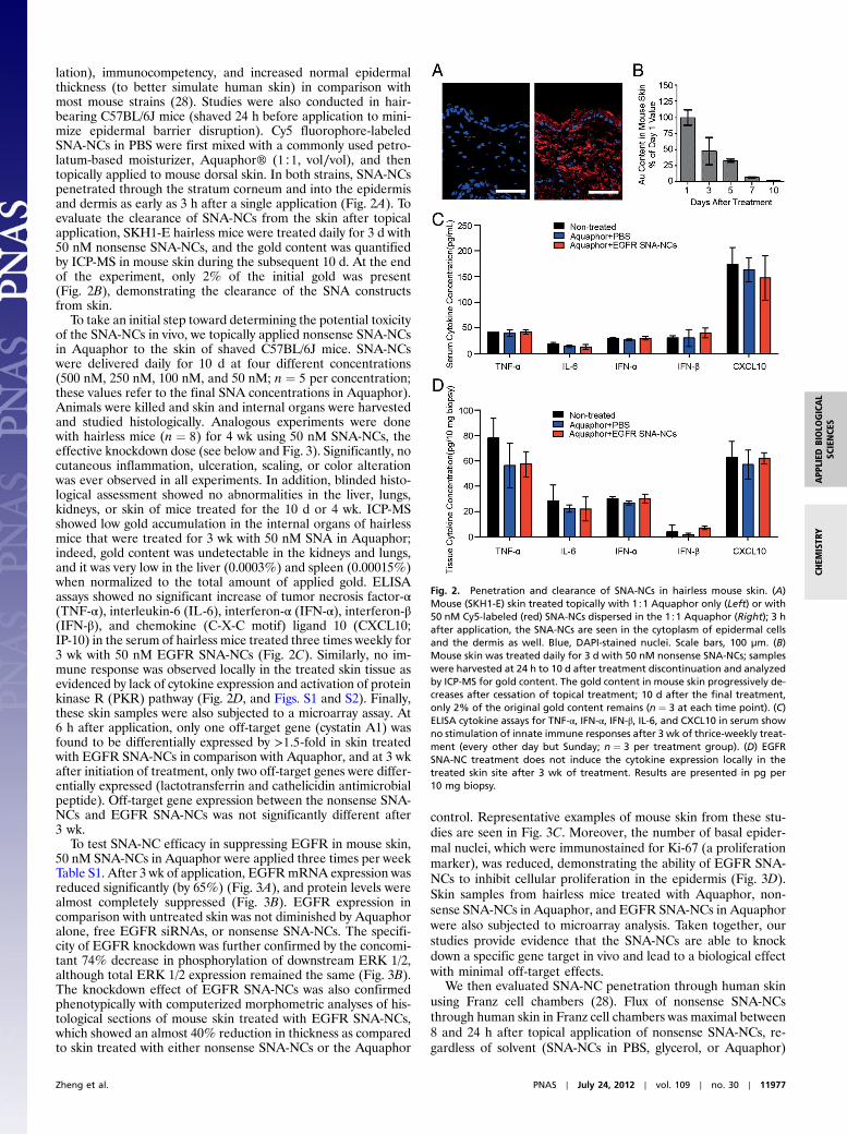
lation), immunocompetency, and increased normal epidermalthickness (to better simulate human skin) in comparison withmost mouse strains (28). Studies were also conducted in hair-bearing C57BL/6J mice (shaved 24 h before application to mini-mize epidermal barrier disruption). Cy5 fluorophore-labeledSNA-NCs in PBS were first mixed with a commonly used petro-latum-based moisturizer, Aquaphor® (1∶1, vol∕vol), and thentopically applied to mouse dorsal skin. In both strains, SNA-NCspenetrated through the stratum corneum and into the epidermisand dermis as early as 3 h after a single application (Fig. 2A). Toevaluate the clearance of SNA-NCs from the skin after topicalapplication, SKH1-E hairless mice were treated daily for 3 d with50 nM nonsense SNA-NCs, and the gold content was quantifiedby ICP-MS in mouse skin during the subsequent 10 d. At the endof the experiment, only 2% of the initial gold was present(Fig. 2B), demonstrating the clearance of the SNA constructsfrom skin.
To take an initial step toward determining the potential toxicityof the SNA-NCs in vivo, we topically applied nonsense SNA-NCsin Aquaphor to the skin of shaved C57BL/6J mice. SNA-NCswere delivered daily for 10 d at four different concentrations(500 nM, 250 nM, 100 nM, and 50 nM; n ¼ 5 per concentration;these values refer to the final SNA concentrations in Aquaphor).Animals were killed and skin and internal organs were harvestedand studied histologically. Analogous experiments were donewith hairless mice (n ¼ 8) for 4 wk using 50 nM SNA-NCs, theeffective knockdown dose (see below and Fig. 3). Significantly, nocutaneous inflammation, ulceration, scaling, or color alterationwas ever observed in all experiments. In addition, blinded histo-logical assessment showed no abnormalities in the liver, lungs,kidneys, or skin of mice treated for the 10 d or 4 wk. ICP-MSshowed low gold accumulation in the internal organs of hairlessmice that were treated for 3 wk with 50 nM SNA in Aquaphor;indeed, gold content was undetectable in the kidneys and lungs,and it was very low in the liver (0.0003%) and spleen (0.00015%)when normalized to the total amount of applied gold. ELISAassays showed no significant increase of tumor necrosis factor-α(TNF-α), interleukin-6 (IL-6), interferon-α (IFN-α), interferon-β(IFN-β), and chemokine (C-X-C motif) ligand 10 (CXCL10;IP-10) in the serum of hairless mice treated three times weekly for3 wk with 50 nM EGFR SNA-NCs (Fig. 2C). Similarly, no im-mune response was observed locally in the treated skin tissue asevidenced by lack of cytokine expression and activation of proteinkinase R (PKR) pathway (Fig. 2D, and Figs. S1 and S2). Finally,these skin samples were also subjected to a microarray assay. At6 h after application, only one off-target gene (cystatin A1) wasfound to be differentially expressed by >1.5-fold in skin treatedwith EGFR SNA-NCs in comparison with Aquaphor, and at 3 wkafter initiation of treatment, only two off-target genes were differ-entially expressed (lactotransferrin and cathelicidin antimicrobialpeptide). Off-target gene expression between the nonsense SNA-NCs and EGFR SNA-NCs was not significantly different after3 wk.
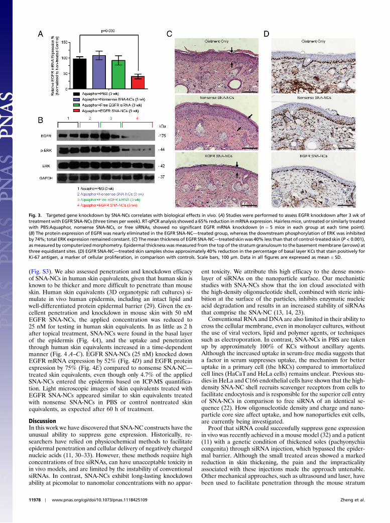
To test SNA-NC efficacy in suppressing EGFR in mouse skin,50 nM SNA-NCs in Aquaphor were applied three times per weekTable S1. After 3 wk of application, EGFRmRNA expression wasreduced significantly (by 65%) (Fig. 3A), and protein levels werealmost completely suppressed (Fig. 3B). EGFR expression incomparison with untreated skin was not diminished by Aquaphoralone, free EGFR siRNAs, or nonsense SNA-NCs. The specifi-city of EGFR knockdown was further confirmed by the concomi-tant 74% decrease in phosphorylation of downstream ERK 1/2,although total ERK 1/2 expression remained the same (Fig. 3B).The knockdown effect of EGFR SNA-NCs was also confirmedphenotypically with computerized morphometric analyses of his-tological sections of mouse skin treated with EGFR SNA-NCs,which showed an almost 40% reduction in thickness as comparedto skin treated with either nonsense SNA-NCs or the Aquaphor
control. Representative examples of mouse skin from these stu-dies are seen in Fig. 3C. Moreover, the number of basal epider-mal nuclei, which were immunostained for Ki-67 (a proliferationmarker), was reduced, demonstrating the ability of EGFR SNA-NCs to inhibit cellular proliferation in the epidermis (Fig. 3D).Skin samples from hairless mice treated with Aquaphor, non-sense SNA-NCs in Aquaphor, and EGFR SNA-NCs in Aquaphorwere also subjected to microarray analysis. Taken together, ourstudies provide evidence that the SNA-NCs are able to knockdown a specific gene target in vivo and lead to a biological effectwith minimal off-target effects.
We then evaluated SNA-NC penetration through human skinusing Franz cell chambers (28). Flux of nonsense SNA-NCsthrough human skin in Franz cell chambers was maximal between8 and 24 h after topical application of nonsense SNA-NCs, re-gardless of solvent (SNA-NCs in PBS, glycerol, or Aquaphor)
Fig. 2. Penetration and clearance of SNA-NCs in hairless mouse skin. (A)Mouse (SKH1-E) skin treated topically with 1∶1 Aquaphor only (Left) or with50 nM Cy5-labeled (red) SNA-NCs dispersed in the 1∶1 Aquaphor (Right); 3 hafter application, the SNA-NCs are seen in the cytoplasm of epidermal cellsand the dermis as well. Blue, DAPI-stained nuclei. Scale bars, 100 μm. (B)Mouse skin was treated daily for 3 d with 50 nM nonsense SNA-NCs; sampleswere harvested at 24 h to 10 d after treatment discontinuation and analyzedby ICP-MS for gold content. The gold content in mouse skin progressively de-creases after cessation of topical treatment; 10 d after the final treatment,only 2% of the original gold content remains (n ¼ 3 at each time point). (C)ELISA cytokine assays for TNF-α, IFN-α, IFN-β, IL-6, and CXCL10 in serum showno stimulation of innate immune responses after 3 wk of thrice-weekly treat-ment (every other day but Sunday; n ¼ 3 per treatment group). (D) EGFRSNA-NC treatment does not induce the cytokine expression locally in thetreated skin site after 3 wk of treatment. Results are presented in pg per10 mg biopsy.
Zheng et al. PNAS ∣ July 24, 2012 ∣ vol. 109 ∣ no. 30 ∣ 11977
CHEM
ISTR
YAPP
LIED
BIOLO
GICAL
SCIENCE
S
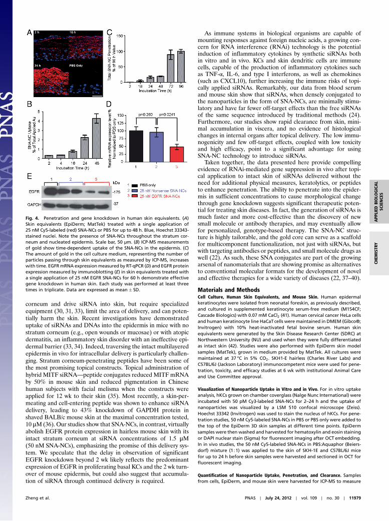
(Fig. S3). We also assessed penetration and knockdown efficacyof SNA-NCs in human skin equivalents, given that human skin isknown to be thicker and more difficult to penetrate than mouseskin. Human skin equivalents (3D organotypic raft cultures) si-mulate in vivo human epidermis, including an intact lipid andwell-differentiated protein epidermal barrier (29). Given the ex-cellent penetration and knockdown in mouse skin with 50 nMEGFR SNA-NCs, the applied concentration was reduced to25 nM for testing in human skin equivalents. In as little as 2 hafter topical treatment, SNA-NCs were found in the basal layerof the epidermis (Fig. 4A), and the uptake and penetrationthrough human skin equivalents increased in a time-dependentmanner (Fig. 4 A–C). EGFR SNA-NCs (25 nM) knocked downEGFR mRNA expression by 52% (Fig. 4D) and EGFR proteinexpression by 75% (Fig. 4E) compared to nonsense SNA-NC—treated skin equivalents, even though only 4.7% of the appliedSNA-NCs entered the epidermis based on ICP-MS quantifica-tion. Light microscopic images of skin equivalents treated withEGFR SNA-NCs appeared similar to skin equivalents treatedwith nonsense SNA-NCs in PBS or control nontreated skinequivalents, as expected after 60 h of treatment.
DiscussionIn this work we have discovered that SNA-NC constructs have theunusual ability to suppress gene expression. Historically, re-searchers have relied on physicochemical methods to facilitateepidermal penetration and cellular delivery of negatively chargednucleic acids (11, 30–33). However, these methods require highconcentrations of free siRNAs, can have unacceptable toxicity inin vivo models, and are limited by the instability of conventionalsiRNAs. In contrast, SNA-NCs exhibit long-lasting knockdownability at picomolar to nanomolar concentrations with no appar-
ent toxicity. We attribute this high efficacy to the dense mono-layer of siRNAs on the nanoparticle surface. Our mechanisticstudies with SNA-NCs show that the ion cloud associated withthe high-density oligonucleotide shell, combined with steric inhi-bition at the surface of the particles, inhibits enzymatic nucleicacid degradation and results in an increased stability of siRNAsthat comprise the SNA-NC (13, 14, 23).
Conventional RNA and DNA are also limited in their ability tocross the cellular membrane, even in monolayer cultures, withoutthe use of viral vectors, lipid and polymer agents, or techniquessuch as electroporation. In contrast, SNA-NCs in PBS are takenup by approximately 100% of KCs without ancillary agents.Although the increased uptake in serum-free media suggests thata factor in serum suppresses uptake, the mechanism for betteruptake in a primary cell (the hKCs) compared to immortalizedcell lines (HaCaTand HeLa cells) remains unclear. Previous stu-dies in HeLa and C166 endothelial cells have shown that the high-density SNA-NC shell recruits scavenger receptors from cells tofacilitate endocytosis and is responsible for the superior cell entryof SNA-NCs in comparison to free siRNA of an identical se-quence (22). How oligonucleotide density and charge and nano-particle core size affect uptake, and how nanoparticles exit cells,are currently being investigated.
Proof that siRNA could successfully suppress gene expressionin vivo was recently achieved in a mouse model (32) and a patient(11) with a genetic condition of thickened soles (pachyonychiacongenita) through siRNA injection, which bypassed the epider-mal barrier. Although the small treated areas showed a markedreduction in skin thickening, the pain and the impracticalityassociated with these injections made the approach untenable.Other mechanical approaches, such as ultrasound and laser, havebeen used to facilitate penetration through the mouse stratum
Fig. 3. Targeted gene knockdown by SNA-NCs correlates with biological effects in vivo. (A) Studies were performed to assess EGFR knockdown after 3 wk oftreatmentwith EGFR SNA-NCs (three times per week). RT-qPCR analysis showed a 65% reduction inmRNA expression. Hairlessmice, untreated or similarly treatedwith PBS:Aquaphor, nonsense SNA-NCs, or free siRNAs, showed no significant EGFR mRNA knockdown (n ¼ 5 mice in each group at each time point).(B) The protein expression of EGFR was nearly eliminated in the EGFR SNA-NC—treated group, whereas the downstream phosphorylation of ERK was inhibitedby 74%; total ERK expression remained constant. (C) Themean thickness of EGFR SNA-NC—treated skinwas 40% less than that of control-treated skin (P < 0.001),as measured by computerizedmorphometry. Epidermal thickness was measured from the top of the stratumgranulosum to the basement membrane (arrows) atthree equidistant sites. (D) EGFR SNA-NC—treated skin samples show approximately 40% reduction in the percentage of basal layer KCs that stain positively forKi-67 antigen, a marker of cellular proliferation, in comparison with controls. Scale bars, 100 μm. Data in all figures are expressed as mean� SD.
11978 ∣ www.pnas.org/cgi/doi/10.1073/pnas.1118425109 Zheng et al.
corneum and drive siRNA into skin, but require specializedequipment (30, 31, 33), limit the area of delivery, and can poten-tially harm the skin. Recent investigations have demonstrateduptake of siRNAs and DNAs into the epidermis in mice with nostratum corneum (e.g., open wounds or mucosae) or with atopicdermatitis, an inflammatory skin disorder with an ineffective epi-dermal barrier (33, 34). Indeed, traversing the intact multilayeredepidermis in vivo for intracellular delivery is particularly challen-ging. Stratum corneum-penetrating peptides have been some ofthe most promising topical constructs. Topical administration ofhybrid MITF siRNA—peptide conjugates reduced MITF mRNAby 50% in mouse skin and reduced pigmentation in Chinesehuman subjects with facial melisma when the constructs wereapplied for 12 wk to their skin (35). Most recently, a skin-per-meating and cell-entering peptide was shown to enhance siRNAdelivery, leading to 43% knockdown of GAPDH protein inshaved BALB/c mouse skin at the maximal concentration tested,10 μM (36). Our studies show that SNA-NCs, in contrast, virtuallyabolish EGFR protein expression in hairless mouse skin with itsintact stratum corneum at siRNA concentrations of 1.5 μM(50 nM SNA-NCs), emphasizing the promise of this delivery sys-tem. We speculate that the delay in observation of significantEGFR knockdown beyond 2 wk likely reflects the predominantexpression of EGFR in proliferating basal KCs and the 2 wk turn-over of mouse epidermis, but could also suggest that accumula-tion of siRNA through continued delivery is required.
As immune systems in biological organisms are capable ofmounting responses against foreign nucleic acids, a growing con-cern for RNA interference (RNAi) technology is the potentialinduction of inflammatory cytokines by synthetic siRNAs bothin vitro and in vivo. KCs and skin dendritic cells are immunecells, capable of the production of inflammatory cytokines suchas TNF-α, IL-6, and type I interferons, as well as chemokines(such as CXCL10), further increasing the immune risks of topi-cally applied siRNAs. Remarkably, our data from blood serumand mouse skin show that siRNAs, when densely conjugated tothe nanoparticles in the form of SNA-NCs, are minimally stimu-latory and have far fewer off-target effects than the free siRNAsof the same sequence introduced by traditional methods (24).Furthermore, our studies show rapid clearance from skin, mini-mal accumulation in viscera, and no evidence of histologicalchanges in internal organs after topical delivery. The low immu-nogenicity and few off-target effects, coupled with low toxicityand high efficacy, point to a significant advantage for usingSNA-NC technology to introduce siRNAs.
Taken together, the data presented here provide compellingevidence of RNAi-mediated gene suppression in vivo after topi-cal application to intact skin of siRNAs delivered without theneed for additional physical measures, keratolytics, or peptidesto enhance penetration. The ability to penetrate into the epider-mis in sufficient concentrations to cause morphological changethrough gene knockdown suggests significant therapeutic poten-tial for treating skin diseases. In fact, the generation of siRNAs ismuch faster and more cost-effective than the discovery of newsmall molecule or antibody therapies, and may eventually allowfor personalized, genotype-based therapy. The SNA-NC struc-ture is highly tailorable, and the gold core can serve as a scaffoldfor multicomponent functionalization, not just with siRNAs, butwith targeting antibodies or peptides, and small molecule drugs aswell (22). As such, these SNA conjugates are part of the growingarsenal of nanomaterials that are showing promise as alternativesto conventional molecular formats for the development of noveland effective therapies for a wide variety of diseases (22, 37–40).
Materials and MethodsCell Culture, Human Skin Equivalents, and Mouse Skin. Human epidermalkeratinocytes were isolated from neonatal foreskin, as previously described,and cultured in supplemented keratinocyte serum-free medium (M154CF;Cascade Biologics) with 0.07 mM CaCl2 (41). Human cervical cancer HeLa cellsand human keratinocyte line HaCaTcells were maintained in DMEM (Gibco®;Invitrogen) with 10% heat-inactivated fetal bovine serum. Human skinequivalents were generated by the Skin Disease Research Center (SDRC) atNorthwestern University (NU) and used when they were fully differentiatedas intact skin (42). Studies were also performed with EpiDerm skin modelsamples (MatTek), grown in medium provided by MatTek. All cultures weremaintained at 37 °C in 5% CO2. SKH1-E hairless (Charles River Labs) andC57BL/6J (Jackson Laboratory) immunocompetent mice were used for pene-tration, toxicity, and efficacy studies at 6 wk with institutional Animal Careand Use Committee approval.
Visualization of Nanoparticle Uptake in Vitro and in Vivo. For in vitro uptakeanalysis, hKCs grown on chamber coverglass (Nalge Nunc International) wereincubated with 50 pM Cy3-labeled SNA-NCs for 2–24 h and the uptake ofnanoparticles was visualized by a LSM 510 confocal microscope (Zeiss).Hoechst 33342 (Invitrogen) was used to stain the nucleus of hKCs. For pene-tration studies, 50 nM Cy5-labeled SNA-NCs in PBS or PBS only were added tothe top of the EpiDerm 3D skin samples at different time points. EpiDermsamples were then washed and harvested for hematoxylin and eosin stainingor DAPI nuclear stain (Sigma) for fluorescent imaging after OCT embedding.In in vivo studies, the 50 nM Cy5-labeled SNA-NCs in PBS:Aquaphor (Beiers-dorf) mixture (1∶1) was applied to the skin of SKH-1E and C57BL/6J micefor up to 24 h before skin samples were harvested and sectioned in OCT forfluorescent imaging.
Quantification of Nanoparticle Uptake, Penetration, and Clearance. Samplesfrom cells, EpiDerm, and mouse skin were harvested for ICP-MS to measure
Fig. 4. Penetration and gene knockdown in human skin equivalents. (A)Skin equivalents (EpiDerm; MatTek) treated with a single application of25 nM Cy5-labeled (red) SNA-NCs or PBS for up to 48 h. Blue, Hoechst 33343-stained nuclei. Note the presence of SNA-NCs throughout the stratum cor-neum and nucleated epidermis. Scale bar, 50 μm. (B) ICP-MS measurementsof gold show time-dependent uptake of the SNA-NCs in the epidermis. (C)The amount of gold in the cell culture medium, representing the number ofparticles passing through skin equivalents as measured by ICP-MS, increaseswith time. EGFRmRNA expressionmeasured by RT-qPCR (D) and EGFR proteinexpression measured by immunoblotting (E) in skin equivalents treated witha single application of 25 nM EGFR SNA-NCs for 60 h demonstrate effectivegene knockdown in human skin. Each study was performed at least threetimes in triplicate. Data are expressed as mean� SD.
Zheng et al. PNAS ∣ July 24, 2012 ∣ vol. 109 ∣ no. 30 ∣ 11979
CHEM
ISTR
YAPP
LIED
BIOLO
GICAL
SCIENCE
S
gold content (SI Text), which quantitatively represents nanoparticle uptake(43). Final values were normalized to the total amount of applied SNA-NCs.
Immune Response Studies. To evaluate immune responses to nanoparticletreatment, SKH1-E hairless mice were treated for 3 wk as described earlier.Blood was collected in HEMATO-CLAD Hematocrit Tubes (1-000-7500-HC/5;Drummond Scientific). Isolated serum samples were used for ELISA cytokinetesting for TNF-α, IL-6, IFN-α, IFN-β, and CXCL10 (ELISA Kit KMC3011,KMC0061, and KMC4041; Invitrogen; Elisa Kit BMS6018 and BMS6027;eBioscience). RNA samples from treated mouse skin were analyzed by RT-qPCR to measure mRNA expression.
Cell Proliferation Assay and Gene Microarray Analysis. hKCs (3 · 103 cells∕well)in 48-well plates were treated with 0.05 or 0.1 nM nonsense SNA-NCs or PBSfor 48 h. Proliferation was assessed daily using a Guava EasyCyte flow cyt-ometer (Millipore). Studies were performed four times in triplicate. Microar-ray expression analyses were performed at the NUGenomics Core Facility andanalyzed at the Bioinformatics Core Facility (44) (SI Text).
mRNA Analysis by RT-qPCR. Total RNA was extracted and 1 μg of RNA wasreverse transcribed using qScript cDNA SuperMix (Quanta BioSciences).Real-time reverse-transcription PCR was performed on the cDNA with Light-Cycler®480 SYBR Green I Master on a LightCycler®480 system (Roche). Therelative abundance of each mRNA transcript was normalized to GAPDHexpression, and compared to untreated cells. Primers were purchased fromIntegrated DNA Technologies (Table S2). Each study was performed threetimes in triplicate.
Immunoblotting. Immunoblotting was performed as previously described (45)(SI Text).
Histology and Immunohistologic Analyses. Human skin equivalents and trea-ted mouse skin, distant skin, and internal organs (liver, lung, and kidney)were harvested, embedded in paraffin, prepared for routine histology, andstained with hematoxylin and eosin. The thickness of mouse epidermistreated with EGFR SNA-NCs, nonsense SNA-NCs, or PBS, each mixed withAquaphor, was measured (three equidistant sites per specimen) from thebasement membrane to the top of the stratum granulosum using the ZeissAxioPlan2 microscope at 20 × magnification and AxioVision software. Sam-ples from treatedmouse skin were also stained for Ki-67 using a rabbit mono-clonal antibody (SP6; Cell Marque) as a marker for proliferation.
Statistical Analysis. Multifactor ANOVA was used to assess in vivo study re-sults. Posthoc comparisons after ANOVA and significance testing for in vitrostudies were performed by Student’s t test. With all analyses, P < 0.05 wasconsidered significant.
ACKNOWLEDGMENTS. The authors acknowledge the National Institute ofArthritis and Musculoskeletal and Skin Diseases Grant R01 AR060810 (to A.P.and C.M.), National Center for Research Resources Grant UL1 RR025741 (toA.P.), Center for Cancer Nanotechnology Excellence Grant U54CA151880(to C.M.) from National Cancer Institute, and Army Research Office (C.M.).Core resources and a pilot grant were provided by the Northwestern SkinDisease Research Center (P30AR057216) with support from National Insti-tutes of Health/National Institute of Arthritis and Musculoskeletal and SkinDiseases. Expression analyses were performed at Northwestern UniversityGenomics and Bioinformatics Consulting Cores, and supported by a CancerCenter Support Grant (NCI CA060553) and the National Center for ResearchResources (UL1 RR025741). Imaging work and metal analysis were performedat the Northwestern University Quantitative Bioelemental Imaging Center,generously supported by National Science Foundation Grant CHE-9810378/005 and National Aernoautics and Space Administration Ames ResearchCenter Grant NNA04CC36G.
1. Flaherty KT, et al. (2010) Inhibition of mutated, activated BRAF in metastatic melano-ma. New Engl J Med 363:809–819.
2. Von Hoff DD, et al. (2009) Inhibition of the hedgehog pathway in advanced basal-cellcarcinoma. New Engl J Med 361:1164–1172.
3. Griffiths CEM, et al. (2010) Comparison of ustekinumab and etanercept for moderate-to-severe psoriasis. New Engl J Med 362:118–128.
4. Mountzios G, Syrigos KN (2011) A benefit-risk assessment of erlotinib in non-small-celllung cancer and pancreatic cancer. Drug Safety 34:175–186.
5. Adams GP, Weiner LM (2005) Monoclonal antibody therapy of cancer. Nat Biotechnol23:1147–1157.
6. Prausnitz MR, Langer R (2008) Transdermal drug delivery. Nat Biotechnol26:1261–1268.
7. Aigner A (2006) Gene silencing through RNA interference (RNAi) in vivo: Strategiesbased on the direct application of siRNAs. J Biotechnol 124:12–25.
8. Rayburn ER, Zhang RW (2008) Antisense, RNAi, and gene silencing strategies fortherapy: Mission possible or impossible? Drug Discov Today 13:513–521.
9. Proksch E, Brandner JM, Jensen J (2008) The skin: An indispensable barrier. Exp Der-matol 17:1063–1072.
10. Geusens B, Sanders N, Prow T, Van Gele M, Lambert J (2009) Cutaneous short-inter-fering RNA therapy. Expert Opinion Drug Deliv 6:1333–1349.
11. Leachman SA, et al. (2010) First in-humanmutation-targeted siRNA phase Ib trial of aninherited skin disorder. Mol Ther 18:442–446.
12. Rosi NL, et al. (2006) Oligonucleotide-modified gold nanoparticles for intracellulargene regulation. Science 312:1027–1030.
13. Giljohann DA, Seferos DS, Prigodich AE, Patel PC, Mirkin CA (2009) Gene regulationwith polyvalent siRNA-nanoparticle conjugates. J Am Chem Soc 131:2072–2073.
14. Cutler JI, Auyeung E, Mirkin CA (2012) Spherical nucleic acids. J Am Chem Soc134:1376–1391.
15. Patel PC, et al. (2010) Scavenger receptors mediate cellular uptake of polyvalent oli-gonucleotide-functionalized gold nanoparticles. Bioconjugate Chem 21:2250–2256.
16. Whitehead KA, Langer R, Anderson DG (2009) Knocking down barriers: Advances insiRNA delivery. Nat Rev Drug Discov 8:129–138.
17. Malone RW, Felgner PL, Verma IM (1989) Cationic liposome-mediated RNA transfec-tion. Proc Natl Acad Sci USA 86:6077–6081.
18. Akinc A, et al. (2008) A combinatorial library of lipid-like materials for delivery of RNAitherapeutics. Nat Biotechnol 26:561–569.
19. McNamara JO, II, et al. (2006) Cell type-specific delivery of siRNAs with aptamer-siRNAchimeras. Nat Biotechnol 24:1005–1015.
20. Song E, et al. (2005) Antibody mediated in vivo delivery of small interfering RNAs viacell-surface receptors. Nat Biotechnol 23:709–717.
21. Giljohann DA, et al. (2007) Oligonucleotide loading determines cellular uptake ofDNA-modified gold nanoparticles. Nano Lett 7:3818–3821.
22. Giljohann DA, et al. (2010) Gold nanoparticles for biology and medicine. Angew ChemInt Edit 49:3280–3294.
23. Seferos DS, Prigodich AE, Giljohann DA, Patel PC, Mirkin CA (2009) Polyvalent DNAnanoparticle conjugates stabilize nucleic acids. Nano Lett 9:308–311.
24. MassichMD, Giljohann DA, Schmucker AL, Patel PC, Mirkin CA (2010) Cellular responseof polyvalent oligonucleotide-gold nanoparticle conjugates. ACS Nano 4:5641–5646.
25. Dickens S, et al. (2010) Nonviral transfection strategies for keratinocytes, fibroblasts,and endothelial progenitor cells for ex vivo gene transfer to skin wounds. Tissue EngPart C Methods 16:1601–1608.
26. Massich MD, et al. (2009) Regulating immune response using polyvalent nucleic acid-gold nanoparticle conjugates. Mol Pharm 6:1934–1940.
27. Cutler JI, et al. (2011) Polyvalent nucleic acid nanostructures. J Am Chem Soc133:9254–9257.
28. Benavides F, Oberyszyn TM, VanBuskirk AM, Reeve VE, Kusewitt DF (2009) The hairlessmouse in skin research. J Dermatol Sci 53:10–18.
29. Batheja P, Song YF, Wertz P, Michniak-Kohn B (2009) Effects of growth conditions onthe barrier properties of a human skin equivalent. Pharm Res 26:1689–1700.
30. Tran MA, et al. (2008) Targeting V600EB-Raf and Akt3 using nanoliposomal-small inter-fering RNA inhibits cutaneousmelanocytic lesion development. Cancer Res 68:7638–7649.
31. Lee WR, Shen SC, Zhuo RZ, Wang KC, Fang JY (2009) Enhancement of topical smalliinterfering RNA delivery and expression by low-fluence erbium: YAG laser pretreat-ment of skin. Hum Gene Ther 20:580–588.
32. Garcia M, et al. (2011) Development of skin-humanized mouse models of pachyony-chia congenita. J Invest Dermatol 131:1053–1060.
33. Ritprajak P, Hashiguchi M, Azuma M (2008) Topical application of cream-emulsifiedCD86 siRNA ameliorates allergic skin disease by targeting cutaneous dendritic cells.Mol Ther 16:1323–1330.
34. Uchida T, Kanazawa T, Kawai M, Takashima Y, Okada H (2011) Therapeutic effectson atopic dermatitis by anti-RelA short interfering RNA combined with functionalpeptides Tat and AT1002. J Pharmacol Exp Ther 338:443–450.
35. Yi XA, et al. (2011) MITF-siRNA formulation is a safe and effective therapy for humanmelasma. Mol Ther 19:362–371.
36. Hsu T, Mitragotri S (2011) Delivery of siRNA and other macromolecules into skin andcells using a peptide enhancer. Proc Natl Acad Sci USA 108:15816–15821.
37. Kolishetti N, et al. (2010) Engineering of self-assembled nanoparticle platform for pre-cisely controlled combination drug therapy. Proc Natl Acad Sci USA 107:17939–17944.
38. Enlow EM, Luft JC, Napier ME, DeSimone JM (2011) Potent engineered PLGA nano-particles by virtue of exceptionally high chemotherapeutic loadings. Nano Lett11:808–813.
39. Davis ME, et al. (2010) Evidence of RNAi in humans from systemically administeredsiRNA via targeted nanoparticles. Nature 464:1067–1070.
40. BowmanMC, et al. (2008) Inhibition of HIV fusion withmultivalent gold nanoparticles.J Am Chem Soc 130:6896–6897.
41. Boyce ST, Ham RG (1983) Calcium-regulated differentiation of normal human epider-mal-keratinocytes in chemically defined clonal culture and serum-free serial culture.J Invest Dermatol 81:S33–S40.
42. Getsios S, et al. (2009) Desmoglein 1-dependent suppression of EGFR signaling pro-motes epidermal differentiation and morphogenesis. J Cell Biol 185:1243–1258.
43. ZhengD, Seferos DS, GiljohannDA, Patel PC,Mirkin CA (2009) Aptamer nano-flares formolecular detection in living cells. Nano Lett 9:3258–3261.
44. Brazma A, et al. (2001) Minimum information about a microarray experiment(MIAME)-toward standards for microarray data. Nat Genet 29:365–371.
45. Wang XQ, et al. (2007) Suppression of epidermal growth factor receptor signaling byprotein kinase C-alpha activation requires CD82, Caveolin-1, and ganglioside. CancerRes 67:9986–9995.
11980 ∣ www.pnas.org/cgi/doi/10.1073/pnas.1118425109 Zheng et al.