topographic characterization of micropipes in sic thin ... · (b) topograph taken after a thin sic...
TRANSCRIPT
Topographic Characterization of Micropipes in SiC Thin Films X. Huang and M. Dudley (SUNY, Stony Brook)
Abstract No. Huan6106 Beamline(s): X19C
Introduction: Micropipes in silicon carbide (SiC) are hollow-core superscrew dislocations with large Burgers
vectors. Since the formation of micropipes and close-core elementary screw dislocations is a serious and persistent problem, characterization of these dislocations is very important for SiC growth and device fabrication. In our systematical study of micropipes using synchrotron white beam X-ray topography (SWBXT), we have developed the specific back-reflection geometry, which can clearly reveal individual micropipes as well as the close-core elementary screw dislocations in bulk SiC crystal.1, 2 Our study also shows that the imaging principles of this technique are based on that the diffraction intensities mainly come from a thin layer near the crystal surface. Thus the back-reflection topography can also be used to image dislocations in SiC epitaxial layers.
Methods and Materials: Back-reflection SWBXT was performed at beamline X19C to image the (0001) surface of a 4H-SiC single crystal. Afterwards, a 4H-SiC thin film with thickness about 1µm was epitaxially grown on this crystal (with a different doping level). Then the (0001) surface was imaged again under the same diffraction geometry.
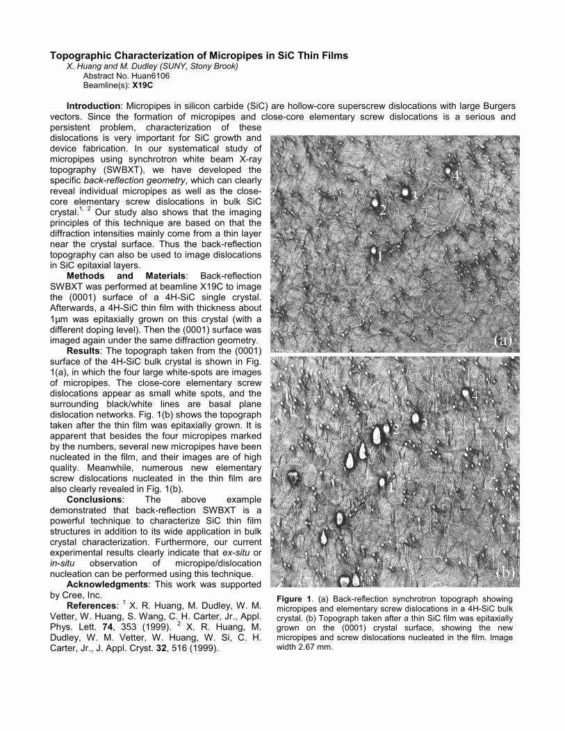
Results: The topograph taken from the (0001) surface of the 4H-SiC bulk crystal is shown in Fig. 1(a), in which the four large white-spots are images of micropipes. The close-core elementary screw dislocations appear as small white spots, and the surrounding black/white lines are basal plane dislocation networks. Fig. 1(b) shows the topograph taken after the thin film was epitaxially grown. It is apparent that besides the four micropipes marked by the numbers, several new micropipes have been nucleated in the film, and their images are of high quality. Meanwhile, numerous new elementary screw dislocations nucleated in the thin film are also clearly revealed in Fig. 1(b).
Conclusions: The above example demonstrated that back-reflection SWBXT is a powerful technique to characterize SiC thin film structures in addition to its wide application in bulk crystal characterization. Furthermore, our current experimental results clearly indicate that ex-situ or in-situ observation of micropipe/dislocation nucleation can be performed using this technique.
Acknowledgments: This work was supported by Cree, Inc.
References: 1 X. R. Huang, M. Dudley, W. M. Vetter, W. Huang, S. Wang, C. H. Carter, Jr., Appl. Phys. Lett. 74, 353 (1999). 2 X. R. Huang, M. Dudley, W. M. Vetter, W. Huang, W. Si, C. H. Carter, Jr., J. Appl. Cryst. 32, 516 (1999).
Figure 1. (a) Back-reflection synchrotron topograph showingmicropipes and elementary screw dislocations in a 4H-SiC bulkcrystal. (b) Topograph taken after a thin SiC film was epitaxiallygrown on the (0001) crystal surface, showing the newmicropipes and screw dislocations nucleated in the film. Imagewidth 2.67 mm.