torque measurement at the single-molecule...
TRANSCRIPT
BB42CH25-Wang ARI 3 April 2013 18:3
Torque Measurementat the Single-Molecule LevelScott Forth,1 Maxim Y. Sheinin,2 James Inman,2
and Michelle D. Wang2,3
1Laboratory of Chemistry and Cell Biology, The Rockefeller University, New York,New York 10065; email: [email protected] of Physics, Laboratory of Atomic and Solid State Physics, 3Howard HughesMedical Institute, Cornell University, Ithaca, New York 14853;email: [email protected], [email protected], [email protected]
Annu. Rev. Biophys. 2013. 42:583–604
The Annual Review of Biophysics is online atbiophys.annualreviews.org
This article’s doi:10.1146/annurev-biophys-083012-130412
Copyright c© 2013 by Annual Reviews.All rights reserved
Keywords
angular optical trapping, rotor bead tracking, magnetic tweezers, DNAphase transitions, rotary molecular motors
Abstract
Methods for exerting and measuring forces on single molecules have revo-lutionized the study of the physics of biology. However, it is often the casethat biological processes involve rotation or torque generation, and these pa-rameters have been more difficult to access experimentally. Recent advancesin the single-molecule field have led to the development of techniques thatadd the capability of torque measurement. By combining force, displace-ment, torque, and rotational data, a more comprehensive description of themechanics of a biomolecule can be achieved. In this review, we highlighta number of biological processes for which torque plays a key mechanicalrole. We describe the various techniques that have been developed to di-rectly probe the torque experienced by a single molecule, and detail a varietyof measurements made to date using these new technologies. We concludeby discussing a number of open questions and propose systems of study thatwould be well suited for analysis with torsional measurement techniques.
583
Ann
u. R
ev. B
ioph
ys. 2
013.
42:5
83-6
04. D
ownl
oade
d fr
om w
ww
.ann
ualr
evie
ws.
org
by C
orne
ll U
nive
rsity
on
05/1
4/13
. For
per
sona
l use
onl
y.
BB42CH25-Wang ARI 3 April 2013 18:3
Contents
INTRODUCTION . . . . . . . . . . . . . . . . . . . . . . . . . . . . . . . . . . . . . . . . . . . . . . . . . . . . . . . . . . . . . . . 584TORQUE IN BIOLOGICAL SYSTEMS. . . . . . . . . . . . . . . . . . . . . . . . . . . . . . . . . . . . . . . . . . 584
DNA and DNA-Based Motors . . . . . . . . . . . . . . . . . . . . . . . . . . . . . . . . . . . . . . . . . . . . . . . . . . 585Rotary Protein Motors . . . . . . . . . . . . . . . . . . . . . . . . . . . . . . . . . . . . . . . . . . . . . . . . . . . . . . . . . . 587
METHODS OF TORQUE DETECTION. . . . . . . . . . . . . . . . . . . . . . . . . . . . . . . . . . . . . . . . 588Electrorotation . . . . . . . . . . . . . . . . . . . . . . . . . . . . . . . . . . . . . . . . . . . . . . . . . . . . . . . . . . . . . . . . . 588Viscous Drag of a Rotating Body . . . . . . . . . . . . . . . . . . . . . . . . . . . . . . . . . . . . . . . . . . . . . . . . 589Optical Trapping . . . . . . . . . . . . . . . . . . . . . . . . . . . . . . . . . . . . . . . . . . . . . . . . . . . . . . . . . . . . . . . 589Magnetic Tweezers . . . . . . . . . . . . . . . . . . . . . . . . . . . . . . . . . . . . . . . . . . . . . . . . . . . . . . . . . . . . . 592Comparison of Different Techniques . . . . . . . . . . . . . . . . . . . . . . . . . . . . . . . . . . . . . . . . . . . . 593
TORQUE MEASUREMENTS ON BIOLOGICAL SYSTEMS. . . . . . . . . . . . . . . . . . . . 594Introduction to DNA Mechanics . . . . . . . . . . . . . . . . . . . . . . . . . . . . . . . . . . . . . . . . . . . . . . . . 594B-DNA Torsional Modulus . . . . . . . . . . . . . . . . . . . . . . . . . . . . . . . . . . . . . . . . . . . . . . . . . . . . . 595Plectonemic DNA . . . . . . . . . . . . . . . . . . . . . . . . . . . . . . . . . . . . . . . . . . . . . . . . . . . . . . . . . . . . . . 595Underwound DNA . . . . . . . . . . . . . . . . . . . . . . . . . . . . . . . . . . . . . . . . . . . . . . . . . . . . . . . . . . . . . 597Overwound DNA and Twist-Stretch Coupling . . . . . . . . . . . . . . . . . . . . . . . . . . . . . . . . . . . 597DNA with Bound Proteins and Small Molecules . . . . . . . . . . . . . . . . . . . . . . . . . . . . . . . . . 598Motor Proteins . . . . . . . . . . . . . . . . . . . . . . . . . . . . . . . . . . . . . . . . . . . . . . . . . . . . . . . . . . . . . . . . . 598
CONCLUSIONS AND FUTURE DIRECTIONS . . . . . . . . . . . . . . . . . . . . . . . . . . . . . . . . 599
INTRODUCTION
Over the past several decades, techniques for observing and manipulating single biologicalmolecules have opened up new fields of study, allowing researchers in the biological and bio-physical sciences to understand how the components of life behave, not only biochemically butalso mechanically. The direct manipulations of physical parameters, such as force, have elucidatedthe mechanisms of action for many biological machines and structures that compose each livingcell. Although force is certainly a key physical coordinate, it is not the only parameter of impor-tance. Indeed, for a host of cellular machinery, rotation and torque may be even more crucial. Todate, these variables have been much harder to observe directly, leading to gaps in our knowledgeregarding how key biomolecules function. In recent years, however, researchers have developed avariety of techniques and methodologies that enable direct access to these mechanical parameters,allowing for a more comprehensive understanding of the behavior of certain biological compo-nents. In this review, we detail the major technical advances made to date, which allow for themeasurement of rotation and torque in single-molecule systems. We also discuss a variety of resultsobtained from experiments on biomolecules subjected to torsional strain and detail the mannerin which the direct acquisition of torque data has led to new and important insights. Finally, wepropose a range of topics for future study with these new methodologies.
TORQUE IN BIOLOGICAL SYSTEMS
Rotation and the corresponding torque generation are common, though often underappreciated,features of a number of cellular processes. One can broadly identify two kinds of processes that havebeen of particular interest to researchers: those involving DNA, one of the major chiral molecules
584 Forth et al.
Ann
u. R
ev. B
ioph
ys. 2
013.
42:5
83-6
04. D
ownl
oade
d fr
om w
ww
.ann
ualr
evie
ws.
org
by C
orne
ll U
nive
rsity
on
05/1
4/13
. For
per
sona
l use
onl
y.
BB42CH25-Wang ARI 3 April 2013 18:3
of the cell, and its related processing machinery; and those involving the so-called rotary motors,such as F0F1-ATPase and the bacterial flagellar motor. In this section, we provide an overview ofthe in vivo relevance of both torque and rotation.
DNA and DNA-Based Motors
Since the discovery of the double-helical nature of DNA by Watson & Crick in 1953 (117), re-searchers have come to realize that the cell must be able to overcome a set of topological challengesin order to perform some of its basic functions. For example, initiation of replication and tran-scription requires opening of the double helix (57), architectural proteins directionally wrap theDNA (71), and torsional stress can be generated as translocases move along the molecule (26, 68).Alteration of the topological state of DNA, which can include structural changes, is called DNAsupercoiling. Supercoiling is often characterized by means of the superhelical density σ, definedas the number of extra turns in the DNA normalized by the total number of superhelical turnsin relaxed DNA. Superhelical density is tightly regulated in cells and is maintained at approxi-mately −0.05 in both prokaryotes and eukaryotes (92). In prokaryotes, approximately half of thesupercoiling is unconstrained, and the other half is constrained by various proteins such as HU,HNS, and RNA polymerase (RNAP) (30). Conversely, in eukaryotes, on average all supercoil-ing is constrained by the nucleosomes (54), although local unconstrained supercoiling can arise(10, 69). The supercoiling balance is maintained by a special class of enzymes—topoisomerases—that are able to relax (and sometimes introduce) supercoiling.
Supercoiling can drastically alter the DNA molecule. Both positive and negative supercoilingcan induce the formation of intertwined loop-like structures called plectonemes (111). Negativesupercoiling, in particular, has been biochemically shown to disrupt the double-helical structureof DNA, producing a variety of non-B-DNA conformations. Strand separation is the most generalconsequence of underwinding; however, specific sequences can produce more exotic structures:cruciforms at palindromic sites, left-handed Z-DNA, and G-quadruplexes within certain GC-richtracts (55). Although initially these structures were considered a mere in vitro curiosity, researchin past decades has demonstrated their existence in vivo and has provided examples of importantregulatory roles they can play, particularly in transcription.
Transcription initiation requires melting of ∼9 bp of the promoter region (13) and is thusmodulated, in part, by torsion. The effects of supercoiling on the transcription of individual geneshave been documented extensively both in vitro (62) and in vivo (44). Supercoiling can also act as aglobal regulator of transcription, as has been strikingly illustrated by the studies of circadian geneexpression in cyanobacteria (110). In addition to the melting of the promoter region, supercoilingcan promote formation of non-B-DNA structures that can attract regulatory proteins (15).
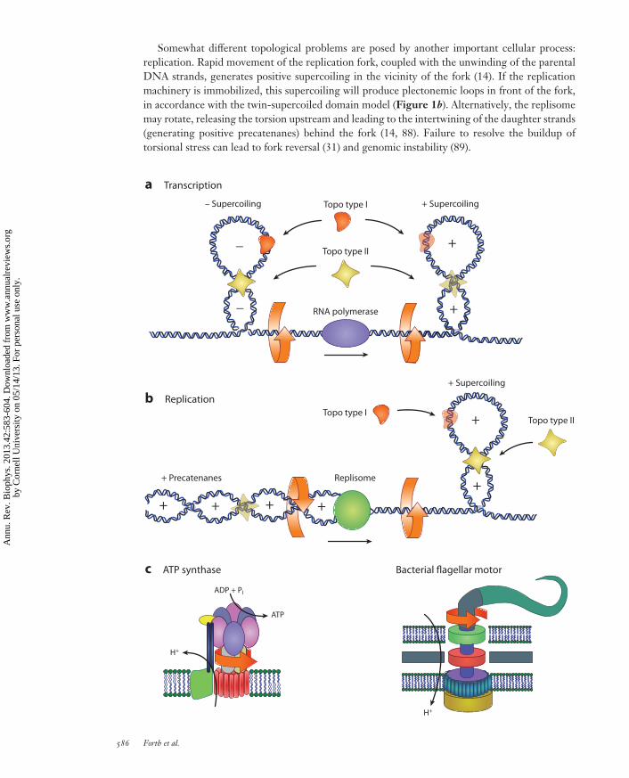
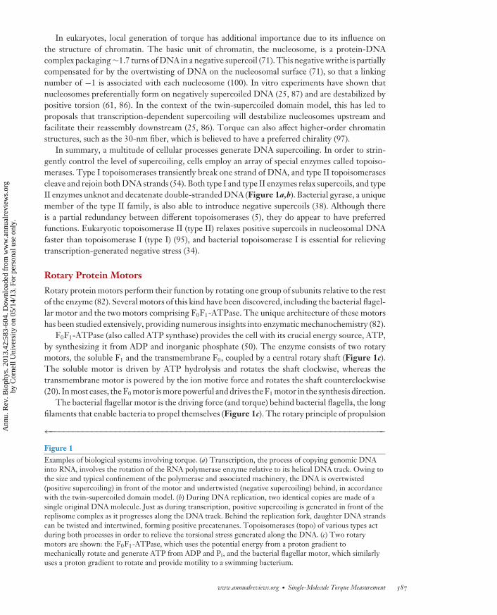
Torque affecting transcription is only half the story. Transcription can also be a source oftorque. During elongation, the RNAP machinery tracks the helical groove of DNA, which requiresrotation of the enzyme relative to DNA (Figure 1a). However, as suggested by Liu & Wang (68),RNAP can become immobilized, requiring DNA to rotate instead and thus generating positivesupercoiling in front of, and negative supercoiling behind, the polymerase. The free rotation ofpolymerase can be prevented by a large viscous drag due to the RNA transcript and associatedfactors, such as ribosomes in prokaryotes and spliceosomes in eukaryotes, as well as tethering tothe cell membrane and other cellular structures (9). This so-called twin-supercoiled domain modelhas been confirmed by numerous in vitro and in vivo experiments (56, 58, 109). Transcription-generated supercoiling can modulate transcription of the same or neighboring genes (23, 56),facilitate formation of RNA-DNA hybrids known as R-loops (1), and potentially affect the structureof chromatin (see below).
www.annualreviews.org • Single-Molecule Torque Measurement 585
Ann
u. R
ev. B
ioph
ys. 2
013.
42:5
83-6
04. D
ownl
oade
d fr
om w
ww
.ann
ualr
evie
ws.
org
by C
orne
ll U
nive
rsity
on
05/1
4/13
. For
per
sona
l use
onl
y.
BB42CH25-Wang ARI 3 April 2013 18:3
Somewhat different topological problems are posed by another important cellular process:replication. Rapid movement of the replication fork, coupled with the unwinding of the parentalDNA strands, generates positive supercoiling in the vicinity of the fork (14). If the replicationmachinery is immobilized, this supercoiling will produce plectonemic loops in front of the fork,in accordance with the twin-supercoiled domain model (Figure 1b). Alternatively, the replisomemay rotate, releasing the torsion upstream and leading to the intertwining of the daughter strands(generating positive precatenanes) behind the fork (14, 88). Failure to resolve the buildup oftorsional stress can lead to fork reversal (31) and genomic instability (89).
_
_
+
+
+
+
++++
– Supercoiling Topo type I
Topo type I
+ Precatenanes Replisome
ADP + PI
ATP
H+
H+
Topo type II
RNA polymerase
+ Supercoiling
+ Supercoiling
a Transcription
b Replication
c ATP synthase Bacterial flagellar motor
Topo type II
586 Forth et al.
Ann
u. R
ev. B
ioph
ys. 2
013.
42:5
83-6
04. D
ownl
oade
d fr
om w
ww
.ann
ualr
evie
ws.
org
by C
orne
ll U
nive
rsity
on
05/1
4/13
. For
per
sona
l use
onl
y.
BB42CH25-Wang ARI 3 April 2013 18:3
In eukaryotes, local generation of torque has additional importance due to its influence onthe structure of chromatin. The basic unit of chromatin, the nucleosome, is a protein-DNAcomplex packaging ∼1.7 turns of DNA in a negative supercoil (71). This negative writhe is partiallycompensated for by the overtwisting of DNA on the nucleosomal surface (71), so that a linkingnumber of −1 is associated with each nucleosome (100). In vitro experiments have shown thatnucleosomes preferentially form on negatively supercoiled DNA (25, 87) and are destabilized bypositive torsion (61, 86). In the context of the twin-supercoiled domain model, this has led toproposals that transcription-dependent supercoiling will destabilize nucleosomes upstream andfacilitate their reassembly downstream (25, 86). Torque can also affect higher-order chromatinstructures, such as the 30-nm fiber, which is believed to have a preferred chirality (97).
In summary, a multitude of cellular processes generate DNA supercoiling. In order to strin-gently control the level of supercoiling, cells employ an array of special enzymes called topoiso-merases. Type I topoisomerases transiently break one strand of DNA, and type II topoisomerasescleave and rejoin both DNA strands (54). Both type I and type II enzymes relax supercoils, and typeII enzymes unknot and decatenate double-stranded DNA (Figure 1a,b). Bacterial gyrase, a uniquemember of the type II family, is also able to introduce negative supercoils (38). Although thereis a partial redundancy between different topoisomerases (5), they do appear to have preferredfunctions. Eukaryotic topoisomerase II (type II) relaxes positive supercoils in nucleosomal DNAfaster than topoisomerase I (type I) (95), and bacterial topoisomerase I is essential for relievingtranscription-generated negative stress (34).
Rotary Protein Motors
Rotary protein motors perform their function by rotating one group of subunits relative to the restof the enzyme (82). Several motors of this kind have been discovered, including the bacterial flagel-lar motor and the two motors comprising F0F1-ATPase. The unique architecture of these motorshas been studied extensively, providing numerous insights into enzymatic mechanochemistry (82).
F0F1-ATPase (also called ATP synthase) provides the cell with its crucial energy source, ATP,by synthesizing it from ADP and inorganic phosphate (50). The enzyme consists of two rotarymotors, the soluble F1 and the transmembrane F0, coupled by a central rotary shaft (Figure 1c).The soluble motor is driven by ATP hydrolysis and rotates the shaft clockwise, whereas thetransmembrane motor is powered by the ion motive force and rotates the shaft counterclockwise(20). In most cases, the F0 motor is more powerful and drives the F1 motor in the synthesis direction.
The bacterial flagellar motor is the driving force (and torque) behind bacterial flagella, the longfilaments that enable bacteria to propel themselves (Figure 1c). The rotary principle of propulsion
←−−−−−−−−−−−−−−−−−−−−−−−−−−−−−−−−−−−−−−−−−−−−−−−−−−−−−−−−−−−−−−−−−−−−−−−−Figure 1Examples of biological systems involving torque. (a) Transcription, the process of copying genomic DNAinto RNA, involves the rotation of the RNA polymerase enzyme relative to its helical DNA track. Owing tothe size and typical confinement of the polymerase and associated machinery, the DNA is overtwisted(positive supercoiling) in front of the motor and undertwisted (negative supercoiling) behind, in accordancewith the twin-supercoiled domain model. (b) During DNA replication, two identical copies are made of asingle original DNA molecule. Just as during transcription, positive supercoiling is generated in front of thereplisome complex as it progresses along the DNA track. Behind the replication fork, daughter DNA strandscan be twisted and intertwined, forming positive precatenanes. Topoisomerases (topo) of various types actduring both processes in order to relieve the torsional stress generated along the DNA. (c) Two rotarymotors are shown: the F0F1-ATPase, which uses the potential energy from a proton gradient tomechanically rotate and generate ATP from ADP and Pi, and the bacterial flagellar motor, which similarlyuses a proton gradient to rotate and provide motility to a swimming bacterium.
www.annualreviews.org • Single-Molecule Torque Measurement 587
Ann
u. R
ev. B
ioph
ys. 2
013.
42:5
83-6
04. D
ownl
oade
d fr
om w
ww
.ann
ualr
evie
ws.
org
by C
orne
ll U
nive
rsity
on
05/1
4/13
. For
per
sona
l use
onl
y.

BB42CH25-Wang ARI 3 April 2013 18:3
was demonstrated in the 1970s (7), and incidentally, this was also the first observation of the actionof a single molecular motor. This 11-MDa, 45-nm-diameter machine can rotate at up to 1,700 Hzand generate up to a remarkable 4,500 pN · nm of torque (72, 103, 104). Like the F1-ATPasemotor, the bacterial flagellar motor is a transmembrane protein driven by the ion motive force.However, owing to its large size and complexity, the exact principle of operation of the motorremains elusive (103).
Although the importance of rotation has been well established for the enzymes described above,the list is not exhaustive. The molecular motors myosin and kinesin have been observed to partiallyrotate their cargo (42, 53). It has also been suggested that certain members of the AAA family, suchas helicases and DNA packaging proteins, are capable of generating rotation (82). The abundanceof torsion-generating enzymes underscores the importance of torque for cellular functions.
METHODS OF TORQUE DETECTION
Despite the importance of torque as a key mechanical regulator in a significant number of bio-logical processes, understanding how torsional stress regulates cellular functions has proven tobe experimentally challenging. For example, the most commonly employed method to study reg-ulation by DNA supercoiling has been one- or two-dimensional gel electrophoresis. However,this biochemical method does not measure torque directly and also has limited ability to discerndynamical behavior and population heterogeneities. As Cozzarelli et al. (26) pointed out, a reasonthat “twist and torque changes have been underappreciated is that until recently they were notdirectly measurable.”
Single-molecule techniques developed in the past two decades have proven to be powerfulapproaches for the investigation of the response of biological systems to torsional stress. Individ-ual DNA molecules can now be twisted, and molecular motors, acting on these DNA molecules,can be monitored under physiologically relevant conditions. Initial efforts focused on generat-ing and measuring rotation of a torsionally constrained biological molecule. More recent effortshave resulted in the ability to precisely control the torque exerted on a molecule and to directlymeasure the torque generated by the molecule, permitting more quantitative measurements ofrotational motions and torsional properties in biology. This review is not intended to be exhaus-tive in the coverage of all methods of torque measurement. Instead, we focus on methods thathave demonstrated impact and applications at the single-molecule level. We highlight four majorcategories of torque measurement techniques in a roughly chronological fashion of their develop-ment: (a) electrorotation, (b) viscous drag of a rotating body, (c) optical trapping, and (d ) magnetictweezers.
Electrorotation
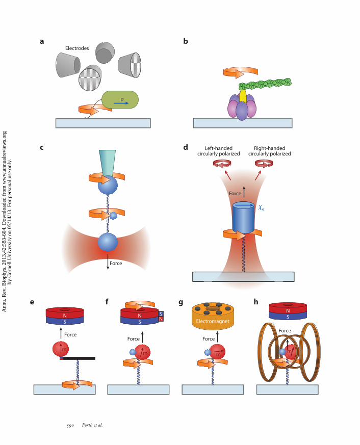
Direct application of an external torque on a single molecule was first demonstrated by the methodof electrorotation, which applies a constant torque to a polarizable and conductive particle by arotating electric field (8, 115). Briefly, a megahertz rotating electric field is created by applying ACvoltage to radially oriented metallic probes. The induced dipole of a particle in the field rotates atthe same frequency, but it lags or leads the electric field due to the conductance and/or dielectricloss of the particle. This phase delay produces a constant torque on the particle, which dependson the field intensity, rotation rate, and properties of the particle and the solution. It is importantto note this relation is complex, and rotation of the particle in the opposite sense of field rotationis possible (49). Studies of the bacterial flagellar motor were enhanced by applying electrorotationto the tethered bacterial cells (8, 115) (Figure 2a). This assay is not limited to the rotation of cells;
588 Forth et al.
Ann
u. R
ev. B
ioph
ys. 2
013.
42:5
83-6
04. D
ownl
oade
d fr
om w
ww
.ann
ualr
evie
ws.
org
by C
orne
ll U
nive
rsity
on
05/1
4/13
. For
per
sona
l use
onl
y.

BB42CH25-Wang ARI 3 April 2013 18:3
it can also be used to rotate typical dielectric probe particles (108). The combination of opticaltrapping and electrorotation has been demonstrated and allows for high-bandwidth detection ofrotation by back focal plane interferometry (93).
Viscous Drag of a Rotating Body
The earliest reported measurements of torque on single biological molecules were made by exploit-ing the drag torque experienced by a body rotating through solution (6, 8). The visual observationof a rotating probe can give sufficient estimates of torque generation, either by estimation of theviscous drag coefficient or by calibration of the viscous drag. The exerted torque can only be variedvia the probe geometry and the viscosity of the solution. Thus, the main limitation of this methodis that torque and rotational velocity are never fully decoupled. It is therefore difficult to exert auser-defined constant torque, which would be advantageous when probing important parameterssuch as a motor’s stalling torque.
The viscous drag torque method was first used to study single rotary motors. The torquegenerated by a single bacterial flagellar motor was estimated on the basis of the viscous torqueexperienced by the bacterium (70). Subsequently, the torque generated by a single F1-ATPasemotor was estimated using an actin filament as the rotational probe coupled to the motor (78)(Figure 2b). The viscous drag torque can be calculated for the rotating body as the product of itsviscous drag coefficient and rotation rate.
Recent developments in the use of the viscous drag method for DNA-based torque measure-ments have greatly improved the precision and control of this methodology. Bryant et al. (19) devel-oped a clever rotor bead tracking (RBT) method that utilized the rotation of a small bead attachedto the side of a DNA tether in order to both apply and directly measure torque (Figure 2c). Therotation of wound DNA, or the action of DNA binding proteins, is observed by visually trackingthe spatial position of the rotor bead as it revolves about the long axis of the DNA strand. Torqueis determined by measuring the angular velocity of the rotor bead and multiplying it by the viscousdrag factor for a sphere rotating about an axis on its edge. The accuracy of the method is enhancedby the calibration of the viscous drag factor for each rotor bead. In order to maintain DNA as a lin-ear rotation axis, the DNA needs to be extended by optical traps, conventional magnetic tweezers,or micropipettes, while passively observing rotation and extension change (39, 40). RBT can also beused to exert a controlled torque by applying twist with one of the force probes, such that the rotorbead rotates at a constant speed while the force is independently controlled by the force probe (80).
Optical Trapping
Since its introduction to biology by Ashkin et al. (3), optical trapping has proven to be an invaluabletool for single-molecule research, permitting the dynamics of motor proteins and their substratesto be examined mechanically one molecule at a time. Systems that have been investigated underforce include, but are not limited to, RNAP (113), DNA polymerase (119), helicase (48), theribosome (118), nucleosomes (16), DNA (102), RNA (67), and viral motors (101). However, untilrather recently, measurements were limited to forces and displacements.
In order to use optical trapping to investigate rotational motions, a trapped particle needs tobe rotated by the trapping beam. Conventional optical traps employ a Gaussian laser beam andan optically isotropic microsphere that cannot be rotated by a trap with either a linear or circularpolarization. Therefore, many of the early demonstrations to rotate the trapping particle reliedon breaking the rotational symmetry of the particle and/or the input trapping beam (12, 35, 37,79). In a seminal work by Friese et al. (35), a calcite particle, which is optically birefringent, was
www.annualreviews.org • Single-Molecule Torque Measurement 589
Ann
u. R
ev. B
ioph
ys. 2
013.
42:5
83-6
04. D
ownl
oade
d fr
om w
ww
.ann
ualr
evie
ws.
org
by C
orne
ll U
nive
rsity
on
05/1
4/13
. For
per
sona
l use
onl
y.
BB42CH25-Wang ARI 3 April 2013 18:3
a b
c
e f g h
d
Electrodes
Left-handed circularly polarized
Right-handed circularly polarized
p
Force
Force
χe
ForceForce Force
Electromagnet
Force
mm m
m
NS
NS
NS
NS
590 Forth et al.
Ann
u. R
ev. B
ioph
ys. 2
013.
42:5
83-6
04. D
ownl
oade
d fr
om w
ww
.ann
ualr
evie
ws.
org
by C
orne
ll U
nive
rsity
on
05/1
4/13
. For
per
sona
l use
onl
y.
BB42CH25-Wang ARI 3 April 2013 18:3
rotated with both linear and circularly polarized light. This work provided the inspiration for anew instrument for single-molecule studies that is described below.
A recent advance in optical trapping techniques, namely the angular optical trap (AOT), alsotermed the optical torque wrench, has enabled direct torque and rotation detection of individualbiological molecules (29, 59) (Figure 2d ). There are three core features of this instrument.
First, the trapping particle is a nanofabricated quartz cylinder, which has its extraordinaryoptical axis perpendicular to its cylinder axis and one of its ends chemically derivatized forattachment to a biological molecule of interest (29). Quartz has positive optical anisotropy, witha single axis more polarizable than the other two, so that a quartz particle is angularly confinedby a linearly polarized light in two of its three Euler angles. [In contrast, a calcite particle (35)can be confined in only one of its three Euler angles.] The remaining Euler angle of the quartzcylinder is confined by the shape anisotropy intrinsic to an elongated cylinder. When a quartzcylinder is trapped by a linearly polarized laser, its cylinder axis aligns with the direction of lightpropagation so that the cylinder can be rotated about its axis by rotation of the laser polarization.Attaching a biological molecule specifically to one end of the cylinder allows the applicationof force to the molecule along the laser propagation direction, permitting independent controlof force and torque. Nanofabrication techniques allow for the mass production of cylinders ofuniform size, shape, and optical properties, as well as specific chemical derivatization of only oneend of each cylinder. The cylinders may be fabricated by optical lithography (29) or, for moreselective localization of a molecule’s attachment point, by electron beam lithography (45).
The second feature of an AOT is a rapid and flexible control of the input linear polarizationof the trapping laser beam, so that the instrument may function in different modes of operation(59). The use of a pair of acousto-optic modulators (AOMs) provides continuous and rapidcontrol (∼100 kHz) of the input polarization (59). Alternatively, an electro-optic modulator maybe used to rotate the input polarization (41). In the rotation mode, the particle is rotated by simplerotation of the polarization, with the particle’s optical axis closely tracking the electric field of thelaser beam (35, 59). In the active torque wrench mode, a constant torque on the trapped particleis maintained via active feedback on the input polarization angle (59). This mode is best suitedfor applications at high torques. In the passive torque wrench mode, a constant optical torqueis achieved by rotating the polarization at a rate much faster than the particle is able to respond,resulting in a minute constant torque exerted on the particle (46). This technique establishes aclear relationship between the rapid polarization rotation rate and the value of the torque actingon the particle, thereby allowing for an easily controllable torque. This passive torque wrench
←−−−−−−−−−−−−−−−−−−−−−−−−−−−−−−−−−−−−−−−−−−−−−−−−−−−−−−−−−−−−−−−−−−−−−−−−−−−−−−−−−−−−−−−−−−Figure 2Experimental configurations for single-molecule torque measurements. (a) Electrorotation of a single bacterium with its flagellumbound to the surface (8). (b) Rotation of the F1-ATPase can be observed by monitoring the rotational orientation of a fluorescent actinfilament (78). (c) The rotor bead tracking assay utilizes the rotation of a small bead to monitor and/or generate torque. Force andexternally imposed twist can be controlled at the DNA ends by microsphere handles (19). (d ) The angular optical trap angularly orientsa quartz cylinder with the linear polarization of the input trapping laser beam. The polarization state of the transmitted beam, asmeasured by the transmitted light intensities in the right- and left-handed circular polarizations, directly determines the applied torqueon a double-stranded DNA (dsDNA) molecule and the angular orientation of the DNA (29, 59). (e) A magnetic tweezers setup byCeledon et al. (21) consists of a magnetic bead and nanorod torque arm to angularly orient one end of a dsDNA tether; twist isintroduced by rotating the microscope cover glass. ( f ) Magnetic torque tweezers incorporate a supplementary side magnet to add asmall horizontal perturbation to the vertical magnetic field created by a cylindrical magnet, producing a low-stiffness angular trap toorient a magnetic bead (65). ( g) Soft magnetic tweezers use a six pole electromagnet to create a rapidly rotating horizontal magneticfield of varying intensity in order to produce either a constant torque or a magnetic angular trap of tunable stiffness (77).(h) Electromagnetic torque tweezers use two pairs of Helmholtz coils for dynamic control of the horizontal magnetic field (47).
www.annualreviews.org • Single-Molecule Torque Measurement 591
Ann
u. R
ev. B
ioph
ys. 2
013.
42:5
83-6
04. D
ownl
oade
d fr
om w
ww
.ann
ualr
evie
ws.
org
by C
orne
ll U
nive
rsity
on
05/1
4/13
. For
per
sona
l use
onl
y.

BB42CH25-Wang ARI 3 April 2013 18:3
operates as if the AOT has zero torsional stiffness, and as the torque approaches zero the particlecan freely rotate in the trap. This mode is optimal for zero- and low-torque applications.
Finally, torque detection in an AOT is based on the change in the ellipticity of the trappingbeam after it interacts with the trapping particle, a method independently demonstrated by twodifferent groups (12, 59). In an AOT, a quartz cylinder is trapped such that its extraordinaryaxis, which is more polarizable than the other two axes, is aligned with the input beam’s linearpolarization. If the cylinder is rotated away from this stable trapping orientation, there will be arestoring torque, which arises due to misalignment of the particle’s polarization and the electricfield. Direct torque measurements are subsequently made by measuring the change in the angularmomentum of the transmitted beam downstream of the trapped particle, which is accomplished bysplitting the beam into its left- and right-circular components and determining their differentialintensities (12, 59).
In an AOT, the same trapping beam is used for both torque and angle detection of the trappedparticle, without the need for a secondary detection beam or imaging method. Such a detectionmethod is exceedingly direct, relying solely on conservation of angular momentum, and thusdistinguishes the AOT from other methods described in this review. During a typical experiment,force, displacement, torque, and angle of the cylinder are simultaneously measured at kilohertzfrequencies, making this method well suited for the study of fast events.
An AOT takes advantage of a combination of optical and shape anisotropy to angularly orienta trapped particle. Particle orientation may also be achieved solely via shape anisotropy withparticles made as disks (81), long rods (12), or more complex objects (37). In particular, Orosziet al. (81) trapped a disk-shaped particle with linearly polarized light and measured the torsionalstiffness of DNA by imaging the particle’s angular deflection. In addition, although AOT employsa linearly polarized trapping beam to rotate a particle, rotation may also be achieved via circular andelliptical (35, 36) polarizations, laser beams carrying both spin and orbital momentums (84, 85),and asymmetrical trapping beams (79). Future studies may reveal whether these types of variationswill further lend themselves to direct torque measurements in single-molecule experiments.
Magnetic Tweezers
Magnetic tweezers are the most well-known technique used to apply twist to a single biologicalmolecule. In an elegant demonstration by Strick et al. (105), single DNA molecules were super-coiled, via rotation of a magnetic bead, using a pair of permanent magnets oriented transverse tothe DNA molecule. This technique is relatively simple to employ, and a major advantage is theability to monitor the behavior of many molecules simultaneously, a feature typically lacking inmany other rotation methods. Even though torque was not directly measured, this technique hasproven to be a powerful tool to study torsional properties of DNA (105), chromatin (4), RNAP(91), and topoisomerases (107).
Conventional magnetic tweezers used for rotational experiments tightly confine a magneticbead’s angular orientation, resulting in a stiff angular trap (52). Measuring torque in this scenariowould require the detection of a minute angular deviation between the applied field and themagnetic bead, well below the typical resolution of an optical microscopy–based method. Torquedetection with magnetic tweezers thus requires dramatic reduction in the torsional stiffness aboutthe axis parallel to the applied force. There has been a recent surge of magnetic tweezers–baseddevices suitable for making such torque measurements.
A solution is to orient the magnets axially instead of transversely, dramatically reducing the hori-zontal component of the magnetic force. Indeed Harada et al. (43) used this approach to track DNArotation generated by Escherichia coli RNAP as visualized by attaching small fluorescent beads to
592 Forth et al.
Ann
u. R
ev. B
ioph
ys. 2
013.
42:5
83-6
04. D
ownl
oade
d fr
om w
ww
.ann
ualr
evie
ws.
org
by C
orne
ll U
nive
rsity
on
05/1
4/13
. For
per
sona
l use
onl
y.

BB42CH25-Wang ARI 3 April 2013 18:3
the magnetic bead. Although direct torque measurement was not obtained in this study, it demon-strated that torsional stiffness can indeed be greatly reduced by orienting the magnets axially.
The first realization of direct torque measurements using magnetic tweezers with axiallyoriented magnets was made by Celedon et al. (21, 22). Their assay involved an axially orientedcylindrical magnet to apply force on a magnetic bead coupled to a nanorod torque arm(Figure 2e). A small force on the nanorod kept the probe aligned horizontally, and rotation wasapplied mechanically by moving the sample stage. The torsional stiffness of the probe was madesufficiently low to allow optical microscopy measurements of the angular deviations, and themethod was capable of resolving single pN · nm scale torques.
Lipfert et al. (65) developed magnetic torque tweezers (MTT), a simpler configuration thatdoes not require nanofabricated handles. MTT utilizes a cylindrical magnet to produce an axialmagnetic field and a side-located magnet for a small horizontal field to orient the magnetic bead,and rotation is achieved by rotating the magnets (Figure 2f ). Kauert et al. (51) showed that smallmagnetic field asymmetries generated in the main magnets oriented axially can also be sufficientto orient the bead for torque measurements. Lipfert et al. (66) further demonstrated that whenthe magnetic bead is located in the exact center of the field of a cylindrical magnet, the beadwill rotate freely about the axis of force application, and they referred to this approach as freelyorbiting magnetic tweezers (FOMT).
More recent efforts for torque detection with magnetic tweezers have focused on the use ofelectromagnets to provide more precise control of the magnetic field. Mosconi et al. (77) developedthe soft magnetic tweezers apparatus that used electromagnets to rapidly rotate the field in such away as to simultaneously apply and measure an arbitrary torque on a magnetic bead (Figure 2g).Janssen et al. (47) replaced the side magnet of the MTT with two pairs of Helmholtz coils toachieve full control of the transverse magnetic field. This instrument, named electromagnetictorque tweezers (eMTT), combines the features of MTT and FOMT and allows independentcontrol of the vertical force and torsional stiffness (Figure 2h).
Although magnetic tweezers for torque measurement come in different configurations, theyshare the same torque measurement principle. Torque is determined by observing the angularorientation of the magnetic particle relative to the applied magnetic field with image-trackingtechniques and multiplying by a calibrated angular trap stiffness to produce physical torque units(21, 64, 65, 77).
Comparison of Different Techniques
Each method of torque measurement described above has its advantages and disadvantages.Electrorotation has been the method of choice to exert a user-defined constant torque in single-molecule experiments (though other techniques in principle also possess this capability). However,electrorotation has not been adapted to incorporate force control, and associated heating can be se-vere (116). On the other hand, RBT, angular optical trapping, and magnetic tweezers–based tech-niques are all suited for simultaneous torque and force measurements and manipulation. Torqueresolution, one of the critical parameters in investigating minute biological torques, is limited bythe viscous drag coefficient of the probe particle, which scales as the cube of the probe’s dimension(18). A smaller probe, however, limits the amount of force that can be exerted. This limitation iscircumvented in the RBT assay, which decouples force and torque probes. One of the prerequisitesto probe fast dynamics of biological systems is a high data acquisition rate. Because the detectionof the linear and angular parameters in an AOT is performed by directly monitoring the trans-mitted laser beam with photodiodes, acquisition rates in the kilohertz range can be achieved.In comparison, RBT and magnetic tweezers rely on video-based imaging, generally limiting
www.annualreviews.org • Single-Molecule Torque Measurement 593
Ann
u. R
ev. B
ioph
ys. 2
013.
42:5
83-6
04. D
ownl
oade
d fr
om w
ww
.ann
ualr
evie
ws.
org
by C
orne
ll U
nive
rsity
on
05/1
4/13
. For
per
sona
l use
onl
y.
BB42CH25-Wang ARI 3 April 2013 18:3
acquisition rates to, at most, several hundred hertz. On the one hand, methods employing magnetictweezers to exert force do not suffer from potential laser-induced damage and heating. On theother hand, AOT offers flexible control of both force and torque, enabling rapid switching betweendifferent modes of operation (46). In summary, all these methods offer benefits and challenges toa user, and the needs of the experimental system of study should dictate which choice is preferred.
TORQUE MEASUREMENTS ON BIOLOGICAL SYSTEMS
To date, there have been a number of reported measurements of biological torques. These canbe parsed into two broad categories: measurements performed on DNA and DNA-based systems,and measurements performed on rotary molecular motors. In this section, we detail the majorfindings made with the techniques described above.
Introduction to DNA Mechanics
DNA has long been of great interest to biophysicists who have sought to detail its mechanicalbehavior utilizing the terms of polymer physics. For example, the elastic behavior of B-DNAunder tension has been described successfully by the well-known modified Marko-Siggia modelusing two parameters, the bending and stretching moduli (74, 114). In contrast to tension, theDNA response to torsion can be described by a simple harmonic potential Etwist = Cθ2
2L , where Lis the DNA contour length, θ is the added twist, and C is the torsional modulus, a measure of thetorsional stiffness of the molecule. Although early biochemical experiments were able to estimatethe torsional modulus (28), direct measurements have become possible only recently due to theexperimental advances detailed above. By measuring the change in torque �τ as additional twist�θ is added to the molecule, an effective torsional modulus can be experimentally measured asCeff = L �τ
�θ. Interestingly, Ceff has been observed to increase with force and approach C at ∼3 pN
(65, 76); this behavior has been explained by taking into account the writhe fluctuations of theDNA molecule (75). While a harmonic approximation describes the torsional response of B-DNArather well, sensitive experiments have detected nonzero coupling between twist and tension (39,63, 99), leading to a more precise formulation of the torsional energy Etwist = Cθ2
2L + gθ zL . Here, z
is the DNA extension and g is the so-called twist-stretch coupling. Surprisingly, this coupling isnegative for forces up to 30 pN, signifying that DNA overwinds when stretched (39).
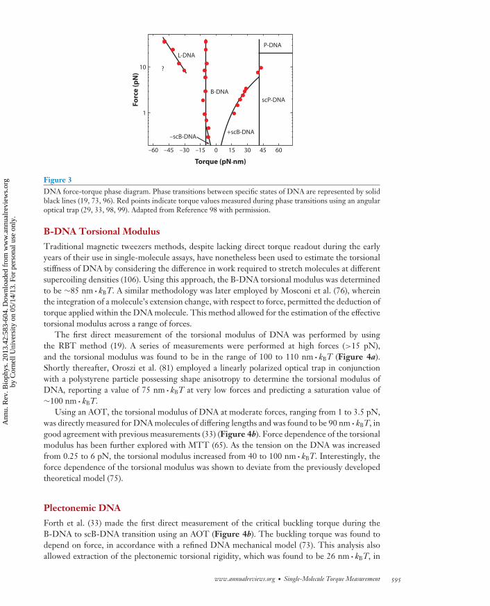
Note that the above discussion relates solely to B-DNA, a very important, but by no meansunique, state of DNA. The past two decades of biophysical experiments have witnessed a plethoraof DNA structures that can be formed under the impact of torsion and tension. For example, undermoderate tension and torque DNA can absorb additional twist by buckling, forming intertwinedsupercoiled loops (also called plectonemes). Such supercoiled DNA (scB-DNA) can exist at lowforces under both positive and negative torque (105). Undertwisting DNA at somewhat higherforces torsionally breaks the base-paired interactions, leading to a state known as L-DNA, whichdiffers in important ways from thermally melted DNA (19, 98). Substantial overtwisting of DNAcreates an exotic state termed P-DNA, which has a smaller helical repeat than standard B-DNAand unpaired bases extruding to the exterior of the molecule (2, 19).
Direct torque measurements are crucial for the identification of these DNA structural transi-tions, which display a phase transition behavior so that the phase coexistence state at a set forceis characterized by a constant torque (73). It is therefore convenient to introduce a force-torquephase diagram (Figure 3). Below, we describe key studies that have characterized the torsionalproperties of various DNA states.
594 Forth et al.
Ann
u. R
ev. B
ioph
ys. 2
013.
42:5
83-6
04. D
ownl
oade
d fr
om w
ww
.ann
ualr
evie
ws.
org
by C
orne
ll U
nive
rsity
on
05/1
4/13
. For
per
sona
l use
onl
y.
BB42CH25-Wang ARI 3 April 2013 18:3
Fo
rce
(p
N)
–60 –45 –30 –15 0 15 30 45 60
1
10
Torque (pN·nm)
L-DNA
–scB-DNA
B-DNA
?
+scB-DNA
scP-DNA
P-DNA
Figure 3DNA force-torque phase diagram. Phase transitions between specific states of DNA are represented by solidblack lines (19, 73, 96). Red points indicate torque values measured during phase transitions using an angularoptical trap (29, 33, 98, 99). Adapted from Reference 98 with permission.
B-DNA Torsional Modulus
Traditional magnetic tweezers methods, despite lacking direct torque readout during the earlyyears of their use in single-molecule assays, have nonetheless been used to estimate the torsionalstiffness of DNA by considering the difference in work required to stretch molecules at differentsupercoiling densities (106). Using this approach, the B-DNA torsional modulus was determinedto be ∼85 nm · kBT. A similar methodology was later employed by Mosconi et al. (76), whereinthe integration of a molecule’s extension change, with respect to force, permitted the deduction oftorque applied within the DNA molecule. This method allowed for the estimation of the effectivetorsional modulus across a range of forces.
The first direct measurement of the torsional modulus of DNA was performed by usingthe RBT method (19). A series of measurements were performed at high forces (>15 pN),and the torsional modulus was found to be in the range of 100 to 110 nm · kBT (Figure 4a).Shortly thereafter, Oroszi et al. (81) employed a linearly polarized optical trap in conjunctionwith a polystyrene particle possessing shape anisotropy to determine the torsional modulus ofDNA, reporting a value of 75 nm · kBT at very low forces and predicting a saturation value of∼100 nm · kBT.
Using an AOT, the torsional modulus of DNA at moderate forces, ranging from 1 to 3.5 pN,was directly measured for DNA molecules of differing lengths and was found to be 90 nm · kBT, ingood agreement with previous measurements (33) (Figure 4b). Force dependence of the torsionalmodulus has been further explored with MTT (65). As the tension on the DNA was increasedfrom 0.25 to 6 pN, the torsional modulus increased from 40 to 100 nm · kBT. Interestingly, theforce dependence of the torsional modulus was shown to deviate from the previously developedtheoretical model (75).
Plectonemic DNA
Forth et al. (33) made the first direct measurement of the critical buckling torque during theB-DNA to scB-DNA transition using an AOT (Figure 4b). The buckling torque was found todepend on force, in accordance with a refined DNA mechanical model (73). This analysis alsoallowed extraction of the plectonemic torsional rigidity, which was found to be 26 nm · kBT, in
www.annualreviews.org • Single-Molecule Torque Measurement 595
Ann
u. R
ev. B
ioph
ys. 2
013.
42:5
83-6
04. D
ownl
oade
d fr
om w
ww
.ann
ualr
evie
ws.
org
by C
orne
ll U
nive
rsity
on
05/1
4/13
. For
per
sona
l use
onl
y.

BB42CH25-Wang ARI 3 April 2013 18:3
–200 –100 0 100 200
–20
0
20
40
60
80
0 200 400 6000
1,000
2,000
3,000
4,000
5,000
0 4 8 12 16
–10
–5
0
5
10
15
20
–6
–5
–4
–3
–2
–1
0
0 2 4 6 8 10
a
d
b
e
c
f
Torq
ue
(p
N·n
m)
Torq
ue
(p
N·n
m)
Torq
ue
(p
N·n
m)
Torq
ue
(p
N·n
m)
Torq
ue
(p
N·n
m)
Ro
tati
on
(tu
rns)
Twist (turns)
Twist (relative turns) Rotation rate (Hz) Time (s)
Force (pN)Twist (turns)
Torque (pN·nm)
TorquescB-DNA
Measured
Predicted (not a fit)
TorqueL-DNA
TorqueP-DNA
DNA DNA
–10
0
10
20
30
40
1 pN2 pN3 pN
0 5 100
10
20
30 Holliday junction
RecA-DNA filament Flagellar motor F1-ATPase
–100 –50 0 50 100
50 mM NaCl
10 mM NaCl
3 mM NaCl
017.831.240.149.0
Figure 4Torque measurements on single biological molecules. (a) Averaged torque trace obtained using rotor bead tracking (RBT) duringunder- and overwinding of 14.8-kb DNA molecules held at high force. Torque plateaus correspond to transitions to P-DNA (positivetwist) or melted L-DNA (negative twist). The torsional modulus was extracted from the slope of the linear region. Adapted fromReference 19 with permission. (b) Individual torque traces obtained with an angular optical trap (AOT) as a single 2.2-kb DNAmolecule was overwound under tension. The torsional modulus was extracted from the slopes of the linear regions. Torque plateauedas the DNA buckled to form plectonemes (supercoiled B-DNA, scB-DNA). Adapted from Reference 33 with permission. (c) Thetorque-force relationship during the migration of a fully homologous Holliday junction, as determined by an AOT. Shown are themean torque values as a function of force (black points) and theoretical prediction (red line; not a fit). Adapted from Reference 32 withpermission. (d ) Torsional response of a RecA filament at 3.5 pN, obtained with magnetic torque tweezers. The wide spread in torquevalues reflects the dynamic nature of the RecA filament ( J. Lipfert, personal communication). Adapted from Reference 65 withpermission. (e) Torque-speed relationship measured for the Na+-driven flagellar motor of Vibrio alginolyticus by monitoring rotation ofa bead attached to the flagellum. Adapted from Reference 104 with permission. ( f ) Rotational trajectories of a single F1-ATPasemotor, under external torque, in the presence of ATP, ADP, and Pi. At the stalling torque (∼31 pN · nm), the motor still exhibitedbidirectional stepwise fluctuations. Adapted from Reference 108 with permission.
good agreement with previous bulk experiments (94). Further analysis of the torque measurementsin the pre- and postbuckling states revealed an overshoot of torque of ∼3 pN · nm at the bucklingtransition; such an overshoot was predicted on the basis of an elastic rod theory (27). Subsequently,Celedon et al. (21) utilized a special magnetic tweezers apparatus to determine the buckling torquesat forces as low as 0.3 pN, further validating the Marko model (73).
Forth et al. (33) also discovered that the buckling transition takes place abruptly and is highlydynamic. At the buckling transition, the DNA extension hops rapidly between two distinctivestates: an extended prebuckled state and a plectonemic postbuckled state. Furthermore, the ini-tial plectonemic loop absorbed approximately twice as much extension as each subsequent turn.Interestingly, such an abrupt transition was absent in previous magnetic tweezers measurements,
596 Forth et al.
Ann
u. R
ev. B
ioph
ys. 2
013.
42:5
83-6
04. D
ownl
oade
d fr
om w
ww
.ann
ualr
evie
ws.
org
by C
orne
ll U
nive
rsity
on
05/1
4/13
. For
per
sona
l use
onl
y.

BB42CH25-Wang ARI 3 April 2013 18:3
where instead a smooth and gradual transition was observed (105). The angular trapping methodallowed for the detection of this abrupt transition, due to higher bandwidth, increased spatial reso-lution, and the use of shorter DNA tethers. More recent experiments, using an upgraded magnetictweezers setup, provided systematic measurements of the buckling transition under different DNAlengths and salt conditions (17).
Underwound DNA
Under moderate forces (>0.6 pN) negative torque induces DNA strand separation to melt DNAwithout undergoing buckling transition. Bryant et al. (19) first directly measured the meltingtorque using the rotary bead assay and found it to be approximately −10 pN · nm (Figure 4a). Thisvalue was further supported with measurements by Sheinin et al. (98) using an AOT. Torsionallymelted DNA was long thought to be equivalent to a thermally melted DNA bubble. Interestingly,Bryant et al. (19) and Sheinin et al. (98) independently discovered that melted DNA actuallyexists in a unique left-handed configuration, termed L-DNA, with mechanical properties distinctfrom those of both B-DNA and Z-DNA and inconsistent with the predicted mechanical behaviorof thermally melted parallel strands (2). Sheinin et al. (98) also revealed that under low forcedifferent DNA sequences exhibited drastically different behavior when underwound and that theunderwinding process was not reversible under the experimental timescale. These results suggestthat the transition from B-DNA to L-DNA at low force occurs along a complex pathway, withmultiple secondary structures formed off equilibrium. Subsequent to this discovery, Oberstrasset al. (80) studied the torsional behavior of a range of DNA sequences using a feedback-enhancedRBT method. The L-DNA state was also observed and was found to possess mechanical parameterssimilar to those measured previously (98). In addition, GC-rich tracts formed Z-DNA undermoderate negative torsional stress of approximately −3 pN · nm, whereas tracts of mismatchedDNA behave similarly to a B-DNA-like helical structure during underwinding.
Besides Z-DNA, a number of other sequence-specific non-B-DNA structures can form undernegative torsion (83). One notable example is a DNA cruciform, or Holliday junction, which isfavored at palindromic or near-palindromic sequences. Forth et al. (32) directly measured thetorque generated during Holliday junction migration for both fully homologous and single-baseheterologous sequences using an AOT. The minute torques observed during smooth migration,on the order of 1 pN · nm, were found to depend on force in a predictable manner (Figure 4c).However, it took ∼7 pN · nm (consistent with the magnitude of torque needed to melt DNA) tomigrate through just a single base mismatch.
Overwound DNA and Twist-Stretch Coupling
While the existence of highly overwound P-DNA was first inferred on the basis of extensionmeasurements using magnetic tweezers (2) and micropipettes (60), crucial additional confir-mation was provided by rotary bead experiments (19). Under high force, B-DNA transitionsto P-DNA at ∼35 pN · nm (Figure 4a) and the helical pitch of P-DNA was found to be∼2.7 bp per turn. Deufel et al. (29) measured a similar torque value for the transition fromB-DNA to supercoiled P-DNA (scP-DNA), which is P-DNA shortened by secondary structureformation (2).
Conventional wisdom predicts that DNA should shorten when overwound under tension.Surprisingly, Gore et al. (39) discovered that DNA lengthens instead, achieving a maximumextension before beginning to shorten again. These findings yielded a negative twist-stretch cou-pling coefficient of −22 kBT. The extension peak was thought to correspond to the location of
www.annualreviews.org • Single-Molecule Torque Measurement 597
Ann
u. R
ev. B
ioph
ys. 2
013.
42:5
83-6
04. D
ownl
oade
d fr
om w
ww
.ann
ualr
evie
ws.
org
by C
orne
ll U
nive
rsity
on
05/1
4/13
. For
per
sona
l use
onl
y.
BB42CH25-Wang ARI 3 April 2013 18:3
a phase transition between B-DNA and scP-DNA (63). Sheinin et al. (99) further investigatedthe twist-stretch coupling using an AOT and measured a value of −21 kBT, in agreement withGore et al. (39). However, examination of the concurrently measured torque signal showed thatthe phase transition did not occur at the peak of the extension but instead at a higher degree ofsupercoiling. This result underscores the importance of direct experimental access to all possiblevariables.
DNA with Bound Proteins and Small Molecules
In addition to measurements of the torsional properties of naked DNA, several studies havebeen performed on protein-bound DNA filaments and DNA intercalated with small molecules.Celedon et al. (21) directly measured the torque required to twist nucleosomal arrays. Althougha wide experimental variability in both extension and torque behavior existed among moleculesdue to variations in nucleosome occupancy, it was still unambiguously shown that chromatin has atorsional stiffness much softer than that of naked DNA. Subsequent work by the same group alsofound that ethidium bromide intercalation of DNA leads to torsional softening of the molecule,but the bending stiffness remains largely unchanged (22). In contrast, magnetic torque tweezersexperiments showed (65) that RecA filaments were approximately twice as torsionally stiff as bareDNA, with a torsional modulus of ∼175 nm · kBT (Figure 4d ).
Motor Proteins
Torque measurements have also been performed on several molecular motors. The E. coli flagellarmotor has been studied extensively with a variety of techniques, including viscous drag on cells (70)or beads (24, 90), optical trapping (11), and electrorotation (8). Although a range of torque valueshas been obtained, a recent work has determined the maximum torque to be ∼1,300 pN · nm(90). In comparison, the flagellar motor of Vibrio alginolyticus was shown to generate torques ofup to 4,000 pN · nm during its rotation (104). The flagellar motor also displayed a nonlineartorque-speed relationship (8, 24, 104), as the torque remained nearly constant for speeds up toseveral hundred hertz and subsequently decreased quickly (Figure 4e). More experimental andtheoretical work is still required to further understand the mechanochemical cycle of the flagellarmotor (103).
The significantly smaller rotary motor F1-ATPase has also been studied in single-moleculedetail. By attaching a fluorescent actin filament to a surface-immobilized motor and observing theresulting rotation rate, Noji et al. (78) estimated that a single rotary unit can generate torques ofup to ∼40 pN · nm. This finding, together with the discovery that the rotary shaft of F1-ATPasemakes 120◦ steps (120), allowed Kinosita and coworkers to conclude that nearly 100% of theenergy from ATP hydrolysis is expended to perform work against the viscous drag. This makesF1-ATPase a highly efficient molecular machine (120). Note that the nonconservative nature ofthe viscous drag force complicates the interpretation of these results in relationship to the truethermodynamic efficiency (112). To overcome this limitation, Toyabe et al. (108) employed theelectrorotation method to investigate the rotational behavior of the F1-ATPase under a constantexternally imposed torque (Figure 4f ). The motor stalled at a torque of 31 pN · nm, whichcorresponded to a thermodynamic efficiency of ∼80% (108).
Similarly, RNAP is capable of generating torques of at least 5 pN · nm, determined by visual-izing the rotation of a DNA-bound bead (43). However, the upper limit of torque generation hasnot yet been directly reported. Such experiments for RNAP and other motor proteins could berepeated using one of the more precise and controllable methodologies outlined above, yieldinga more refined insight into the operation of motors that twist.
598 Forth et al.
Ann
u. R
ev. B
ioph
ys. 2
013.
42:5
83-6
04. D
ownl
oade
d fr
om w
ww
.ann
ualr
evie
ws.
org
by C
orne
ll U
nive
rsity
on
05/1
4/13
. For
per
sona
l use
onl
y.
BB42CH25-Wang ARI 3 April 2013 18:3
CONCLUSIONS AND FUTURE DIRECTIONS
Moving forward, the ability to directly measure the minute torques relevant to biologicalstructures will become increasingly important. Applying these innovative techniques to studythe torsional properties of protein-bound DNA, as well as the enzymes that process DNAduring transcription, replication, and packaging, will be paramount to our understanding oftheir operation. Determining the behavior of chromatin under twist will give insight into therole of torque in chromatin assembly and stability in vivo. Investigation of the AAA family ofDNA-processing machines, such as helicases and phage-packaging motors, should resolve thelong-standing issue of their torque-generating potential.
Although DNA has been studied extensively owing to the relative ease of adapting the biologicalsubstrate to direct torque measurement techniques, a wide class of other biomolecules will mostcertainly be examined as well. The stepping behavior of microtubule-based molecular motors suchas kinesins and dyneins can be modulated by force, and such motors are believed to also be capableof generating and withstanding torsional loads during cargo transport. The direct measurementof stall torques and motor behavior under constant torque will further our understanding of howthese motors behave in vivo. Similarly, studying the kinetics under torsional load of processivemyosins (such as myosin V) as they walk along their actin substrates should lead to insights intotheir mechanics.
Single-molecule methods are incredibly effective at elucidating mechanisms of action thattraditional ensemble methods cannot probe. Direct manipulation of the mechanical propertiesof molecules is a rich and vibrant pursuit, leading to a greater understanding of our biologicalworld. With the addition of direct torque measurement capabilities, we can move even further inour efforts to understand the physical principles underlying biological systems and the role thatmechanics plays in regulating the building blocks of life.
DISCLOSURE STATEMENT
The authors are not aware of any affiliations, memberships, funding, or financial holdings thatmight be perceived as affecting the objectivity of this review.
ACKNOWLEDGMENTS
We would like to acknowledge Drs. Z. Bryant, S. Toyabe, J. Lipfert, and A. Ishijima for sharingdata for figure reproduction, and Drs. K. Kinosita, H. Berg, M. Yoshida, and F. Oberstrass for help-ful communication. We thank Dr. S. Fellman for critical comments on the manuscript. We wish toacknowledge postdoctoral support to S.F. from the National Institutes of Health NRSA fellowship(F32GM099380), graduate traineeship support to J.I. from Cornell University’s Molecular Bio-physics Training Grant (T32GM008267), and support to M.D.W. from the National Institutesof Health grant (GM059849) and National Science Foundation grant (MCB-0820293).
LITERATURE CITED
1. Aguilera A, Garcıa-Muse T. 2012. R loops: from transcription byproducts to threats to genome stability.Mol. Cell 46:115–24
2. Allemand JF, Bensimon D, Lavery R, Croquette V. 1998. Stretched and overwound DNA forms aPauling-like structure with exposed bases. Proc. Natl. Acad. Sci. USA 95:14152–57
3. Ashkin A, Dziedzic JM, Yamane T. 1987. Optical trapping and manipulation of single cells using infrared-laser beams. Nature 330:769–71
www.annualreviews.org • Single-Molecule Torque Measurement 599
Ann
u. R
ev. B
ioph
ys. 2
013.
42:5
83-6
04. D
ownl
oade
d fr
om w
ww
.ann
ualr
evie
ws.
org
by C
orne
ll U
nive
rsity
on
05/1
4/13
. For
per
sona
l use
onl
y.
BB42CH25-Wang ARI 3 April 2013 18:3
4. Bancaud A, Conde e Silva N, Barbi M, Wagner G, Allemand JF, et al. 2006. Structural plasticity of singlechromatin fibers revealed by torsional manipulation. Nat. Struct. Mol. Biol. 13:444–50
5. Baranello L, Levens D, Gupta A, Kouzine F. 2012. The importance of being supercoiled: how DNAmechanics regulate dynamic processes. Biochim. Biophys. Acta 1819:632–38
6. Berg HC. 2003. The rotary motor of bacterial flagella. Annu. Rev. Biochem. 72:19–547. Berg HC, Anderson RA. 1973. Bacteria swim by rotating their flagellar filaments. Nature 245:380–828. Berg HC, Turner L. 1993. Torque generated by the flagellar motor of Escherichia coli. Biophys. J. 65:2201–
169. Bermejo R, Lai MS, Foiani M. 2012. Preventing replication stress to maintain genome stability: resolving
conflicts between replication and transcription. Mol. Cell 45:710–1810. Bermudez I, Garcıa-Martınez J, Perez-Ortın JE, Roca J. 2010. A method for genome-wide analysis of
DNA helical tension by means of psoralen-DNA photobinding. Nucleic Acids Res. 38:e18211. Berry RM, Berg HC. 1997. Absence of a barrier to backwards rotation of the bacterial flagellar motor
demonstrated with optical tweezers. Proc. Natl. Acad. Sci. USA 94:14433–3712. Bishop AI, Nieminen TA, Heckenberg NR, Rubinsztein-Dunlop H. 2003. Optical application and
measurement of torque on microparticles of isotropic nonabsorbing material. Phys. Rev. A 68:813. Borukhov S, Nudler E. 2003. RNA polymerase holoenzyme: structure, function and biological implica-
tions. Curr. Opin. Microbiol. 6:93–10014. Branzei D, Foiani M. 2010. Maintaining genome stability at the replication fork. Nat. Rev. Mol. Cell Biol.
11:208–1915. Brooks TA, Hurley LH. 2009. The role of supercoiling in transcriptional control of MYC and its
importance in molecular therapeutics. Nat. Rev. Cancer 9:849–6116. Brower-Toland BD, Smith CL, Yeh RC, Lis JT, Peterson CL, Wang MD. 2002. Mechanical disruption
of individual nucleosomes reveals a reversible multistage release of DNA. Proc. Natl. Acad. Sci. USA99:1960–65
17. Brutzer H, Luzzietti N, Klaue D, Seidel R. 2010. Energetics at the DNA supercoiling transition. Biophys.J. 98:1267–76
18. Bryant Z, Oberstrass FC, Basu A. 2012. Recent developments in single-molecule DNA mechanics.Curr. Opin. Struct. Biol. 22:304–12
19. Bryant Z, Stone MD, Gore J, Smith SB, Cozzarelli NR, Bustamante C. 2003. Structural transitions andelasticity from torque measurements on DNA. Nature 424:338–41
20. Capaldi RA, Aggeler R. 2002. Mechanism of the F1F0-type ATP synthase, a biological rotary motor.Trends Biochem. Sci. 27:154–60
21. Celedon A, Nodelman IM, Wildt B, Dewan R, Searson P, et al. 2009. Magnetic tweezers measurementof single molecule torque. Nano Lett. 9:1720–25
22. Celedon A, Wirtz D, Sun S. 2010. Torsional mechanics of DNA are regulated by small-molecule inter-calation. J. Phys. Chem. B 114:16929–35
23. Chen D, Bowater R, Dorman CJ, Lilley DM. 1992. Activity of a plasmid-borne Leu-500 promoterdepends on the transcription and translation of an adjacent gene. Proc. Natl. Acad. Sci. USA 89:8784–88
24. Chen X, Berg HC. 2000. Torque-speed relationship of the flagellar rotary motor of Escherichia coli.Biophys. J. 78:1036–41
25. Clark DJ, Felsenfeld G. 1991. Formation of nucleosomes on positively supercoiled DNA. EMBO J.10:387–95
26. Cozzarelli NR, Cost GJ, Nollmann M, Viard T, Stray JE. 2006. Giant proteins that move DNA: bulliesof the genomic playground. Nat. Rev. Mol. Cell Biol. 7:580–88
27. Daniels BC, Forth S, Sheinin MY, Wang MD, Sethna JP. 2009. Discontinuities at the DNA supercoilingtransition. Phys. Rev. E 80:4
28. Depew DE, Wang JC. 1975. Conformational fluctuations of DNA helix. Proc. Natl. Acad. Sci. USA72:4275–79
29. Deufel C, Forth S, Simmons CR, Dejgosha S, Wang MD. 2007. Nanofabricated quartz cylinders forangular trapping: DNA supercoiling torque detection. Nat. Methods 4:223–25
30. Drlica K. 1992. Control of bacterial DNA supercoiling. Mol. Microbiol. 6:425–33
600 Forth et al.
Ann
u. R
ev. B
ioph
ys. 2
013.
42:5
83-6
04. D
ownl
oade
d fr
om w
ww
.ann
ualr
evie
ws.
org
by C
orne
ll U
nive
rsity
on
05/1
4/13
. For
per
sona
l use
onl
y.
BB42CH25-Wang ARI 3 April 2013 18:3
31. Fierro-Fernandez M, Hernandez P, Krimer DB, Stasiak A, Schvartzman JB. 2007. Topological lockingrestrains replication fork reversal. Proc. Natl. Acad. Sci. USA 104:1500–5
32. Forth S, Deufel C, Patel SS, Wang MD. 2011. Direct measurements of torque during Holliday junctionmigration. Biophys. J. 101:L5–7
33. Forth S, Deufel C, Sheinin MY, Daniels B, Sethna JP, Wang MD. 2008. Abrupt buckling transitionobserved during the plectoneme formation of individual DNA molecules. Phys. Rev. Lett. 100:4
34. French SL, Sikes ML, Hontz RD, Osheim YN, Lambert TE, et al. 2011. Distinguishing the roles oftopoisomerases I and II in relief of transcription-induced torsional stress in yeast rRNA genes. Mol. Cell.Biol. 31:482–94
35. Friese MEJ, Nieminen TA, Heckenberg NR, Rubinsztein-Dunlop H. 1998. Optical alignment andspinning of laser-trapped microscopic particles. Nature 394:348–50
36. Funk M, Parkin SJ, Stilgoe AB, Nieminen TA, Heckenberg NR, Rubinsztein-Dunlop H. 2009. Constantpower optical tweezers with controllable torque. Opt. Lett. 34:139–41
37. Galajda P, Ormos P. 2003. Orientation of flat particles in optical tweezers by linearly polarized light.Opt. Express 11:446–51
38. Gellert M, Mizuuchi K, O’Dea MH, Nash HA. 1976. DNA gyrase: an enzyme that introduces superhe-lical turns into DNA. Proc. Natl. Acad. Sci. USA 73:3872–76
39. Gore J, Bryant Z, Noellmann M, Le MU, Cozzarelli NR, Bustamante C. 2006. DNA overwinds whenstretched. Nature 442:836–39
40. Gore J, Bryant Z, Stone MD, Nollmann MN, Cozzarelli NR, Bustamante C. 2006. Mechanochemicalanalysis of DNA gyrase using rotor bead tracking. Nature 439:100–4
41. Gutierrez-Medina B, Andreasson JOL, Greenleaf WJ, LaPorta A, Block SM. 2010. An optical apparatusfor rotation and trapping. Methods Enzymol. 475:377–404
42. Gutierrez-Medina B, Fehr AN, Block SM. 2009. Direct measurements of kinesin torsional proper-ties reveal flexible domains and occasional stalk reversals during stepping. Proc. Natl. Acad. Sci. USA106:17007–12
43. Harada Y, Ohara O, Takatsuki A, Itoh H, Shimamoto N, Kinosita K. 2001. Direct observation of DNArotation during transcription by Escherichia coli RNA polymerase. Nature 409:113–15
44. Hatfield GW, Benham CJ. 2002. DNA topology-mediated control of global gene expression in Escherichiacoli. Annu. Rev. Genet. 36:175–203
45. Huang ZX, Pedaci F, van Oene M, Wiggin MJ, Dekker NH. 2011. Electron beam fabrication of bi-refringent microcylinders. ACS Nano 5:1418–27
46. Inman J, Forth S, Wang MD. 2010. Passive torque wrench and angular position detection using asingle-beam optical trap. Opt. Lett. 35:2949–51
47. Janssen XJA, Lipfert J, Jager T, Daudey R, Beekman J, Dekker NH. 2012. Electromagnetic torquetweezers: a versatile approach for measurement of single-molecule twist and torque. Nano Lett. 12:3634–39
48. Johnson DS, Bai L, Smith BY, Patel SS, Wang MD. 2007. Single-molecule studies reveal dynamics ofDNA unwinding by the ring-shaped T7 helicase. Cell 129:1299–309
49. Jones TB. 2003. Basic theory of dielectrophoresis and electrorotation. IEEE Eng. Medicine Biol. Mag.22:33–42
50. Junge W, Sielaff H, Engelbrecht S. 2009. Torque generation and elastic power transmission in the rotaryF0F1-ATPase. Nature 459:364–70
51. Kauert DJ, Kurth T, Liedl T, Seidel R. 2011. Direct mechanical measurements reveal the materialproperties of three-dimensional DNA origami. Nano Lett. 11:5558–63
52. Klaue D, Seidel R. 2009. Torsional stiffness of single superparamagnetic microspheres in an externalmagnetic field. Phys. Rev. Lett. 102:4
53. Komori Y, Iwane AH, Yanagida T. 2007. Myosin-V makes two Brownian 90 degrees rotations per 36-nmstep. Nat. Struct. Mol. Biol. 14:968–73
54. Koster DA, Crut A, Shuman S, Bjornsti MA, Dekker NH. 2010. Cellular strategies for regulating DNAsupercoiling: a single-molecule perspective. Cell 142:519–30
55. Kouzine F, Levens D. 2007. Supercoil-driven DNA structures regulate genetic transactions. Front. Biosci.12:4409–23
www.annualreviews.org • Single-Molecule Torque Measurement 601
Ann
u. R
ev. B
ioph
ys. 2
013.
42:5
83-6
04. D
ownl
oade
d fr
om w
ww
.ann
ualr
evie
ws.
org
by C
orne
ll U
nive
rsity
on
05/1
4/13
. For
per
sona
l use
onl
y.
BB42CH25-Wang ARI 3 April 2013 18:3
56. Kouzine F, Sanford S, Elisha-Feil Z, Levens D. 2008. The functional response of upstream DNA todynamic supercoiling in vivo. Nat. Struct. Mol. Biol. 15:146–54
57. Kowalski D, Eddy MJ. 1989. The DNA unwinding element: a novel, cis-acting component that facilitatesopening of the Escherichia coli replication origin. EMBO J. 8:4335–44
58. Krasilnikov AS, Podtelezhnikov A, Vologodskii A, Mirkin SM. 1999. Large-scale effects of transcriptionalDNA supercoiling in vivo. J. Mol. Biol. 292:1149–60
59. La Porta A, Wang MD. 2004. Optical torque wrench: angular trapping, rotation, and torque detectionof quartz microparticles. Phys. Rev. Lett. 92:4
60. Leger J, Romano G, Sarkar A, Robert J, Bourdieu L, et al. 1999. Structural transitions of a twisted andstretched DNA molecule. Phys. Rev. Lett. 83:1066–69
61. Levchenko V, Jackson B, Jackson V. 2005. Histone release during transcription: displacement of the twoH2A–H2B dimers in the nucleosome is dependent on different levels of transcription-induced positivestress. Biochemistry 44:5357–72
62. Lim HM, Lewis DE, Lee HJ, Liu M, Adhya S. 2003. Effect of varying the supercoiling of DNA ontranscription and its regulation. Biochemistry 42:10718–25
63. Lionnet T, Joubaud S, Lavery R, Bensimon D, Croquette V. 2006. Wringing out DNA. Phys. Rev. Lett.96:4
64. Lipfert J, Kerssemakers JJW, Rojer M, Dekker NH. 2011. A method to track rotational motion for usein single-molecule biophysics. Rev. Sci. Instrum. 82:103707
65. Lipfert J, Kerssemakers JWJ, Jager T, Dekker NH. 2010. Magnetic torque tweezers: measuring torsionalstiffness in DNA and RecA-DNA filaments. Nat. Methods 7:977–80
66. Lipfert J, Wiggin M, Kerssemakers JWJ, Pedaci F, Dekker NH. 2011. Freely orbiting magnetic tweezersto directly monitor changes in the twist of nucleic acids. Nat. Commun. 2:9
67. Liphardt J, Onoa B, Smith SB, Tinoco I, Bustamante C. 2001. Reversible unfolding of single RNAmolecules by mechanical force. Science 292:733–37
68. Liu LF, Wang JC. 1987. Supercoiling of the DNA template during transcription. Proc. Natl. Acad. Sci.USA 84:7024–27
69. Ljungman M, Hanawalt PC. 1992. Localized torsional tension in the DNA of human cells. Proc. Natl.Acad. Sci. USA 89:6055–59
70. Lowe G, Meister M, Berg HC. 1987. Rapid rotation of flagellar bundles in swimming bacteria. Nature325:637–40
71. Luger K, Mader AW, Richmond RK, Sargent DF, Richmond TJ. 1997. Crystal structure of the nucle-osome core particle at 2.8 A resolution. Nature 389:251–60
72. Magariyama Y, Sugiyama S, Muramoto K, Maekawa Y, Kawagishi I, et al. 1994. Very fast flagellarrotation. Nature 371:752
73. Marko JF. 2007. Torque and dynamics of linking number relaxation in stretched supercoiled DNA.Phys. Rev. E 76:13
74. Marko JF, Siggia ED. 1995. Stretching DNA. Macromolecules 28:8759–7075. Moroz JD, Nelson P. 1997. Torsional directed walks, entropic elasticity, and DNA twist stiffness. Proc.
Natl. Acad. Sci. USA 94:14418–2276. Mosconi F, Allemand JF, Bensimon D, Croquette V. 2009. Measurement of the torque on a single
stretched and twisted DNA using magnetic tweezers. Phys. Rev. Lett. 102:477. Mosconi F, Allemand JF, Croquette V. 2011. Soft magnetic tweezers: a proof of principle. Rev. Sci.
Instrum. 82:1278. Noji H, Yasuda R, Yoshida M, Kinosita K. 1997. Direct observation of the rotation of F1-ATPase. Nature
386:299–30279. O’Neil AT, Padgett MJ. 2002. Rotational control within optical tweezers by use of a rotating aperture.
Opt. Lett. 27:743–4580. Oberstrass FC, Fernandes LE, Bryant Z. 2012. Torque measurements reveal sequence-specific cooper-
ative transitions in supercoiled DNA. Proc. Natl. Acad. Sci. USA 109:6106–1181. Oroszi L, Galajda P, Kirei H, Bottka S, Ormos P. 2006. Direct measurement of torque in an optical trap
and its application to double-strand DNA. Phys. Rev. Lett. 97:4
602 Forth et al.
Ann
u. R
ev. B
ioph
ys. 2
013.
42:5
83-6
04. D
ownl
oade
d fr
om w
ww
.ann
ualr
evie
ws.
org
by C
orne
ll U
nive
rsity
on
05/1
4/13
. For
per
sona
l use
onl
y.
BB42CH25-Wang ARI 3 April 2013 18:3
82. Oster G, Wang H. 2003. Rotary protein motors. Trends Cell Biol. 13:114–2183. Palecek E. 1991. Local supercoil-stabilized DNA structures. Crit. Rev. Biochem. Mol. Biol. 26:151–22684. Parkin S, Knoner G, Nieminen TA, Heckenberg NR, Rubinsztein-Dunlop H. 2006. Measurement of
the total optical angular momentum transfer in optical tweezers. Opt. Express 14:6963–7085. Paterson L, MacDonald MP, Arlt J, Sibbett W, Bryant PE, Dholakia K. 2001. Controlled rotation of
optically trapped microscopic particles. Science 292:912–1486. Pfaffle P, Gerlach V, Bunzel L, Jackson V. 1990. In vitro evidence that transcription-induced stress
causes nucleosome dissolution and regeneration. J. Biol. Chem. 265:16830–4087. Pfaffle P, Jackson V. 1990. Studies on rates of nucleosome formation with DNA under stress. J. Biol.
Chem. 265:16821–2988. Postow L, Crisona NJ, Peter BJ, Hardy CD, Cozzarelli NR. 2001. Topological challenges to DNA
replication: conformations at the fork. Proc. Natl. Acad. Sci. USA 98:8219–2689. Postow L, Ullsperger C, Keller RW, Bustamante C, Vologodskii AV, Cozzarelli NR. 2001. Positive
torsional strain causes the formation of a four-way junction at replication forks. J. Biol. Chem. 276:2790–96
90. Reid SW, Leake MC, Chandler JH, Lo CJ, Armitage JP, Berry RM. 2006. The maximum number oftorque-generating units in the flagellar motor of Escherichia coli is at least 11. Proc. Natl. Acad. Sci. USA103:8066–71
91. Revyakin A, Ebright RH, Strick TR. 2004. Promoter unwinding and promoter clearance by RNA poly-merase: detection by single-molecule DNA nanomanipulation. Proc. Natl. Acad. Sci. USA 101:4776–80
92. Roca J. 2011. The torsional state of DNA within the chromosome. Chromosoma 120:323–3493. Rowe AD, Leake MC, Morgan H, Berry RM. 2003. Rapid rotation of micron and submicron dielectric
particles measured using optical tweezers. J. Mod. Opt. 50:1539–5494. Rybenkov VV, Vologodskii AV, Cozzarelli NR. 1997. The effect of ionic conditions on DNA helical
repeat, effective diameter and free energy of supercoiling. Nucleic Acids Res. 25:1412–1895. Salceda J, Fernandez X, Roca J. 2006. Topoisomerase II, not topoisomerase I, is the proficient relaxase
of nucleosomal DNA. EMBO J. 25:2575–8396. Sarkar A, Leger JF, Chatenay D, Marko JF. 2001. Structural transitions in DNA driven by external force
and torque. Phys. Rev. E 63:1097. Scheffer MP, Eltsov M, Frangakis AS. 2011. Evidence for short-range helical order in the 30-nm chro-
matin fibers of erythrocyte nuclei. Proc. Natl. Acad. Sci. USA 108:16992–9798. Sheinin MY, Forth S, Marko JF, Wang MD. 2011. Underwound DNA under tension: structure, elasticity,
and sequence-dependent behaviors. Phys. Rev. Lett. 107:599. Sheinin MY, Wang MD. 2009. Twist-stretch coupling and phase transition during DNA supercoiling.
Phys. Chem. Chem. Phys. 11:4800–3100. Simpson RT, Thoma F, Brubaker JM. 1985. Chromatin reconstituted from tandemly repeated cloned
DNA fragments and core histones: a model system for study of higher order structure. Cell 42:799–808101. Smith DE, Tans SJ, Smith SB, Grimes S, Anderson DL, Bustamante C. 2001. The bacteriophage ϕ29
portal motor can package DNA against a large internal force. Nature 413:748–52102. Smith SB, Cui YJ, Bustamante C. 1996. Overstretching B-DNA: the elastic response of individual
double-stranded and single-stranded DNA molecules. Science 271:795–99103. Sowa Y, Berry RM. 2008. Bacterial flagellar motor. Q. Rev. Biophys. 41:103–32104. Sowa Y, Hotta H, Homma M, Ishijima A. 2003. Torque-speed relationship of the Na+-driven flagellar
motor of Vibrio alginolyticus. J. Mol. Biol. 327:1043–51105. Strick TR, Allemand JF, Bensimon D, Bensimon A, Croquette V. 1996. The elasticity of a single super-
coiled DNA molecule. Science 271:1835–37106. Strick TR, Bensimon D, Croquette V. 1999. Micro-mechanical measurement of the torsional modulus
of DNA. Genetica 106:57–62107. Strick TR, Croquette V, Bensimon D. 2000. Single-molecule analysis of DNA uncoiling by a type II
topoisomerase. Nature 404:901–4108. Toyabe S, Watanabe-Nakayama T, Okamoto T, Kudo S, Muneyuki E. 2011. Thermodynamic efficiency
and mechanochemical coupling of F1-ATPase. Proc. Natl. Acad. Sci. USA 108:17951–56
www.annualreviews.org • Single-Molecule Torque Measurement 603
Ann
u. R
ev. B
ioph
ys. 2
013.
42:5
83-6
04. D
ownl
oade
d fr
om w
ww
.ann
ualr
evie
ws.
org
by C
orne
ll U
nive
rsity
on
05/1
4/13
. For
per
sona
l use
onl
y.
BB42CH25-Wang ARI 3 April 2013 18:3
109. Tsao YP, Wu HY, Liu LF. 1989. Transcription-driven supercoiling of DNA: direct biochemical evidencefrom in vitro studies. Cell 56:111–18
110. Vijayan V, Zuzow R, O’Shea EK. 2009. Oscillations in supercoiling drive circadian gene expression incyanobacteria. Proc. Natl. Acad. Sci. USA 106:22564–68
111. Vologodskii AV, Cozzarelli NR. 1994. Conformational and thermodynamic properties of supercoiledDNA. Annu. Rev. Biophys. Biomol. Struct. 23:609–43
112. Wang H, Oster G. 2002. The Stokes efficiency for molecular motors and its applications. Europhys. Lett.57:134–40
113. Wang MD, Schnitzer MJ, Yin H, Landick R, Gelles J, Block SM. 1998. Force and velocity measuredfor single molecules of RNA polymerase. Science 282:902–7
114. Wang MD, Yin H, Landick R, Gelles J, Block SM. 1997. Stretching DNA with optical tweezers. Biophys.J. 72:1335–46
115. Washizu M, Kurahashi Y, Iochi H, Kurosawa O, Aizawa S, et al. 1993. Dielectrophoretic measurementof bacterial motor characteristics. Biomicrofluidics pii:022808
116. Watanabe-Nakayama T, Toyabe S, Kudo S, Sugiyama S, Yoshida M, Muneyuki E. 2008. Effect ofexternal torque on the ATP-driven rotation of F1-ATPase. Biochem. Biophys. Res. Commun. 366:951–57
117. Watson JD, Crick FH. 1953. Molecular structure of nucleic acids: a structure for deoxyribose nucleicacid. Nature 171:737–38
118. Wen J-D, Lancaster L, Hodges C, Zeri A-C, Yoshimura SH, et al. 2008. Following translation by singleribosomes one codon at a time. Nature 452:598–603
119. Wuite GJL, Smith SB, Young M, Keller D, Bustamante C. 2000. Single-molecule studies of the effectof template tension on T7 DNA polymerase activity. Nature 404:103–6
120. Yasuda R, Noji H, Kinosita K Jr, Yoshida M. 1998. F1-ATPase is a highly efficient molecular motor thatrotates with discrete 120◦ steps. Cell 93:1117–24
604 Forth et al.
Ann
u. R
ev. B
ioph
ys. 2
013.
42:5
83-6
04. D
ownl
oade
d fr
om w
ww
.ann
ualr
evie
ws.
org
by C
orne
ll U
nive
rsity
on
05/1
4/13
. For
per
sona
l use
onl
y.
BB42-Frontmatter ARI 13 April 2013 7:12
Annual Review ofBiophysics
Volume 42, 2013Contents
Doing Molecular Biophysics: Finding, Naming, and Picturing SignalWithin ComplexityJane S. Richardson and David C. Richardson � � � � � � � � � � � � � � � � � � � � � � � � � � � � � � � � � � � � � � � � � � � � � � � � 1
Structural Biology of the ProteasomeErik Kish-Trier and Christopher P. Hill � � � � � � � � � � � � � � � � � � � � � � � � � � � � � � � � � � � � � � � � � � � � � � � � � � � �29
Common Folds and Transport Mechanisms of SecondaryActive TransportersYigong Shi � � � � � � � � � � � � � � � � � � � � � � � � � � � � � � � � � � � � � � � � � � � � � � � � � � � � � � � � � � � � � � � � � � � � � � � � � � � � � � � � � � � � �51
Coarse-Graining Methods for Computational BiologyMarissa G. Saunders and Gregory A. Voth � � � � � � � � � � � � � � � � � � � � � � � � � � � � � � � � � � � � � � � � � � � � � � � � �73
Electrophysiological Characterization of MembraneTransport ProteinsChristof Grewer, Armanda Gameiro, Thomas Mager, and Klaus Fendler � � � � � � � � � � � � � � �95
Entropy-Enthalpy Compensation: Role and Ramifications inBiomolecular Ligand Recognition and DesignJohn D. Chodera and David L. Mobley � � � � � � � � � � � � � � � � � � � � � � � � � � � � � � � � � � � � � � � � � � � � � � � � � � � 121
Molecular Mechanisms of Drug Action: An Emerging ViewJames M. Sonner and Robert S. Cantor � � � � � � � � � � � � � � � � � � � � � � � � � � � � � � � � � � � � � � � � � � � � � � � � � � � 143
The Underappreciated Role of Allostery in the Cellular NetworkRuth Nussinov, Chung-Jung Tsai, and Buyong Ma � � � � � � � � � � � � � � � � � � � � � � � � � � � � � � � � � � � � � 169
Structural Insights into the Evolution of the Adaptive Immune SystemLu Deng, Ming Luo, Alejandro Velikovsky, and Roy A. Mariuzza � � � � � � � � � � � � � � � � � � � � � 191
Molecular Mechanisms of RNA InterferenceRoss C. Wilson and Jennifer A. Doudna � � � � � � � � � � � � � � � � � � � � � � � � � � � � � � � � � � � � � � � � � � � � � � � � � � 217
Molecular Traffic Jams on DNAIlya J. Finkelstein and Eric C. Greene � � � � � � � � � � � � � � � � � � � � � � � � � � � � � � � � � � � � � � � � � � � � � � � � � � � � 241
vii
Ann
u. R
ev. B
ioph
ys. 2
013.
42:5
83-6
04. D
ownl
oade
d fr
om w
ww
.ann
ualr
evie
ws.
org
by C
orne
ll U
nive
rsity
on
05/1
4/13
. For
per
sona
l use
onl
y.
BB42-Frontmatter ARI 13 April 2013 7:12
Advances, Interactions, and Future Developments in the CNS, Phenix,and Rosetta Structural Biology Software SystemsPaul D. Adams, David Baker, Axel T. Brunger, Rhiju Das, Frank DiMaio,
Randy J. Read, David C. Richardson, Jane S. Richardson,and Thomas C. Terwilliger � � � � � � � � � � � � � � � � � � � � � � � � � � � � � � � � � � � � � � � � � � � � � � � � � � � � � � � � � � � � � � 265
Considering Protonation as a Posttranslational ModificationRegulating Protein Structure and FunctionAndre Schonichen, Bradley A. Webb, Matthew P. Jacobson, and Diane L. Barber � � � � � 289
Energy Functions in De Novo Protein Design: Current Challengesand Future ProspectsZhixiu Li, Yuedong Yang, Jian Zhan, Liang Dai, and Yaoqi Zhou � � � � � � � � � � � � � � � � � � � � � 315
Quantitative Modeling of Bacterial Chemotaxis: Signal Amplificationand Accurate AdaptationYuhai Tu � � � � � � � � � � � � � � � � � � � � � � � � � � � � � � � � � � � � � � � � � � � � � � � � � � � � � � � � � � � � � � � � � � � � � � � � � � � � � � � � � � � � 337
Influences of Membrane Mimetic Environments on MembraneProtein StructuresHuan-Xiang Zhou and Timothy A. Cross � � � � � � � � � � � � � � � � � � � � � � � � � � � � � � � � � � � � � � � � � � � � � � � � 361
High-Speed AFM and Applications to Biomolecular SystemsToshio Ando, Takayuki Uchihashi, and Noriyuki Kodera � � � � � � � � � � � � � � � � � � � � � � � � � � � � � � � � 393
Super-Resolution in Solution X-Ray Scattering and Its Applications toStructural Systems BiologyRobert P. Rambo and John A. Tainer � � � � � � � � � � � � � � � � � � � � � � � � � � � � � � � � � � � � � � � � � � � � � � � � � � � � � 415
Molecular Basis of NF-κB SignalingJohanna Napetschnig and Hao Wu � � � � � � � � � � � � � � � � � � � � � � � � � � � � � � � � � � � � � � � � � � � � � � � � � � � � � � � � 443
Regulation of Noise in Gene ExpressionAlvaro Sanchez, Sandeep Choubey, and Jane Kondev � � � � � � � � � � � � � � � � � � � � � � � � � � � � � � � � � � � � 469
Evolution in MicrobesEdo Kussell � � � � � � � � � � � � � � � � � � � � � � � � � � � � � � � � � � � � � � � � � � � � � � � � � � � � � � � � � � � � � � � � � � � � � � � � � � � � � � � � � � 493
Protein Structure Determination by Magic-Angle Spinning Solid-StateNMR, and Insights into the Formation, Structure, and Stability ofAmyloid FibrilsGemma Comellas and Chad M. Rienstra � � � � � � � � � � � � � � � � � � � � � � � � � � � � � � � � � � � � � � � � � � � � � � � � � 515
Structural Studies of RNase PAlfonso Mondragon � � � � � � � � � � � � � � � � � � � � � � � � � � � � � � � � � � � � � � � � � � � � � � � � � � � � � � � � � � � � � � � � � � � � � � � � � 537
On the Universe of Protein FoldsRachel Kolodny, Leonid Pereyaslavets, Abraham O. Samson, and Michael Levitt � � � � � � 559
viii Contents
Ann
u. R
ev. B
ioph
ys. 2
013.
42:5
83-6
04. D
ownl
oade
d fr
om w
ww
.ann
ualr
evie
ws.
org
by C
orne
ll U
nive
rsity
on
05/1
4/13
. For
per
sona
l use
onl
y.
BB42-Frontmatter ARI 13 April 2013 7:12
Torque Measurement at the Single-Molecule LevelScott Forth, Maxim Y. Sheinin, James Inman, and Michelle D. Wang � � � � � � � � � � � � � � � � 583
Modeling Gene Expression in Time and SpacePau Rue and Jordi Garcia-Ojalvo � � � � � � � � � � � � � � � � � � � � � � � � � � � � � � � � � � � � � � � � � � � � � � � � � � � � � � � � � 605
Mechanics of Dynamin-Mediated Membrane FissionSandrine Morlot and Aurelien Roux � � � � � � � � � � � � � � � � � � � � � � � � � � � � � � � � � � � � � � � � � � � � � � � � � � � � � � 629
Nanoconfinement and the Strength of BiopolymersTristan Giesa and Markus J. Buehler � � � � � � � � � � � � � � � � � � � � � � � � � � � � � � � � � � � � � � � � � � � � � � � � � � � � � 651
Solid-State NMR of Nanomachines Involved in PhotosyntheticEnergy ConversionA. Alia, Francesco Buda, Huub J.M. de Groot, and Jorg Matysik � � � � � � � � � � � � � � � � � � � � � � 675
Index
Cumulative Index of Contributing Authors, Volumes 38–42 � � � � � � � � � � � � � � � � � � � � � � � � � � � 701
Errata
An online log of corrections to Annual Review of Biophysics articles may be found athttp://biophys.annualreviews.org/errata.shtml
Contents ix
Ann
u. R
ev. B
ioph
ys. 2
013.
42:5
83-6
04. D
ownl
oade
d fr
om w
ww
.ann
ualr
evie
ws.
org
by C
orne
ll U
nive
rsity
on
05/1
4/13
. For
per
sona
l use
onl
y.