total-body ct scanning in trauma patients
TRANSCRIPT

ToTal-body CT sCanning
in Trauma paTienTs
Benefits and Boundaries
Joanne sierink

ToTal-body CT sCanning in Trauma paTienTs
benefits and boundaries
Joanne sierink

Publication of this thesis was obtained by kind contributions of:
Academisch Medisch Centrum (AMC), Wetenschappelijk Fonds Chirurgie AMC, TraumaNet
AMC, Nederlandse Vereniging voor Traumachirurgie, Chipsoft B.V., ABN AMRO.
© JC Sierink, Amsterdam, The Netherlands, 2014. No parts of this thesis may be produced,
stored or transmitted in any form by any means, without prior permission of the author.
IBSN: 978-94-91602-35-1
Cover design and lay-out: Sinds 1961 I Grafisch Ontwerp
Printed by: Print Service Ede

ToTal-body CT sCanning in Trauma paTienTs
benefits and boundaries
aCademisCH proeFsCHriFT
ter verkrijging van de graad van doctor
aan de Universiteit van Amsterdam
op gezag van de Rector Magnificus
prof. dr. D.C. van den Boom
ten overstaan van een door het college voor promoties
ingestelde commissie, in het openbaar te verdedigen in de Agnietenkapel
op vrijdag 27 maart 2015, om 10:00 uur.
door
Joanne Corine sierink
geboren te Assen

promoTieCommissie
Promotores: Prof. dr. J.C. Goslings
Prof. dr. M.J.R. Edwards
Co-promotores: Dr. N.W.L. Schep
Dr. M.G.W. Dijkgraaf
Overige leden: Prof. dr. O.M. van Delden
Dr. J. Deunk
Prof. dr. R.J. de Haan
Prof. dr. M.W. Hollmann
Prof. dr. C. van Kuijk
Prof. dr. D.A. Legemate
Faculteit der Geneeskunde

ConTenTs
General introduction and outline of the thesis
Chapter 1 Systematic review and meta-analysis of immediate total-body computed
tomography compared with selective radiological imaging of injured patients.
British Journal of Surgery 2012
Chapter 2 Routinely recorded versus dedicated time registrations during trauma work-up.
Journal of Trauma Management and Outcomes 2014
Chapter 3 Split bolus technique in polytrauma: a prospective study on scan
protocols for trauma analysis.
Acta Radiologica 2014
Chapter 4 A case-matched series of immediate total-body CT scanning versus the
standard radiological work-up in trauma patients.
World Journal of Surgery 2013
Chapter 5 Incidental findings on total-body CT scans in trauma patients.
Injury 2013
Chapter 6 Radiation exposure before and after the introduction of a dedicated total-body
CT protocol in multi trauma patients.
Emergency Radiology 2013
Chapter 7 A multicenter, randomized controlled trial of immediate total-body CT
scanning in trauma patients (REACT-2); study protocol.
BMC Emergency Medicine 2012
Chapter 8 Immediate total-body CT scanning versus conventional imaging and selective CT
scanning in severe trauma patients: A randomized controlled trial (REACT-2 trial)
Submitted
Summary and future perspectives
Nederlandse samenvatting
Portfolio
List of publications
Dankwoord
Curriculum Vitae
7
17
33
47
65
81
97
111
129
157
165
173
177
181
187


general inTroduCTion and ouTline oF THe THesis

8
general inTroduCTion
Trauma is the third cause of death across all age groups (after cardiovascular diseases and cancer),
but it is the number one cause in North Americans aged between 1 and 44 years.1 Every two
minutes one European citizen dies of a traumatic injury.2 Many others are disabled by accidents
or violence. Since most trauma patients are in their working-age years, the economic burden is
reflected not only by health care costs, but also in lost productivity. When inital trauma care can
be improved, lives might be saved.
The origin of protocollized trauma work-up lies in the late seventies when James K. Styner,
an orthopedic surgeon, crashed his plane into rural Nebraska.3 His wife was killed instantly.
Three of his four children were severely injured and he was appalled by the abominable care
they received in the local hospital. Various medical and nursing groups began to work together
to provide a protocol for the management of severely injured patients. The American College
of Surgeons modified the set of protocols into the first Advanced Trauma Life Support (ATLS)
book, published in 1980.4 Currently, the ATLS® course is used worldwide to train docters in
the primary management of trauma victims, whether they are admitted to a rural hospital with
limited resources or to an academic level-1 trauma center with a broad range of diagnostic and
management possibilities.
in-hospital trauma evaluationThe ATLS® course is based upon the principle ‘treat first what kills first’.4 Protocollized clinical
examination and diagnostic tests are performed and the trauma patient is managed by a
multidisciplinary team of surgeons, anesthesiologists and radiologists. The primary survey consists
of an ABCDE approach, an acronym for Airway, Breathing, Circulation, Disability and Exposure.
When vital functions are normal or stabilized the secondary survey follows, which consists of a
complete head to toe examination supplemented by radiological imaging and other adjuncts.
The past decades, there has been a major shift in the trauma care setting. First, specialized
care in designated trauma centers has improved trauma outcome.5,6 Secondly, clinically relevant
time intervals are more often used as a quality indicator, although there is no scientific evidence
to support the correlation between time intervals and quality of care.7-9 Lastly, the Computed
Tomography (CT) scan has established its crucial role as a supplemental tool to conventional
radiologic imaging or even as its replacement during trauma survey.
Conventional radiological imaging and selective CT scanningConventional radiological imaging of severely injured patients routinely consists of plain X-rays of
the chest and pelvis. Ultrasound of the abdomen is done by Focussed Assessment of Sonography
for Trauma (FAST), which is used as a rapid screening tool for the presence of intra-abdominal
and intrapericardial fluid.10,11 Since 2009, the Eastern Association for the Surgery of Trauma

introduction 9
(EAST) in the United States, advocates CT scanning of the cervical spine as a replacement for
cervical X-rays.12 The clinical decision to perform imaging of the cervical spine, is based upon the
Nexus criteria or Canadian C-spine rules.13,14 Recently, the current standard of care for imaging
of the thoracolumbar spine (TLS) is also redefined in an EAST guideline and CT scanning is
recommended as the screening modality of choice.15 In general, TLS imaging is performed when
there is a clinical suspicion for spine injuries or when there is a trauma mechanism prone to
injuries of the thoracolumbar spine (e.g. axial trauma).
Plain X-rays are widely available, have a high specificity for the detection of fractures and are
relatively inexpensive. Radiation doses of plain X-rays expressed in milliSievert (mSv) are negligible
compared to CT (eg. a posteroanterior chest X-ray is 0.02mSv and an adult chest CT is 5mSv).16
However, the sensitivity of plain X-rays for the detection of severe injuries is low. For example,
chest X-ray has a sensitivity ranging from 10-45% for the detection of a pneumothorax and
about 50% for the detection of rib fractures17. The sensitivity of a pelvic X-ray for the detection
of significant pelvic fractures varies between 50-70%.18-22
In the past decades, CT scanning is increasingly used in the assesment of trauma patients.23-26
Primarily, selective CT scans of certain body regions were performed as a supplement to
conventional imaging. CT scanners became faster, more detailed and more available in the
trauma care setting. Since the introduction of the multidetector-row technology in the 1990s,
CT scanning has been used more often as a replacement for conventional imaging.27 Image
quality was further refined by investigating different patterns of intraveneous contrast infusion28.
Furthermore, it was shown that image quality could be increased by repositioning the patient
with his arms raised beside the head.29,30
CT has a high sensitivity for the detection of injuries to most body regions.31-34 For example, CT
images greatly improve the detection of thoracic injuries and in 20% will reveal more extensive
injuries compared with abnormal plain radiographs, necessitating a change of management.35
CT scanning is also valuable for the diagnosis of abdominal injuries and proved to perfectly
identify patients with active bleeding or bowel, mesenteric or pancreatic injuries.36
It was shown that the location of the CT scanner in or near the trauma room, as opposed to at the
Radiology Department, could also have a beneficial effect on outcome.37,38 A higher availability
of the CT scanner in the trauma room facilitates its routine use.39 The Nijmegen trauma research
group performed a study to compare routine CT scanning of the chest and abdomen with a
selective CT algorithm in severely injured patients. It was shown that with a routinely performed
CT scan of the chest, in almost 10 percent of the patients additional injuries were found that led
to a change of treatment.40 For the routine CT scan of the abdomen this percentage was about
6 percent.41

10
Despite its favorable characteristics, CT scanning is still associated with a high radiation dose42,43
and might affect health care costs.44
Total-body CT scanning A landmark article on the role of total-body CT (TBCT) scanning in trauma patients was published
by Huber-Wagner and colleagues in 2009.45 This retrospective analysis of a subset of data (2002-
2004) from the German Trauma Registry showed an increase in the probability of survival in patients
who received a total-body CT scan (n=1494) compared to those who received no CT scan at all
or a selective CT scan (n=3127). The authors conclude that “Total-body CT is recommended as a
standard diagnostic method during the early resuscitation phase for patients with polytrauma.”
The results may however be confounded by the so called ‘immortal time bias’.46 This means that
patients that were included in the TBCT cohort, had to survive until the scan was completed.
Subsequently, the patients who died before the scan was performed were assigned to the non-TBCT
cohort, which might overestimate the number of fatal events in the non-TBCT cohort. Secondly,
there were no differences in crude mortality rates found between the TBCT and control group, but
CT scanning was associated with a favorable difference between expected and observed deaths.
Given the fact that TBCT scanning detects more injuries than a standard imaging strategy, the
subsequently increased Injury Severity Score (ISS) and Trauma-ISS (TRISS)47 might have artificially
increased the survival rate of patients with an apparently poorer probability of survival48.
Several retrospective and prospective studies followed this landmark study, all together assessed
in six systematic reviews.49-54 In summary, all reviews agreed on a time benefit in favor of TBCT
scanning, but no consensus was obtained regarding a possible survival benefit. All systematic
reviews concluded their manuscript with saying that solid scientific evidence is needed. Despite the
lack of proper scientific evidence, there are more and more trauma centers that use a TBCT scan
during trauma survey, either as a supplement to or as a replacement for conventional imaging.31,55-57
With the increased use of CT scanning, incidental (trauma-unrelated) findings are also detected
more often. Incidence numbers are found to be around 50% for either selective or TBCT
scanning.58-61 In previous studies, indications for a TBCT scan were not clearly described and the
clinical consequences of the incidental findings are unclear.60,61 Incidental findings might result in
increased patients’ anxiety and health care costs in case of additional work-up for abnormalities that
ultimately might not affect patients’ health. Therefore, it is useful to know the clinical consequences
of the incidental findings in a well-defined study population.
Lastly, several studies have compared radiation doses between pre- and post-total-body CT scan
protocol cohorts.23,42,43 However, in all these studies the number of polytrauma patients (Injury
Severity Score ≥16) was relatively low, while this is the population we are most interested in with
regard to radiation dose.23,43,62

introduction 11
ouTline oF THe THesis
The aim of this thesis was to clarify the role of immediate TBCT scanning in severely injured
patients, considering its benefits and boundaries. Therefore, this thesis is divided into 8 chapters.
Chapter 1 provides a systematic review of the literature regarding TBCT scanning in trauma
patients. Clinical relevant time intervals were assessed, but moreover patient outcome in terms
of mortality was described. If time intervals are used to determine quality of care, it is relevant
to know how reliable those intervals can be measured.
Chapter 2 contains a study on the topic of clinically relevant time intervals in trauma care in a
convenience sample of 100 patients. Subsequently we were interested in which TBCT scanning
protocol would suit best for the detection of injuries in trauma patients.
In Chapter 3 we describe a prospective pilot study in which three different TBCT scanning
protocols are compared with regard to optimal image quality. Three radiologists independently
evaluated protocol quality scores, parenchymal and vascular enhancement and artifacts.
In Chapter 4 a historical cohort of patients who underwent immediate TBCT scanning, without
previous conventional imaging, was case-matched with patients who underwent conventional
imaging supplemented by selective CT scanning. Groups were compared with regard to thirty-
day mortality.
Chapter 5 shows an overview of incidental (eg. trauma-unrelated) findings accompanied by
TBCT scanning.
Chapter 6 examines the amount of radiation exposure that polytrauma patients (i.e. ISS≥16)
were exposed to before and after the introduction of a dedicated total-body CT scan protocol.
Chapter 7 comprises the study protocol of the Randomized clinical trial of Early Assessment with
CT scanning in trauma patients (REACT-2) study. The REACT-2 trial is the first randomized clinical
trial on this topic worldwide. Trauma patients are randomized to either conventional imaging
with X-rays and FAST, supplemented by a selective CT scan, or to an immediate TBCT scan. In
this chapter background, eligible patients and (statistical) methods are described extensively.
Lastly, in Chapter 8 the results of the REACT-2 study are presented. Results regarding the primary
outcome measure (in-hospital mortality) are described as well as the most relevant secondary
outcome measures (clinically relevant time intervals, radiation dose and cost-utility analysis).

12
reFerenCes
1. National Trauma Institute. Trauma statistics.
Website on the Internet 2014; Available
from: http://www.nationaltraumainstitute.
org/home/trauma_statistics.html. Assessed
November 13, 2014.
2. European health data. Website on the
Internet 2014; Available from: http://
ec.europa.eu/health/data_col lect ion/
docs/idb_report_2013_en.pdf. Assessed
November 13, 2014.
3. ACS. Trauma.org. Website on the Internet
2014; Available from: //www.trauma.org/
archive/conferences/confatls.html. Assessed
November 13, 2014.
4. American College of Surgeons Committee
on Trauma. ATLS advanced trauma life
support program for doctors. Student
Course Manual. Chigago, IL: 2008.
5. Champion HR, Sacco WJ, Copes WS.
Improvement in outcome from trauma
center care. Arch Surg. 1992; 127:333-338.
6. Sampalis JS, Lavoie A, Boukas S et al. Trauma
center designation: initial impact on trauma-
related mortality. J Trauma 1995; 39:232-
237.
7. Stelfox HT, Bobranska-Artiuch B, Nathens
A et al. Quality indicators for evaluating
trauma care: a scoping review. Arch Surg.
2010; 145:286-295.
8. Stelfox HT, Straus SE, Nathens A et al.
Evidence for quality indicators to evaluate
adult trauma care: a systematic review. Crit
Care Med. 2011; 39:846-859.
9. Evans C, Howes D, Pickett W et al. Audit filters
for improving processes of care and clinical
outcomes in trauma systems. Cochrane
Database Syst Rev. 2009;CD007590.
10. Rozycki GS, Ochsner MG, Jaffin JH et al.
Prospective evaluation of surgeons’ use
of ultrasound in the evaluation of trauma
patients. J Trauma. 1993; 34:516-526.
11. McElveen TS, Collin GR. The role of
ultrasonography in blunt abdominal trauma:
a prospective study. Am Surg. 1997; 63:184-
188.
12. Como JJ, Diaz JJ, Dunham CM et al. Practice
management guidelines for identification
of cervical spine injuries following trauma:
update from the eastern association for the
surgery of trauma practice management
guidelines committee. J Trauma. 2009;
67:651-659.
13. Griffith B, Vallee P, Krupp S et al. Screening
cervical spine CT in the emergency
department, phase 3: increasing
effectiveness of imaging. J Am Coll Radiol.
2014; 11:139-144.
14. Michaleff ZA, Maher CG, Verhagen AP et
al. Accuracy of the Canadian C-spine rule
and NEXUS to screen for clinically important
cervical spine injury in patients following
blunt trauma: a systematic review. CMAJ.
2012; 184:E867-E876.
15. Sixta S, Moore FO, Ditillo MF et al. Screening
for thoracolumbar spinal injuries in blunt
trauma: an Eastern Association for the
Surgery of Trauma practice management
guideline. J Trauma Acute Care Surg. 2012;
73:S326-S332.
16. Mettler FA, Jr., Huda W, Yoshizumi TT et al.
Effective doses in radiology and diagnostic
nuclear medicine: a catalog. Radiology.
2008; 248:254-263.
17. Hoffstetter P, Dornia C, Schafer S et al.

introduction 13
Diagnostic significance of rib series in minor
thorax trauma compared to plain chest
film and computed tomography. J Trauma
Manag Outcomes. 2014; 8:10.
18. Holmes JF, Akkinepalli R. Computed
tomography versus plain radiography to
screen for cervical spine injury: a meta-
analysis. J Trauma. 2005; 58:902-905.
19. Hauser CJ, Visvikis G, Hinrichs C et al.
Prospective validation of computed
tomographic screening of the thoracolumbar
spine in trauma. J Trauma. 2003; 55:228-
234.
20. Duane TM, Tan BB, Golay D et al. Blunt
trauma and the role of routine pelvic
radiographs: a prospective analysis. J
Trauma. 2002; 53:463-468.
21. Elmali M, Baydin A, Nural MS et al. Lung
parenchymal injury and its frequency in
blunt thoracic trauma: the diagnostic value
of chest radiography and thoracic CT. Diagn
Interv Radiol. 2007; 13:179-182.
22. Inaba K, Munera F, McKenney M et al.
Visceral torso computed tomography for
clearance of the thoracolumbar spine in
trauma: a review of the literature. J Trauma.
2006; 60:915-920.
23. Inaba K, Branco BC, Lim G et al. The
increasing burden of radiation exposure
in the management of trauma patients. J
Trauma. 2011; 70:1366-1370.
24. Leidner B, Beckman MO. Standardized
whole-body computed tomography as a
screening tool in blunt multitrauma patients.
Emergency Radiology 2001; 8:20-8.
25. Sampson MA, Colquhoun KB, Hennessy
NL. Computed tomography whole body
imaging in multi trauma: 7 years experience.
Clin Radiol. 2006; 61:365-369.
26. Ptak T, Rhea JT, Novelline RA. Experience
with a continuous, single-pass whole-body
multidetector CT protocol for trauma: The
three-minute multiple trauma CT scan.
Emergency Radiology. 2001; 8:250-256.
27. Linsenmaier U, Krotz M, Hauser H et al.
Whole-body computed tomography in
polytrauma: techniques and management.
Eur Radiol. 2002; 12:1728-1740.
28. Johnson PT, Christensen GM, Fishman
EK. I.v. contrast administration with dual
source 128-MDCT: a randomized controlled
study comparing 18-gauge nonfenestrated
and 20-gauge fenestrated catheters for
catheter placement success, infusion rate,
image quality, and complications. AJR Am J
Roentgenol. 2014; 202:1166-1170.
29. Kahn J, Grupp U, Maurer M. How does arm
positioning of polytraumatized patients in
the initial computed tomography (CT) affect
image quality and diagnostic accuracy? Eur J
Radiol. 2014; 83:e67-e71.
30. Brink M, de LF, Oostveen LJ et al. Arm
raising at exposure-controlled multidetector
trauma CT of thoracoabdominal region:
higher image quality, lower radiation dose.
Radiology. 2008; 249:661-670.
31. Salim A, Sangthong B, Martin M et al.
Whole body imaging in blunt multisystem
trauma patients without obvious signs of
injury: results of a prospective study. Arch
Surg. 2006; 141:468-473.
32. Weninger P, Mauritz W, Fridrich P et al.
Emergency room management of patients
with blunt major trauma: evaluation of the
multislice computed tomography protocol
exemplified by an urban trauma center. J
Trauma. 2007; 62:584-591.
33. Gralla J, Spycher F, Pignolet C et al. Evaluation

14
of a 16-MDCT scanner in an emergency
department: initial clinical experience and
workflow analysis. AJR Am J Roentgenol.
2005; 185:232-238.
34. Kanz KG, Paul AO, Lefering R et al. Trauma
management incorporating focused
assessment with computed tomography in
trauma (FACTT) - potential effect on survival.
J Trauma Manag Outcomes. 2010; 4:4.
35. Oikonomou A, Prassopoulos P. CT imaging
of blunt chest trauma. Insights Imaging.
2011; 2:281-295.
36. Fang JF, Wong YC, Lin BC et al. Usefulness
of multidetector computed tomography for
the initial assessment of blunt abdominal
trauma patients. World J Surg. 2006;
30:176-182.
37. Saltzherr TP, Bakker FC, Beenen LFM et
al. Randomized clinical trial comparing
the effect of computed tomography in
the trauma room versus the radiology
department on injury outcomes. British
Journal of Surgery. 2012; 99:105-113.
38. Huber-Wagner S, Mand C, Ruchholtz S et al.
Effect of the localisation of the CT scanner
during trauma resuscitation on survival-A
retrospective, multicenter study. Injury.
2014; 45 Suppl 3:S76-S82.
39. Fung Kon Jin PH, Goslings JC, Ponsen KJ et
al. Assessment of a new trauma workflow
concept implementing a sliding CT scanner
in the trauma room: the effect on workup
times. J Trauma. 2008; 64:1320-1326.
40. Brink M, Deunk J, Dekker HM et al. Added
value of routine chest MDCT after blunt
trauma: evaluation of additional findings
and impact on patient management. AJR
Am J Roentgenol. 2008; 190:1591-1598.
41. Deunk J, Brink M, Dekker HM et al. Routine
versus selective computed tomography of
the abdomen, pelvis, and lumbar spine in
blunt trauma: a prospective evaluation. J
Trauma. 2009; 66:1108-1117.
42. Brenner DJ, Hall EJ. Computed tomography-
-an increasing source of radiation exposure.
N Engl J Med. 2007; 357:2277-2284.
43. Ahmadinia K, Smucker JB, Nash CL et al.
Radiation exposure has increased in trauma
patients over time. J Trauma Acute Care
Surg. 2012; 72:410-415.
44. Lee WS, Parks NA, Garcia A et al. Pan
computed tomography versus selective
computed tomography in stable, young
adults after blunt trauma with moderate
mechanism: A cost-utility analysis. J Trauma
Acute Care Surg. 2014; 77:527-533.
45. Huber-Wagner S, Lefering R, Qvick LM et
al. Effect of whole-body CT during trauma
resuscitation on survival: a retrospective,
multicenter study. Lancet. 2009; 373:1455-
1461.
46. Andersohn F. Effect on survival of whole-
body CT during trauma resuscitation.
Lancet. 2009; 374:197-199.
47. van Vugt R, Deunk J, Brink M et al. Influence of
routine computed tomography on predicted
survival from blunt thoracoabdominal
trauma. Eur J Trauma Emerg Surg. 2011;
37:185-190.
48. Stengel D, Frank M, Matthes G et al.
Primary pan-computed tomography for
blunt multiple trauma: can the whole be
better than its parts? Injury. 2009; 40 Suppl
4:S36-S46.
49. Sierink JC, Saltzherr TP, Reitsma JB et
al. Systematic review and meta-analysis
of immediate total-body computed
tomography compared with selective

introduction 15
radiological imaging of injured patients. Br
J Surg. 2012; 99 Suppl 1:52-58.
50. van Vugt R, Kool DR, Deunk J et al. Effects on
mortality, treatment, and time management
as a result of routine use of total body
computed tomography in blunt high-energy
trauma patients. J Trauma Acute Care Surg.
2012; 72:553-559.
51. Caputo ND, Stahmer C, Lim G et al. Whole-
body computed tomographic scanning leads
to better survival as opposed to selective
scanning in trauma patients: A systematic
review and meta-analysis. J Trauma Acute
Care Surg. 2014; 77:534-539.
52. Healy DA, Hegarty A, Feeley I et al.
Systematic review and meta-analysis of
routine total body CT compared with
selective CT in trauma patients. Emerg Med
J. 2014; 31:101-108.
53. Surendran A, Mori A, Varma DK et al.
Systematic review of the benefits and harms
of whole-body computed tomography in the
early management of multitrauma patients:
are we getting the whole picture? J Trauma
Acute Care Surg. 2014; 76:1122-1130.
54. Jiang L, Ma Y, Jiang S et al. Comparison
of whole-body computed tomography vs
selective radiological imaging on outcomes
in major trauma patients: a meta-analysis.
Scand J Trauma Resusc Emerg Med. 2014;
22:54.
55. Wurmb TE, Quaisser C, Balling H et
al. Whole-body multislice computed
tomography (MSCT) improves trauma care
in patients requiring surgery after multiple
trauma. Emerg Med J. 2011; 28:300-304.
56. Huber-Wagner S, Biberthaler P, Haberle S
et al. Whole-body CT in hemodynamically
unstable severely injured patients--a
retrospective, multicenter study. PLoS One.
2013; 8:e68880.
57. Hilbert P, zur Nieden K, Hofmann GO et
al. New aspects in the emergency room
management of critically injured patients: a
multi-slice CT-oriented care algorithm. Injury.
2007; 38:552-558.
58. van Vugt R, Dekker HM, Deunk J et
al. Incidental Findings on Routine
Thoracoabdominal Computed Tomography
in Blunt Trauma Patients. J Trauma. 2011.
59. Paluska TR, Sise MJ, Sack DI et al. Incidental
CT findings in trauma patients: incidence
and implications for care of the injured. J
Trauma. 2007; 62:157-161.
60. Barrett TW, Schierling M, Zhou C et al.
Prevalence of incidental findings in trauma
patients detected by computed tomography
imaging. Am J Emerg Med. 2009; 27:428-
435.
61. Hoffstetter P, Herold T, Daneschnejad M
et al. [Non-trauma-associated additional
findings in whole-body CT examinations in
patients with multiple trauma]. Rofo. 2008;
180:120-126.
62. Asha S, Curtis KA, Grant N et al. Comparison
of radiation exposure of trauma patients
from diagnostic radiology procedures before
and after the introduction of a panscan
protocol. Emerg Med Australas. 2012;
24:43-51.


1sysTemaTiC review and meTa-analysis oF immediaTe
ToTal-body CompuTed TomograpHy Compared wiTH
seleCTive radiologiCal imaging oF inJured paTienTs
JC Sierink, TP Saltzherr, JB Reitsma, OM van Delden, JSK Luitse, JC Goslings
British Journal of Surgery 2012

Chapter 118
absTraCT
objective The aim of this review was to assess the value of immediate total-body computed
tomography (CT) during the primary survey of injured patients compared with conventional
radiographic imaging supplemented with selective CT.
methods A systematic search of the literature was performed in MEDLINE, Embase, Web of
Science and Cochrane Library databases. Reports were eligible if they contained original data
comparing immediate total-body CT with conventional imaging supplemented with selective
CT in injured patients. The main outcomes of interest were overall mortality and time in the
emergency room (ER).
results Four studies were included describing a total of 5470 patients; one study provided 4621
patients (84.5 percent). All four studies were non-randomized cohort studies with retrospective
data collection. Mortality was reported in three studies. Absolute mortality rates differed
substantially between studies, but within studies mortality rates were comparable between
immediate total-body CT and conventional imaging strategies (pooled odds ratio 0.91, 95
percent confidence interval 0.79 to 1.05). Time in the ER was described in three studies. In two
it was significantly shorter in patients who underwent immediate total-body CT: 70 vs.104 min
(P = 0.025) and 47 vs. 82 min (P < 0.001) respectively.
Conclusion This review showed differences in time in the ER in favour of immediate total-
body CT during the primary trauma survey compared with conventional radiographic imaging
supplemented with selective CT. There were no differences in mortality. The substantial reduction
in time in the ER is a promising feature of immediate total-body CT, but well designed and larger
randomized studies are needed to see how this will translate into clinical outcomes.
SyStematic review tBct 19
1
inTroduCTion
The initial diagnostic evaluation of injured patients is frequently based on Advanced Trauma
Life Support (ATLS®) principles, including a fast and priority-based physical examination as well
as screening radiographs supplemented with selective computed tomography (CT).1 Since the
introduction of spiral CT in the early 1990s2, CT scanning has become more important in trauma
care.
The introduction of multi-detector-CT (MDCT) scanners made total body CT (TBCT) technically
feasible and its high diagnostic accuracy makes it an attractive diagnostic tool for the initial
radiographic imaging of trauma patients. 3-5 Furthermore, the amount of scanning time needed
to obtain a TBCT appears to be acceptable.4,6-10 An increasing number of trauma centers
encourages the use of immediate TBCT in the diagnostic phase of primary trauma care.4,11-19 The
number of time-consuming transfers (and associated dangers) will be decreased with the use
of immediate total-body CT. Furthermore, rapid total-body CT in an environment that enables
resuscitation may streamline clinical pathways. Whether the advantages of such scanning justify
the higher radiation dose given remains controversial. 15,20,21 The most important remaining
question is whether the use of immediate TBCT improves survival.
The primary aim of this systematic review was to assess whether immediate TBCT scanning during
primary survey is associated with a lower mortality than conventional imaging supplemented
with CT scanning. The second goal was to determine its effect on the time in the emergency
room (ER).
maTerials and meTHods
The guidelines for the Preferred Reporting Items for Systematic Reviews and Meta-Analyses
(PRISMA) were followed.22
in- and exclusion criteriaStudies comparing immediate total-body CT during the primary survey of injured patients with
conventional imaging and selective CT in a control group were included. Methods of analysis
and inclusion criteria were specified in advance. Both randomized and observational studies
were included. Only studies with a mainly adult study population were included (defined as
median age of the study group above 16 years). Case reports, reviews, editorials, meeting
abstracts and theses were excluded. Publications in a language other than English or German
were also excluded.

Chapter 120
outcomeThe main outcome of interest was overall mortality rate. The secondary outcome measure was
time spent in the ER. Missed injury rates, complications and total length of hospital stay were
also analyzed.
search strategy The MEDLINE, Embase Web of Science and Cochrane Library databases were searched for
articles published between 1947 and November 2010 (cut-off date 1 November 2010). The
search terms consisted of [[‘fbct’ or ‘tbct’ or ‘whole body ct’ or ‘total body ct’ or ‘full body ct’]
OR [[‘whole body’ or ‘full body’ or ‘total body’] AND [‘ct’ and ‘scan*’ or ‘tomograph*’ or ‘ct
scan’]]. These terms were combined with the following terms: [‘trauma’ or ‘injur*’ or ‘shock*’
or ‘emergen*’].
In addition, reference lists of each eligible article and reviews selected for abstract screening
were scanned for additional references. The last search was performed in October 2010 and
was conducted with the help of a clinical librarian.
study selection Two reviewers independently assessed titles or abstracts of all studies identified by the initial
search and excluded irrelevant studies. The full text of potentially relevant studies was obtained.
Then full-text articles were assessed to determine whether they met the inclusion criteria for
this review. Any discrepancies in inclusion were resolved by discussion between the reviewers. If
necessary, an independent third reviewer was consulted.
data extraction and methodological quality Two reviewers extracted the following data from each included paper on a data extraction
sheet: publication year, sample size, language in which the paper was written, study design,
patient characteristics, type of intervention and outcomes. Disagreements were resolved by
discussion between the two reviewers; if no agreement could be reached, a third reviewer made
the final decision. Furthermore, the corresponding author of an original study was contacted if
the reported data were unclear or incomplete.
The methodological quality of the studies was described using the Newcastle–Ottawa Scale,
designed for assessing the quality of non-randomized studies in meta-analyses. It scores
potential sources of bias and variation in cohort studies regarding selection, comparability and
outcome.23

SyStematic review tBct 21
1
statistical analysis Patient characteristics, mortality rates and time in the ER for each included study were
summarized using descriptive statistics. For mortality, data were extracted to calculate the
odds ratio and its standard error for each study. Random-effects meta-analysis of the logit-
transformed proportion of mortality was done using the NLMIXED procedure (non-linear mixed
model) (SAS® version 9.2; SAS Institute, Cary, North Carolina, USA).
resulTs
search strategy and selectionThe computerized search resulted in 796 titles from the MEDLINE database, 396 titles from the
EMBASE database, 382 from the Web of Science database and 11 titles from the Cochrane
database. Following application of inclusion and exclusion criteria, eight full-text articles were
reviewed (Figure 1). The cross-reference search added one additional paper, giving a total of 9
articles for full-text review. 6,13,15,20,24-28 Five of these were found to be irrelevant to this systematic
review (three made a comparison between single-pass TBCT and multi-detector TBCT; two had
irrelevant outcomes).6,13,15,20,24 Therefore, the remaining four studies were included. 25-28
Figure 1 Flow chart for the review.
Titles identifiedN = 1585
Excludedn = 1514
Excludedn = 63
Excluded n = 5
Single-pass CT versus MDCT n=3Outcome not relevant n=2
Cross reference
n = 1
Abstracts selected
n = 71
Potentially eligible
n = 8
Articles for reviewn = 9
Included cohort studies
n = 4
Abbreviations: CT, computed tomography; MDCT, multidetector CT.

Chapter 122
These studies all had a non-randomized cohort design with retrospective data collection. They
reported on mortality as well as time in the ER26, mortality and time to the operating room28,
mortality alone25 and time in the ER alone.27 Huber-Wagner and colleagues25 were contacted
and provided additional information on time in the ER. Wurmb and co-investigators27, 28 provided
their raw data with mean ISS scores.
The included studies scored 6 or more (maximum 8) on the Newcastle-Ottawa scale. All studies
achieved the maximum amount of points regarding the ‘selection’ category. Comparability of
the cohorts was not always assured because three of the studies did not adjust for possible
confounders in the analysis. Outcome was generally recorded well, although most of the studies
made no comments regarding the follow-up time. Furthermore, all included studies lacked
randomization, a power calculation, long-term mortality reports and quality-of-life data. None
of the studies was excluded because of poor methodological quality. The level of evidence was
2b according to the Oxford Level of Evidence scale.29
data extractionThe four studies described a total of 5470 patients.25-28 One series, with a population of 4621,
provided 84.5 percent of the total number of patients.25 The median sample size of the other
three studies was 318. The study characteristics are summarized in Table 1. All reports provided
data on the comparison between injured patients analyzed by immediate total-body CT and a
control group that had conventional imaging supplemented with CT.
Table 1 Characteristics of the four eligible studies
reference year Country study design newcastle-
ottawa
scale
n
Huber-wagner25 2009 Germany, Austria
and Switzerland
Non-randomized cohort 7 of 8 4621
weninger26 2007 Austria Non-randomized cohort 7 of 8 370
wurmb2 7 2009 Germany Non-randomized cohort 6 of 8 161
wurmb28 2011 Germany Non-randomized cohort 7 of 8 318
Total-body CT was performed with MDCT scanners, and comprised unenhanced imaging of
the head followed by contrast-enhanced CT of the chest, abdomen and pelvis. The scanner
was located in the ER in three studies, whereas information on its location was not available
in one report.25 In three studies a 16-slice MDCT instrument was used; one multicenter study
provided no information about the scanners.25 The scanning protocols for CT, when described,

SyStematic review tBct 23
1
varied regarding the slice thickness, which ranged from 0.75 to 5 mm for the head and neck,
and from 1 to 5 mm for the torso. Detailed information on rotation time, table speed and delay
after injection of contrast material was not described routinely. Variations were also seen in the
workflow; some centers performed focussed assessment with sonography for trauma (FAST)
in hemodynamically unstable patients to examine the abdomen for the presence of free fluid
before starting total-body CT. Conventional evaluation strategies were not described routinely
in each study, but in general consisted of plain X-ray of the chest, cervical spine and pelvis, a
check of the abdomen by FAST and, finally, selective CT when necessary.

Chapter 124
Tab
le 2
Pat
ient
dem
ogra
phic
s
mea
n a
ge
(yea
rs)
med
ian
iss
(po
ints
)
ref
eren
cen
Trau
ma
mec
han
ism
Tota
l-b
od
y
CT
Co
nve
nti
on
al
imag
ing
p-va
lue
Tota
l-b
od
y
CT
Co
nve
nti
on
al
imag
ing
p-va
lue
Hub
er-W
agne
r2546
21bl
unt
42.5
42
.70.
8532
.4*
28.4
*<
0.00
1
Wen
inge
r2637
0bl
unt
43.5
40.7
nsˆ
26.6
*27
.6*
nsˆ
Wur
mb27
161
blun
t39
36ns
ˆ24
22
ns
ˆ
Wur
mb28
318
blun
t an
d pe
netr
atin
g38
38ns
ˆ27
24
0.
001
Abb
revi
atio
ns: I
SS, I
njur
y Se
verit
y Sc
ore;
NS,
not
sig
nific
ant.
*Va
lues
are
mea
n. C
onve
ntio
nal i
mag
ing
com
pris
ed c
onve
ntio
nal i
mag
ing
stra
tegi
es s
uppl
emen
ted
with
sele
ctiv
e co
mpu
ted
tom
ogra
phy
(CT)
.

SyStematic review tBct 25
1
Table 2 shows the demographics of the study groups. Three studies included only patients
with blunt trauma25-27, and one also included patients with penetrating trauma.28 The mean
age varied from 36 to 44 years, and did not differ significantly between the total-body CT
and control groups in any of the studies. In two studies, the Injury Severity Score (ISS) was
comparable between the two groups26;27, whereas in the other two series patients who received
immediate total-body CT had a significantly higher ISS.25;28 One study included only patients
who underwent emergency surgery immediately after trauma resuscitation and diagnosis in the
trauma room.28
Data on outcome are summarized in Table 3. Mortality was reported in three studies.25;26;28
Huber-Wagner and colleagues25 described an overall mortality rate of 20.5 percent among
patients who had total-body CT versus 22.1 percent in the group evaluated with conventional
imaging strategies (P = 0.21). Weninger et al.26 reported similar in-hospital mortality rates in the
two groups (16.2 versus 16.8 percent), and Wurmb et al.28 found no significant difference in 30-
day mortality rates (8.6 versus 9.0 percent). The absolute mortality rates varied widely between
studies. Within studies, however, mortality rates were comparable between immediate total-
body CT and conventional imaging strategies (pooled odds ratio 0.91, 95 percent confidence
interval 0.79 to 1.05) (Figure 2). The result was the same when patients with an ISS of 0–15 in
one study28 were excluded (pooled odds ratio 0.91, 0.78 to 1.05).25;26;28
Table 3 Outcomes
median time in er (min) overall mortality (%)
reference Total-
body CT
Conventional
imaging
p-value Total-
body CT
Conventional
imaging
p-value
Huber-Wagner25 70* (tER) 78* (tER) nsˆ 21% 22% 0.21
Weninger26 70 (tER) 104 (tER) 0.025 16%‡ 17%‡ nsˆ
Wurmb27 47 (tER) 82 (tER) <0.001 na na -
Wurmb28 105 (tOR) 120 (tOR) <0.05 8.5%▪ 9%▪ nsˆ
Abbreviations: Total-body CT, total body computed tomography; Conventional imaging, conventional
imaging strategies supplemented with selective CT; tER, time in the emergency room; tOR, time to the
operating room; ISS, injury severity score; na, not available; ns, not significant. * mean, ˆ P-value not
mentioned, ‡ in-hospital mortality rate, ▪ 30-day mortality rate
Time in the ER was registered in three studies.25-27 Huber-Wagner and colleagues (personal
communication) reported no difference between the total-body CT and conventional imaging
groups (70 versus 78 min respectively), whereas time in the ER was significantly shorter in the
immediate total-body CT group in the studies by Weninger et al. 26 (70 versus 104 min; P =
0.025) and Wurmb and co-workers27 (47 versus 82 min; P < 0.001). One study reported time

Chapter 126
to the operating room, which was significantly shorter among patients who had total-body CT
(105 versus 120 min; P < 0.050).28
None of the included studies described missed injury or complication rates. Total length of
hospital stay was described in two studies. Huber-Wagner and colleagues31 reported a mean
hospital stay of 28.2 days in the total-body CT group versus 25.0 days in the conventional group
(P = 0.002), whereas Weninger and co-workers26 reported 29.0 and 32.5 days respectively (P
= 0.046).
Figure 2 Meta-analysis of overall mortality.
A random-effects model was used. Odds ratios are shown with 95 percent confidence intervals on a
logarithmic scale. * No mortality data reported.
disCussion
Mortality rates did not differ between patients who were evaluated with immediate total-body
CT and those who had conventional imaging supplemented by selective CT. However, the
studies differed markedly in their absolute mortality rates and the meta-analysis was dominated
by one large study.25 Time in the ER, registered in three studies, was significantly shorter in
patients who underwent immediate total-body CT in two studies and showed a non-significant
difference in favor of this approach in the third report. Missed injury and complication rates were
not described in the included studies. Although two studies described a significant difference in
length of hospital stay between the groups, these results were inconclusive.
All reviewed studies had a retrospective non-randomized design. Because of their retrospective
nature, they showed associations rather than causalities. Characteristics of included injured
patients, especially ISS, determined prognosis and this could have caused selection bias.
Wurmb and colleagues27;28, for example, included patients with an ISS of 0–15, which probably
accounted for the lower mortality rates in their studies compared with the other series. However,
reanalysis of the original data from Wurmb et al.27;28, after exclusion of patients with an ISS of
0–15, showed a mean ISS and trends in outcome variables comparable with those of the other
studies (data not shown).

SyStematic review tBct 27
1
Differences in time in the ER between the two groups may have depended on factors other
than the one under study. Selection bias among patients subjected to total-body CT and CT
protocols (arm-raising before contrast-enhanced CT of the torso is time-consuming) may have
affected the measured time intervals. The experience of the trauma team, imaging interpretation
by the radiologist and different institutional levels may also have played a role. The indications
for CT were well defined in most reports, but in one study the indications were chosen by each
participating hospital and were not mentioned separately.25
Overall, the smaller number of cohort studies identified, the small sample sizes (with the exception
of one study) and the many differences in study protocols and methods hampered interpretation
of the results. Several studies reported data on time factors related to the use of immediate total-
body CT, but fewer studies compared the effects of immediate total-body CT versus conventional
imaging supplemented with selective CT. Even less is known about the effects on survival.
Injured patients are exposed to significant radiation doses during diagnostic imaging with total-
body CT.21 The effective radiation dose is assumed to be 10–20 mSv for one examination.30
However, conventional imaging protocols supplemented with CT account for significant radiation
doses as well31, and so the burden in terms of radiation dose of immediate total-body CT remains
controversial.15;20;21
Although immediate total-body CT has proved to be highly accurate in detecting a range of
significant injuries3;4;6-9;14;32, its effect on clinical outcome remains unclear. Some studies have
suggested a trend towards lower mortality when immediate total-body CT is used. In the
large study by Huber-Wagner and colleagues25 the patients in the total-body CT group had
a significantly higher ISS than those in the control group. Despite this unfavorable prognostic
characteristic, mortality rates were comparable with those among less severely injured patients
who underwent conventional imaging. However, it is uncertain whether this was a consequence
of use of the total-body scan. Furthermore, the same study reported a significant increase in
probability of survival for patients who had immediate total-body CT compared with those who
underwent non-total-body CT.25 Hilbert and co-workers18 described a decrease in mortality rate
from 15 to 8.6 percent after introduction of a clinical algorithm using immediate total-body
CT in the clinical care of seriously injured patients. Whether this was due to the scan or to the
clinical care algorithm, and whether the study groups were comparable, remains unclear.
Larger and higher-quality studies are needed to further examine the potential role and value
of immediate total-body CT in the primary trauma survey.33 Future studies should randomize
patients with comparable prognosis to either immediate total-body CT in the trauma room
or conventional imaging supplemented with selective CT. It is crucial to select patients who
will benefit the most from immediate total-body CT. Outcomes of interest are (24-h or in-

Chapter 128
hospital) mortality, several clinical relevant time intervals, missed injuries, complication rates,
radiation exposure during the hospital stay and cost-effectiveness of the intervention in both
cohorts. For the CT protocol, use of a MDCT scanner is mandatory and availability of multiplanar
reconstructions is strongly recommended. The direct evaluation and structured reporting of
images by the radiologist should be guaranteed. To equalize study protocols and increase the
study population, a multicenter and international study design is preferable.
aCknowledgemenT
The authors thank Huber-Wagner and colleagues (Munich University Hospital, Munich,
Germany) and Wurmb and co-workers (Universitätsklinikum Würzburg, Würzburg, Germany)
for providing the original data from their studies. The authors declare no conflict of interest.

SyStematic review tBct 29
1
reFerenCes
1. American College of Surgeons Committee
on Trauma. ATLS advanced trauma life
support program for doctors. Student
Course Manual. Chigago, IL: 2008.
2. Kalender WA, Seissler W, Klotz E et al.
Spiral volumetric CT with single-breath-
hold technique, continuous transport, and
continuous scanner rotation. Radiology.
1990; 176:181-183.
3. Leidner B, Beckman MO. Standardized
whole-body computed tomography as a
screening tool in blunt multitrauma patients.
Emergency Radiology. 2001; 8:20-8.
4. Sampson MA, Colquhoun KB, Hennessy
NL. Computed tomography whole body
imaging in multi-trauma: 7 years experience.
Clinical Radiology. 2006; 61:365-369.
5. Saltzherr TP, Goslings JC. Effect on survival of
whole-body CT during trauma resuscitation.
Lancet. 2009; 374:198-199.
6. Ptak T, Rhea JT, Novelline RA. Experience
with a continuous, single-pass whole-body
multidetector CT protocol for trauma: The
three-minute multiple trauma CT scan.
Emergency Radiology. 2001; 8(5):250-256.
7. Philipp MO, Kubin K, rmann M et al.
Radiological emergency room management
with emphasis on multidetector-row CT.
[Review] [22 refs]. European Journal of
Radiology. 2003; 48:2-4.
8. Kanz KG, rner M, Linsenmaier U et al.
[Priority-oriented shock trauma room
management with the integration of
multiple-view spiral computed tomography].
[German]. Unfallchirurg. 2004; 107(10):937-
44.
9. Prokop A, tte H, ger K et al. [Multislice
CT in diagnostic work-up of polytrauma].
[German]. Unfallchirurg. 2006; 109:545-
550.
10. Okamoto K, Norio H, Kaneko N et al. Use
of early-phase dynamic spiral computed
tomography for the primary screening
of multiple trauma. American Journal of
Emergency Medicine. 2002; 20:528-534.
11. Albrecht T, Von SJ, Stahel PF et al. The role of
whole body spiral CT in the primary work-up
of polytrauma patients - Comparison with
conventional radiography and abdominal
sonography. [German]. RoFo Fortschritte auf
dem Gebiet der Rontgenstrahlen und der
Bildgebenden Verfahren. 2004; 176:1142-
1150.
12. Heyer CM, Rduch GJ, Wick M et al.
Evaluation of multiple trauma victims with
16-row multidetector CT (MDCT): A time
analysis. Rofo-Fortschritte Auf dem Gebiet
der Rontgenstrahlen und der Bildgebenden
Verfahren. 2005; 177:1677-1682.
13. Wurmb T, Fruhwald P, Brederlau J et al. The
Wurzburg polytrauma algorithm. Concept
and first results of a sliding-gantry-based
computer tomography diagnostic system.
[German]. Anaesthesist. 2005; 54:763-772.
14. Salim A, Sangthong B, Martin M et al.
Whole body imaging in blunt multisystem
trauma patients without obvious signs of
injury: results of a prospective study. Arch
Surg. 2006; 141:468-473.
15. Fanucci E, Fiaschetti V, Rotili A et al. Whole
body 16-row multislice CT in emergency
room: Effects of different protocols on
scanning time, image quality and radiation
exposure. Emergency Radiology. 2007;

Chapter 130
13(5):251-257.
16. Hoffstetter P, Herold T, Daneschnejad M
et al. Non-trauma-associated additional
findings in whole-body CT examinations in
patients with multiple trauma. [German].
RoFo Fortschritte auf dem Gebiet der
Rontgenstrahlen und der Bildgebenden
Verfahren. 2008; 180:120-126.
17. Bayer J, Pache G, Strohm PC et al. Influence
of Arm Positioning on Radiation Dose for
Whole Body Computed Tomography in
Trauma Patients. J Trauma. 2010.
18. Hilbert P, zur Nieden K, Hofmann GO et
al. New aspects in the emergency room
management of critically injured patients: a
multi-slice CT-oriented care algorithm. Injury.
2007; 38:552-558.
19. Leidner B, Adiels M, Aspelin P et al.
Standardized CT examination of the
multitraumatized patient. Eur Radiol. 1998;
8:1630-1638.
20. Ptak T, Rhea JT, Novelline RA. Radiation
dose is reduced with a single-pass whole-
body multi-detector row CT trauma protocol
compared with a conventional segmented
method: Initial experience. Radiology. 2003;
229:902-905.
21. Ruchholtz S, Waydhas C, Schroeder T et al.
[The value of computed tomography in the
early treatment of seriously injured patients].
[German]. Chirurg. 2002; 73:1005-1012.
22. Moher D, Liberati A, Tetzlaff J et al. Preferred
reporting items for systematic reviews and
meta-analyses: the PRISMA statement. BMJ.
2009; 339:b2535.
23. Wells GA, Shea B, O’Connell D et al. The
Newcastle-Ottawa Scale (NOS) for assessing
the quality of nonrandomized studies in
meta-analyses. Ottawa Hospital Research
Institute. 2010; Available from: http://www.
ohri.ca/programs/clinical_epidemiology/
oxford.asp [accessed 22 November 2010].
24. Wedegartner U, Lorenzen M, Nagel HD
et al. Diagnostic Imaging in polytrauma:
Comparison of radiation exposure from
whole-body MSCT and conventional
radiography with organ-specific CT.
Rofo-Fortschritte Auf dem Gebiet der
Rontgenstrahlen und der Bildgebenden
Verfahren. 2004; 176:1039-1044.
25. Huber-Wagner S, Lefering R, Qvick LM et
al. Effect of whole-body CT during trauma
resuscitation on survival: a retrospective,
multicenter study. Lancet. 2009; 373:1455-
1461.
26. Weninger P, Mauritz W, Fridrich P et al.
Emergency room management of patients
with blunt major trauma: evaluation of the
multislice computed tomography protocol
exemplified by an urban trauma center. J
Trauma. 2007; 62:584-591.
27. Wurmb TE, Fruhwald P, Hopfner W et
al. Whole-body multislice computed
tomography as the first line diagnostic tool
in patients with multiple injuries: the focus
on time. J Trauma. 2009; 66:658-665.
28. Wurmb TE, Quaisser C, Balling H et
al. Whole-body multislice computed
tomography (MSCT) improves trauma care
in patients requiring surgery after multiple
trauma. Emerg Med J. 2010; 28(4):300-304.
29. Center for Evidence Based Medicine. Levels
of Evidence. Oxford Center for Evidence-
Based Medicine. 2009; http://www.cebm.
net/index.aspx?o=1025 [accessed 1 April
2011].
30. Brenner DJ, Elliston CD. Estimated radiation
risks potentially associated with full-body CT

SyStematic review tBct 31
1
screening. Radiology 2004; 232:735-738.
31. Tien HC, Tremblay LN, Rizoli SB et al.
Radiation exposure from diagnostic imaging
in severely injured trauma patients. J Trauma.
2007; 62:151-156.
32. Rieger M, Sparr H, Esterhammer R et al.
[Modern CT diagnosis of acute thoracic and
abdominal trauma]. Anaesthesist. 2002;
51:835-842.
33. Goslings JC. A multi-center, randomized
study of early assessment by CT scanning
in severely injured trauma patients. Dutch
Trial Register, 2011. http://www.trialregister.
nl/trialreg/admin/retview.asp?TC=2607
[accessed 17 October 2011].


2rouTinely reCorded versus dediCaTed Time
regisTraTions during Trauma work-up
JC Sierink, EWM de Jong, NWL Schep, JC Goslings
Journal of Trauma Management and Outcomes, 2014

Chapter 234
absTraCT
objective Since time intervals are used to determine quality of trauma care, it is relevant to
know how reliable those intervals can be measured. The aim of our study is to assess the
reliability of time intervals as recorded in our hospital databases.
methods We conducted a prospective study on time intervals in our level-1 trauma center
and compared those with the routinely recorded data from February 2012 to June 2012. A
convenience sample of all trauma patients admitted to our trauma room was included. The
routinely recorded time intervals were retrieved from computerized hospital databases. The
dedicated time registration was done on a standardized form on which five time intervals
considered clinically relevant were evaluated for each patient by a dedicated person: trauma
room time, time to start CT, imaging time, time from trauma room to ICU and time from trauma
room to intervention.
results In a sample of 100 trauma patients dedicated registered trauma room time was median
47 minutes (IQR = 32-63), compared to 42 minutes (IQR = 28-56) in routinely recorded time
intervals (P < 0.001). Time to start of CT scanning differed significantly as well, with again an
increased time interval measured dedicatedly (median 20 minutes (IQR = 15-28)) compared
to the routinely recorded time registration (median 13 minutes (IQR = 4-21)). The other time
intervals recorded did not differ between the dedicated and routinely recorded registration.
Bland-Altman plots also showed that there is considerable discrepancy between the two
measurement methods with wide limits of agreement.
Conclusion This study shows that routinely recorded time intervals in the trauma care setting
differ statistically significant from dedicatedly registered intervals.

time registration during trauma work-up 35
2
inTroduCTion
Time is one of the important issues in trauma and acute care surgery. Optimal pre-hospital and
in-hospital time management can be of life-saving importance. Although the Golden Hour
concept is based upon an expert opinion rather than solid scientific evidence1, national trauma
databases register time intervals to be able to analyze time-management in the acute trauma
care setting.2
Time intervals are therefore also used as a quality indicator in trauma care,3,4 although there is
no high-level evidence to support the correlation between time intervals and quality of care.5,6
Clearly defined and based on solid scientific evidence are fundamental prerequisites for useful
performance indicators.3 In the evaluation of trauma care however a wide diversity in quality
indicators is used and there is no clear set of broadly accepted indicators.3 In order to improve
performance measurement by means of quality indicators, the American College of Surgeons
(ACS) Committee on Trauma has set up a National Surgical Quality Improvement Program
(NSQIP).7 In the NSQIP, several time intervals, such as time to CT and time to laparotomy or
craniotomy, are used as quality indicators.7,8 If time intervals are used to determine quality of
care, it is relevant to know how reliable those intervals can be measured.
In the Dutch Trauma Registry, admission time and time of departure from the trauma room
are the only time points that are registered. For quality control, performance improvement and
research purposes however, other clinically relevant time points can be retrieved from hospital
databases. The reliability and usability of time intervals routinely recorded in several hospital
databases is not clear.
Therefore, the aim of our study is to assess the reliability of time intervals as recorded in our
hospital databases.

Chapter 236
paTienTs and meTHods
patient selection and settingWe conducted a prospective study on time intervals in our level-1 trauma center and compared
those with the routinely recorded data from February 2012 to June 2012. A convenience sample
of all trauma patients admitted to our trauma room was included. All trauma patients admitted
to the trauma room during office hours were enrolled (Monday to Friday, 8 am-6 pm). Patients
admitted during night and weekend shifts were occasionally enrolled, depending on the
availability of the researcher. To assure that the convenience sample taken was representative
for the population as a whole, baseline characteristics between in- and excluded patients were
compared.
The study setting was a level-1 trauma center in The Netherlands with approximately 750
trauma room admissions each year of which approximately 200 multi trauma patients. Trauma
work-up is done according to ATLS® guidelines.9 Radiologic imaging consists of the standard
evaluation with chest and pelvic X-rays, FAST and selective CT scanning. A second trial (REACT-2)
was conducted during the study period. Patients included in the REACT-2 trial are randomized
between the standard evaluation and an immediate total-body CT scan.10 A movable 64-slice
CT scanner (SOMATOM Sensation 4; Siemens Medical Systems, Erlangen, Germany) is located
in the trauma room.11,12
definitions and time registrationTime intervals that are routinely recorded as a standard operational procedure (either fully
computerized or by nursing staff) are further mentioned ‘routinely recorded’. The routinely
recorded time intervals were retrieved from the following databases: admission time and time of
departure from the trauma room are routinely registered in the computerized hospital database
by nursing staff. Start and end of radiologic imaging and time of arrival at the angiography
suite are registered in a radiologic database (acquisition times of images). Time of arrival at the
operating room is routinely registered by the OR nursing staff in the computerized operating
report and time of arrival at the ICU is routinely registered in the computerized ICU database
when a patient is connected to a ventilator or other monitoring device.
The dedicated time registration was registered on a standardized form on which the five time
intervals considered clinically relevant were registered. The definitions for starting and stopping
the time registration are depicted in Table 1. These definitions are based upon the routinely
recorded time registration. The same definitions were used for the dedicated time registration.

time registration during trauma work-up 37
2
Table 1 Definitions of starting and stopping time registrations
patient leaves trauma room patient leaves trauma room patient leaves trauma room
Trauma room time Patient enters trauma room Patient leaves trauma room
Time to start CT Patient enters trauma room First CT image obtained
Imaging time First image obtained during trauma
work up
Last image obtained during
trauma-workup
Time from admission on
Trauma room-ICU
Patient enters trauma room Patient arrives at the ICU
Time from admission on
Trauma room-intervention*
Patient enters trauma room Patient arrives at angio suite/OR
The definitions are based upon the routinely recorded time registration. The same definitions were used for
the dedicated recorded time registration. Time registration was done by an independent researcher who
was not involved in actual trauma care. Recording was started and stopped when the patient crossed the
doorstep.
* Either angiographical or surgical intervention.
Time registration was done by an independent researcher who was not involved in actual
trauma care. The researcher was on call during office hours (8 am to 6 pm) and occasionally
during weekends and nights. Times were recorded using a smart phone with a stopwatch
application. Since the times in the computerized databases are rounded to the minute, the same
was done to the times measured with the stopwatch application.
statistical analysisAll data were imported in SPSS (version 19.0; SPSS Inc, Chicago, IL). Descriptive statistics were
used to describe the data. The Wilcoxon matched-pairs signed-ranks test was used to analyse
the time differences between the dedicated and routinely recorded time registration. A P-value
less the 0.05 is considered significant. Furthermore, the Bland-Altman plot13 was used to assess
the relative agreement between the dedicated and routinely recorded time measurements. The
‘limits of agreement’ are defined by Bland-Altman as the mean of the difference between the
two measurement methods plus or minus 1.96 times the standard deviation of the mean.

Chapter 238
resulTs
In total, 338 patients were admitted to the trauma room during the study period. The analyzed
convenience sample consisted of 100 trauma patients (30% of the total population admitted to
the trauma room in the study period). There were no statistically significant differences found
in age, sex, trauma mechanism, ISS, ICU stay and trauma-related mortality of included patients
versus excluded patients, except for the length of total hospital stay (2 days (IQR = 1-7) versus
2 days (IQR = 1-5), P = 0.019).
Characteristics of the convenience sample are depicted in Table 2. Median age was 40 years, the
majority of patients was male (68%) sustaining blunt trauma (97%) and median ISS was 5 (IQR
= 1-13). There were 20 multi trauma patients in the convenience sample and trauma related
mortality was 5%.
Table 2 Patient characteristics
n=100
Age (years) 40.4 (IQR=22.7-66.3)
Men 68 (68%)
Blunt trauma 97 (97%)
Mechanism of injury
fall from height
motor vehicle collision
bicycle accident
penetrating
other
26 (26%)
36 (36%)
16 (16%)
2 (2%)
20 (20%)
ISS
Multitrauma patients (defined as ISS>15)
Hospital stay (days)
ICU stay (days)
Ventilation time (days)
Trauma-related mortality
4.5 (IQR = 1-13)
20 (20%)
2 (IQR = 1-7)
2 (IQR = 1-5)
2 (IQR = 1-4.8)
5 (5%)
Data are number (%) or median (interquartile range (IQR)) unless otherwise indicated. Abbreviations: ICU=
Intensive Care Unit; ISS= Injury severity score.
* Mean (SD) † Two patients had combined blunt and penetrating trauma
The dedicatedly and routinely recorded time registrations are shown in Table 3. Total trauma
room time was median 47 minutes (IQR = 32-63) in the dedicated time registration and median

time registration during trauma work-up 39
2
42 minutes (IQR = 28-56) in the routinely recorded time registration (P < 0.001). Time to start
CT differed significantly as well, with again an increased time interval measured dedicatedly
(median 20 minutes (IQR = 15-28)) compared to the routinely recorded time registration
(median 13 minutes (IQR = 4-21)). The other time intervals recorded did not differ between the
dedicated and routinely recorded registration.
Table 3 Time registration in minutes dedicatedly versus routinely recorded
dedicated time
registration
routinely recorded p-value
TR time (n = 100) 46.5 (32.3-62.8) 41.5 (28–55.8) <0.001
Time to start CT (n = 77) 20 (14.5-27.5) 13 (3.5-21) <0.001
Imaging time (n = 100) 18 (7.3-25) 18.5 (8–25) 0.180
Time from TR to ICU (n = 21)* 56 (47.8-91.5) 58 (49.5-96) 0.410
Time from TR to intervention (n = 17)* 199 (78–261) 201 (88–256) 0.379
Data are number (%) or median (interquartile range (IQR)). Abbreviations: TR = trauma room; ICU =
intensive care unit. Trauma Room time is time between arrival at and departure from the trauma room.
*Other patients were admitted to the general ward or discharged from the ED.
Figure 1 depicts the Bland-Altman plots of the levels of agreement for the two time measurement
methods. The plots showed a random nature of the spreads with biases in each plot. However,
each time interval shows wide ‘limits of agreement’, reflected by the small sample size and
great variation of the differences.13 For example, the routinely recorded total trauma room time
may be 45 minutes below or 57 minutes above the dedicatedly recorded time. Although most
observations are within the limits of agreement, we assumed that the wideness of the limits
would be relevant for research purposes. This was the case for time to CT as well (routinely
recorded time may be 22 minutes below or 47 minutes above the dedicatedly recorded time).
The range was less wide in total imaging time with 21 minutes below and 17 minutes above
which might be acceptable for research purposes. For the time intervals trauma room to ICU
and trauma room to intervention there were wide intervals, but those are difficult to interpret
due to the small sample sizes.
Table 4 sets out the time intervals measured according to ISS. Patients with an ISS between 16
and 24 have the longest trauma room time with both measurement methods (52 minutes with
the dedicated measurement and 43 minutes with the routinely recorded measurement) while
patients with an ISS above 24 have the shortest time at the trauma room (44 minutes with the
dedicated measurement and 38 minutes with the routinely recorded measurement).

Chapter 240
Figure 1 Bland-Altman plots (difference against mean)
Average TR time by two measuring methods
Average time to CT by two measuring methods
Average total imaging time by two measuring methods
Average TR-ICU time by two measuring methods
Average TR-intervention time by two measuring methods
Average TR room time by two measuring methods
140120100806040200
Diff
in T
R tim
e (p
rosp
ectiv
e - a
utom
atic
caly
reco
rded
) (m
in)
120
100
80
60
40
20
0
-20
-40
-60
-80
-100
Mean (5.73)
Mean + 1.96SD (56.67)
Mean - 1.96SD (-45.21)
GET
FILE='\\amc.intra\users\J\jcsierink\home\Tijdenregistratie traumakamer\Database\Database_tijdenregistratie_12-11-2012_COMPLEET_vJCS.sav'.
DATASET NAME DataSet1 WINDOW=FRONT.
COMPUTE MMEAN_CTtime=(Tijd_tot_CT_scan+Tijd_tot_CT_scan_AZD)/2.
VARIABLE LABELS MMEAN_CTtime 'Eigen tijd tot start CT minus AZD tijd tot start CT gedeeld door 2'.
EXECUTE.
DATASET ACTIVATE DataSet1.
T-TEST
/TESTVAL=0
/MISSING=ANALYSIS
/VARIABLES=Verschil_tot_start_CT_scan
/CRITERIA=CI(.95).
T-Test
Page 4
Average TR-ICU time by two measuring methods
250200150100500
Diff
in T
R-IC
U ti
me
(pro
spec
tive
- aut
omat
icca
ly re
cord
ed) (
min
)
60
40
20
0
-20
-40
-60
-80
Mean (-0.52)
Mean + 1.96SD (41.13)
Mean - 1.96SD (-42.18)
COMPUTE MMEAN_TR_ICUtime=(Tijd_tot_IC+Tijd_tot_IC_AZD)/2.
VARIABLE LABELS MMEAN_TR_ICUtime 'Eigen TR-ICU time plus AZD TR-ICU time gedeeld door 2'.
EXECUTE.
COMPUTE Verschil_in_tijd_van_TR_naar_ICU=DATEDIFF(Tijd_tot_IC,Tijd_tot_IC_AZD,"minutes").
EXECUTE.
COMPUTE Verschil_in_tijd_van_TR_naar_ICU=Tijd_tot_IC-Tijd_tot_IC_AZD.
EXECUTE.
COMPUTE Verschil_in_tijd_van_TR_naar_ICU=Tijd_tot_IC-Tijd_tot_IC_AZD.
EXECUTE.
T-TEST
/TESTVAL=0
/MISSING=ANALYSIS
/VARIABLES=Verschil_in_tijd_van_TR_naar_ICU
/CRITERIA=CI(.95).
COMPUTE MMEAN_TR_Intervention=(Tijd_tot_interventie+Tijd_tot_interventie_AZD)/2.
VARIABLE LABELS MMEAN_TR_Intervention 'Eigen TR-INterventie tijd plus AZD TR-tijd tot interventie tijd gedeeld door 2'.
EXECUTE.
T-TEST
/TESTVAL=0
/MISSING=ANALYSIS
/VARIABLES=Verschil_in_tijd_tot_interventie
Page 17
Average time to CT by two measuring methods
100806040200-20
Diff
in ti
me
to C
T (p
rosp
ectiv
e - a
utom
atic
caly
reco
rded
) (m
in)
110
90
70
50
30
10
-10
-30
Mean (12.18)
Mean + 1.96SD (46.52)
Mean - 1.96SD (-22.16)
COMPUTE MMEAN_Imagingtime=(Duur_beeldvorming+Duur_beeldvorming_AZD)/2.
VARIABLE LABELS MMEAN_Imagingtime 'Eigen imaging time plus AZD imaging time gedeeld door 2'.
EXECUTE.
T-TEST
/TESTVAL=0
/MISSING=ANALYSIS
/VARIABLES=Verschil_duur_beeldvorming
/CRITERIA=CI(.95).
T-Test
Page 8
Average total imaging time by two measuring methods
806040200
Diff
in im
agin
g tim
e (p
rosp
ectiv
e - a
utom
atic
caly
reco
rded
) (m
in)
30
20
10
0
-10
-20
-30
Mean (-2.45)
Mean + 1.96SD (17.01)
Mean - 1.96SD (-21.91)
* Chart Builder.
GGRAPH
/GRAPHDATASET NAME="graphdataset" VARIABLES=MMEAN_TR_Intervention
Verschil_in_tijd_tot_interventie MISSING=LISTWISE REPORTMISSING=NO
/GRAPHSPEC SOURCE=INLINE.
BEGIN GPL
SOURCE: s=userSource(id("graphdataset"))
DATA: MMEAN_TR_Intervention=col(source(s), name("MMEAN_TR_Intervention"))
DATA: Verschil_in_tijd_tot_interventie=col(source(s), name("Verschil_in_tijd_tot_interventie"))
GUIDE: axis(dim(1), label("Eigen TR-INterventie tijd plus AZD TR-tijd tot interventie tijd ",
"gedeeld door 2"))
GUIDE: axis(dim(2), label("Verschil in tijd tot interventie"))
ELEMENT: point(position(MMEAN_TR_Intervention*Verschil_in_tijd_tot_interventie))
END GPL.
T-Test
Page 12
Average TR-intervention time by two measuring methods
5004003002001000
Diff
in T
R-In
t tim
e (p
rosp
ectiv
e - a
utom
atic
caly
reco
rded
) (m
in)
60
40
20
0
-20
-40
-60
-80
-100
Mean (-2.88)
Mean + 1.96SD (42.93)
Mean - 1.96SD (-48.69)
SORT CASES BY Verschil_in_tijd_tot_interventie (D).
End of job: 2 command lines 1 errors 1 warnings 1 CPU seconds
Page 20
A)
B)
C)
D)
E)
Abbreviations: SD, standard deviation; TR, trauma room; ICU, intensive care unit; Int, intervention.

time registration during trauma work-up 41
2
Table 4 ISS versus trauma room time (dedicatedly registered) in minutes
iss dedicated time
registration
routinely recorded p-value
1-15 (n = 80) 46 (32–62) 42 (27–56) 0.001
16-24 (n = 11) 53 (40–71) 43 (37–90) 0.756
25-75 (n = 9) 44 (35–53) 38 (28–47) 0.075
Data are number (%) or median (interquartile range (IQR)). Abbreviations: ISS = injury severity score; TR =
trauma room. Trauma Room time is time between arrival at and departure from the trauma room.
disCussion
This study shows that routinely recorded time intervals in the trauma care setting differ statistically
significant from dedicatedly registered intervals. In a convenience sample of a general trauma
population, dedicated registered trauma room time is 47 minutes compared to 42 minutes
routinely recorded in hospital databases. Time to start CT is longer when dedicated registered
as well. Bland-Altman plots also show that there is considerable discrepancy between the two
measurement methods with wide limits of agreement. It depends on the research topic whether
wide intervals are acceptable.
We believe that most hospitals would argue that time points registered in their hospital
databases are in fact dedicatedly collected data. Although this should ideally be the case, we
hypothesized that it is well possible that time points retrieved from hospital databases are less
prospective and less accurate then we assume. For example, admission and departure times
are registered by hand in the medical record by personnel which has other (potentially more
important) duties in patient care as well. Therefore we compared those time points with purely
dedicatedly collected time points. This dedicated and purely prospective form of data collection
is performed in several centers in Germany as well, by using dedicated software to collect data
including time intervals for the national trauma registry.14
Since clinically relevant time intervals in trauma care are used as quality indicators in the ACS
NSQIP program, we wondered whether time points that are registered in hospital databases
are reliable enough to be used as such. We did not formulate an a priori assumption about the
relevant difference between recordings since this is highly dependent on the specific purpose
of the measurement the recordings are used for. In case of life-saving measures differences of
minutes could be relevant while greater differences could be accepted in case of other research
topics.

Chapter 242
Time intervals are useful as quality indicators when they reflect the efficiency of the provided
trauma care. It should be fully clear that gaining time in trauma care should not be an aim in
itself. Trauma care is suited to the unique needs of each patient and all medically indicated
diagnostic and interventional procedures should be performed, regardless of the time it takes.
This is reflected by our finding that patients with an ISS between 16 and 24 have the longest
trauma room time: these patients are mostly hemodynamically stable enough to remain at the
trauma room where central lines can be placed, tubes and drains can be inserted and most
diagnostics can be realized. However, during the current economic challenging times in health
care, efficient time management in the trauma room is desirable. This will make the trauma room
available for new admissions and it will allow medical, nursing and other personnel involved to
shift their attention (back) to other, more or less urgent patients, or other (non-clinical) duties.
To raise the awareness of time management during trauma care in our hospital, a specially
developed trauma clock is attached to a wall in the trauma room (Figure 2). The colours of the
LED light in the outer circle represent time intervals relevant during trauma care and correspond
with the adjoining poster. The following target time points were set up: the primary survey should
be finished in 10 minutes (orange), another 10 minutes are needed to do radiologic imaging
(yellow), the consecutive 15 minutes are used for secondary survey (green) and preferably, after
35 minutes a patient should be ready for transport (red). Although no formal research on this
topic has been done yet, we have the impression that the clock raises the awareness of time
management during trauma care. Especially young residents, for whom the learning experience
of being the trauma team leader is demanding itself, mention that they are more aware of the
time they spend in the trauma room with each patient. The Trauma Clock is currently being
further refined and made commercially available (adjustments are possible according to local
specifications and wishes).
Figure 2 Trauma Clock and adjoining poster
Dedicatedly registered intervals might be preferred above routinely recorded time intervals when
used as quality indicators, but this method is labour-intensive. An alternative is improving the

time registration during trauma work-up 43
2
routinely registered time intervals. This could be done by linking routinely recorded time intervals
to routinely executed actions at the trauma room. A pressure plate in the entrance of the trauma
room, that automatically records time of arrival, for example. An automatically recorded time
of arrival when the patient is connected to the monitoring device is an inexpensive alternative.
Besides registering time intervals dedicatedly or by linking routinely recorded time intervals to
routinely executed actions there is a third option. This is the use of Real-Time Location Systems
like radio-frequency identification (RFID).15 The way RFID works is simple. A small tag on a device
or person emits a radio wave that is detected by a network of receivers around the hospital.
Software states the position of the patient and puts the location into a hospital information
system. The same software can link time intervals to the location. This creates a very accurate
way of recording time intervals. Though it is expensive to build such an infrastructure, it can help
the staff to work more efficiently by providing them with real-time information.
limitations and strengthsThe main limitation of our study is the size of the patient sample and the subsequent relatively
small absolute amount of multi trauma patients. Differences might be greater than we assume in
a larger study population, although the characteristics of the study population are representative
for trauma patients in our center and included patients did not differ in baseline characteristics
from excluded patients.
Another limitation is that the compared time intervals are both at least partially biased by human
factors. Not all routinely recorded time intervals are therefore strictly ‘routinely recorded’, trauma
room time and ICU time for example depend on human factors at least partially. However,
our aim was to assess the reliability of time intervals as recorded in our hospital databases.
These time intervals are used for research purposes and were therefore not corrected for bias
in human factors. Furthermore, we could have validated the dedicated time registrations by a
second independent observer or video recording. However, video recording might be even more
subjective than ‘on-scene’ registrations, since not all actions might be visible. The independent
researcher was not involved in trauma care and his only task was to register the time intervals
thereby reducing the risk of bias.
Strength of our study is that it reflects daily practice. Most retrospective studies use routinely
recorded time intervals under the assumption that these intervals correspond with the real
intervals. To our knowledge, this is the first study on the topic of trauma patients that questions
this assumption. Especially when time intervals are used as quality indicators, it is of major
importance to know whether these time intervals are realistic enough to be judged on.
Furthermore, we developed a device which can be useful in increasing the awareness of the
passing of time during trauma work-up.

Chapter 244
ConClusion
This study shows that routinely recorded time intervals in the trauma care setting differ
statistically significant from dedicatedly registered intervals.

time registration during trauma work-up 45
2
reFerenCes
1. Lerner EB, Moscati RM: The golden hour:
scientific fact or medical “urban legend”?
Acad Emerg Med. 2001;8:758–760.
2. John F, Karen G, Clay Mann N, Melanie
N, Paige N, Dagan W. ACS NTDB National
Trauma Data Standard Data Dictionary.
National Trauma Data Bank. 2015:59-80.
3. Stelfox HT, Bobranska-Artiuch B, Nathens A,
Straus SE: Quality indicators for evaluating
trauma care: a scoping review. Arch Surg.
2010;145:286–295.
4. Stelfox HT, Straus SE, Nathens A, Bobranska-
Artiuch B: Evidence for quality indicators
to evaluate adult trauma care: a systematic
review. Crit Care Med. 2011;39:846–859.
5. Evans C, Howes D, Pickett W, Dagnone
L: Audit filters for improving processes
of care and clinical outcomes in trauma
systems. Cochrane Database Syst Rev.
2009;7:CD007590.
6. Di Bartolomeo S, Valent F, Rosolen V, Sanson
G, Nardi G, Cancellieri F, Barbone F : Are pre-
hospital time and emergency department
disposition time useful process indicators for
trauma care in Italy? Injury 2007, 38:305–
311.
7. Shafi S, Nathens AB, Parks J, Cryer HM,
Fildes JJ, Gentilello LM: Trauma quality
improvement using risk-adjusted outcomes.
J Trauma. 2008;64:599–604.
8. Glance LG, Dick AW, Mukamel DB, Osler
TM: Association between trauma quality
indicators and outcomes for injured patients.
Arch Surg. 2012;147:308–315.
9. American College of Surgeons Committee
on Trauma: ATLS Advanced Trauma Life
Support Program for Doctors. Chigago, IL:
Student Course Manual. 2008.
10. Sierink JC, Saltzherr TP, Beenen LF, Luitse
JS, Hollmann MW, Reitsma JB, Edwards MJ,
Hohmann J, Beuker BJ, Patka P, Suliburk JW,
Dijkgraaf MG, Goslings JC: A multicenter,
randomized controlled trial of immediate
total-body CT scanning in trauma patients
(REACT-2). BMC Emerg Med. 2012;12:4.
11. Fung Kon Jin PH, Goslings JC, Ponsen
KJ, Van KC, Hoogerwerf N, Luitse JS:
Assessment of a new trauma workflow
concept implementing a sliding CT scanner
in the trauma room: the effect on workup
times. J Trauma. 2008; 64:1320–1326.
12. Saltzherr TP, Bakker FC, Beenen LF, Dijkgraaf
MG, Reitsma JB, Goslings JC: Randomized
clinical trial comparing the effect of
computed tomography in the trauma room
versus the radiology department on injury
outcomes. Br J Surg. 2012; 99(Suppl 1):105–
113.
13. Bland JM, Altman DG: Statistical methods
for assessing agreement between two
methods of clinical measurement. Lancet.
1986;1:307–310.
14. Helm M, Kulla M, Fischer S, Lampl L:
Trauma watch. A modular concept of data
reporting following major trauma. Notfall &
Rettungsmedizin. 2012;7:328–333.
15. Drazen E, Rhoads J: Using Tracking Tools
to Improve Patient Flow in Hospitals, Issue
Brief. California HealthCare Foundation.
2011;4:1–21.


3spliT bolus TeCHnique in polyTrauma:
a prospeCTive sTudy on sCan proToCols For Trauma
analysis
LFM Beenen, JC Sierink, S Kolkman, C Yung Nio, TP Saltzherr, MGW Dijkgraaf, JC Goslings
Acta Radiologica, 2014

Chapter 348
absTraCT
objective For the evaluation of severely injured trauma patients a variety of total-body CT
scanning protocols exist. Frequently multiple pass protocols are used. A split bolus contrast
protocol can reduce the number of passes through the body, and thereby radiation exposure, in
this relatively young and vitally threatened population. The purpose of this study is to evaluate
three protocols for single pass total-body scanning in a 64-slice MDCT on optimal image quality.
methods Three total-body CT protocols were prospectively evaluated in three series of 10
consecutive trauma patients. In Group A unenhanced brain and cervical spine CT was followed
by chest–abdomen-pelvis CT in portovenous phase after repositioning of the arms. Group
B underwent brain CT followed by a one-volume contrast CT from skull base to the pubic
symphysis without arm repositioning. Group C was identical to Group A, but the torso was
scanned with a split bolus technique. Three radiologists independently evaluated protocol
quality scores (5 point Likert scale), parenchymal and vascular enhancement and artifacts.
results Overall image quality was good (4.10) in Group A, more than satisfactory (3.38) in
Group B, and nearly excellent (4.75) in Group C (P<0.001). Interfering artifacts were mostly
reported in Group B in the liver and spleen.
Conclusion In single pass total-body CT scanning a split bolus technique reached the highest
overall image quality compared to conventional total-body CT and one volume contrast CT.

CT sCanning proToCols 49
3
inTroduCTion
Computed Tomography (CT) imaging in trauma has become increasingly important and
prevalent.1-8 Recently the beneficial effect of total-body CT scanning on mortality in severe trauma
patients was reported when compared to selective CT scanning after conventional work up.9
Although rapid diagnosis led to an increase in probability of survival, some criticism followed,
focussing on the increased radiation exposure in this relatively young population.10-12
In most centers that perform a total-body CT in trauma patients, the technique comprises a
multidetector CT (MDCT) of the head, cervical spine, chest and abdomen. Despite this consensus
in the scanned volume, a considerable variation in the scan protocols is seen in different trauma
centers around the world.2,4-9,13,14 This variation includes contrast timing and number of phases.
Of note, the term pass reflects merely the acquisition of a single scan series, whereas contrast
phase points at a certain time after administration of contrast medium targeted at the specific
enhancement of organs of interest. When up to four passes through the body (non-contrast,
arterial, portovenous phase and excretion phase) are used, this could lead to higher and
unnecessary radiation exposure. However, only a paucity on data on the quality of protocols
exist13,14, and no prospective study so far for a 64-slice CT scanner.
In this study we evaluated three different scan protocols (portovenous contrast phase, with and
without arm repositioning and split bolus contrast technique) with respect to quality and scan
times. The aim of this study was to evaluate three protocols for single pass total-body scanning
in a 64-slice MDCT on optimal image quality.
paTienTs and meTHods
The present study was a single center prospective study performed at a level-1 university trauma
center in The Netherlands as a pilot study before the start of a multicenter, randomized trial
on total-body CT in trauma patients (April 2011). Our trauma resuscitation room has a sliding
gantry 64-slice CT-scanner (Sensation 64, Siemens Medical Solutions, Forchheim, Germany) with
a multifunctional, radiolucent trauma resuscitation table.15,16 All consecutive polytrauma patients
who were admitted during day time were eligible. Inclusion criteria for total-body CT scanning
were: life-threatening problems (respiratory rate >29 or <10, or pulse >120/min, systolic blood
pressure < 100 mmHg, exterior blood loss > 500 ml, or Glasgow Coma Score ≤ 13 or abnormal
pupils), or clinical signs of flail chest, open chest, multiple rib fractures, pelvic fracture, unstable
vertebral fractures, spinal cord compression or fractures of at least two long bones. Exclusion
criteria were: age <18 years, known pregnancy, patients referred from other hospitals or any
patient judged too unstable to undergo scanning and requiring resuscitation or immediate
operation. All patients were followed during the complete hospital stay. The study was approved
by the local Ethics Committee, with a waiver of informed consent.

Chapter 350
imaging protocolThree different trauma scan protocols were evaluated. Inclusion took only place during office
hours when at least one of the investigators was present to control the work flow and perform
time registration. Three series of 10 patients were included. No randomization was performed;
after every 10 consecutive inclusions the protocol was changed for the next 10 patients.
All patients received a CT of the brain, cervical spine, chest and abdomen/pelvis. Scan
parameters were equal in the three groups: collimation 64 * 0.6 mm with 120 kV and 380 mAs
for brain, reference mAs of 250 for cervical spine and 200 for body, rotation time 1.0 sec for
brain and cervical spine and 0.5 for chest/abdomen, with standard pitch of 0.85, 0.9 and 1.4,
for the respective body parts (median Dose Lenght Product (DPL) of the torso was 1125, 1125
and 1128 mGy-cm for the three respective groups). The protocols for cervical spine and torso
used a 4D automatic tube current modulation (CARE dose 4D Automatic Exposure Control,
Siemens, Forchheim, Germany). During scanning no gantry tilt was used. Intravenous contrast
(Optiray® 350 125 ml Pre fill, Covidien Mallinckrodt, Cincinnati, OH, USA) was administered
via a 18G peripheral cannula in the right antecubital vein. Preset contrast medium protocols
were programmed in the injection device (Optivantage DH injector, Covidien Mallinckrodt). In
all three groups administration of intravenous contrast medium was followed by a saline chase
of 40 ml at 4 ml/s. Brain reconstruction was in axial planes with 5 mm head kernel and 1 mm
bone kernel, cervical spine in axial, sagittal and coronal planes 1 mm bone kernel. Torso was
reconstructed at 3 mm axial and coronal slices in soft and bone kernel.
The following scanning protocols were compared:
Group A. Conventional total-body trauma CT. Non-contrast enhanced CT brain and cervical
spine with arms alongside the patient, after which arms were elevated and positioned alongside
the head followed by CT of chest / abdomen/ pelvis after administration of 100 ml intravenous
contrast medium at a rate of 4 ml/s in the venous phase, started after 60 seconds.
Group B. One volume contrast CT. Non-contrast enhanced CT of the brain, followed by a
contrast enhanced volume-CT from skull base until the pubic symphysis, 4 ml/s with fixed delay
of 30 seconds and arms alongside the body. Cervical spine was included into this torso scan,
with the upper abdomen generally scanned in a late arterial phase.
Group C. Split Bolus. Equal to Group A, but with split bolus technique: non-contrast enhanced
CT of the brain and cervical spine, followed by repositioning of the arms alongside the head
and scanning the torso with a fixed delay split bolus: at 60 sec before start of the CT 80 ml
intravenous contrast medium at a rate of 4 ml/s and saline chase, followed at 20 seconds before
start of the CT by 40 ml contrast medium at a rate of 5 ml/s and saline chase.

CT sCanning proToCols 51
3
evaluationSubjective image quality was assessed on a standardized form independently by three radiologists
with 6, 8 and 12 years of experience in trauma imaging. These observers were blinded for
patient data and scanning protocols. All studies were evaluated using a picture archiving and
communication system PACS (Impax 4.5, AGFA Gevaert, Mortsel, Belgium). Qualitative image
assessment focused primarily on organ/vessel delineation in combination with its homogeneity
of enhancement. A lower score was attributed when artifacts significantly hindered this
evaluation. Subjective scores for image quality were recorded for the overall quality of the total-
body CT scan and on specific body regions: brain, cervical spine, thoracolumbar spine, lung
parenchyma, mediastinum, liver, spleen, kidney, pelvis, and aortic arch, abdominal aorta at level
of the superior mesenteric artery, and portal vein. For this assessment a 5 point Likert scale was
used: 1- non diagnostic image quality; 2- poor image quality; 3- satisfactory; 4- good image
quality and 5- excellent image quality. Hounsfield Units (HU) attenuation determined by setting
a region of interest (ROI) half of the vessel caliber for the aortic arch, abdominal aorta and the
portal vein were registered, as well as in the parenchyma of the liver, spleen and renal cortex
using a 1 cm ROI. In case artifacts were present, the type of artifact, location and interference
with evaluation were noted.
Several time points were registered: time of admission, start and end of CT acquisition (scout
view and last axial image respectively), time of diagnosis for treatment planning and time of
departure from the trauma resuscitation room.
statistical analysisDifferences between patient series were assessed by Fisher’s exact test (gender) and one-
way analysis of variance (age and injury severity score (ISS)). Differences between the three
investigated protocols in image quality scores were assessed by balanced univariate analyses of
covariance with adjustment for differences among radiologists. Differences between protocols
by contrast enhancement values and acquisition times were assessed by balanced univariate
analysis of variance. A value of P < 0.05 was considered statistically significant. Bonferroni
correction was applied during post hoc comparisons.
The intra-class correlation coefficient (ICC) was used to measure inter-observer absolute
agreement among the three reviewers on image quality. The ICC values with a 95% confidence
interval (95% CI) were calculated using a two-way mixed-effects model with single measures.
The ICC is an index of concordance that indicates the degree of agreement beyond that
expected by chance alone, and is appropriate when assessing agreement between two or more
observers. ICC values higher than 0.8 were considered to represent almost perfect concordance,
values between 0.61 and 0.8 as substantial, between 0.41 and 0.6 as moderate, between 0.21
and 0.4 as fair, between 0.0 and 0.2 as slight, and below 0.0 as poor according to the Landis

Chapter 352
and Koch classification.(17) Statistical analysis was performed using the Statistical Package for the
Social Sciences (SPSS version 18.0, SPSS Inc. Chicago, IL, USA).
resulTs
Thirty consecutive patients were included, of which the demographics are displayed in Table 1.
The three groups differed by age, with older patients in Group C and younger patients in Group
B.
Table 1 Demographics for conventional (Group A), one volume contrast (Group B) and split
bolus protocol (Group C) total body trauma CT
group a
n=10
group b
n=10
group C
n=10
p-value
Age (mean, SD) 52.6 (23.2) 41.3 (21.0) 60.5 (21.1) <0.001
M:F 6/4 8/2 8/2 0.67
Mechanism of injury 1.00
Blunt 9 10 10
Sharp 1 0 0
ISS (mean, SD) 17.3 (15.4) 28.7 (20.9) 32.2 (20.9) 0.21
Mortality
(cause of death)
2
(TBI, stroke)
2
(TBI)
4
(TBI n=2, SCI,
pulmonary edema)
Abbreviations: M, male; F, female; ISS, injury severity score; TBI, Traumatic Brain Injury; SCI, Spinal Cord
Injury

CT sCanning proToCols 53
3
Figure 1 Axial and coronal reformatted images (3 mm soft kernel) of 3 trauma patients of
Group A (conventional total-body trauma CT with CT scanning of the body in venous phase
after repositioning of the arms), Group B (one volume contrast enhanced CT from skull base
until the pubic symphysis) and Group C (split bolus technique).
Table 2 shows that overall quality was rated nearly excellent (4.75) in the split bolus Group C,
good (4.1) in the conventional Group A, and more than satisfactory in the one volume Group
B (3.38), all differences being significant. For parenchymal evaluation of the liver and spleen
Group B was of significantly lesser quality (P<0.001) compared to Group A and C. Image quality
of the kidney and pelvis were higher in Group C compared to Group B (P=0.002 and P=0.031,
respectively). Image quality of the abdominal aorta was higher in Group C than in the other two
groups (P=0.014) and of the portal vein higher than in Group B (P=0.01). No quality differences
were recorded for brain, cervical and thoracolumbar spine, lung, and mediastinum between the
three groups.

Chapter 354
Table 2 Quality evaluation and main vessel enhancement in total-body CT using conventional
(Group A), one volume contrast (Group B) and split bolus protocol (Group C)
group a
n=30
group b
n=30
group C
n=30
F-test; p
Quality
Overall 4.10 (0.76) 3.38 (0.87) 4.75 (0.43) 27.6; <0.001
Brain 3.40 (0.67) 3.50 (0.51) 3.23 (0.73) 2.5; 0.09
C-spine 3.83 (0.84) 3.97 (0.77) 3.93 (0.83) 0.3; 0.73
Lung 4.10 (0.80) 3.87 (0.94) 4.10 (0.80) 0.9; 0.40
Aortic arch 3.27 (1.08) 3.83 (1.02) 4.17 (0.99) 6.5; 0.002
Mediastinum 4.00 (0.79) 4.03 (0.67) 4.17 (0.65) 0.9; 0.39
TL-spine 4.27 (0.64) 4.10 (0.66) 4.33 (0.61) 1.3; 0.27
Abdominal aorta 3.77 (0.93) 3.83 (0.91) 4.47 (0.86) 7.3; 0.001
Portal vein 3.70 (1.09) 3.13 (1.36) 3.93 (0.91) 4.9; 0.01
Liver 3.80 (1.13) 2.83 (0.99) 3.93 (0.94) 14.1; <0.001
Spleen 3.77 (1.13) 2.93 (1.08) 4.10 (0.80) 13.7; <0.001
Kidney 3.90 (0.96) 3.63 (0.85) 4.30 (0.53) 6.2; 0.003
Pelvis 4.13 (0.86) 4.00 (0.79) 4.43 (0.57) 3.6; 0.031
Enhancement (HU)
Aortic arch 177.8 (86.7) 226.1 (54.2) 275.9 (74.4) 13.3; <0.001
Abdominal aorta 172.8 (88.7) 174.9 (43.2) 241.4 (81.4) 8.2; 0.001
Portal vein 140.3 (55.2) 155.6 (34.9) 155.5 (33.2) 1.3; 0.28
Liver 82.9 (30.1) 89.2 (17.8) 78.0 (18.8) 1.8; 0.17
Spleen 103.6 (26.8) 118.4 (24.3) 120.4 (24.8) 3.8; 0.025
Kidney 165.1 (46.1) 170.4 (40.5) 177.2 (40.4) 0.6; 0.54
Data are expressed as mean (standard deviation). Abbreviations: C-spine, cervical spine; TL-spine, thoracolumbar
spine; HU, Hounsfield Unit. A 5 point Likert scale was used ranging between 1- non diagnostic image quality, and
5 –excellent image quality. *Three radiologists each assessed the same 10 patients per protocol.
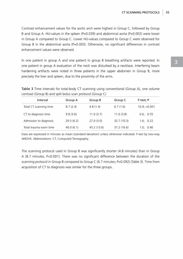
CT sCanning proToCols 55
3
Contrast enhancement values for the aortic arch were highest in Group C, followed by Group
B and Group A. HU-values in the spleen (P=0.039) and abdominal aorta (P=0.002) were lower
in Group A compared to Group C. Lower HU-values compared to Group C were observed for
Group B in the abdominal aorta (P=0.003). Otherwise, no significant differences in contrast
enhancement values were observed.
In one patient in group A and one patient in group B breathing artifacts were reported. In
one patient in group A evaluation of the neck was disturbed by a necklace. Interfering beam
hardening artifacts were noted in three patients in the upper abdomen in Group B, more
precisely the liver and spleen, due to the proximity of the arms.
Table 3 Time intervals for total-body CT scanning using conventional (Group A), one volume
contrast (Group B) and split bolus scan protocol (Group C)
interval group a group b group C F-test; p
Total CT scanning time 8.7 (2.4) 4.8 (1.4) 6.7 (1.6) 10.9; <0.001
CT to diagnosis time 9.8 (3.6) 11.0 (3.7) 11.6 (3.8) 0.6; 0.55
Admission to diagnosis 29.5 (6.2) 27.0 (3.0) 32.7 (10.3) 1.6; 0.22
Total trauma room time 40.0 (6.1) 45.2 (13.6) 37.2 (16.6) 1.0; 0.40
Data are expressed in minutes as mean (standard deviation) unless otherwise indicated. F-test by two-way
ANOVA. Abbreviations: CT, Computed Tomography.
The scanning protocol used in Group B was significantly shorter (4.8 minutes) than in Group
A (8.7 minutes; P<0.001). There was no significant difference between the duration of the
scanning protocol in Group B compared to Group C (6.7 minutes; P=0.092) (Table 3). Time from
acquisition of CT to diagnosis was similar for the three groups.

Chapter 356
Table 4 Inter-observer absolute agreement among the three observers on image quality in
total-body CT using conventional (Group A), one volume contrast (Group B) and split bolus
protocol (Group C).
group a
n=10
group b
n=10
group C
n=10
Radiologists (n) 3 3 3
Overall 0.61 (0.24, 0.87) 0.17 (-0.12, 0.60) 0.28 (-0.06, 0.68)
Brain 0.30 (-0.01, 0.69) 0.00 (-0.07, 0.20) 0.11 (-0.04, 0.43)
C-spine 0.28 (-0.02, 0.67) 0.15 (-0.19, 0.61) 0.10 (-0.03, 0.41)
Lung 0.00 (-0.12, 0.31) 0.21 (-0.11, 0.63) 0.37 (0.03, 0.75)
Aortic arch 0.60 (0.24, 0.86) 0.11 (-0.12, 0.51) 0.46 (0.10, 0.8)
Mediastinum -0.01 (-0.11, 0.26) 0.02 (-0.09, 0.31) 0.09 (-0.06, 0.42)
TL-spine 0.12 (-0.10, 0.52) 0.13 (-0.16, 0.57) 0.03 (-0.15, 0.41)
Abdominal aorta 0.55 (0.15, 0.85) 0.31 (0.00, 0.70) 0.44 (0.08, 0.79)
Portal vein 0.42 (0.06, 0.77) 0.05 (-0.17, 0.46) 0.12 (-0.08, 0.50)
Liver 0.30 (-0.01, 0.69) 0.37 (0.00, 0.75) 0.09 (-0.03, 0.38)
Spleen 0.29 (-0.01, 0.68) 0.60 (0.24, 0.86) 0.04 (-0.05, 0.27)
Kidney 0.22 (-0.08, 0.64) 0.38 (0.01, 0.75) 0.00 (-0.07, 0.21)
Pelvis 0.02 (-0.17, 0.41) 0.2 (-0.05, 0.59) -0.06 (-0.18, 0.25)
Data are expressed as intraclass correlations coefficients (95% confidence interval).
Abbreviations: C-spine, cervical spine; TL-spine, thoracolumbar spine.
The inter-observer absolute agreement among the three reviewers on overall image quality was
substantial for the conventional protocol Group A, slight for the one volume Group B, and fair
for the split bolus Group C (Table 4). For head and neck it ranged between slight and fair, and
for chest agreement ranged between poor and moderate with the highest value (0.6) for the
aortic arch in Group A. For the abdomen agreement also varied between poor and moderate
with the highest agreement (0.6) achieved for Group B on the spleen.

CT sCanning proToCols 57
3
disCussion
This study shows that the split bolus technique for single pass total-body CT scanning had the
highest overall image quality, compared to the conventional total-body trauma CT and the
one volume contrast CT protocol. A split bolus technique combines different contrast phases
into one acquisition, thereby diminishing radiation exposure with only limited increase of the
amount of contrast medium. It is most frequently used in renal imaging.18,19 In trauma only three
reports on a multi-phasic scan protocol in MDCT have been published.20-22 Loupatatzis et al.
compared in 16-slice MDCT a tri-phasic injection scheme (70 ml at 3 ml/s, followed by 0.1 ml/s
for 8 seconds, and 70 ml at 4 ml/s) to their standard CT angiography protocol.20 The tri-phasic
protocol achieved similarly high image quality for arteries compared to standard CT angiography
protocol, parenchymatous organs had better image quality compared to specialized protocols.
As in our study arm, artifacts reduced the enhancement of spleen and liver parenchyma. In
a retrospective study, Yaniv and colleagues compared the same contrast protocol set up for
64-slice MDCT with an arterial-phase contrast-enhanced CT of the thorax and a portovenous
scan of the abdomen and pelvis, but added a preceding unenhanced CT of the abdomen.21
The tri-phasic injection protocol enabled better vascular and abdominal parenchymal imaging,
although mean enhancement values in the aorta were significantly greater with the conventional
protocol. Nguyen et al. compared a standard injection protocol with an one-volume acquisition
from the circle of Willis to the pubic symphysis in 16-slice MDCT using a biphasic (150 ml, at 6
and 4 ml/s) or mono-phasic (110 ml at 4 ml/s, 400 mg I/ml) injection.22 No significant differences
were found in mean enhancement values in the aorta, liver, spleen, and kidney for the three
protocols. Quality scores were significantly higher for liver, spleen and kidney with the arms
above the head compared to arms alongside the body. Single-pass protocols had significantly
shorter median acquisition times than the conventional protocol. Hence, our findings for 64-slice
MDCT are in concordance with these previous CT studies.
In blunt abdominal trauma an arterial phase is more sensitive for the detection of intrasplenic
pseudoaneurysms, but a portal venous phase is more sensitive for the detection of parenchymal
injuries and active bleeding of the spleen.23 Dual phase CT can be considered as a complete
work up as it has better overall diagnostic performance than single phase CT. However, this
is at the cost of doubling radiation exposure to the abdomen. As group B is scanned in a
late arterial phase, it could be inferior in case of active bleeding and parenchymal injuries.
Whether the theoretical superposition of an arterial on a portal venous phase in split bolus
provides a comparable complete performance as dual phase imaging has to be proven in a large
prospective study.
Total-body CT can also be useful to diagnose blunt cerebrovascular injuries.24-26 Only protocol
B offered the possibility to screen for these injuries. The split bolus protocol can theoretically

Chapter 358
also fulfill these requirements, though was not investigated in our study. It was, however,
satisfactorily used in later patients if during CT acquisition a skull base or upper cervical spine
fracture was seen. Although injuries of the supra-aortic vessels are rare after blunt trauma, these
cerebrovascular injuries are associated with fractures of the cervical spine or skull base.27
Time is essential in trauma, as underlined by the ATLS® philosophy “time is life”. We observed
a time difference between the protocol without repositioning of the arms and the other two
protocols with arm repositioning. Remarkably, only few reports have focussed on this important
work flow aspect in correlation with scan protocols.14,28-30 Whether the slight time gain can
outweigh the observed lower diagnostic quality remains to be seen.
We did not focus on radiation exposure in our study. However, Brink et al. found that scanning
with the arms alongside the body resulted in a 45% increase in radiation exposure and a
decrease in image quality compared to scanning patients with a cranial position of the arms.31
Recently this has been confirmed by two other studies.32, 33
limitationsOur study has several limitations. First, there are different combinations and settings possible
when considering contrast medium phases (non-enhanced, arterial, venous, delayed phases)
and body regions (head, neck, thorax, abdomen/pelvis and extremities). We only studied three
different contrast protocols, and therefore we cannot make a statement on other variations.
Secondly, patients’ BMI or weight was not known. Since contrast enhancement of the liver
parenchyma is influenced by body weight, differences in liver enhancement and subsequently
in quality of liver imaging may be caused by differences in body weight between patients. For
the split bolus group ROI triggering in the thoracic aorta probably could have resulted in higher
arterial enhancement values. On the other hand, targeted planning by the technician would have
resulted in a more complicated procedure and longer examination times. Fixed delay appears to
be more practical in this stressful circumstance and therefore less error-prone. Further, diagnosis
time, admission to diagnosis time, and total room time are not only influenced by the type of
CT scan protocol but also by the specific types of injuries and by emergency room management
factors. Total-body CT scanning time is therefore the most relevant time factor comparing
group A, B and C. A final limitation concerns the restricted sample size for the assessments
of differences among the imaging protocols. After checking whether the data fulfilled the test
conditions, it was noted that homogeneity of variances could not be assumed (i) for the contrast
enhancement values for portal vein and liver, and (ii) the acquisition time between admission
and diagnosis. The corresponding P-values of the ANOVA tests are well above 0.05, but should
nevertheless be interpreted with some caution. Although unlikely, these results may be false
negative. Of note, in our series no important missed diagnosis during follow up was recorded.
Since the end of the study we used the split bolus technique in all following cases as part of an

CT sCanning proToCols 59
3
international multicenter randomized trial on total-body CT in severe trauma patients.34 Further
experience requiring more prospective studies are needed to evaluate its clinical accuracy.
ConClusion
In conclusion, evaluation of three scanning protocols for single pass total-body CT in severe
trauma patients showed that the split bolus technique reached the highest scores in image
quality and vascular and parenchymal enhancement. The one volume contrast CT protocol was
quickest and can probably be used in selected cases where time gain of some minutes could
potentially outweigh the reduced image quality.

Chapter 360
reFerenCes
1. Hauser H, Bohndorf K. Radiologic emergency
management in multiple trauma cases.
Radiologe. 1998; 38: 637-644.
2. Ptak T, Rhea JT, Novelline RA. Radiation
dose is reduced with a single-pass whole-
body multi-detector row CT trauma protocol
compared with a conventional segmented
method: initial experience. Radiology.
2003;229:902-905
3. Philipp MO, Kubin K, Hormann M, et al.
Radiological emergency room management
with emphasis on multidetector-row CT. Eur
J Radiol. 2003;48:2-4.
4. Gralla J, Spycher F, Pignolet C, et al.
Evaluation of a 16-MDCT scanner in an
emergency department: initial clinical
experience and workflow analysis. Am J
Roentgenol. 2005;185:232-238.
5. Prokop A, Hötte H, Krüger K, et al. Multislice
CT in diagnostic work-up of polytrauma.
Unfallchirurg. 2006;109:545-550.
6. Rieger M, Czermak B, El Attal R, et al. Initial
clinical experience with a 64-MDCT whole-
body scanner in an emergency department:
better time management and diagnostic
quality? J Trauma. 2009;66:648-657.
7. Van Vugt R, Deunk J, Brink M, et al.
Influence of routine computed tomography
on predicted survival from blunt
thoracoabdominal trauma. Eur J Trauma
Emerg Surg. 2011;37:185-190.
8. Stengel D, Ottersbach C, Matthes G, et
al. Accuracy of single-pass whole-body
computed tomography for detection of
injuries in patients with major blunt trauma.
CMAJ. 2012;184:869-876.
9. Huber-Wagner S, Lefering R, Qvick LM, et
al. Effect of whole body CT during trauma
resuscitation on survival: a retrospective,
multicenter study. Lancet. 2009;373:1455-
1461.
10. Brenner DJ, Hall EJ. Computed tomography-
an increasing source of radiation exposure.
N Engl J Med. 2007;357: 2277-2284.
11. Tien HC, Tremblay LN, Rizoli SB, et al.
Radiation exposure from diagnostic imaging
in severely injured trauma patients. J Trauma.
2007;62:151-156.
12. Inaba K, Branco BC, Lim G, et al. The
increasing burden of radiation exposure
in the management of trauma patients. J
Trauma. 2011;70:1366-1370.
13. Awai K, Imuta M, Utsunomiya D, et al.
Contrast enhancement for whole-body
screening using multidetector row helical
CT: comparison between uniphasic and
biphasic injection protocols. Radiat Med.
2004;22:303-309.
14. Fanucci E, Fiaschetti V, Rotili A, et al. Whole
body 16-row multislice CT in emergency
room: effects of different protocols on
scanning time, image quality and radiation
exposure. Emerg Radiol. 2007;13:251-257.
15. Saltzherr TP, Bakker FC, Beenen LF, et
al. Randomized clinical trial comparing
the effect of computed tomography in
the trauma room versus the radiology
department on injury outcomes. Br J Surg.
2012;99 Suppl 1:105-113.
16. Fung Kon Jin PH, Dijkgraaf MG, Alons CL, et
al. Improving CT scan capabilities with a new
trauma workflow concept: Simulation of
hospital logistics using different CT scanner
scenarios. Eur J Radiol. 2011; 80: 504-509.

CT sCanning proToCols 61
3
17. Landis JR, Koch GG. The measurement of
observer agreement for categorical data.
Biometrics. 1977; 33: 159-174.
18. Van Der Molen AJ, Cowan NC, Mueller-
Lisse UG, et al. CT urography: definition,
indications and techniques. A guideline for
clinical practice. Eur Radiol. 2008;18:4-17.
19. Gandrup KL, Nordling J, Balslev I, et al.
Upper urinary tract tumors: how does the
contrast enhancement measured in a split-
bolus CTU correlate to histological staging?
Acta Radiol. 2013;55:761-768
20. Loupatatzis C, Schindera S, Gralla J, et al.
Whole-body computed tomography for
multiple traumas using a triphasic injection
protocol. Eur Radiol. 2008;18:1206-1214.
21. Yaniv G, Portnoy O, Simon D, et al. Revised
protocol for whole-body CT for multi-trauma
patients applying triphasic injection followed
by a single-pass scan on a 64-MDCT. Clin
Radiol. 2013;68:668-675.
22. Nguyen D, Platon A, Shanmuganathan K,
et al. Evaluation of a single-pass continuous
whole-body 16-MDCT protocol for patients
with polytrauma. Am J Roentgenol.
2009;192:3-10.
23. Boscak AR, Shanmuganathan K, Mirvis
SE, et al. Optimizing trauma multidetector
CT protocol for blunt splenic injury: need
for arterial and portal venous phase scans.
Radiology. 2013;268:79-88.
24. Sliker CW, Shanmuganathan K, Mirvis
SE. Diagnosis of blunt cerebrovascular
injuries with 16-MDCT: accuracy of
whole-body MDCT compared with neck
MDCT angiography. Am J Roentgenol.
2008;190:790-799.
25. Chokshi FH, Munera F, Rivas LA, et al.
64-MDCT angiography of blunt vascular
injuries of the neck. Am J Roentgenol.
2011;196:309-315.
26. Borisch I, Boehme T, Butz B, et al. Screening
for carotid injury in trauma patients: image
quality of 16-detector-row computed
tomography angiography. Acta Radiol.
2007;48:798-805.
27. Biffl WL, Egglin T, Benedetto B, et al. Sixteen-
slice computed tomographic angiography
is a reliable noninvasive screening test for
clinically significant blunt cerebrovascular
injuries. J Trauma. 2006;60:745-752.
28. Roos JE, Desbiolles LM, Willmann JK, et
al. Multidetector-row helical CT: analysis
of time management and workflow. Eur
Radiol. 2002;12:680-685.
29. Heyer C, Rduch G, Kagel T, et al. Prospective
randomized trial of a modified standard
multislice CT protocol for the evaluation
of multiple trauma patients. Rofo.
2005;177:242-249.
30. Wurmb TE, Frühwald P, Hopfner W, et
al. Whole-body multislice computed
tomography as the first line diagnostic tool
in patients with multiple injuries: the focus
on time. J Trauma. 2009;66:658-665.
31. Brink M, de Lange F, Oostveen LJ, et al. Arm
raising at exposure-controlled multidetector
trauma CT of thoracoabdominal region:
higher image quality, lower radiation dose.
Radiology. 2008;249:661-670.
32. Bayer J, Pache G, Strohm PC, et al. Influence
of arm positioning on radiation dose for
whole body computed tomography in
trauma patients. J Trauma. 2011;70:900-
905.
33. Karlo C, Gnannt R, Frauenfelder T, et al.
Whole-body CT in polytrauma patients:
effect of arm positioning on thoracic and

Chapter 362
abdominal image quality. Emerg Radiol.
2011;18:285-293.
34. Sierink JC, Saltzherr TP, Beenen LF, et al. A
multicenter, randomized controlled trial of
immediate total-body CT scanning in trauma
patients (REACT-2). BMC Emerg Med. 2012;
12:4.


4a Case-maTCHed series oF immediaTe ToTal-body CT
sCanning versus THe sTandard radiologiCal work-up
in Trauma paTienTs
JC Sierink, TP Saltzherr, LFM Beenen, MJAM Russchen,
JSK Luitse, MGW Dijkgraaf, JC Goslings
Word Journal of Surgery, 2013

Chapter 466
absTraCT
objective In recent years Computed Tomography (CT) has become faster and more available
in the acute trauma care setting. The aim of the present study was to compare injured patients
who underwent immediate total-body CT scanning with patients who underwent the standard
radiological work-up with respect to 30-day mortality.
methods Between January 2009 and April 2011, 152 consecutive patients underwent
immediate total-body CT scanning as part of a prospective pilot study. These patients were
case-matched by age, gender and Injury Severity Score category with control patients from a
historical cohort (July 2006 – November 2007) who underwent X-rays and FAST followed by
selective CT scanning.
results Despite comparable demographics, TBCT patients had a lower median Glasgow Coma
Score than controls (10 vs. 15, P<0.001) and on-scene endotracheal intubation was performed
more often (33% vs. 19%, P=0.004). Thirty day mortality was 13% in the TBCT patient group
vs. 13% in the control group (P=1.000). A generalized linear mixed model analysis showed that
a higher in-hospital Glasgow Coma Score (Odds Ratio (OR) 0.8, 95% confidence interval (CI)
0.745-0.86; P<0.001) and immediate total body CT scanning (OR 0.46, 95%CI 0.236-0.895;
P=0.022) were associated with decreased 30-day mortality, while a higher Injury Severity Score
(OR 1.054, 95%CI 1.028-1.08; P<0.001) was associated with increased 30-day mortality.
Conclusion Trauma patients who underwent immediate total body CT scanning had similar
absolute 30-day mortality rates compared to patients who underwent conventional imaging
and selective CT scanning. However, immediate TBCT scanning was associated with a decreased
30-day mortality after correction for the impact of differences in raw ISS and in-hospital GCS.

Case-matChed series 67
4
inTroduCTion
Traumatic injuries are an important cause of death among people 15-60 years of age, and
the effects on the lives of those who survive may be devastating.1,2 Safe, accurate and rapid
diagnostic procedures make sure that treatment can be planned and carried out as soon as
possible. If we can speed up the diagnostic work-up in injured patients, this may help to prevent
deaths.
In recent years Computed Tomography (CT) has become faster, more detailed and more
available in the acute trauma care setting.3-5 Hence, the standard radiological work-up (X-rays
and Focussed Assessment with Sonography for Trauma (FAST) followed by selective CT
scanning) may no longer be the optimal choice of primary diagnostics. One area in particular
that has gained interest in trauma care is an immediate total-body CT scan without previous
conventional imaging.3, 6-8
Previous cohort studies have shown that immediate total-body CT scanning provides
faster diagnosis in injured patients than the standard radiological workup.9-13 In the largest
retrospective study performed on this topic, patients who underwent immediate total-body
CT-scanning were found to have an increased probability of survival compared to patients who
underwent the standard radiological work-up.10 This survival benefit is, however, not confirmed
in absolute mortality numbers in other studies.11, 13, 14 Wurmb and colleagues11, 14 depicted their
triage scheme but all other studies collected data of patients with an Injury Severity Score (ISS)
of at least 16 who underwent a total-body CT scan. For daily practice it is of major importance
to know what the selection criteria were for a patient to undergo a total-body CT and to make
sure that those patients are comparable to patients who underwent the standard work-up.
The aim of the present study was to compare severely injured patients who underwent
immediate total-body CT scanning according to predefined criteria with matched controls who
underwent the standard radiological work-up with respect to thirty-day mortality.
paTienTs and meTHods
immediate Total-body CT scanning: TbCT patientsBetween January 2009 and April 2011 patients in whom severe injury was suspected, based on
predefined vital signs and clinically suspicious diagnoses, underwent immediate total-body CT
scanning as a pilot study for the REACT-2 trial.15 In- and exclusion criteria for this pilot study are
given in Table 1.

Chapter 468
This study was approved by the Institutional Review Board with a waiver of informed consent
for including 50 patients. After the inclusion of 50 patients, total-body CT scanning in patients
fulfilling the inclusion criteria became routine in our level-1 trauma center. All consecutive
injured patients who received total-body CT scanning in this period according to the criteria
defined in Table 1 were therefore included in this study and will be described as ‘TBCT patients’.
During the primary survey the vital functions were checked and, when necessary, corrected. The
necessary corrections and interventions during the primary survey consisted of intubation or
performing a cricothyrotomy, chest tube drainage or pericardiocenthesis and taking hemorrhage-
controlling measurements such as applying a pelvic binder or external pressure. Furthermore,
at least one working infusion system should be available and blood could be drawn for analysis
before making the CT scan.
Total-body CT scanning consisted of a two-step whole-body acquisition (from vertex to pubic
symphysis) starting with Head and Neck Non Enhanced CT (NECT) with arms alongside the
body. For the second complementary scan a split-bolus intravenous contrast protocol was used
directly after repositioning the arms alongside the head. This scan covered chest, abdomen and
pelvis.
standard radiological work-up: control patientsPatients admitted between July 2006 and November 2007 underwent the standard radiologic
work-up. During this period, the REACT-1 trial16 was recruiting all blunt trauma patients who
were admitted to our hospital. Standard radiological work-up consisted of chest X-ray, pelvic
X-ray and FAST followed by selective CT scanning based on local imaging guidelines.
Conventional digital radiographs were made by a mobile X-ray machine and archived in the
Picture Archiving and Communication System PACS (Impax 4.5, AGFA Gevaert, Mortsel,
Belgium). Portable ultrasound was available to perform FAST. The trauma resuscitation room
was further equipped with a sliding gantry CT-scanner (since 2008 a 64-slice CT scanner, before
that period a 4-slice CT scanner was used) (Sensation 64, Siemens Medical Solutions, Forchheim,
Germany) with a multifunctional, radiolucent trauma resuscitation table.

Case-matChed series 69
4
Table 1 Criteria for immediate total-body CT-scanning in TBCT patients
Trauma patients with the presence of one of the following vital parameters:
o respiratory rate >29/min or <10/min;
o pulse >120/min;
o systolic blood pressure < 100 mmHg;
o estimated exterior blood loss > 500 ml;
o Glasgow Coma Score ≤ 13;
o abnormal pupillary reaction on site.
or patients with one of the following clinically suspicious diagnoses:
o fractures from at least two long bones;
o flail chest, open chest or multiple rib fractures;
o pelvic fracture;
o unstable vertebral fractures;
o spinal cord compression.
Trauma patients not receiving total-body CT scanning:
o known age <18 years;
o known pregnancy;
o referred from another hospital;
o any patient who is judged to be too unstable to undergo a CT scan and requires (cardiopulmonary)
resuscitation or immediate operation.
data collection and processingData concerning patient demographics, radiologic imaging, type of treatment and clinical
outcome were prospectively registered in a database by a dedicated research nurse for the
control patients and by a trained medical student under supervision of a physician for the
TBCT patients. Pre-hospital parameters (hypotension defined as systolic blood pressure below
90 mmHg, on-scene Glasgow Coma Score and presence of endotracheal intubation) were
registered as well. On-scene pre-hospital GCS was registered by the ambulance personnel. In
case of on-scene intubation, in-hospital GCS was scored as 3.
Injury severity of every trauma patient was scored by trained trauma surgeons using the
Abbreviated Injury Scale (AIS) per body region. Severity scores (1 to 6) within the AIS range
from minor (code 1), moderate, serious, severe, critical to unsurvivable injury (code 6). AIS body
regions head, chest, abdomen and extremities were described.
General practitioners and discharge locations were contacted for information concerning
follow-up and hospital- and ICU days in case the patient was discharged to another hospital.
In case of a deceased patient, predominant cause of death was abstracted from patient charts,
computerized hospital databases and/or from information of general practitioners.

Chapter 470
outcome measureThirty-day mortality.
matchingEach TBCT patient was matched 1:1 to a control patient with SPSS-syntax (from David Marso;
SPSSX-L archives http://listserv.uga.edu/cgi-bin/wa?A2=ind1103&L=spssx-l&D=0&P=64620; March
30th, 2011), adapted for the following matching variables. Patients were matched by age within
5 years, gender and Injury Severity Score (ISS) category (according to the American College
of Surgeons’ categories of ISS: 1-9, 10-15, 16-24, >24). Matching by ISS category rather
than matching by raw ISS allowed for small measurement bias to occur that resulted from
the different CT scanning protocols applied, with immediate total body CT scanning tending
towards slightly higher ISS values.17
With this method, 139 patients of 156 could be matched. Four of the 17 non-matched patients
had an ISS of 75. Three died shortly after admission and one unexpectedly survived a C3-fracture
with a traumatic disc hernia. Hand-matching these patients with control patients who were
non-comparable in age and sex would potentially have caused selection bias. These patients
were therefore excluded.
Thirteen of the non-matched patients were hand-matched by age and ISS. Age was comparable
in these patients with a mean difference of 7.6 years and a maximum difference of 23 years
(between females of 84 and 61 years of age). ISS was similar in these patients with a maximum
difference of 2 points except in 1 patient in which the difference was 12 points (ISS of 50 and
38 in males of 93 and 80 years of age respectively). In total 152 TBCT patients were matched
with 152 control patients.
The study flowchart is depicted in Figure 1.

Case-matChed series 71
4
Figure 1 Study flow chart
Immediate total-body CT scanningJan 2009 – April 2011
n=156
Standard work-upJuly 2006 – Nov 2007
n=1376
Immediate total-body CT scanningJan 2009 – April 2011
n=152
Standard work-upJuly 2006 – Nov 2007
n=152
1:1 case-matchingon age, sex and ISS
Succesful matching in139 out of 156
4 pilot patients with an ISS of75 were excluded
13 patients were hand-matched
Standard work-up: chest and pelvic X-ray, FAST, and selective CT. Abbreviations: CT, Computed Tomography;
ISS, Injury Severity Score.
statistical analysis Data are presented as median and interquartile ranges for not normally distributed data and
as mean ± standard deviation for normally distributed data. Considering the matching of TBCT
with control patients, testing was done with the McNemar test for dichotomous variables,
the McNemar-Bowker test or extended McNemar test for categorical variables and with the
Wilcoxon signed ranks test for continuously but not normally distributed variables.
The Revised Trauma Score (RTS) was calculated following the formula described by Champion et
al.18,19 The Trauma and Injury Severity Score (TRISS) method was used to calculate the probability
of survival. 20
Generalized linear mixed modelling was done to identify the independent predictive value of
type of CT scanning for 30-day mortality, accounting for (1) TBCT patients being matched

Chapter 472
with control patients, (2) potential confounders, and (3) multicollinearity in the predictor set. A
backward stepwise approach was applied excluding predictors with the highest non-significant
P-value one-by-one. A P-value below 0.05 was considered to indicate statistical significance.
Statistical analyses were performed using SPSS (version 20.0.0.1; IBM Coorperation) and PEPI
(version 1.11; Abramson/Gahlinger, Salt Lake City, USA, 2000-2004).
resulTs
Patient characteristics are summarized in Table 2. TBCT patients were comparable to controls
with respect to mechanism of injury and laboratory results. Pre- and inhospital median Glasgow
Coma Scores (GCS) were lower, while on-scene endotracheal intubation too was performed
more often in TBCT patients than in controls.
Table 2 Characteristics of injured patients in the matched cohorts.
TbCT*
n=152
Controls†
n=152
missing‡ p-value
Age in years § 43.91 (19.67) 43.63 (18.61) 0 0.324||
Men 107 (70.4%) 109 (71.7%) 0 0.687||
Blunt trauma 147 (96.7%) 144 (94.7%) 0 0.581
Prehospital
hypotension(SBP<90 mmHg)
intubation
GCS
4 (7.8%)
48 (32.9%)
12 (4-15)
1 (2.0%)
28 (19.2%)
15 (9-15)
101
6
17
0.375
0.004
<0.001
Trauma room
hypotension(SBP<90 mmHg)
GCS
5 (3.4%)
10 (3-15)
7 (4.7%)
15 (7.3-15)
3
26
0.774
<0.001
Laboratory results on admission
Hemoglobin in g/dl §
Ph
base excess in mmol/L§
12.50 (2.00)
7.36 (7.31-7.39)
-2.81 (4.34)
12.52 (1.91)
7.37 (7.31-7.42)
-3.33 (4.97)
3
6
6
0.911
0.471
0.326
ISS (points) 18 (9-29) 18 (8-29) 0 0.528
ISS in matching categories
0-9
10-15
16-24
≥ 25
49 (32.2%)
7 (4.6%)
34 (22.4%)
62 (40.8%)
49 (32.2%)
7 (4.6%)
34 (22.4%)
62 (40.8%)
0 1.000||
Data are number (%) or median (interquartile range (IQR)) unless otherwise indicated. Abbreviations: CT,
Computed Tomography; SBP, systolic blood pressure; GCS, Glasgow Coma Score; AIS, Abbreviated Injury
Score; ISS, Injury Severity Score.
*Immediate total-body CT from head to pelvis. †Chest and pelvic X-ray, FAST and selective CT
‡Missing pairs, if no data were available for at least one patient in the matched pair.
§Mean (SD). ||Matching parameters.

Case-matChed series 73
4
Of the 152 patients in the control group, 132 patients (87%) underwent selective CT scanning
of one or more body regions after conventional imaging. Twenty-two control patients (14%)
underwent TBCT scanning after imaging with X-rays and FAST. Cranial CT scans were obtained
in 73 patients (48%), cervical spine CT scans in 92 patients (61%), thoracic CT scans in 13
patients (9%), abdominal CT scans in 18 patients (12%), pelvic CT scans in 3 patients (2%) and
thoracic or lumbar spine CT scans in 29 patients (19%).
Figure 2 shows the prevalence of severe injuries (AIS ≥3) per body region in both study groups.
The TBCT patients had serious head injuries more often than the control patients did (63% vs.
37%, P<0.001). The prevalence of severe injuries to the chest, abdomen and extremities was
comparable between TBCT and control patients.
Figure 2 Presence of severe injuries (AIS≥3) in the matched cohorts
* Immediate total-body CT from head to pelvis. † Chest and pelvic X-ray, FAST and selective CT.
main resultsOutcome parameters are listed in Table 3. The crude thirty-day mortality rates were 13%
for both TBCT and control patients (P=1.000). Cause of death differed significantly between
TBCT patients and controls (P=0.002). Five control patients who died from hemorrhage were
hemodynamically unstable on admission and died at the operating room due to uncontrolled
traumatic bleeding; 2 patients had no cardiac output on admission. The last patient was a
drowning victim who underwent cardiopulmonary resuscitation on-scene. He was admitted to
the hospital with a marked decrease in hemoglobin level with a radiologically occult bleeding

Chapter 474
site other than subcutaneously. Other causes of death in TBCT patients were hypoxia after
drowning (n=1), hypoxia after a suicide attempt by hanging (n=1), septic shock (n=1) and
unexpected asystole most likely due to fat embolism (n=1). In control patients, other causes of
death were out-of-hospital cardiac arrest after drowning (n=1), traumatic spinal cord lesion with
complete paraplegia (n=1), fall from height while recovering from a cerebral contusion (n=1),
respiratory insufficiency in combination with marginal neurologic function (n=1) and traumatic
hemorrhagic complications due to anticoagulant treatment (n=1).
Type of treatment did not differ between TBCT en control patients (P=0.422). Median hospital
stay was 9 days in TBCT as well as control patients (9 (IQR=3-25) vs. 9 (IQR=1-23), P=0.358).
Table 3 Outcome parameters of injured patients in the matched cohorts.
TbCT*
n=152
Controls†
n=152
missing‡ p-value
Treatment
no intervention
operative
interventional radiology
71 (47.3%)
74 (49.3%)
5 (3.3%)
75 (50.0%)
67 (44.7%)
8 (5.3%)
2 0.422
Hospital stay (days)
overall
ICU stay
ventilation time
9 (3-25)
2 (0-6)
1 (0-3)
8.5 (1-22.8)
1 (0-5)
0 (0-1.3)
0
0
10
0.358
0.022
0.134
Discharge location
home
other hospital
rehabilitation center
nursing home
other
76 (50.0%)
22 (14.5%)
14 (9.2%)
11 (7.2%)
29 (19.1%)
88 (57.9%)
18 (11.8%)
17 (11.2%)
9 (5.9%)
20 (13.2%)
0 0.605
Mortality
24h
30-day
11 (7.25)
20 (13.2%)
10 (6.6%)
20 (13.2%)
0
1.000
1.000
Predominant cause of death
no death
TBI
haemorrhage
other
128 (84.2%)
17 (11.2%)
0 (0%)
7 (4.6%)
129 (84.9%)
8 (5.3%)
8 (5.3%)
7 (4.6%)
0 0.002
Data are number (%) or median (interquartile range (IQR)) unless otherwise indicated. Abbreviations: CT,
Computed Tomography; SBP, systolic blood pressure; GCS, Glasgow Coma Score.
*Immediate total-body CT from head to pelvis.
†Chest and pelvic X-ray, FAST and selective CT.
‡Missing pairs, if no data were available for at least one patient in the matched pair

Case-matChed series 75
4
A generalized linear mixed model analysis was applied to assess the predictive value of type of
CT scanning for 30-day mortality. Pre-hospital intubation, pre-hospital and in-hospital GCS, and
raw ISS were identified as potential confounders (see Table 2). With pre-hospital and in-hospital
GCS being highly correlated (Spearman’s rho: 0.87), only in-hospital GCS was included in the
multivariable model. Further, pre-hospital intubation was associated with higher ISS values and
was dropped during the backward stepwise approach. Table 4 shows the final model, indicating
that immediate total body CT scanning was associated with a lower 30-day mortality (OR 0.46,
95%CI 0.236-0.895; P=0.022) after correction for the impact of differences in raw ISS (OR
1.054, 95%CI 1.028-1.08) P<0.001) and in-hospital GCS (OR 0.8, 95%CI 0.745-0.86; P<0.001).
Table 4 Independent predictors of 30-day mortality (N=278) with GLMM
odds ratio 95% Ci p-value
Total-body CT scanning 0.46 0.236-0.895 0.022
Trauma room GCS 0.80 0.745-0.860 <0.001
ISS 1.054 1.028-1.080 <0.001
Abbreviations: GLMM, generalized linear mixed model; CI, Confidence Interval; CT, Computed Tomography;
GCS, Glasgow Coma Score; ISS, Injury Severity Score.
disCussion
This case-matched series shows that patients who underwent immediate total-body CT scanning
had similar absolute 30-day mortality rates as patients who underwent conventional imaging and
selective CT scanning. Patients were comparable in important prognostic factors for mortality
such as age and ISS category. To correct for any non-comparable pre-hospital characteristics, a
generalized linear mixed model was used, and this analysis showed that immediate TBCT was
associated with decreased 30-day mortality.
The advantages of TBCT scanning in the clinical setting seem clear: TBCT scanning saves time.
A rapid overview of all threatened body regions can be obtained in 15 minutes, which leads to
rapid decision making in treatment. As a result of goal-directed and earlier start of treatment,
mortality could be reduced. Previous studies were too focussed on TBCT scanning and mortality
in injured patients, but whether this scan was ‘immediate’, as was the case in the present study,
that fact was not described routinely. Differences in overall, 24h of 30-day mortality in favor
of immediate TBCT have been described previously8,10,21,22, but there is no reproducible level 1
scientific evidence.
All these studies compared two different cohorts of patients, and did not match the patients
on age, gender and ISS. Furthermore, patients in the standard radiological work-up group

Chapter 476
in these studies were not described in detail. The present study showed however, that these
patients underwent total-body CT scanning after conventional imaging in 14% of the cases.
In these patients, only time is a factor of importance when comparing them to patients in the
immediate TBCT group. All available evidence suggests that total-body CT scanning in injured
patients leads to fast and accurate diagnosis and treatment.3, 10-13, 23-25 Previous studies showed
that diagnostic work-up time was significantly longer in patients who underwent the standard
radiological work-up.12,26,27 However, the clinical relevance of speeding up the diagnostic process
cannot be described based on these retrospective series.
RTS and TRISS could be calculated in 39 TBCT patients (26%) and 35 control patients (23%).
RTS differed significantly between TBCT and control patients (7.55 vs. 7.84, P=0.008) as well
as TRISS (0.98 vs. 0.99, P=0.017). However, patients in whom TRISS could not be calculated
were more severely injured than patients in whom TRISS could be calculated. This was reflected
by a higher on-scene endotracheal intubation rate (30% vs. 16%, P=0.025), a lower median
GCS pre-hospital (13 vs. 15, P=0.036) and a higher median ISS (20 vs. 16, P=0.009). Age,
sex, mechanism of injury, pre- and in hospital hypotension rates and in-hospital GCS were
comparable between patients in whom TRISS could not be calculated and patients in whom
TRISS could be calculated.
The major limitation of our study is that patients were compared retrospectively. Although
matching parameters were carefully chosen and ISS is strongly related to mortality28, there were
differences in pre-hospital parameters and the incidence of serious head injuries between the
groups. Preferably, we would have selected control patients based on the same inclusion criteria
as TBCT patients. However, these data could not be retrieved retrospectively in a reliable manner.
We therefore used matching to select a cohort of control patients that is more comparable to the
patients selected in the TBCT group than would have been the case when using all control patients
admitted between 2006 and 2007. This would have yielded more control patients who were not
comparable to TBCT patients, for example with a lower ISS and a lower prevalence of mortality.
Although it would have made the logistic regression analysis easier, there would have been more
‘noise’ that had to be explained by the prediction model. Furthermore, fewer variables could have
been used in the prediction model since the prevalence of mortality will be lower when using all
control patients. By matching, the number of confounding variables in our prediction model was
reduced. This was done in other studies as well, although in those studies retrospective selection
of patients was used to create groups with an ISS of 16 and higher10 or patients who were
admitted to the ICU.22 In several studies on this topic similar differences in characteristics between
study groups were found and statistical methods were used to correct for these differences.10, 21,
22 Obviously, matching possibly only partially compensates for the difference in selection methods
of TBCT patients and the subsequent regression analysis has been restricted to a limited number
of predictors. Consequently, residual confounding may remain like in most observational studies.

Case-matChed series 77
4
Another limitation of the present study is that the ISS in the TBCT patients might be influenced
by the more extensive classification that is made possible by the total-body CT scan.17 This
phenomenon has been described in patients with occult pulmonary contusions, in whom
the median ISS was increased without significant consequences for complication rate and
mortality.29 Because the majority of control patients had a considerable number of CT scans
after conventional imaging as well, we think that this effect is marginal.
To investigate the causal relationship between initial imaging and mortality in injured patients,
a multicenter randomized clinical study is underway.15 This randomized trial does not only focus
on clinical outcome, but will analyse radiation dose and cost-effectiveness as well. An analysis
of all these factors together is required to provide a complete overview of the advantages and
disadvantages of using immediate total-body CT in the initial evaluation of injured patients.
ConClusion
In conclusion, this case-matched series showed that patients who underwent immediate total
body CT scanning had comparable absolute mortality rates when compared with trauma
patients who underwent conventional imaging and selective CT scanning. After correction for
confounders, a decrease in 30-day mortality in TBCT patients was apparent.

Chapter 478
reFerenCes
1. World Health Organization (2008) The global
burden of disease: 2004 update. Available
at: http://www who int/healthinfo/global_
burden_disease/2004_report_update/en/
index html [Accessed December 14, 2011].
2. Sethi D, Racioppi F, Baumgarten I, Bertollini R.
Reducing inequalities from injuries in Europe.
Lancet. 2006; 368:2243-2250.
3. Sampson MA, Colquhoun KB, Hennessy NL.
Computed tomography whole body imaging
in multi-trauma: 7 years experience. Clin
Radiol. 2006; 61:365-369.
4. Leidner B, Beckman MO. Standardized whole-
body computed tomography as a screening
tool in blunt multitrauma patients. Emergency
Radiology. 2001; 8:20-8.
5. Rieger M, Czermak B, El AR, Sumann
G, Jaschke W, Freund M. Initial clinical
experience with a 64-MDCT whole-body
scanner in an emergency department: better
time management and diagnostic quality? J
Trauma. 2009; 66:648-657.
6. Salim A, Sangthong B, Martin M, Brown C,
Plurad D, Demetriades D. Whole body imaging
in blunt multisystem trauma patients without
obvious signs of injury: results of a prospective
study. Arch Surg. 2006; 141:468-473.
7. Fanucci E, Fiaschetti V, Rotili A, Floris R,
Simonetti G. Whole body 16-row multislice
CT in emergency room: effects of different
protocols on scanning time, image quality
and radiation exposure. Emerg Radiol. 2007;
13:251-257.
8. Hilbert P, zur Nieden K, Hofmann GO, Hoeller
I, Koch R, Stuttmann R. New aspects in the
emergency room management of critically
injured patients: a multi-slice CT-oriented care
algorithm. Injury. 2007; 38:552-558.
9. Sierink JC, Saltzherr TP, Reitsma JB, Van Delden
OM, Luitse JSK, Goslings JC. Systematic
review and meta-analysis of immediate total-
body tomography compared with selective
radiological injured patients. British Journal of
Surgery Society. 2011; 99:52-58.
10. Huber-Wagner S, Lefering R, Qvick LM, et
al. Effect of whole-body CT during trauma
resuscitation on survival: a retrospective,
multicenter study. Lancet. 2009; 373:1455-
1461.
11. Wurmb TE, Quaisser C, Balling H, et al. Whole-
body MSCT improves trauma care in patients
requiring surgery after multiple trauma. Emerg
Med J. 2011; 28:300-304.
12. Wurmb TE, Fruhwald P, Hopfner W, et al.
Whole-body multislice computed tomography
as the first line diagnostic tool in patients with
multiple injuries: the focus on time. J Trauma.
2009; 66:658-665.
13. Weninger P, Mauritz W, Fridrich P, et al.
Emergency room management of patients
with blunt major trauma: evaluation of the
multislice computed tomography protocol
exemplified by an urban trauma center. J
Trauma. 2007; 62:584-591.
14. Wurmb T, Balling H, Fruhwald P, et al.
[Polytrauma management in a period of
change: time analysis of new strategies for
emergency room treatment]. Unfallchirurg.
2009; 112:390-399.
15. Sierink JC, Saltzherr TP, Beenen LF, et al. A
multicenter, randomized controlled trial of
immediate total-body CT scanning in trauma
patients (REACT-2). BMC Emerg Med. 2012;
12:4.

Case-matChed series 79
4
16. Saltzherr TP, Bakker FC, Beenen LFM, et al.
Randomized clinical trial comparing the effect
of computed tomography in the trauma room
versus the radiology department on injury
outcomes. British Journal of Surgery. 2012;
99:105-113.
17. van Vugt R, Deunk J, Brink M, et al. Influence
of routine computed tomography on predicted
survival from blunt thoracoabdominal trauma.
Eur J Trauma Emerg Surg. 2011; 37:185-190.
18. Champion HR, Sacco WJ, Carnazzo AJ,
Copes W, Fouty WJ. Trauma score. Crit Care
Med.1981; 9:672-676.
19. Champion HR, Sacco WJ, Copes WS, Gann
DS, Gennarelli TA, Flanagan ME. A revision of
the Trauma Score. J Trauma. 1989; 29:623-
629.
20. Boyd CR, Tolson MA, Copes WS. Evaluating
trauma care: the TRISS method. Trauma Score
and the Injury Severity Score. J Trauma. 1987;
27:370-378.
21. Hutter M, Woltmann A, Hierholzer C,
Gartner C, Buhren V, Stengel D. Association
between a single-pass whole-body computed
tomography policy and survival after blunt
major trauma: a retrospective cohort study.
Scand J Trauma Resusc Emerg Med. 2011;
19:73.
22. Yeguiayan JM, Yap A, Freysz M, et al. Impact
of whole-body computed tomography on
mortality and surgical management of severe
blunt trauma. Crit Care. 2012; 16:R101.
23. Ptak T, Rhea JT, Novelline RA. Experience
with a continuous, single-pass whole-body
multidetector CT protocol for trauma: The
three-minute multiple trauma CT scan.
Emergency Radiology. 2001; 8:250-256.
24. Kanz KG, Korner M, Linsenmaier U, et
al. [Priority-oriented shock trauma room
management with the integration of
multiple-view spiral computed tomography].
Unfallchirurg. 2004; 107:937-944.
25. Prokop A, Hotte H, Kruger K, Rehm KE,
Isenberg J, Schiffer G. [Multislice CT
in diagnostic work-up of polytrauma].
Unfallchirurg. 2006; 109:545-550.
26. Fung Kon Jin PH, van Geene AR, Linnau KF,
Jurkovich GJ, Ponsen KJ, Goslings JC. Time
factors associated with CT scan usage in
trauma patients. Eur J Radiol. 2009; 72:134-
138.
27. Fung Kon Jin PH, Goslings JC, Ponsen KJ, van
KC, Hoogerwerf N, Luitse JS. Assessment of a
new trauma workflow concept implementing
a sliding CT scanner in the trauma room:
the effect on workup times. J Trauma. 2008;
64:1320-1326.
28. Baker SP, O’Neill B, Haddon W, Jr., Long
WB. The injury severity score: a method for
describing patients with multiple injuries and
evaluating emergency care. J Trauma. 1974;
14:187-196.
29. Deunk J, Poels TC, Brink M, et al. The clinical
outcome of occult pulmonary contusion on
multidetector-row computed tomography
in blunt trauma patients. J Trauma. 2010;
68:387-394.
30. Brenner DJ, Hall EJ. Computed tomography--
an increasing source of radiation exposure. N
Engl J Med. 2007; 357:2277-2284.


5inCidenTal Findings on ToTal-body CT sCans
in Trauma paTienTs
JC Sierink, TP Saltzherr, MJAM Russchen, SMM de Castro, LFM Beenen, NWL Schep,
JC Goslings
Injury, 2013

Chapter 582
absTraCT
objective Total-body Computed Tomography (CT) scans are increasingly used in trauma care.
Herewith the observation of incidental findings, trauma unrelated findings, is also increased.
The aim of this study was to evaluate the number of incidental findings in adult trauma patients.
methods All consecutive trauma patients that underwent total-body CT scanning between
January 2009 and December 2011 were analyzed. Incidental findings were divided in three
categories: category I (potentially severe condition, further diagnostic work-up is required),
category II (diagnostic work-up dependent on patients’ symptoms) and category III (findings of
minor concern, no diagnostic work-up required).
results There were 2248 trauma room presentations; 321 patients underwent a total-body CT
scan (14.3%). In 143 patients (44.5%), 186 incidental findings were reported. There were 13
category I findings (7%), 45 category II findings (24%) and 125 category III incidental findings
(67%). Overall, 18 patients (5.6%) required additional diagnostic work-up. Four patients
underwent work-up by additional radiologic imaging. Three patients required further invasive
work-up or treatment. Three patients were transferred to another hospital, no extended follow-
up was performed. In three patients, there was no documentation of follow-up. Five patients
deceased before diagnostic work-up of the incidental finding could start.
Conclusion Total-body CT scanning as part of the evaluation of trauma patients leads to a
substantial amount of incidental findings. Documentation of incidental findings and their
clinical consequences was incomplete. Therefore, the findings of this study have prompted us
to add an item to our electronic trauma room report that obliges residents to report whether or
not incidental findings are found during trauma imaging.

IncIdental fIndIngs 83
5
inTroduCTion
Total-body Computed Tomography (CT) scanning is increasingly used as a diagnostic technique
in trauma patients. The main advantage of this total-body imaging concept is a quick and
complete overview of traumatic injuries in all relevant body regions (head to pelvis).1-3 One
of the most important disadvantages is the associated radiation exposure.4,5 Another point of
interest is the observation of incidental findings on total-body CT scanning. These are defined
as findings that are totally unrelated to the clinician’s reasons for requesting the radiological
examination.6 These incidental findings might be beneficial to patients in case of earlier detection
of a malignancy. Conversely, it might result in increased patients’ anxiety and health care costs in
case of additional work-up for abnormalities that ultimately might not affect patients’ health.7,8
The total amount of incidental findings on selective CT scans of one or more body regions
in trauma patients varies between 34%9 and 45%.10,11 A recent study reported a 36%
incidental findings rate on routine thoracoabdominal CT in trauma patients.14 Only two studies
evaluated incidental findings in patients that underwent a total-body CT scan as part of the
initial trauma evaluation and incidental finding numbers of 50% and 53% were found.15,16
However, indications for a total-body CT scan were not described in both studies and the clinical
consequences of these incidental findings were unclear.15
Therefore, the aim of this study was to evaluate the percentage of incidental findings and
their clinical consequences in a cohort of adult trauma patients who underwent total-body CT
scanning.
paTienTs and meTHods
patient selection All consecutive trauma patients admitted to our academic Level I trauma center that underwent
total-body CT scanning between January 2009 and December 2011 were included in this study.
All patients fulfilling the in- and exclusion criteria listed in Table 1 underwent immediate total-
body CT scanning. Prior conventional imaging was not an exclusion criterion for the present
study.
Total-body CT All patients were evaluated on a sliding gantry 64-slice CT-scanner (Sensation 64, Siemens
Medical Solutions, Forchheim, Germany) located in our trauma resuscitation room. Total-body
CT scanning consisted of a two-step total-body acquisition (from vertex to pubic symphysis)
starting with Head and Neck Non-Enhanced CT with arms alongside the body. Directly after
repositioning the arms alongside the head for the second part of the scan covering chest,

Chapter 584
abdomen and pelvis, a split-bolus intravenous contrast protocol was injected (Optiray® 350 125
ml Pre fill, Covidien Mallinckrodt, Cincinnati, USA). The reconstruction thickness was 5mm head
and 1 mm bone kernel for the brain, 1mm bone kernel for the cervical spine and 3mm soft
tissue and bone kernel for the torso.
Table 1 Criteria for immediate total-body CT scanning
Trauma patients with the presence of one of the following vital parameters:
o respiratory rate >29/min or <10/min;
o pulse >120/min;
o systolic blood pressure < 100 mmHg;
o estimated exterior blood loss > 500 ml;
o Glasgow Coma Score ≤ 13;
o abnormal pupillary reaction on site.
or Trauma patients with one of the following clinically suspicious diagnoses:
o fractures from at least two long bones;
o flail chest, open chest or multiple rib fractures;
o pelvic fracture;
o unstable vertebral fractures;
o spinal cord compression.
Trauma patients not receiving total-body CT scanning:
o known age ≤18 years;
o known pregnancy;
o referred from another hospital;
o any patient who is judged to be too unstable to undergo a CT scan and requires (cardiopulmonary)
resuscitation or immediate operation.
data collectionAll trauma room patients were registered in a prospective database. A cohort that underwent
total-body CT scanning was selected and data were collected anonymously.
Double-reading of radiologic imaging by the resident on call and by a senior radiologist with
expertise in trauma is common practice in our center. By protocol, if previous radiology reports
are present they are compared with current CT findings. The presence of incidental findings in the
radiology report and the clinical consequences were determined by an unblinded investigator.
Clinical consequences were defined as actions (i.e. diagnostic work-up or treatment) following
the incidental finding. Information was extracted from computerized hospital databases
containing admission and discharge letters, radiological reports, surgery reports and pathology
reports.

IncIdental fIndIngs 85
5
The Institutional Review Board evaluated the study protocol and declared that the need for
informed consent was waived. Data collection was done solely in our level-1 trauma center. Due
to privacy reasons, there was no extended follow-up.
incidental finding in categories Incidental findings were divided in three categories: category I is an incidental finding that is
a potentially severe condition, further diagnostic work-up is required. Work-up consisted of
additional blood tests, consultation of other specialties, radiologic imaging or invasive diagnostic
procedures. In category II diagnostic work-up is dependent of patients’ symptoms and category
III are findings of minor concern for which no diagnostic work-up is required. The definitions of
the categories were comparable to categories described in previous studies9,14,15 and combine
the potential severity of the abnormalities found with the respective clinical consequences.
The following incidental findings were excluded: degenerative joint diseases, stenotic
atherosclerotic vessel disease, sinusitis, age-related brain cerebral atrophy and signs of earlier
operation. These conditions were considered clinically known or irrelevant for the research
question, in accordance with previous studies.9,14 CT findings which were already known from
previous (radiological) examinations were excluded.
statistical analysisDescriptive statistics with SPSS software (IBM Corp. Released 2010. IBM SPSS Statistics for
Windows, Version 19.0. Armonk, NY: IBM Corp.) was used to describe the data. For continuous
data, mean and standard deviation (parametric data) or median and interquartile ranges (non-
parametric data) were calculated.
resulTs
There were 2248 trauma room presentations between 2009 and 2011 of which 321 patients
underwent a total-body CT scan (14%) during trauma survey. Patient characteristics of the
study population are shown in Table 2. In 143 patients (45%), 186 incidental findings were
found. Incidental findings per body region according to category are depicted in Figure 1. Most
incidental findings were located in the abdomen.

Chapter 586
Table 2 Patient characteristics
n=321
Age (years) 43.8 (21.4)*
Men 231 (71.7%)
Blunt trauma 308 (95.7%)†
Mechanism of injury
motor vehicle collision
fall from height
bicycle accident
pedestrian accident
penetrating
other
116 (36.0%)
90 (28.0%)
42 (13.0%)
20 (6.2%)
12 (3.7%)†
43 (13.0%)
ISS 22 (IQR=10-29)
Multitrauma patients (defined as ISS ≥16) 226 (70.2%)
Hospital stay (days) 9 (IQR=2-22.3)
Data are number (%) or median (interquartile range (IQR)) unless otherwise indicated. Abbreviations: ICU=
Intensive Care Unit; ISS= Injury severity score.
* Mean (SD) † Two patients had combined blunt and penetrating trauma
Figure 1 Incidental findings per body region
Head / Neck
Chest
Abdomen
Skeletal0%
10%20%30%40%50%60%70%80%90%
100%n=9
n=4
n=5
n=109
n=15
n=5
n=9
n=29 n=1
Category I
Category III
Category II
Category I, potentially severe condition that requires further diagnostic work-up; category II, diagnostic work-up dependent of patients’ symptoms; category III, findings of minor concern, no diagnostic work-up required

IncIdental fIndIngs 87
5
Table 3 shows the incidental findings of category I (potentially severe condition, further
diagnostic work-up required) and their respective interventions and final diagnoses (n=13).
Three patients underwent work-up by radiologic imaging (ultrasound and CT) and one or
more visits to a medical specialist. One patient required further invasive work-up (lymph node
resection with a final diagnosis of disseminated non-seminoma testicular carcinoma; Figure 2)
and two patients underwent surgery (endovascular repair of an aneurysm of the abdominal
aorta and a laparoscopic adnex extirpation). One patient with a 2.9 cm large adrenal mass
was, on his own request, referred to a specialist in another hospital; no further follow-up was
obtained. Three patients deceased before diagnostic work-up and three patients were lost to
follow-up. All patients that were lost to follow-up had category I findings with the advice for
further diagnostic work-up in the correspondence, but there was no documentation of follow-
up in the medical chart.
Table 3 Incidental findings of Category I (potentially severe condition that requires further diagnostic work-up, n=13)
incidental finding intervention Final diagnosis
Pulmonary mass (n=3) Deceased before intervention (n=2)
Follow-up by CT (n=1) -
Mass decreased, no malignancy
Mediastinal
lymphadenopathy (n=1)
Lymph node resection Non-seminoma testicular tumor
Liver mass (n=1) Deceased before intervention Autopsy: suspicious for HCC
Aneurysm abdominal aorta
> 5cm (n=1)
Endovascular repair Abdominal aneurysm
Pancreatic mass suspect for
main-duct type IPMN (n=1)
MRI was advised Lost to follow-up*
Renal mass suspect for
malignancy (n=1)
Follow-up by CT Mass decreased, no malignancy
Hydronephrosis (n=1) Follow-up was advised Lost to follow-up†
Adrenal mass (n=2) Follow-up was advised Referral to other hospital
Lost to follow-up‡
Ovarian cyst > 5cm (n=1) Laparoscopic adnex extirpation Epithelial cyst
Irregular aspect uterus and
adnex (n=1)
Follow-up by ultrasonography No irregularities diagnosed
Abbreviations: HCC, hepatocellular carcinoma; IPMN, intraductal papillary mucinous neoplasm.
*In the ICU letter, a visit to the surgical outpatient clinic was advised. There was no documentation of
follow-up in the medical chart.
† The radiologist recommended an adrenal protocol CT scan. There was no documentation of follow-up in
the medical chart.
‡ The radiologist recommended follow-up. In the ICU letter the incidental finding was mentioned, but after
transfer to another hospital, there was no documentation of follow-up.

Chapter 588
Figure 2 In a 27-year old male, right paratracheal and para aortic lymphadenopathy was found
incidentally on a total-body CT scan that was made during trauma work-up. After lymph node
resection, pathology showed metastasis of a non-seminoma testicular tumor.
Table 4 Incidental findings of Category II (diagnostic work-up dependent of patients’ symptoms,
n=45)
Pleural plaques (n=6) Diaphragmatic hernia (n=5)
Pulmonary emphysema (n=3) Umbilical hernia (n=1)
Cardiomegaly (n=3) Renal pelvic dilatation (n=1)
Slight mediastinal lymphadenopathy (n=2) Renal atrophy (n=1)||
Calcified mediastinal lymph nodes (n=1)* Adrenal hyperplasia (n=1)
Aneurysm abdominal aorta <5cm (n=1)* Adrenal adenoma (n=2)
Fatty liver infiltration (n=4) Infrarenal aortic dissection (n=1)
Liver enlargement (n=1)† Disc herniation L5-S1 (n=1)
Hyperdense liver lesions (n=1)‡ Prostatic hypertrophy (n=1)
Cholecystolithiasis (n=6) Uterine lipoleiomyoma (n=1)
Common bile duct dilatation (n=2)
Diagnostic or therapeutic consequences:
* Patient deceased before (diagnostic or therapeutic) intervention.
† In the ICU discharge letter the incidental finding was mentioned, follow-up by additional blood tests was
advised and the patient was transferred to another hospital.
‡ Follow-up by MRI was advised by the radiologist. No further documentation.
|| Renal ultrasound performed. Final diagnosis: congenital renal atrophy or atrophy due to nephropathy.

IncIdental fIndIngs 89
5
Incidental findings of category II (diagnostic work-up dependent of patients’ symptoms) are
outlined in Table 4 (n=45). In 40 patients, no further diagnostic work-up was done. In one
patient work-up by radiologic imaging was done (renal ultrasound). Two patients deceased
before diagnostic work-up and two patients were lost to follow-up. In one of the patients that
were lost to follow-up, hepatomegaly was detected. In the ICU letter the incidental finding
was mentioned and follow-up by additional blood tests was recommended. Subsequently, the
patient was transferred to another hospital. The other patient had liver lesions that required
follow-up by MRI as advised by the radiologist. There was no documentation of follow-up in
the medical chart.
Table 5 Incidental findings of Category III (findings of minor concern, no diagnostic work-up
required, n=128)
brain
Brain calcifications (n=1)
Brain cysts (n=4)
Spica of sinus maxillaris and mastoid (n=1)
abdomen
Hypodense noduli liver parenchyma (n=3)
Liver cyst (n=24)
Liver hemangioma (n=7)
Hypodense splenic nodule (n=3)
Calcifications in the spleen (n=1)
Splenic hemangioma (n=1)
Accessory spleen (n=1)
Fatty changes of the pancreas (n=1)
Calcifications in the pancreas (n=2)
Diverticulosis coli (n=15)
Calcified mesenterial lymph nodes (n=1)
Follicle cyst (n=1)
Corpus luteum cyst (n=1)
Renal angiomyolipoma (n=1)
Horse-shoe kidney (n=1)
Renal cyst (n=39)
Thinning of the renal cortex (n=1)
Renal calcifications (n=2)
Adrenal cyst (n=1)
Slightly enlarged adrenal gland (n=1)
Incidentaloma adrenal gland (n=1)
Adrenal myolipoma (n=1)
neck
Thyroid nodule (n=2)
Retrosternal thymus rest (n=1)
Chest
Non-specific small pulmonary nodus (n=3)
Aortic calcification (n=2)
skeletal
Vertebral hemangioma (n=3)
Bone cyst (n=2)

Chapter 590
Incidental findings of category III (findings of minor concern, no diagnostic work-up required)
are shown in Table 5 (n=125). None of the patients with incidental findings in category III
underwent further diagnostic work-up as was expected. All incidental findings located in
craniofacial or in the neck region were of category III.
Based on definitive diagnosis the prevalence of malignant tumors in the total trauma population
was 0.6%.
disCussion
This study shows that the percentage of incidental findings in a cohort of adult trauma
patients that underwent total-body CT scanning is 45%. Most incidental findings were located
in the abdomen. Overall, 18 patients (6%) received complementary diagnostic work-up or
therapeutic intervention because of a potentially severe condition. In three patients, there was
no documentation of follow-up found in the medical chart despite recommendation for follow-
up of their category I incidental finding.
Although there are several studies describing incidental findings on selective CT scans, few
studies addressed the topic in total-body CT scanning. One study reported on incidental findings
in a large cohort of over 3000 patients, however, consequences of the incidental findings
were not described.15 In another study, incidental findings in 304 patients were described
including clinical consequences.16 Strength of the present study is that a considerable amount
of prospectively registered patients was included and that all incidental findings and their
consequences are described.
Strikingly, an important number of patients that required complementary diagnostic work-
up, had insufficient documentation of follow-up in their medical chart (3 out of 18 patients).
Poor documentation has been described previously.9-13 It was shown that less than 20% of
the patients with an incidental finding of moderate to severe concern had evidence of follow-
up.11 More recently, Paluska et al.9 found that only in 48% of the incidental findings requiring
attention before discharge, there was chart documentation of treatment, follow-up or referral.
Insufficient documentation of incidental findings and effectuation of diagnostics and/or follow-
up have ethical and potential legal ramifications. Therefore, the findings of this study have
prompted us to add an item to our electronic trauma room report that obliges residents to
report whether or not incidental findings are found during trauma imaging. Furthermore,
which actions should be taken and by whom should also be reported on this electronic report.
Since this information is now required to be described in the admission papers, it is therefore
also recorded in the discharge letter to the general practitioner. We believe that this dedicated

IncIdental fIndIngs 91
5
protocol that is incorporated into our trauma algorithm, will make residents more aware of the
presence of incidental findings and subsequent actions to be taken. That dedicated attention
results in improved capture, documentation and management of incidental findings have been
shown previously.17
The detection of a disease in an earlier stage could be a possible advantage related to incidental
findings. A recent study showed that the number of CT scans per trauma patient has more
than doubled over six years.18 There is a risk that more radiologic imaging is done than strictly
needed and subsequently more incidental findings are found, while benefits for the patient
remain uncertain. This is supported by Ekeh et al.19 who found 35% incidental findings on
trauma abdominal CT scans, while in 75% of the patients no concomitant traumatic injuries
were present. Munk and colleagues10 found that physicians were more likely to detect an
incidental finding than a traumatic injury on abdominal CT scans for trauma in a general trauma
population.
Overdiagnosis, in terms of the detection of a condition that would otherwise not cause
symptoms or death, might lead to further diagnostic tests that are not strictly necessary6.
Patients might be overexposed to ionising radiation with subsequent life-time risk for developing
cancer. Currently, medical imaging is responsible for 50% of the total radiation exposure to the
population in the United States.20 It is estimated that approximately 29 000 future cancers every
year could be the result of past CT scan use.21 Furthermore, invasive diagnostic procedures
following an incidental finding have their own subsequent risks for complications and might
increase health care costs extensively.6,8 In addition to the physical hazards, increased concern
awaiting results of diagnostic tests might unnecessarily affect patients. Whether the advantages
of this total-body imaging concept outweigh the disadvantages for trauma patients, is currently
under investigation in the REACT-2 trial.22
limitationsThe retrospective part of the study design is a limitation of this study. Preferably, we would have
included information concerning all pre-existent findings that were already known by the patient
before trauma evaluation. To reduce this problem we have checked previous documentation in
our own hospital but this does not rule out known diseases in other institutions. Even more
important is the verification of all clinical consequences. It is possible that we have missed
clinical consequences, because of poor documentation and because patients were not followed
after discharge from the hospital.
The reason not to perform a follow-up also based on information from other institutions was
based on our widespread (inter)national distribution of our trauma population and on former
experiences in trauma follow-up studies.
Chapter 592
Third, it is possible that not all incidental findings were described in the radiological reports.
When reviewing a total-body CT scan in trauma setting, the focus of the resident radiology
is on possible life-threatening traumatic injuries and therefore less relevant incidental findings
might be missed. However, the double-reading system by a senior radiologist that is applied in
our hospital should minimize the risk for missing incidental findings.23
ConClusion
Total-body CT scanning as part of the evaluation of trauma patients leads to a substantial
amount of incidental findings. The majority of findings does not require further diagnostic
work-up or therapeutic intervention although some life-threatening findings were detected.
Documentation of incidental findings and their clinical consequences was incomplete. Therefore,
the findings of this study have prompted us to add an item to our electronic trauma room report
that obliges residents to report whether or not incidental findings are found during trauma
imaging. We suggest that other institutions consider doing the same.

IncIdental fIndIngs 93
5
reFerenCes
1. van Vugt R, Kool DR, Deunk J, Edwards MJ.
Effects on mortality, treatment, and time
management as a result of routine use of total
body computed tomography in blunt high-
energy trauma patients. J Trauma Acute Care
Surg. 2012;72:553-559.
2. Sierink JC, Saltzherr TP, Reitsma JB, Van
Delden OM, Luitse JS, Goslings JC. Systematic
review and meta-analysis of immediate total-
body computed tomography compared with
selective radiological imaging of injured
patients. Br J Surg. 2012;99 Suppl 1:52-58.
3. Healy DA, Hegarty A, Feeley I, Clarke-Moloney
M, Grace PA, Walsh SR. Systematic review
and meta-analysis of routine total body
CT compared with selective CT in trauma
patients. Emerg Med J. 2013.
4. Brenner DJ, Elliston CD. Estimated radiation
risks potentially associated with full-body CT
screening. Radiology. 2004;232:735-738.
5. Loewenhardt B, Buhl M, Gries A et al.
Radiation exposure in whole-body computed
tomography of multiple trauma patients:
bearing devices and patient positioning. Injury.
2012;43:67-72.
6. Berlin L. The incidentaloma: a medicolegal
dilemma. Radiol Clin North Am. 2011;49:245-
255.
7. Berlin L. Potential legal ramifications of
whole-body CT screening: taking a peek
into Pandora’s box. AJR Am J Roentgenol.
2003;180:317-322.
8. Maizlin ZV, Barnard SA, Gourlay WA, Brown
JA. Economic and ethical impact of extrarenal
findings on potential living kidney donor
assessment with computed tomography
angiography. Transpl Int. 2007;20:338-342.
9. Paluska TR, Sise MJ, Sack DI, Sise CB, Egan
MC, Biondi M. Incidental CT findings in trauma
patients: incidence and implications for care of
the injured. J Trauma. 2007;62:157-161.
10. Munk MD, Peitzman AB, Hostler DP, Wolfson
AB. Frequency and follow-up of incidental
findings on trauma computed tomography
scans: experience at a level one trauma center.
J Emerg Med. 2010;38:346-350.
11. Messersmith WA, Brown DF, Barry MJ. The
prevalence and implications of incidental
findings on ED abdominal CT scans. Am J
Emerg Med. 2001;19:479-481.
12. Devine AS, Jackson CS, Lyons L, Mason JD.
Frequency of incidental findings on computed
tomography of Trauma patients. J Emerg Med.
2010; 11(1):24-27.
13. Thompson RJ, Wojcik SM, Grant WD, Ko
PY. Incidental Findings on CT Scans in the
Emergency Department. Emerg Med Int;
2011: 624847.
14. van Vugt R, Dekker HM, Deunk J et al. Incidental
Findings on Routine Thoracoabdominal
Computed Tomography in Blunt Trauma
Patients. J Trauma. 2012;72: 416–421.
15. Barrett TW, Schierling M, Zhou C et al.
Prevalence of incidental findings in trauma
patients detected by computed tomography
imaging. Am J Emerg Med. 2009;27:428-435.
16. Hoffstetter P, Herold T, Daneschnejad M et al.
[Non-trauma-associated additional findings in
whole-body CT examinations in patients with
multiple trauma]. Rofo. 2008;180:120-126.
17. Sperry JL, Massaro MS, Collage RD et al.
Incidental radiographic findings after injury:
dedicated attention results in improved
capture, documentation, and management.

Chapter 594
Surgery. 2010;148:618-624.
18. Ahmadinia K, Smucker JB, Nash CL, Vallier HA.
Radiation exposure has increased in trauma
patients over time. J Trauma Acute Care Surg.
2012;72:410-415.
19. Ekeh AP, Walusimbi M, Brigham E, Woods RJ,
McCarthy MC. The prevalence of incidental
findings on abdominal computed tomography
scans of trauma patients. J Emerg Med.
2010;38:484-489.
20. Brenner DJ. Medical imaging in the 21st
century--getting the best bang for the rad. N
Engl J Med. 2010;362:943-945.
21. Berrington de GA, Mahesh M, Kim KP et
al. Projected cancer risks from computed
tomographic scans performed in the
United States in 2007. Arch Intern Med.
2009;169:2071-2077.
22. Sierink JC, Saltzherr TP, Beenen LF et al.
A multicenter, randomized controlled trial
of immediate total-body CT scanning in
trauma patients (REACT-2). BMC Emerg Med.
2012;12:4.
23. Cheng T, Dumire R, Golden S, Gregory J.
Impact on patient care of discordance in
radiology readings between external overnight
radiology services and staff radiology readings
at a level 1 trauma center. Am J Surg.
2013;205:280-283.


6radiaTion exposure beFore and aFTer THe inTroduCTion
oF a dediCaTed ToTal-body CT proToCol
in mulTi Trauma paTienTs
JC Sierink, TP Saltzherr, MR Wirtz, GJ Streekstra,
LFM Beenen, JC Goslings
Emergency Radiology, 2013

Chapter 698
absTraCT
objective Total-body CT scanning in trauma patients is being increasingly used in trauma
assessment. One of the major disadvantages of CT scanning is the amount of radiation exposure
involved. The aim of this study was to assess the number of radiological investigations and their
associated radiation exposure in multi trauma patients before and after the introduction of a
total-body CT protocol as a primary diagnostic tool.
methods The Trauma Registry was used to identify trauma patients admitted to our Level-1
trauma center in 2008 (pre-TBCT protocol) and 2010 (post-TBCT protocol). Consecutive
patients with an Injury Severity Score of ≥16 were included. Patients aged 16 or under, referrals
from other hospitals and patients with specific low-energy injury mechanisms were excluded.
Subsequent effective doses were estimated from literature and from dose calculations.
results 301 patients were included, 150 patients pre- and 151 post-introduction of the TBCT
protocol. Demographics were comparable. In 2008, 20% of severely injured patients underwent
a total-body CT scan, compared with 46% of the patients in 2010. Trauma room radiation
doses for conventional radiographs were significantly higher in 2008, while doses for CT-scans
were significantly lower. The total effective dose of trauma room radiological investigations was
16 milliSieverts (mSv) in 2008 vs. 24 mSv in 2010 (P=0.223). The overall effective dose during
the total hospital admission was not significantly different between 2008 and 2010 (20 mSV
vs. 24 mSV, P=0.509).
Conclusion After the introduction of a dedicated TBCT protocol the TBCT rate was more than
doubled. Although this increased the CT induced trauma room radiation dose, the overall
radiation dose during total hospital admission was comparable between patients in 2008 and
2010.

Radiation exposuRe 99
6
inTroduCTion
Computed tomography (CT) is a valuable tool in the assessment of trauma patients.1,2 It is a fast
and highly accurate modality for the identification of various injuries3-5 and it enables a rapid
response to life-threatening problems.6 Therefore, total-body CT scanning, comprising a CT
scan of the head, neck, chest, abdomen and pelvis, is becoming increasingly popular.7 However,
the main disadvantage of CT scanning is the considerable amount of radiation exposure.8
Several studies have compared radiation doses between pre- and post-total-body CT scan
(TBCT) protocol cohorts.7,9 A recent study10 has shown that after the introduction of a TBCT
scanning protocol, the risk of receiving a higher radiation dose during the trauma evaluation
was increased. An increase in radiation exposure both during the first 24 hours in the hospital9
as well as during the total hospital stay7 was shown. This was mainly attributed to an increase
in the use of CT scans.7,9,11 However, in all these studies the number of multi trauma patients
(Injury Severity Score [ISS] ≥16) was relatively low. In addition, severely injured patients will more
often undergo a total-body CT scan and radiation exposure is higher than in patients with a
lower ISS.12-14
The aim of this study was to assess the amount of radiation exposure that severely injured
patients were exposed to before and after the introduction of a dedicated total-body CT scan
protocol. It was our hypothesis that multi trauma patients admitted during a period when a
dedicated TBCT protocol was in force received a higher dose of radiation in the trauma room,
but a lower total dose of radiation throughout the hospital admission period compared with the
doses received by patients admitted before the introduction of the TBCT protocol.
paTienTs and meTHods
Since 2009, a policy of carrying out immediate total-body CT scanning has been in force at our
level-1 trauma center. All patients fulfilling criteria regarding life-threatening vital parameters on
admission and who were clinically suspected of having severe injury or certain injury mechanisms
immediately underwent a TBCT scan instead of conventional imaging supplemented by selective
CT scanning (Table 1). Before 2009, multi trauma patients were evaluated in accordance with
the ATLS® principles using primary conventional imaging (X-rays and ultrasonography) and
selective CT scanning on indication. During the study period, the trauma evaluation setting,
equipment and the imaging techniques did not differ.
data extractionThe Dutch Hospital Trauma Registry, a prospective national database of all trauma patients kept
up to date by trained data managers, was used to identify trauma patients admitted in 2008

Chapter 6100
(pre-total-body CT protocol) and 2010 (post-total-body CT protocol). Patients with an Injury
Severity Score of 16 and higher were selected for inclusion in the study. Patients aged 16 or
under, referrals from other hospitals and patients with specific, low-energy injury mechanisms
(i.e. drowning, carbon monoxide intoxication, water intoxication, hanging or burn injury) were
excluded.
The numbers and types of radiologic examination carried out were extracted retrospectively from
computerized hospital databases. Double data entry was done independently by two authors.
Any discrepancies in the number or type of imaging were resolved by discussion between the
reviewers.
Table 1 Criteria for total-body CT-scanning in trauma patients
Trauma patients with the presence of one of the following vital parameters:
o respiratory rate >29/min or <10/min;
o pulse >120/min;
o systolic blood pressure < 100 mmHg;
o estimated exterior blood loss > 500 ml;
o Glasgow Coma Score ≤ 13;
o abnormal pupillary reaction on site.
or patients with one of the following clinically suspicious diagnoses:
o fractures from at least two long bones;
o flail chest, open chest or multiple rib fractures;
o pelvic fracture;
o unstable vertebral fractures;
o spinal cord compression.
Trauma patients not receiving total-body CT scanning:
o known age <18 years;
o known pregnancy;
o referred from another hospital;
o any patient who is judged to be too unstable to undergo a CT scan and requires (cardiopulmonary)
resuscitation or immediate operation.
effective dose calculations After ascertaining the numbers and types of radiologic examination, we calculated the
subsequent doses of radiation in accordance with standard effective doses. We assigned an
effective dose value to every type of radiological investigation, instead of to every individual
patient to exclude possible effect on dose estimates due to differences in patient weight and
scan length between the cohorts. Dose catalogues published by Mettler et al.15 were used to
determine the effective doses for X-ray examinations and interventional radiology procedures

Radiation exposuRe 101
6
(Table 2). X-rays of clavicle and shoulder were regarded as X-rays of the extremities, pelvic ala
and obturator X-ray views were regarded as pelvic X-rays and sternum and rib X-rays were
regarded as chest X-rays. The dose for X-rays of the thoracolumbar transition is not described
in Mettler et al.15, and was therefore estimated as the average for a thoracic spine X-ray and a
lumbar spine X-ray (1.25 milliSieverts (mSv)).
Table 2 Effective doses for conventional X-ray imaging
examination average effective dose (msv)
Skull 0.1
Chest 0.02
Cervical spine 0.2
Thoracic spine 1
Lumbar spine 1.5
Abdomen 0.7
Acetabulum 0.7
Pelvis 0.6
Extremity (other) 0.001
Abbreviations: mSv, milliSievert.
As average dose values for the CT protocols used in a trauma setting were not readily available
in literature, we calculated representative radiation doses for the various CT scans based on
optimized trauma CT protocols at our level-1 trauma center (Table 3). Doses of CT scans of the
extremities were excluded in the analysis. To calculate effective doses, we used the ImPACT CT
Dosimetry spreadsheet (www.impactscan.org/ctdosimetry.htm).
Fluoroscopy is excluded from the present study since those were not used for diagnostic
examinations for treatment decision which is the scope of this paper.15

Chapter 6102
Table 3 Effective doses for Computed Tomography procedures
examination average effective dose (msv)
Total-body 24
Brain 1.8
CTA brain 2.5
Sinuses 0.6
Mastoid 0.36
Cervical spine 3
CTA carotids 4.4
Chest 5.1
CTA chest 3.4
Thoracic spine 12
Shoulder 1
Abdomen 11
Upper abdomen 6.5
Kidney 11
Lumbar spine 12
Pelvis 4.5
Abbreviations: CTA, Computed Tomography Angiography; mSv, milliSievert.
statistical analysisContinuous data are presented as medians and interquartile ranges (IQR). Categorical data are
presented as frequencies and percentages. To compare dichotomous outcomes, the Chi-square
test was used. Continuous outcomes with a skewed distribution were analyzed by means of the
Mann-Whitney U test. A P-value < 0.05 was considered to be statistically significant. Statistical
analyses were performed with PASW statistics for Windows (IBM Corp. Released 2010. IBM
SPSS Statistics for Windows, Version 19.0. Armonk, NY: IBM Corp.)
resulTs
There were 301 patients who fitted the criteria for the study, 150 patients before (2008) and 151
patients after (2010) the introduction of the TBCT scan protocol. A flowchart of the selection
process is depicted in Figure 1.

Radiation exposuRe 103
6
Figure 1 Flowchart of the patient selection process
2010† ISS ≥ 16(n=225)
Excluded:younger than 16 years of age (n=19)
traumamechanism (n=8)referred from another hospital (n=24)
immediate resuscitation or operation (n=2)missing data (n=2)
Excluded:younger than 16 years of age (n=22)
traumamechanism (n=12)referred from another hospital (n=33)
immediate resuscitation or operation (n=7)
2008* ISS ≥ 16(n=205)
Hospital Trauma Registry ISS ≥ 16 2008 and 2010
(n=430)
patients included in analysis 2010(n=151)
patients included in analysis 2008(n=150)
* 2008 cohort: pre-total-body CT scan protocol
† 2010 cohort: post-total-body CT scan protocol
Patient demographics and clinical outcomes were comparable between the two groups of
patients (Table 4). A TBCT scan was performed less in 2008 than in 2010 (20% vs. 46%,
P<0.001).
The number of radiological examinations is presented in Table 5. More conventional radiographs
and fewer CT scans were carried out in 2008 than in 2010, both in the trauma room and
throughout the total hospital admission period.

Chapter 6104
Table 4 Patient demographics
2008
n=150
2010
n=151
p-value
Age (years) 40.5 (29-62.3) 42 (26-62) 0.965
Sex (male) 111 (74%) 104 (68.9%) 0.325
Blunt trauma 142 (94.7%) 139 (92.1%) 0.363
Total-body CT scan 30 (20%) 70 (46.4%) <0.001
Injury Severity Score (ISS) 22 (18-27) 25 (17-29) 0.383
ISS categories 0.404
16-24 84 (56%) 73 (48.3%)
25-49 63 (42%) 75 (49.7%)
50-75 3 (2%) 3 (2%)
Body region with AIS ≥3
chest 87 (25.8%) 73 (21%) 0.135
abdomen 37 (11%) 38 (10.3%) 0.788
extremities 63 (18.7%) 64 (18.4%) 0.919
Blood transfusion 54 (36%) 53 (35.1%) 0.870
ICU stay (days) 2 (0-4) 2 (0-4) 0.506
Hospital stay (days) 10 (4-22) 9 (4-18) 0.295
Mortality rate 0.816
no mortality 134 (89.3%) 138 (91.4%)
24h 8 (5.3%) 6 (4%)
30 days 8 (5.3%) 7 (4.6%)
All data are number (%) or median (interquartile range).
Abbreviations: AIS, Abbreviated Injury Scale; ISS, Injury Severity Score, CT, Computed Tomography; ICU,
Intensive Care Unit.
Table 6 shows the calculated radiation doses in 2008 and 2010 for patients in the trauma room
and during hospital admission (trauma room excluded and trauma room included). Trauma
room doses for conventional radiographs were significantly higher in 2008 while doses for CT-
scans were significantly lower. The total amount of trauma room radiation dose did not differ
significantly between 2008 and 2010 (16 mSv (IQR=12-25) vs. 24 mSv (IQR=12-25), P=0.223).
The calculated amount of radiation dose during hospital admission (trauma room excluded)
was marginally different between 2008 and 2010 (1.8 mSv (IQR=0-9) vs. 1.8 mSv (IQR=0-5),
P=0.043). Overall, the total radiation dose during hospital admission did not differ between
2008 and 2010 (20 mSV (IQR=13-32) vs. 24 mSV (IQR=13-30), P=0.509).

Radiation exposuRe 105
6
Table 5 Number of radiological examinations performed before (2008) and after (2010) the
introduction of a TBCT protocol
2008
n=150
2010
n=151
p-value
Trauma room
X-rays
CT scans*
8.5 (3-11)
3 (2-4)
3 (0-9)
4 (2-5)
<0.001
0.012
Hospital admission
X-rays
CT scans*
Angiography†
4 (1-9.3)
0.5 (0-1)
3 (1-5)
2 (0-7)
1 (0-2)
2 (1.5-6)
0.045
0.604
0.402
Total
X-rays
CT scans*
13 (8-18)
3 (2-5)
9 (2-14)
5 (3-6)
<0.001
0.016
All data are number (%) or median (interquartile range).
*A total-body CT is calculated as 4 CT-scans (head/chest/abdomen/pelvis).
†2008 cohort: abdominal angiography n=12, head and/or neck angiography n=1, thoracic angiography of
pulmonary artery or aorta n=5, angiography pelvis minor n=2, angiography of iliac artery n=3.
†2010 cohort: abdominal angiography n=8, thoracic angiography of pulmonary artery or aorta n=3,
angiography of iliac artery n=6.
Table 6 Radiation dose before (2008) and after (2010) the introduction of a TBCT protocol
2008
n=150
2010
n=151
p-value
Trauma room
X-ray radiation dose
CT radiation dose
Total radiation dose
3.5 (0.6-8.1)
9.9 (4.8-19.1)
16.1 (12.3-24.6)
0.6 (0-6.2)
16.8 (4.8-24)
24 (11.7-24.6)
<0.001
0.005
0.223
Hospital admission
X-ray radiation dose
CT radiation dose
Total radiation dose
0.1 (0-1.7)
0 (0-5.1)
1.8 (0-9.2)
0 (0-0.5)
0 (0-3.6)
1.8 (0-4.8)
0.011
0.838
0.043
Total radiation dose hospital admission 20.1 (12.9-31.9) 24 (12.9-29.9) 0.509
All data are medians (interquartile range).
Radiation doses are in milliSievert.

Chapter 6106
disCussion
In 2008, 20% of the severely injured patients underwent a total-body CT scan, compared with
46% of these patients in 2010. Although in 2010, patients received a higher radiation dose on
CT scanning in the trauma room, as hypothesized by the present study, the total trauma room
radiation dose was not significantly higher. Furthermore, the total hospital admission radiation
dose did not significantly differ between patients in 2008 and 2010.
These findings are partially in accordance with others in the current literature. One previous
study shows that the number of CT-scans more than doubled over six years, generating more
radiation exposure9. However, only diagnostic imaging during the first 24 hours of hospitalization
has been included. The outcome that radiation dose from CT scanning in the trauma room was
higher is also shown by our study. However, our study also shows that the total amount of
radiation dose received by our study groups during hospital admission was comparable.
Another study in a substantial cohort of 1,280 patients shows an increase in the proportion of
patients who received a radiation dose of more than 20 mSv after the introduction of a total-
body CT scan protocol.10 The dose of 20 mSv was based on the theoretical risk of cancer, which
rises above one in 1,000.10 A limitation of this study was that radiological examinations in the
trauma room were included and repeat examinations were excluded from the analysis. This
amount of more 20 mSv was also found in the present study. However, the most important
finding was that the overall radiation dose did not significantly differ throughout total hospital
admission. The introduction of a dedicated TBCT protocol does not seem to increase the risk of
radiation-induced risk of cancer.
Although this study shows that there is no increase of total hospital admission radiation dose
after the introduction of a dedicated TBCT protocol for trauma evaluation, we would like to
emphasise the importance of knowledge on radiation exposure. Several studies show that this
knowledge by non-radiologist but also by radiologists is poor. By presenting this data, we would
like to raise awareness of radiation exposure.16,17
One potential limitation of the present study was that no differentiation was made between
patients with a blunt and penetrating trauma. Normally, different imaging and evaluation
strategies are used for these two populations. Since only seven patients in 2008 and 12
patients in 2010 presented with a penetrating trauma, we felt that a separate analysis was
not meaningful. Another limitation was that data on radiological examinations were retrieved
retrospectively although risk of entry errors was minimized by means of double data entry by
two independent authors. Finally, due to the retrospective study design, rather than giving
the precise doses of radiation exposure for each individual patient, the effective doses were

Radiation exposuRe 107
6
calculated. We chose this model, because it makes data less dependent on the specific CT-
scanner settings and different approaches to scan protocols, i.e. number of passes through the
body. In addition, if individual doses had been calculated, certain patient characteristics such
as weight could be an unconsciously confounder as data on weight (and thus the distribution
between 2008 and 2010) were not present in the trauma registry nor could they reliably be
obtained at presentation.
The strengths of our study are that a considerable number of multi trauma patients with similar
characteristics is included, double data entry was done to minimize risk of data entry errors and
a clear and reproducible description of the effective dose estimates was given.
ConClusion
We conclude that the TBCT rate and CT-induced radiation dose during trauma room evaluation
of multi trauma patients were higher after the introduction of a dedicated TBCT protocol.
However, the overall radiation dose during the total hospital admission was comparable for
these severely injured trauma patients.

Chapter 6108
reFerenCes
1. Sierink JC, Saltzherr TP, Reitsma JB et
al. Systematic review and meta-analysis
of immediate total-body computed
tomography compared with selective
radiological imaging of injured patients. Br
J Surg. 2012; 99 Suppl 1:52-58.
2. van Vugt R, Kool DR, Deunk J et al. Effects on
mortality, treatment, and time management
as a result of routine use of total body
computed tomography in blunt high-energy
trauma patients. J Trauma Acute Care Surg.
2012; 72:553-559.
3. Salim A, Sangthong B, Martin M et al.
Whole body imaging in blunt multisystem
trauma patients without obvious signs of
injury: results of a prospective study. Arch
Surg. 2006; 141:468-473.
4. Weninger P, Mauritz W, Fridrich P et al.
Emergency room management of patients
with blunt major trauma: evaluation of the
multislice computed tomography protocol
exemplified by an urban trauma center. J
Trauma. 2007; 62:584-591.
5. Gralla J, Spycher F, Pignolet C et al. Evaluation
of a 16-MDCT scanner in an emergency
department: initial clinical experience and
workflow analysis. AJR Am J Roentgenol.
2005; 185:232-238.
6. Kanz KG, Paul AO, Lefering R et al. Trauma
management incorporating focused
assessment with computed tomography in
trauma (FACTT) - potential effect on survival.
J Trauma Manag Outcomes. 2010; 4:4.
7. Inaba K, Branco BC, Lim G et al. The
increasing burden of radiation exposure
in the management of trauma patients. J
Trauma. 2011; 70:1366-1370.
8. Brenner DJ, Hall EJ. Computed tomography-
an increasing source of radiation exposure.
N Engl J Med. 2007; 357:2277-2284.
9. Ahmadinia K, Smucker JB, Nash CL et al.
Radiation exposure has increased in trauma
patients over time. J Trauma Acute Care
Surg. 2012; 72:410-415.
10. Asha S, Curtis KA, Grant N et al. Comparison
of radiation exposure of trauma patients
from diagnostic radiology procedures before
and after the introduction of a panscan
protocol. Emerg Med Australas. 2012;
24:43-51.
11. Sharma OP, Oswanski MF, Sidhu R et al.
Analysis of radiation exposure in trauma
patients at a level I trauma center. J Emerg
Med. 2011; 41:640-648.
12. Huber-Wagner S, Lefering R, Qvick LM et
al. Effect of whole-body CT during trauma
resuscitation on survival: a retrospective,
multicenter study. Lancet. 2009; 373:1455-
1461.
13. Prasarn ML, Martin E, Schreck M et al.
Analysis of radiation exposure to the
orthopaedic trauma patient during their
inpatient hospitalisation. Injury. 2012;
43:757-761.
14. Ott M, McAlister J, VanderKolk WE et al.
Radiation exposure in trauma patients. J
Trauma. 2006; 61:607-609.
15. Mettler FA, Jr., Huda W, Yoshizumi TT et al.
Effective doses in radiology and diagnostic
nuclear medicine: a catalog. Radiology.
2008; 248:254-263.
16. Lee RK, Chu WC, Graham CA et al.
Knowledge of radiation exposure in common
radiological investigations:a comparison

Radiation exposuRe 109
6
between radiologists and non radiologists.
Emer Med J. 2012; 29:306–308.
17. Soye JA, Paterson A. A Survey of awareness
of radiation dose among health professionals
in Northern Ireland. Br J Radiol. 2008;
81:725–729.

Chapter 6110

7a mulTiCenTer, randomized ConTrolled Trial
oF immediaTe ToTal-body CT sCanning in Trauma
paTienTs (reaCT-2)
JC Sierink, TP Saltzherr, LFM Beenen, JSK Luitse,
MW Hollmann, JB Reitsma, MJR Edwards, J Hohmann,
BJA Beuker, P Patka, JW Suliburk, MGW Dijkgraaf, JC Goslings
BMC Emergency Medicine 2012

Chapter 7112
absTraCT
objective Computed tomography (CT) scanning has become essential in the early diagnostic
phase of trauma care because of its high diagnostic accuracy. The introduction of multi-slice CT
scanners and infrastructural improvements made total-body CT scanning technically feasible
and its usage is currently becoming common practice in several trauma centers. However,
literature provides limited evidence whether immediate total-body CT leads to better clinical
outcome then conventional radiographic imaging supplemented with selective CT scanning in
trauma patients. The aim of the REACT-2 trial is to determine the value of immediate total-body
CT scanning in trauma patients.
methods The REACT-2 trial is an international, multicenter randomized clinical trial. All
participating trauma centers have a multi-slice CT scanner located in the trauma room or at
the Emergency Department (ED). All adult, non-pregnant, severely injured trauma patients
according to predefined criteria will be included. Patients in whom direct scanning will hamper
necessary cardiopulmonary resuscitation or who require an immediate operation because of
imminent death (both as judged by the trauma team leader) are excluded. Randomization will
be computer assisted. The intervention group will receive a contrast-enhanced total-body CT
scan (head to pelvis) during the primary survey. The control group will be evaluated according
to local conventional trauma imaging protocols (based on ATLS® guidelines) supplemented with
selective CT scanning. Primary outcome will be in-hospital mortality. Secondary outcomes are
differences in mortality and morbidity during the first year post trauma, several trauma work-up
time intervals, radiation exposure, general health and quality of life at 6 and 12 months post
trauma and cost-effectiveness.
discussion The REACT-2 trial is a multicenter randomized clinical trial that will provide evidence
on the value of immediate total-body CT scanning during the primary survey of severely injured
trauma patients. If immediate total-body CT scanning is found to be the best imaging strategy
in severely injured trauma patients it could replace conventional imaging supplemented with CT
in this specific group.
Trial registration ClinicalTrials.gov (NCT01523626).

REACT-2 sTudy pRoToCol 113
7
inTroduCTion
Injuries are the cause of 5.8 million deaths annually which accounts for almost 10% of global
mortality.1 Among adults aged 15–59 years the proportion of injuries as cause of death is
even higher, ranging from 22% to 29%.1 Injuries, whether unintentional or intentional, may
have devastating effects on the lives of individuals and poses a great burden on public-health
budgets.2 This burden may even increase in the future, since the World Health Organization
(WHO) predicted a 28% increase in global deaths due to injury between 2004 and 2030.1
Specialized trauma centers all over the world provide initial trauma care and diagnostic work-up
of trauma patients. This work-up is standardized and frequently based on the Advanced Trauma
Life Support (ATLS®) guidelines which include a fast and priority-based physical examination as
well as screening radiographs supplemented with selective Computed Tomography (CT).3 ATLS®
guidelines advise to routinely perform X-rays of chest and pelvis and Focussed Assessment
with Sonography for Trauma (FAST) in trauma patients. X-rays of the spine and extremities are
performed based on clinical suspicion during the secondary survey. Whether or not to perform
CT scanning following conventional imaging is defined less clearly in the ATLS® guidelines and
depends upon national guidelines and local protocols.
In recent years CT has become faster, more detailed and more available in the acute trauma care
setting. CT shows high accuracy for a wide range of injuries4-7 which is reflected by a low missed
diagnosis rate.5,8-10 Hence, the conventional radiological work-up according to the ATLS may not
be the optimal choice of primary diagnostics anymore. Furthermore, severely injured patients
frequently require secondary CT scanning of many parts of the body after conventional imaging.
Modern multi-detector CT scanners (MDCT) can perform imaging of the head, cervical spine,
chest, abdomen and pelvis in a single examination (total-body CT scanning). The past few years
this total-body imaging concept gained popularity as a possible alternative to the conventional
imaging strategy. With the use of immediate total-body CT scanning in trauma patients, rapid
and detailed information of organ and tissue injury becomes available and a well-founded plan
for further therapy can be made.
In the past, CT scanners were located in the radiology department, frequently even on
another floor than the emergency department (ED) were the trauma patient is admitted. The
past assumption that total-body CT scanning in severely injured trauma patients is too time
consuming may no longer be held, since an increasing number of trauma centers have a CT
scanner available at the ED or even in the trauma room itself.11,12 Several studies evaluated time
intervals associated with total-body CT usage in severely injured patients.4,5,8,13-18 Time intervals
focussed on are scanning time, time to all diagnosis known and time in the ED. Some studies
compare different scanning protocols19-21, some evaluate the effects of a total-body CT scan in

Chapter 7114
one group trauma patients5,8,9, while others make a comparison in two cohorts trauma patients,
one evaluated with an immediate total-body CT scan and one evaluated with ATLS based
imaging protocols and selective CT scanning.22-25 Although these studies are incomparable with
respect to design, CT scanners used, diagnostic work-up protocols and trauma populations26,
the main conclusion is clear. Total-body CT scanning in trauma patients is not as time consuming
as was once expected and may even be time saving compared to conventional imaging protocols
supplemented with selective CT.
The most important question remains whether immediate total-body CT scanning can be
translated into improved clinical outcome. A recent study in 4621 trauma patients reported
a significant increase in the probability of survival for patient given immediate total-body
CT scanning compared with conventional imaging strategies supplemented with selective
CT scanning.25 However, since the study was retrospective in nature, no correction for all
confounding variables could have been made. Patients who underwent immediate total-body
CT scanning were on average more severely injured than those who did not receive total-body
CT scanning. Differences between participating centers and protocols used for diagnostic work-
up were not described. Whether the positive effect in survival in patients who underwent total-
body CT scanning can be attributed solely to the total-body CT scan itself, remains therefore
unclear.
Although literature provides limited evidence for the usage of an immediate total-body CT
scan in the work-up of trauma patients, more and more trauma centers encourage and are
implementing immediate total-body CT scanning in the diagnostic phase of primary trauma
care. Since the burden of total-body CT scanning in terms of costs and radiation dose is at least
controversial20,27,28, the advantage of performing an immediate total-body CT scan should be
proven in high quality studies resulting in high level evidence in order to make its implementation
justifiable.
In order to assess the value of immediate total-body CT scanning in severely injured trauma
patients, the Academic Medical Center (AMC) in Amsterdam, the Netherlands, has initiated
an international multicenter randomized controlled trial. Severely injured patients, who are
thought to benefit the most from a total-body imaging concept, will be included. Such a trial
has never been done before and is crucial to provide evidence whether or not the usage of
immediate total-body CT scanning in the diagnostic phase of primary trauma care is justifiable.

REACT-2 sTudy pRoToCol 115
7
meTHods/design
study objectivesThe primary objective is to determine the effects of immediate total-body CT scanning during
the primary trauma survey on clinical outcomes compared to patients who are evaluated with
standard conventional Advanced Trauma Life Support (ATLS®) based radiological imaging. The
secondary objectives are to assess the effects of total-body CT scanning on long term clinical
outcomes, quality of life, clinically relevant time intervals in the early phase of trauma care and
the differences in treatment strategies used.
study designThe REACT-2 trial is an international, multicenter randomized clinical trial in six high-volume
trauma centers that will compare the effects of immediate total-body CT scanning in severely
injured trauma patients with conventional imaging protocols.
settingIn total four trauma centers in The Netherlands, one Swiss and one American trauma center
will participate in the REACT-2 trial. All participating hospitals are level-1 trauma centers with a
multi-slice CT scanner located in the trauma resuscitation room or at the ED.
When a patient arrives in the trauma room a brief report of the pre-hospital circumstances,
medical assessment and clinically suspected injuries is presented to the trauma team leader
by the ambulance personnel. The initial evaluation of trauma patients will be done according
to the ATLS® guidelines for the primary survey. Potential life-saving interventions during the
primary survey and before any imaging include securing the airway by intubation or performing
a cricothyrotomy, chest tube insertion, pericardiocenthesis or taking hemorrhage controlling
measurements such as applying a pelvic binder or external pressure on bleeding sites to
(temporarily) stabilize the vital functions. Usually, peripheral intravenous access is taken care of
by the ambulance personnel, but if not, at least one intravenous catheter will be inserted before
radiologic imaging takes place. Based on information received from the ambulance personnel
and the findings during primary survey, the trauma team leader decides on the eligibility of the
patient to participate in the trial. If the patient is found to be eligible randomization takes place.
Figure 1 depicts the study flow chart.

Chapter 7116
Figure 1 Study flow chart REACT-2 trial
The intervention group will receive a total-body CT scan from head to pelvis. In the intervention
group conventional radiography of the torso and FAST will be completely omitted. The CT
protocol consists of a two-step whole-body acquisition (from vertex to pubic symphysis) starting
with Head and Neck Non Enhanced CT (NECT) with arms alongside the body. The preferred
technique for the second complementary scan is a split-bolus intravenous contrast directly after

REACT-2 sTudy pRoToCol 117
7
repositioning of the arms alongside the head, and this second scan covers thorax, abdomen
and pelvis. Participating centers however are free to choose their own technique as long as
intravenous contrast is given for the chest and abdominal part of the total-body CT.
The control group will be evaluated according to a conventional trauma protocol with selective
CT scanning. The REACT-2 trial has been designed to maximize the applicability of the trial’s
results to usual care settings. Therefore, the technical details of the CT scanning done in the
control group are not specified and participating centers follow their own protocols. Indications
for the selective CT scanning however are pre-defined based on the combined local protocols
of the participating centers. These standardized protocols provide a basis for the comparison of
the two imaging approaches.
study populationAll non-pregnant trauma patients aged 18 years and older having life-threatening (respiratory,
circulatory or neurologically) conditions with compromising vital parameters, with clinical
suspicion on specific injuries or with specific injury mechanisms are included. Patients in
whom the scanning will hamper necessary (cardiopulmonary) resuscitation or who require an
immediate operation because of imminent death (both as judged by the trauma team leader)
are excluded. Detailed in- and exclusion criteria are summarized below:
inclusion criteriaTrauma patients with the presence of life-threatening vital problems defined as at least one of
the following:
o respiratory rate ≥30 min of ≤10/min;
o pulse ≥120/min;
o systolic blood pressure ≤ 100mmHg;
o estimated exterior blood loss ≥ 500 ml;
o Glasgow Coma Score ≤ 13;
o Abnormal papillary reaction on site.
OR
Patients with one of the following clinically suspicious diagnoses:
o flail chest, open chest or multiple rib fractures;
o severe abdominal injury;
o pelvic fracture;
o unstable vertebral fractures/spinal cord compression;
o fractures from at least two long bones.

Chapter 7118
OR
Patients with one of the following injury mechanisms:
o fall from height (>3 m or >10 ft);
o ejection from the vehicle;
o death occupant in same vehicle;
o severely injured patient in same vehicle;
o wedged or trapped chest/abdomen.
exclusion criteriaTrauma patients with one of the following characteristics will be excluded:
o known age <18 years;
o known pregnancy;
o referred from another hospital;
o clearly low-energy trauma with blunt injury mechanism;
o penetrating injury in 1 body region (except gun shot wounds) as the clearly isolated injury;
o any patient who is judged to be too unstable to undergo a CT scan and requires
(cardiopulmonary) resuscitation or immediate operation because death is imminent
according to the trauma team leader in mutual agreement with the other leading care
givers.
endpointsThe primary outcome criterion for this trial is in-hospital mortality.
Secondary parameters focus on additional clinical consequences for the patients and cost-
effectiveness and cost-utility:
o mortality (24-h, 30-day and 1-year mortality);
o morbidity (complications and total number of (re-)interventions and re-admissions up
to 6 months post trauma; transfusion requirements, length of ICU stay and number of
ventilation days);
o several time intervals during initial evaluation (time of arrival, time to CT, scanning time,
time to diagnosis and time in the ED);
o radiation exposure;
o quality of life 6 and 12 months after the trauma as recorded by completing the EuroQol-6D;
o general health 6 and 12 months after the trauma as recorded by completing the HUI-3;
Economic parameters/endpoints:
o total costs of imaging during the initial/index hospital stay;
o total direct and indirect medical and non-medical costs during the first half year posttrauma;
o quality adjusted life-years (QALY’s).

REACT-2 sTudy pRoToCol 119
7
randomizationIf a patient is eligible for the trial the diagnostic imaging pathway for initial assessment in
the trauma resuscitation room will be determined by randomization. The randomization will
be performed immediately after inclusion at computers located in the trauma room of the
participating hospitals. Randomization will be performed using a ‘one-click’ computer program
on a 1:1 basis per hospital with varying block sizes of 2, 4, 6, 8, 10 and 12. The trauma team
will be directly informed on the outcome of the randomization so that imaging can be started.
A standardized case record from (CRF) will be used. This CRF is totally web-based via a secured
internet module.
sample size calculation and data analysisA previous study reported a reduction in mortality from 15% to 8.6% with total-body CT scanning
as the single diagnostic procedure during trauma evaluation as compared to historical control
data.29 Analysis on the large German polytrauma registration database performed by Huber-
Wagner et al. showed a significant reduction in the 24-h mortality in patient who underwent
immediate total-body CT compared to the conventional group (10% vs. 12%, P = 0.038).25
Historical AMC data show a mortality rate of 12% for trauma patients matching the current
trial inclusion criteria. Based on the combination of the AMC data and the participation of the
other trauma centers with comparable trauma populations, it is expected to find a reduction in
mortality from 12% to 7%. The detection of such a difference requires 539 patients per group
using a power of 80% and a two-sided alpha of 5%. Based on the historical and estimated
data of the participating centers the inclusion period will take about 1.5 years and the follow-up
period will take an additional year.
The main analyses of primary and secondary outcomes will be conducted for all randomized
patients according to the result of the randomization (intention-to-treat). Data are expressed as
percentages for categorical data, as mean and standard deviation (SD) for normally distributed
numerical data and as median, range, and, where appropriate, inter-quartile range (IQR = 25 to
75%) for non-normally distributed numerical data.
The following subgroups will be used for subgroup analysis:
o multitrauma patients (defined as Injury Severity Score (ISS) ≥16);
o severe traumatic brain injury patients (defined as admission Glasgow Coma Scale (GCS) ≤8
and an Abbreviated Injury Score (AIS)-head of ≥3);
o penetrating versus blunt trauma.
A P-value less than 0.05 is considered statistically significant. If appropriate, predictive values
between variables are calculated. Predictive values in continuous outcome variables are assessed
using a multivariate regression model, and binary outcome measures are assessed using a

Chapter 7120
multivariate logistic regression model. In case of binary outcome measures, predictive values are
expressed as Odds Ratio’s (OR) with 95% Confidence Intervals (CI). Data are analyzed using the
Statistical Package for the Social Sciences (SPSS) version 18.0 SPSS Inc., Chicago, IL.
economic evaluation and cost analysisTotal-body CT scanning will be evaluated economically from a societal perspective against a
conventional diagnostic strategy consisting of X-ray, FAST and selective CT scanning according
to the ATLS® guidelines. Cost-effectiveness analyses will be performed with the costs per patient
alive and costs per patient alive without serious morbidity as outcome measures. Additionally, a
cost-utility analysis will be done with the cost per QALY as outcome measure. Incremental cost-
effectiveness ratios will be calculated, expressing the extra costs per (i) extra patients alive, (ii) extra
patients alive and without serious morbidity, and (iii) additional QALY. Sampling variability will
be accounted for by (bias-corrected and accelerated) non-parametric bootstrapping. Sensitivity
analyses will be directed at applied QALY algorithms (generic, country-specific; uniform, linear,
curvilinear interpolations between measurements), unit costs of major cost components, and
the (country-specific) friction period in case of production loss. Subgroup analyses will be
performed by the predefined subgroups. The time horizon for the cost-effectiveness analysis will
be six months following trauma. Because of this time horizon, no discounting will take place.
The economic evaluation will take all direct and indirect medical and non-medical costs into
account. The direct and indirect medical costs include the costs of initial trauma care, ICU-
care and care at the general ward during the index admission - including all diagnostic and
therapeutic procedures – as well as the costs of repeat hospital admissions, other intramural
care like rehabilitation and extramural care during the first 6 months post trauma. Direct
and indirect non-medical costs of, respectively, out-of-pocket expenses and production loss
during the first 6 months will also be estimated. Volume data will be collected by case report
form, institutional administrative databases and by patient questionnaires at 3 and 6 months,
depending on the cost category. The patient questionnaire will be derived from the Dutch
Health and Labour Questionnaire and adapted for international use. Unit costing will be based
on activity based costing and hospital ledger data concerning the major diagnostic procedures
in this trial. Unit costing of other health care components will be based on available national
guidelines. In case of absence of national guidelines in specific countries, available unit costs from
abroad will be recalculated using Organisation for Economic Co-operation and Development
(OECD) purchasing power parities. Out-of-pocket expenses will be estimated as supplied by the
patients. Indirect costs of production loss will be calculated according to the Dutch perspective
by following the friction cost method, while applying the most recent friction cost period known
at the time of analysis. Costs will be calculated for the base year 2012. Unit costs of other base
years will be price-indexed.

REACT-2 sTudy pRoToCol 121
7
safety monitoringAn independent Data and Safety Monitoring Board (DSMB), consisting of three members (2
physicians and 1 clinical epidemiologist), is installed for this trial. On regular intervals, this
committee will review accumulating trial data and provide advice on the conduct of the trial to
the trial leader and Steering Committee. The DSMB will focus both on safety and effectiveness
data. Standard Operating Procedures (SOP) will be used with respect to the schedule and format
of DSMB meetings and with respect to the format and timing of presenting data. The DSMB can
recommend the Steering Committee to terminate the trial when there is clear and substantial
evidence of harm.
The role of the DSMB is to perform an interim review of the trial’s progress including updated
figures on main outcomes and safety data. This review would include, but not be restricted to,
the following:
o monitor compliance with the protocol by participants and investigators;
o monitor evidence for treatment differences in the main efficacy outcome measures;
o monitor evidence for treatment harm (e.g. SAEs, deaths);
o decide whether to recommend that the trial continues to recruit participants or whether
recruitment should be terminated either for everyone or for some treatment groups and/
or some participant subgroups;
o suggest additional data analyses;
o monitor compliance with previous DSMB recommendations;
o consider the ethical implications of any recommendations made by the DSMB;
o assess the impact and relevance of external evidence as supplied by the Chief Investigator.
The DSMB will evaluate these safety and efficacy parameters at regular intervals. After 275
(25%), 550 (50%) and 700 (65%) included patients, non-blinded interim-analyses for evaluation
of safety rules will be performed. No formal stopping rules based on statistical criteria alone will
be used. The DSMB decides after evaluation of all necessary interim data whether the trial will
be continued or terminated. Other investigators, designated by the Board of Direct of the AMC
to control the trial will have the authority to gain insight in all the confidential data relevant for
the trial as well.
ethicsThis trial is conducted in accordance with the principles of the Declaration of Helsinki30, the
Medical Research Involving Human Subjects Act (WMO) and ‘Good Clinical Practice’ guidelines.
The Medical Ethical Committee of the Academic Medical Center in Amsterdam has approved
the protocol on January 6 2011. The Ethical Committees of the participating centers approved
for local feasibility.

Chapter 7122
To participate in a research project the subjects must be volunteers and informed participants
according to ethical principles stated in the Declaration of Helsinki. However, the acute life-
threatening situation of severely injured trauma patients hinders a considered decision. Neither
a legal guardian nor a legal representative of the patient can make a decision because of the
time pressure or because they simply do not arrive in time. A temporary waiver of informed
consent during randomization and the consecutive diagnostic phase during trauma survey was
approved by the Medical Ethical Committee of the Academic Medical Center in Amsterdam.
In all cases informed consent will be asked afterwards from the patient or the legal guardian/
representative of the patient, as soon as reasonably possible.
disCussion
The need for prospective studies to measure the effect of immediate total-body CT scanning in
trauma care has been stressed recently by several authors.8,22,23,25,29 Retrospective studies have
shown the possible benefits in time and outcome of immediate total-body CT scanning in
trauma patients. The next step is to compare its usage to the current best imaging strategy
according to ATLS guidelines in a prospective trial.
The primary question that needs to be answered is whether immediate total-body CT scanning
in severely injured trauma patients decreases mortality and significant morbidity when compared
to conventional imaging strategies supplemented with CT. Therefore, randomization is within
the hospital, ensuring that a comparison between imaging protocols is made per hospital
instead of between hospitals. The design of the trial is multi-centered, with participating centers
in The Netherlands, Switzerland and North America. This design assures that differences in
trauma populations, trauma mechanisms and workflow in different parts of the world are taken
into account as well. This is important to make sure that if an effect on outcome is seen that this
can solely be attributed to the usage of a total-body CT scan.
The in- and exclusion criteria assure that only potentially severely injured trauma patients
are included and over triage is minimized. Especially severely injured patients are thought to
benefit the most from fast and detailed information that becomes available with total-body
CT scanning. Selecting the right patients for immediate total-body CT scanning is therefore
crucial. Since the excluded trauma patients will be registered as well, final analysis will show
whether the chosen inclusion criteria led to an appropriate selection of patients. Furthermore,
severely injured patients are those patients in whom the radiation dose may be justifiable since
their possible life-threatening injuries require accurate treatment as fast as possible. Trauma
patients are exposed to a great amount of radiation and it is well known that CT scanning is a
significant contributor to iatrogenic radiation exposure.31 The mean effective dose received by
trauma patients evaluated by conventional imaging protocols supplemented with CT scanning

REACT-2 sTudy pRoToCol 123
7
was found to be 22.7 milliSievert (mSv).32 A single total-body CT scan accounts for 14–21
milliGray (mGy), which in medical X-ray studies is equal to mSv.31 However, cumulative doses
for all the radiological examinations undertaken during hospitalization may be much higher.33
The long-term effects of the radiation exposure are based upon estimations, but the most
concerning is an increased cancer risk. For a single total-body CT examination the estimated
lifetime attributable cancer mortality risk is thought to be around 0.08%.31
After conventional imaging in terms of X-rays and ultrasound has been finished, the trauma
leader has to decide whether or not selective CT should take place. The ATLS® guidelines provide
some decision rules but to some extent it is susceptible to individual judgment. Experience of
the trauma leader and local infrastructures may influence these decisions. Furthermore, the
randomization between total-body CT and conventional imaging supplemented with CT within
each center holds the risk of a learning curve experienced by trauma leaders. If the trauma
leader suspects detecting more injuries with a total-body CT scan than was expected on clinical
grounds, performing selective CT scanning in the conventional arm could become more easily
accessible and may lower the possible differences in outcome between the study groups. That is
why the indication for selective CT scanning in the conventional arm are pre-defined, based on
combined local protocols of the participating centers. The standardization of the conventional
arm will lower the aforementioned risks.
This trial aims to determine the optimal diagnostic strategy for severely injured trauma patients
in the ED. If immediate total-body CT scanning is found to be the best imaging strategy in
severely injured trauma patients it could replace conventional imaging supplemented with CT in
this specific group. This will probably minimize the total diagnostic work-up time of the initial
trauma evaluation. How this reflects in outcome needs to be analyzed in this trial. Furthermore,
severely injured patients are already likely to receive selective CT scanning after conventional
imaging according to ATLS® guidelines or according to local trauma protocols. Segmented
CT scanning in these patients, added to the conventional work-up, will result in a high total
radiation dose because of overlapping radiation fields. It could therefore even be possible that
an immediate total-body CT results in a lower total effective radiation dose compared to the
conventional work-up with selective CT scanning.27
The trial not only focusses on clinical outcome in terms of mortality and morbidity. Since
radiation exposure and cost-effectiveness will be taken into account as well, the REACT-2 trial
will provide a detailed overview of considerations that should be taken into account when
discussing the efficacy of immediate total-body CT scanning in trauma patients. The large
sample size will make sure that results are reliable and can be generalized to all international
trauma populations and centers.

Chapter 7124
ConclusionThe REACT-2 trial is an international multicenter randomized clinical trial (ClinicalTrials.gov/
NCT01523626) to compare immediate total-body CT scanning during the primary survey of
severely injured trauma patients with conventional imaging strategies supplemented by selective
CT scanning.
prospectiveThe REACT-2 inclusion has started in April 2011. Results are expected in mid 2014.
abbreviationsATLS, Advanced Trauma Life Support; AIS, Abbreviated Injury Score; AMC, Academic Medical
Center; ED, Emergency Department; FAST, Focused Assessment with Sonography for Trauma;
GCS, Glascow Coma Scale; ICU, Intensive Care Unit; mGy, Milligray; ISS, Injury Severity Score;
mSv, milliSievert; REACT-2, Randomized study of Early Assessment by CT scanning in Trauma
patients −2; CT, Computed Tomography.
Competing interestsJ.C. Sierink, MD, is a Ph.D.-student at the Trauma Unit Department of Surgery, employed by
the AMC Medical Research B.V., and supported by an unrestricted grant from ZonMw, the
Netherlands organisation for health research and development (grant number: 171102023). All
authors declare that they have no competing interests.
authors’ contributionsJCS drafted the manuscript, TPS and JCG co-authored the writing of the manuscript. All authors
participated actively in the design of the trial and critically appraised the manuscript. All authors
read and approved the final manuscript.
acknowledgementsZonMw, the Netherlands organisation for health research and development (grant number:
171102023) funded the REACT-2 trial.

REACT-2 sTudy pRoToCol 125
7
reFerenCes
1. World Health Organization. The global burden
of disease: 2004 update. http://www who
int/healthinfo/global_burden_disease/2004_
report_update/en/index html 2008; Available
from: http://www.who.int/healthinfo/global_
burden_disease/2004_report_update/en/
index.html [accessed 6 December 2010].
2. Sethi D, Racioppi F, Baumgarten I et al.
Reducing inequalities from injuries in Europe.
Lancet. 2006; 368:2243-2250.
3. American College of Surgeons Committee on
Trauma. ATLS advanced trauma life support
program for doctors. Student Course Manual.
Chigago, IL: 2008.
4. Leidner B, Beckman MO. Standardized whole-
body computed tomography as a screening
tool in blunt multitrauma patients. Emergency
Radiology. 2001; 8:20-8.
5. Sampson MA, Colquhoun KB, Hennessy NL.
Computed tomography whole body imaging
in multi-trauma: 7 years experience. Clin
Radiol. 2006; 61:365-369.
6. Linsenmaier U, Krotz M, Hauser H et al.
Whole-body computed tomography in
polytrauma: techniques and management.
Eur Radiol. 2002; 12:1728-1740.
7. Smith CM, Woolrich-Burt L, Wellings R et al.
Major trauma CT scanning: the experience of
a regional trauma center in the UK. Emerg
Med J. 2010.
8. Rieger M, Czermak B, El AR et al. Initial clinical
experience with a 64-MDCT whole-body
scanner in an emergency department: better
time management and diagnostic quality? J
Trauma. 2009; 66:648-657.
9. Salim A, Sangthong B, Martin M et al. Whole
body imaging in blunt multisystem trauma
patients without obvious signs of injury:
results of a prospective study. Arch Surg.
2006; 141:468-473.
10. Albrecht T, von SJ, Stahel PF et al. [The role
of whole body spiral CT in the primary work-
up of polytrauma patients--comparison with
conventional radiography and abdominal
sonography]. Rofo. 2004; 176:1142-1150.
11. Saltzherr TP, Bakker FC, Beenen LFM et al.
Randomized clinical trial comparing the effect
of computed tomography in the trauma room
versus the radiology department on injury
outcomes. British Journal of Surgery. 2012;
99:105-113.
12. Fung Kon Jin PH, Goslings JC, Ponsen KJ et
al. Assessment of a new trauma workflow
concept implementing a sliding CT scanner in
the trauma room: the effect on workup times.
J Trauma. 2008; 64:1320-1326.
13. Ptak T, Rhea JT, Novelline RA. Experience
with a continuous, single-pass whole-body
multidetector CT protocol for trauma: The
three-minute multiple trauma CT scan.
Emergency Radiology. 2001; 8:250-256.
14. Philipp MO, Kubin K, Hormann M et al.
Radiological emergency room management
with emphasis on multidetector-row CT. Eur
J Radiol. 2003; 48:2-4.
15. Kanz KG, Korner M, Linsenmaier U et
al. [Priority-oriented shock trauma room
management with the integration of
multiple-view spiral computed tomography].
Unfallchirurg. 2004; 107:937-944.
16. Prokop A, Hotte H, Kruger K et al. [Multislice
CT in diagnostic work-up of polytrauma].
Unfallchirurg. 2006; 109:545-550.
17. Gralla J, Spycher F, Pignolet C et al. Evaluation

Chapter 7126
of a 16-MDCT scanner in an emergency
department: initial clinical experience and
workflow analysis. AJR Am J Roentgenol.
2005; 185:232-238.
18. Fung Kon Jin PH, van Geene AR, Linnau KF
et al. Time factors associated with CT scan
usage in trauma patients. European Journal of
Radiology. 72(1):134-8, 2009.
19. Leidner B, Adiels M, Aspelin P et al.
Standardized CT examination of the
multitraumatized patient. Eur Radiol. 1998;
8:1630-1638.
20. Fanucci E, Fiaschetti V, Rotili A et al. Whole
body 16-row multislice CT in emergency
room: effects of different protocols on
scanning time, image quality and radiation
exposure. Emerg Radiol. 2007; 13:251-257.
21. Nguyen D, Platon A, Shanmuganathan K et
al. Evaluation of a single-pass continuous
whole-body 16-MDCT protocol for patients
with polytrauma. AJR Am J Roentgenol. 2009;
192:3-10.
22. Weninger P, Mauritz W, Fridrich P et al.
Emergency room management of patients
with blunt major trauma: evaluation of the
multislice computed tomography protocol
exemplified by an urban trauma center. J
Trauma. 2007; 62:584-591.
23. Wurmb T, Balling H, Fruhwald P et al.
Polytrauma management in a period of
change. Unfallchirurg. 2009; 112:390-+.
24. Wurmb TE, Quaisser C, Balling H et al. Whole-
body multislice computed tomography
(MSCT) improves trauma care in patients
requiring surgery after multiple trauma.
Emerg Med J. 2010.
25. Huber-Wagner S, Lefering R, Qvick LM et
al. Effect of whole-body CT during trauma
resuscitation on survival: a retrospective,
multicenter study. Lancet. 2009; 373:1455-
1461.
26. Sierink JC, Saltzherr TP, Reitsma JB et al.
Systematic review and meta-analysis of
immediate total-body computed tomography
compared with selective radiological imaging
of injured patients. British Journal of Surgery.
2012; 99:52-58.
27. Ptak T, Rhea JT, Novelline RA. Radiation dose is
reduced with a single-pass whole-body multi-
detector row CT trauma protocol compared
with a conventional segmented method:
initial experience. Radiology. 2003; 229:902-
905.
28. Ruchholtz S, Waydhas C, Schroeder T et al.
[The value of computed tomography in the
early treatment of seriously injured patients].
Chirurg. 2002; 73:1005-1012.
29. Hilbert P, zur Nieden K, Hofmann GO et
al. New aspects in the emergency room
management of critically injured patients: a
multi-slice CT-oriented care algorithm. Injury.
2007; 38:552-558.
30. WMA Declaration of Helsinki - Ethical
Principles for Medical Research Involving
Human Subjects. http://www.wma.net/
en/30publications/10policies/b3/ [accessed
February 2011].
31. Brenner DJ, Elliston CD. Estimated radiation
risks potentially associated with full-body CT
screening. Radiology. 2004; 232:735-738.
32. Tien HC, Tremblay LN, Rizoli SB et al.
Radiation exposure from diagnostic
imaging in severely injured trauma
patients. J Trauma. 2007; 62:151-156.
33. Kim PK, Gracias VH, Maidment AD et
al. Cumulative radiation dose caused by
radiologic studies in critically ill trauma
patients. J Trauma. 2004; 57:510-514.


immediaTe ToTal-body CT sCanning versus
ConvenTional imaging and seleCTive CT sCanning
in severe Trauma paTienTs: a randomized ConTrolled
Trial (reaCT-2 Trial)
JC Sierink, K Treskes, MJR Edwards, BJA Beuker, D den Hartog
J Hohmann, MGW Dijkgraaf, JSK Luitse, LFM Beenen,
MW Hollmann, JC Goslings
Submitted
8

Chapter 8130
absTraCT
background: Recent literature suggests a survival benefit for trauma patients when they are
evaluated with total-body Computed Tomography (TBCT) scanning during the initial trauma
evaluation. However, level-1 evidence is lacking.
methods: In this multicenter clinical trial, we randomly assigned 541 trauma patients to
immediate TBCT scanning and 542 patients to the standard workup with conventional
imaging supplemented with selective CT scanning. Trauma patients having compromized vital
parameters, clinical suspicion of life-threatening injuries or severe injury mechanisms were
eligible. The primary endpoint was in-hospital mortality. Secondary endpoints were radiation
exposure, clinically relevant time intervals, missed injuries and direct medical costs.
Findings: The in-hospital mortality rate was similar in both groups (TBCT 15.9% vs. standard
15.7%, P=0.923). There was a limited absolute increase in median radiation dose during the
total hospital stay in the TBCT group, but substantially more patients in the standard workup
group received a lower effective radiation dose, as reflected by the wide interquartile ranges
(IQR) (21.0mSv [IQR=20.9-25.2] versus 20.6mSv [IQR=11.8-27.6], P<0.001). Imaging time in
the trauma room was decreased in the TBCT group (30 min vs. 37 min, P<0.001). The number
of missed injuries found during the tertiary survey was similar in both groups (45 [8.8%] vs. 53
[10.1%], P=0.448). The medical costs were €24,967 (95% CI: €21,880– €28,752) for the TBCT
group and €26,995 (95% CI: €23,326–€30,908) for the standard workup group (P=0.439).
interpretation: Total-body CT scanning was safe, shortened the imaging time and did not
increase the medical costs, but it did not improve survival, and most patients in the standard
workup group received a lower radiation dose.
Funding: an unrestricted grant from ZonMw, the Netherlands Organisation for Health Research
and Development (grant number: 171102023).

REACT-2 sTudy REsulTs 131
8
baCkground
Total-body computed tomography (TBCT) scanning is increasingly used in the primary
evaluation of trauma patients, and is performed according to Advanced Trauma Life Support
(ATLS)® guidelines.1 CT scanning is accurate and safe for the detection of traumatic injuries.2 A
significant advantage of the total-body imaging concept as compared to the standard workup
with X-rays, ultrasound and selective CT scanning is the rapid and complete overview of life-
threatening traumatic injuries. Time benefits in favor of TBCT scanning compared with the
standard workup,3-6 changes in treatment associated with the TBCT scanning7 and potential
survival benefits3,8-13 have been described previously.
A potential disadvantage of TBCT scanning of trauma patients is the increased radiation
exposure.14,15 As a side effect, incidental (i.e., trauma-unrelated) findings are more frequently
found with TBCT scanning.16-18 Despite the lack of level-1 scientific evidence for the use of TBCT
scanning in the evaluation of trauma patients,2,19,20 an increasing number of trauma centers
have incorporated this imaging strategy in their daily practice.6,13,21,22 The TBCT scan could be
used as a supplemental tool to the standard radiologic imaging or even as a total replacement,
without prior conventional imaging (e.g., X-rays and ultrasound).
Most previously performed studies retrospectively included a specific cohort of patients (e.g.,
polytrauma patients defined as patients with an Injury Severity Score (ISS) of 16 and above).3,13
Because the ISS is calculated retrospectively at discharge, this parameter is not available in
daily clinical practice and cannot be used as a triage method for the selection of severely
injured patients. Therefore, intrinsic methodological limitations and the risk of selection bias are
confounders in these studies. The need for a randomized clinical trial was highlighted3 and was
the primary conclusion of all systematic reviews.2,23-27
We conducted this randomized clinical trial of early assessment with CT scanning in trauma
patients (REACT-2) to examine the effect of immediate TBCT scanning as part of the primary
evaluation of severe trauma patients on in-hospital mortality, and compared it with that of the
standard workup employing conventional imaging supplemented with selective CT scanning.
Secondary objectives included radiation exposure, clinically relevant time intervals, missed
injuries and direct medical costs of the institutionalized care associated with immediate total-
body CT scanning.

Chapter 8132
meTHods
study design and oversightREACT-2 was designed as an international, randomized controlled multicenter trial in which
immediate total-body CT scanning in severe trauma patients was compared with a standard
workup with conventional imaging supplemented by selective CT scanning. The design of the
REACT-2 study has been previously described (ClinicalTrials.gov: NCT01523626).(28) The study
was approved by the Medical Ethics Committees at all participating centers (MEC 10/145).
study populationEligibility criteria were chosen with the aim of enrolling a trauma population with potentially
severe injuries. Those with compromised vital parameters, clinical suspicion of life-threatening
injuries or severe injury mechanisms were eligible. The complete list of inclusion and exclusion
criteria is available in the Appendix, Table 1.
randomization and study treatmentAt the initial presentation in the trauma room and in the presence of a temporary waiver of
informed consent, eligible patients were identified and randomly assigned in a center-stratified
1:1 ratio to either immediate total-body CT scanning without prior conventional imaging or to
the standard workup. Potential life-saving interventions during the primary survey and prior to
imaging included securing the airway by intubation, obtaining intravenous access, chest tube
insertion, pericardiocentesis or taking hemorrhage control measures. Indications for selective CT
scanning in the standard workup group were predefined according to local protocols (Appendix,
Table 2). The multidetector CT scanner was located in the trauma room or in a room adjacent
to the Emergency Department (ED). Subsequent medical care was provided according to local
protocols based on current international trauma care standards.
At the earliest possible moment after the trauma workup, the patient or his legal representative
was informed about the REACT-2 trial. Written informed consent was requested. All patients
for whom written informed consent could be obtained were sent 3 questionnaires (EuroQol-
6D, HUI-3 and a questionnaire derived from the Dutch Health and Labour Questionnaire for
cost-effectiveness analysis)28 at 3, 6 and 12 months post trauma. Patients for whom written
informed consent could not be obtained, despite all efforts, were included in the intention-to-
treat analysis, except for outcomes based on patient questionnaires (n=170; approved by the
Medical Ethics Committee and the Dutch Central Committee on Research Involving Human
Subjects). Data for the 3-, 6- and 12-month follow-up were prospectively collected from clinical
and outpatient reports in the hospital databases. If no information could be obtained from this
database, the patient and/or their general practitioner were contacted by telephone by one of
the authors or research nurses. If a patient was transferred to another hospital, data from this
hospital were also included in the analyses.

REACT-2 sTudy REsulTs 133
8
radiological imagingThe protocol for the intervention (TBCT) group consisted of a two-step acquisition (from vertex
to pubic symphysis) without gantry angulations, starting with a non-enhanced CT of the head
and neck (NECT) with arms alongside the trunk. The second scan covers the chest, abdomen
and pelvis. The preferred technique for the second scan was a split-bolus intravenous contrast
imaging directly after raising the arms alongside the head.29
The standard radiologic trauma workup was performed according to ATLS® guidelines.1 Chest
and pelvic x-rays and Focussed Assessment with Sonography for Trauma (FAST) ultrasound
imaging were performed during the ATLS® based primary survey. Following further assessment
and resuscitation during the secondary survey, a selective CT-scan could be made of individual
body regions with segmented acquisition of the respective body regions.
endpointsThe primary endpoint was in-hospital mortality, defined as mortality during the index hospital
admission after trauma (including patients who were transferred to another hospital following
initial admission at one of the participating sites). Secondary endpoints were clinically relevant
time intervals during trauma survey, radiation exposure, missed injuries and direct medical costs.
The cumulative radiation dose was defined as the sum of all effective doses from all radiologic
imaging strategies (e.g. x-rays and CT scans), expressed in milliSievert (mSv) and calculated for all
radiologic examinations performed in the trauma room and for the complete index admission.
The radiation dose was estimated based on the dose catalogue of Mettler et al.30 With respect
to the radiation dose, X-rays of the clavicle were regarded as X-rays of the extremities; X-rays
of the face and dental panoramic orthopantomography were regarded as X-rays of the skull;
and a retrograde urethrogram was regarded as a pelvic X-ray. The dose for X-rays of the
thoracolumbar transition was not provided by Mettler, and was therefore estimated as the
average for a thoracic spine X-ray and a lumbar spine X-ray (1.25 mSv). Because average doses
for the CT protocols used in a trauma setting were not readily available from the literature, we
calculated representative radiation doses for single-pass CT scans of various body regions based
on optimized trauma CT protocols at one of the study sites (i.e. AMC, Amsterdam, see Appendix,
Table 2).31 This trauma resuscitation room has a sliding gantry 64-slice CT-scanner (Sensation 64,
Siemens Medical Solutions, Forchheim, Germany) with a multifunctional, radiolucent trauma
resuscitation table. Doses of CT scans of the extremities were excluded from the analysis. To
calculate effective doses, we used the ImPACT CT Dosimetry spreadsheet (www.impactscan.
org/ctdosimetry.htm). Fluoroscopies were excluded because they were not used for diagnostic
examinations leading to treatment decisions, which was the limit of the scope of our study.
The direct medical costs were assessed for Dutch patients only (89.3%) and included the
costs for all diagnostic and therapeutic procedures in the trauma room, ICU and general ward

Chapter 8134
during the index admission. We further included the costs of inpatient and outpatient hospital
consultations, repeat hospital admissions and diagnostic and therapeutic procedures during
the 6 months of follow-up. Costs for a stay in a nursing home or rehabilitation center (other
than rehabilitation in the index hospitals) were excluded from this analysis. Data on health care
volume in the Dutch index hospitals (for all hospitalisations) were gathered uniformly from the
hospital information systems. Unit costs were expressed in euros for the base year 2013; unit
costs from other calendar years were price indexed using the national general consumer price
indices as published by Statistics Netherlands.32
definitionsHypotension was defined as a systolic blood pressure below 90 mmHg upon arrival at the
trauma room.33 Traumatic brain injury patients were defined as having a Glasgow Coma Score
below 9 at presentation and an Abbreviated Injury Scale head score of 3 or above. Polytrauma
patients were defined as patients with an Injury Severity Score (ISS) of 16 or above. Trauma-
Injury Severity Score (TRISS) was used to calculate the probability of survival (coefficients for blunt
trauma: b0=-0.4499, b1=0.8085, b2=-0.0835 and b3=-1.7430. Coefficients for penetrating
trauma: b0=-2.5355, b1=0.9934, b2=-0.0651, b3=-1.1360).34
Clinically relevant time intervals were defined as imaging time, time to diagnosis of life-
threatening injuries and total time spent in the trauma room. Time intervals were prospectively
registered by the trauma team (on a PC or tablet) starting immediately after the patient entered
the trauma room. Imaging time was defined as the time from arrival in the trauma room until
the end of imaging in the trauma room. Time to diagnosis was defined as the time at arrival
to the time all life-threatening injuries were diagnosed according to the trauma team leader, in
accordance with the radiologist.
A Serious Adverse Event (SAE) was defined as a life-threatening event during scanning. Every
SAE was reported to the research coordinator and the Medical Ethics Committee within 24
hours.
Post-randomization exclusions were defined as patients who were included by mistake because
they did not fulfill the inclusion criteria as assessed by a member of the study group as soon as
possible after the moment of inclusion. When a mistake was suspected, the trauma team leader
and the research investigator at the specific sites were contacted. In consultation with these
persons, the decision was made whether a patient should be regarded as a post-randomization
exclusion.
Missed injuries were defined as injuries not detected during the primary trauma survey and were
prospectively registered by research staff (research nurses and clinical investigators).

REACT-2 sTudy REsulTs 135
8
Median length of stay (LoS) during total hospital admission was based on data from admitted
patients only. Intensive Care Unit (ICU) LoS and ventilation days were calculated for patients
admitted to the ICU.
Complications were classified according to the Clavien-Dindo Classification for surgical
patients.35
statistical analysisThe detection of a difference in mortality of 5% with a power of 80% and a two-sided alpha
of 5% required 539 patients per group. Details of the power analysis have been described
previously.28
The statistical analyses were performed by the authors and independently by a clinical
epidemiologist not involved in the trial. The continuous data with a normal distribution are
expressed as means and standard deviation, whereas the non-normally distributed data are
presented as medians with interquartile ranges. Independent sample t-tests and Mann-Whitney
U Tests were used to compare the continuous data, and the chi-squared test and Fisher’s Exact
test were used to compare the categorical variables. A P-value of less than 0.05 was considered
statistically significant.
The primary analyses were performed according to the intention-to-treat principle. Per-protocol
analyses, excluding crossovers (i.e. patients who received the opposite intervention to which they
had been allocated), were also performed. Subgroups were specified in advance (polytrauma
and severe traumatic brain injury patients) and were also analyzed.
Differences in health effects and costs between TBCT scanning and the standard workup
of trauma patients were assessed by calculating the 95% confidence intervals for the mean
differences after correction for bias and using accelerated non-parametric bootstrapping,
drawing 1,000 samples of the same size as the original sample separately for each group and
with replacement.36
After 275 (25%), 550 (50%) and 700 (65%) patients were included, non-blinded interim
analyses for the evaluation of safety rules were performed. No formal stopping rules were
predetermined. Instead, the data and safety monitoring board evaluated the data and each
serious adverse event and decided whether the trial should be continued.
To comply with Good Clinical Practice guidelines, a monitoring plan was made.37 Data monitoring
was performed in February 2013, and was repeated after enrollment ended in February 2014.

Chapter 8136
resulTs
Figure 1 Flowchart of the study patients.
Enrollment
Allocation
Follow-up
Analysis
Abbreviations: CT; Computed Tomography, GSW; Gun Shot Wounds, cp resuscitation; cardiopulmonary
resuscitation.
*3860 patients were excluded, but could have more than 1 reason to be excluded; therefore, the numbers
do not total 3860.

REACT-2 sTudy REsulTs 137
8
study populationPatient enrollment began on April 22, 2011, and ended on January 1, 2014 at 4 hospitals in the
Netherlands and one hospital in Switzerland. According to the CONSORT statement, the number
of patients involved in the trial from assessment of eligibility to analysis of the primary endpoint
is shown in Figure 1. Of all the randomized patients, 203 were excluded after randomization
(details are described in the Appendix, Table 4). In total, 541 patients were randomized to an
immediate total-body CT scan and 542 patients to the standard workup group.
There were 6 crossovers (1.1%) in the TBCT group versus 18 (3.3%) in the standard workup
group (P=0.21). Other protocol violations, not classified as crossovers by the Steering Committee,
were found in 49 (9.1%) TBCT patients versus 62 (11.4%) of the standard workup patients
(P=0.196). Details are described in the Appendix, Table 5.
Table 1 shows the baseline demographics and clinical characteristics of the included patients.
The groups were comparable for all characteristics except for the number of polytrauma patients
(TBCT versus standard workup) (362 [66.9%] vs. 331 [61.1%], P=0.045), hemoglobin level (129
vs. 133 g/dl, P=0.003) and hematocrit level (0.38 vs. 0.39 l/l, P=0.003). Median ISS (20 vs. 19,
P=0.405) and the number of patients who received blood transfusions (147 [27%] vs. 150
[28%], P=0.867) were similar between groups. The distribution of ISS in 4 categories is shown
in Figure 2.
primary and secondary endpointsData on the primary and secondary endpoints are shown in Table 2. For the primary outcome
of in-hospital mortality, no significant difference was found. Subgroup analyses of polytrauma
patients and Traumatic Brain Injury (TBI) patients also revealed similar mortality rates between
the two randomized cohorts.
Radiation exposure in the ED was increased in TBCT patients (20.9 mSv [IQR 20.6-20.9] vs.
20.6 mSv [IQR 9.9-22.1]), and was slightly increased during total hospital admission (21.0 mSv
[IQR 20.9-25.2] vs. 20.6 mSv [11.8-27.6]). In the standard workup group, more patients were
exposed to a lower radiation dose: 40% had a radiation dose that was below the lowest dose
of patients who underwent a TBCT scan.

Chapter 8138
Table 1 Baseline demographic and clinical characteristics of the patients*
CharacteristicTbCT (nvs.
standard (n)†
Total-body CT
(n=541)
standard workup
(n=542)Age (years) 541 vs. 542 42 (27-59) 45 (26-59)
Male sex, n (%) 541 vs. 542 413 (76.3) 411 (75.8)
Blunt trauma, n (%) 541 vs. 542 530 (98.0) 533 (98.3)
Trauma mechanism blunt trauma, n (%)
Fall from height
MVC – patient as occupant
MVC – patient as cyclist
MVC – patient as pedestrian
Other
530 vs. 534
170 (32.1)
201 (37.9)
65 (12.3)
29 (5.5)
65 (12.3)
178 (33.3)
190 (35.6)
60 (11.2)
45 (8.4)
61 (11.4)
Comorbidity, n (%)
ASA I or II
ASA III, IV or V
517 vs. 521
495 (95.7)
22 (4.3)
501 (96.2)
20 (3.8)
Relevant medication, n (%)
Coumarin derivatives
Thrombocyte aggregation inh.
Insulin
505 vs. 516
17 (3.4)
38 (7.5)
4 (0.8)
14 (2.7)
28 (5.4)
3 (0.6)
Pre-hospital vital parameters
Respiratory rate (per minute)
Pulse (bpm‡)
Systolic BP (mmHg‡)
GCS (points)
323 vs. 317
470 vs. 478
451 vs. 459
528 vs. 533
17 (14-20)
90 (25)
133 (31)
14 (6-15)
16 (14-20)
88 (24)
134 (31)
14 (6-15)
Triage Revised Trauma Score 316 vs. 302 6.90 (5.03-7.84) 7.69 (5.03-7.84)
In-hospital vital parameters
Respiratory rate (per minute)
Pulse (bpm‡)
Systolic BP (mmHg‡)
Hypotensive at admission, n (%)
GCS (points)
330 vs. 339
528 vs. 531
530 vs. 530
-
541 vs. 542
16 (14-20)
88 (22)
131 (26)
38 (7.2)
13 (3-15)
16 (13-20)
87 (22)
131 (29)
44 (8.3)
13 (3-15)
Revised Trauma Score 322 vs. 329 6.90 (4.09-7.84) 7.55 (4.09-7.84)
Laboratory results
Hemoglobin level (g/dl)*
Hematocrit (l/l)*
pH
Base excess (mmol/l)
531 vs. 537
478 vs. 488
491 vs. 488
491 vs. 490
129 (113-142)
0.38 (0.34-0.41)
7.34 (7.28-7.38)
-2.1 (-4.7- -0.5)
133 (120-145)
0.39 (0.35-0.42)
7.35 (7.29-7.39)
-2.1 (-5.1- -0.1)
Abbreviated Injury Scale ≥3, n (%)
Head
Chest
Abdomen
Extremities
541 vs. 542
247 (45.7)
229 (42.3)
49 (9.1)
150 (27.7)
218 (40.2)
206 (38.0)
67 (12.4)
154 (28.4)
Injury Severity Score (points) 541 vs. 542 20 (10-29) 19 (9-29)
Polytrauma patients, n (%)*§ 541 vs. 542 362 (66.9) 331 (61.1)
TBI patients, n (%)§ 541 vs. 542 178 (32.9) 151 (27.9)
TRISS, survival probability 317 vs. 301 0.93 (0.65-0.98) 0.94 (0.70-0.99)

REACT-2 sTudy REsulTs 139
8
*P>0.05 for all between-group comparisons except for hemoglobin level (P=0.003), hematocrit level
(P=0.002) and polytrauma patients (P=0.045).
All data are number (%) or median (interquartile range) unless otherwise specified.
† This column displays the number of patients that was analyzed for each specific variable.
‡ Mean (SD).
§Polytrauma patients are defined as ISS ≥16. Traumatic Brain Injury (TBI) patients are defined as GCS <9 at
presentation and AIS Head ≥3.
MVC denotes Motor Vehicle Collision, ASA denotes American Society of Anaesthesiologists, BP denotes
Blood Pressure and TRISS denotes Trauma and Injury Severity Score.
Imaging time in the trauma room (30 min vs. 37 min, P<0.001) and time to diagnosis (50 min
vs. 58 min, P<0.001) were decreased in TBCT patients. Time spent in the trauma room showed a
trend towards less time spent at the ED for TBCT patients (63 minutes vs. 72 minutes, P=0.067).
The number of missed injuries found during the tertiary survey was similar in both groups (45
[8.8%] vs. 53 [10.1%], P=0.448).
The direct medical costs of the institutional stay were €24,967 (95% CI: €21,880– €28,752)
for the TBCT group and €26,995 (95% CI: €23,326–€30,908) for the standard workup group
(P=0.439).
Figure 2 Distribution of ISS between randomization groups

Chapter 8140
Table 2 Primary and secondary endpoints
CharacteristicTbCT (n)vs. standard (n)
Total-body CT(n=541)
standard workup(n=542)
p-value
mortality
In-hospital mortality, n (%)
All patients, intention-to-treat§
Polytrauma patients ||
Patients with severe TBI ||
541 vs. 542
362 vs. 331
178 vs. 151
86 (15.9)
81 (22.4)
68 (38.2)
85 (15.7)
82 (24.8)
66 (43..7)
0.923†
0.457†
0.311†
24-hour mortality, n (%)
All patients, intention-to-treat
Polytrauma patients
Patients with severe TBI
541 vs. 542
362 vs. 331
178 vs. 151
43 (7.9)
41 (11.3)
37 (20.8)
33 (6.1)
33 (10.0)
27 (17.9)
0.231†
0.564†
0.507†
Time intervals
Time to end of imaging (minutes)
All patients, intention-to-treat
Polytrauma patients
Patients with severe TBI
429 vs. 424
289 vs. 253
148 vs. 117
30 (24-40)
32 (24-41)
31 (23-41)
37 (28-52)
38 (29-53)
35 (27-47)
<0.001*
<0.001*
0.007*
Time to diagnosis (minutes)
All patients, intention-to-treat
Polytrauma patients
Patients with severe TBI
415 vs. 410
276 vs. 245
141 vs. 114
50 (38-68)
52 (40-69)
49 (39-63)
58 (42-78)
63 (45-81)
54 (41-73)
0.001*
0.001*
0.070*
Time at ED (minutes)
All patients, intention-to-treat
Polytrauma patients
Patients with severe TBI
423 vs 416
285 vs. 252
144 vs. 119
63 (47-102)
69 (49-109)
66 (49-95)
72 (50-109)
82 (57-119)
74 (52-114)
0.067*
0.011*
0.083*
radiation exposure¶
Trauma resuscitation room (mSv)
All patients, intention-to-treat
Polytrauma patients
Patients with severe TBI
520 vs. 531
346 vs. 323
172 vs. 146
20.9 (20.6-20.9)
20.9 (20.1-20.9)
20.9 (20.0-20.9)
20.6 (9.9-22.1)
20.6 (17.6-22.7)
20.6 (10.5-22.4)
<0.001*
0.272*
0.040*
Total during hospital stay (mSv)
All patients, intention-to-treat
Polytrauma patients
Patients with severe TBI
520 vs. 531
346 vs. 323
172 vs. 146
21.0 (20.9-25.2)
22.3 (20.7-26.5)
22.7 (20.6-26.4)
20.6 (11.8-27.6)
22.5 (20.0-33.1)
21.4 (15.1-29.1)
<0.001*
0.766*
0.068*
direct medical costs - € (95% CI) 479 vs. 488 24,967
(21,880-28,752)
26,995
(23,326-30,908)
0.439
Complications, n (%)
541 vs. 540 129 (23.8) 124 (23.0) 0.732†
Transfusions in-hospital, n (%)** 540 vs. 542 148 (27.4) 150 (27.7) 0.907†
length of stay (days††
Total hospital stay
ICU stay
Ventilation days
483 vs. 494
286 vs. 295
286 vs. 295
10 (4-20)
3 (1-8)
2 (1-5)
9 (3-19)
3 (1-8)
1 (1-6)
0.110*
0.825*
0.779*
Tertiary survey
Missed injuries found, n (%)** 541 vs. 542 45 (8.8) 53 (10.1) 0.448†
serious adverse events, n (%)‡‡ 541 vs. 542 3 (0.6%) 1 (0.2%) 0.374‡

REACT-2 sTudy REsulTs 141
8
Data are number (%) or median (interquartile range [IQR]) unless otherwise indicated.
TBI denotes Traumatic Brain Injury. ED denotes Emergency Department.
*Mann-Whitney U test; †Chi2 test; ‡Fisher’s Exact Test.
§ Outcome remained similar in all endpoints with a per-protocol analysis in which crossovers were excluded.
|| Subgroups were specified in advance. Polytrauma patients are patients with ISS ≥16. TBI was defined as an
admission Glasgow Coma Scale (GCS) ≤8 and an Abbreviated Injury Score (AIS)-head of ≥3.
¶ Excluded are patients who died at the ED (6/1.1% TBCT patients vs. 4/0.7% standard workup patients)
and patients with incomplete follow-up for radiation exposure (15/2.8% TBCT patients vs. 7/1.3% standard
workup patients).
** Number of patients who received blood transfusions during hospital stay (i.e. packed cells, thrombocytes,
plasma) and the number of patients for whom missed injuries were detected during the tertiary survey.
††Total hospital stay is unknown in case a patient is transferred to another hospital and date of discharge
to own housing conditions could not be retrieved.
‡‡ One other SAE occurred in the post-randomisation patients. Details of the SAEs are described in the
Appendix.
per-protocol analysisIn the per-protocol analysis, 24 crossovers (i.e., patients who received the opposite intervention
to which they had been allocated) were excluded. No significant differences in outcome were
found for all endpoints.
serious adverse eventsFive serious adverse events (SAEs) were reported during the course of the trial. Three were
reported in TBCT patients (0.6%) and one SAE occurred in the standard workup group (0.2%).
One SAE was reported in a post-randomization excluded patient. Details of these SAEs can be
found in the Appendix.
disCussion
In this randomized multicenter trial, we found no significant difference in immediate total-body
CT scanning compared with the standard workup with conventional imaging and selective CT
scanning with respect to in-hospital mortality in severe trauma patients. The radiation dose was
slightly increased in the TBCT patients, and substantially more patients in the standard workup
group received a lower radiation dose. There is a time benefit for time to diagnosis and imaging
time in the trauma room in favour of TBCT scanning. The number of missed injuries found
during the tertiary survey and direct medical costs was similar in both groups.
During the last few years, several mostly retrospective studies showed an association between
TBCT scanning and survival in trauma patients, as was summarized in 6 systematic reviews.2,23-27
Huber-Wagner et al. repeatedly showed a decrease in absolute mortality rates and an increase
in the probability of survival in polytrauma patients (ISS ≥16) who had received a total-body

Chapter 8142
CT scan (compared with non-TBCT) in a large cohort of patients from the German Trauma
Registry.3,13 The major difference between this study and the REACT-2 trial is that the study of
Huber-Wagner and colleagues is retrospective. The authors had to use a risk-adjusted approach
and multivariate analysis to adjust for possible confounders. As yet, there is no consensus on the
appropriate selection criteria for patients eligible for a TBCT scan. In some centers, the trauma
team leader decides whether to select a patient for total-body CT,11,38 whereas in other centers
the selection is based on a 3-tier structure with vital parameters, clinical suspicion of specific
injuries and injury mechanisms, as was used in the present study.5,39 As a consequence of our
randomized design, we included a considerable number of patients with an ISS lower than 16
(35%). This reflects daily practice and the difficulties in preventing over- or undertriage, but
possibly confounds the association between survival and total-body CT scanning. Although
severely injured patients can be expected to benefit most from the rapid and detailed overview
of the TBCT scan, the differences between the two randomized groups might be narrowed due
to the relatively high number of patients with less severe injuries. Nevertheless, the subgroup
analyses of polytrauma patients and traumatic brain injury patients also revealed no differences
with regard to in-hospital mortality. Further refinement of appropriate selection criteria is a
challenge for future studies.
The need to limit the amount of the radiation dose is another important factor in determining
which patients might benefit from an immediate TBCT scan. CT scanning is associated with a
high radiation dose, which could contribute to an increased lifetime cancer risk.15 The present
study shows that 40% of the patients in the standard workup group had a radiation dose below
the lowest radiation dose of patients who underwent a TBCT scan. The substantial number of
patients in the standard workup group having had a low radiation dose might have been due
to having 35% non-polytrauma patients in our study population.
The time benefit with the use of TBCT has been shown previously.28 This benefit is in line with the
results of the present study, which showed a time benefit of 7 minutes in time to diagnosis in all
patients, and a time benefit of 11 minutes in the polytrauma patients. Although a time benefit
of 7 minutes might seem marginal, it is in fact relevant because it comprises 10% of the median
time spent in the trauma room (69 minutes for all included patients). Nevertheless, all registered
time intervals appear to be relatively long because a TBCT scan can technically be obtained in
5 minutes. Explanations might be that patient transfers, ATLS® primary surveys and life-saving
interventions in the trauma room are time-consuming, particularly in severely injured patients, or
that registered time intervals are lagging behind the real time intervals.
It must be noted that confidence in the safety of a TBCT scan is a concern of the complete
multidisciplinary trauma team. We found a low number of SAEs during the course of the trial.
Although all the SAEs occurred during CT scanning, a high risk of SAE was noted by the entire

REACT-2 sTudy REsulTs 143
8
trauma team in these specific cases. We hypothesized that in the case of severe injuries combined
with increased age and a compromized medical history, with associated very low probability of
survival, the trauma team sometimes accepts extra minutes of diagnostic time and proceeds
with CT scanning to exclude salvageable injuries instead of performing potentially futile invasive
procedures.
limitations and strengths The limitations of this study should be acknowledged. First, the results show that 250 (46%) of
the patients in the standard workup group underwent sequential segmental CT scans of all body
regions, comprising a TBCT scan in the end. This high percentage might introduce a bias in the
interpretation of our results, given the differences between the groups with respect to mortality
might be narrowed by the increased amount of non-immediate TBCT scans in the standard
workup group. Although we discussed this phenomenon, we aimed to keep the study as close
to daily practice as possible and therefore did not impose on the participating centers to change
their local protocols for obtaining CT scans of specific body regions.
Second, the number of TBCT scans in the standard workup group might have been increased
due to a learning curve experienced by trauma team members during the course of the trial. An
alternative to the present study design with respect to imaging protocols would have been that
all participating hospitals had to perform a specific imaging and contrast administration protocol
in both study arms. However, there is no solid scientific basis for the choice and preference of
one imaging protocol over another, and participating hospitals would have had to change their
current practice. This requirement likely would have increased the risk of protocol violations. Also,
the introduction of a new protocol is associated with the usual learning curve disadvantages.
In addition, such a forced use of imaging protocols would ignore the wide variation in imaging
protocols used worldwide, thereby limiting the external validity of the trial results.
Third, a certain degree of subjectivity could not be prevented with respect to the inclusion and
exclusion criteria. For example, determining whether a trauma mechanism was high- or low-
energetic is not a measurable criterion. Defining these criteria gives an appearance of objectivity
(e.g., a high-energy trauma defined as “MVC with >50km/hr”), whereas in daily practice
individual interpretations by witnesses or ambulance personnel will occur. Our pragmatic design
has led to a considerable number of post-randomization exclusions because of inappropriate
enrollment. We did find a wide variety in the amount of post-randomization exclusions between
the participating sites (shown in the Appendix, Table 4), that might be explained by differences
in experience with the use of a TBCT scan. Nevertheless, being too strict with regard to the
inclusion criteria in an acute setting will lead to a higher rate of excluded patients who otherwise
might have potentially benefitted from the TBCT scan.

Chapter 8144
Lastly, similar arguments are applicable to the number of protocol violations. More experience
with the use of a TBCT scan might decrease the number of protocol violations. Protocol violations
are not routinely described in previous studies, but that does not mean that they did not occur.
The strength of the present study is the clear and detailed description of these violations, which
were justified based on clinical grounds in 24% of the cases.
Considering strengths, this is the first international multicenter randomized study of immediate
TBCT scanning in severe trauma patients with prospective clinically based inclusion criteria. Its
overall design and randomization stratification protocol per hospital ensured that the randomized
cohorts were equal in patient characteristics, treatment modalities and prior probability of
survival. Furthermore, the pragmatic design described above will facilitate trial results being
generalizable and thus applicable to the various trauma imaging settings worldwide. Finally,
the detailed description of the study method, including the missing variables lacking in most
studies, increases the chance of reproduction of the trial, which we would warmly encourage.
ConClusion
Total-body CT scanning is safe, shortens the time to end of imaging and does not increase
direct medical costs; however, it does not improve survival. Also, many patients in the standard
workup group received lower total radiation doses.

REACT-2 sTudy REsulTs 145
8
ConTribuTors
All authors contributed to the study design, writing of the manuscript, and the decision to
submit for publication. KT and JCS gathered and analyzed the data. MGD performed the cost-
analysis. LFB contributed to the radiation dose calculation. JCS wrote the manuscript and created
the figures under supervision of JCG. All authors contributed equally in editing the manuscript
and accepted the manuscript in its present form.
aCknowledgemenTs
The authors would like to thank the following institutes and people:
o All included patients and their family;
o All surgeons, radiologists, anesthesiologists, residents, radiological technicians and
emergency department personnel at the participating sites for patient inclusion;
o ZonMw, the Netherlands Organisation for Health Research and Development, for providing
a grant for the REACT-2 trial (grant number: 171102023);
o M.A.G. Poeze, MD, PhD (trauma surgeon, Maastricht University Medical Center, Maastricht,
Netherlands), Prof. C. van Kuijk, MD, PhD (radiologist, Chair Department of Radiology,
VU University Medical Center, Amsterdam, Netherlands) and R.B. Geskus, PhD (scientific
staff member, department of Clinical Epidemiology, Biostatistics and Bioinformatics AMC,
Amsterdam, Netherlands), members of the Data Safety and Monitoring Board, for their
meaningful advice, time and effort;
o S. van Dieren (clinical epidemiologist, Department of Surgery AMC, Amsterdam,
Netherlands) for the independent data analysis;
o G.P. Clerx, T. Tromp, B. Bos, E. Baard, B. Visser, C. Bathelt and S. Purschke, research nurses
at the participating sites, for their continuous and much appreciated efforts in including
patients and data completion;
o M.J.A.M. Russchen and M.R. Wirtz, research students, for their assistance in data
completion.

Chapter 8146
reFerenCes
1 American College of Surgeons Committee
on Trauma. ATLS advanced trauma life
support program for doctors. Student
Course Manual. 9th. ed. Chigago, IL: 2012.
2 Sierink JC, Saltzherr TP, Reitsma JB, Van
Delden OM, Luitse JS, Goslings JC. Systematic
review and meta-analysis of immediate total-
body computed tomography compared with
selective radiological imaging of injured
patients. Br J Surg. 2012 January;99 Suppl
1:52-8.
3 Huber-Wagner S, Lefering R, Qvick LM et
al. Effect of whole-body CT during trauma
resuscitation on survival: a retrospective,
multicenter study. Lancet. 2009 April
25;373(9673):1455-61.
4 Weninger P, Mauritz W, Fridrich P et al.
Emergency room management of patients
with blunt major trauma: evaluation of the
multislice computed tomography protocol
exemplified by an urban trauma center. J
Trauma. 2007 March;62(3):584-91.
5 Wurmb TE, Fruhwald P, Hopfner W et
al. Whole-body multislice computed
tomography as the first line diagnostic tool in
patients with multiple injuries: the focus on
time. J Trauma. 2009 March;66(3):658-65.
6 Wurmb TE, Quaisser C, Balling H et al. Whole-
body multislice computed tomography
(MSCT) improves trauma care in patients
requiring surgery after multiple trauma.
Emerg Med J. 2011 April;28(4):300-4.
7 Salim A, Sangthong B, Martin M et al. Whole
body imaging in blunt multisystem trauma
patients without obvious signs of injury:
Results of a prospective study. Archives of
Surgery. 2006 May;141(5):468-75.
8 Kanz KG, Paul AO, Lefering R et al. Trauma
management incorporating focused
assessment with computed tomography in
trauma (FACTT- potential effect on survival. J
Trauma Manag Outcomes. 2010;4(1):4.
9 Kimura A, Tanaka N. Whole-body computed
tomography is associated with decreased
mortality in blunt trauma patients with
moderate-to-severe consciousness
disturbance: a multicenter, retrospective
study. J Trauma Acute Care Surg. 2013
August;75(2):202-6.
10 Hutter M, Woltmann A, Hierholzer C,
Gartner C, Buhren V, Stengel D. Association
between a single-pass whole-body
computed tomography policy and survival
after blunt major trauma: a retrospective
cohort study. Scand J Trauma Resusc Emerg
Med. 2011;19:73.
11 Wada D, Nakamori Y, Yamakawa K et al.
Impact on survival of whole-body computed
tomography before emergency bleeding
control in patients with severe blunt trauma.
Crit Care. 2013 August 27;17(4):R178.
12 Yeguiayan JM, Yap A, Freysz M et al. Impact
of whole-body computed tomography
on mortality and surgical management of
severe blunt trauma. Crit Care. 2012 June
11;16(3):R101.
13 Huber-Wagner S, Biberthaler P et al.
Whole-body CT in hemodynamically
unstable severely injured patients--a
retrospective, multicenter study. PLoS One.
2013;8(7):e68880.
14 Brenner DJ, Elliston CD. Estimated
radiation risks potentially associated with
full-body CT screening. Radiology. 2004

REACT-2 sTudy REsulTs 147
8
September;232(3):735-8.
15 Brenner DJ, Hall EJ. Computed tomography-
-an increasing source of radiation
exposure. N Engl J Med. 2007 November
29;357(22):2277-84.
16 Barrett TW, Schierling M, Zhou C et al.
Prevalence of incidental findings in trauma
patients detected by computed tomography
imaging. Am J Emerg Med. 2009
May;27(4):428-35.
17 Hoffstetter P, Herold T, Daneschnejad M
et al. [Non-trauma-associated additional
findings in whole-body CT examinations in
patients with multiple trauma]. Rofo. 2008
February;180(2):120-6.
18 Sierink JC, Saltzherr TP, Russchen MJ et al.
Incidental findings on total-body CT scans in
trauma patients. Injury. 2013 October 30.
19 Healy DA, Hegarty A, Feeley I, Clarke-
Moloney M, Grace PA, Walsh SR. Systematic
review and meta-analysis of routine total
body CT compared with selective CT in
trauma patients. Emerg Med J. 2013 January
12.
20 van Vugt R, Kool DR, Deunk J, Edwards MJ.
Effects on mortality, treatment, and time
management as a result of routine use of
total body computed tomography in blunt
high-energy trauma patients. J Trauma
Acute Care Surg. 2012 March;72(3):553-9.
21 Salim A, Sangthong B, Martin M, Brown
C, Plurad D, Demetriades D. Whole body
imaging in blunt multisystem trauma
patients without obvious signs of injury:
results of a prospective study. Arch Surg.
2006 May;141(5):468-73.
22 Hilbert P, zur Nieden K, Hofmann GO, Hoeller
I, Koch R, Stuttmann R. New aspects in the
emergency room management of critically
injured patients: a multi-slice CT-oriented
care algorithm. Injury. 2007 May;38(5):552-
8.
23 van Vugt R, Kool DR, Deunk J, Edwards MJ.
Effects on mortality, treatment, and time
management as a result of routine use of
total body computed tomography in blunt
high-energy trauma patients. J Trauma
Acute Care Surg. 2012 March;72(3):553-9.
24 Caputo ND, Stahmer C, Lim G, Shah
K. Whole-body computed tomographic
scanning leads to better survival as
opposed to selective scanning in trauma
patients: A systematic review and meta-
analysis. J Trauma Acute Care Surg. 2014
October;77(4):534-9.
25 Healy DA, Hegarty A, Feeley I, Clarke-
Moloney M, Grace PA, Walsh SR. Systematic
review and meta-analysis of routine total
body CT compared with selective CT in
trauma patients. Emerg Med J. 2014
February;31(2):101-8.
26 Surendran A, Mori A, Varma DK, Gruen RL.
Systematic review of the benefits and harms
of whole-body computed tomography in the
early management of multitrauma patients:
are we getting the whole picture? J Trauma
Acute Care Surg. 2014 April;76(4):1122-30.
27 Jiang L, Ma Y, Jiang S et al. Comparison
of whole-body computed tomography vs
selective radiological imaging on outcomes
in major trauma patients: a meta-analysis.
Scand J Trauma Resusc Emerg Med. 2014
September 2;22(1):54.
28 Sierink JC, Saltzherr TP, Beenen LF et al. A
multicenter, randomized controlled trial
of immediate total-body CT scanning in
trauma patients (REACT-2). BMC Emerg
Med. 2012;12:4.
29 Beenen LF, Sierink JC, Kolkman S et al. Split
bolus technique in polytrauma: a prospective

Chapter 8148
study on scan protocols for trauma analysis.
Acta Radiol. 2014 July 17.
30 Mettler FA, Jr., Huda W, Yoshizumi TT,
Mahesh M. Effective doses in radiology
and diagnostic nuclear medicine: a catalog.
Radiology. 2008 July;248(1):254-63.
31 Sierink JC, Saltzherr TP, Wirtz MR, Streekstra
GJ, Beenen LF, Goslings JC. Radiation
exposure before and after the introduction
of a dedicated total-body CT protocol in
multitrauma patients. Emerg Radiol. 2013
December;20(6):507-12.
32 Statline. Website on the Internet , 2014.
Assessed November 15, 2014. Available
from: http://statline.cbs.nl/StatWeb/
publication/?PA=71311ned
33 Bruns B, Gentilello L, Elliott A, Shafi S.
Prehospital hypotension redefined. J Trauma.
2008 December;65(6):1217-21.
34 Boyd CR, Tolson MA, Copes WS. Evaluating
trauma care: the TRISS method. Trauma
Score and the Injury Severity Score. J Trauma.
1987 April;27(4):370-8.
35 Dindo D, Demartines N, Clavien PA.
Classification of surgical complications: a
new proposal with evaluation in a cohort of
6336 patients and results of a survey. Ann
Surg. 2004 August;240(2):205-13.
36 Barber JA, Thompson SG. Analysis of cost
data in randomized trials: an application of
the non-parametric bootstrap. Stat Med.
2000 December 15;19(23):3219-36.
37 ICH Harmonised Tripartite Guideline.
Guideline for Good Clinical Practice E6 (R1),
1996. Assessed December 2, 2014. Available
from: http://www.ich.org/fileadmin/Public_
Web_Site/ICH_Products/Guidelines/Efficacy/
E6/E6_R1_Guideline.pdf
38 Venugopal K, Kinghorn AF, Emordi CE,
Atkinson PR, Kendall RJ. An evaluation
of the use of whole-body computed
tomography in trauma patients at a United
Kingdom trauma center. Eur J Emerg Med.
2012 June;19(3):193-5.
39 Sedlic A, Chingkoe CM, Tso DK, Galea-Soler
S, Nicolaou S. Rapid imaging protocol in
trauma: a whole-body dual-source CT scan.
Emerg Radiol. 2013 October;20(5):401-8.

REACT-2 sTudy REsulTs 149
8
appendix
Table 1 Inclusion and exclusion criteria
inclusion criteriaTrauma patients with one of the following parameters at hospital arrival:
o respiratory rate ≥30/min or ≤10/min
o pulse ≥120/min
o systolic blood pressure ≤100 mmHg
o estimated exterior blood loss ≥500 ml
o Glasgow Coma Score ≤13
o abnormal pupillary reaction
OR
Patients with a clinical suspicion of one of the following diagnoses:
o fractures from at least two long bones
o flail chest, open chest or multiple rib fractures
o severe abdominal injury
o pelvic fracture
o unstable vertebral fractures / spinal cord compression
OR
Patients with one of the following injury mechanisms:
o fall from a height (>3 meters / >10 feet)
o ejection from a vehicle
o death of occupant in same vehicle
o severely injured patient in same vehicle
o wedged or trapped chest / abdomen
exclusion criteriaTrauma patients with one of the following characteristics will be excluded:
o known age <18 years
o known pregnancy
o referred from another hospital
o clearly low-energy trauma with blunt injury mechanism
o any patient with a stab wound in one body region
o any patient who is judged to be too unstable to undergo a CT scan and requires
(cardiopulmonary resuscitation or immediate operation because death is imminent)

Chapter 8150
appendix
Table 2 Effective doses for single-pass computed tomography procedures in trauma.
examination average effective dose (msv)
Total-body 20.9*
Brain 1.8
Face 1.8
Sinuses 0.6
Mastoid 0.36
Cervical spine 3
Chest 5.1
Thoracic spine 12
Shoulder 1
Abdomen 11
Upper abdomen 6.5
Kidney 11
Lumbar spine 12
Pelvis 4.5
Abbreviations: CTA, Computed Tomography Angiography; mSv, millisievert. *Calculated as the sum of CT-
brain, cervical spine, chest and abdomen.

REACT-2 sTudy REsulTs 151
8
appendix
Table 3 Indications for Selective CT scanning after conventional imaging
CT-brain
A patient with trauma of the head and with at least:
1 major criterion:
- EMV ≤13
- loss of consciousness >30 minutes
- hemodynamically unstable
- age ≥60 years
- high-risk trauma
- vomiting
- posttraumatic seizure
- coagulopathy risk factors (primary or by
medication)
- focal neurological deficit
- >1 point decline in EMV after 1 hour
- posttraumatic amnesia >4 hours
- clinical suspicion for skull base or facial fractures
and/or at least 2 minor criteria:
- age between 40-60 years
- posttraumatic loss of consciousness
- posttraumatic amnesia 2-4 hours
- externally facial injuries without
signs of fractures
- 1 point decline in EMV after 1 hour
CT of the cervical spine
1. Always when CT-brain is performed
2. In all patients unless they meet all the Nexus
criteria:
- no posterior midline cervical spine tenderness
- no focal neurological deficit
- a normal level of alertness
- no evidence of intoxication
- no painful distracting injuries
x-cervical spine
Never indicated. If Nexus deviant: cervical-CT.
abdominal CT (with iv contrast)
1. Penetrating injuries in abdomen, chest and/or
flank
2. Deficits found with FAST
- intra-abdominal free fluid
- suspicion organ injury
- suspicion retroperitoneal injury
3. Dislocated pelvic ring fracture and/or dislocated
acetabulum fracture
4. Clinical suspicion of intra-abdominal injury
at physical examination
5. Subjective judgment of severity of injury by
trauma leader
- combined thoracic and pelvic injury
- ‘seatbelt sign’
- chance fracture
x-thoracic and lumbar spine
Not indicated when chest or abdominal CT
is performed (reconstructions can be made)
1. Complaints of the thoracic and lumbar spine
2. Tenderness of the thoracic and lumbar spine
in the midline
3. Loss of consciousness
4. Deficits in peripheral neurologic examination
5. Painful distracting injuries
pelvic CT (with iv contrast
1. All pelvic ring and acetabulum fractures unless
conventional imaging is sufficient for adequate
diagnosis and treatment
2. After reposition of hip luxation with suspicion
of femoral head fractures and/or acetabulum
fracture.
When CT-abdomen is performed, CT-pelvis is not
necessary.

Chapter 8152
Table 3 Continued
Chest CT (with iv contrast)
1. Chest gunshot wound with suspicion of
transmediastinal route
2. Acute aortic injury
3. Abnormal mediastinum seen at chest
radiography.
- mediastinal widening
- pleural cap (‘apical cap’)
- aorta arc unclear enclosed
- left main bronchus removed downwards
- deviated trachea or esophagus
- filled aortopulmonary window
- widened paraspinal line
- widened paratracheal line right
4. Relative indications:
- type and severity of trauma
- fractures of costa 1 or 2
- thoracic spine fracture
- posterior sternoclavicular luxation
- hesitation about the existence of pneumothorax /
pneumomediastinum or pneumopericardium
- fractures of the clavicle and shoulder
retrograde urethrogram
1. Male patient with severe pelvic injury (type B
and C)
2. Bleeding from the meatus, perineal injury or
injury of the outer genital organs
3. Penetrating abdominal injury
4. In women only selectively after inspection
imaging of the extremities
When fractures/dislocations are suspected:
conventional imaging and selective CT.

REACT-2 sTudy REsulTs 153
8
ap
pen
dix
Tab
le 4
Pos
t-ra
ndom
izat
ion
excl
usio
ns p
er c
ente
r
Cen
ter
a
n=35
4
Cen
ter
b
n=29
1
Cen
ter
C
n=36
3
Cen
ter
d
n=14
5
Cen
ter
e
n=13
3
Tota
l
n=12
86
ran
do
miz
ed t
o s
tan
dar
d w
ork
up
Kno
wn
age
<18
yea
rs2
2
Kno
wn
preg
nanc
y0
Refe
rred
fro
m a
noth
er h
ospi
tal
12
3
Cle
arly
low
-ene
rgy
trau
ma
with
blu
nt in
jury
mec
hani
sm9
2612
53
55
Pene
trat
ing
inju
ry in
one
bod
y re
gion
(exc
ept
GSW
)1
12
Too
unst
able
to
unde
rgo
CT
scan
*1
33
7
Seco
nd e
nrol
men
t in
tria
l1
12
Doe
s no
t fu
lfil i
nclu
sion
crit
eria
148
25
29
ran
do
miz
ed t
o T
bC
T
Kno
wn
age
<18
yea
rs1
1
Kno
wn
preg
nanc
y0
Refe
rred
fro
m a
noth
er h
ospi
tal
11
Cle
arly
low
-ene
rgy
trau
ma
with
blu
nt in
jury
mec
hani
sm17
1317
552
Pene
trat
ing
inju
ry in
one
bod
y re
gion
(exc
ept
GSW
)2
13
Too
unst
able
to
unde
rgo
CT
scan
*2
34
41
14
Seco
nd e
nrol
men
t in
tria
l1
1
Doe
s no
t fu
lfil i
nclu
sion
crit
eria
210
86
531
Tota
l33
(9.3
%)
69 (2
3.7%
)59
(16.
3%)
25 (1
7.2%
)17
(12.
8%)
203
(15.
8%)
CT
deno
tes
Com
pute
d To
mog
raph
y, G
SW d
enot
es g
unsh
ot w
ound
s.
*Too
uns
tabl
e fo
r C
T sc
an ((
card
iopu
lmon
aryr
esus
cita
tion
or im
med
iate
ope
ratio
n is
req
uire
d be
caus
e de
ath
is im
min
ent)
.

Chapter 8154
ap
pen
dix
Tab
le 5
Pro
toco
l vio
latio
ns
Cen
ter
a
n=
321
Cen
ter
b
n=
222
Cen
ter
C
n=
304
Cen
ter
d
n=
120
Cen
ter
e
n=
116
Tota
l
n=
1083
ran
do
miz
ed t
o s
tan
dar
d w
ork
up
No
ches
t X
-ray
per
form
ed1
1
No
pelv
ic X
-ray
per
form
ed4
55
111
26
No
FAST
per
form
ed2
13
6
No
ches
t an
d pe
lvic
X-r
ay p
erfo
rmed
34
411
No
pelv
ic X
-ray
and
FA
ST p
erfo
rmed
23
510
TBC
T af
ter
conv
entio
nal i
mag
ing
12
3
Sele
ctiv
e C
T pr
ior
to s
tand
ard
wor
kup
12
3
No
spin
al X
-ray
s pe
rfor
med
, spi
nal C
T in
stea
d1
12
ran
do
miz
ed t
o T
bC
T
TBC
T no
t co
mpl
eted
2
21
5
TBC
T no
t co
mpl
eted
+ p
rior
FAST
22
Che
st X
-ray
per
form
ed b
efor
e TB
CT
32
21
8
Pelv
ic X
-ray
per
form
ed b
efor
e TB
CT
11
FAST
per
form
ed b
efor
e TB
CT
36
110
Che
st X
-ray
and
FA
ST p
erfo
rmed
bef
ore
TBC
T1
23
Che
st a
nd p
elvi
c X
-ray
per
form
ed b
efor
e TB
CT
22
11
6
Che
st a
nd p
elvi
c X
-ray
+ F
AST
per
form
ed b
efor
e TB
CT
75
214
Tota
l14
(4.
4%)
28 (
12.6
%)
33 (
10.9
%)
15 (
12.5
%)
21 (
18.1
%)
111
(10.
2%)
pro
toco
l cro
sso
vers
TBC
T in
stea
d of
sta
ndar
d w
orku
p
Stan
dard
wor
kup
inst
ead
of T
BCT
4 3 1
5 5 0
4 0 4
11 10 1
24 18 6
Tota
l pro
toco
l vio
latio
ns14
(4.4
%)
32 (1
4.4%
)38
(12.
5%)
19 (1
5.8%
)32
(27.
6%)
(12.
5%)
Tota
l pro
toco
l vio
latio
ns w
ith u
nkno
wn
reas
ons*
11 (3
.4%
)28
(12.
6%)
19 (6
.3%
)13
(10.
8%)
32 (2
7.6%
)
103
(9.5
%)
*Tot
al n
umbe
r of
pro
toco
l vio
latio
ns w
hen
excl
udin
g pa
tient
s fo
r w
hom
dev
iatio
n of
pro
toco
l had
a c
lear
clin
ical
rea
son
(e.g
., tr
ansp
orta
tion
to O
R or
ang
iosu
ite)

REACT-2 sTudy REsulTs 155
8
appendix. Serious Adverse Events (SAEs described in detail)
Patient A: An 81-year-old woman was randomized for immediate total-body CT. Because of
respiratory and hemodynamic instability (blood pressure [BP] 80/40 mmHg, pulse [P] 80/min,
Glasgow Coma Score [GCS] 3, on-scene endotracheal intubation) the randomization result was
not followed and chest and pelvic X-rays and FAST were performed. A pneumothorax was seen
on the chest x-ray and two chest tubes were placed. Before starting the CT brain the trauma
team decided not to resuscitate in case of cardiac arrest. CT scanning was interrupted because
of respiratory instability. The patient died shortly after.
Patient B: A 75-year-old man was randomized for immediate total-body CT. Because blood
pressure and pulse were considered acceptable (BP 130/100 mmHg, P 110/min, GCS 3, on-scene
endotracheal intubation), the trauma team decided to proceed with the CT scan. During the
scanning, the clinical condition of the patient deteriorated and the scan was interrupted. Physical
examination and ultrasound did not reveal a cardiac tamponade or tension pneumothorax. The
patient died due to his traumatic injuries after a short period of cardiopulmonary resuscitation
and insertion of a chest tube.
Patient C: A 74-year-old woman was randomized for immediate total-body CT. There was a
discrepancy between the detection of carotid and femoral pulsations, but the vital parameters
were considered acceptable (BP 128/95 mmHg, P 80/min, GCS 3, on-scene endotracheal
intubation). Therefore, the trauma team decided to proceed with a CT scan to guide therapeutic
interventions. When the total-body CT scan was almost finished, the patient developed severe
bradycardia and the scan was interrupted. During cardiopulmonary resuscitation, the CT brain
was evaluated and the traumatic brain injury was diagnosed as incompatible with life. The
cardiopulmonary resuscitation was discontinued and the patient died.
Patient D: an 86-year-old woman was randomized for conventional imaging. Upon arrival at the
Emergency Department the vital parameters were marginal (BP 94/52 mmHg, P 69/min, GCS 3,
on-scene endotracheal intubation). Given her compromized medical history, the decision was
made not to resuscitate in case of an event during scanning. After conventional imaging there
was an indication for TBCT. During the scan there was no cardiac output. The patient died in
the trauma room.
There was also 1 SAE reported in the post-randomization exclusions. This 86-year-old woman
was randomized for immediate total-body CT, but should have been excluded. During the
scanning, a massive pneumothorax was found and the scan was interrupted. Two chest tubes
were inserted and shortly after this procedure the patient died, following a short period of
cardiopulmonary resuscitation.


summary oF THe THesis and FuTure perspeCTives

158
summary oF THe THesis
Computed tomography (CT) scanning has become essential in the early diagnostic phase of
trauma care. It is a fast and highly accurate modality for the identification of various injuries and
it enables a rapid response to life-threatening problems. Especially total-body CT (TBCT) scanning
is increasingly used. The TBCT scan can be used as a supplement to conventional imaging
(i.e. chest and pelvic x-rays, FAST and selective CT scanning), but also as a total replacement.
The aim of this thesis was to clarify the role of (immediate) TBCT scanning in severely injured
patients, considering its benefits and boundaries.
In Chapter 1 a systematic review and meta-analysis on the significance of immediate TBCT
scanning was described. The main outcomes were overall mortality and time spent in the
emergency room (ER). After a systematic search of the literature, four studies were included
describing a total of 5470 patients. All four studies were non-randomized retrospective cohort
studies. Mortality was reported in three studies. Absolute mortality rates differed substantially
between studies, but within studies mortality rates were comparable between immediate TBCT
scanning and conventional imaging strategies. Time in the ER was described in three studies,
and was significantly shorter in two studies in patients who underwent immediate TBCT (70
vs 104 minutes and 47 vs 82 minutes respectively). The substantial reduction in time in the ER
is a promising feature of immediate TBCT scanning, but well designed and larger randomized
studies are needed to see how this will translate into clinical outcomes.
In Chapter 2, we examined the reliability of time intervals recorded in an academic Level-1
hospital database during trauma survey. Time intervals (such as time spent at the trauma room,
or time to intervention) are often mentioned in studies on the topic of TBCT scanning, and their
reliability is therefore an important topic. Furthermore, time intervals are increasingly used as
performance indicators. Dedicatedly recorded time intervals were compared with the routinely
recorded data from February 2012 to June 2012. Dedicated time registration was done by an
independent researcher who was not involved in actual trauma care. Time intervals that were
done as a standard operational procedure (either fully computerized or by nursing staff), were
retrieved from several hospital databases and were called ‘routinely recorded’. In a convenience
sample of 100 trauma patients dedicatedly registered median trauma room time was 47
minutes, compared to 42 minutes in routinely recorded in hospital databases (P < 0.001). Time
to start of CT scanning differed significantly as well, with a larger dedicatedly registered time
interval compared to the routinely recorded time registration. Bland-Altman plots showed wide
‘limits of agreement’, reflected by the small sample size and great variation of the differences.
For example, the routinely recorded total trauma room time may be 45 minutes below or 57
minutes above the dedicatedly recorded time. Although most observations are within the limits
of agreement, we assumed that the wideness of the limits would be relevant for research

159summary
purposes. For the time intervals trauma room to ICU and trauma room to intervention there
were wide intervals, but those are difficult to interpret due to the small sample sizes. This study
showed that routinely recorded time intervals in the trauma care setting might not be very
reliable.
The study described in Chapter 3 compared different scanning protocols in TBCT scanning
regarding to the sequence and timing of intravenous contrast administration in three series of
10 consecutive trauma patients. A total-body CT scan comprises a scan from head to symphysis
pubic (i.e. CT brain, cervical spine, chest and abdomen incuding the pelvis). Intravenous contrast
is given to assure that bleeding sites can be identified. This study compared three different
scanning protocols. In Group A unenhanced brain and cervical spine CT was followed by
chest–abdomen-pelvis CT in portovenous phase after raising the arms beside the head. Group
B underwent brain CT followed by a one-volume contrast CT from skull base to the pubic
symphysis, without arm repositioning. Group C was identical to Group A, but the trunk was
scanned with a split bolus technique. Three radiologists independently evaluated protocol
quality scores (5 point Likert scale), parenchymal and vascular enhancement and artifacts. It was
shown that in single-pass total body CT scanning a split bolus technique reached the highest
overall image quality, compared to conventional TBCT and one-volume contrast CT.
In a retrospective case-matched series described in Chapter 4, it was shown that trauma
patients who underwent immediate TBCT scanning (n=152) had similar crude 30-day mortality
rates (13% in both groups, P=1.000) compared to patients who underwent conventional
imaging supplemented by selective CT scanning (n=152). However, immediate TBCT scanning
was associated with decreased 30-day mortality after correction for the impact of differences in
Injury Severity Score (ISS) and in-hospital Glasgow Coma Score (GCS).
The study described in Chapter 5 investigated incidental findings associated with immediate
TBCT scanning. TBCT scanning was performed in 321 patients between 2009 and 2011.
Incidental findings were divided in three categories: category I (potentially severe condition,
further diagnostic work-up is required), category II (diagnostic work-up dependent on patients’
symptoms and category III (findings of minor concern, no diagnostic work-up required). In 143
patients (44.5%), 186 incidental findings were reported. There were 13 category I findings
(7%), 45 category II findings (24%) and 125 category III incidental findings (67%). Overall, 18
patients (5.6%) required additional diagnostic work-up. However, in 3 of these patients, there
was no documentation of follow-up. In conclusion, the majority of incidental findings did not
have clinical consequences. It was shown that documentation of incidental findings and their
clinical consequences was incomplete and the findings of this study prompted us to add an item
to our electronic trauma room report that obliges residents to report whether or not incidental
findings are found during trauma imaging.

160
The difference in radiation dose before and after the introduction of a liberal policy towards
TBCT scanning in polytrauma patients was determined in Chapter 6. Before the introduction
of the TBCT scanning protocol (2008), 20% of the patients underwent a TBCT scan compared
to 46% of the patients post-introduction of the TBCT scanning protocol (2010). Despite the
increased amount of TBCT scans, the overall radiation dose during total hospital stay was
comparable between patients in 2008 and 2010. This is most likely due to the fact that a TBCT
scan during trauma survey provides more information than the conventional imaging strategy,
thereby lowering the need for additional radiological imaging during hospital admission.
In Chapter 7, the REACT-2 study protocol is described. A previous trial, the REACT-1 trial,
found that a CT scanner located in the trauma room reduces the time to acquire CT images
and improves workflow, but does not lead to substantial improvements in clinical outcomes in
a general trauma population. REACT stands for Randomized clinical trial of Early Assessment by
CT scanning in trauma patients. The REACT-2 study aimed to determine the value of immediate
TBCT scanning in trauma patients. It was an international, multicenter randomized clinical
trial. All participating trauma centers had a multi-slice CT scanner located in the trauma room
or at the Emergency Department (ED). Adult, non-pregnant, severely injured trauma patients
according to predefined criteria were included. By randomization two groups were selected:
the intervention group received a contrast-enhanced TBCT scan (head to pelvis) during the
primary survey without prior conventional imaging. The control group was evaluated according
to local conventional trauma imaging protocols consisting of X-rays and ultrasound (based on
ATLS guidelines supplemented with selective CT scanning). Primary outcome was in-hospital
mortality. Secondary outcomes included differences in mortality and morbidity during the first
year post trauma, several trauma work-up time intervals, radiation exposure, general health and
quality of life at 6 and 12 months post trauma and cost-effectiveness.
Finally, in Chapter 8, the results of the REACT-2 trial are described. In-hospital mortality rate
was similar in the randomized groups (TBCT 15.9% vs standard 15.7%, P=0.923). Subgroups
analyses in polytrauma patients (22.1% vs 24.8%) and Traumatic Brain Injury (TBI) (37.6%
vs 43.7%) patients also showed similar in-hospital mortality rates between the randomized
cohorts. Imaging time at the trauma room was decreased in the TBCT group (30 min vs 37 min,
P<0.001). The limited absolute increase in median radiation dose during total hospital stay in
the TBCT group masked the observation that substantially more patients in the standard work-
up group received a lower radiation dose (21.0mSv (IQR=20.9-25.2) versus 20.6mSv (IQR=11.8-
27.6), P<0.001). There were five serious adverse events (SAEs). Although four SAEs occurred
during CT scanning, the high risk of a SAE was foreseen by the entire trauma team in advance
in all these specific cases. The direct medical costs were €24,967 (95% CI: €21,880 – €28,752)
for the TBCT group and €26,995 (95% CI: €23,326 - €30,908) for the standard work-up group
(P=0.439). In conclusion, TBCT scanning was safe, shortened the time to end of imaging and

161summary
did not increase the medical costs, but it did not improve survival and many patients in the
standard work-up eventually received lower radiation doses.
general ConClusion and FuTure perspeCTives
Ongoing developments in the field of diagnostic imaging in trauma patients propose a major
challenge to physicians. The TBCT scan provides a rapid and complete overview of possible life-
threatening injuries that trauma patients can sustain, particularly internal bleeding. This total-
body imaging concept is theoretically so promising that numerous trauma centers worldwide
incorporated the TBCT scan in their daily practice, without Level 1 evidence. On the other
side, TBCT scanning is associated with a considerable amount of radiation exposure and health
care costs. Point of interest is whether it is safe to perform an immediate TBCT scan, with the
omission of conventional imaging (X-rays and FAST). This thesis aimed to answer the question:
will the advantages of a TBCT scan in trauma patients outweigh the disadvantages? The first
randomized trial on this topic worldwide, the REACT-2 trial, partially provided us with answers:
No, there is no difference in survival in trauma patients that undergo an immediate TBCT scan.
Yes, 40% of the patients in the standard work-up had a radiation dose below the minimum
radiation dose of patients who underwent a TBCT scan.
Yes, TBCT scanning is safe, shortens imaging time at the trauma room and does not increase
the medical costs.
Future perspectivesFuture studies should be directed to optimize the selection criteria for severely injured patients.
The REACT-2 trial was one of the first trials that listed inclusion criteria to select severely injured
(i.e. polytrauma patients) immediately after admission to the trauma room. Polytrauma patients
are defined as patients with an Injury Severity Score (ISS) of 16 or above. In the REACT-2 trial,
we aimed to include polytrauma patients solely, but in fact only 64% of the patients was
severely injured. The study showed how difficult it is to determine which patients are severely
injured on beforehand, as opposed to select polytrauma patients retrospectively, when results
of radiography are known and an ISS is already attributed to the patient. The vital parameters,
clinical suspicion of potential injuries and injury mechanisms used in the REACT-2 trial are a
good, but certainly not the perfect starting point for future studies.
Theoretically, these less injured patients will benefit less from a TBCT scan. When the benefit
decreases, disadvantages in terms of radiation dose and costs become more prominent. The
REACT-2 showed that 40% of the patients in the standard work-up group had a radiation
dose below the minimum radiation dose of patients who underwent a TBCT scan. Thus, if an
individual patient is suspected to have a total radiation dose of 20 mSv or higher, this patient
will have a lower radiation dose when evaluated with an immediate TBCT scan without previous

162
conventional imaging than when evaluated with the standard work-up. However, giving a proper
estimation of the amount of radiation exposure a patient will receive, will be a challenge for a
trauma team leader. Polytrauma patients, with multi-region injuries, are candidates for a higher
radiation dose. Future studies should focus on how to identify these patients on beforehand,
i.e. prior to diagnostic imaging. Until that time, the trauma team leader has to decide whether
or not a TBCT scan is indicated in each individual trauma patient. Furthermore, improvements
in scan algorithms may also help to further decrease the radiation dose.
Another point of interest is if the TBCT scan should be used as a supplement to or as a replacement
of conventional imaging. In 9% of the REACT-2 patients randomized to an immediate TBCT
scan, previous conventional imaging, such as a chest X-ray or FAST, was done. Whether this
was strictly necessary, can be doubted. In our experience, the need for conventional imaging
decreases with the increase of experience and confidence in the safety of the TBCT scan. Even
in hemodynamically unstable patients, the TBCT scan can be a safe or even preferred imaging
modality. If conventional imaging can be omitted, the radiation exposure in TBCT patients will
further decline and more time will be saved. Since various level-1 trauma centers worldwide
have already incorporated the use of the TBCT scan in their daily practice, it is unlikely that more
randomized clinical trials on this topic will follow. However, prospective analysis of a cohort of
hemodynamically unstable patients can provide information on the transition point between
‘unstable but stable enough for a TBCT scan’ and ‘too unstable to perform a TBCT scan’.
It has to be noted that close cooperation among the entire trauma team (trauma surgeons,
anesthesiologists, radiologists) is essential to make an immediate TBCT scan possible. Also an
optimal infrastructure and workflow are necessary to provide safe and rapid care to a trauma
patient during the first diagnostic phase of trauma survey.
In Europe the CT scanner is located in or near the trauma room in almost all level-1 trauma
centers and mostly used for blunt trauma patients. In North America however, penetrating
injuries are more common and in this population, the role of total-body CT scanning is less clear.
The REACT-2 study included only a small percentage of patients with penetrating injury and
larger cohorts are needed to draw conclusions in this subgroup. Given the great variety in type
and severity of penetrating injury (stab or gunshot wound, the presence or absence of an exit
wound, the involvement of one or more body regions), an individual approach for each patient
seems to be more suitable than performing a TBCT scan per se. For severe blunt trauma patients
on the other hand, the REACT-2 trial shows that a TBCT scan is a safe and fast method, with
similar costs involved compared to a standard radiological work-up. A CT scanner in or near the
trauma room is therefore indispensable in a modern Level-1 trauma center, in both Europe and
North America.
An interesting development is the integration of trauma resuscitation and management. The

163summary
hybrid operating room (OR), currently built in several trauma centers including the Academic
Medical Center in Amsterdam, is a great example. In this multifunctional operating room, the
trauma surgeon and the interventional radiologist can work together to provide an optimal form
of care to the severely injured patient, following appropriate selection of patients by a TBCT in
the trauma room. It is well possible that this room will be used as both a resuscitation room
and operating room in the near future. Diagnostic procedures and management by surgeon or
interventional radiologist can go hand in hand, in order to save time and provide the best care
possible to the injured patient.
With these advancements in technology, cooperation between different trauma centers is of
major importance to provide a clear and practical protocol for radiological imaging in severely
injured patients. The continuation of joint scientific research is indispensable to draw useful
conclusions.


nederlandse samenvaTTing en ToekomsTperspeCTieven

166
samenvaTTing
De Computer Tomographie (CT) scan is cruciaal geworden in de vroege diagnostische fase
van traumazorg. Het scannen zelf kan snel worden verricht met een hoge accuratesse
voor het detecteren van veel verschillende letsels. Hierdoor is een snelle behandeling van
levensbedreigende aandoeningen mogelijk. Recent is er steeds meer aandacht gekomen
voor het scannen van het gehele lichaam (total-body CT scanning). Dit wordt dan vergeleken
met de conventionele beeldvormingsstrategie die bestaat uit Röntgenfoto’s, een echo van de
buik en een CT scan van een specifiek lichaamsdeel op indicatie. De total-body CT scan kan
aansluitend aan conventionele beeldvorming verricht worden, maar ook als vervanging van de
conventionele beeldvorming dienen. Het doel van dit proefschrift was het verhelderen van de
rol van de (directe) total-body CT scan bij de opvang van ernstig gewonde patiënten.
In hoofdstuk 1 wordt een systematisch overzicht van de literatuur gegeven over de waarde van
total-body CT scanning. De belangrijkste uitkomstmaten waren mortaliteit en totale tijd op de
traumakamer. Na een systematische zoektocht in elektronische databases werden vier studies
geïncludeerd die gezamenlijk een totaal van 5470 patiënten beschreven. Alle vier de studies
waren niet-gerandomiseerde cohort studies met retrospectieve data collectie. Mortaliteit werd
gerapporteerd in 3 studies. Absolute mortaliteitsaantallen verschilden substantieel tussen de
studies, maar binnen studies waren de mortaliteitsaantallen vergelijkbaar tussen directe total-
body CT scanning en de conventionele beeldvormingsstrategie. Tijd op de traumakamer werd
beschreven in 3 studies, waarbij 2 studies lieten zien dat deze tijd significant korter was bij
patiënten die een directe total-body CT scan kregen. De substantiële vermindering in tijd op de
traumakamer als een directe total-body CT scan wordt gebruikt, is veelbelovend, maar er zijn
beter opgezette en grotere studies nodig om te kunnen bepalen hoe zich dit laat vertalen naar
klinische uitkomsten.
In hoofdstuk 2 wordt de betrouwbaarheid beschreven van tijdsintervallen tijdens de trauma
opvang zoals deze bijgehouden worden in de database van een academisch ziekenhuis (Level-1
trauma centrum). Tijdsintervallen (zoals totale tijd op de traumakamer of tijd tot interventie)
worden vaak genoemd in studies naar het gebruik van een total-body CT scan en daarom
is de betrouwbaarheid van deze tijdsintervallen relevant. Daarnaast worden tijdsintervallen in
toenemende mate gebruikt als kwaliteitsindicatoren. Prospectief bijgehouden tijdsintervallen
werden vergeleken met de automatisch bijgehouden tijdsintervallen tussen februari en juni 2012.
De prospectieve tijdsintervallen werden bijgehouden door een onafhankelijke onderzoeker die
niet betrokken was bij de trauma-opvang. De automatisch bijgehouden tijdsintervallen worden
bijgehouden als standaard procedure (ofwel volledig geautomatiseerd ofwel door verplegend
personeel) en werden verkregen uit ziekenhuis databases. In een groep van 100 patiënten was
de prospectief bijgehouden tijd op de traumakamer mediaan 47 minuten, vergeleken met 42

167samenvaTTing
minuten in de automatisch bijgehouden database (P < 0.001). Tijd tot start scannen was ook
significant verschillend, met opnieuw een groter tijdsinterval bij de prospectief bijgehouden
tijden vergeleken met de automatisch bijgehouden database. Nadere analyse van de data
liet zien dat er een grote spreiding was in de resultaten, hetgeen in de praktijk betekende
dat de automatisch bijgehouden tijdsintervallen 45 minuten onder of 57 minuten boven de
prospectief bijgehouden tijden kon liggen. Ondanks dat de meeste observaties binnen deze
‘limits of agreement’ vielen, concludeerden we dat deze spreiding zeker relevant is als het gaat
om de bruikbaarheid van deze getallen voor onderzoek. Voor wat betreft de tijdsintervallen
van traumakamer naar de Intensive Care Unit (ICU) en van traumakamer tot interventie (bijv.
operatie of embolisatie) golden eveneens zeer wijde intervallen, maar conclusies worden
achterwege gelaten vanwege kleine aantallen. Deze studie liet zien dat automatisch bijgehouden
tijdsintervallen niet zo betrouwbaar zijn als wordt gedacht.
De studie in hoofdstuk 3 vergelijkt drie verschillende scanprotocollen die gebruikt kunnen
worden bij het maken van een total-body CT scan in drie series van 10 trauma patiënten. Met
een total-body CT scan wordt bedoeld een scan van kruin tot onderrand bekken (CT brein,
cervicale wervelkolom, thorax en abdomen, inclusief bekken). Tijdens het scannen kan intra-
veneus contrast toegediend worden om bloedingen aan organen of vaten beter zichtbaar
te maken. De drie series patiënten die vergeleken werden in dit hoofdstuk, werden gescand
met drie verschillende scanprotocollen. In Group A werd een CT scan verricht van brein en
de cervicale wervelkolom (CWK), gevolgd door een CT scan van thorax, abdomen en bekken
in de portoveneuze fase nadat de armen boven het hoofd geplaatst zijn. In Group B werd de
total-body CT scan in één keer uitgevoerd, zonder het verplaatsen van de armen. Group C
was identiek aan groep A, met als verschil dat thorax en abdomen gescand werden met de
zogenaamde ‘split-bolus’ techniek, waarin zowel de arteriële als de veneuze fase zichtbaar
worden gemaakt. Dit laatste scanprotocol gaf de beste beeldkwaliteit.
In een retrospectieve serie die beschreven wordt in hoofdstuk 4, werd aangetoond dat
trauma patiënten die een directe total-body CT scan ondergaan een vergelijkbare 30-dagen
mortaliteit hadden in vergelijking met patiënten die geanalyseerd werden met de conventionele
beeldvormingsstrategie. Wanneer gecorrigeerd werd voor factoren die de ernst van het trauma
aangeven (zoals de Injury Severity Score en de Glasgow Coma Score), bleek een direct total-
body CT scan wel geassocieerd met een lagere mortaliteit.
In hoofdstuk 5 worden ‘toevalsbevindingen’ (een bevinding die niet gerelateerd is aan eventueel
gevonden traumatische afwijkingen) als gevolg van een directe total-body CT scan beschreven.
Tussen 2009 en 2011 kregen 321 patiënten een directe total-body CT scan. Toevalsbevindingen
werden ingedeeld in 3 categorieën: categorie I (potentieel ernstige aandoening, verdere
diagnostiek is nodig), categorie II (diagnostiek alleen indien aandoening symptomatisch is) en

168
categorie III (aandoening niet klinisch relevant, geen diagnostiek nodig). In totaal werden er bij
143 patiënten (45%), 186 toevalsbevindingen gevonden. Er waren 13 categorie I bevindingen
(7%), 45 categorie II bevindingen (24%) en 123 categorie III bevindingen (67%). In totaal was er
bij 18 patiënten (5.6%) additionele follow-up nodig. Bij drie van deze 18 patiënten was echter
geen documentatie van follow-up. Concluderend bleek uit deze studie dat de meerderheid
van de gevonden toevalsbevindingen geen follow-up behoefde. Daarnaast liet de studie zien
dat de documentatie van toevalsbevindingen en hun klinische consequenties incompleet was.
Als gevolg van deze studie werd een extra item toegevoegd aan het bestaande elektronische
dossier, waardoor arts-assistenten die een trauma-opvang doen verplicht worden te rapporteren
of er sprake is van toevalsbevindingen tijdens trauma-opvang.
Het verschil in stralingsdosis voor en na de introductie van een laagdrempelig protocol voor total-
body CT scanning bij trauma patiënten wordt beschreven in hoofdstuk 6. Voor de introductie
van dit protocol (2008) onderging 20% van de patiënten een total-body CT scan vergeleken
met 46% van de patiënten na de introductie van dit protocol (2010). Ondanks de verdubbeling
van het aantal total-body CT scans en de daarmee gepaard gaande hogere stralingsdosis op
de traumakamer zelf, bleek dat de totale stralingsdosis tijdens ziekenhuisopname vergelijkbaar
was tussen patiënten in 2008 en 2010. Hoewel niet onderzocht, is dit meest waarschijnlijk een
gevolg van het feit dat een total-body CT scan op de traumakamer zoveel informatie oplevert,
dat gedurende ziekenhuisopname minder radiologische onderzoeken worden verricht dan
wanneer patiënt op de traumakamer de conventionele beeldvormingsstrategie ondergaat.
In hoofdstuk 7 wordt het REACT-2 studieprotocol beschreven. De REACT-2 studie is een vervolg
op de REACT studie waarin gevonden werd dat een CT scan gelokaliseerd op de traumakamer
zelf in plaats van op de afdeling radiologie, de tijd tot scannen vermindert en daarmee het
opvang-algoritme verbetert. REACT staat voor ‘Randomized clinical trial of Early Assessment
by CT scanning in trauma patients’. Er werd niet aangetoond dat dit de klinische uitkomst van
de patiënt verbeterde. De REACT-2 studie had als doel het bepalen van de waarde van een
directe total-body CT scan bij trauma patiënten. De studie is een internationale, multicentrische,
gerandomiseerde klinische studie. Alle deelnemende centra hadden een CT scanner op de
traumakamer zelf of op de spoedeisende hulp in de buurt van de traumakamer. Volwassen, niet
zwangere, ernstig gewonde trauma patiënten werden aan de hand van vooraf vastgestelde
criteria geïncludeerd. Door middel van randomisatie werden twee groepen gecreëerd. De
interventiegroep kreeg een directe total-body CT scan (van kruin tot en met onderrand van
het bekken) met intraveneus contrast. In de controlegroep vond diagnostiek plaats volgens de
lokaal geldende conventionele beeldvormingsstrategie bestaande uit Röntgenfoto’s, een echo
van de buik en alleen op indicatie een CT scan van één of meerdere lichaamsdelen. Primaire
uitkomst van de studie was ziekenhuis-mortaliteit. Secundaire uitkomstmaten waren verschillen
in mortaliteit en morbiditeit tijdens het eerste jaar na het trauma, bepaalde tijdsintervallen tijdens

169samenvaTTing
trauma-opvang, de hoeveelheid radiologische straling, algemene gezondheid en kwaliteit van
leven na 6 en 12 maanden. Verder werd een kosten-effectiviteitsanalyse verricht.
Tenslotte worden in hoofdstuk 8 de resultaten van de REACT-2 studie beschreven. In totaal
werden 541 patienten geanalyseerd in de total-body CT groep en 542 patienten in de
standaard work-up group (conventionele beeldvorming en een selectieve CT scan). Absolute
mortaliteitscijfers waren vergelijkbaar (TBCT 15.9% versus standaard 15.7%, P=0.923).
Subanalyses in multitrauma patiënten (22.1% versus 24.8%) en patiënten met ernstig
hersenletsel (37.6% versus 43.7%) lieten ook vergelijkbare mortaliteitsaantallen zien tussen de
gerandomiseerde groepen. Tijd van binnenkomst tot einde van de radiologische beeldvorming
was korter in de total-body CT groep (30 minuten versus 37 minuten, P<0.001). Ondanks een
kleine verhoging in stralingsdosis tijdens ziekenhuisopname op groepsniveau in de total-body
CT groep (21.0 milliSievert (IQR=20.9-25.2) versus 20.6 milliSievert (IQR=11.8-27.6)), was er
een substantieel aantal patienten in de standaard groep met een lagere stralingsdosis. De total-
body CT scan was veilig om uit te voeren. Directe medische kosten waren vergelijkbaar in beide
groepen (€24.967 versus €26.995, P=0.439).
algemene ConClusie en ToekomsTperspeCTieven
Ontwikkelingen op het gebied van radiologische beeldvorming bij trauma patiënten stellen
clinici voor een grote uitdaging. De total-body CT scan geeft een snel en compleet overzicht
van levensbedreigende letsels die een trauma patiënt kan hebben, in het bijzonder bloedingen.
Deze scan is theoretisch zo veelbelovend, dat vele trauma centra de total-body CT scan al
gebruiken bij de opvang van trauma patiënten, ook al is er nog niet voldoende betrouwbaar
wetenschappelijk bewijs. Aan de andere kant is de total-body CT scan geassocieerd met een
aanzienlijke hoeveelheid straling en zorgkosten. Deze thesis had tot doel het beantwoorden van de
vraag: wegen de voordelen van een directe total-body CT scan, zonder voorafgaande conventionele
beeldvorming, bij trauma patiënten op tegen de nadelen? De eerste gerandomiseerde klinische
studie naar dit onderwerp wereldwijd, de REACT-2 studie, heeft ons deels van antwoorden voorzien.
Nee, er is geen overlevingswinst voor trauma patiënten die een directe total-body CT ondergaan.
Ja, er zijn aanzienlijk meer patienten met een lagere stralingsdosis in de standaard work-up
groep.
Ja, de directe medische kosten zijn gelijk, er is tijdswinst bij het gebruik van de total-body CT
scan en de directe total-body CT scan is veilig om uit te voeren.

170
ToekomstperspectievenToekomstige studies zouden zich moeten richten op het optimaliseren van de criteria voor het
selecteren van ernstig gewonde patiënten. De REACT-2 studie was één van de eerste studies
die criteria opstelde voor het selecteren van ernstig gewonde patiënten (multitrauma patiënten)
direct na binnenkomst op de traumakamer. Het doel was om in de REACT-2 slechts multitrauma
patienten te includeren, maar in de praktijk was 35% van de patienten minder ernstig gewond
(dat wil zeggen: een Injury Severity Score (ISS) van lager dan 16). De studie liet zien hoe moeilijk
het is om direct na binnenkomst op de traumakamer te bepalen of een patient ernstig gewond is,
in tegenstelling tot het retrospectief selecteren van deze patiëntengroep wanneer de resultaten
van radiologische beeldvorming bekend zijn en een ISS al is toegekend. De vitale parameters,
klinische verdenkingen op letsels en specifieke ongevalsmechanismen zoals die gebruikt zijn in
de REACT-2 studie zijn een goed, maar zeker niet perfect, startpunt voor toekomstige studies.
Theoretisch zullen minder ernstig gewonde patiënten ook minder profiteren van de voordelen
van een total-body CT scan. Juist wanneer de voordelen afnemen, worden de nadelen in
de vorm van stralingsdosis en zorgkosten meer prominent. De REACT-2 studie liet zien dat,
ondanks een vrijwel gelijke mediane stralingdosis in beide groepen, een aanzienlijk deel van de
patienten in de standaard work-up groep een lagere stralingsdosis kregen dan in de total-body
CT groep. Desalniettemin was er ook een aantal patienten die juist een hogere stralingsdosis
kregen in de standaard work-up groep, en juist deze patienten zouden voordeel hebben gehad
van de total-body CT scan met het oog op de stralingsdosis. Hiermee wordt de noodzaak voor
het opstellen van passende selectiecriteria onderstreept. De focus van toekomstige studies zou
moeten liggen op het selecteren van criteria die voorspellend zijn voor ernstig letsel. Tot die tijd
zal de traumateam leider voor elke individuele patient moeten beslissen of een total-body CT
scan geïndiceerd is of niet.
Een ander punt van aandacht is of de total-body CT scan gebruikt zou moeten worden als
toevoeging aan conventionele beeldvorming of als een vervanging hiervan. Bij 9% van de
patienten in de REACT-2 studie die gerandomiseerd werden tot een directe total-body CT scan,
is toch eerst conventionele beeldvorming gedaan (zoals een Röntgenfoto’s van de thorax of
een echo van het abdomen). Men kan zich afvragen of dit strikt noodzakelijk was. De ervaring
leert dat de noodzaak voor conventionele beeldvorming voorafgaand aan een total-body CT
scan afneemt wanneer de ervaring met en het vertrouwen in het gebruik van een total-body CT
scan toeneemt. Ook bij hemodynamisch instabiele patienten kan de total-body CT scan veilig
plaatsvinden of zelfs de voorkeur hebben. Als conventionele beeldvorming overgeslagen wordt,
kan de stralingsdosis in de total-body CT groep verder worden verkleind en kan er meer tijd
worden bespaard. Veel level-1 traumacentra gebruiken de total-body CT scan in hun dagelijkse
trauma praktijk, daarom is het onwaarschijnlijk dat nog meer gerandomiseerde klinische
studies naar dit onderwerp zullen volgen. Desalniettemin zijn grotere prospectieve series van

171samenvaTTing
hemodynamisch instabiele patienten nodig om informatie te verkrijgen over waar de transitie
ligt van ‘instabiel, maar stabiel genoeg voor een total-body CT scan’ naar ‘te instabiel voor een
total-body CT scan’.
Tegenwoordig is de CT scanner in vrijwel alle traumacentra in Europa gelokaliseerd op de
traumakamer of daar dichtbij en is het gebruik ervan niet meer weg te denken uit de klinische
praktijk. In Noord Amerika komen scherpe letsels meer voor dan in de meeste Europese
traumacentra en in deze groep is het minder duidelijk wat de voorkeursmodaliteit van
radiologische beeldvorming is. De REACT-2 studie heeft slechts een klein percentage patienten
met scherpe letsels geincludeerd en grotere cohorten moeten worden onderzocht om zinvolle
conclusies te kunnen trekken. De grote variatie in type en ernst van scherpe letsels (steek- of
schotwond, aan- of afwezigheid van een uitschotopening en de betrokkenheid van één of
meerdere lichaamsregio’s) in het oog genomen, lijkt een individuele aanpak voor elke patient
meer op zijn plaats dan een directe total-body CT scan. Voor stompe letsels daarentegen, laat de
REACT-2 studie zien dat het verrichten van een directe total-body CT scan veilig en snel is, met
vergelijkbare kosten vergeleken met een standaard radiologische work-up. Een CT scan in of
vlakbij de traumakamer is daarom niet meer weg te denken uit een modern ziekenhuis, zowel
in Europa als in Noord Amerika.
Een interessante ontwikkeling is de integratie tussen trauma-opvang en behandeling. De hybride
operatiekamer, die momenteel gebouwd wordt in verschillende traumacentra waaronder
het Academisch Medisch Centrum in Amsterdam, is daar een goed voorbeeld van. In deze
multifuntionele operatiekamer, kunnen de chirurg en interventieradioloog samenwerken om de
optimale zorg te leveren aan de ernstig gewonde trauma patiënten, nadat de juiste patienten
hiervoor geselecteerd zijn middels een total-body CT scan. Mogelijk zal deze kamer in de
toekomst gebruikt worden als kamer waar de traumapatiënt zowel opgevangen als behandeld
kan worden. Zo kunnen diagnostiek en behandeling hand in gaan gaan waardoor tijd bespaard
wordt en de beste zorg geleverd kan worden.
Met deze technologische vooruitgang is het van groot belang dat traumacentra samenwerken
om te komen tot een duidelijk en praktisch protocol voor radiologische beeldvorming bij ernstig
gewonde traumapatienten. Het voortzetten van gezamenlijk wetenschappelijk onderzoek is
onontbeerlijk om waardevolle conclusies voor de dagelijkse praktijk te trekken.


pHd porTFolio

174
name phd student: J.C. sierink
phd period: oct 2010 – april 2013
name phd supervisor: prof. dr. J.C. goslings
1. phd training
year workload
(Hours/eCTs)
general courses
- Practical Biostatistics
- Clinical data Management
- BROK (‘Basiscursus Regelgeving Klinisch Onderzoek’)
- Scientific writing in English for Publication
- Project Management
- Oral presentation in English
2011
2011
2011
2011
2011
2011
1.1
0.3
0.9
1.5
0.6
0.8
specific courses
- Advanced Biostatistics 2013 2.1
seminars, workshops and master classes
- Weekly department seminars
- Master class by Prof. J. Powell
2010-2012
2012
3
0.2
presentations
Oral and posters
- Systematic review and meta-analysis of immediate total-body
computed tomography compared with selective radiological
imaging of injured patients, Ass. Symposium Traumachirurgie
(Soestduinen), ECTES - poster (Milan, Italy)
- Use of flexion-extension cervical spine radiography in a level-1
trauma center, ECTES - poster (Basel, Switzerland)
- Mythbuster: it is safe to omit routine repeat Hb measurement in
hemodynamically stable trauma patients? ECTES - poster (Lyon,
France)
- A case-matched series of immediate total-body CT scanning
versus the standard radiological work-up in injured patients,
Chirurgendagen (Veldhoven), ECTES (Basel, Switzerland), ECR
(Vienna, Austria, presented by LFM Beenen)
- Automatically recorded versus prospective time registrations in
the acute trauma care setting, ECTES (Lyon, France, presented
by EW de Jong)
- Time to intervention in patients with blunt splenic injury in a
Dutch level 1 trauma center, ECTES (Lyon, France, presented by
DC Olthof)
- Multitrauma: to scan or not to scan? Reünistendag Chirurgie
AMC (Amsterdam).
2011
2011
2011
2012
2013
2013
2014
1.0
0.5
0.5
1.5
0.5
0.5
0.5

175pHd porTFolio
(inter)national conferences
- NVT, Amsterdam
- Ass. symposium traumachirurgie, Soestduinen
- Chirurgendagen, Veldhoven
- ESTES, Milaan
- NVT, Amsterdam
- Ass. symposium traumachirurgie, Soestduinen
- Chirurgendagen, Veldhoven
- ESTES, Basel
- NVT, Amsterdam
- Chirurgendagen, Veldhoven
2010
2011
2011
2011
2011
2012
2012
2012
2012
2014
0.25
0.25
0.25
0.25
0.25
0.25
0.25
0.25
0.25
0.25
2. Teaching
year workload
(Hours/eCTs)
Teaching/supervising medical students/research nurses
- M. Russchen, A case-matched series of immediate total-body
CT scanning versus the standard radiological work-up in injured
patients, Department of Surgery
- W. van Lieshout, Systematic review of the use of flexion-extension
radiography of the cervical spine in symptomatic trauma patients,
Department of Surgery
- G. Clerx, research nurse REACT-2 trial
- E. de Jong, Retrospective versus prospective time registrations in
the acute trauma care setting, Department of Surgery
- M. Wirtz, Radiation exposure before and after the introduction of
a liberal policy towards total-body CT scanning in multi trauma
patients, Department of Surgery
2011-2012
2011-2012
2011-2013
2012
2013
2
2
2
2
2
3. parameters of esteem year
grants
- ZonMW Doelmatigheids onderzoek
- Stichting Prof. Boerema reisfonds
2010
2014
awards and prizes
Nominated for best poster at ECTES,
Mythbuster: it is safe to omit routine repeat Hb measurement in
hemodynamically stable trauma patients?
2013


lisT oF publiCaTions

178
THis THesis
Sierink JC, Saltzherr TP, Beenen LFM, Luitse JSK, Hollmann MW, Reitsma JB, Edwards MJ, Patka
P, Beuker BJA, Suliburk JW, Hohmann J, Dijkgraaf MGW, Goslings JC; the REACT-2 study group.
a multicenter, randomized controlled trial of immediate total-body CT scanning in
trauma patients (reaCT-2). BMC Emergency Medicine 2012.
Sierink JC, Saltzherr TP, Reitsma JB, Van Delden OM, Luitse JSK, Goslings JC. systematic review
and meta-analysis of immediate total-body computed tomography compared with
selective radiological imaging of injured patients. British Journal of Surgery 2012.
Sierink JC, Saltzherr TP, Beenen LFM, Russchen MJAM, Luitse JSK, Dijkgraaf MGW, J.C. Goslings
JC. a case-matched series of immediate total-body CT scanning versus the standard
radiological work-up in injured patients. World Journal of Surgery 2013.
Sierink JC, De Jong EW, Schep NWL, Goslings JC. routinely recorded versus dedicated time
registrations during trauma work-up. Journal of Trauma Management and Outcomes 2014.
Sierink JC, Saltzherr TP, Russchen MJAM, De Castro SMM, Beenen LFM, Schep NWL, Goslings
JC. incidental findings on total-body CT scans in trauma patients. Injury 2013.
Syrian JC, Saltzherr TP, Wirtz MR, Streekstra GJ, Beenen LFM, Goslings JC. radiation exposure
before and after the introduction of a dedicated total-body CT protocol in multi trauma
patients. Emergency Radiology 2013.
Beenen LFM, Sierink JC, Kolkman S, Yung Nio C, Saltzherr TP, Dijkgraaf MGW, Goslings JC.
split bolus technique in polytrauma: a prospective study on scan protocols for trauma
analysis. Acta Radiologica 2014.
Sierink JC, Treskes K, Edwards MJR, Beuker BJA, Den Hartog D, Hohmann J, Dijkgraaf MGW,
Luitse JSK, Beenen LFM, Hollmann MW, Goslings JC; the REACT-2 study group. immediate
total-body CT scanning versus conventional imaging and selective CT scanning in severe
trauma patients: a randomized controlled trial (reaCT-2 trial). Submitted.
oTHers
Sierink JC, Saltzherr TP, Edwards MJ, Beuker BJ, Patka P, Goslings JC, namens de REACT-2
studiegroep. directe total-body CT bij multitrauma patiënten – vooraankondiging
lopend onderzoek. Ned Tijdschr Geneeskd. 2012.

179lisT oF publiCaTions
Sierink JC, Saltzherr TP, Beenen LFM, Luitse JSK, Hollmann MW, Reitsma JB, Edwards MJR, Patka
P, Beuker BJA, Suliburk JW, Hohmann J, Dijkgraaf MGW, Goslings JC, the REACT-2 study group.
a randomized, controlled trial of immediate total-body CT scanning in trauma patients
– letter to the editor. Emergency Medicine Australasia 2012.
Sierink JC, Schep NWL, Terra MP, Luitse JSK, Goslings JC. a Case of sequester and involucrum
Formation of the Fibula.
J Med Cases 2012.
Sierink JC, Van Lieshout WAM, Beenen LFM, Schep NWL, Vandertop WP, Goslings JC. systematic
review of flexion-extension radiography of the cervical spine in trauma patients. Journal
of European Radiology 2013.
Olthof DC, Sierink JC, van Delden OM, Luitse JSK, Goslings JC. Time to intervention in
patients with blunt splenic injury in a dutch level 1 trauma center. Injury 2013.
Sierink JC, Joosse P, De Castro SMM, Schep NWL, Goslings JC. does repeat Hb measurement
within 2 hours after a normal initial Hb in stable trauma patients add value to trauma
evaluation? International Journal of Emergency Medicine 2014.
Sierink JC, De Castro SMM, Russell NS, Geenen MM, Steller EPh, Vrouenraets BC. Treatment
strategies in elderly breast cancer patients: is there a need for surgery? The Breast 2014.
Sierink JC, Goslings JC. a case-matched series of total-body CT scanning in trauma
patients – letter to the editor. World Journal of Surgery 2014.


dankwoord

182
Wat mooi om nu, in navolging van mijn vader, te kunnen zeggen: ‘Het is geklaard!’. Dit was niet
gelukt zonder de hulp van velen. Ik wil graag een aantal mensen persoonlijk bedanken, maar
mijn grootste dank gaat uit naar de patiënten die dit onderzoek mogelijk hebben gemaakt.
Mijn promotor Prof. J.C. Goslings, beste Carel. Jouw gedrevenheid en snelheid zijn ongekend.
Het lijkt erop dat jouw dag meer dan 24 uur heeft. Stukken komen zonder uitzondering binnen
enkele dagen terug, minutieus nagekeken met de rode pen. Ik heb veel bewondering voor de
manier waarop jij je werk doet, als chirurg en wetenschapper, maar ook als persoon. Dank voor
je vertrouwen en steun.
Mijn promotor Prof. M. Edwards, beste Michael. Onze eerste ontmoeting in het UMC Sint
Radboud herinner ik me nog goed. De REACT-2 al in de steigers, maar de details over hoe de
samenwerking moest verlopen nog niet rond. Onze felle discussie bleek garant te staan voor
een vruchtbare samenwerking. Jouw betrokkenheid en passie voor onderzoek, met nadruk
op toepasbaarheid in de praktijk, is van grote waarde geweest voor de REACT-2 studie en de
totstandkoming van mijn proefschrift. Dank daarvoor.
Mijn co-promotor dr. M.G.W. Dijkgraaf, beste Marcel. Jouw theorieën zijn soms onnavolgbaar,
net als onze telefoongesprekken voor anderen. Jouw vermogen om complexe materie als
kosteneffectiviteit helder op papier te krijgen is ongekend. Het was een waar plezier om van je
te mogen leren.
Mijn co-promotor dr. N.W.L. Schep, beste Niels. In mei 2014 mailde je me al: ‘Nu snel een kaft,
een strik en een feestje’. Met jouw enthousiasme en gedrevenheid als hulp, lijkt een proefschrift
afronden bijna eenvoudig. Dankjewel voor je steun.
De leden van de promotiecommissie, Prof. dr. O.M. van Delden, Dr. J. Deunk, Prof. dr. R.J. de
Haan, Prof. dr. M.W. Hollmann, Prof. dr. C. van Kuijk, Prof. dr. D.A. Legemate. Hartelijk dank
voor uw kritische beoordeling van mijn proefschrift.
Mijn voorganger Teun Peter. De running gag was dat ik op wereldreis ging en alleen nog even
de kleur van mijn kaft hoefde uit te zoeken als ik terug kwam. En het was nog bijna waar ook.
Al die jaren mijn sparring partner, op het voetenplein of op een terras, en onmisbare co-auteur
op vrijwel al mijn stukken. Dankjewel voor al je hulp! Kaij voegt zich nu in het rijtje van illustere
trauma-CT onderzoekers, hopelijk zullen er nog vele mooie publicaties volgen.
De research nurses van de REACT-2. Zonder jullie geen data. Tjarda, Bianca, Evelien, Brenda,
Silke, Cemile: thank you so much for all your efforts! Guido, jouw flexibiliteit en betrokkenheid
zijn van grote waarde geweest voor de REACT-2. Dank daarvoor (en voor de oneindige stroom
aan mailtjes met foto’s van ongelukken door het hele land).

183dankwoord
G4 onderzoekers. Wat moet promoveren saai zijn op een andere plek dan G4. Bedankt voor de
afleiding! Rachel, Shanna, Sanne en Didi, als laatste in de rij ben ik nu ook klaar. Bedankt voor
al jullie hulp en vooral gezelligheid buiten werktijd. Tjibbe en Maarten, zonder de groentequiz
was promoveren niet hetzelfde geweest. En een speciaal dankjewel voor het oude trauma-team
Pieter, Suus en bambino Do, voor een onvergetelijk congres in Milaan! Mooi om te zien dat het
team van trauma-onderzoekers inmiddels zo uitgebreid is.
Jacqueline, Joke, Els, Indra, Ingrid en Coos, zonder jullie werd geen enkel proefschrift gedrukt.
En Jacq, ik ben blij dat er ook altijd ruimte was voor het uitwisselen van een goed pastinaak
recept.
De chirurgen en assistenten van het SLAZ. Bij jullie heb ik geleerd om dokter te zijn. Als je je
collega’s buiten werktijd ook veel ziet, dan weet je dat je goed zit. En Steve, mijn held. Je weet
het hè, waar jij gaat werken, kom ik me melden over een aantal jaren!
De chirurgen en assistenten van het ZMC. Wat een warm bad was komen werken bij jullie. Ik
ben heel blij dat ik in het ZMC mijn opleiding mag volgen. Bedankt voor jullie steun bij de laatste
loodjes.
De SIZ-doccers, voor de leukste co-schap tijd in Zwolle en alles wat daaruit voortgevloeid is.
De domino’s. Zodat donderdagavonden nooit meer hetzelfde zijn.
Mijn schoonfamilie. Jos, Lineke, Arjen, Lenneke, Jelle, Cathelijne, Ties en Rens. Voor uitwaaien
op de dijk, vrijdagavondcafé’s en de hottub. Dank voor jullie belangstelling.
Ca, Wil en Sjaan. Omdat jullie de liefste vriendinnetjes zijn!!
Paranymfen Juul en Edin. Juul. Van een stroom aan lieve berichtjes, ontelbare hoeveelheid
muffins en lunches die altijd te lang duurden tot data invoeren voor één van mijn studies aan
toe. Wat heb jij niet gedaan. Ik ben heel blij dat je naast me wilt staan vandaag. En straks
ben jij aan de beurt! Edin. In 2004 vonden we elkaar in de galacommissie, in 2014 was je
ceremoniemeester voor Joris en mij en nu sta je naast me op deze belangrijke dag. Wat een
feest dat jij er bent!
De brandweermannetjes Kim en Jap. Voor alles wat vooral niks met promoveren te maken
heeft. Ik verheug me op de rest van het leven met jullie!
Min, Siem, Ninte en Juste. Gewoon omdat jullie er zijn, en dat al zo lang.

184
Papa, het is niet alleen vandaag een groot gemis dat jij er niet bij bent. Wat heb ik vaak gedacht
hoe fijn het was geweest als ik je had kunnen bellen voor advies. Jij als ervaringsdeskundige, die in
de afzondering van je studeerkamer je proefschrift voltooide. Op een ouderwetse typemachine,
type-ex ernaast en pijp in de hand. Ik ben blij dat ik oud genoeg was om je gekend te hebben
en om te kunnen bedenken wat je adviezen geweest zouden zijn. Dankjewel voor wie je was.
Mama, ik kan me geen leven voorstellen zonder jouw niet aflatende steun en enthousiasme
voor eigenlijk alles wat ik doe. Met onze dagelijkse telefoontjes heb je meegeleefd met veel
promotie-beslommeringen en minutieus keek je de proefversie na op fouten. Dankjewel dat je
mijn voorbeeld bent.
Joris. In 2004, op de dijk in Friesland, liet jij mij geloven dat ik alles kon. De switch naar
geneeskunde was een feit en hoe fijn was het dat jij naast me stond op de vele cruciale
momenten die naar dit proefschrift leidden. Gelukkig zijn we nooit vergeten om ook allerlei
andere mooie plannen uit te voeren. Van een handel in yakjes tot een reis door Zuid-Amerika,
van trouwen in de tuin van de Welstand tot het kopen van een brandweerauto. Wat een mooi
leven samen!


abouT THe auTHor

188
abouT THe auTHor
Joanne Sierink werd geboren op 24 december 1983 in Assen. Het Gymnasium diploma
behaalde zij aan CS Vincent van Gogh te Assen. Na twee jaar Psychologie gestudeerd te hebben
aan Rijksuniversiteit Groningen, begon zij in 2004 met haar studie Geneeskunde aan dezelfde
universiteit. Haar coassistentschappen deed zij in de Isala Klinieken te Zwolle, Igogwe Hospital
te Tanzania en het Sint Lucas Andreas Ziekenhuis te Amsterdam. Vanaf september 2010 heeft
zij gedurende twee en een half jaar als arts-onderzoeker gewerkt bij de Trauma Unit, Afdeling
Chirurgie onder leiding van prof. Dr. J.C. Goslings hetgeen heeft geresulteerd in dit proefschrift.
Na een half jaar reizen door Zuid-Amerika, heeft zij een jaar gewerkt als arts niet in opleiding tot
specialist (ANIOS) op de afdeling chirurgie van het Sint Lucas Andreas Ziekenhuis te Amsterdam.
Op 1 januari 2015 is zij gestart met haar opleiding tot chirurg in het VUmc/Zaans Medisch
Centrum (Dr. D.L. van der Peet/ dr. F.C. den Boer).

