tracheal transplantation clinical trial protocol version a
TRANSCRIPT
Tracheal transplantation research plan
Confidential
Version A
1
The Ministry of Health and Social Development of the
Russian Federation
Ministry of Education and Science of the
Russian Federation
February 5, 2012
Dr. Paolo Macchiarini
Tracheal Transplantation Clinical Trial Protocol
in "Molecular and Cellular Biology, Biotechnology, Regenerative Medicine" as part of the Russian Government Grant Federation for governmental support of scientific
research conducted under the supervision of leading scientists at Russian institutions of
higher educational training according to contract "19" October 2011 N2 11.034.31.0065
between the Ministry of Education and Science of the Russian Federation and the State
Budgetary Educational Institution of Higher Professional Education "Kuban State Medical
University," the Ministry of Health and Social Development of the Russian Federation and
the leading scientist Paolo Macchiarini performing scientific research for the time period
October 19, 2011 to December 31, 2013

Tracheal transplantation research plan
Confidential
Version A
2
CONTENT
1.0 Clinical doctors-researchers
2.0 Monitoring system
3.0 Introduction / Brief overview
4.0 Approval and adoption of the clinical trial plan
5.0 Brief description of previous research in humans
6.0 Rationale for not cancelling
7.0 Signing of the voluntary informed consent by the patient
8.0 General study plan
9.0 Expected risks
10.0 Adverse events
11.0 Introduction of amendments
12.0 Publication policy
13.0 Case Report Form

Tracheal transplantation research plan
Confidential
Version A
3
1.0 Clinical doctors - researchers
1.1 Leading researcher: Dr. Paolo Macchiarini, Karolinska Institutet, Stockholm, Sweden.
1.2 Clinical research coordinator: Professor Vladimir Porkhanov A., chief physician
GBUZ "Krasnodar Regional Clinical Hospital No. 1, n.a. S.V. Ochapovsky Department of
Health Care Krasnodar Krai.
1.3 Other researchers: Ph.D. Polyakov IS, Ph.D. IA Pashkov, Gilewicz IV, Fedorenko TV.
1.4 Research centers / clinics:
Clinics: State budget institution of Higher Professional Education "Kuban State Medical
University" Ministry of Health and Social Development Federation Krasnodar Regional
Clinical Hospital No. 1, n.a. S.V. Ochapovsky Department of Health Care Krasnodar Krai.
1.5 Funding: Ministry of Health and Social Development, The Russian Federation, The
Russian Ministry of Education and Science.
2.0 Monitorering system
This protocol is designed as a request for permission to transplant trachea as an
intraoperative solution for obstructive tracheal tumors and other conditions requiring
replacement of the native trachea (table 1). The procedure involves the use of bioengineered
synthetic scaffold seeded with autologous mononuclear cells, which is considered to be the
only treatment option in some patients. Lead researcher, Dr. Paolo Macchiarini, will oversee
the process with the assistance of a team of doctors and researchers who, together with the
funding organization, will be responsible for monitoring of the patients before, during and
after the procedure. These data are recorded in accordance to the requirements adopted for
individual registration cards, which can be individually reviewed by an independent data
monitoring committee or similar committee.

Tracheal transplantation research plan
Confidential
Version A
4
3.0 Introduction / brief overview
Tracheal transplantation is the only therapeutic alternative when endoscopic and other
examinations shows that localization and extension of the obstruction (approximately 6 cm
or more than 50% of the total length of the airway) make it impossible to perform surgery to
remove the abnormal segment with adequate remaining length of healthy airway (table. 1).
In the interim, patients can get temporary relief by endotracheal curettage and / or by
inserting a T-tube into the trachea to maintain an open airway, but without surgical
transplantation the disease will usually lead to death of the patient.
Table 1 - Indications for airway transplantation
Type of Disease Rationale Contraindications to
Transplantation Primary malignant tracheal tumors
(benign and malignant)
Extension of the affected
area beyond the limits for
standard trachea resectability *
The presence of systemic
metastases and mediastinal lymph
nodes (malignant tumors);
Conventional functional and
psychological contraindications
Tracheal and
esophageal fistula
Tracheo-esophageal defect
exceeding the limits for standard
trachea resectability
Malignant neoplasm
Tracheal stenosis Extension of the affected
area beyond the limits for standard
resectability
Conventional functional and
psychological contraindications
Tracheobronchial
malacia (primary
or secondary)
Extension of the defect area
beyond the limits for standard
trachea resectability treatment
(luminal or stenting dilatation)
Conventional functional and
psychological contraindications
*(6 cm of the entire length of the respiratory tract)
The proposed protocol involves replacement of the trachea of terminal patients by
transplantation of bioengineered synthetic skeleton, seeded with autologous mononuclear
cells.

Tracheal transplantation research plan
Confidential
Version A
5
3.1 Special training / experience
In addition to surgical methods for trachea transplantation the protocol requires the
knowledge and the experience of preparation of autologous cells, as well as the cell seeding
procedure of bioengineered synthetic scaffolds. Operation and appropriate training will be
carried out by the lead researcher; cell preparation is monitored by a specialist from
Karolinska Institutet, Stockholm, Sweden.
4.0 Approval of the clinical trial protocol
The protocol and the informed consent were approved by the Ethics Committee of the
Kuban State Medical University at the meeting on December 21, 2011 (protocol no. 8). The
new version of the protocol, the study booklet, voluntary informed consent and the patient
registration card were approved by the Ethics Committee of the Kuban State Medical
University at the meeting of February 15, 2012 (protocol no. 9) and by the local Ethics
Committee at GBUZ "Krasnodar Regional Clinical Hospital No. 1, n.a. S.V. Ochapovsky
Department of Health Care Krasnodar Krai" University on January 24, 2012 (protocol no.
45).
5.0 Brief description of previous research in humans
Clinical success of similar operations with transplantation of artificial tracheobronchial
airway, performed at Karolinska University Hospital in Stockholm, Sweden, on June 6,
2011 and November 17, 2011, have shown that tracheobronchial transplantation using
bioengineered nanocomposite scaffold and autologous mononuclear cells can offer the only
chance for the recovery in incurable patients.1
Previous studies have confirmed the ability of mononuclear cells to stimulate the migration
of peripheral blood stem cells into the different tracheal layers and make them differentiate
_________________________ 1Jungebluth Р, Alici Е, Baiguera S, et al. Tracheobronchial transplantation with а stem-cell-seeded bioartificial
nanocomposite: proof-of-concept study. Lancet 2011 Dec 10; 378 (9808): 1997-2004.
Tracheal transplantation research plan
Confidential
Version A
6
into respiratory epithelium and cartilage cells.2 In carrying out this transplantation
procedure, we can not only fully remove the affected airway, but also give the patient an
optimistic chance for cure and normal quality of life.
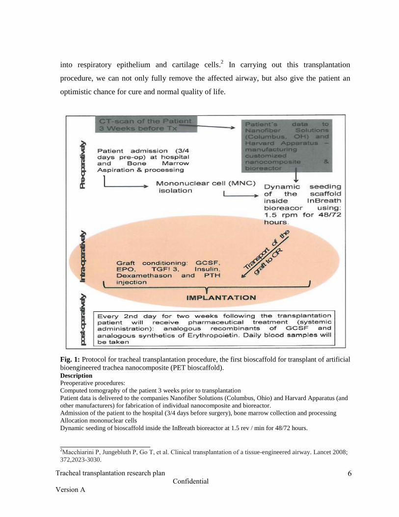
Fig. 1: Protocol for tracheal transplantation procedure, the first bioscaffold for transplant of artificial
bioengineered trachea nanocomposite (PET bioscaffold). Description Preoperative procedures:
Computed tomography of the patient 3 weeks prior to transplantation
Patient data is delivered to the companies Nanofiber Solutions (Columbus, Ohio) and Harvard Apparatus (and
other manufacturers) for fabrication of individual nanocomposite and bioreactor.
Admission of the patient to the hospital (3/4 days before surgery), bone marrow collection and processing
Allocation mononuclear cells
Dynamic seeding of bioscaffold inside the InBreath bioreactor at 1.5 rev / min for 48/72 hours.
_______________________ 2Macchiarini Р, Jungebluth Р, Go Т, et al. Clinical transplantation of a tissue-engineered airway. Lancet 2008;
372,2023-3030.
Tracheal transplantation research plan
Confidential
Version A
7
Intraoperative procedures:
Preparation of graft: an injection of granulocyte colony stimulating factor G-CSF, erythropoietin, transforming
growth factor beta-3, insulin, dexamethasone and parathyroid hormone.
Transportation of the transplant to the operating room
Transplantation
Postoperative procedures:
The patient receives the drugs (systemic application): recombinant G-CSF factor analogues and synthetic
analogs of erythropoietin every second day for two weeks after transplantation
transplant. Blood sampling is performed daily.
To date, two tracheal transplantations with bioengineered synthetic scaffolds have been
successfully performed by Dr. Macchiarini together with colleagues from Karolinska
Institutet in Stockholm, Sweden. In the first operation (June 2011) a nanocomposite
bioengineered synthetic scaffold made of POSS-PCU (polyhedral oligomeric
silsesquioxane) was used, while in the second operation (November 2011) a nanocomposite
bioengineered synthetic scaffold made of PET (polyethylene terephthalate) was used. In
both cases luminal ingrowth with healthy cells of respiratory epithelium was observed.
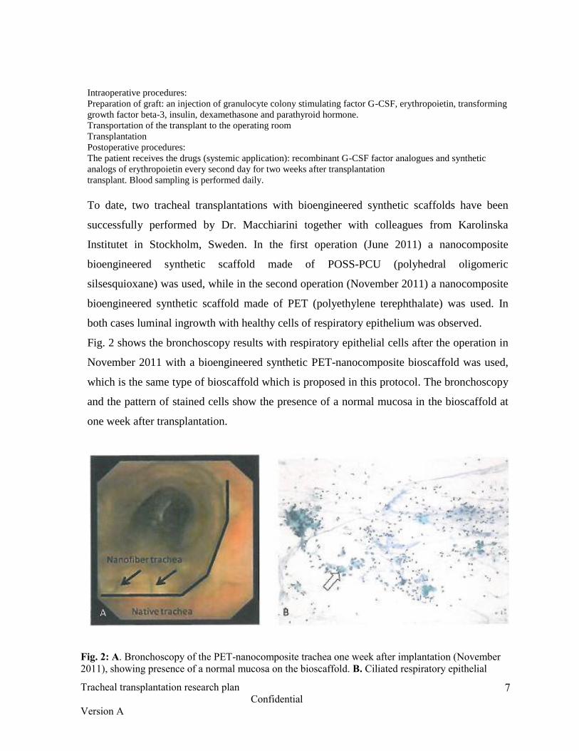
Fig. 2 shows the bronchoscopy results with respiratory epithelial cells after the operation in
November 2011 with a bioengineered synthetic PET-nanocomposite bioscaffold was used,
which is the same type of bioscaffold which is proposed in this protocol. The bronchoscopy
and the pattern of stained cells show the presence of a normal mucosa in the bioscaffold at
one week after transplantation.
Fig. 2: A. Bronchoscopy of the PET-nanocomposite trachea one week after implantation (November
2011), showing presence of a normal mucosa on the bioscaffold. B. Ciliated respiratory epithelial
Tracheal transplantation research plan
Confidential
Version A
8
cells (white arrow) obtained from a brush biopsy from the center of the transplanted trachea one
week after implantation. The fact that this biopsy was done almost immediately after transplantation,
allows us to assume that the cells derive from the differentiated stem cells, and not from the spread
of normal epithelial cells from the proximal or distal end of the transplant.
6.0 Rationale for not cancelling
In the previous research it has not been any negative effects or complications which would
cause rejection of the proposed research plan (protocol).
7.0 The procedure for obtaining voluntary informed consent
Patient information is presented by informed consent (attached to the protocol). The form of
informed consent for reference is issued to patients before the procedures and is necessary
for inclusion in the study protocol. The patient is given the opportunity to familiarize
themselves with the content of the informed consent in a separate room or bring it home to
familiarize themselves with it, get answers to their questions and sign with the date and time
of signing. All preparing of the patient for transplantation provided by the protocol starts
after the agreement has been signed. Researchers register the date and time of its completion
in the patient card.
8.0 General study plan
The tracheal transplant procedure will be performed sequentially, as shown in Fig. 1
"Protocol for tracheal transplantation procedure, transplantation for the first time applied for
the with artificial bioengineered trachea nanocomposite (PET bioscaffold)".
Preoperative assessment will include the following procedures:
Tomography of the neck and chest including a three-dimensional reconstruction of the
respiratory tract.
Rigid / Flexible fiberbronchoscopy.
Evaluation of cardiac function (scanning with thallium during exercise).
Assessment of respiratory function (spirometry).
Analysis of blood, including blood coagulation factors.
Evaluation of liver and kidney function.
Immunogenic assess of peripheral blood sample for the determination of the phenotype
HLA and serologic infections (HIV, syphilis, EBV, etc.).

Tracheal transplantation research plan
Confidential
Version A
9
Evaluation of the patients hematopoietic stem cells baseline levels; approximately 30 ml
of peripheral blood will be taken at admission for assessment basic level of
hematopoietic stem cells, and a portion of this sample is frozen for further analyzes.
Evaluation of endogenous baseline erythropoietin levels in the peripheral blood.
In case of successful preoperative evaluation bone marrow samples will be selected for
approximately 1-4 weeks prior to surgery.
Harvesting of mononuclear cells from bone marrow and seeding of the bioscaffold [After 48 or 72 hours before transplantation depending on the necessary shape of
bioscaffold (tubular or bifurcated shape)].
The procedure will be performed under general anesthesia.
Around 250-300 ml of bone marrow (BM) will be aspirated. BM will be passed to the
Department of Hematology or other department to isolate mononuclear
cells (MNCs).
Cell medium DMEM (Dulbecco Modified Eagle´s Medium) * +
autologous serum (10%) + antibiotics.
Peripheral blood sample (50 ml) will be aspirated with heparin (operating) and
transferred to the laboratory for cell culture in compliance with good manufacturing
practices. These MNCs will be isolated and frozen in liquid nitrogen.
Bronchoscopy and bronchoalveolar lavage (BAL). It is necessary to keep the BAL (take
away the supernatant, add PBS, pelleted by cell centrifugation and
freeze).
Bioreactor will be sterilized beforehand (locally with using plasma sterilization
according to manufacturer).
Synthetic bioscaffold will be sterilized with alcohol (ethanol) or by
gamma-irradiation, and then incubated in cell medium in 2 hours before
adding cells.
Necessary materials: stitching and forceps, sterilized scissors
Incubator.

Tracheal transplantation research plan
Confidential
Version A
10
Seeding and growing the cells on a synthetic bioscaffold (scanning
electron microscopy (SAM), microscopic studies of living cells,
histology).
* - environment and their manufacturers may differ.
48 hours before transplantation:
The patients receive treatment to stimulate mobilization of cells by intravenous injection of
recombinant granulocyte colony stimulating factor (Filgrastim, 10 µg/kg, not more than 30
million IU), and erythropoietin (EPO alpha or beta, not more than 40000 IU) 3, 4, 5
.
The patient will receive full and accurate information through the informed consent form,
orally and in writing, about the risks of the therapeutic procedure.
Cell preparation procedure
Bone marrow separation and further manipulation
72/48 hours before transplantation (depending on the desired shape of the bioscaffold:
tubular or bifurcated) bone marrow samples (BM) will be selected by bilateral repeated
aspiration from the iliac crest, general volume 250-300 ml. This procedure will be carried
out under general anesthesia and lasts about 20 minutes.
Explanted aspirate will be transferred to the hematology unit (or to another department) to
isolate mononuclear cells (MNCs). MNCs are obtained by Ficoll gradient separation, at
density 1.077 g/ml. After separation the cells are washed three times with saline (with the
addition of 5% human albumin) to remove the residual ficoll and left in a solution
consisting solely of components approved for clinical use. The whole procedure will be
carried out in a closed system (Sepax 3 Biosafe America Inc, Houston,
____________________ 3Haas R, Murea S. The role of granulocyte colony-stimulating factor in mobilization and transplantation of
peripheral blood progenitor and stem cells. Cytokines Mol. Ther. 1995; 1:249-70. 4Jia Wu, Warin R, Yu X, Epstein R, Noguchi CT. Erythropoietin signaling promotes cell progenitor transplanted
survival. FASEB J. 2009; 23 (9): 3089-99. 5Brines M, Cerami A. Erythropoietin-mediated tissue protection: reducing collateral damage from the primary
injury response. J Intern Med. 2008; 264 (5): 405-32.

Tracheal transplantation research plan
Confidential
Version A
11
Texas) to ensure sterility and a complete automatic process6. Isolated MNCs are transferred
into a bag with 600 ml medium (Dulbecco’s Modified Eagle Medium [DMEM 10% human
albumin) and transported from operation at temperature 4°C to a laboratory working under
the principles of GMP for filling synthetic bioscaffold (sterilized by ethanol or gamma
radiation). After incubation (DMEM) for 2 hours the bioscaffold will be secured in the
bioreactor. MNCs (medium DMEM) are seeded on the surface of the graft. Then
corresponding medium and growth factors is added (10μg/cm2 of recombinant human
transforming growth factor β-3 (R & Systems, Minneapolis, Minnesota, United States), 10
nmol/l recombinant parathyroid hormone-related peptide (PeproTech), 100 nmol/l
dexamethasone and 10 µg/ml insulin (Sigma-Aldrich, Dorset, United Kingdom). The
bioreactor is placed into a incubator running with an initial rate of 1 cycle per minute for 18
hours, then the speed is gradually increased to 1.5 cycles per minute. After 48/72 hours the
chamber is placed in a sterile container and carefully transferred to the operating room.
Isolated MNCs will be checked for the following indicators:
The number of mononuclear cells (MNCs): the minimum amount of 2x10⁶ cells/ml.
Cell viability by fluorescence microscopy analysis (7-AAD): range 94-98%.
Evaluation of mesenchymal progenitor cells CFU-F.
Evaluation of hematopoietic progenitor cells CD34+.
Altogether 15x106
cells are placed in three separate cryo-vials and frozen in DMSO, in
accordance with the standard procedure for quality control analysis. The sterile graft is then
re-inoculated with
________________________ 6Dal Pozzo 6 S, Urbani S, Mazzanti B, et.al. High recovery of mesenchymal progenitor cells with non-density
gradient separation of human bone marrow. Cytotherapy. 2010; 12 (5): 579-86.

Tracheal transplantation research plan
Confidential
Version A
12
cells in the operating room immediately before implantation (see Transplantation airway,
page 14).
Artificial nanocomposite airway transplantation
The company Nanofiber Solutions (Doctor Jed Johnson, Columbus, Ohio) developed the
graft trachea and tracheobronchial airways which is made of polyethylene terephthalate
(PET). PET has been used successfully for more than 10 years for production of
components to surgical implants and medical devices, ranging from non-absorbable suture
thread including vascular transplants and orthopedic implants. Proposed polymer for the
manufacture of a tracheal transplant is the biologically non-absorbable polyethylene
terephthalate, that has been transformed into nanofibers (intermediate diameter of 350 nm),
embedded in a semicircular spacer made of the material Dacron and forms a nanocomposite
being completely biocompatible having the nanofiber structure of a natural trachea (Fig. 3
and 4).
Fig. 3. Transformed nanofibers with a diameter in the range 300-400 nm.
Tracheal transplantation research plan
Confidential
Version A
13
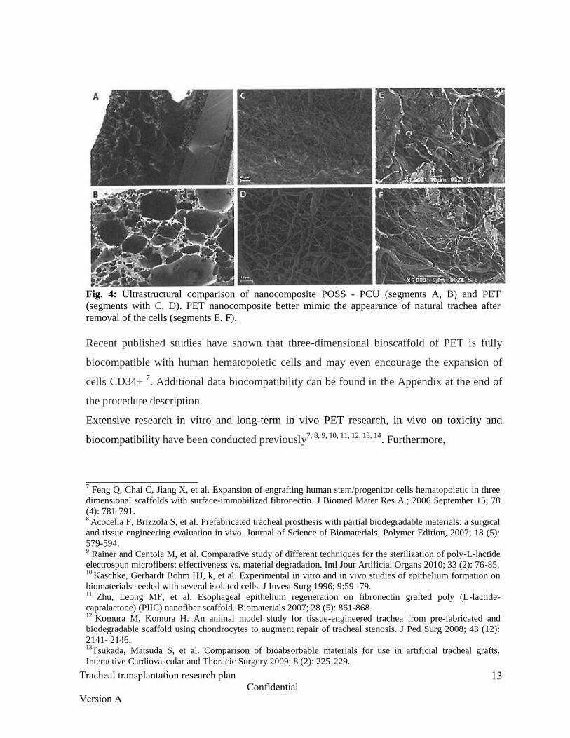
Fig. 4: Ultrastructural comparison of nanocomposite POSS - PCU (segments A, B) and PET
(segments with C, D). PET nanocomposite better mimic the appearance of natural trachea after
removal of the cells (segments E, F).
Recent published studies have shown that three-dimensional bioscaffold of PET is fully
biocompatible with human hematopoietic cells and may even encourage the expansion of
cells CD34+ 7. Additional data biocompatibility can be found in the Appendix at the end of
the procedure description.
Extensive research in vitro and long-term in vivo PET research, in vivo on toxicity and
biocompatibility have been conducted previously7, 8, 9, 10, 11, 12, 13, 14
. Furthermore,
____________________ 7 Feng Q, Chai C, Jiang X, et al. Expansion of engrafting human stem/progenitor cells hematopoietic in three
dimensional scaffolds with surface-immobilized fibronectin. J Biomed Mater Res A.; 2006 September 15; 78
(4): 781-791. 8 Acocella F, Brizzola S, et al. Prefabricated tracheal prosthesis with partial biodegradable materials: a surgical
and tissue engineering evaluation in vivo. Journal of Science of Biomaterials; Polymer Edition, 2007; 18 (5):
579-594. 9 Rainer and Centola M, et al. Comparative study of different techniques for the sterilization of poly-L-lactide
electrospun microfibers: effectiveness vs. material degradation. Intl Jour Artificial Organs 2010; 33 (2): 76-85. 10
Kaschke, Gerhardt Bohm HJ, k, et al. Experimental in vitro and in vivo studies of epithelium formation on
biomaterials seeded with several isolated cells. J Invest Surg 1996; 9:59 -79. 11
Zhu, Leong MF, et al. Esophageal epithelium regeneration on fibronectin grafted poly (L-lactide-
capralactone) (PIIC) nanofiber scaffold. Biomaterials 2007; 28 (5): 861-868. 12
Komura M, Komura H. An animal model study for tissue-engineered trachea from pre-fabricated and
biodegradable scaffold using chondrocytes to augment repair of tracheal stenosis. J Ped Surg 2008; 43 (12):
2141- 2146. 13
Tsukada, Matsuda S, et al. Comparison of bioabsorbable materials for use in artificial tracheal grafts.
Interactive Cardiovascular and Thoracic Surgery 2009; 8 (2): 225-229.

Tracheal transplantation research plan
Confidential
Version A
14
biocompatible nanocomposite has been successfully used for transplantation, which was
done in Sweden, at Karolinska University Hospital in November 2011. In the November
study mononuclear cells were isolated from bone marrow aspirates and plated on
bioscaffold via plasma sterilized bioreactor, which is described below. The results showed
improved survival rate of cells (increased number of cells, improved orientation and the
accumulation of extracellular matrix) in the PET bioscaffold compared to the POSS
bioscaffold (Fig. 5 and 6).
Fig.5: Structure of PET-nanocomposite after seeding with autologous progenitor cells
________________________________________________________________________________________________________________________________________
14 Kanzaki M, Yamato M, et al Tissue engineered epithelial cell sheets for the creation of a bioartificial
trachea. Tissue Engineering 2006; 12 (5): 1275-1283.

Tracheal transplantation research plan
Confidential
Version A
15
Fig. 6: Bioscaffold of PET gives a higher cell acceptability compared to bioscaffold of POSS -
PCU.
We offer manufacturing of synthetic bioengineered trachea transplant for individual patients
based on the results of recent computed tomography and endoscopic studies; the graft will
be made of polymeric nanofibers transformed (Nanofiber Solutions®, Columbus, Ohio,
United States), having mechanical and structural properties that mimic the natural
respiratory tract (fig. 4). The company Nanofiber Solutions will manufacture the cartilage
rings of the trachea with mechanical properties similar to those of a natural trachea with its
resistance of mechanical collapse. The cartilaginous rings are sandwiched between the
nanofibers and placed at regular intervals in a special form, exactly reproduced after the
shape of the patient´s trachea, and then transformed nanofibers will be used to cover each
ring inside and outside, respectively.

Tracheal transplantation research plan
Confidential
Version A
16
The company Nanofiber Solutions does not see any problems in terms of manufacturing the
device. As previously described, the inert nature of the polymer, combined with biomimetic
topography transformed graft nanofibers provides the necessary surface properties for
improved engraftment of cells, including mononuclear and epithelial cells specific to the
trachea.
Bioreactor InBreath
This protocol includes the design of the bioreactor design previously used by our group
during the first successful human implantation of a bioengineered trachea. The device,
known by the market name InBreath 3D Organ Bioreactor (Harvard Bioscience, Holliston,
Massachusetts), is placed inside the incubator for cell culture and consists of a single
chamber of polysulfone, where the artificial organs are placed, as well as motor and remote
control. Information about the materials used for the construction of the bioreactor can be
found in Appendix 2.
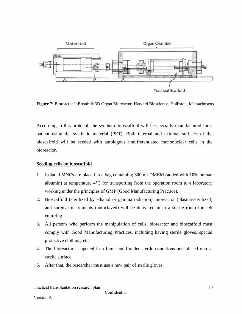
The bioreactor InBreath chamber is easily separated from the motor block and can be
subjected to plasma sterilization. The motor provides constant rotation of the cellular basis
inside the chamber, thereby providing controlled effect of the hydrodynamic forces on the
growing tracheal transplant. Protective chassis fully covers the brushless electric motor,
protecting it from the corrosive action of moisture generated inside the incubator (fig. 7).
Remote controller placed outside of the incubator, giving the possibility to adjust the speed
rotation, without affecting the incubator.
Tracheal transplantation research plan
Confidential
Version A
17
Figure 7: Bioreactor InBreath ® 3D Organ Bioreactor, Harvard Bioscience, Holliston, Massachusetts
According to this protocol, the synthetic bioscaffold will be specially manufactured for a
patient using the synthetic material (PET). Both internal and external surfaces of the
bioscaffold will be seeded with autologous undifferentiated mononuclear cells in the
bioreactor.
Seeding cells on bioscaffold
1. Isolated MNCs are placed in a bag containing 300 ml DMEM (added with 10% human
albumin) at temperature 4°C for transporting from the operation room to a laboratory
working under the principles of GMP (Good Manufacturing Practice).
2. Bioscaffold (sterilized by ethanol or gamma radiation), bioreactor (plasma-sterilized)
and surgical instruments (autoclaved) will be delivered in to a sterile room for cell
culturing.
3. All persons who perform the manipulation of cells, bioreactor and bioscaffold must
comply with Good Manufacturing Practices, including having sterile gloves, special
protective clothing, etc.
4. The bioreactor is opened in a fume hood under sterile conditions and placed onto a
sterile surface.
5. After that, the researcher must use a new pair of sterile gloves.

Tracheal transplantation research plan
Confidential
Version A
18
6. The bioscaffold will be removed from the primary sterile packaging and secured within
the bioreactor at the respective fixtures.
7. After the bioscaffold has been docked within the bioreactor, MNCs (+ DMEM) will be
seeded on the surface of bioscaffold.
8. 250 ml volume of medium (with the addition of autologous plasma and human
albumin) will be added to the bioreactor chamber.
9. The following ingredients are added to the medium (see Appendix 3 "List of biological
agents and characterization TGF β-3"): 10 mcg/ml of recombinant human transforming
growth factor β-3 (R & D Systems, Minneapolis, Minnesota, United States), 10 nmol/l
of recombinant parathyroid hormone-related peptide (PeproTech), 100 nmol/l
dexamethasone, 10 µg/ml of insulin (Sigma-Aldrich, Dorset, United Kingdom).
(Note: hereinafter products from other manufacturers may be used than the products
from companies which have been used in previous transplants referred to).
10. Thereafter, the bioreactor will be placed in an incubator and the chamber closed
(containing the bioscaffold, MNCs + 250 ml of medium).
11. NOTE: a bifurcate tracheobronchial bioscaffold does not perfectly fit to the shape of
the chamber and this can lead to dynamic tension in the bioreactor. This tension (shear)
will be transmitted through the external connection to the electric motor and can lead to
termination of the chamber rotations. In that case, the bioreactor must manually be
monitored every 20 minutes. In the case of a low speed rate due to influence of
dynamic tension, it is possible to balance the chamber with an object to ensure
continuous communication between these two components. In the event of a significant
dynamic tension (shift), it is necessary to understand additional action to secure the
bioscaffold. As a rule, such an event is adverse for tubular bioscaffolds.
12. The bioreactor is started with an initial speed of 1 rpm for 18 hours and then the rate
will gradually be increased to 1.5 rpm.

Tracheal transplantation research plan
Confidential
Version A
19
13. After 24 hours, 50ml of the above medium is added into the chamber to a total volume
of 300 ml.
14. After 48/72 hours, the chamber will be placed in a sterile container and gently moved to
the operating room.
Transplantation of the airway
The patient will be subject to general anesthesia and intubated with an endotracheal tube.
Any existing t-tube will be removed. The patient will also be subject to bone marrow
aspiration, according to the procedure for preparing cells (see above). Median sternotomy
will be carried out in supine position. After resection of the damaged airway segment the
airway graft will be introduced (conditioning) with growth factors, including 10 ng/ml
recombinant human transforming growth factor β-3 (R & D Systems, Minneapolis,
Minnesota, United States), 10 nmol/l recombinant parathyroid hormone-related peptide
(PeproTech), 100 nmol/l dexamethasone, 10 µg/ml insulin ( Sigma-Aldrich, Dorset, United
Kingdom, G-CSF (of 1 µg/kg) and erythropoietin (40000 IU), to stimulate the mobilization
of peripheral hematopoietic cells. 3,14
The graft will be adjusted in size, and then proximally
and distally anastomosed to correct the defect of the respiratory tract by using non-
absorbable sutures, such as Cardionyl 3/0 (Peters Surgical). The graft will then be covered
by major omental flap wrapping (vascularized adipose tissue is separated from large
stomach bend and the right gastro-omental artery and diaphragmatically or substernal
transferred to the mediastinum) to provide long-term protection of the graft anastomoses,
and indirect stimulate neovascularization of the graft.
Sterility tests
The risk of bacterial/fungal medium contamination will be assessed be microscopic
examination prior to the transplantation. The quality of all components and medium are
controlled by the manufacturers, and remain sealed until the cell seeding procedure.
Tracheal transplantation research plan
Confidential
Version A
20
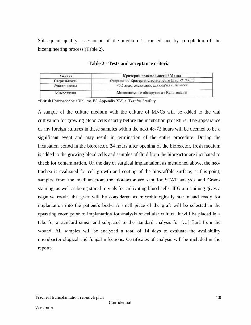
Subsequent quality assessment of the medium is carried out by completion of the
bioengineering process (Table 2).
Table 2 - Tests and acceptance criteria
*British Pharmacopoeia Volume IV. Appendix XVI a. Test for Sterility
A sample of the culture medium with the culture of MNCs will be added to the vial
cultivation for growing blood cells shortly before the incubation procedure. The appearance
of any foreign cultures in these samples within the next 48-72 hours will be deemed to be a
significant event and may result in termination of the entire procedure. During the
incubation period in the bioreactor, 24 hours after opening of the bioreactor, fresh medium
is added to the growing blood cells and samples of fluid from the bioreactor are incubated to
check for contamination. On the day of surgical implantation, as mentioned above, the neo-
trachea is evaluated for cell growth and coating of the bioscaffold surface; at this point,
samples from the medium from the bioreactor are sent for STAT analysis and Gram-
staining, as well as being stored in vials for cultivating blood cells. If Gram staining gives a
negative result, the graft will be considered as microbiologically sterile and ready for
implantation into the patient´s body. A small piece of the graft will be selected in the
operating room prior to implantation for analysis of cellular culture. It will be placed in a
tube for a standard smear and subjected to the standard analysis for […] fluid from the
wound. All samples will be analyzed a total of 14 days to evaluate the availability
microbacteriological and fungal infections. Certificates of analysis will be included in the
reports.

Tracheal transplantation research plan
Confidential
Version A
21
Postoperative procedures
To stimulate the process of regeneration in the postoperative period, the patient will receive
pharmacological agents with following systemic injections:
a) Recombinant analogues of G-CSF (Filgrastim, 10mcg/kg/day, no more than 30
mcg/kg/day)
b) Synthetic analogues of erythropoietin (EPO alpha or beta, max40000 IU).
Both of these factors will be administered in adequate concentrations (in reduced doses not
associated with any side effects) for stimulating mobilization and transformation of
progenitor stem3,4,5,15,16
and bone marrow cells […] day automatic controlled plasma
erythropoietin level and calculation of whole blood (including leukocyte blood count).
Levels above 50000-60000 cells in the blood will be considered a manifestation of toxicity
and as a result will be reduced dose or discontinued. The treatment is carried out every other
day for two weeks after transplantation..
Follow-up
Follow-up will include:
Endoscopy (flexible or rigid bronchoscopy) of the transplanted respiratory system every
day or every other day (when clinically indicated) during the first week and at least once
before discharge from the hospital.
Bronchoscopy is performed on a monthly basis for the first six months and then every 6
months for the first 5 years. Samples from the respiratory airway mucosa shall be
collected and stored for quality analysis.
Counting of blood cells, (including leukocyte blood count) every day during the first two
weeks.
________________________ 15
Bader, A. Macchiarini P. Moving towards in situ tracheal regeneration: the bionic tissue engineered
transplantation approach. J Cell Mol Med 2010; 14 (7): 1877-89. 16
Jungebluth P, Moll G, Baiguera S, Macchiarini P. Tissue engineered airway: a regenerative solution. Clin
Pharm Ther 20 12; 91:81-93

Tracheal transplantation research plan
Confidential
Version A
22
Evaluation and calculation of mobilized progenitor cells according to the graph below:
Assessment of immunogenicity. After 3, 7 and 30 days after transplantation blood
samples will be taken for analysis of HLA by OIA antibodies. Subsequent immunogenic
studies will also be performed on 3, 6 and 12 months after transplantation.
Computer tomography of the neck and chest with three-dimensional reconstruction of the
transplanted airway will be done during the first, third, and sixth months during follow-
up, and then every 6 months within the first 5 years.
Subsequent cancer surveillance in children will be performed throughout the patient´s
life and include standard examinations.

Tracheal transplantation research plan
Confidential
Version A
23
9.0 Expected risks
The positive effect of this operation is supposed to be greater than the risk since this
procedure may be the only possible chance of cure for some patients. Training will be held
in appropriate cell laboratory in compliance with good manufacturing practices. In addition,
numerous sterility tests will be carried out on all cells and materials before tracheal
transplantation. If the sterility of cells and materials will be called into question, which
seems unlikely, the whole procedure will immediately be terminated.
10.0 Adverse events
Possible complications of the transplant are postoperative bleeding, injury to the nervus
recurrence, respiratory infections, anastomotic leak, wound infection, respiratory failure and
the need for mechanical ventilation. All adverse events should be documented and
communicated to the lead researcher, the sponsor and the Ethics Committees of the
University and the Hospital. Address and phone number of the clinic for emergency GBUZ
"Regional clinical hospital (N) 2 (l) S.V. Ochapovsky Department of Health Care Krasnodar
Krai "at the address: 350086, Krasnodar, street. May Day, d. 167, Tel.:( 861) 252-73-02,
260-35-11, Head of the Oncology Department, doctor of higher category, PhD Polyakov,
Igor Stanislavovich
11.0 Introduction of amendments
The clinical trial plan cannot be changed without the written permission from the lead
researcher, the funder, the Ethical Committee of the University and the Clinic. Amendments
may require regulatory approval prior to their entry into force. All the amendments to the
protocol, the patient´s informed consent map, must be approval by the Ethical Committee of
the University and the Clinic.
Tracheal transplantation research plan
Confidential
Version A
24
12.0 Publication policy
The results of this research can be used for publication.
13.0 Individual registration card (separate application)

Tracheal transplantation research plan
Confidential
Version A
25
Appendix 1: Biocompatibility, acceptability / cell proliferation and mechanical
characteristics of PET medical devices available on the market at present
time.
Bioscaffold material: polyethylene terephthalate (PET) is not absorbable.
Summary on biocompatibility: polyethylene terephthalate has for more than a decade
successfully been used in fabrication of numerous of surgical implants and medical devices,
from intracardiac and vascular grafts to threads for surgical sutures. The company
Nanofiber Solutions (Columbus, Ohio) has developed tracheal and tracheobronchial
bioscaffolds imitating the nanofiber structure of the natural trachea (Fig. 8). These
bioscaffolds are made of non-absorbable polyethylene terephthalate transformed into
nanofibers with an mean diameter of 350 nm.
Fig. 8. Electron micrograph scanning of purified human tracheal cells (A) and artificial PET
trachea manufactured by the company Nanofiber Solutions (B).
An artificial tracheobronchial graft using bioscaffold from PET-material has already been
successfully transplanted at Karolinska University Hospital, Stockholm, Sweden, in
November 2011. The clinical success of this operation indicates that the tracheobronchial
graft of bioengineered nanocomposite and autologous mononuclear cells may be the only
chance of cure for some patients.
We propose to use the same material for the bioscaffold and the same procedure for its
production, which has been successfully used for the tracheal transplantation in November,
Tracheal transplantation research plan
Confidential
Version A
26
2011, using this Protocol. The polymer nanocomposite PET has carefully been studied on
cell compatibility, and recent surgery, performed at Karolinska University Hospital,
demonstrated its acceptability, ability to allow for proliferation of autologous mononuclear
cells and early (7 days) re-epithelialization with respiratory epithelium.
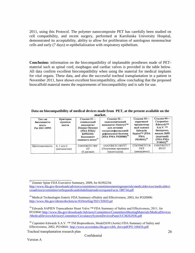
Conclusion: information on the biocompatibility of implantable prostheses made of PET-
material such as spinal cord, esophagus and cardiac valves is provided in the table below.
All data confirm excellent biocompatibility when using the material for medical implants
for vital organs. These data, and also the successful tracheal transplantation in a patient in
November 2011, have shown excellent biocompatibility, allow concluding that the proposed
bioscaffold material meets the requirements of biocompatibility and is safe for use.
Data on biocompatibility of medical devices made from PET, at the present available on the
market.
______________________ 17
Zimmer Spine FDA Executive Summary, 2009, for KO92234:
http://www.fda.gov/downloads/advisorycommittees/committeesmeetingmaterials/medicaldevices/medicaldevi
cesadvisorycommittee/orthopaedicandrehabilitationdevicespanel/ucm 188734.pdf
18
Medical Technologies Enteric FDA Summary ofSafety and Effectiveness, 2003, for PO20006:
http://www.fda.gov/ohrms/dockets/ac/03/briefing/3921/SSED.pd
19
Edwards SAPIEN Transcatheter Heart Valve ™ FDA Summary of Safety and Effectiveness, 2011, for
PO10041:http://www.fda.gov/downloads/AdvisoryCommittees/CommitteesMeetingMaterials/MedicalDevices
/MedicalDevicesAdvisoryCommittee/CirculatorySystemDevicesPanei/UCM262936.pdf
20
Capentier-Edwards S.A.V. ™ TM Bioprosthesis, Model2650 (Aortic) FDA Summary of Safety and
Effectiveness, 2002, PO10041: http://www.accessdata.fda.gov/cdrh_docs/pdf/PO 10041b.pdf

Tracheal transplantation research plan
Confidential
Version A
27

Tracheal transplantation research plan
Confidential
Version A
28

Tracheal transplantation research plan
Confidential
Version A
29

Tracheal transplantation research plan
Confidential
Version A
30
Engraftment and cell proliferation
PET polymer has been extensively analyzed for cellular compatibility and proved its ability
to effectively support the implantation of cells, their dispersion and attachment for
exogenous agents and preservation of the phenotype of cells, and was shown to have
biomechanical dynamic ability to handle stress.21
In addition, bioscaffold, consisting of

Tracheal transplantation research plan
Confidential
Version A
31
transformed nanofibers have demonstrated the ability to provide relevant micro-habitation
for […] and […] differentiation of progenitor cells. 22
Fig. 9. PET bioscaffold of transformed nanofibers shows: (A) original microstructure fiber
network (B) nano-pores on the surface of the fibers that contribute to cell attachment, (C)
section fiber-demonstrates a lack of pores within it, (D) relatively dense flat bottom surface
bioscaffold for slower cell migration cell in the spot with medium.
_________________________ 21
Nam J, Rath in Knobloch, TJ, Lannutti JJ, Agarwa1 S.Nove1 e1ectrospun scaffo1ds for the mo1ecu1ar
ana1ysis of chondrocytes under dynamic compression. Tissue Eng Part a. 2009; 15 (3): 513-23. 22
Nam J, Johnson J, Lannutti JJ, Agarwa1 S. Modu1ation of embryonic progenitor cell differentiation
mesenchyma1via contro1 over pure mechanica1 modu1us in e1ectrospun nanofibers. Acta Biomater. 2011; 7
(4): 1516-24.

Tracheal transplantation research plan
Confidential
Version A
32
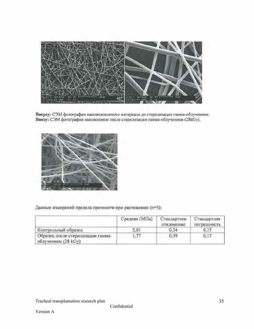
Mechanical characteristics of the bioscaffold
SPECIMEN Electrospun bifurcated * scaffold produced by Nanofiber Solutions,
Columbus, OH.
TEST Uniaxial tensile test.
CONDITIONS Universal testing machine Lloyd LRX
Load cell: 2500 N
Preload: 1 N
Speed of testing: 1 mm/s
Stainless steel custom-made grips fixed to the rigid rings of the scaffold.
In order to prevent slippage, grips were pre-glued to the sample.
Tests were carried out on both as-received scaffolds and scaffolds sterilized by
immersion in ethanol overnight and then dried for 24 h.
For each specimen 5 measurements were performed.
Measured parameters: Force at break (Fmax).
Elongation at break, defined as the percentage
increase in length, before the break occurs, with
respect to the initial length of the specimen.

Tracheal transplantation research plan
Confidential
Version A
33

Tracheal transplantation research plan
Confidential
Version A
34
Tracheal transplantation research plan
Confidential
Version A
35

Tracheal transplantation research plan
Confidential
Version A
36

Tracheal transplantation research plan
Confidential
Version A
37

Tracheal transplantation research plan
Confidential
Version A
38
Tracheal transplantation research plan
Confidential
Version A
39
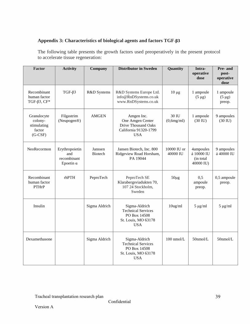
Appendix 3: Characteristics of biological agents and factors TGF-β3
The following table presents the growth factors used preoperatively in the present protocol
to accelerate tissue regeneration:
Factor Activity Company Distributor in Sweden Quantity Intra-
operative
dose
Pre- and
post-
operative
dose
Recombinant
human factor
TGF-β3, CF*
TGF-β3
R&D Systems
R&D Systems Europe Ltd.
www.RnDSystems.co.uk
10 μg
1 ampoule
(5 μg)
1 ampoule
(5 μg)
preop.
Granulocyte
colony-
stimulating
factor
(G-CSF)
Filgastrim
(Neupogen®)
AMGEN
Amgen Inc.
One Amgen Center
Drive Thousand Oaks
California 91320-1799
USA
30 IU
(0,6mg/ml)
1 ampoule
(30 IU)
9 ampoules
(30 IU)
NeoRecormon
Erythropoietin
and
recombinant
Epoetin α
Janssen
Biotech
Jansen Biotech, Inc. 800
Ridgeview Road Horsham,
PA 19044
10000 IU or
40000 IU
4ampoules
á 10000 IU
(in total
40000 IU)
9 ampoules
á 40000 IU
Recombinant
human factor
PTHrP
rhPTH
PeproTech
PeproTech SE
Klarabergsviadukten 70,
107 24 Stockholm,
Sweden
50μg
0,5
ampoule
preop.
0,5 ampoule
preop.
Insulin
Sigma Aldrich
Sigma-Aldrich
Technical Services
РО Вох 14508
St. Louis, МО 63178
USA
10ug/ml
5 μg/ml
5 μg/ml
Dexamethasone
Sigma Aldrich
Sigma-Aldrich
Technical Services
РО Вох 14508
St. Louis, МО 63178
USA
100 nmol/L
50nmol/L
50nmol/L

Tracheal transplantation research plan
Confidential
Version A
40
* TGF-β3: calculation of the necessary dose of TGF-β3 based on the size of the human
trachea and as a rule, it is based on the average lateral area of the cartilage as shown below
in figure 1B.