transactions of the royal society of edinburgh: earth ...orca.cf.ac.uk/10510/1/edwards 2003.pdf ·...
TRANSCRIPT
Transactions of the Royal Society of Edinburgh: EarthScienceshttp://journals.cambridge.org/TRE
Additional services for Transactions of the Royal Society of Edinburgh: EarthSciences:
Email alerts: Click hereSubscriptions: Click hereCommercial reprints: Click hereTerms of use : Click here
Embryophytic sporophytes in the Rhynie and Windyeld cherts
Dianne Edwards
Transactions of the Royal Society of Edinburgh: Earth Sciences / Volume 94 / Issue 04 / December 2003, pp 397 - 410DOI: 10.1017/S0263593300000778, Published online: 26 July 2007
Link to this article: http://journals.cambridge.org/abstract_S0263593300000778
How to cite this article:Dianne Edwards (2003). Embryophytic sporophytes in the Rhynie and Windyeld cherts. Transactions of the Royal Societyof Edinburgh: Earth Sciences, 94, pp 397-410 doi:10.1017/S0263593300000778
Request Permissions : Click here
Downloaded from http://journals.cambridge.org/TRE, IP address: 131.251.254.13 on 25 Feb 2014
Embryophytic sporophytes in the Rhynie andWindyfield cherts
Dianne Edwards
ABSTRACT: Brief descriptions and comments on relationships are given for the seven embryo-phytic sporophytes in the cherts at Rhynie, Aberdeenshire, Scotland. They are Rhynia gwynne-vaughanii Kidston & Lang, Aglaophyton major D. S. Edwards, Horneophyton lignieri Barghoorn &Darrah, Asteroxylon mackiei Kidston & Lang, Nothia aphylla Lyon ex Høeg, Trichopherophytonteuchansii Lyon & Edwards and Ventarura lyonii Powell, Edwards & Trewin. The superb preserva-tion of the silica permineralisations produced in the hot spring environment provides remarkableinsights into the anatomy of early land plants which are not available from compression fossils andother modes of permineralisation. They include soft tissues, such as those surrounding stomata,rhizoids, apical and lateral meristems, and diversity in conducting cells, with inferences forpalaeoecophysiology, including water use efficiency, transport and absorption, and for growthprocesses and patterns.
KEY WORDS: Exceptional preservation, Lycopsida, meristems, palaeoecophysiology,Rhyniopsida, silicification, substomatal chamber, water absorption, xylem, Zosterophyllopsida.
The higher plant taxa in the Rhynie chert, Aberdeenshire,Scotland, although contributing relatively little to LowerDevonian numerical diversity, surely compensate in theircontribution to our understanding of the anatomy of earlyland plants and their roles in early terrestrial ecosystems. Thepresent author’s brief is to review current knowledge on thesporophytes of the seven embryophytic taxa – five of whichwere known to Kidston & Lang – and to summarise, some-what idiosyncratically, the unique insights afforded by thesuperb preservation of the silica permineralisations. Thesehave been revealed by the meticulous descriptions of thou-sands of laboriously prepared peels and sections which showanatomical information far in excess of that derived from moreconventional compression and permineralised fossils.
1. Descriptions of sporophytes
1.1. Rhynia gwynne-vaughanii Kidston & Lang (Figs1b, b#, 2a–c, m, o, 3a, j, m)
1.1.1. Descriptions. Rhynia gwynne-vaughanii was originallyreconstructed by Kidston & Lang (1921) as a rather simplevascular plant, c. 20 cm high, with smooth bifurcating axesbearing numerous hemispherical projections, adventitiousbranches, terminal sporangia and a limited horizontal rhizome(possibly belonging to Aglaophyton) (Kidston & Lang 1917,1921). Its xylem was a terete central bundle of tracheids withbroad annular or helical thickenings (Fig. 2a). In olderassumed decaying examples, these thickenings disappear andthe xylem is darkly stained. In contrast, some of the lateralbuds and short adventitious branches, possible agents ofvegetative propagation, lacked vascularisation when shed,their positions marked by a patch of darkened tissue, while inmore developed adventitious branches, there was no continuityof tracheids between those in the branches and those in theaxes bearing them. The hemisperical projections are nevervascularised. They were produced in maturing axes by deep-seated cortical cell divisions with some additional compensat-ing epidermal divisions. Stomata are present on all except themost proximal (basal) projections, which bear rhizoids. D. S.
Edwards’ (1980) reconstruction of the plant shows moreoverall complexity, particularly in the abundance of adventi-tious branches – dichotomous branching being comparativelyrare. He also provided detailed anatomical information onchanges in the xylem as it passed through a zone of darkenedcells before it terminated in a pad of tissue at the base of thesporangium (Fig. 2b). He speculated that, after sporangialabscission through the darkened zone, growth was resumed bya more proximal adventitious branch, a form of overtopping.Data on extraxylary tissues are given in D. Edwards, Kerp &Hass (1998). Homosporous eusporangia, normally found iso-lated, were fusiform, <5·1 mm long and contained spores,<53 �m diameter, with granular ornamentation. Bhutta (1969)had described the sporangial wall in detail, and in contrast toAglaophyton had noticed a lack of stomata. D. S. Edwards’reconstruction lacked a basal region because he and others hadfailed to find convincing evidence. Bhutta (1969) had illus-trated two possible examples, one with clusters of rhizoids onthe lower surface of a short presumed rhizome, bearing thebases of a number of upwardly directed axes, the latter lackingany hemispherical projections, although in another example,rhizoids were confined to the lower surface of the latteralongside an area with a more extensive rhizoidal region.The specimen illustrated here (Fig. 2c) was identified asR. gwynne-vaughanii by A. G. Lyon.
1.1.2. Relationships. Rhynia gwynne-vaughanii was origi-nally placed with R. major, Hornea and Psilophyton princeps(in part) in the Rhyniaceae in the Psilophytales (Kidston &Lang 1920a). Banks (1968, 1975) made the genus Rhynia thetype of his new subdivision, the Rhyniophytina, and includedit in the Rhyniaceae, together with a number of taxa showingterminal sporangia, dichotomous branching and probablyterete centrarch xylem strands. Edwards & Edwards (1986)reduced the number to three (Rhynia plus Taeniocrada dech-eniana and Renalia in part), whilst Banks (1992) united theeponymous taxon with Uskiella spargens and Cooksonia. In themeantime, the discovery of S-type tracheids in R. gwynne-vaughanii, Stockmansella and Huvenia (Kenrick & Crane 1991;Kenrick et al. 1991) which also share fusiform sporangiacharacterised by an abscission or isolation layer in their
Transactions of the Royal Society of Edinburgh: Earth Sciences, 94, 397–410, 2004 (for 2003)
pedicels, led initially to their inclusion in the Rhyniaceae(Kenrick & Crane 1991), whilst in a further cladistic analysis(Kenrick & Crane 1997), the three taxa were united in anextinct clade (named the Rhyniopsida) and considered a sistergroup to all other vascular plants (Hass & Remy 1991).
The description of the fertile gametophytes of R. gwynne-vaughanii (Kerp et al. 2004) finally brings to an end earlyspeculation that the taxon, as originally described, was agametophyte (see discussion in D. S. Edwards 1980).
1.2. Aglaophyton major Edwards=Rhynia majorKidston & Lang Figs 1a, a#, 2d, e, j, k, n, 3h, i)
1.2.1. Description. Kidston & Lang (1917) included bothsterile and fertile parts of Aglaophyton in their original descrip-tion of R. gwynne-vaughanii, but subsequently (1920a) erecteda new species of Rhynia, to accommodate them. Rhynia majorwas conceived as larger and simpler than R. gwynne-vaughanii(lacking hemispherical projections or adventitious branches)with homosporous terminal eusporangia reaching a length of12 mm. Kidston & Lang (1920a) described the central xylemstrand as comprising narrower central tracheids and widerperipheral ones (Fig. 2d), and despite their apparent perfectionof preservation, could find no indications of tracheidal thick-enings (Fig. 2e). They believed that the uniform appearanceresulted from decay of the original wall.
Further vegetative anatomical data (phloem with putativesieve areas) were obtained by Satterthwait & Schopf (1972),Bhutta (1973a), Remy & Remy (1977), Lemoigne & Zdebska(1980), Remy & Hass (1996) and D. Edwards, Kerp & Hass(1998). D. S. Edwards (1986) produced a new reconstructionbased on serial sections characterised by a basal rhizomatoussystem in which patches of rhizoids occur on bulges on thelower surfaces of undulating (U-shaped) rhizomes presumablywhere they had come in contact with the substrate. Details ofthe vasculature of these rhizoidal bulges, and far more complexgrowth patterns involving arrested apices, were given in Remy& Hass (1996, pp. 182–9).
Some subsequent studies have concentrated on sporangiaand dehiscence. Thus, Remy (1978) in describing the anatomyof the sporangium wall emphasised the detailed distribution ofthickenings in the majority of epidermal cells (cohesion cells)with outer periclinal walls being the thinnest walls and theanticlinal walls, the thickest. Contraction of these cells ispurported to have resulted in longitudinal splitting, althoughwhether or not this was along a predetermined line withmodified tissue could not be determined. Remy (1978) de-scribed a longitudinal hinge-zone of rectangular to square cellsopposite the slit, and considered the spiralling in the walls to bea consequence of dehiscence. In contrast, Kerp and Hass (pers.comm. 2003) described one to several spirally aligned longitu-dinal slits with the tip remaining intact. Remy & Hass (1996)suggested that spores (<64 �m in diameter) were shed intetrads and in clumps, the latter possibly indicative of passivedispersal.
1.2.2. Relationships. Originally named Rhynia major, itwas placed with the type species in the Rhyniaceae of thePsilophytales (Kidston & Lang 1920a). D. S. Edwards (1986)had emphasised the lack of conventional tracheidal thicken-ings in the xylem (see Hebant 1977), and considered thisdifference along with sporangial abscission in R. gwynne-vaughanii sufficient to merit the erection of a new genusAglaophyton. Aglaophyton then appeared to have a far simplergrowth pattern compared with R. gwynne-vaughanii, at least inits dichotomously branching aerial axes (but for complexityin basal parts, see Remy & Hass 1996). Similarities in thevesiculate nature of some xylem elements (Fig. 3i, j) betweenAglaophyton and Rhynia (Remy & Hass 1996; D. Edwards
2003) would seem to narrow the differences between the twogenera, and therefore, possibly invalidate the necessity forgeneric changes. It certainly casts doubt on the placement ofAglaophyton outside the Tracheophyta, as had been contem-plated in perceived similarities of its water-conducting cellswith those of bryophytes (Hebant 1973; D. S. Edwards 1986).In Kenrick & Crane’s (1997) cladistic analysis, Aglaophyton isresolved along with Horneophyton as a basal polysporangiate,the critical character being the smooth-walled, water-conducting cells (but see below for tracheidal thickenings inHorneophyton).
1.3. Horneophyton lignieri Barghoorn & Darrah(=Hornea lignieri Kidston & Lang) (Figs 1d, d#,2f, g, l)
1.3.1. Description. The plant was about 20 cm high, withaerial stems up to 2 mm in diameter showing dichotomousbranching that increased in frequency distally. The basalregion comprised a ‘lobed tuberous rhizome’ from which arosea number of aerial stems. Kidston & Lang (1920a) comparedthe basal structures with the protocorms of certain Lycopo-dium species. They lacked tracheids and bore bands of verylong unicellular rhizoids on the lower surfaces. Althoughessentially parenchymatous, they contained darkened cells in abell-shaped configuration, from the upper surface of whichstrands extended into the aerial axes. Frey, Hilger & Hofmann(1996) suggested that the tubers were gametophytic, based on asingle section showing a possible gametophyte/sporophytejunction, the latter represented by the base of an aerial axis.Although often poorly preserved, the terete strands in thelatter show unequivocal narrow interconnected annular orspiral thickenings, with the smaller elements situated centrallywhere breakdown frequently occurred. The tracheids weresurrounded by elongate, thin-walled cells interpreted asphloem. Details of extra vascular tissues are given in Hass(1991) and Edwards et al. (1998). Particularly striking werethe exceedingly long (<1000 �m) and wide epidermal cells(Fig. 2f, l) surrounding occasional stomata and corticalcells with pronounced intercellular spaces resembling palisademesophyll, but with long axes directed distally at an angle tothe surface. Similarly orientated photosynthesising cells occurin stems and needle-like leaves of certain angiosperms (Meyer1962). Fewer intercellular spaces are seen between the elongateand overlapping inner cortical cells.
The terminal homosporous eusporangia are unusual in thatthey were columellate and branched with a single continuoussporangial cavity. Cells of the columella resemble those of thephloem. While Kidston & Lang (1920a) recorded mainly singleand some branched examples, later authors described two,four or even five lobed sporangia (Bhutta 1973b; Eggert 1974;El-Saadawy & Lacey 1979). Lobes had truncated tips whichcould be slightly convex, concave or smooth, but all showedepidermal cells at the summit with outer and anticlinal wallsthickened. Spore dispersal mechanisms remain conjectural.Kidston & Lang (1920a) thought that the sporangia wereindehiscent, but others have described the existence of one ormore slits (Bhutta 1973b) or an apical pore (Eggert 1974).El-Saadawy & Lacey (1979) suggested that such a pore devel-oped when cells in the central area became displaced and thesurrounding cuticle curved inwards. Hass (1991) recordedsplitting along the middle lamellae of these central cells.El-Saadawy & Lacey (1979) also described emergences on thesporangia, each terminating in a single stoma whose guardcells were relatively thinly cuticularised compared with theremaining epidermal cells. Similar structures, called roundedelevations, were described by Hass (1991), but were outnum-bered by ‘secretory organs’ occurring in the flatter areas
398 DIANNE EDWARDS
(Fig. 2g). These were groups of irregularly shaped cells radiat-ing from a central pore filled with darkly staining material.Similar structures also occurred on the aerial stems.
Spores were of the same size (c. 57 �m diameter) andshowed a number of surface features, but recent investigationshave suggested that they bore proximal ridges, and thus,belong to the dispersed taxon Emphanisporites (Edward &Richardson 2000; Wellman et al. 2004).
1.3.2. Relationships. Hornea was placed with Rhynia in theRhyniaceae (Psilophytales) by Kidston & Lang (1920a), andbroad comparisons were made with bryophytes in its posses-sion of a columella and with the Lower Devonian fossil,Sporogonites exuberans Halle (1916). Since subsequent investi-gations were inconclusive on the presence of a columella in thelatter (Halle 1936), Horneophyton remains the only LowerDevonian vascular plant with this structure. It was classifiedin the Rhyniaceae in the Rhyniophytina by Banks (1975),an assignation later questioned by Edwards & Edwards(1986) on its uniquely and prominently branching sporangia(El-Saadawy & Lacey 1979). Based on an assumption thatHorneophyton lacked tracheidal thickenings, in Kenrick andCrane’s (1997) cladistical analysis, Horneophyton is resolved asa protracheophyte (this grade also including Aglaophyton) thatis basal to all other polysporangiates. It was placed in its owndivision, Horneophytopsida, tentatively along with Caia andTortilicaulis, compression fossils for which no vascular dataare known, but which sometimes show bifurcating sporangia(Kenrick & Crane 1997).
1.4. Asteroxylon mackiei Kidston & Lang (Figs 1e, e#,e2, 3b, c)
1.4.1. Description. Kidston & Lang’s (1921) reconstructionshows upright leafy shoots arising from horizontal smoothaxes which themselves produce downwardly directed dichoto-mously branching axes, the leafless system interpreted asrhizomes not roots. Kidston & Lang (1920b) acknowledgedthat concepts of the whole plant were based not on a singlespecimen, but on a series showing a concatenation of charac-ters, and advocated that ‘caution is therefore necessary inmentally reconstructing the plant as it grew’. There were alsosimilarities in the secondary thickenings (close-set annular orhelical) of the tracheids in all fragments. A similar reconstruc-tion with less branching in aerial axes and unbranched rhizomewas published by Chaloner & MacDonald (1980). In contrast,an unpublished reconstruction in Bhutta’s (1969) thesis wasbased on a series of sections from one individual. It showsan upright main stem of maximum height c. 30 cm lackingbranching, but bearing abundant dichotomously branchinglaterals. He found no evidence for pronounced horizontalrhizomes, but showed branching leafless axes extending down-wards, rather similar to the system in Huperzia selago. Thestellate xylem with protoxylem embedded in the ends of thearms is also similar to that in the extant species (Kidston &Lang 1920b), and contrasts with the terete strand surroundedby a broad uninterrupted zone of phloem in the presumedrhizomes. The leaves varied in size and shape, but differedfrom microphylls in that leaf traces, when present, did notenter the free parts of the leaves. On leaving the arm of thexylem, each trace girdled the cortex before entering the leafbase and ended in a club-shaped mass of rounded, darklystained cells rather than aligned tracheids. Bhutta (1969)showed variation in leaf tip shape from rounded to moreacuminate, and in section from circular to oval to moremarkedly dorsi-ventral. Some shoots had leaves with papillateepidermal cells, but these were absent on others. The very closeinsertion of persistent leaves on Asteroxylon stems hindersprecise elucidation of phyllotaxis. Kidston & Lang’s (1921)
illustration of free leaves close to an apex hints at a helicalphyllotaxis (two sets of opposed parastichies), the arrangementmost favoured by later authors (e.g. Chaloner & MacDonald1980; Kenrick & Crane 1997). Bhutta (1969) described theleaves as in spiral or ‘complex spiral’ arrangements, but lesscrowded than in Kidston & Lang’s (1921) reconstruction andwith space varying between specimens.
In the transition region between leafy shoot and rhizome,scale leaves, sometimes completely lacking vascularisation,were present. They contained fewer stomata (usually sunken)than in the more conventional microphylls and their poreswere longer. Depressed guard cells were also present on stemsand dorsi-ventral leaves, while more conventional examplescharacterised stems and the central parts of leaf bases wherethe leaves were long and fleshy, and oval in transverse section(Edwards et al. 1998). Stomata were always absent in distalparts of leaves. Anatomically the leafy shoots were notable inthe presence of a pronounced trabeculate zone with abundantair spaces in the inner cortex. A single specimen contained aputative endodermis. This would be the only record of anendodermis in early land plants. The same leafy specimenshows an unusual form of endogenous branching, but the fateof the branch could not be determined.
The cauline lateral sporangia of Asteroxylon were inter-spersed between leaves close to the tip of the leafy shoot, butwithout any consistent relationships with the microphylls(Lyon 1964). The tangentially flattened sporangia, up to 7 mmin diameter, were bivalved with a slit extending around thedistal margin. In face view, they were either reniform or had acentral distal concavity (Bhutta 1969). The eusporangium washomosporous with spores about 50 �m in diameter, althoughmost sporangia are empty. They had long stalks with pro-nounced vascular supply. Each strand became spindle-shapedbelow the sporangium before fanning out into a kind oftransfusion tissue at its base. The apex of stalk formed a smallpad of tissue in the bottom of the sporangial cavity (Lyon1964; Bhutta 1969). The most detailed and accurate recon-structions of fertile shoots are in Bhutta’s (1969) thesis. Herecorded one with 19 sporangia, some in small groups, someisolated, but with no indications of a strobilus.
1.4.2. Relationships. Kidston & Lang (1920b), while recog-nising major similarities in the vegetative shoots of Asteroxylonand extant Lycopodium, were influenced in their deliberationson affinities, by information both from Dawson’s Gaspecompression assemblages (Dawson 1871) and the associated‘psilophyte’-like fertile branches (see Nothia) in the Chert.Consequently, they placed Asteroxylon, together with Psilo-phyton princeps in a new family, Asteroxylaceae, alongside theRhyniaceae in the Psilophytales. Scott (1923) concluded that,again based on the association of fertile and vegetative parts,in the absence of the former, Asteroxylon could have beeneither an early lycopod or a lycopod precursor, but that itresembled the Psilotales in lacking roots.
Confirmation of the fertile region in which lateral caulinesporangia are randomly distributed among leaves (Lyon 1964;Bhutta 1969) reinforced the lycophyte affinities, although forsome authors, the incomplete leaf venation and lack of preciserelationship between single adaxial sporangium and micro-phyll (sporophyll) led to the appelation ‘prelycophyte’ or‘prelycopods’ (Niklas & Banks 1990; Gensel 1992; Gensel &Berry 2001). Indeed, Rayner (1984) had earlier erected a newclass – Drepanophycopsida – for plants with the vegetativeorganisation of lycopods and zosterophyll reproduction. Incontrast, Hueber (1992) appears to have accepted Asteroxylonas a lycopod.
In the cladistic analysis of Kenrick & Crane (1997), Aster-oxylon, Baragwanathia and Drepanophycus, grouped in the
EMBRYOPHYTIC SPOROPHYTES 399
400 DIANNE EDWARDS
Drepanophycaceae, were recognised as the stem group in theLycopsida, while in a cladogram of Gensel (1992), Asteroxylonwas resolved as sister to all other lycopod (s.l.) taxa.
Such publications demonstrate that Asteroxylon provides anexcellent example of how the fossil record, and in this case thefossil record at its best, can yield what has traditionally beencalled an intermediate taxon and can reinforce, in cladisticphylogenetic reconstructions, the value of the inclusion ofextinct taxa in analyses.
1.5. Nothia aphylla Lyon 1964 ex Høeg 1967 (Figs 1c,c#, 2h, i, p, 3l)
1.5.1. Description. The recent reconstruction of Nothia(Kerp et al. 2001) was based on the combined studies of theseauthors (mainly basal vegetative regions) and of El-Saadawy& Lacey (1979; aerial reproductive parts). It shows a plantc. 20 cm tall with dichotomous branching in upright axes, butvery complex branching in basal regions commensurate withclonal and intermittent growth – each aerial part being called aplantlet (Kerp et al. 2001). The most striking morphologicalfeatures of all except basal aerial axes and rhizomes are thenumerous overlapping elliptical to lens-shaped emergencescovering the surface – each terminating in a stoma (Fig. 2h).They were <350 �m high and <1·2mm long. Anatomicallydistinguishing features are an epidermis composed of elongategiant cells (<1·6 mm long) and alternating files of smaller cells,and an exarch xylem strand whose tracheids lack distinctivethickenings (Fig. 2i). Kerp et al. (2001) provided a detailedaccount of the vascularisation of the axes in the rhizomatousregions, and particularly, the linking of the vascular core withthe elongate ridges (connective) bearing rhizoids on the lowersurfaces. Reniform to oval bivalved lateral eusporangia(c. 3 mm wide and 1·5 mm high) were arranged in looseterminal strobili. Sporangial arrangement were very variable,and described as spiral to semiverticillate or, occasionally, asterminal clusters (El-Saadawy & Lacey 1979). Their stalkswere at least as long as sporangial height and strongly vascu-larised. Dehiscence was by means of an extensive slit, but notquite as long as the convex margin (details are given in Kerpet al. 2001). The plant was homosporous. Trilete spores(c. 65 �m diameter) are often seen in tetrads and have a faintsurface reticulum or are smooth.
1.5.2. Relationships. Since Kidston & Lang (1920b) brieflydescribed ‘peculiar’ fossils with leafless, slender, branched axesand terminal pear-shaped sporangia, showing distal dehis-cence, as possible fertile regions of Asteroxylon, further abun-dant material investigated in detail by El-Saadawy (1966),El-Saadawy & Lacey (1979) and Kerp et al. (2001) has madethis independent sporophyte, now known as Nothia aphylla,among the most completely known plant in the Rhynie chert,but its affinities are by no means clear cut. In its bivalvedlateral sporangia and exarch xylem, it appears to have affinitieswith the zosterophylls, as was originally concluded by El-Saadawy (1966) and reiterated following a cladistic analysis byKenrick & Crane (1997), in which lack of tracheidal thicken-ings was interpreted as an evolutionary loss. Hueber (1992)had not included it in his reinterpretation of the Zosterophyl-lophytina. Banks (1975) had included it as a questionablerhyniophyte, but Edwards & Edwards (1986) omitted it from
that subdivision. El-Saadawy & Lacey (1979) had earlier listedcharacters found in both zosterophylls and rhyniophytes.
Detailed scrutiny of the character states provides someexplanation for the indecision. Considering tracheidal ultra-structure, tracheids in Nothia have a uniformly thickened walland, thus, appear to lack the G-type tracheids which typify thelycophyte/zosterophyll clade. The taxonomic significance ofthis difference is perhaps lessened by the comments on Aglao-phyton and Rhynia water-conducting cells. Considering spo-rangial position and distribution, these are certainly not asregular and, hence, predictable as in most zosterophylls. Thevascularised sporangial stalks variously inserted, but curvingadaxially, are longer than those in the majority of zostero-phylls and appear more stem-like particularly when, in acluster of ultimate bifurcations, each branch terminates ina single sporangium. A further example of less determinategrowth is seen in the sporangia which have united stalks, but asingle cavity and two dehiscence slits. In the original diagnosis,sporangial arrangement was described as opposite to semiver-ticillate, but this oversimplifies the great variety and irregular-ity described by El-Saadawy & Lacey (1979), which may ormay not be associated with dichotomies of the main axialsystem. The sporangia themselves do indeed possess the zos-terophyll character of two valves, showing splitting around thedistal convex margin, but the mechanism of dehiscence at thecellular level was probably quite different. In the zosterophylls,where sporangia are anatomically preserved, a zone of thick-walled cells (marked in compression fossils by an increasedthickness of coal) border the eventual dehiscence line (e.g.Ventarura here). However in Nothia, there is a decrease in wallthickness close to the dehiscence slit (El-Saadawy & Lacey1979), which is created in a band of very small cells (Kerp et al.2001) perhaps by shrinkage of the cup-shaped thickenings inthe latter (cf. a fern annulus) and schizogenous breakdown ina central line of slightly elongate cells. This superficial slitcontrasts with the zosterophyll split that occurs at the base ofa groove created by the thickened borders. Thus, mere scoringfor presence or absence of sporangial characters in cladisticanalyses can mask major developmental and histologicaldifferences, and in this case, sporangial characters may behomoplasous. Therefore, Nothia’s taxonomic position remainsunresolved.
1.6. Trichopherophyton teuchansii Lyon & Edwards(Figs 2q, r, 3d, e)
1.6.1. Description. Described from very fragmentary re-mains (Lyon & Edwards 1991), even the approximate height ofthis plant remains obscure, although stems reached 2·5 mm indiameter. They are unique among the Rhynie chert plantsbecause they bore unicellular, rigid hairs (?spines) and termi-nated circinately. The stems branched isotomously and possi-bly pseudomonopodially, and contain terete exarch xylemstrands with annular and spiral tracheids. Individual erectsporangia were unequally bivalved, with smaller and thinneradaxial valves apparently unarmed. Dehiscence was by meansof a split at the base of a groove formed by the sporangialmargins. Laevigate trilete spores were c. 55 �m in diameter.Sporangial stalks were vascularised with a diffuse tracheidalsheath entering the base of the abaxial valve. The presence of
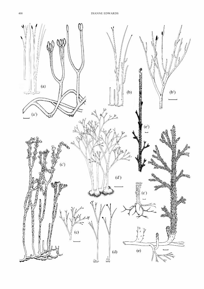
Figure 1 Reconstructions of Rhynie chert sporophytes. (a, a#) Aglaophyton major: (a) After Kidston & Lang 1921; (a#) After D. S. Edwards 1986(scale bar=10 mm). Reproduced by permission of the Linnean Society of London. (b, b#) Rhynia gwynne-vaughanii: (b) After Kidston & Lang 1921;(b#) After D. S. Edwards 1980 (scale bar=5 mm). Reproduced with permission of Elsiever. (c, c#) Nothia aphylla: (c) After Kidston & Lang 1921;(c#) After Kerp et al. 2001 (scale bar=10 mm). Reproduced with permission of Columbia University Press. (d, d#) Horneophyton lignieri: (d) AfterKidston & Lang 1921; (d#) After Eggert 1974 (scale bar=5 mm). Reproduced by permission of the Botanical Society of America. (e, e#, e2)Asteroxylon mackiei: (e) After Kidston & Lang 1921; (e#, e2) Redrawn from Bhutta 1969 (scale bar=1 cm).
EMBRYOPHYTIC SPOROPHYTES 401
402 DIANNE EDWARDS

a strobilus cannot be verified; it seems more likely that therewere zones of sporangia. Smooth rhizoidal axes with similartracheidal anatomy, some penetrating (growing through)spinous examples, are possibly the rhizomes of Trichophero-phyton (see section 2.3).
1.6.2. Relationships. The lateral bivalved sporangia andexarch xylem indicate affinities with the zosterophylls. Closescrutiny of longitudinal sections, particularly of the presumedrhizomes, reveals some indication of additional structuresbetween the secondary thickenings which are suggestive ofG-type tracheids. Evidence for fertile characteristics, both insporangial anatomical detail and arrangement, is limited.However sporangial orientations with distally directed ‘up-right’ sporangia with abaxial (slightly smaller) and adaxialvalves resemble those in Zosterophyllum, but with little evi-dence in the Rhynie plant of a distinct stalk. In the longestwell-preserved specimen, sporangia were arranged in closeproximity in one row, although not directly ‘superpositioned’.A sterile axis shows a circinate tip, a possible indicator ofindeterminate growth following a period of reproduction. Onlyone Zosterophyllum species, Z. divaricatum, unequivocallypossesses circinate tips, although this might relate to non-preservation of immature axes in the remainder. Niklas &Banks (1990) distinguished two major growth patterns withinthe zosterophylls: The first, characterised by Zosterophyllum,had distal spikes with a terminal sporangium and, hence,determinate growth, with pronounced vasculature in thesporangium stalk. The second was more diverse, with circinatetips and sporangial zones indicative of indeterminate growth,xylem elliptical in cross-section and a variety of surfaceoutgrowths. Trichopherophyton possessed characters of bothgroups with its terete strand, strong vascularised stalks and,from circinate tips, presumed indeterminate growth.
1.7. Ventarura lyonii Powell, Edwards & Trewin (Fig.3f, g, k)
1.7.1. Description. This latest new genus comes from theWindyfield chert (Powell et al. 2000). It is based on dispersedfragments, the longest being c. 120 mm long. Aerial stems weresmooth and dichotomously branched. The terete exarch xylemstrand consisted of G-type tracheids. Circum-xylary tissuescomprised thin-walled fusiform cells lacking intercellularspaces. The cortex is exceptional in that two zones of paren-chymatous cells were separated by a conspicuous completecylinder (between two and seven cells thick) of amber-coloured, thick-walled cells. These were elongate with trun-cated or tapering ends. The walls were three-layered and thelumina appear empty. Such features characterise scleren-chyma, a tissue not previously documented in the Rhyniechert, although cells in the peripheral regions of ?mature axesof Aglaophyton have thickened but not layered walls. The
outer cortex in Ventarura is poorly preserved, and an epider-mal layer has not been seen, although a cuticle with inwardprojections is present. The inner cortical cells have granularcontents and abundant intercellular spaces. Eusporangia wereborne laterally and were broadly attached to the stem, with apossibly fleshly abaxial valve and narrower adaxial one. In fullvalve view, they were circular to slightly pear-shaped. Theplant was homosporous with mean spore diameter c. 67 �m.No trilete marks have been recorded. The exospore wasprobably bilayered, the reticulate or folded outer layer possess-ing a coarse granular outer surface. Dehiscence was via a slitsituated at the base of a groove flanked by thickened valveborders which extended around much of the convex margin,but not to the bases of the valves. Sporangia were quite closelyspaced, probably inserted in two rows and possibly forming astrobilus. Associated axes with similar tracheidal anatomy, butsometimes showing xylem medullation, bear unicellular ?rhiz-oids on all surfaces (Fig. 3f, g) and are possibly Ventarura’srhizomes. Branching was less regular and more frequent thanin the aerial stems.
1.7.2. Relationships. The lateral bivalved sporangia, exarchxylem and G-type tracheids unequivocally relate this genus tothe zosterophylls. However the distribution of sporangia andthe symmetry of their arrangement remain equivocal. Limitedinformation indicates that the sporangia were arranged in tworows, and their spacing suggests either a fertile zone or perhapsmore likely a strobilus in which broadly attached sporangia,with abaxial and slightly smaller adaxial valves, were slightlyinclined distally and show very little overlap. Thus, theygenerally resemble the genus Zosterophyllum in their orienta-tion, but lack the well-defined stalks of the latter. Whether ornot the fertile growth pattern was indeterminate or determi-nate sensu Niklas & Banks (1990) cannot be resolved. Thisdilemma highlights both the advantages of (well-preserved)compression fossils for information on gross morphology andthe frustrations caused by their lack of anatomical detail,which prevents detailed sporangial comparisons, in this caseparticularly with the compressed, coalified sporangia withapparently similar morphology, seen in Sawdonia ornata(Hueber 1992).
2. The added value factors of the Rhynie chert
In this section, some examples are presented of data revealedonly by silicification of entire plant organs.
2.1. Soft tissue preservationFor the plant anatomist, the most important and uniquecontribution of the Rhynie chert to studies on early tracheo-phyte evolution must surely relate to parenchymatous tissues.
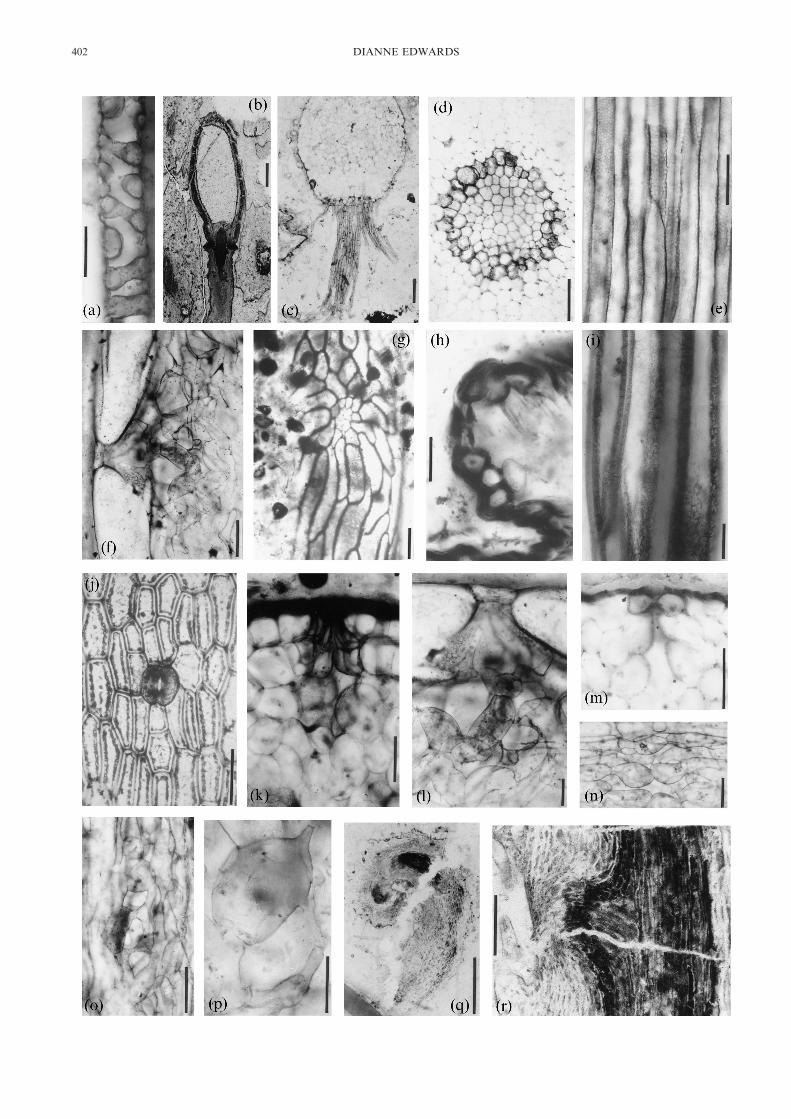
Figure 2 Anatomy in Rhynie Chert sporophytes, based on ground sections except 2b, c, q, r which are peels: (a) LS tracheid of Rhyniagwynne-vaughanii (scale bar=10 �m); (b) LS sporangium and subtending axis in R. gwynne-vaughanii (scale bar=500 �m); (c) TS rhizoid-bearing axisof R. gwynne-vaughanii (scale bar=200 �m); (d) TS central region of Aglaophyton major (scale bar=100 �m); (e) LS central region of A. major. Notefaint reticulate patterning (scale bar=100 �m); (f) LS outer region of aerial axis of Horneophyton lignieri with giant epidermal cells and sub-stomatalregion (scale bar=100 �m); (g) LS emergence on H. lignieri sporangium showing a secretory organ (scale bar=100 �m); (h) LS emergence on Nothiaaphylla with stoma at tip (scale bar=50 �m;) (i) LS presumed water-conducting cells of N. aphylla (scale bar=20 �m); (j) paradermal section of A.major (scale bar=100 �m); (k) TS stoma and substomatal region in A. major (scale bar=100 �m); (l) LS stoma and substomatal cavity in H. lignieri(magnified from (f) (scale bar=100 �m); (m) TS stoma and substomatal cavity in R. gwynne-vaughanii (scale bar=100 �m); (n) LS spongyassimilatory tissue in outer cortex of A. major (scale bar=100 �m); (o) LS similar region in R. gwynne-vaughanii (scale bar=100 �m); (p) LS spongyassimilatory in N. aphylla emergence (scale bar=50 �m); (q) LS hooked tip of Trichopherophyton teuchansii. Dark area is vascular strand (scalebar=1 mm; (r) Lateral meristems in T. teuchansii. Note vascular strand and hairs (scale bar=200 �m).(a), (e) & (i): First published in Edwards 2003 and reproduced by permission of Blackwell Publishing Ltd.(b), (q) & (r): First published in Edwards 1994 and reproduced by permission of the Linnean Society of London.(c) & (j): First published in Edwards 1986 and reproduced by permission of Blackwell Publishing Ltd.(d): First published in Edwards 1993 and reproduced courtesy of the New Phytologist Trust.(f)–(h), (k)–(p): First published in Edwards et al. 1998 and reproduced by permission of Oxford University Press.
EMBRYOPHYTIC SPOROPHYTES 403

Dermal and vascular tissues (especially xylem) are also wellrepresented in compressions and other forms of permineralisa-tion. In the chert, the three-dimensional preservation
of ‘soft tissues’ has been particularly informative as regardsintercellular air space systems and presumed assimilatorytissues. Corner (1964) had regarded these aeration systems and
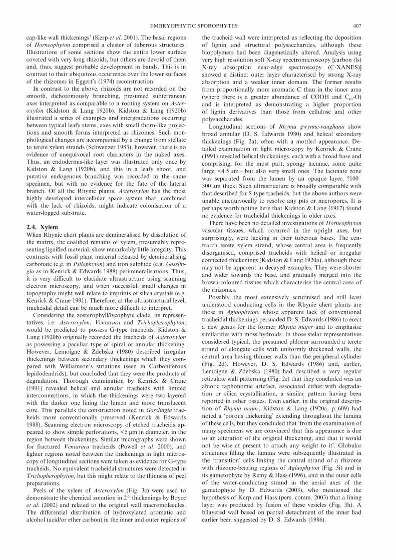
Figure 3 Anatomy of Rhynie and Windyfield cherts’ sporophytes. All are ground sections except (d) and (e)which are peels. (a) LS apex of Rhynia gwynne-vaughanii (scale bar=100 �m); (b) LS apex of Asteroxylon mackiei(courtesy of F. M. Hueber) (scale bar=100 �m). (c) LS tracheids of A. mackiei. Munster 3471 (scale bar=10 �m);(d) TS axis associated with Trichopherophyton teuchansii (scale bar=100 �m); (e) presumed rhizoids in axesassociated with T. teuchansii (scale bar=100 �m); (f, g) presumed rhizoids in axes associated with Ventarura lyonii(f) GLAHM 11402 (g) GLAHM 114037 (scale bars=100 �m); (h) LS outer ‘thickened’ cells of central conductingstrands of A. major showing coalescence of vesicles. Munster 346 (scale bar=20 �m); (i) ‘transition’ cells in A.major rhizome. Munster 1876 (scale bar=50 �m); (j) ‘transition’ tracheids in R. gwynne-vaughanii. Munster 2278(scale bar=50 �m); (k) tracheidal cells in rhizomes associated with V. lyonii. GLAHM 114041 (scale bar=50 �m);(l) ‘transition’ cells in Nothia aphylla. Munster 2926 (scale bar=20 �m); (m) paradermal section through apex ofR. gwynne-vaughanii (scale bar=10 �m).(a) & (b): First published in Edwards 1994 and reproduced by permission of the Linnean Society of London.(c), (h)–(l): First published in Edwards 2003 and reproduced by permission of Blackwell Publishing Ltd.
404 DIANNE EDWARDS

internalised photosynthetic cells as mere details in the progres-sion of plants from water to land, but the Rhynie chert plantsdemonstrate extensive sophistication early in the conquest.Guard cell anatomy (Fig. 2j) and stomatal frequencies havebeen acquired from variously preserved fossils (see Edwardset al. 1998), but only in the Rhynie chert silicifications have weinformation on substomatal cavities and presumed assimila-tory tissues (‘mesophyll’). Aglaophyton and Rhynia are broadlysimilar in that hypodermal cells produce a short canal/channelbelow the guard cells leading into a deep sub-stomatal cavity(Fig. 2k, m). Aglaophyton (Remy & Hass 1996) differs fromRhynia in that the hypodermal cells are more elongate andthickened on walls adjacent to the canal, but in both, the latterhas a cuticularised lining (Edwards et al. 1998). In Horneophy-ton, the exceedingly long epidermal cells cradle the guard cells,again forming a short canal above the substomatal cavity (Fig.2f, l). An extensive aerating system is seen in the parenchyma-tous cells lining the sides and base of these cavities. Cells,matching the diversity in spongy mesophyll of angiosperms(e.g. Meyer 1962), were sausage-shaped to globular and alsocharacterised by a number of extensions (<6 in Aglaophyton)presumably creating maximum surface area for CO2 absorp-tion in an internalised humid atmosphere (Fig. 2h, o). Suchconcentrations of cells formed lens-shaped areas of assimila-tory tissue below the stomata. The interstomatal areas (moreextensive in these Rhynie plants because of low stomatalfrequencies) show configurations of outer cortical cells whichare reminiscent of certain types of palisade mesophyll. Thus,Remy & Hass (1996) described files of cubic to cuboid cellsseparated by large intercellular spaces which are inserted at anangle to the surface in Aglaophyton. Similarly orientated cellsoccur in the depressed areas between the characteristic emer-gences of Nothia (Kerp et al. 2001) and in the outer cortex ofHorneophyton (Edwards et al. 1998). Today, such arrange-ments are seen in photosynthesising tissues in linear leaves andstems of certain flowering plants (Meyer 1962). In Nothia, theouter cortical cells in the emergences themselves resemble thoselining the substomatal cavities. They vary in size and length,and numbers of protrusions (Fig. 2p), with the largest intercel-lular spaces occurring towards the centre of the emergence.The guard cells occurring on the summits of the emergences(Fig. 2h) are somewhat atypical among Rhynie chert examplesin that they usually open directly into the substomatal cavity.Nothia is also characterised by a unique epidermis in whichbetween two and 12 files of � ‘normal’ short epidermal cellsalternate with rows of giant cells <1600 �m long and up to200 �m deep. They are of similar width, but in surface view,they appear much narrower (10–20 �m wide) because the bulkof each cell is embedded in hypodermis (Kerp et al. 2001).Detailed observations such as these allow speculation on thefunctional anatomy of the structures (e.g. as water-storagestructures).
Stomatal configurations and frequencies were used to inferhigh water use efficiency in several Rhynie plants in quantita-tive approaches to consideration of water relations (McElwain& Chaloner 1995; Edwards et al. 1998). Similar conclusionswere reached by Konrad et al. (2000) in calculations using thespatial data measurements on the tissues. While it has beeninferred that low stomatal frequencies confirm modelled highconcentrations of atmospheric CO2 (McElwain & Chaloner1995), anatomical characters which appear to minimise H2Oloss were somewhat unexpected in plants growing in relativelywet habitats. For example, Aglaophyton was further hypoth-esised to possess adaptations (wall thickening and cutinisation)in basal regions of aerial axes to protect against flooding anddefence against aquatic fungi (Remy & Hass 1996). Theabsence of very extensive intercellular airspace systems remi-
niscent of aquatic plants, where lack of oxygen in absorbingareas might interfere with water uptake, leaves the possibilitythat high silica concentration in ground water might havecaused the latter. Physiological drought is experienced by anumber of angiosperms colonising areas close to hot springstoday. Adaptations to seasonal drought, as might be antici-pated from the locality’s palaeolatitude, seem unlikely in sucha hot spring system.
Such deliberations show how detailed anatomical informa-tion of the type provided by the Rhynie chert sporophytesallow reasoned speculation of the plants’ eco-physiology (e.g.Edwards et al. 1998; Konrad et al. 2000).
2.2. Branching: from process to pattern (or lack of it!)Permineralisations involving large volumes of decaying orstanding vegetation have the potential of providing veryaccurate three-dimensional reconstructions of extensive re-gions, both aerial and subaerial, of the higher plant sporo-phytes. The physical efforts involved in the production ofaccurate models as exemplified in the theses of Bhutta (1969)and D. S. Edwards (1973) are tedious and time-consuming, butto date, computer-based techniques such as those recentlydeveloped in the reconstruction of arthropods within nodules(Sutton et al. 2001), for example, have not been successfullyemployed (e.g. work in progress in Cardiff on Trichopherophy-ton spikes). Nevertheless, reconstructions produced by, forexample, D. S. Edwards for R. gwynne-vaughanii (1980) andAglaophyton (1986) and Kerp et al. (2001) for Nothia, show fargreater complexity in branching than those produced byKidston & Lang (1921). The latter reflected the patterns nowconventionally regarded as typical of the earliest land plantlineages, in which apical dichotomy produced isotomous oranisotomous branching, the latter usually accompanied bysubsequent unequal growth resulting in varying extents ofovertopping (e.g. Gosslingia breconensis), and eventually, apseudomonopodial aerial system (e.g. Psilophyton spp.) or theK- or H-branching more characteristics of basal parts (e.g.Zosterophyllum spp.) (Edwards 1994). Further variation isexemplified by the development of lateral branching againshowing regularity because it is associated with branch points.Examples include: axillary/subaxillary tubercles, as in Gosslin-gia (Edwards 1970) and parts of Anisophyton potonei (angular-organs: Remy et al. 1986); more extensive branching systems,downwardly directed in spiny Deheubarthia splendens(Edwards et al. 1989) and erect in Anisophyton gothani (Remyet al. 1986); smaller protuberances (?dormant structures) onthe lower surfaces at the base of lateral branches in Sawdoniaornata (Rayner 1983); and aborted branches forming partsof trichotomies in Psilophyton dawsonii (Banks et al. 1975). Arare example of less ordered, clustered branches in a LowerDevonian tracheophyte was illustrated in the enigmatictracheophyte, Bitelaria dubjanskii (Johnson & Gensel 1992). Inall these cases, anatomical information on the apices is lacking.Until recently, only the Rhynie chert plants provided this – thenew examples being some naked tips in minute charcoalifiedaxes, in which cells are visible but poorly preserved (Edwardset al. 2003).
Superb preservation is seen in the relatively few recordedtips of the main axes of R. gwynne-vaughanii (Fig. 3a, m). Firstillustrated by Kidston & Lang (1917), they show in longitudi-nal section a cuticularised, dome-shaped tip with more or lessequant cells comprising a bean-shaped area (c. 220 �m deep)below the dermal layer, and in an adjacent tip, sectionedparadermally, packets of cells demonstrating the derivatives ofmultiple mitotic divisions. Intercellular spaces are lacking, as isany indication of an apical cell. The longitudinal, presumablymedian, section shows no distinct zonation, as might indicate a
EMBRYOPHYTIC SPOROPHYTES 405

procambium in the w1 mm of axis preserved, although cellsbecome less distinct and longer more proximally, whereas axisdiameter decreases.
Less well preserved at the cellular level, but of similaroverall construction, apices were described in Nothia (Kerpet al. 2001) and in Asteroxylon (Hueber 1992). The latter wasbased on a single�median longitudinal ground sectionthrough a lateral branch showing a flat apex with several leafprimordia at various stages of development (Fig. 3b). Theseinclude small mounds of poorly defined apical initials toadaxially curved linear primordia with evidence of vasculardevelopment ranging from procambial strands to well-developed leaf traces. In the same paper, Hueber (1992)illustrated a young leaf showing an apparently isolated undif-ferentiated procambial strand extending downwards towardsthe leaf trace and upwards into the microphyll. Whether or notthis demonstrates centripetal and centrifugal developmentof the leaf trace remains equivocal since the route of thetrace may have deviated from the plane of the section,notwithstanding it was ground rather than a peel.
Far more common in the chert sporophytes are lateralmeristems, which although tending to be less well preserved atthe cellular level, are more informative on branching processes.Trichopherophyton teuchansii is unique in that apical growthis achieved via uncoiling of the apex (Fig. 2q). In the rareexample of a circinate tip, the apex itself is obscured byprecociously developed unicellular spines, similar to those thatcover the aerial axes (Lyon & Edwards 1991). A group ofmeristematic cells occurs flush with the stem surface somedistance below the apex, but facing the coiled tip (Fig. 2r). Itsfate can only be conjectured. Although lateral sporangia mightbe anticipated to be initiated in this region, the presence of asubstantial, but undifferentiated, vascular strand continuouswith that of the main axis (and arising at right angles to it) ismore suggestive of its being the beginnings of a stem. Thisinterpretation may have implications for growth processes inzosterophylls with circinate vernation: in Trichopherophyton,overtopping was probably not preceded by the division of anapical meristem.
Lateral growth has also been inferred for R. gwynne-vaughanii. D. S. Edwards (1980) suggested that apically pro-duced sporangia may have been shed, with subsequent axialgrowth facilitated by the development of adventitious branchesstimulated by the removal of the sporangium. D. S. Edwards(1980) also emphasised that adventitious branching, as origi-nally discovered by Kidston & Lang (1917), was far morecommon than dichotomous branching. Kidston & Lang (1917)had described small examples as lateral buds associated withvegetative reproduction. Sometimes lacking vascular tissueswhen shed, their original positions were then marked by darklystaining wound reaction tissues terminating short ‘stalks’.Similar regions were recorded, if rarely, on aerial axes ofAglaophyton by Remy & Hass (1996), and were associated withsaucer- or disc-like organs with numerous tuberculate out-growths on the upper margins. These were interpreted asbulbil-like structures for vegetative propagation that haddeveloped from arrested lateral meristems.
However, the latter are far more common on the basalregions and rhizomes of Aglaophyton and Nothia (Kerp et al.2001), with significance for clonal and seasonal growth, regen-eration and vegetative reproduction. The meristems them-selves, represented by poliferations of cells occurring flush tothe surface or as small mounds, were interpreted as nascent ordormant lateral apices. In Aglaophyton, Remy & Hass (1996)described small, dome-shaped structures characterised byradiating files of cells as arrested apices. They occurred mostfrequently on basal (especially U-shaped) regions of the aerial
axes and developed into second-order branches and bulbil-likeorgans. Those which developed two growing points (one withrhizoids) gave rise to the H-shaped branching that typifieszosterophylls. In Nothia, Kerp et al. (2001) elucidated, withimmense precision, a basal system of great complexity, inwhich they recognised at least three types of patches ofmeristematic cells on the dorsal surface of prostrate rhizomes.These were distinguished in terms of regularity of cell arrange-ment and size. Groups of large prismatic cells were comparedwith dormant or bud apices in Selaginella, while a second typecomprising exclusively rectangular cells with no distinct initialswere interpreted as potential primary axes. While demonstrat-ing versatility in development in these early land plants, thesestudies reinforce the importance of growth from lateral mer-istems rather than by division of the apex in many of theRhynie plants. Such a strategy has implications for vasculartissue development and there are numerous examples of a lack,at least initially, of continuity between daughter and subtend-ing axes (e.g. in Aglaophyton; Remy & Hass 1996), and evenbetween rhizome and aerial stems (e.g. in Horneophyton;Kidston & Lang 1920a) with presumed major consequences forwater transport in these sporophytes.
2.3. Water absorptionThe Rhynie chert plants provide the only evidence for thecellular nature of the tissues responsible for water absorptionin early land plants. With the exception of Asteroxylon,rhizoids have been recorded in all the taxa known to Kidstonand Lang, and on rhizomes associated with the two zostero-phylls. In all cases, they are non-septate, and thus, morecharacteristic of tracheophytes than bryophytes. Their distri-bution varies with taxa. Only in the rhizomes associated withVentarura and Trichopherophyton (Fig. 3d–g) are rhizoidsdeveloped on all sides/surfaces of the rhizome, suggesting thatthe organs were completely buried in the substrate. In thesecases, the rhizoids themselves were quite short, <450 �m in‘Ventarura rhizomes’ (Fig. 3f, g), where they were eitherisolated or in clumps, or <250 �m in Trichopherophyton-typerhizomes, where certain papillate epidermal cells may bedeveloping examples (Fig. 3d, e). Longer examples have blunttips. In Trichopherophyton, all are extensions to cells similar tothe remaining epidermal cells, while the unicellular rigidspinous outgrowths on the aerial axes are borne on cells withoval outlines, rather than the usual longitudinally elongatetypes. Ventarura rhizoids have expanded bases, which reflectthe markedly convex surfaces of the remaining epidermal cells,from whose apices the extensions developed. In Aglaophyton,patches of rhizoids <1000 �m long occur on lower surfaces ofrhizomes, as reported by D. S. Edwards (1986) and Remy &Hass (1996), and appear to be concentrated on the bulges onthe bases of the U-shaped axes where they were in contact withthe substrate. The upper surfaces of these axes have stomata.Remy & Hass (1996) further reported the development of therhizoidal areas from arrested apices marked by radiatingwart-like configurations of cells. What little is known about thepresumed rhizomatous region of R. gwynne-vaughanii is basedon Kidston & Lang (1917) (a dichotomously branching struc-ture) and Bhutta’s (1969) thesis. The latter showed thatrhizoids were not confined to the hemispherical projectionswhich characterise this taxon (see Fig. 2c), although themajority of the latter bore tufts of rhizoids towards the basesof the aerial axes.
In Nothia, rhizoids were borne on the lower surface of anaxial rhizome and concentrated on the crest of an elongateridge. They are c. 30 �m wide and up to 1500 �m long. Shorter,smaller examples have tapering tips, but in older ones, thelatter are enlarged (<50 �m wide and 70 �m long) ‘with
406 DIANNE EDWARDS
cap-like wall thickenings’ (Kerp et al. 2001). The basal regionsof Horneophyton comprised a cluster of tuberous structures.Illustrations of some sections show the entire lower surfacecovered with very long rhizoids, but others are devoid of themand, thus, suggest probable development in bands. This is incontrast to their ubiquitous occurrence over the lower surfacesof the rhizomes in Eggert’s (1974) reconstruction.
In contrast to the above, rhizoids are not recorded on thesmooth, dichotomously branching, presumed subterraneanaxes interpreted as comparable to a rooting system on Aster-oxylon (Kidston & Lang 1920b). Kidston & Lang (1920b)illustrated a series of examples and intergradations occurringbetween typical leafy stems, axes with small thorn-like projec-tions and smooth forms interpreted as rhizomes. Such mor-phological changes are accompanied by a change from stellateto terete xylem strands (Schweitzer 1983); however, there is noevidence of unequivocal root characters in the naked axes.Thus, an endodermis-like layer was illustrated only once byKidston & Lang (1920b), and this in a leafy shoot, andputative endogenous branching was recorded in the samespecimen, but with no evidence for the fate of the lateralbranch. Of all the Rhynie plants, Asteroxylon has the mosthighly developed intercellular space system that, combinedwith the lack of rhizoids, might indicate colonisation of awater-logged substrate.
2.4. XylemWhen Rhynie chert plants are demineralised by dissolution ofthe matrix, the coalified remains of xylem, presumably repre-senting lignified material, show remarkably little integrity. Thiscontrasts with fossil plant material released by demineralisingcarbonate (e.g. in Psilophyton) and iron sulphide (e.g. Gosslin-gia as in Kenrick & Edwards 1988) perimineralisations. Thus,it is very difficult to elucidate ultrastructure using scanningelectron microscopy, and when successful, small changes intopography might well relate to imprints of silica crystals (e.g.Kenrick & Crane 1991). Therefore, at the ultrastructural level,tracheidal detail can be much more difficult to interpret.
Considering the zosterophyll/lycophyte clade, its represen-tatives, i.e. Asteroxylon, Ventarura and Trichopherophyton,would be predicted to possess G-type tracheids. Kidston &Lang (1920b) originally recorded the tracheids of Asteroxylonas possessing a peculiar type of spiral or annular thickening.However, Lemoigne & Zdebska (1980) described irregularthickenings between secondary thickenings which they com-pared with Williamson’s striations (seen in Carboniferouslepidodendrids), but concluded that they were the products ofdegradation. Thorough examination by Kenrick & Crane(1991) revealed helical and annular tracheids with limitedinterconnections, in which the thickenings were two-layeredwith the darker one lining the lumen and more translucentcore. This parallels the construction noted in Gosslingia trac-heids more conventionally preserved (Kenrick & Edwards1988). Scanning electron microscopy of etched tracheids ap-peared to show simple perforations, <5 �m in diameter, in theregion between thickenings. Similar micrographs were shownfor fractured Ventarura tracheids (Powell et al. 2000), andlighter regions noted between the thickenings in light micros-copy of longitudinal sections were taken as evidence for G-typetracheids. No equivalent tracheidal structures were detected inTrichopherophyton, but this might relate to the thinness of peelpreparations.
Peels of the xylem of Asteroxylon (Fig. 3c) were used todemonstrate the chemical zonation in 2( thickenings by Boyceet al. (2002) and related to the original wall macromolecules.The differential distribution of hydroxylated aromatic andalcohol (acid/or ether carbon) in the inner and outer regions of
the tracheid wall were interpreted as reflecting the depositionof lignin and structural polysaccharides, although thesebiopolymers had been diagenetically altered. Analysis usingvery high resolution soft X-ray spectromicroscopy [carbon (ls)X-ray absorption near-edge spectroscopy (C-XANES)]showed a distinct outer layer characterised by strong X-rayabsorption and a weaker inner domain. The former resultsfrom proportionally more aromatic C than in the inner area(where there is a greater abundance of COOH and Cm-O)and is interpreted as demonstrating a higher proportionof lignin derivatives than those from cellulose and otherpolysaccharides.
Longitudinal sections of Rhynia gwynne-vaughanii showbroad annular (D. S. Edwards 1980) and helical secondarythickenings (Fig. 2a), often with a mottled appearance. De-tailed examination in light microscopy by Kenrick & Crane(1991) revealed helical thickenings, each with a broad base andcomprising, for the most part, spongy lacunae, some quitelarge <4·5 �m – but also very small ones. The lacunate zonewas separated from the lumen by an opaque layer, ?100–300 �m thick. Such ultrastructure is broadly comparable withthat described for S-type tracheids, but the above authors wereunable unequivocally to resolve any pits or micropores. It isperhaps worth noting here that Kidston & Lang (1917) foundno evidence for tracheidal thickenings in older axes.
There have been no detailed investigations of Horneophytonvascular tissues, which occurred in the upright axes, butsurprisingly, were lacking in their tuberous bases. The cen-trarch terete xylem strand, whose central area is frequentlydisorganised, comprised tracheids with helical or irregularconnected thickenings (Kidston & Lang 1920a), although thesemay not be apparent in decayed examples. They were shorterand wider towards the base, and gradually merged into thebrown-coloured tissues which characterise the central area ofthe rhizomes.
Possibly the most extensively scrutinised and still leastunderstood conducting cells in the Rhynie chert plants arethose in Aglaophyton, whose apparent lack of conventionaltracheidal thickenings persuaded D. S. Edwards (1986) to erecta new genus for the former Rhynia major and to emphasisesimilarities with moss hydroids. In those stelar representativesconsidered typical, the presumed phloem surrounded a teretestrand of elongate cells with uniformly thickened walls, thecentral area having thinner walls than the peripheral cylinder(Fig. 2d). However, D. S. Edwards (1986) and, earlier,Lemoigne & Zdebska (1980) had described a very regularreticulate wall patterning (Fig. 2e) that they concluded was anabiotic taphonomic artefact, associated either with degrada-tion or silica crystallisation, a similar pattern having beenreported in other tissues. Even earlier, in the original descrip-tion of Rhynia major, Kidston & Lang (1920a, p. 609) hadnoted a ‘porous thickening’ extending throughout the luminaof these cells, but they concluded that ‘from the examination ofmany specimens we are convinced that this appearance is dueto an alteration of the original thickening, and that it wouldnot be wise at present to attach any weight to it’. Globularstructures filling the lumina were subsequently illustrated inthe ‘transition’ cells linking the central strand of a rhizomewith rhizome-bearing regions of Aglaophyton (Fig. 3i) and inits gametophyte by Remy & Hass (1996), and in the outer cellsof the water-conducting strand in the aerial axes of thegametophyte by D. Edwards (2003), who mentioned thehypothesis of Kerp and Hass (pers. comm. 2003) that a lininglayer was produced by fusion of these vesicles (Fig. 3h). Abilayered wall based on partial detachment of the inner hadearlier been suggested by D. S. Edwards (1986).
EMBRYOPHYTIC SPOROPHYTES 407
In contrast there is general consensus that the elongatestrongly tapering central cells (Fig. 2i) in Nothia aphylla wereuniformly (2–3 �m) thick-walled. Exceptional examples showseparation of an inner wall except at the corners: dark stainingis interpreted as indicating the presence of phenolic substances(?lignin).
2.5. Short distance transportThe preceding account has concentrated on water-conductingtissues extending longitudinally in axial systems and concludedto have been involved in long distance transport. The Rhyniechert sporophytes show that tissues, broadly equivalent totransfusion tissues, had already evolved by Lower Devoniantimes and have been termed transfusion (Remy & Hass 1996:Aglaophyton), transition (Kerp et al. 2001: Nothia) or linking(Bhutta 1969: R. gwynne-vaughanii) cells. The most convincingexamples are short cells, two to three times as long as wide,with irregular outlines, which often mould the convex curva-ture of surrounding thin-walled cells. In all, the wall thickeningis identical to that in the sporophyte xylem. Variation occurs ingroupings and location. Thus, in rhizomes associated withVentarura, short lineages of cells show lighter reticulate areascharacteristic of G-type tracheids (Fig. 3k). They occur inrhizomes showing closely spaced bifurcations, the anomalousbranching probably reflecting a tufted growth form. A farcloser relationship to water absorption itself is seen in Nothiaand Aglaophyton. In the latter, strands (less than six) of cellsfilled with vesicles (Fig. 3i) or lining the lumen (transfusion ortransition cells) connect the rhizome xylem with the hypoder-mis of rhizoidal bulges. The number of strands is the same asthe number of surface initials over the surface of the bulge(Remy & Hass 1996). Similarly shaped cells filled withvesicles form ‘transition’ tracheids, emanating from the mainxylem strands of aerial axes of R. gwynne-vaughanii (seeEdwards 2003. Fig. 3j), marking the positions of adventitiousbranches although are not continuous with their vascularsystem.
In Nothia (Kerp et al. 2001), uniformly thickened walls(Fig. 3l) of similar thickness (2–3 �m) to the fusiform cells inthe main axes are arranged in longitudinal files forming abridge of tissue (the connective) between the hypodermis of therhizoidal ridge and the stele. The thick-walled cells are inter-mingled with thin-walled examples lacking intercellular spaces.The latter are intermediate in size and shape between thelongitudinally elongate ‘phloem’ cells of the stele and cuboidalhypodermal cells, and have been assumed to have had a waterand nutrient-conducting function. The darkly stained, poorlypreserved cells at the centre of the bulbous rhizomes ofHorneophyton may also act in short distance transport, con-necting with the more conventional tracheids of the uprightaxes.
The more detailed accounts of these tissues (e.g. Kerp et al.2001) clearly demonstrate that they are consequent to thedevelopment of lateral structures and are not apical meristemderivatives. In the case of Nothia, for example, the short,water-conducting cells traversing the ‘phloem region’ musthave been produced by de-differentiation of phloem cells,either food-conducting or parenchyma. Dedifferentiation,often accompanied by cell division, is also seen in woundresponses. Thus, in Aglaophyton (Remy & Hass 1996), wherevascular tissues may have been destroyed, probably by fungalor bacterial infection, new anomalous vascular tissues maydevelop in the phloem or in the inner cortex, while in R.gwynne-vaughanii, new vascular tissues are differentiatedwithin the infected region.
3. And the down side?
Persistent searching through systematic sectioning and screen-ing has filled in many of the questions unanswered by Kidstonand Lang’s pioneering work. Particularly impressive in termsof completeness is our information on the ‘slender, branched,leafless axes with peculiar structure and pear-shaped sporan-gia’ thought by the above authors to be probably the fertileregion of Asteroxylon, but now known as Nothia aphylla (aneven more equivocal affinity!). Revisiting of R. gwynne-vaughanii and Rhynia (Aglaophyton) major has added infor-mation on anatomy and gross morphology, leading to aquestioning of the distinction of the two genera.
Detailed ultrastructure of presumed phloem still remainsobscure, perhaps understandably so as sieve pores fall into thesize range of silica crystals (Satterthwait & Schopf 1972).However, as our understanding of silicification improves(Channing 2001; Channing & Edwards 2004), it becomesopportune to reexamine in detail the cells surrounding thexylem in all the chert plants.
Fortuitous sections will surely eventually lead to wholeplant reconstructions of Ventarura and Trichopherophyton,and a better understanding of the rooting systems of Asteroxy-lon. Such studies combine serendipity with hard work!
The present author started by commenting on the compara-tively few taxa in the chert. The missing plants (at leastcompared with coeval assemblages preserved in clastic rocks ofthe Old Red Continent) are characterised by an abundanceof thick-walled tissues (D. Edwards & Richardson 2004).Ventarura has a cortical zone of cells with beautifully preservedthree-layered walls – even here different because it is deepseated compared with peripheral stereomes of other coevalzosterophylls. The absent taxa also encompass the very smallplants now known to have formed a turf-like vegetation atleast until the end of the Lochkovian (D. Edwards 1996), andto include the permanent tetrad and dyad producers. InOrdovician and Silurian times, these spores were thought tohave been produced by bryophytes, and whilst their parentsmay have become extinct before the Rhynie hot springs wereactive, it is surprising that there are no records to date ofbryophytes in the cherts.
Such gaps in our knowledge provide the stimulus for furtherresearch. As is obvious from this symposium, the Rhyniecherts continues to astound. The present author concludes byreiterating the words of Kidston & Lang (1920a) who, innaming the specific epithet of Horneophyton in honour ofProfessor Lignier, revealed that they had been of the opinionthat, in his theories relating to the appearance of the earliestvascular plants, he had indulged in ‘acute morphologicalspeculations’ (Lignier 1908), ‘some of which gain confirma-tion and reality from the discovery of these simple types ofVascular Cryptogams’. Long may this continue.
4. Acknowledgements
I remain exceedingly grateful to Hans Kerp and Hagen Hassfor the generous and unselfish use of the information they havegathered, and for supplying me with illustrations. I thankLindsey Axe for her expert technical support.
5. References
Banks, H. P. 1968. The early history of land plants. In Drake, E.T.(ed.) Evolution and Environment: A Symposium Presented on theOccasion of the One Hundredth Anniversary of the Foundation ofthe Peabody Museum of Natural History at Yale University,73–107. New Haven, Connecticut: Yale University Press.
408 DIANNE EDWARDS
Banks, H. P. 1975. Reclassification of Psilophyta. Taxon 24, 401–13.Banks, H. P. 1992. The classification of early land plants – revisited.
The Palaeobotanist 41, 36–50.Banks, H. P., Leclercq, S. & Hueber, F. M. 1975. Anatomy and
morphology of Psilophyton dawsonii, sp.n. from the late LowerDevonian of Quebec (Gaspe), and Ontario, Canada. Palaeonto-graphica Americana 8, 77–127.
Bhutta, A. A. 1969. Studies on the flora of the Rhynie Chert. Unpub-lished Ph.D. thesis, University of Wales, Cardiff.
Bhutta, A. A. 1973a. On the spores (including germinating spores) ofRhynia major Kidston and Lang. Biologia 19, 47–57.
Bhutta, A. A. 1973b. On the spores (including germinating spores) ofHorneophyton (Hornea) lignieri (Kidston and Lang) Barghoornand Darrah (1938). Pakistan Journal of Botany 5, 45–55.
Boyce, C. K., Cody, G. D., Feser, M. Jacobsen, C., Knoll, A. H. &Wirick, S. 2002. Organic chemical differentiation within fossilplant cell walls detected with x-ray spectromicroscopy. Geology30, 1039–42.
Chaloner, W. G. & MacDonald, P. 1980. Plants Invade the Land.Glasgow: HMSO for the Royal Scottish Museum, Edinburgh.
Channing, A. 2001. Processes and environments of vascular plantsilicification. Unpublished PhD thesis, Cardiff University.
Channing, A. & Edwards, D. 2004. Experimental taphonomy:silification of plants in Yellowstone hot-spring environments.Transactions of the Royal Society of Edinburgh: Earth Sciences 94(for 1993), 503–21.
Corner, E. J. H. 1964. The Life of Plants. London: Weidenfeld andNicolson.
Dawson, J. W. 1871. Report on the fossil land plants of the Devonianand Upper Silurian formations of Canada. Geological Survey ofCanada Publication 428, 1–92.
Edwards, D. 1970. Further observations on the Lower Devonianplant, Gosslingia breconensis Heard. Philosophical Transactions ofthe Royal Society of London B 258, 225–43.
Edwards, D. 1986. Preservation in early vascular plants. GeologyToday 2, 176–81.
Edwards, D. 1993. Tansley Review No. 53. Cells and tissues in thevegetative sporophytes of early land plants. New Phytologist 125,225–47.
Edwards, D. 1994. Towards an understanding of pattern and processin the growth of early vascular plants. In Ingram, D. S. &Hudson, A. (eds) Shape and Form in Plants and Fungi. LinneanSociety Symposium Series No. 16, 39–59. London: AcademicPress.
Edwards, D. 1996. New insights into early land ecosystems: a glimpseof a Lilliputian world. Review of Palaeobotany and Palynology 90,159–74.
Edwards, D. 2003. Xylem in early tracheophytes. Plant, Cell andEnvironment 26, 57–72.
Edwards, D., Kerp, H. & Hass, H. 1998. Stomata in early land plants:an anatomical and ecophysiological approach. Journal of Experi-mental Botany 49 (Special Issue), 255–78.
Edwards, D., Kenrick, P. & Carluccio, L. M. 1989. A reconsiderationof cf. Psilophyton princeps (Croft and Lang, 1942), a zosterophyllwidespread in the Lower Old Red Sandstone of South Wales.Botanical Journal of the Linnean Society 100, 293–318.
Edwards, D., Axe L. & Duckett, J. G. 2003. Diversity in conductingcells in early land plants and comparisons with extant bryophytes.Botanical Journal of the Linnean Society 141, 297–347.
Edwards, D. & Edwards, D. S. 1986. A reconsideration of theRhyniophytina, Banks. In Spicer, R. A. & Thomas, B. A. (eds).Systematic and Taxonomic Approaches in Palaeobotany. The Sys-tematics Association Special Volume No. 31, 199–220. Oxford:Clarendon Press.
Edwards, D. & Richardson, J. B. 2000. Progress in reconstructingvegetation on the Old Red Sandstone Continent: two Em-phanisporites producers from the Lochkovian sequence ofthe Welsh Borderland. In Friend, P. F. & Williams, B. P. J. (eds)New Perspectives on the Old Red Sandstone. Geological Society,London, Special Publication 180, 355–70.
Edwards, D. & Richardson, J. 2004. Silurian and Lower Devonianplant assemblages from the Anglo–Welsh Basin a palaeobotanicaland palynological synthesis. In Williams, B. P. J., Marriott, S. B.& Hillier, R. D. (eds) The Lower Old Red Sandstone of the WelshBasin. Geological Journal Special Issue.
Edwards, D. S. 1973. Studies on the flora of the Rhynie Chert.Unpublished Ph.D. thesis, University of Wales, Cardiff.
Edwards, D. S. 1980. Evidence for the sporophytic status of the LowerDevonian plant Rhynia gwynne-vaughanii Kidston and Lang.Review of Palaeobotany and Palynology 29, 177–88.
Edwards, D. S. 1986. Aglaophyton major, a non-vascular land plantfrom the Devonian Rhynie Chert. Botanical Journal of the Lin-nean Society 93, 173–204.
Eggert, D. A. 1974. The sporangium of Horneophyton lignieri (Rhynio-phytina). American Journal of Botany 61, 405–13.
El-Saadawy, W. El-S. 1966. Studies on the flora of the Rhynie Chert.Unpublished Ph.D. thesis, University of Wales, Cardiff.
El-Saadawy, W. El-S. & Lacey, W. S. 1979. Observations on Nothiaaphylla Lyon ex Høeg. Review of Palaeobotany and Palynology 27,119–47.
Frey, W., Hilger, H. H. & Hofmann, M. 1996. Water-conducting cellsof extant Symphyogyna-type metzgerialean taxa: ultrastructureand phylogenetic implications. Nova Hedwigia 63, 471–81.
Gensel, P. G. 1992. Phylogenetic relationships of the zosterophylls andlycopsids: evidence from morphology, paleoecology and cladisticmethods of inference. Annals of the Missouri Botanical Garden 79,450–73.
Gensel, P. G. & Berry, C. 2001. Early lycophyte evolution. AmericanFern Journal 91, 74–98.
Halle, T. G. 1916. A fossil sporogonium from the Lower Devonian ofRoragen in Norway. Botaniska Notiser, 79–81.
Halle, T. G. 1936. Notes on the Devonian genus Sporogonites. SvenskBotanisk Tidskrift 30, 613–23.
Hass, H. 1991. Die Epidermis von Horneophyton lignieri (Kidston &Lang) Barghoorn & Darrah. Neues Jahrbuch fur Geologie undPalaontologie Abhandlungen 183, 61–85.
Hass, H. & Remy, W. 1991. Huvenia kleui nov. gen., nov. spec. – eineRhyniophyte aus dem hohoren Siegen des Rheinischen Schiefer-gebirges. Argumenta Palaeobotanica 8, 131–68.
Hebant, C. 1973. Diversity of structure of the water-conductingelements in liverworts and mosses. Journal of the Hattori Botani-cal Laboratory 37, 229–34.
Hueber, F. M. 1992. Thoughts on the early lycopsids and zostero-phylls. Annals of the Missouri Botanical Garden 79, 474–99.
Johnson, N. G. & Gensel, P. G. 1992. A reinterpretation of the EarlyDevonian land plant, Bitelaria Istchenko and Istchenko, 1979,based on new material from New Brunswick, Canada. Review ofPalaeobotany and Palynology 74, 109–38.
Kenrick, P., Remy, W. & Crane, P. R. 1991. The structure of thewater-conducting cells in the enigmatic early land plants Stock-mansella langii Fairon-Demaret, Huvenia kleui Hass et Remy andSciadophyton sp. Remy et al. 1980. Argumenta Palaeobotanica 8,179–91.
Kenrick, P. & Crane, P. R. 1991. Water-conducting cells in early landplants: implications for the early evolution of tracheophytes.Botanical Gazette 152, 335–56.
Kenrick, P. & Crane, P. R. 1997. The Origin and Early Diversificationof Land Plants. Washington, DC: Smithsonian Institution Press.
Kenrick, P. & Edwards, D. 1988. The anatomy of Lower DevonianGosslingia breconensis Heard based on pyritized axes, with somecomments on the permineralization process. Botanical Journal ofthe Linnean Society 97, 95–123.
Kerp, H., Hass, H. & Mosbrugger, V. 2001. New data on Nothiaaphylla Lyon 1964 ex El-Saadawy et Lacey 1979, a poorly knownplant from the Lower Devonian Rhynie Chert. In Gensel, P. G. &Edwards, D. (eds) Plants Invade the Land: Evolutionary andEnvironmental Perspectives, 52–82. New York, NY: ColumbiaUniversity Press.
Kerp, H., Trewin, N. H. & Hass, H. 2004. New gametophytes from theEarly Devonian Rhynie chert. Transactions of the Royal Society ofEdinburgh: Earth Sciences 94 (for 2003), 411–28.
Kidston, R. & Lang, W. H. 1917. On Old Red Sandstone plantsshowing structure, from the Rhynie Chert Bed, Aberdeenshire.Part I. Rhynia gwynne-vaughani, Kidston and Lang. Transactionsof the Royal Society of Edinburgh 51, 761–84.
Kidston, R. & Lang, W. H. 1920a. On Old Red Sandstone plantsshowing structure, from the Rhynie Chert Bed, Aberdeenshire.Part II. Additional notes on Rhynia gwynne-vaughani, Kidstonand Lang; with descriptions of Rhynia major, n.sp. and Hornealignieri, n.g., n.sp. Transactions of the Royal Society of Edinburgh52, 603–27.
Kidston, R. & Lang, W. H. 1920b. On Old Red Sandstone plantsshowing structure, from the Rhynie Chert Bed, Aberdeenshire.Part III. Asteroxylon mackiei, Kidston and Lang. Transactions ofthe Royal Society of Edinburgh 52, 643–80.
Kidston, R. & Lang, W. H. 1921. On Old Red Sandstone plantsshowing structure, from the Rhynie Chert Bed, Aberdeenshire.Part IV. Restorations of the vascular cryptogams, and discussionof their bearing on the general morphology of the Pteridophytaand the origin of the organisation of land plants. Transactions ofthe Royal Society of Edinburgh 52, 831–54.
EMBRYOPHYTIC SPOROPHYTES 409
Konrad, W., Roth-Nebelsick, A., Kerp, H. & Hass, H. 2000. Transpi-ration and assimilation of Early Devonian land plants with axiallysymmetric telomes – simulations on the tissue level. Journal ofTheoretical Biology 206, 91–107.
Lemoigne, Y. & Zdebska, D. 1980. Structures problematiques ob-servees dans des axes provenant du Chert Devonien de Rhynie.Acta Palaeobotanica 21, 3–8.
Lignier, O. 1908. Sur l’evolution morphologique du regne vegetal.Assocation Francais Avancement des Sciences 1908, 530–42.
Lyon, A. G. 1964. Probable fertile region of Asteroxylon mackiei K.and L. Nature 203, 1082–3.
Lyon, A. G. & Edwards, D. 1991. The first zosterophyll from theLower Devonian Rhynie Chert, Aberdeenshire. Transactions ofthe Royal Society of Edinburgh: Earth Sciences 82, 323–32.
McElwain, J. C. & Chaloner, W. G. 1995. Stomatal density and indexof fossil plants track atmospheric carbon dioxide in the Palaeo-zoic. Annals of Botany 76, 389–95.
Meyer, F. J. 1962. Das trophische Parenchym A. Assimilationsgewebe. In Zimmermann, W. & Ozenda, P. G. (eds) Handbuch derPflanzenanatomie. Gebruder Borntraeger. Berlin: Nikolassee.
Niklas, K. J. & Banks, H. P. 1990. A reevaluation of the Zosterophyl-lophytina with comments on the origin of lycopods. AmericanJournal of Botany 77, 274–83.
Powell, C. L., Edwards, D. & Trewin, N. H. 2000. A new vascularplant from the Lower Devonian Windyfield Chert, Rhynie, N.E.Scotland. Transactions of the Royal Society of Edinburgh, EarthSciences 90 (for 1999), 331–49.
Rayner, R. J. 1983. New observations on Sawdonia ornata fromScotland. Transactions of the Royal Society of Edinburgh: EarthSciences 74, 79–93.
Rayner, R. J. 1984. New finds of Drepanophycus spinaeformis Goppertfrom the Lower Devonian of Scotland. Transactions of the RoyalSociety of Edinburgh: Earth Sciences 75, 353–63.
Remy, W. 1978. Der Dehiszenzmechanismus der Sporangien vonRhynia. Argumenta Palaeobotanica 5, 23–30.
Remy, W., Hass, H. & Schultka, S. 1986. Anisophyton potoniei nov.spec. aus den Kuhlbacher Schichten (Emsian) vom SteinbruchUfersmuhle, Wiehltalsperre. Argumenta Palaeobotanica 7, 123–38.
Remy, W. & Hass, H. 1996. New information on gametophytes andsporophytes of Aglaophyton major and inferences about possibleenvironmental adaptations. Review of Palaeobotany and Palynol-ogy 90, 175–93.
Remy, W. & Remy, R. 1977. Die Floren des Erdaltertums. Essen:Verlag Gluckauf GmbH.
Satterthwait, D. F. & Schopf, J. W. 1972. Structurally preservedphloem zone tissues in Rhynia. American Journal of Botany 59,373–6.
Schweitzer, H.-J. 1983. Die Unterdevonflora des Rheinlandes. Palae-ontographica B 189, 1–138.
Scott, D. H. 1923. Studies in Fossil Botany, 3rd edn, Vol. II. London:A. & C. Black Ltd.
Sutton, M. D., Briggs, D. E. G., Siveter, D. J. & Siveter, D. J. 2001.Methodologies for the visualization and reconstruction of three-dimensional fossils from the Silurian Herefordshire lagerstatte.Palaeontologia Electronica 4, Issue 1. [www. document.] URLhttp://palaeo-electronica.org/2001 1/s2/s2.pdf
Wellman, C. H., Kerp, H. & Hass, H. 2004. Spores of the Rhynie chertplant Horneophyton lignieri (Kidston & Lang) Barghoorn &Darrah, 1938. Transactions of the Royal Society of Edinburgh:Earth Sciences 94 (for 2003), 429–43.
DIANNE EDWARDS, School of Earth, Ocean and Planetary Sciences, Cardiff University,Cardiff CF10 3YE, UK.
MS received 14 December 2003. Accepted for publication 10 June 2004.
410 DIANNE EDWARDS