transcatheter valve implantation for patients with aortic ... · future science group 291...
TRANSCRIPT

289ISSN 1755-5302Interv. Cardiol. (2010) 2(3), 289–30010.2217/ICA.10.19 © 2010 Future Medicine Ltd
Transcatheter valve implantation for patients with aortic stenosis
review
Aortic stenosis (AS) is the most frequent valve disease in Europe. It has a predominantly degen-erative origin and is therefore mostly present in elderly patients. Valve replacement is the defini-tive therapy [1]; however, the risk of this treatment may be high in elderly patients with comorbidi-ties. Several registries show that as many as a third of patients with severe valve disease and severe symptoms are not being considered for surgery. Thus, there is a role for less invasive alternatives. Balloon aortic valvuloplasty (BAV) is now rarely used in isolation, mainly due to its limited long-term efficacy [2]. Conversely, transcatheter aortic valve implantation (TAVI) currently represents a dynamic field of research and development 7 years after its introduction in clinical practice by Cribier et al. [3].
This article describes the main steps for patient selection, the technical aspects of prosthesis implantation, summarizes the current results of TAVI and finally elaborates on future perspectives.
Patient selection As a general principle, the selection of candidates for TAVI and the performance of the procedure requires the cooperation of a multidisciplinary team including cardiologists, surgeons, imaging specialists and anesthesiologists, all with expe-rience in the management of valve disease [4]. Patient selection consists of several steps, which will be described in the following sections.
� Confirmation of the diagnosisTranscatheter aortic valve implantation should be performed only in patients with severe symp-toms that can definitely be attributed to severe
AS as assessed by echocardiography [4]. This rela-tionship could be difficult to establish in elderly patients, especially in the two following situations:
� In patients with concomitant severe respiratory disease, the medical history, in particular the chronology of dyspnea, as well as dosage of biomarkers such as brain natriuretic p eptide, is useful.
� In patients with low left ventricular (LV) ejec-tion fraction and low gradient, the evaluation of the degree of calcification and low-dose dobutamine echocardiography are useful adjuncts to differentiate between severe and ‘pseudosevere’ AS.
� Evaluation of comorbidities Transcatheter aortic valve implantation should not be performed in patients whose life expec-tancy is very limited in terms of duration and quality of life (<1 year has been proposed as an acceptable threshold).
Life expectancy is most significantly influ-enced by comorbidities, which should be care-fully looked for. In addition to clinical evalua-tion, semiquantitative scoring systems, such as those used in geriatrics [5], may be helpful for identifying patients whose life expectancy is com-promised more by comorbidities than by heart disease itself. In these latter patients, AS should be managed conservatively.
� Evaluation of the risk of surgeryTranscatheter aortic valve implantation should currently be restricted to patients at high-risk or with contraindications for surgery. The key
Transcatheter aortic valve implantation (TAVI) has expanded the field of treatment of aortic stenosis in high-risk patients. However, the selection of candidates for TAVI and the performance of the procedure require the cooperation of a multidisciplinary team. The current results suggest that TAVI is feasible and provides midterm hemodynamic and clinical improvement in patients with severe symptomatic aortic stenosis at high risk or with contraindications for surgery. Pending questions mainly concern safety and long-term durability. This article describes the main steps for patient selection, the technical aspects of prosthesis implantation, summarizes the current results of TAVI and finally elaborates on future perspectives.
KEYWORDS: aortic stenosis � multidisciplinary team � patient selection � transcatheter aortic valve implantation
Gregory Ducrocq1, Dominique Himbert1, Eric Brochet1 & Alec Vahanian†1
1Département de Cardiologie, Hôpital Bichat-Claude Bernard, 46 rue Henri Huchard, 75877 Paris Cedex 18, Paris, France †Author for correspondence:Tel.: +33 1 40 25 86 61 Fax: +33 1 40 25 67 32 [email protected]

Interv. Cardiol. (2010) 2(3)290 future science group
review Ducrocq, Himbert, Brochet & Vahanian
element in establishing whether patients are at high-risk for surgery is clinical judgment. However, this evaluation might be complex in elderly patients who represent a heterogeneous population and require balanced and individu-alized analysis. A more quantitative assessment of the operative mortality risk, based on the combination of several scores, is used by many teams. The EuroSCORE is user-friendly and widely used. However, it has been consistently shown to overestimate operative mortality in high-risk patients with valve disease. Other scores have been specifically developed for
valvular diseases, such as the Society of Thoracic Surgeons Predicted Risk of Mortality (STS-PROM) score, which appears to be more reli-able than the EuroSCORE in high-risk patients [6]. Various thresholds have been proposed; for example, expected mortality greater than 20% with the logistic EuroSCORE and greater than 10% with STS-PROM score. However, it is important to realize that all scoring systems have limitations; in particular, they do not take into account the surgical results in the given institu-tion. Moreover, it must be taken into account that some risk factors are not covered in scores but often presented in practice (e.g., previous aorto-coronary bypass with patent grafts, chest radiation, porcelain aorta and liver cirrhosis).
Feasibility of transcatheter aortic valve implantationWhen TAVI is envisaged, the following steps should be taken to assess its feasibility:
� Coronary angiography should be performed. If associated coronary artery disease requires revascularization, whether to proceed percu-taneously as well as the chronology of inter-vention should be the subject of individualized discussion based on the patient’s clinical c ondition and anatomy. In practice, without
4.3 mm
8.6 mm
Figure 1. Risk of coronary obstruction: combination of low coronary ostia and narrow aortic root.
Figure 2. Multimodal assessment of aortic annulus diameter. (A) Transthoracic echocardiography: measurement of the annulus diameter. (B) Transesophageal echocardiography: measurement of annulus diameter, defined by the distance from hinge point to hinge point. (C) Multislice computed tomography, showing the oval shape of the aortic annulus, with a small and a large diameter.

www.futuremedicine.com 291future science group
Transcatheter valve implantation for patients with aortic stenosis review
any robust evidence to support it, the trend is to use higher thresholds for combined revas-cularization than in surgical candidates: revas-cularization is performed only in cases with severe lesions of the left main coronary trunk, left anterior descending or dominant right coronary artery. In such cases, percutaneous revascularization is usually performed a few weeks before TAVI. Finally, in order to detect coronary implantation that is too low and avoid subsequent coronary obstruction, the position of the coronary arteries relative to the aortic cusps is best assessed using m ultislice computed tomography (MSCT) (F igure 1).
� Sizing of the valve is critical to minimize the potential for paravalvular leakage and to avoid prosthesis migration after placement or annu-lus rupture. Several methods are available, but today, the most accurate remains to be deter-mined (Figure 2) [7]. Transthoracic echocardio-graphy is the most popular method, while transesophageal echocardiography (TEE), which has been found to show larger values than transthoracic echocardiography, is advised if borderline values lead to doubt of the feasibility of the procedure. MSCT offers the opportunity to assess the complex 3D structure of the aortic annulus and confirms its oval shape [8]. The measurements obtained by MSCT are somewhat larger than those shown by echocardiography. Finally, measure-ments of aortography performed d uring BAV are also advocated by a few teams.
� The morphology of the valve and its length, the importance and location of calcification and the dimensions of the aortic root will also be assessed by echocardiography and MSCT.
� Echocardiography will eliminate the p resence of dynamic subvalvular LV obstruction and assess the mitral valve in quantifying the importance of regurgitation and establishing whether mitral disease is organic or functional.
� Evaluation of the peripheral arteries by angio graphy and, even better, MSCT ( Figure 3) will guide the choice of the approach, either retro grade transfemoral or anterograde
Figure 3. Evaluation of the femoroiliac axes. (A) Conventional angiography, showing the general anatomy of the arterial axes and allowing quantitative assessment of the minimal luminal diameters between the origin of the common iliac arteries and the access site on the common femoral arteries. (B) Multislice computed tomography (sagital view) to detect eccentric calcifications.
Box 1. Contraindications for transcatheter aortic valve implantation.
General contraindications � Aortic annulus of less than18 mm or greater than 25 mm for balloon-expandable and less than
20 mm or greater than 27 mm for self-expandable devices � Bicuspid valves (relative contraindication) � Presence of asymmetric heavy valvular calcification � Aortic root dimension greater than 45 mm at the sinotubular junction for self-expandable prostheses � Low position of coronary ostia (<8 mm from the aortic annulus) � Dynamic subvalvular obstruction � Severe organic mitral regurgitation � Apical left ventricular thrombus
Specific contraindications for the transfemoral approach � Iliac arteries: severe calcification, tortuosity, small diameter (<6–9 mm depending on the device
used) or previous aortofemoral bypass � Aorta: severe angulation, severe atheroma of the arch, coarctation or aneurysm of the abdominal
aorta with protruding mural thrombus � The presence of bulky atherosclerosis of the ascending aorta and arch detected by transesophageal
echocardiography.Contraindications for the transapical approach � Severe respiratory insufficiency � Major chest deformity � Previous surgery of the left ventricular using a patch

Interv. Cardiol. (2010) 2(3)292 future science group
review Ducrocq, Himbert, Brochet & Vahanian
transapical (F igure 4). Size, tortuosity, degree and location of calcification of peripheral arteries will be evaluated in this endeavor.
Contraindications for transcatheter aortic valve implantationThe contraindications for TAVI, either general or approach- or device-specific, are shown in Box 1. Some specific issues must be considered:
� Bicuspid aortic valve, especially when severely calcified, is currently a relative contraindica-tion because of the risk of asymmetric deployment of the prosthesis owing to the calcification and the large size of the annulus;
� Asymmetric heavy valvular calcification may compress the coronary arteries during TAVI and should be detected before TAVI on MSCT and also during BAV;
Severe AS (TEE)Severe symptoms
Yes No
Surgery
High risk for surgery? (EuroSCORE/STS)Contraindication to surgery?
Yes No
Contraindications for TAVI?Life expectancy less than 1 year, poor QOL, technical contraindications
No Yes Medical treatment
TAVI
Evaluation of the peripheral arteries(diameter, calcification and tortuosity)MSCT/angiography
Diameter <6 mm
Consider:– Transapical (Edwards-Sapien)– Reteroperitoneal (Edwards-Sapien)– Axillary (CoreValve)
Diameter >6 mm
18 Fr compatible:– Transfemoral CoreValve– Transfemoral Edwards-Sapien XT Novaflex 23 mm
Diameter >7 mm
19 or 22 Fr compatible:– Transfemoral Edwards-Sapien Retroflex 23 mm– Transfemoral Edwards-Sapien XT Novaflex 23/26 mm– CoreValve
Diameter >8 mm
24 Fr compatible:– Transfemoral possible for every kind of prosthesis
Evaluation of the aorticannulus diameter
18–21 mm
Edwards-Sapien 23 mm
21–25 mm
Edwards-Sapienor CoreValvecompatible
25–27 mm
CoreValve 29 mm
(Reconsidered)
Figure 4. Algorithm of decision for selection of patients and approach.AS: Aortic stenosis; MSCT: Multislice computed tomography; QOL: Quality of life; STS: Society of Thoracic Surgeons; TAVI: Transcatheter aortic valve implantation; TEE: Transesophageal echocardiography.

www.futuremedicine.com 293future science group
Transcatheter valve implantation for patients with aortic stenosis review
� Dynamic subvalvular obstruction, which may lead to severe hypotension when the valvular obstacle is relieved, should also be looked for carefully, as well as the presence of severe sep-tal hypertrophy localized in the close vicinity of the aortic cusps;
� Severe organic mitral regurgitation is a con-traindication for TAVI. Conversely, func-tional MR is not, because it is likely to decrease in the case of a successful procedure.
Techniques of implantation � General considerations
The performance of TAVI should be restricted to high volume centers that have both cardiology and cardiac surgery departments, with expertise in structural heart disease intervention and high-risk valvular surgery and facilities for cardiac support such as availability of femoral– femoral bypass systems in particular when treating patients with depressed LV ejection fraction [4].
The optimal environment for TAVI should be spacious and sterile and feature high qual-ity imaging equipment and facilities for cardiac support. A hybrid suite is ideal, but in practice very few centers in Europe have the availabil-ity of such equipment and most procedures are performed in catheterization laboratories.
Most teams perform the procedure under gen-eral anesthesia, although sedation and analgesia may be sufficient for the transfemoral approach. However, it should be kept in mind that the pres-ence of anesthesiologists with specific expertise in cardiology is mandatory for periprocedural care owing to the severe clinical condition of these patients.
The use of general anesthesia allows for peri-procedural TEE monitoring, to help correctly position the valve and even more so to detect complications.
� DevicesTwo devices are currently commercialized for TAVI (Figure 5); The Edwards–Sapien™ valve (Edwards Lifesciences, CA, USA), and the Medtronic CoreValve® System (Medtronic, MN, USA). The Edwards–Sapien valve consists of three bovine pericardial leaflets mounted within a tubular, slotted, stainless-steel, balloon-expandable stent. It is currently available in 23 and 26 mm sizes, necessitating, for the transfemoral approach, 22 and 24 Fr, respectively, introducer sheaths for the Retroflex 3™ (Edwards Lifesciences) catheter or 18 and 19 Fr, respectively, introducer sheaths for the Novaflex Catheter™ (Edwards Lifesciences). The
transapical approach is performed through a 26 Fr catheter. The Medtronic CoreValve System con-sists of three porcine pericardial leaflets mounted in a self-expanding nitinol frame. It is available in 26 and 29 mm sizes and goes through 18 Fr i ntroducers for transfemoral or transaxillary use.
Figure 5. Commercially available prostheses. (A) Edwards-Sapien™ transcatheter heart valve and the third generation of RetroFlex™ catheter with the balloon and its distal nose cone. (B) Medtronic CoreValve® System and the third generation 18 Fr catheter.
Figure 6. Possible approaches for transcatheter aortic valve implantation. (A) Transfemoral. The common femoral artery is exposed surgically. The sheath is then passed through the skin to provide a firm anchor. (B) Transapical (Edwards-Sapien). An anterolateral minithoracotomy and a puncture of the left ventricle using a needle through purse-string sutures are performed. (C) Subclavian (Medtronic CoreValve). (D) Transiliac retroperitoneal using an iliac conduit.

Interv. Cardiol. (2010) 2(3)294 future science group
review Ducrocq, Himbert, Brochet & Vahanian
� ApproachTranscatheter aortic valve implantation is cur-rently carried out using two different approaches
(Figure 6): retrograde transfemoral or transaxil-lary and anterograde transapical. The trans-septal approach has been a bandoned. Specific issues are related to the different approaches.
In the transfemoral approach, the common femoral artery can be either prepared surgically or approached percutaneously. Closure of the vascu lar access can be effected surgically or using a percutaneous closure device depending on the size of the device and the experience of the team. The same principles hold to be true for the trans axillary approach (Figure 6). Finally, a retroperitoneal approach of the iliac artery (Figure 6) has been used in a few cases.
The transapical approach requires an antero-lateral minithoracotomy, pericardiotomy, iden-tification of the apex, then puncture of the left ventricle using a needle through purse-string sutures [9]. Subsequently, an introductory sheath is positioned in the LV and the p rosthesis is implanted using the anterograde route.
� Balloon aortic valvuloplasty After crossing the aortic valve, BAV is performed to predilate the native valve just before pros-thesis implantation. Similar to the procedure performed when using surgical valve replace-ment, BAV could be used as a bridge to TAVI in patients presenting with very low LV ejection fraction or in acute heart failure. This stepwise approach performed at an interval of a few days or weeks may decrease the risk of intervention.
� Prosthesis positioningPositioning the prosthesis at the level of the a ortic valve is a crucial step and may use fluo-roscopy and echocardiography in combination:
� Fluoroscopy is useful to assess the level of valve calcification and aortography to determine the position of the valve and the plane of alignment of the aortic cusps.
� Transesophageal echocardiography is helpful, in particular, in cases with moderate calcifica-tion. The additional value of 3D real-time TEE is currently being evaluated. According to the limited current experience with intra-cardiac echography, it does not appear to enhance TEE in this setting.
� Prosthesis implantationWhen positioning is considered correct, the pros-thesis is released. Rapid pacing is used at this stage in balloon-expandable valves but not in self-expanding devices to decrease c ardiac o utput and stabilize the prosthesis during i nflation (Figure 7).
Figure 8. Postimplantation evaluation. Edwards-Sapien valve: (A) angiogram and (B) transesophageal echocardiography. Medtronic CoreValve System: (C) angiogram and (D) transesophageal echocardiography.
Balloon inflation
Figure 7. Rapid ventricular pacing used to decrease cardiac output and stabilize the prosthesis during inflation.

www.futuremedicine.com 295future science group
Transcatheter valve implantation for patients with aortic stenosis review
Immediately after TAVI, aortography and, whenever available, TEE are performed to assess the location and degree of aortic regur-gitation (AR), the patency of the coronary arteries and also to rule out complications such as hemopericardium and aortic dissec-tion (Figure 8). The hemodynamic results are assessed using p ressure recordings and/or echocardiography.
� Postprocedural careAfter the procedure, the patients should stay in intensive care for at least 24 h and h emodynamics, vascular access and rhythm d isturbances, e specially late atrioventricu-lar blockage, should be closely monitored for s everal days.
On an empirical basis, dual antiplatelet t herapy is usually administered for 3–6 months and a spirin is continued for the rest of the patients life. If vitamin K blockers are required, the duration of dual antiplatelet treatment should be made shorter or a single agent should be considered.
Results � General considerations
More than 10,000 cases of TAVI have been performed worldwide; however, it should be noted that the evidence in this field remains limited. A number of single center reports were published in peer-reviewed journals but they seldom included more than 100 cases and were a mix of early and late experience as well as first and subsequent generation devices [10–12]. The second source of information is regis-tries [13–16], mostly reported in oral presenta-tions with all the inherent limitations. In addi-tion, the series are heterogeneous with regard to the approach or device used. In most reports, the transfemoral approach is the default approach and, therefore, the patients treated using the transapical approach usually have a higher-risk profile and more comorbidities, which should be kept in mind when analyz-ing the results. There are currently no studies comparing either one device against the other or one approach against the other. The overall results are shown in TaBles 1–3.
Table 1. Baseline characteristics of patients treated by transcatheter aortic valve implantation.
Edwards-Sapien Edwards-Sapien Edwards-Sapien + CoreValve CoreValve CoreValve
Webb† (n = 168) SOURCE‡ (n = 1038)
Bichat§ (n = 120) Piazza¶ (n = 646)
Post-CE registry# (n = 1483)
Age (years) 84 81 ± 7 81 ± 9 81 ± 7 81 ± 6
Female sex 81 (48.2) 575 (55) 54 (45) 348 (54) 816 (55)
NYHA class III/IV 145 (86.3) – 116 (97) 532 (85) 1246 (84)
Coronary artery disease:– Previous MI– Previous PCI– Previous CABG
–123 (73.2)–62 (36.9)
539 (52)––235 (23)
71 (59)24 (20)26 (22)34 (28)
367 (57)77 (12)187 (30)130 (20)
890 (60)–415 (28)311 (21)
Peripheral artery disease 60 (35.7) 208 (20) 18 (15) 144 (23) 356 (24)
Renal failure 20 (11.9) 310 (30) 42 (35) – 400 (27)
Diabetes mellitus 39 (23) – 26 (22) 172 (27) 386 (26)
Severe COPD 35 (20.8) 286 (28) 32 (27) – 371 (25)
Previous cerebrovascular event
30 (17.9) – – 48 (8) 133 (9)
Porcelain aorta 36 (21.4) 87 (8) 14 (12) 33 (7) 119 (8)
Aortic valve area (cm²) 0.6 (0.5–0.7) – 0.7 ± 0.2 0.6 ± 0.2 0.6 ± 0.2
Mean gradient (mmHg, mean ± SD)
46 (34–55) – 52 ± 15 49 ± 14 49 ± 6
LVEF (%) – – 51 ± 15 51 ± 14 52 ± 14
Logistic EuroSCORE (%) 29 (22–27) 27 ± 15 27 ± 14 23 ± 14 23 ± 14
STS-PROM (%) 9 (6–13) – 16 ± 9 – –
Pulmonary hypertension >60 mmHg
45 (26.8) – 29 (24) – –
†Data taken from [10]. ‡Data taken from EuroPCR 2009. §Data taken from European Society Cardiology Barcelona 2009. ¶Data taken from [16]. #Data taken from EuroPCR 2009. Values are expressed as n (%), mean ± SD or median (Q1–Q3). CABG: Coronary artery bypass grafting; COPD: Chronic obstructive pulmonary disease; LVEF: Left ventricular ejection fraction; MI: Myocardial infarction; NYHA: New York Heart Association; PCI: Percutaneous coronary intervention; SD: Standard deviation; STS-PROM: Society of Thoracic Surgeons Predicted Risk of Mortality.

Interv. Cardiol. (2010) 2(3)296 future science group
review Ducrocq, Himbert, Brochet & Vahanian
� ScreeningThe reports considering the management of severe AS in the era of TAVI by recent obser-vation demonstrated that approximately 60% of the patients screened by a multidisciplinary team for the procedure are currently treated by TAVI if both approaches are available, while 10–20% of patients are operated on and, finally, approximately 30% of patients are treated medi-cally. These respective percentages would vary according to whether only one device or both devices were used and the availability of all the approaches versus the availability of transfemo-ral approach alone. Overall, the availability of TAVI has increased the number of referrals for intervention in patients with severe AS,
which is a good thing, taking into consider-ation the c urrent underuse of surgery in this population [17].
� ProcedureThe two types of devices are used more or less equally. Overall, two out of three of cases have been performed using the transfemoral approach and only preliminary reports describe the experi-ence of the transaxillary route in a pproximately 200 patients.
The most recent registries report procedural success rates exceeding 90% in experienced centers [10–13,16]. However, it is important to acknowledge that all reports consistently show a learning curve effect, both on the success
Table 3. Transapical aortic valve implantation (Edwards-Sapien) 30-day results.
Webb† (n = 55)
SOURCE‡ (n = 575)
Bichat§ (n = 37)
France¶ (n = 71)
PARTNER EU# (n = 69)
Implant success (%) – 93 100 97 91
Death (%) 18 10 11 17 19
Stroke (%) 2 3 0 3 3
Coronary obstruction (%) – 1 0 – 3
Permanent pacemaker (%) 7 7 3 4 4
Vascular complications (%) 4 3 5 7 3
Aortic regurgitation >2/4 (%) – 2 8 1 1
Conversion to AVR (%) 2 3 0 1 1
Tamponade (%) 4 – 11 – –†Data taken from [10]. ‡Source Registry, EuroPCR 2009. §Data taken from [12]. ¶France Registry, Eltchaninoff et al., American Health Association 2009. #PARTNER EU trial, EuroPCR 2009.AVR: Aortic valve replacement.
Table 2. The 30-day outcomes in patients treated by transfemoral aortic valve implantation.
Edwards-Sapien
Edwards-Sapien
Edwards-Sapien + CoreValve
CoreValve CoreValve
Webb† (n = 113)
SOURCE‡ (n = 463)
Bichat§ (n = 83) Piazza¶ (n = 646) Post-CE registry# (n = 1483)
Implantation success – 95.2 94 97.2 98.5
Aortic valve area (cm²) – – 1.7 – 1.5
Mean gradient (mmHg) – 11 3 9
Paravalvular aortic regurgitation > grade 2
– 1.5 3 0 –
Valve-in-valve implantation – 0.6 2 2.6 –
Major vascular complications 8.0 10.6 12 1.9 3.8
Stroke 5.3 2.4 5 0.6 2.2
Tamponade 1.8 – 2 1.4 3.6
Valve embolization – 0 0 – –
Coronary obstruction – 0.7 1.2 0 –
Renal failure requiring dialysis 1.8 1.3 0 – 2.2
Need for new pacemaker 5.4 6.7 12 9.3 25
Conversion to open heart surgery 0 – 0 0.5 0.8
Mortality at 30 days 8 6.3 8 8 10.3Values are expressed as % unless otherwise stated. †Data taken from [10]. ‡Data taken from EuroPCR 2009. §Data taken from European Society of Cardiology Barcelona 2009. ¶Data taken from [16]. #Data taken from EuroPCR 2009.
www.futuremedicine.com 297future science group
Transcatheter valve implantation for patients with aortic stenosis review
rate and the incidence and severity of compli-cations, which emphasizes the importance of careful training.
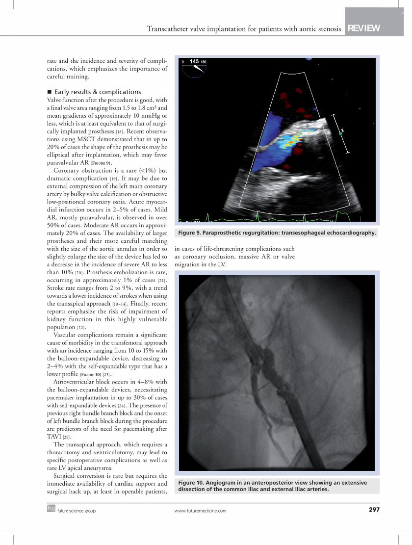
� Early results & complicationsValve function after the procedure is good, with a final valve area ranging from 1.5 to 1.8 cm² and mean gradients of approximately 10 mmHg or less, which is at least equivalent to that of surgi-cally implanted prostheses [18]. Recent observa-tions using MSCT demonstrated that in up to 20% of cases the shape of the prosthesis may be elliptical after implantation, which may favor paravalvular AR (Figure 9).
Coronary obstruction is a rare (<1%) but dramatic complication [19]. It may be due to external compression of the left main coronary artery by bulky valve calcification or obstructive low-positioned coronary ostia. Acute myocar-dial infarction occurs in 2–5% of cases. Mild AR, mostly paravalvular, is observed in over 50% of cases. Moderate AR occurs in approxi-mately 20% of cases. The availability of larger prostheses and their more careful matching with the size of the aortic annulus in order to slightly enlarge the size of the device has led to a decrease in the incidence of severe AR to less than 10% [20]. Prosthesis embolization is rare, occurring in approximately 1% of cases [21]. Stroke rate ranges from 2 to 9%, with a trend towards a lower incidence of strokes when using the transapical approach [10–14]. Finally, recent reports emphasize the risk of impairment of kidney function in this highly vulnerable p opulation [22].
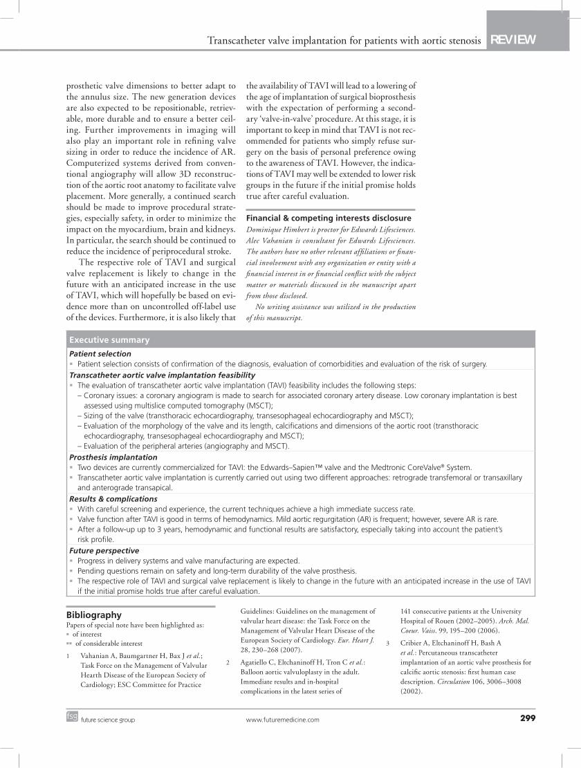
Vascular complications remain a significant cause of morbidity in the transfemoral approach with an incidence ranging from 10 to 15% with the balloon-expandable device, decreasing to 2–4% with the self-expandable type that has a lower profile (Figure 10) [23].
Atrioventricular block occurs in 4–8% with the balloon-expandable devices, necessitating pacemaker implantation in up to 30% of cases with self-expandable devices [24]. The presence of previous right bundle branch block and the onset of left bundle branch block during the procedure are predictors of the need for pacemaking after TAVI [25].
The transapical approach, which requires a thoracotomy and ventriculotomy, may lead to specific postoperative complications as well as rare LV apical aneurysms.
Surgical conversion is rare but requires the immediate availability of cardiac support and surgical back up, at least in operable patients,
in cases of life-threatening complications such as coronary occlusion, massive AR or valve m igration in the LV.
Figure 9. Paraprosthetic regurgitation: transesophageal echocardiography.
Figure 10. Angiogram in an anteroposterior view showing an extensive dissection of the common iliac and external iliac arteries.

Interv. Cardiol. (2010) 2(3)298 future science group
review Ducrocq, Himbert, Brochet & Vahanian
Overall, mortality at 30 days ranges from 5 to 18% for the transfemoral approach and from 10 to 19% when using the transapical approach [10–14,16].
Finally, case reports have shown the feasibil-ity of ‘valve-in-valve implantation’ (Figure 11) for either acute failure of TAVI caused by intra-prosthetic AR or for either stented or stentless degenerated valve prostheses [26]; however, it is too early to draw any definite conclusions on this attractive potential indication.
� Late resultsThe majority of late adverse clinical events are due to comorbidities. Anecdotal cases of valve endocarditis or thromboembolism have been reported. The risk of bleeding could be a con-cern in elderly patients when receiving a com-bination of antiplatelet agents plus vitamin K blockers. The degree of AR remains stable over
time and mild-to-moderate AR did not require reintervention or cause severe hemolysis during this limited follow-up. Serial echocardiographic studies have consistently shown good prosthetic valve function and no structural deteriora-tion of valve tissue has been reported so far. A few cases of secondary surgical intervention have been performed, mostly in cases of inad-equate valve positioning. Preliminary reports have shown that LV ejection fraction improves after TAVI while the degree of functional MR decreases [10].
Long-term results up to 6 years (although only 1 year to a maximum of 3 years in most studies) demonstrate a survival rate of 70% at 1 year and 60% at 2 years with a significant improvement in clinical condition and qual-ity of life parameters in most cases, which is of utmost importance in the elderly population [27].
� Upcoming dataIt is necessary to accumulate more evidence on the results of TAVI. The results of the first randomized trial (PARTNER US), compar-ing TAVI with either medical therapy or sur-gical valve replacement according to patients’ c ondition, will be reported during the coming year. Data should be accumulated from regis-tries with longer follow-up to assess safety and durability with special focus on the timing and the mode of valve failure, the consequences of mild-to-moderate AR and the feasibility of either p ercutaneous or surgical reintervention. These data will help to better define the indica-tions of the t echnique and the respective place of each approach.
ConclusionThe current results suggest that TAVI is f easible and provides hemodynamic and clini-cal improvement for up to 3 years in patients with severe symptomatic AS at high-risk or with contra indications for surgery. Pending questions mainly concern safety and long-term durabil-ity. Surgeons and cardiologists must work as a team to select the best candidates, perform the procedure and, finally, evaluate the results. At p resent, these t echniques are targeted at high-risk patients.
Future perspectiveProgress in delivery systems and valve manu-facturing will lead to overcoming what are currently the main limitations of TAVI; lower profile enabling a decrease of the sheath size for the transfemoral approach and a wider range of
Figure 11. Valve-in-valve implantation. A CoreValve System was implanted within a degenerated bioprosthesis.
www.futuremedicine.com 299future science group
Transcatheter valve implantation for patients with aortic stenosis review
prosthetic valve dimensions to better adapt to the annulus size. The new generation devices are also expected to be repositionable, retriev-able, more durable and to ensure a better ceil-ing. Further improvements in imaging will also play an important role in refining valve sizing in order to reduce the incidence of AR. Computerized systems derived from conven-tional angiography will allow 3D reconstruc-tion of the aortic root anatomy to facilitate valve placement. More generally, a continued search should be made to improve procedural strate-gies, especially safety, in order to minimize the impact on the myocardium, brain and kidneys. In particular, the search should be continued to reduce the i ncidence of periprocedural stroke.
The respective role of TAVI and surgical valve replacement is likely to change in the future with an anticipated increase in the use of TAVI, which will hopefully be based on evi-dence more than on uncontrolled off-label use of the devices. Furthermore, it is also likely that
the availability of TAVI will lead to a lowering of the age of implantation of surgical bioprosthesis with the expectation of performing a second-ary ‘valve-in-valve’ procedure. At this stage, it is important to keep in mind that TAVI is not rec-ommended for patients who simply refuse sur-gery on the basis of personal preference owing to the awareness of TAVI. However, the indica-tions of TAVI may well be extended to lower risk groups in the future if the initial promise holds true after careful evaluation.
Financial & competing interests disclosureDominique Himbert is proctor for Edwards Lifesciences. Alec Vahanian is consultant for Edwards Lifesciences. The authors have no other relevant affiliations or finan-cial involvement with any organization or entity with a financial interest in or financial conflict with the subject matter or materials discussed in the manuscript apart from those disclosed.
No writing assistance was utilized in the production of this manuscript.
Executive summary
Patient selection � Patient selection consists of confirmation of the diagnosis, evaluation of comorbidities and evaluation of the risk of surgery.
Transcatheter aortic valve implantation feasibility � The evaluation of transcatheter aortic valve implantation (TAVI) feasibility includes the following steps:
– Coronary issues: a coronary angiogram is made to search for associated coronary artery disease. Low coronary implantation is best assessed using multislice computed tomography (MSCT); – Sizing of the valve (transthoracic echocardiography, transesophageal echocardiography and MSCT); – Evaluation of the morphology of the valve and its length, calcifications and dimensions of the aortic root (transthoracic echocardiography, transesophageal echocardiography and MSCT); – Evaluation of the peripheral arteries (angiography and MSCT).
Prosthesis implantation � Two devices are currently commercialized for TAVI: the Edwards–Sapien™ valve and the Medtronic CoreValve® System. � Transcatheter aortic valve implantation is currently carried out using two different approaches: retrograde transfemoral or transaxillary
and anterograde transapical.
Results & complications � With careful screening and experience, the current techniques achieve a high immediate success rate. � Valve function after TAVI is good in terms of hemodynamics. Mild aortic regurgitation (AR) is frequent; however, severe AR is rare. � After a follow-up up to 3 years, hemodynamic and functional results are satisfactory, especially taking into account the patient’s
risk profile.
Future perspective � Progress in delivery systems and valve manufacturing are expected. � Pending questions remain on safety and long-term durability of the valve prosthesis. � The respective role of TAVI and surgical valve replacement is likely to change in the future with an anticipated increase in the use of TAVI
if the initial promise holds true after careful evaluation.
BibliographyPapers of special note have been highlighted as:� of interest�� of considerable interest
1 Vahanian A, Baumgartner H, Bax J et al.; Task Force on the Management of Valvular Hearth Disease of the European Society of Cardiology; ESC Committee for Practice
Guidelines: Guidelines on the management of valvular heart disease: the Task Force on the Management of Valvular Heart Disease of the European Society of Cardiology. Eur. Heart J. 28, 230–268 (2007).
2 Agatiello C, Eltchaninoff H, Tron C et al.: Balloon aortic valvuloplasty in the adult. Immediate results and in-hospital complications in the latest series of
141 consecutive patients at the University Hospital of Rouen (2002–2005). Arch. Mal. Coeur. Vaiss. 99, 195–200 (2006).
3 Cribier A, Eltchaninoff H, Bash A et al.: Percutaneous transcatheter implantation of an aortic valve prosthesis for calcific aortic stenosis: first human case description. Circulation 106, 3006–3008 (2002).

Interv. Cardiol. (2010) 2(3)300 future science group
review Ducrocq, Himbert, Brochet & Vahanian
4 Vahanian A, Alfieri OR, Al-Attar N et al.: Transcatheter valve implantation for patients with aortic stenosis: a position statement from the European Association of Cardio-Thoracic Surgery (EACTS) and the European Society of Cardiology (ESC) in collaboration with the European Association of Percutaneous Cardiovascular Interventions (EAPCI). Eur. Heart J. 29, 1463–1470 (2008).
�� Document issued by both medical and surgical scientific societies to set the stage for the use of transcatheter aortic valve implantation.
5 Lee SJ, Lindquist K, Segal MR, Covinsky KE: Development and validation of a prognostic index for 4-year mortality in older adults. JAMA 295, 801–808. (2006); erratum in: JAMA 295, 1900 (2006).
6 Dewey TM, Brown D, Ryan WH et al.: Reliability of risk algorithms in predicting early and late operative outcomes in high-risk patients undergoing aortic valve replacement. J. Thorac. Cardiovasc. Surg. 135, 180–187 (2008).
�� Comparative evaluation of the predictive value of the risk scores for aortic valve replacement.
7 Messika-Zeitoun D, Serfaty JM, Brochet E et al.: Multimodal assessment of the aortic annulus diameter: implications for transcatheter aortic valve implantation. J. Am. Coll. Cardiol. 55, 186–194 (2010).
� Comparative evaluation of transthoracic echocardiography, transesophageal echocardiography and multislice computed tomography performances for aortic annulus diameter evaluation.
8 Tops L, Wood D, Delgado V et al.: Non-invasive evaluation of the aortic root with multislice computed tomography: implications for transcatheter aortic valve replacement. J. Am. Coll. Cardiol. Img. 1, 321–330 (2008).
�� Pioneering work on the role of multislice computed tomography during transcatheter aortic valve implantation.
9 Walther T, Dewey T, Borger MA et al.: Transapical aortic valve implantation: step by step. Ann. Thorac. Surg. 87, 276–283 (2009).
10 Webb JG, Altwegg L, Boone RH et al.: Transcatheter aortic valve implantation: impact on clinical and valve-related outcomes. Circulation 119, 3009–3016 (2009).
�� Largest single center series using the balloon-expandable prosthesis.
11 Grube E, Buellesfeld L, Mueller R et al.: Progress and current status of percutaneous aortic valve replacement: results of three device generations of the CoreValve revalving system. Circ. Cardiovasc. Intervent. 1, 167–175 (2008).
� Largest single center study using the self-expandable device.
12 Himbert D, Descoutures F, Al-Attar N et al.: Results of transfemoral or transapical aortic valve implantation following a uniform assessment in high-risk patients with aortic stenosis. J. Am. Coll. Cardiol. 54, 303–311 (2009).
13 Rodés-Cabau J, Webb J, Cheung A et al.: Transcatheter aortic valve implantation for the treatment of severe symptomatic aortic stenosis in patients at very high or prohibitive surgical risk: acute and late outcomes of the multicenter Canadian experience. J. Am. Coll. Cardiol. 55, 1080–1090 (2010).
14 Walther T, Simon P, Dewey T et al.: Transapical minimally invasive aortic valve implantation: multicenter experience. Circulation 116(Suppl. 11), I240–I245 (2007).
� Largest report on the transapical approach.
15 Svensson L, Dewey T, Kapadia S et al.: United States feasibility study of transcatheter insertion of a stented aortic valve by the left ventricular apex. Ann. Thorac. Surg. 86, 46–55 (2008).
� Largest North American registry on the transapical approach.
16 Piazza N, Grube E, Gerckens U et al.: Procedural and 30-day outcomes following transcatheter aortic valve implantation using the third generation (18 Fr) CoreValve ReValving System: results from the multicentre, expanded evaluation registry 1-year following CE mark approval. EuroIntervention 4, 242–249 (2008).
17 Iung B, Cachier A, Baron G et al.: Decision-making in elderly patients with severe aortic stenosis: why are so many denied surgery? Eur. Heart J. 26, 2714–2720 (2005).
18 Clavel MA, Webb JG, Pibarot P: Comparison of the hemodynamic performance of percutaneous and surgical bioprostheses for the treatment of severe aortic stenosis. J. Am. Coll. Cardiol. 53(20), 1883–1891 (2009).
19 Kapadia SR, Svensson L, Tuzcu EM: Successful percutaneous management of left main trunk occlusion during percutaneous aortic valve replacement. Catheter Cardiovasc. Interv. 73, 966–972 (2009).
20 Detaint D, Lepage L, Himbert D et al.: Determinants of significant paravalvular regurgitation after transcatheter aortic valve: implantation impact of device and annulus discongruence. JACC Cardiovasc. Interv. 2, 821–827 (2009).
21 Clavel MA, Dumont E, Pibarot P et al.: Severe valvular regurgitation and late prosthesis embolization after percutaneous aortic valve implantation. Ann. Thorac. Surg. 87, 618–621 (2009).
22 Bagur R, Webb JG, Nietlispach F et al.: Acute kidney injury following transcatheter aortic valve implantation: predictive factors, prognostic value, and comparison with surgical aortic valve replacement. Eur. Heart J. 31(7), 865–874 (2009).
23 Ducrocq G, Francis F, Serfaty JM et al.: Vascular complications of transfemoral aortic valve implantation with the Edwards SAPIEN prosthesis: incidence and impact on outcome. EuroIntervention 5, 666–672 (2010).
24 Piazza N, Onuma Y, Jesserun E et al.: Early and persistent intraventricular conduction abnormalities and requirements for pacemaking after percutaneous replacement of the aortic valve. JACC Cardiovasc. Interv. 1, 310–316 (2008).
25 Jilaihawi H, Chin D, Vasa-Nicotera M et al.: Predictors for permanent pacemaker requirement after transcatheter aortic valve implantation with the CoreValve® bioprosthesis. Am. Heart J. 157, 860–866 (2009).
26 Wenaweser P, Buellesfeld L, Gerckens U, Gruber E: Percutaneous aortic valve replacement for severe aortic regurgitation in degenerated bioprosthesis: the first valve in valve procedure using the CoreValve® revalving system. Catheter Cardiovasc. Interv. 70, 760–764 (2007).
27 Ussia GP, Mulè M, Barbanti M et al.: Quality of life assessment after percutaneous aortic valve implantation. Eur. Heart J. 30, 1790–1796 (2009).