transcripto vitiligo jid a transcriptional network linking wnt signal-ing and melanogenesis pathways...
TRANSCRIPT
Transcriptional Analysis of Vitiligo Skin Reveals theAlteration of WNT Pathway: A Promising Target forRepigmenting Vitiligo PatientsClaire Regazzetti1,6, Florence Joly2,6, Carine Marty2, Michel Rivier2, Bruno Mehul2, Pascale Reiniche2,Carine Mounier2, Yves Rival2, David Piwnica2, Marine Cavalié3, Bérengère Chignon-Sicard4, Robert Ballotti5,Johannes Voegel2 and Thierry Passeron1,3
Vitiligo affects 1% of the worldwide population. Halting disease progression and repigmenting the lesional skinrepresent the two faces of therapeutic challenge in vitiligo. We performed transcriptome analysis on lesional,perilesional, and non-depigmented skin from vitiligo patients and on matched skin from healthy subjects. Wefound a significant increase in CXCL10 in non-depigmented and perilesional vitiligo skin compared with levels inhealthy control skin; however, neither CXCL10 nor other immune factors were deregulated in depigmentedvitiligo skin. Interestingly, the WNT pathway, which is involved in melanocyte differentiation, was alteredspecifically in vitiligo skin. We demonstrated that oxidative stress decreases WNT expression/activation inkeratinocytes and melanocytes. We developed an ex vivo skin model and confirmed the decrease activation ofthe WNT pathway in human skin subjected to oxidative stress. Finally, using pharmacological agents that activatethe WNT pathway, we treated ex vivo depigmented skin from vitiligo patients and successfully induceddifferentiation of resident stem cells into pre-melanocytes. Our results shed light on the previouslyunrecognized role of decreased WNT activation in the prevention of melanocyte differentiation in depigmentedvitiligo skin. Furthermore, these results support further clinical exploration of WNT agonists to repigment vitiligolesions.
Journal of Investigative Dermatology advance online publication, 24 September 2015; doi:10.1038/jid.2015.335
INTRODUCTIONVitiligo is a disease of acquired depigmentation of the skin,sometimes affecting hair follicles. This condition affects0.5–1% of the world population. Previous studies haveclearly demonstrated that vitiligo greatly impairs the qualityof life of affected individuals, and, as such, the developmentof an effective therapy is critical (Radtke et al., 2009;Silverberg and Silverberg, 2013). The pathophysiology iscomplex and involves many cellular players. Both oxidativestress and the immune system have roles in vitiligo ingenetically predisposed individuals (Jin et al., 2012;Passeron and Ortonne, 2012; Spritz, 2012; Bellei et al.,2013; Schallreuter et al., 2013). One of the main challenges of
studying vitiligo is that the affected cells, the melanocytes, areno longer present in the affected lesional skin. Animal modelsusing reactive T cells against melanocyte antigens haveprovided interesting data about the immune reaction that isthought to be involved in the depigmentation of vitiligo skin,but these models are not adapted for studying the mecha-nisms of melanocyte differentiation and repigmentationin vitiligo skin (Mosenson et al., 2013; Rashighi et al.,2014). Although currently available treatments can providecosmetically acceptable repigmentation (475%; Taieb et al.,2013), repigmentation consisting of the differentiation andproliferation of new melanocytes in depigmented vitiligo skinremains difficult to achieve in most cases.In order to further characterize the pathophysiological
mechanisms involved in vitiligo, we performed transcriptome(Affymetrix) analysis in combination with expression profi-ling of cytokines and chemokines in stratum corneumfrom lesional, perilesional, and non-depigmented skinsamples from 10 vitiligo patients as well as fromlocalization-matched skin samples from 10 healthy volun-teers. Functional analyses were then performed to furtherexplore our findings using in vitro experiments and an ex vivoskin model of vitiligo.
ORIGINAL ARTICLE
1C3M, INSERM U1065, team 12, Nice, France; 2Galderma R&D, Sophia-Antipolis, France; 3Department of Dermatology, University Hospital Center ofNice, Nice, France; 4Department of Plastic Surgery, University Hospital Centerof Nice, Nice, France and 5C3M, INSERM U1065, team 1, Nice, France
Correspondence: Thierry Passeron, Department of Dermatology, Archet 2hospital, University Hospital Center of Nice, 151, Route de St Antoine deGinestière, 06200, Nice, France. E-mail: [email protected] authors contributed equally to this work.
Received 7 April 2015; revised 1 July 2015; accepted 19 July 2015; acceptedarticle preview online 31 August 2015
© 2015 The Society for Investigative Dermatology www.jidonline.org 1

RESULTSTranscriptome analysis revealed a low-level immune reaction innon-depigmented and perilesional vitiligo skin and repression ofthe WNT pathway in depigmented skinA total of 118 genes were upregulated, and 138 weredownregulated in lesional vitiligo skin compared with theskin from healthy controls (Figure 1a). When perilesional andnon-depigmented vitiligo skin samples were compared withsamples from healthy controls, 110 and 98 annotated geneswere found to be upregulated, and 21 and 18 annotatedgenes were downregulated, respectively. Ingenuity pathwayanalysis of overlapping canonical pathways revealed significant
deregulation of melanocyte development and pigmentationsignaling, circadian signaling, and the WNT/β-catenin path-way in vitiligo lesions compared with control skin (Figure 1b).Hierarchical clustering of genes that were differentiallyexpressed between lesional and healthy skin from all sampleshighlighted a transcriptional signature of melanocyte loss withan almost complete extinction of the expression of melanocytemarkers in lesional samples (Figure 1c). Although we sought toidentify modulations of genes reported to be expressed invarious immune cell subsets, we observed very few variationsamong the vitiligo samples, and none reached the level ofstatistical significance (Supplementary Table S1 online).
Gene symbol
GPM6BGPM6BGPM6BGPM6BIRF4ARNT2SLC45A2CA14CAPN3CAPN3CAPN3DCT
DCTDCT
MYEF2
MLANAMLANATYRTYRP1PMELTYRTRPM1OCA2PCSK2PCSK2GPR143VEPH1INPP4BMYEF2MYEF2PLP1APBA2LPPR4TRIM63TRPM1TRPM1CRYMRNF144AMITFMITF
Probeset
209168_at209167_at209170_s_at209169_at204562_at202986_at220245_at219464_at210944_s_at211890_x_at214475_x_at216512_s_at222153_at205338_s_at205337_at206426_at206427_s_at206630_at205694_at209848_s_at1555505_a_at206479_at206498_at204869_at204870_s_at206696_at232122_s_at235046_at222771_s_at232676_x_at210198_s_at209871_s_at213496_at236972_at237069_s_at237070_at205489_at204040_at207233_s_at226066_at
FC
–3.4–3.5–3.7–3.2–3.1–2.2–2.3–4.4–3.4–3.3–3.6–17.2–18.2–41.8–30.5–36.6–24.3–21.6–27.2–6.6–14.1–7.6–4.1–6.8–4.9–6.4–7.0–2.0–3.7–2.8–3.4–2.6–3.5–2.0–4.3–10.0–2.2–1.8–1.7–1.8
FDR adjP value
2.0E–043.0E–041.0E–042.0E–043.4E–062.2E–059.3E–036.5E–051.2E–031.3E–053.0E–042.4E–055.2E–033.6E–063.2E–063.2E–061.6E–069.1E–064.0E–071.8E–061.3E–051.4E–054.5E–076.5E–071.2E–061.2E–065.0E–071.6E–062.9E–078.4E–076.9E–064.6E–071.1E–061.2E–032.2E–039.9E–066.1E–073.0E–061.2E–063.6E–06
Healthy skin (HS) Non-lesional skin (NLs) Perilesional skin (PLS) Lesional skin (LS)
c
150
100
50
0
1
5
112
44
22
72
Num
ber
of genes
Upregulated
LS vs HV
Downregulated
LS vs HV
Melanocyte development and
pigmentation signaling
Circadian rhythm signaling
Wnt/β-catenin signaling
4.5E–07
1.7E–06
3.2E–03
9
6
7
TYRP1, ADCY2, MITF, TYR, SOX10,
PAX3, KIT, DCT, MC1R
PER3, PER1, ARNTL, NR1D1,
BHLHE41, CLOCK
TP53, CDH2, CDH3, SOX10, DVL1,
TLE4, LEF1
>2.5
>2 and �2.5
�1.5 and �2
< –2.5
<–2 and �–2.5
�–1.5 and �–2
a b
Ingenuity canonical pathways P-value Count Genes
Figure 1. Transcriptomic analysis of vitiligo patients highlights the disappearance of melanocytes and the involvement of the WNT signaling pathway in the
absence of immune system activation. (a) Histogram of differentially expressed genes between lesional and healthy samples depicts a similar number of
significantly upregulated and downregulated genes. (b) Ingenuity-based canonical pathways that were enriched in vitiligo lesional skin. Genes highlighted in bold
are upregulated, and those in italics were downregulated. (c) Cluster analysis of the microarray data from healthy skin (HS), non-depigmented non-lesional skin
(NLS), perilesional skin (PLS), and lesional skin (LS) from vitiligo subjects. All probes significantly modulated between HS and LS samples (|FC|⩾ 1.5 with a
FDRo0.05, n=333) were included in the cluster analysis, and the transcriptional signature corresponding to melanocyte loss was the focus. Gene symbol, probe
set number, fold change between LS and HS samples, and FDR-adjusted P-values of melanocytic genes are presented in the accompanying table. FDR, false
discovery rate.
C Regazzetti et al.Targeting the WNT Pathway for Repigmenting Vitiligo
2 Journal of Investigative Dermatology (2015), Volume 00
Interestingly, a transcriptional network linking WNT signal-ing and melanogenesis pathways was observed usingingenuity pathway analysis (Supplementary Figure S1conline). Lesional vitiligo skin is characterized by down-regulated expression of lymphoid enhancer binding factor 1(LEF1), the key transducer of the WNT signaling pathway, andof downstream effectors, such as cadherin 2 and cadherin 3and INF regulatory factor 4 (IRF4). Lesional skin is alsocharacterized by upregulation of negative regulators of theWNT signaling pathway, such as p53, which is involved insignal transduction of WNT players; TLE4, a Groucho familymember; and ZBTB33/Kaiso, which is involved in thetranscriptional repression of WNT target genes. Furthermore,the transcription factor LEF1 directly induces expressionof microphthalmia-associated transcription factor (MITF),which accordingly is downregulated in vitiligo skin. IRF4has recently been identified as a direct target of LEF1 andMITF. At the functional level, IRF4 transactivates MITFfunction by binding to shared melanocytic gene promotors(Praetorius et al., 2013). Furthermore, LEF1 and β-cateninregulate melanogenesis through SOX10 transcriptionalinduction, which in turn regulates the expression of theEDNRB, PCSK2, and PLP1 genes.Our transcriptional analysis also revealed modulation of
the circadian pathway in the vitiligo samples. Because ofthe known variation of these genes during the day, weinvestigated whether these modulations were due to the timeof sampling or were linked directly to vitiligo pathogenesis.We analyzed the expression of the key circadian genes(ARNTL/BMAL1, CLOCK, PER1, and NR1D1) with respect tothe hour when the samples were taken. A strong correlationwas found between the hour of sampling and gene expres-sion, indicating that modulation of expression of thesecircadian genes was not linked to vitiligo pathology butrather to sequential biopsy sampling at 1-h intervals(Supplementary Figure S2a online). Following synchroniza-tion of melanocytes, LEF1 expression was not correlated withARNTL expression (Supplementary Figure S2b online), andthis finding was confirmed at the messenger RNA (mRNA)level for several WNT members examined during the courseof the circadian cycle (data not shown).The results of the transcriptional analysis were then
examined with quantitative real-time reverse-transcriptase–PCR using Taqman Low Density Array (Life Technology,Carlsbad, CA, USA). A marked decrease in expression of allthe melanocytic genes in vitiligo skin compared with that incontrol skin confirmed the loss of melanocytes in the affectedvitiligo skin (Figure 2a). Analysis of the expression of LEF1,which serves as a key marker of the activation of the WNTpathway, revealed that this factor was downregulated inlesional vitiligo skin, in accord with the results of thetranscriptional analysis (Figure 2b). Finally, because CXCL10has been implicated in a mouse model of vitiligo (Rashighiet al., 2014), we also analyzed CXCL10 expression in our skinsamples using Taqman Low Density Array. We observed asignificant increase in CXCL10 expression in perilesionalskin but also in non-depigmented skin of vitiligo patientscompared with healthy controls; however, the level of
expression of CXCL10 in depigmented vitiligo lesions wasnot different than that in healthy skin (Figure 2c).These results indicated insignificant modulation of genes
involved in the immune reaction, although CXCL10was significantly upregulated in perilesionaland in non-depigmented skin of vitiligo patients. Thus, these findingshighlighted the absence of an immune reaction in lesionalskin devoid of melanocytes and interestingly revealed adownregulation of the WNT/β-catenin pathway in lesionalvitiligo skin.
Analysis of stratum corneum cytokines shows no significantdysregulation in vitiligo skinA total of 62 cytokines were examined in the stratum corneumof subjects with vitiligo and healthy volunteers via tapestripping. Twelve cytokines were detected and quantified withconcentrations ranging from 1 to 7,000 pgmg−1 of protein(Supplementary Table S2 online). No significant difference inthe cytokine profiles between pathological and non-pathological samples was observed (Supplementary TableS3 online). The concentration of CXCL10 protein in thestratum corneum was compared with the mRNA expressionobtained from skin biopsies. Although CXCL10 was found tobe significantly upregulated within the non-lesional andperilesional vitiligo skin compared with healthy controls atthe mRNA level, no significant modulation was observedat the protein level in the stratum corneum between thesesamples (Supplementary Table S4 online). These resultsdemonstrate that cytokines do not accumulate in the stratumcorneum of lesional skin and that the immune reaction invitiligo therefore occurs at a very low level and is no longerdetectable in depigmented skin regions.
Oxidative stress decreases WNT pathway activity in the skinAs the WNT pathway appeared to be affected in vitiligo skin,we sought to determine the specific factor responsible for thisdysregulation. Oxidative stress has been reported to inhibitthe WNT/β-catenin pathway in kidney cells (Shin et al., 2004),and other studies support a role for oxidative stress in vitiligo(Bellei et al., 2013; Schallreuter et al., 2013). We, therefore,examined the impact of oxidative stress on WNT/β-cateninactivation in an ex vivo skin model. H2O2 decreased theexpression of LEF1 and CDH3 and of most WNT familymembers, whereas the level of β-catenin remained stable(Figure 3a and Supplementary Figure S3c online). H2O2 alsodecreased the expression of the LEF1 and WNT familymembers in melanocyte and keratinocyte cultures (Figure 3b)and the activity of the TCF/LEF promoter in melanocyteculture (Figure 3c). In agreement to the results obtained withthe transcriptome analysis, after 8 days of treatment withH2O2, LEF1 and CDH3 remained decreased in ex vivo skin,whereas the WNT members tended to exhibit increasedexpression (Figure 3d and Supplementary Figure S3d online).
Development of an ex vivo model to study vitiligo skinThe difficulty in studying vitiligo lies in the absence of amodel that mimics the in vivo conditions and also containsstem cells that could be targeted to induce repigmentation.
C Regazzetti et al.Targeting the WNT Pathway for Repigmenting Vitiligo
www.jidonline.org 3

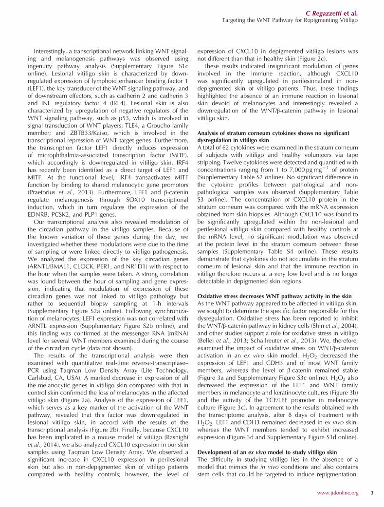
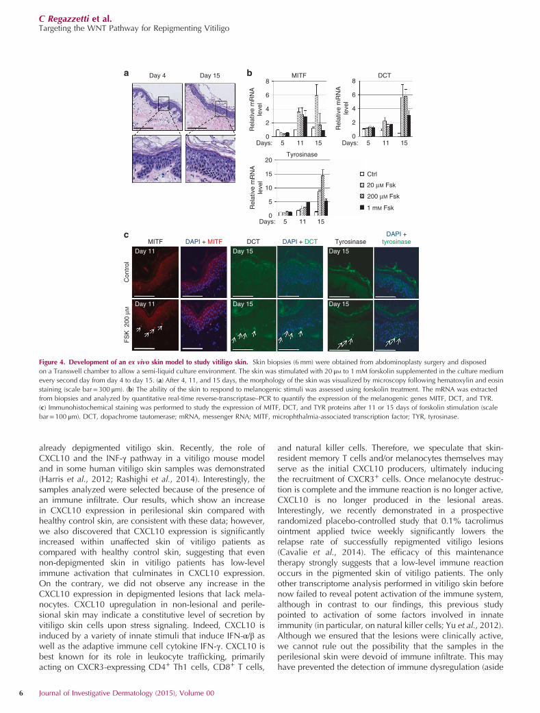
Thus, we developed an ex vivo skin model that is viable longenough to induce the differentiation of melanocyte stem cellsin vitiligo skin. In clinical practice, vitiligo lesions usuallyrequire months to achieve complete or almost completerepigmentation; however, in the best cases, the onset ofpigmentation can sometimes be observed after 15 days oftreatment. We therefore examined the morphologic charac-teristics of skin after treatment with the pigmentation inducerforskolin after 15 days of ex vivo culture following abdomi-noplasty skin surgery. At this time, the morphology of the skinremained stable, compared with the initial conditions. Wenoted only a flattening of the dermal–epidermal junction, andsuch flattening may be owing to a difference of tension in theskin (Figure 4a). The ability of the skin to respond to forskolinwas analyzed by quantitative PCR and immunofluorescence
by studying the expression of the melanogenic genes MITF,dopachrome tautomerase (DCT), and tyrosinase. In order toassess the dose response of forskolin, the skin was stimulatedby forskolin systemically every other day. As expected, theinitial response induced upregulation of MITF mRNA(Figure 4b) and protein (Figure 4c) after 11 days of stimulation.This phenomenon was transient, although expressionremained high after 15 days at a low forskolin concentration(Figure 4b). After 15 days, we observed a strong increase ofthe melanogenic enzymes DCT and tyrosinase at both themRNA and protein levels (Figures 4b and c). Despite theactivation of the melanogenesis pathway, we did not detectan increase in melanin contain in the cells (data not shown).In conclusion, our model of ex vivo skin culture was viableand functional for as many as 15 days in culture.
mR
NA
rela
tive e
xpre
ssio
n
NLS PLS LS
mR
NA
re
lative
exp
ressio
n
MLANA
3
mR
NA
re
lative
expre
ssio
n
2
1
0NLS PLS LS
L1CAM
***
3
mR
NA
re
lative
exp
ressio
n
2
1
0NLS PLS LS
MITF
***
3
2
1
0NLS PLS LS
***
3
mR
NA
re
lative
expre
ssio
n
2
1
0NLS PLS LS
PLXNC1
***
3
mR
NA
re
lative
exp
ressio
n
2
1
0NLS PLS LS
KIT
***
mR
NA
re
lative
exp
ressio
n
TYRP1
3
2
1
0NLS PLS LS
***
3
mR
NA
re
lative
expre
ssio
n
2
1
0NLS PLS LS
CDH2
***
3
mR
NA
re
lative
exp
ressio
n
2
1
0NLS PLS LS
NANOG
**
mR
NA
re
lative
exp
ressio
n
DCT3
2
1
0NLS PLS LS
***
*
**
*
*
LEF16
5
4
3
2
1
0
mR
NA
rela
tive e
xpre
ssio
n
NLS PLS LS
6
5
4
3
2
1
0
CXCL10
3m
RN
A r
ela
tive
exp
ressio
n2
1
0NLS PLS LS
PMEL
***
3
mR
NA
re
lative
exp
ressio
n
2
1
0NLS PLS LS
FOXD3
***
3
mR
NA
re
lative
expre
ssio
n
2
1
0NLS PLS LS
PCDH11X
***
a
b c
Figure 2. Gene expression analysis of the key factors emphasized by the transcriptomic analysis. Gene expression was measured by quantitative real-time
reverse-transcriptase–PCR of (a) melanocytic genes, (b) LEF1, and (c) CXCL10 mRNA. Non-lesional skin (NLS), perilesional skin (PLS), and and lesional skin (LS)
sample groups were compared with the healthy skin (HS) group using a t-test linear model without pairing. Differences in gene expression were considered
significant if both the |fold change| was ⩾1.8 and the FDR value was o0.05. The y axis displays the fold change in expression relative to the pool of healthy skin
samples. The data were normalized relative to expression of HPRT, ACTB, and GAPDH. The error bars display the SE of three replicates. *Po0.05; **Po0.01;
***Po0.001. The TLDA validation set encompassed genes related to terminal differentiation of melanocytes (PMEL, MLANA, TYRP1, and DCT), transcription
factors involved in melanogenesis (MITF, FOXD3, KIT, and NANOG), and adhesion molecules expressed in melanocytes (PLXNC1, PCDH11X, CDH2, and
L1CAM). CDH2, cadherin 2; DCT, dopachrome tautomerase; FDR, false discovery rate; mRNA, messenger RNA; LEF1, lymphoid enhancer binding factor 1;
MITF, microphthalmia-associated transcription factor; TLDA, Taqman Low Density Array; TYR, tyrosinase.
C Regazzetti et al.Targeting the WNT Pathway for Repigmenting Vitiligo
4 Journal of Investigative Dermatology (2015), Volume 00

Pharmacological WNT pathway activators induce increasedWNT pathway expression in ex vivo cultured vitiligo skinThe WNT pathway is involved in melanocyte differentiation(Yamada et al., 2013; Fukunaga-Kalabis et al., 2015), andwe have demonstrated that this pathway is altered in vitiligoskin. In order to induce the differentiation of melanocytestem cells, we pharmacologically activated the WNT pathwayin ex vivo vitiligo skin using the WNT agonist (SKL2001) andthe glycogen synthase kinase (GSK)3β inhibitors lithiumchloride (LiCl) and CHIR99021. We obtained biopsies fromnine vitiligo subjects from diverse body locations (three fromthe elbow, two from the trunk, two from the leg, one from thearm, and one from the axilla). We then treated the ex vivocultures for 14 days via systemic stimulation every otherday with these WNT activators. Evaluation of the activationof the WNT pathway indicated that treatment with LiCl,CHIR99021, or SKL2001 induced all WNT members andLEF1 at the mRNA level after 14 days of stimulation (Figure 5aand Supplementary Figure 4 online).
Ex vivo treatment of depigmented skin biopsies from vitiligopatients with WNT activators induces the differentiation ofresident stem cells into pre-melanocytesAs we succeeded in upregulating the WNT pathway in the exvivo vitiligo skin biopsies, we analyzed the expression ofmelanoblast markers following treatment. The mRNA levelsof early melanoblast markers, such as PAX3 and BRN2, wereupregulated in biopsies stimulated with CHIR99021,
SKL2001, and LiCl (Figures 5b and c). This result suggestsinitiation of melanocyte differentiation. MITF levels wereincreased only in the skin treated with SKL2001 (Figure 5d);however, this expression was transient following stimulation,suggesting that the time response is different for the differenttreatments. The pre-melanocyte marker DCT was increased inresponse to all treatments, but this effect was stronger inresponse to LiCl (Figure 5e).We next employed immunofluorescence to study the co-
expression of DCT and PAX3 in order to investigate whetherthese treatments lead to the differentiation of melanocyte stemcells in pre-melanocytes in the skin. In vitiligo skin biopsiescultured under control conditions, we observed no or fewisolated cells expressing PAX3 and DCT (Figure 5f). Followingstimulation with Wnt activators (Figures 5g–i), we found manyclusters of cells co-expressing the two markers in the hairfollicles and in the dermis, representing melanocytes under-going differentiation. Taken together, these results showedthat targeting the deficient WNT pathway of vitiligo skin usingWNT agonists or GSK3β inhibitors leads to the differentiationof melanocyte stem cells into pre-melanocytes.
DISCUSSIONOur results emphasize the complexity of vitiligo pathophy-siology. Although these findings partially support the role ofthe immune system, and notably CXCL10, in depigmentationof the skin in vitiligo patients, these data also indicate thatneither CXCL10 nor other immune factors are deregulated in
Ex vivo skin – 2 days H2O2
Ex vivo skin – 8 days H2O2
1.4
1.2
1
0.8
0.6
0.4
0.2
0
1.4
1.2
1
0.8
0.6
0.4
0.2
0
1.4
1.6
1.2
1
0.8
0.6
0.4
0.2
0
1.2
1
0.8
0.6
0.4
0.2
0
******
***
** **
LEF1 βcatenin CDH3
Re
lative
mR
NA
le
ve
l
Re
lative
mR
NA
le
ve
lR
ela
tive m
RN
A le
ve
l
Melanocytes
Melanocytes
Keratinocytes
*
***
*
*
LEF1
LEF1 CDH3TCF/LEF1 promoter activity
Rela
tive T
CF
/LE
F p
rom
ote
r
activity
a
c d
bCtrl
25 µM H2O2
50 µM H2O2
100 µM H2O2
Figure 3. Oxidative stress decreases WNT pathway activity in the skin. In whole skin in ex vivo culture, NHM and NHK were stimulated for 24 h (b), 2 days (a),
or 8 days (d) with 25–100 μM H2O2. After mRNA extraction, reverse transcription was performed, and the relative gene expression of LEF1, β-catenin, and CDH3
was analyzed using quantitative PCR. Results were normalized with the expression of SB34.(c) NHM were infected with lentivirus luciferase TCF/LEF1 reporter.
After selection, NHM were treated for 24 hours with H2O2, and the relative activity of TCF/LEF was quantified with a firefly luciferase assay and normalized with
Renillaluciferase expression. *Po0.05; **Po0.01, ***Po0.001. mRNA, messenger RNA; LEF1, lymphoid enhancer binding factor 1.
C Regazzetti et al.Targeting the WNT Pathway for Repigmenting Vitiligo
www.jidonline.org 5
already depigmented vitiligo skin. Recently, the role ofCXCL10 and the INF-γ pathway in a vitiligo mouse modeland in some human vitiligo skin samples was demonstrated(Harris et al., 2012; Rashighi et al., 2014). Interestingly, thesamples analyzed were selected because of the presence ofan immune infiltrate. Our results, which show an increasein CXCL10 expression in perilesional skin compared withhealthy control skin, are consistent with these data; however,we also discovered that CXCL10 expression is significantlyincreased within unaffected skin of vitiligo patients ascompared with healthy control skin, suggesting that evennon-depigmented skin in vitiligo patients has low-levelimmune activation that culminates in CXCL10 expression.On the contrary, we did not observe any increase in theCXCL10 expression in depigmented lesions that lack mela-nocytes. CXCL10 upregulation in non-lesional and perile-sional skin may indicate a constitutive level of secretion byvitiligo skin cells upon stress signaling. Indeed, CXCL10 isinduced by a variety of innate stimuli that induce IFN-α/β aswell as the adaptive immune cell cytokine IFN-γ. CXCL10 isbest known for its role in leukocyte trafficking, primarilyacting on CXCR3-expressing CD4+ Th1 cells, CD8+ T cells,
and natural killer cells. Therefore, we speculate that skin-resident memory T cells and/or melanocytes themselves mayserve as the initial CXCL10 producers, ultimately inducingthe recruitment of CXCR3+ cells. Once melanocyte destruc-tion is complete and the immune reaction is no longer active,CXCL10 is no longer produced in the lesional areas.Interestingly, we recently demonstrated in a prospectiverandomized placebo-controlled study that 0.1% tacrolimusointment applied twice weekly significantly lowers therelapse rate of successfully repigmented vitiligo lesions(Cavalie et al., 2014). The efficacy of this maintenancetherapy strongly suggests that a low-level immune reactionoccurs in the pigmented skin of vitiligo patients. The onlyother transcriptome analysis performed in vitiligo skin beforenow failed to reveal potent activation of the immune system,although in contrast to our findings, this previous studypointed to activation of some factors involved in innateimmunity (in particular, on natural killer cells; Yu et al., 2012).Although we ensured that the lesions were clinically active,we cannot rule out the possibility that the samples in theperilesional skin were devoid of immune infiltrate. This mayhave prevented the detection of immune dysregulation (aside
Day 4 Day 15 MITF DCT8
6
4
2
0
8
6
4
2
0Days: 5 11 15
Days: 5 11 15
Days: 5 11 15
Re
lative
mR
NA
leve
lR
ela
tive
mR
NA
leve
l
Re
lative
mR
NA
leve
l
Tyrosinase
Tyrosinase
DAPI +
tyrosinase
20
15
10
5
0
Ctrl
1 mM Fsk
20 µM Fsk
200 µM Fsk
FS
K
200 µ
M
MITF DAPI + MITF DAPI + DCTDCT
Day 11 Day 15 Day 15
Day 15Day 15Day 11
Contr
ol
c
a b
Figure 4. Development of an ex vivo skin model to study vitiligo skin. Skin biopsies (6 mm) were obtained from abdominoplasty surgery and disposed
on a Transwell chamber to allow a semi-liquid culture environment. The skin was stimulated with 20 μM to 1 mM forskolin supplemented in the culture medium
every second day from day 4 to day 15. (a) After 4, 11, and 15 days, the morphology of the skin was visualized by microscopy following hematoxylin and eosin
staining (scale bar=300 μm). (b) The ability of the skin to respond to melanogenic stimuli was assessed using forskolin treatment. The mRNA was extracted
from biopsies and analyzed by quantitative real-time reverse-transcriptase–PCR to quantify the expression of the melanogenic genes MITF, DCT, and TYR.
(c) Immunohistochemical staining was performed to study the expression of MITF, DCT, and TYR proteins after 11 or 15 days of forskolin stimulation (scale
bar=100 μm). DCT, dopachrome tautomerase; mRNA, messenger RNA; MITF, microphthalmia-associated transcription factor; TYR, tyrosinase.
C Regazzetti et al.Targeting the WNT Pathway for Repigmenting Vitiligo
6 Journal of Investigative Dermatology (2015), Volume 00

from CXCL10) and may also explain the fact that we did notobserve activation of natural killer cells as previously shownby Yu et al. (2012).
Recently, the WNT/β-catenin pathway was found to have akey role in UVB-induced melanocyte stem cell differentiation(Yamada et al., 2013). Within the skin, the secretion of WNT
10
5
0
LEF1
–
Re
lative
mR
NA
leve
l
Re
lative
mR
NA
leve
l
CHIR
9902
1SKL2
001
LiCI
CHIR
9902
1SKL2
001
LiCI
6
4
2
0
PAX3
–
a bR
ela
tive m
RN
A
level
10
5
0
Brn2
–
CHIR
9902
1SKL2
001
LiCI
cR
ela
tive m
RN
A
level
–
CHIR
9902
1SKL2
001
LiCI
3
2
1
0
MITFd
Rela
tive m
RN
A
level
–
CHIR
9902
1SKL2
001
LiCI
15
10
5
0
DCTe
SKL2001
DCT DCT
Control
PAX3 PAX3Merge
DCT PAX3 Merge
Merge
DCT PAX3 Merge
LiCICHIR99021
D
E
D E
DE
D E
D E
D
E
D
E
DE
D
D
E
E
D
DE
f g
ih
Figure 5. Treatment of ex vivo depigmented skin samples from vitiligo patients with pharmacological WNT pathway activators induces the differentiation
of resident stem cells into pre-melanocytes. Lesional skin from vitiligo patients (n= 9) was biopsied. The biopsies were stimulated in ex vivo culture for 14 days
with the GSK3β inhibitors CHIR99021 (3 μM) or LiCl (20 μM) or with the WNT agonist SKL2001 (40 μM) every other day. Then, mRNA was extracted, and
quantitative real-time reverse-transcriptase–PCR was performed to quantify the relative expression of LEF1 (a) and the melanocyte markers PAX3 (b), BRN2 (c),
MITF (d), and DCT (e). The response of the vitiligo skin was analyzed by immunohistochemical staining for the pre-melanocyte markers DCT (green) and PAX3
(red). The co-localization (yellow) observed on merge pictures shows melanoblasts in differentiation within the dermis following treatment with the following
agents: (f) control, (g) WNT agonist SKL2001 (40 μM), (h) CHIR99021 (3 μM), and (i) LiCl 20 μM (scale bar=100 μm). D, dermis; DCT, dopachrome tautomerase;
E, epidermis; LEF1, lymphoid enhancer binding factor 1; LiCl, lithium chloride; MITF, microphthalmia-associated transcription factor; mRNA, messenger RNA.
C Regazzetti et al.Targeting the WNT Pathway for Repigmenting Vitiligo
www.jidonline.org 7
mainly by keratinocytes and melanocytes contributesto the differentiation of stem cells in melanocytes. Ourtranscriptional analysis reveals an alteration of the WNT/β-catenin pathway in vitiligo skin with a significant decrease inLEF/TCF expression that was confirmed using quantitativereal-time reverse-transcriptase–PCR. As LEF1 is predominantlyexpressed by melanocytes in the skin, we cannot exclude thepossibility that this decrease merely reflects the absence ofmelanocytes; however, other WNT pathway genes that werefound to be differentially expressed in the transcriptomeanalysis are similarly expressed in keratinocytes and melano-cytes (e.g., CDH3, TLE4, and Kaiso; Supplementary Figure 5online). In addition, our functional analyses further supportthe impact of the decreased activation of the WNT pathway invitiligo as decreased expression of LEF1 and decreasedactivity of the LEF1/TCF promoter were observed afteroxidative stress and differentiation of resident stem cells wasinduced in pre-melanocytes following ex vivo stimulationwith WNT activators.Our transcriptome analysis also demonstrated that adhe-
sion proteins, including cadherins, were also decreased invitiligo skin. The WNT pathway is known to regulateE-cadherin expression, and interestingly, recent data showeddecreased expression of E-cadherin across melanocytemembranes in vitiligo patients, leading to decreased adhe-siveness of these cells to the basal layer under oxidative andmechanical stress (Wagner et al., 2015). Many studies haveemphasized the role of oxidative stress in vitiligo (Marescaet al., 1997; Bellei et al., 2013; Schallreuter et al., 2013).Furthermore, a link between oxidative stress and activation ofthe immune response has recently been uncovered (Passeronand Ortonne, 2012; Toosi et al., 2012), and our data clearlyindicate that oxidative stress decreases WNT pathway activityin melanocytes and keratinocytes. WNT ligands and LEF1 arefirst decreased in both melanocytes and keratinocytes; yet,decreased levels of LEF1 and CDH3 persist in vitiligo skinex vivo despite the augmentation of WNT probably owing tocompensation of the impaired pathway. In vitiligo lesionsdevoid of melanocytes, the keratinocytes are presumablyresponsible for the production of WNT proteins. Takinginto account the central role of the WNT/β-catenin path-way on the differentiation of melanocyte stem cells, wehypothesize that oxidative stress negative impacts thedifferentiation of melanocytes in vitiligo skin. We, therefore,directly addressed the defective differentiation of melanocytestem cells by stimulating the WNT/β-catenin pathway, whichwas altered in vitiligo lesions. Using our ex vivo model fordepigmented skin of vitiligo patients, we demonstrated thattreatment with WNT agonists or GSK3β inhibitors induceincreased expression of melanocyte markers, triggering thedifferentiation of resident melanocyte stem cells in pre-melanocytes expressing PAX3 and DCT. Interestingly, weobserved pre-melanocytes not only in the hair follicles butalso in the dermis, suggesting that this approach may behelpful for the differentiation of dermal stem cells of glabrousskin (Li et al., 2010). The cells that expressed DCT andPAX3 remained in the dermis, and we did not detect thedifferentiated melanocyte marker tyrosinase at either the
mRNA or protein level (using immunohistochemistry) in thesecells (data not shown). Such localization and expressionpatterns of melanocyte markers strongly suggest that thesecells are pre-melanocytes. In all likelihood, however, thelimited timeframe of our ex vivo model is too short to obtainfully differentiated melanocytes.Taken together, our results demonstrate that the immune
reaction in vitiligo occurs only at very low levels. Specifically,an increase in CXCL10 expression in non-depigmented andperilesional skin was observed, and an immune reaction isno longer detectable in vitiligo lesions already devoid ofmelanocytes. These findings also highlight a previouslyunrecognized defect in WNT/β-catenin activation triggeredby oxidative stress, and this defect may effectively prevent thedifferentiation of melanocyte stem cells (Figure 6). Theseresults not only provide a better understanding of the complexpathophysiology of vitiligo but also support further clinicalexploration of WNT activators for repigmenting vitiligolesions.
MATERIALS AND METHODSPatients for transcriptomic analysisTen patients with active non-segmental vitiligo, which is defined by
the occurrence or the worsening of depigmented lesions in the past
3 months and having hypochromic borders upon Wood’s lamp
examination, were enrolled in the study after informed, written
consent was obtained. The study was approved by the local ethics
committee (N12.034). A 4-mm-skin biopsy was taken from each
patient in the center of a vitiligo patch, in the perilesional area
(defined as 5mm outside of the lesion border), and in non-lesional
skin located in the same area but at least 3 cm from a depigmented
lesion. A 4-mm biopsy was also taken from 10 healthy patients and
served as a control that was matched for gender, age, and location.
The procedure for taking samples and the characteristics of the
patient population are described in Supplementary Figures S1a and
S1b online.
Transcriptomic analysisBiopsies for the microarray analysis were stored in RNA Stabilization
Reagent (Qiagen, Venlo, The Netherlands) until use. For RNA
extraction, the samples were homogenized with a potter in Qiagen
lysis buffer (Qiagen). Total RNA was extracted using RNeasy
extraction kits (Qiagen) according to the manufacturer's protocol.
RNA quantity was measured using a Nanodrop Spectrophotometer
ND8000 (Thermo Fisher Scientific, Waltham, MA, USA). RNA
quality was monitored using a 2100 Bioanalyzer (Agilent
Technologies, Waldbronn, Germany). Probes were synthesized and
then hybridized on Affymetrix U133 Plus 2.0 chips (Affymetrix,
Santa Clara, CA, USA). All chips were normalized using the robust
multi-array average method (Bolstad et al., 2003). Only Affymetrix
identifiers (IDs) with expression ⩾ 2exp6(64) for at least 7 out of 10
samples in at least 1 sample group (lesional (LS), non-lesional (NLS),
perilesional (PLS), or healthy) were selected. Finally, 29,906 of
54,675 IDs that were initially present were kept for statistical
analyses, and thresholds of modulation of 1.5 and of − 1.5 were
selected for further analyses. Data analysis was performed on Array
Studio software (OmicSoft, Cary, NC, USA). A two-sided paired
Student’s t-test was performed. The Benjamini-Hochberg procedure
C Regazzetti et al.Targeting the WNT Pathway for Repigmenting Vitiligo
8 Journal of Investigative Dermatology (2015), Volume 00

(1995) was used for correction of multiple testing. The raw data are
available at NCBI GEO, accession number GSE65127.
Functional analysis of gene expression resultsAverage-linkage hierarchical clustering using Pearson correlation
was performed using the Cluster v.3.0 program (Eisen et al., 1998).
Graphic outputs were generated with the JavaTreeView3.0 software
(Saldanha, 2004). Functional annotation and gene networks for the
differentially expressed genes among sample groups were generated
using the QIAGEN IPA (QIAGEN, Redwood City, CA, USA; www.
qiagen.com/ingenuity).
Ex vivo skin cultureSkin from abdominoplasty surgery was used for the development of
the ex vivo skin culture model. The subcutaneous fat was removed,
and biopsies of 6 mm were taken from skin composed of dermis and
epidermis. For vitiligo skin biopsies, after informed consent was
obtained and the absence of melanocytes with Wood’s lamp was
verified, two to three 6-mm biopsies composed of dermis and
epidermis were taken from lesional skin (n= 9). The biopsies are
rapidly placed into a 0.4-μm Transwell chamber (Becton Dickinson,
Franklin Lakes, NJ, USA) and maintained under semi-liquid culture
conditions in “Skin long-term culture medium” (Biopredic, Saint
Grégoire, France). The skin was maintained at 37 °C in a 5% CO2
atmosphere. The culture medium that was supplemented with
forskolin (Sigma-Aldrich, Saint-Louis, MO), LiCl (Sigma-Aldrich),
CHIR99021 (Calbiochem, San Diego, CA, USA), or SKL2001 (Calbio-
chem) was changed every day during the 14-day period.
The methods for Luminex 200 system (Luminex Corporation,
Austin, TX) quantitation of cytokines in stratum corneum, cells,
quantitative real-time reverse-transcriptase–PCR, and histological
analyses are detailed in the Supplementary Methods online.
CONFLICT OF INTERESTThis work was funded in part by Galderma. FJ, CM, MR, BM, PR, CM, YR, DP,and JV are employees of Galderma.
ACKNOWLEDGMENTSThis work was funded in part by a grant from the French Society ofDermatology and by the INSERM. This work was performed in collabo-ration with Galderma Research. The expert technical assistance ofEve Ferrara, Philippe Grundt, Corinne Menigot, Agnès Perrin and Luigi Russois gratefully acknowledged. Light microscopy was performed in the C3MImaging Core Facility (part of Microscopy and Imaging Platform, Côted’Azur, MICA).
SUPPLEMENTARY MATERIAL
Supplementary material is linked to the online version of the paper at http://www.nature.com/jid
REFERENCES
Bellei B, Pitisci A, Ottaviani M et al. (2013) Vitiligo: a possible model ofdegenerative diseases. PloS One 8:e59782
Bolstad BM, Irizarry RA, Astrand M et al. (2003) A comparison of normalizationmethods for high density oligonucleotide array data based on varianceand bias. Bioinformatics 19:185–93
Cavalie M, Ezzedine K, Fontas E et al. (2014) Maintenance therapy of adultvitiligo with 0.1% tacrolimus ointment: a randomized, double blind,placebo-controlled study. J Invest Dermatol 135:970–4.
Healthy skin
Melanocyte
Melanocyte
Keratinocyte
Keratinocyte
Differentiation and
migrationDifferentiation and
migration
Ep
ide
rmis
De
rmis
LEF
WNT
Stem cell
LEF
WNT
Stem cell
Normal renewal of melanocyte Decreased renewal of melanocyte
Vitiligo lesional skin
Immune
system Oxidative stress
CXCL10
Antigen
releaseCell adhesion
Melanocyte
detachment and
melanocyte death
Figure 6. Schematic representation of factors involved in vitiligo pathogenesis. In healthy skin, the stimulation of the WNT pathway by keratinocytes and
melanocytes induces the differentiation and proliferation of melanocyte stem cells, allowing the constant turnover of the pools of epidermal melanocytes. In
vitiligo skin, oxidative stress can trigger the immune reaction in a genetically predisposed individual. The destruction of melanocytes by the immune system
releases melanocyte antigens that stimulate an autoimmune response and ultimately lead to the complete disappearance of melanocyte from the epidermis (and
sometimes the hair follicles). Concomitantly, oxidative stress decreases WNT pathway activity in melanocytes and in keratinocytes. This effectively induces
decreased cell adhesion with detachment of melanocytes and an impaired differentiation of melanocyte stem cells, altering the capacity of melanocyte turnover.
We hypothesize that depending on the patient and the course of the disease, these two mechanisms may be differentially implicated, leading to active
depigmentation of the skin and resistance to repigmenting approaches.
C Regazzetti et al.Targeting the WNT Pathway for Repigmenting Vitiligo
www.jidonline.org 9

Eisen MB, Spellman PT, Brown PO et al. (1998) Cluster analysis and display ofgenome-wide expression patterns. Proc Natl Acad Sci USA 95:14863–8
Fukunaga-Kalabis M, Hristova DM, Wang JX et al. (2015) UV-InducedWnt7a in the Human Skin Microenvironment Specifies the Fate ofNeural Crest-Like Cells via Suppression of Notch. J Invest Dermatol 135:1521–32
Harris JE, Harris TH, Weninger W et al. (2012) A mouse model of vitiligo withfocused epidermal depigmentation requires IFN-gamma for autoreactiveCD8(+) T-cell accumulation in the skin. J Invest Dermatol 132:1869–76
Jin Y, Birlea SA, Fain PR et al. (2012) Genome-wide association analyses identify13 new susceptibility loci for generalized vitiligo. Nat Genet 44:676–80
Li L, Fukunaga-Kalabis M, Yu H et al. (2010) Human dermal stem cellsdifferentiate into functional epidermal melanocytes. J Cell Sci 123:853–60
Maresca V, Roccella M, Roccella F et al. (1997) Increased sensitivity toperoxidative agents as a possible pathogenic factor of melanocyte damagein vitiligo. J Invest Dermatol 109:310–3
Mosenson JA, Zloza A, Nieland JD et al. (2013) Mutant HSP70 reversesautoimmune depigmentation in vitiligo. Sci Transl Med 5:174ra28
Passeron T, Ortonne JP (2012) Activation of the unfolded protein response invitiligo: the missing link? J Invest Dermatol 132:2502–4
Praetorius C, Grill C, Stacey SN et al. (2013) A polymorphism in IRF4 affectshuman pigmentation through a tyrosinase-dependent MITF/TFAP2A path-way. Cell 155:1022–33
Radtke MA, Schafer I, Gajur A et al. (2009) Willingness-to-pay and quality of lifein patients with vitiligo. Br J Dermatol 161:134–9
Rashighi M, Agarwal P, Richmond JM et al. (2014) CXCL10 is critical for theprogression and maintenance of depigmentation in a mouse model ofvitiligo. Sci Transl Med 6:223ra23
Saldanha AJ (2004) Java Treeview–extensible visualization of microarray data.Bioinformatics 20:3246–8
Schallreuter KU, Salem MA, Holtz S et al. (2013) Basic evidence for epidermalH2O2/ONOO(-)-mediated oxidation/nitration in segmental vitiligo issupported by repigmentation of skin and eyelashes after reduction ofepidermal H2O2 with topical NB-UVB-activated pseudocatalase PC-KUS.FASEB J 27:3113–22
Shin SY, Kim CG, Jho EH et al. (2004) Hydrogen peroxide negatively modulatesWnt signaling through downregulation of beta-catenin. Cancer Lett 212:225–31
Silverberg JI, Silverberg NB (2013) Association between vitiligo extent anddistribution and quality-of-life impairment. JAMA Dermatol 149:159–64
Spritz RA (2012) Six decades of vitiligo genetics: genome-wide studies provideinsights into autoimmune pathogenesis. J Invest Dermatol 132:268–73
Taieb A, Alomar A, Bohm M et al. (2013) Guidelines for the management ofvitiligo: the European Dermatology Forum consensus. Br J Dermatol 168:5–19
Toosi S, Orlow SJ, Manga P (2012) Vitiligo-inducing phenols activate theunfolded protein response in melanocytes resulting in upregulation of IL6and IL8. J Invest Dermatol 132:2601–9
Wagner RY, Luciani F, Cario-Andre M et al. (2015) Altered E-cadherin levelsand distribution in melanocytes precede clinical manifestations of vitiligo. JInvest Dermatol 135:1810–9.
Yamada T, Hasegawa S, Inoue Y et al. (2013) Wnt/beta-catenin and kit signalingsequentially regulate melanocyte stem cell differentiation in UVB-inducedepidermal pigmentation. J Invest Dermatol 133:2753–62
Yu R, Broady R, Huang Y et al. (2012) Transcriptome analysis reveals markersof aberrantly activated innate immunity in vitiligo lesional and non-lesional skin. PloS One 7:e51040
C Regazzetti et al.Targeting the WNT Pathway for Repigmenting Vitiligo
10 Journal of Investigative Dermatology (2015), Volume 00