transition from growth cone to functional motor nerve
TRANSCRIPT

Transition from Growth Cone to Functional Motor Nerve Terminal inDrosophila Embryos
Motojiro Yoshihara,1 Mary B. Rheuben,2 and Yoshiaki Kidokoro1
1Gunma University School of Medicine, Maebashi 371, Japan, and 2Department of Anatomy, College of VeterinaryMedicine, Michigan State University, East Lansing, Michigan 48824
As a motor axon grows from the CNS to its target muscle, theterminal has the form of a flattened growth cone with a planarcentral region, lamellipodia, and filopodia. A mature terminalusually has a stereotyped shape that may be elongated withvaricosities, as in several invertebrate species, or have shortbranches with boutons, as in mammals. We examined in Dro-sophila the developmental changes between growth cone andmature terminal using ultrastructural and immunocytochemicalmethods.
The transition period, which occurs 2–3 hr after the firstgrowth cone reaches its target muscle, is marked by the for-mation of “prevaricosities,” smoothly contoured enlargementsof the axons at the point where the nerve trunk first contacts themuscle fiber (MF). There is a 15–30 min ventral-to-dorsal gra-dient in the formation of prevaricosities on the individual ab-dominal MFs. Multineuronal innervation of each MF has oc-
curred by this time, and two or more different axons undergoprevaricosity formation while they are intimately intertwined atthe nerve entry point (NEP). Presynaptic active zones, bothnerve–nerve and nerve–muscle, occur within the prevaricositiesalong broad contact regions. Synaptotagmin immunoreactiveclusters form concurrently.
The first varicosities then develop as a result of constrictionsof the larger prevaricosities rather than as enlargement of dis-crete portions of the filopodia or neurites. The prevaricositystage therefore may include the key steps that lead to thedifferentiation of functional differences in terminal subtypes aswell as those leading to the formation of a stable neuromuscularjunction.
Key words: Drosophila; neuromuscular junction; synaptogen-esis; growth cone; development; immunohistochemistry
Mature neuromuscular junctions of various species are character-ized by long, infrequently branched nerve terminals, chains ofvaricosities, or clusters of boutons that house the specializationsassociated with fast, high quantity transmitter release and recy-cling. Quantitative variations in that morphology, both character-istic and plastic, are associated with changes in functional capa-bilities (for review, see Atwood and Wojtowicz, 1986; Hall andSanes, 1993; Burns and Augustine, 1995).
In Drosophila the subtypes of larval motor terminals havevaricosities of characteristic dimensions and cytoplasmic inclu-sions (Johansen et al., 1989a; Atwood et al., 1993; Jia et al., 1993).Mutations that produce defects in neuronal proteins can directlyor indirectly affect terminal shape. For example, mutations thatmodify the properties of excitable channels, thus altering the levelof neuronal activity, also change the number of branches and the
number of varicosities in mature terminals (Budnik et al., 1990).Similarly, the effect of altered cAMP levels (Zhong et al., 1992),adhesion molecules such as Fasciclin II (Fas II) (Schuster et al.,1996b; Stewart et al., 1996), and specific synaptic proteins such assyntaxin (Broadie et al., 1995) all result in a change in the numberof varicosities formed. In some mutants this is independent of theresulting synaptic strength and may be accompanied by changes innumbers of synaptic structures within the varicosity as well (Jia etal., 1993; Stewart et al., 1996). These mutants thus offer clues asto the mechanisms underlying plasticity.
A motor nerve terminal during outgrowth from the CNS hasthe form of a growth cone, a flared veil-like enlargement of theaxon tip with numerous exploring filopodia, in Drosophila (Good-man et al., 1984) as well as in vertebrates (Ramon y Cajal, 1890;Harrison, 1910). Growth cones explore among potential MFtargets and connect only to specific ones (Johansen et al., 1989b;Halpern et al., 1991; Sink and Whitington, 1991; Van Vactor etal., 1993). A growth cone has given rise to a varicose terminalcharacteristic of the specific MF, albeit with fewer than themature number of varicosities, by the time the embryo hatches(Halpern et al., 1991; Broadie and Bate, 1993). However, little isknown about the interactions between a growth cone and an MFthat determine the final placement of the terminal on a specificpart of the selected fiber, the exchange of the labile form of thegrowth cone for the characteristic branch pattern, or those thatdetermine varicosity size and varicosity number.
To begin an analysis of these steps we examined the cellularreorganization that occurs during the transition from the growthcone to terminal in specified abdominal MFs in Drosophila em-bryos. This period coincides with the time that miniature excita-
Received May 14, 1997; revised Aug. 22, 1997; accepted Aug. 22, 1997.This work was supported by Grants-in-Aid from the Ministry of Education,
Science, Sports, and Culture to Y.K. and M.Y., grants from the Brain ScienceFoundation and Kato Memorial Science Foundation to M.Y., an All-UniversityResearch Initiation Grant from Michigan State University, and grants from theNarishige Neurosciences Foundation and the Yamada Science Foundation toM.B.R., and from the Mitsubishi Foundation to Y.K. We thank Dr. Kazuo Ikedaand Dr. Masahiro J. Go for kind instruction, Dr. Hugo Bellen for the antibody tosynaptotagmin, and Dr. Michihiro Igarashi, Dr. Kensuke Hayashi, Dr. Ryoki Ish-ikawa, Dr. Kei Ito, and Dr. Hiroshi Kuromi for useful discussions. The confocalstudies were performed in Dr. Harunori Ishikawa’s laboratory, and we thank himand the members of his lab for kind help. We also gratefully acknowledge thetechnical assistance of Dawn Autio, Jessica Hoane, Nobuko Yoshihara, FumikoSekiguchi, and Masako Terada, as well as Takeshi Masuda and Kouei Suto of theGunma University electron microscope facility.
M.Y. and M.B.R. contributed equally to this research.Correspondence should be addressed to Dr. Mary B. Rheuben, Department of
Anatomy, A514 East Fee Hall, Michigan State University, East Lansing, MI 48824.Copyright © 1997 Society for Neuroscience 0270-6474/97/178408-19$05.00/0
The Journal of Neuroscience, November 1, 1997, 17(21):8408–8426

tory junctional currents are first recorded (Broadie and Bate,1993; Kidokoro and Nishikawa, 1994) and therefore includes thesynaptic activity important to modulating terminal morphology.We describe a transitional structure containing organelles associ-ated with transmitter release, whose shape first presages thebranch pattern of the mature terminal but whose dimensions arelarger than those of mature varicosities.
MATERIALS AND METHODSFly stocks. For all specimens in this study, we used wild-type Drosophilamelanogaster, strain Canton-Special. The stock was maintained usingstandard fly-rearing techniques (Ashburner, 1989). The artificial dietcontained cornmeal, sugar, yeast, and agar. Incubator temperatures werekept at 25 6 1°C and humidity at .50%, and the flies were reared underuncrowded conditions.
Staging. For the precisely timed embryos needed for the developmen-tal studies, adult male and female flies were placed into a bottle with anagar plate in the bottom and allowed to lay eggs in the incubator for 1 hr.A paste consisting of 1 gm of dry yeast and 1.5 ml grape juice was placedon the surface of the agar plate to stimulate egg laying. After a precol-lection period of 1 hr, the agar plate was exchanged for a new one, andflies were allowed to lay eggs for 10 min. This plate was then transferredto a moist chamber for embryonic development. The temperature wasprecisely controlled at 25 6 0.5°C for egg laying and subsequent devel-opment. Thus, the hours after egg laying (AEL) were accurately timedwithin ;5 min and were approximately the same as hours after fertili-zation. Under these conditions, embryos develop synchronously andhatch at approximately 21 hr AEL (Broadie and Bate, 1993). Themorphology of the intestine, color of Malpighian tubules, presence of airin tracheae, cuticle formation, and ability to move were also used toconfirm stage of development. For the electron microscopy studies, eggswere collected over 30 min time periods.
Preparation and dissection. Embryos and first instar larvae were dis-sected in an osmotically balanced saline (modified from Stewart et al.,1994), 70 mM NaCl, 20 mM KCl, 25 mM MgCl2 , 10 mM NaHCO3 , 2 mMNaH2PO4 , 5 mM L-glutamine, 5 mM trehalose, 40 mM sucrose, 10 mMHEPES buffer, pH 7.1.
Before 17 hr AEL, when the epidermis still stuck to glass, a “flatpreparation” was made according to Bate’s method in Ashburner (1989).Briefly, mechanically dechorionated embryos were placed on a double-side sticky tape. The vitelline membrane was cut with a sharp glassneedle; the embryo was removed from it and put on a clean glass slide.The body wall was cut longitudinally along the embryo’s left side with aglass needle and then uncurled so that its outer surface stuck to the glassslide. The intestine, fat body, and tracheae were removed with fineforceps or sucked away with a fine glass capillary tube.
After 17 hr AEL, dissection was performed with a pair of sharpenedneedles using a method modified from Kidokoro and Nishikawa (1994).The posterior end of the embryo was immobilized by one needle andpierced with a second needle that was ground in the shape of a knife.Then, the knife needle was inserted deeply into the abdomen, and thebody wall (epidermis and muscles) along the left side of the animal wascut by pushing the animal against the bottom of the dish. Care was takennot to damage the right side or the nervous system. Dissected embryosand first instar larvae were mounted on Lux 13 mm Thermanox cover-slips (Nunc, Naperville, IL) using single strands of dragline spider silk.The main support strands of spider webs are not sticky and can bedissociated into individual fibers of sufficient strength and elasticity tobind the filleted embryo to the coverslip. We used silk from Nephilaclavata, a common Japanese spider. The strands of web were insertedinto notches cut in the edges of the plastic coverslips, and their placementwas adjusted to suit the size of the larva being immobilized. Thisparticular type of coverslip and the spider web could be carried throughfixation, dehydration, and embedding without damage, or could be usedas the support planchette for critical point drying and examination in thescanning electron microscope. Wells were made around the specimens tofurther protect them from mechanical damage by gluing a second cov-erslip, with a 4 3 2 mm rectangular hole cut out of the center, over thecoverslip holding the web. For some transmission electron microscopy(TEM) samples, 1–4% agarose (gelling temperature 25°C) (BoehringerMannheim, Indianapolis, IN) was poured over the sample just before theend of aldehyde fixation and then hardened, and fixation finished [mod-ified from Wood and Klomparens (1993)]. The solidified and fixed
agarose block containing the specimen was cut free from the spider webstrands just before embedding. Third instar larvae were held to coverslipseither with dental floss or fine wire or in some instances were carried partway through preparation pinned to Sylgard (Dow Corning, Midland, MI)in a 35 mm petri dish.
Some embryos were prepared for TEM without dissection to minimizethe damage that occurs during filleting. Undissected animals were per-fused directly with fixative via a glass micropipette inserted into theposterior abdomen, with a second small hole in the epidermis havingbeen made anteriorly for exit of the solution. In a control series examinedconfocally, two sets of embryos were prepared with and without dissec-tion, and with or without Ca 21 in the saline and fixative to assess theeffects of mechanical damage and other factors that might affect fragileembryonic structures. None of these treatments affected the appearanceof the prevaricosity in animals at the prevaricosity stage (;16.5 hr AEL).However, as noted in the results section, cutting the intersegmentalnerves (ISNs) did result in the formation of abnormal-appearing balloon-like prevaricosities on the affected side.
Protocols for fluorescence immunohistochemistry. Dissected prepara-tions were fixed in 4% formaldehyde (made from a 37% solution) in PBS(10 mM Na2HPO4 and 130 mM NaCl are mixed with 10 mM NaH2PO4and 130 mM NaCl and titrated to pH 7.2) for 2 hr and washed in PBT(PBS with 0.5% Triton X-100). Goat anti-HRP IgG conjugated to fluo-rescein (Cappel, Durham, NC) was used for staining all neural cells (Janand Jan, 1982). After blocking for 1 hr with 1% BSA in PBT, fixedpreparations were incubated in antibody solution (1:100 dilution forFITC anti-HRP and 1% BSA in PBT) for 2 hr at room temperature withgentle agitation.
Synaptotagmin was localized by rabbit polyclonal antiserum againstsynaptotagmin (Littleton et al., 1993), which was kindly provided by Dr.Hugo Bellen. For double-labeling experiments with both synaptotagminantisera and anti-HRP IgG, fixed and BSA-blocked preparations asabove were incubated in primary antibody solution (1:500 dilution foranti-synaptotagmin; 1:100 dilution for FITC anti-HRP and 1% BSA inPBT) for 2 hr at room temperature with gentle agitation. After it waswashed in PBT, the preparation was incubated in secondary antibodysolution [1:500 dilution for Cy3-conjugated goat anti-rabbit IgG (Chemi-con, Temecula CA) and 1% BSA in PBT] for 2 hr at room temperaturewith gentle agitation and washed again with PBT.
All preparations after staining were mounted with 5% n-propyl gallateand 90% glycerol in PBS on a glass slide.
Confocal microscopy. We used an MRC-600 laser scanning confocalmicroscope (Bio-Rad, Watford, Herts, England) on an Axiophot micro-scope (Carl Zeiss). The objective lens was a Zeiss Plan-APOCHROMAT 1003/1.3 NA oil immersion iris. We used an argonlaser and a filter set for FITC (passing wavelengths of 488 nm forexcitation), or for double excitation, we used an argon laser of 514 nm forexcitation and a filter set for double labeling. Optically sectioned imageswere taken at 0.32 mm intervals. Stereo pairs were made at 69° separa-tion by COMOS software bundled with the MRC-600. x-z sections weremade from the x-y optical section stack by calculation with Thruviewsoftware (Bio-Rad).
Measurement of the thickness of synaptic terminals. We examined thethickness of swellings in developing synaptic terminals as follows. First,by systematically changing focus, we found the region of a prevaricosityin the x-y coordinates where the distance between the highest in-focusoptical section and the lowest in-focus section was the greatest. Then wedetermined from the computed number of sections at that point whetherthat depth was .2 mm. By this criterion, the “thickness” of the early,sheet-like growth cone never exceeded 2 mm, but at later stages welldefined thickenings were observed. Thus, this method is reasonable forquantitative estimation of the presence of prevaricosities but is notnecessarily a measurement of the actual thickness of the terminals.
For measurement of thickness of the terminal swellings of MFs 6 and7, where the growth cone is disposed perpendicular to the plane ofoptical section, each x-y section was examined to determine the dimen-sions of the terminal at the region of greatest diameter.
Fixation protocols for electron microscopy. In the most frequently usedprotocol, specimens intended for electron microscopy were dissected inthe calcium-free saline described above, pH 7.2, modified from Drosoph-ila saline recipes used by Stewart et al. (1994) and those used by Soneaand Rheuben (1992) on Manduca larvae. Solutions over the specimenswere first changed to a fixative containing 4% paraformaldehyde, 1%glutaraldehyde in 0.1 M cacodylate or NaH2PO4 buffer (Millonig’s “C”)for 10 min, and then changed to fresh fixative of the same composition
Yoshihara et al. • Neuromuscular Junction Formation in Drosophila J. Neurosci., November 1, 1997, 17(21):8408–8426 8409
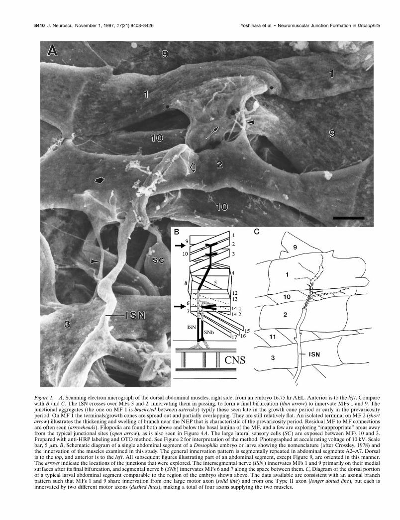
Figure 1. A, Scanning electron micrograph of the dorsal abdominal muscles, right side, from an embryo 16.75 hr AEL. Anterior is to the lef t. Comparewith B and C. The ISN crosses over MFs 3 and 2, innervating them in passing, to form a final bifurcation (thin arrow) to innervate MFs 1 and 9. Thejunctional aggregates (the one on MF 1 is bracketed between asterisks) typify those seen late in the growth cone period or early in the prevaricosityperiod. On MF 1 the terminals/growth cones are spread out and partially overlapping. They are still relatively flat. An isolated terminal on MF 2 (shortarrow) illustrates the thickening and swelling of branch near the NEP that is characteristic of the prevaricosity period. Residual MF to MF connectionsare often seen (arrowheads). Filopodia are found both above and below the basal lamina of the MF, and a few are exploring “inappropriate” areas awayfrom the typical junctional sites (open arrow), as is also seen in Figure 4A. The large lateral sensory cells (SC) are exposed between MFs 10 and 3.Prepared with anti-HRP labeling and OTO method. See Figure 2 for interpretation of the method. Photographed at accelerating voltage of 10 kV. Scalebar, 5 mm. B, Schematic diagram of a single abdominal segment of a Drosophila embryo or larva showing the nomenclature (after Crossley, 1978) andthe innervation of the muscles examined in this study. The general innervation pattern is segmentally repeated in abdominal segments A2–A7. Dorsalis to the top, and anterior is to the lef t. All subsequent figures illustrating part of an abdominal segment, except Figure 9, are oriented in this manner.The arrows indicate the locations of the junctions that were explored. The intersegmental nerve (ISN ) innervates MFs 1 and 9 primarily on their medialsurfaces after its final bifurcation, and segmental nerve b (SNb) innervates MFs 6 and 7 along the space between them. C, Diagram of the dorsal portionof a typical larval abdominal segment comparable to the region of the embryo shown above. The data available are consistent with an axonal branchpattern such that MFs 1 and 9 share innervation from one large motor axon (solid line) and from one Type II axon (longer dotted line), but each isinnervated by two different motor axons (dashed lines), making a total of four axons supplying the two muscles.
8410 J. Neurosci., November 1, 1997, 17(21):8408–8426 Yoshihara et al. • Neuromuscular Junction Formation in Drosophila
but containing 0.1 or 0.05 mM Ca 21 for an additional 1 or 2 hr (depend-ing on age of specimen) at room temperature. Specimens were thenrinsed three times in 0.1 M cacodylate or phosphate buffer from 30 min toovernight at 4°C.
Most of those specimens intended for TEM were post-fixed in 1%OsO4 in 0.1 M phosphate buffer, 0.05 mM Ca 21 for 1 hr, rinsed inphosphate buffer for 30 min, placed in 0.1 M sodium acetate, pH 5.0, for1 hr, block-stained in 1% UrAc in 50 mM NaAc in the dark for 1.5 hr,dehydrated, and embedded in Epon 812 “hard.” Minor variations in thisgeneral protocol, including omitting the UrAc block stain, were con-ducted to improve fixation and staining.
For scanning electron microscopy (SEM), after aldehyde fixation andbuffer wash, specimens were transferred gradually (over 1 hr) to distilledwater. Modifications of protocols described in Kelley et al. (1973) andRheuben and Reese (1978) were used to take advantage of the ability ofthiocarbohydrazide to enhance osmium binding to cellular organelles,the “OTO” method: after 6 changes in distilled water over at least 1 hr,specimens were placed in 1% OsO4 for 1–3 hr and then treated with asaturated solution of thiocarbohydrazide (Polysciences, Warrington, PA)in distilled water for 30 min. The sequence of distilled water washes,thiocarbohydrazide, and osmium treatments was repeated up to threetimes. In some cases, the basal lamina covering the neuromuscularjunction was removed first by collagenase digestion (1 mg/ml Type II,Sigma, St. Louis, MO) applied before fixation and again after a post-fixation treatment with 25% KOH for 5 min at 60°C. Treatment timesvaried because over- and underdigestion were highly unpredictable andoccurred even in the same sample. After osmication, samples werewashed in distilled water, dehydrated in ethanol, and either criticalpoint-dried through CO2 or infiltrated with Peldri (Ted Pella, Redding,CA) according to the manufacturer’s protocol, frozen, and placed in avacuum jar for sublimation. Most specimens were examined in the SEM
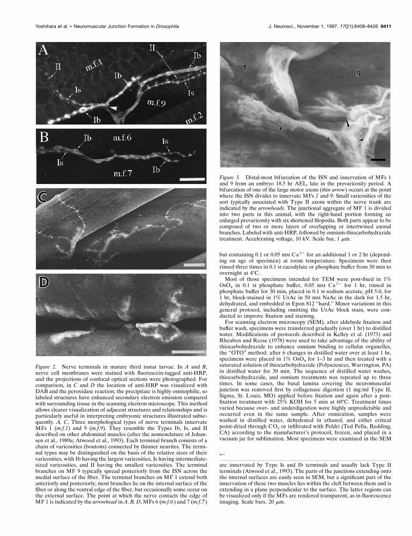
Figure 2. Nerve terminals in mature third instar larvae. In A and B,nerve cell membranes were stained with fluorescein-tagged anti-HRP,and the projections of confocal optical sections were photographed. Forcomparison, in C and D the location of anti-HRP was visualized withDAB and the peroxidase reaction; the precipitate is highly osmiophilic, solabeled structures have enhanced secondary electron emission comparedwith surrounding tissue in the scanning electron microscope. This methodallows clearer visualization of adjacent structures and relationships and isparticularly useful in interpreting embryonic structures illustrated subse-quently. A, C, Three morphological types of nerve terminals innervateMFs 1 (m.f.1) and 9 (m.f.9). They resemble the Types Ib, Is, and IIdescribed on other abdominal muscles (after the nomenclature of Johan-sen et al., 1989a; Atwood et al., 1993). Each terminal branch consists of achain of varicosities (boutons) connected by thinner neurites. The termi-nal types may be distinguished on the basis of the relative sizes of theirvaricosities, with Ib having the largest varicosities, Is having intermediate-sized varicosities, and II having the smallest varicosities. The terminalbranches on MF 9 typically spread posteriorly from the ISN across themedial surface of the fiber. The terminal branches on MF 1 extend bothanteriorly and posteriorly; most branches lie on the internal surface of thefiber or along the ventral edge of the fiber, but occasionally some occur onthe external surface. The point at which the nerve contacts the edge ofMF 1 is indicated by the arrowhead in A. B, D, MFs 6 (m.f.6 ) and 7 (m.f.7 )
Figure 3. Distal-most bifurcation of the ISN and innervation of MFs 1and 9 from an embryo 18.5 hr AEL, late in the prevaricosity period. Abifurcation of one of the large motor axons (thin arrow) occurs at the pointwhere the ISN divides to innervate MFs 1 and 9. Small varicosities of thesort typically associated with Type II axons within the nerve trunk areindicated by the arrowheads. The junctional aggregate of MF 1 is dividedinto two parts in this animal, with the right-hand portion forming anenlarged prevaricosity with six shortened filopodia. Both parts appear to becomposed of two or more layers of overlapping or intertwined axonalbranches. Labeled with anti-HRP, followed by osmium-thiocarbohydrazidetreatment. Accelerating voltage, 10 kV. Scale bar, 1 mm.
4
are innervated by Type Is and Ib terminals and usually lack Type IIterminals (Atwood et al., 1993). The parts of the junctions extending ontothe internal surfaces are easily seen in SEM, but a significant part of theinnervation of these two muscles lies within the cleft between them and isextending in a plane perpendicular to the surface. The latter regions canbe visualized only if the MFs are rendered transparent, as in fluorescenceimaging. Scale bars, 20 mm.
Yoshihara et al. • Neuromuscular Junction Formation in Drosophila J. Neurosci., November 1, 1997, 17(21):8408–8426 8411
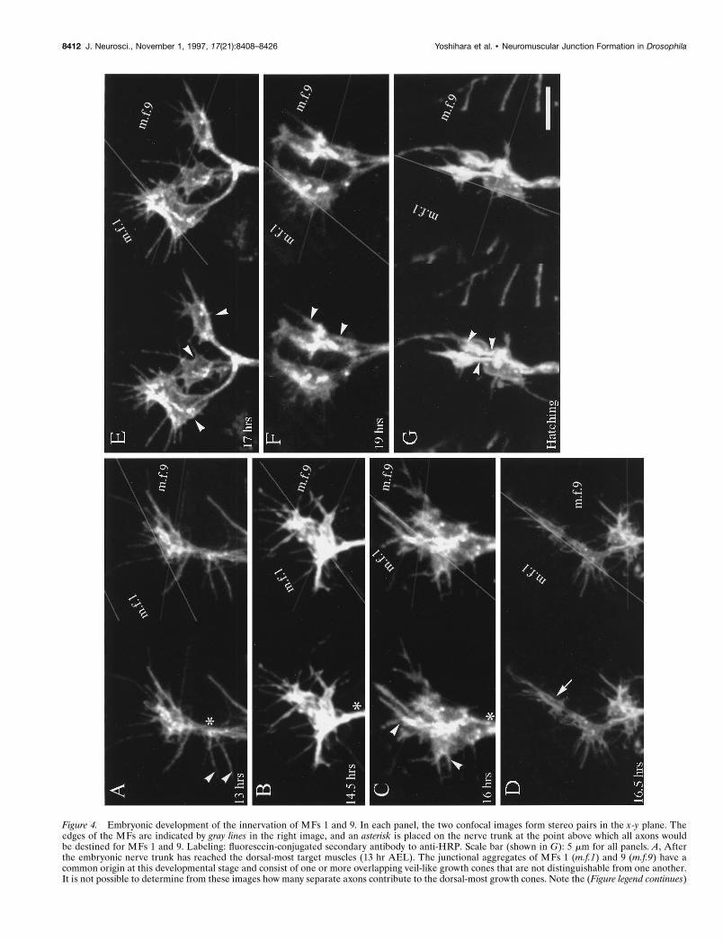
Figure 4. Embryonic development of the innervation of MFs 1 and 9. In each panel, the two confocal images form stereo pairs in the x-y plane. Theedges of the MFs are indicated by gray lines in the right image, and an asterisk is placed on the nerve trunk at the point above which all axons wouldbe destined for MFs 1 and 9. Labeling: fluorescein-conjugated secondary antibody to anti-HRP. Scale bar (shown in G): 5 mm for all panels. A, Afterthe embryonic nerve trunk has reached the dorsal-most target muscles (13 hr AEL). The junctional aggregates of MFs 1 (m.f.1) and 9 (m.f.9) have acommon origin at this developmental stage and consist of one or more overlapping veil-like growth cones that are not distinguishable from one another.It is not possible to determine from these images how many separate axons contribute to the dorsal-most growth cones. Note the (Figure legend continues)
8412 J. Neurosci., November 1, 1997, 17(21):8408–8426 Yoshihara et al. • Neuromuscular Junction Formation in Drosophila

without further treatment after drying; a few (not including those withantibody labels) were coated with a thin layer of platinum to reducecharging.
SEM specimens were photographed at 10–30 kV on either a HitachiS-800 or an S-4100 field emission microscope with lanthanum hexaboridesource in the electron microscopy facility at Gunma University, or witha JEOL 6400 at the Michigan State University Center for Electron
Optics. TEM specimens were photographed with a JEOL 100CX or aPhillips CM-10.
Antibody labeling for electron microscopy. To visualize nerve terminalmembranes more clearly in SEM, anti-HRP was used. Specimens werefixed with 4% paraformaldehyde in PBS (as described above, pH 7.2)overnight, washed in 0.5% Triton X-100 in PBS (PBT), and incubated in0.3% H2O2 in methanol for 30 min. After a rinse in PBT, specimens werelabeled with an antibody to horseradish peroxidase (goat anti-HRP,1:10,000; Cappel, Durham NC) overnight, and rinsed in PBT. An over-night incubation in biotinylated rabbit anti-goat IgG (H1L) (VectorLaboratories, Burlingame, CA) at a dilution of 1:1000 followed by anovernight incubation in an avidin–biotin complex tagged with HRP(ABC Vectastain Peroxidase Standard, Vector) resulted in amplificationof the antibody signal. Specimens were then incubated in a substratesolution containing equal volumes of diaminobenzidine (DAB) (1 mg/ml; Sigma) and 0.02% H2O2 in 0.1 M Tris-HCl, pH 7.2, to visualize thereaction product. They were then osmicated and critical point-dried forSEM as described above. Because the DAB reaction product is highlyosmiophilic, nervous tissue was more clearly outlined.
Data analysis. The types of terminals were distinguished by criteriaderived from the ultrastructural descriptions of third instar larvae pro-vided by Atwood et al. (1993) and Jia et al. (1993) and are consolidatedas follows: Type Ib (CV) terminals have 2–5 mm boutons, 44 nmclear-cored vesicles, active zones with T-shaped, branched dense bodies,and a deep subsynaptic reticulum. Type Is (CVo) terminals have some-what smaller varicosities, clear-cored vesicles of a greater range of sizes,occasional dense-cored vesicles, similar presynaptic dense bodies, and ashallower subsynaptic reticulum. Type II (MV) terminals have varicos-ities ,2 mm, a mixture of clear- and dense-cored vesicles, with the latterbeing elliptical or irregularly shaped, and they lack a subsynaptic retic-ulum. Type III (DV) terminals are thought to contain peptides such as aninsulin-like peptide on MF 12 (Anderson et al., 1988; Gorczyka et al.,1993) or a leucokinin I immunoreactive peptide on MF 8 (Cantera andNassel, 1992); they are characterized by a population of large sphericaldense-cored vesicles and clusters of small, 33 nm clear vesicles at whatappear to be active zones (Jia et al., 1993).
RESULTSThe body wall muscles of Drosophila larvae are composed ofsingle identifiable fibers arranged in a segmentally repeated man-ner (Hertweck, 1931; Crossley, 1978; Bate, 1990). The peripheralnervous system (PNS) innervating these muscles also follows asegmentally repeated pattern, with no apparent differences be-tween abdominal segments A2 through A7 throughout the em-bryonic stages (Fig. 1).
Each of the abdominal MFs is innervated by a consistent set ofaxons, which form junctions with shapes and locations specific toa given fiber. These have been divided by morphological criteriainto Type I junctions, with one or more linear chains of relatively
Figure 5. Scanning electron micrograph of an anti-HRP-labeled junc-tional aggregate at the NEP on MF 1 from an embryo 17 hr AEL, duringthe prevaricosity period. Processes from two or three of the innervatingaxons overlie one another. The filopodia are shorter than those seenduring the growth cone period, and some axonal branches have begun toenlarge at the NEP. Because the animal was dissected and fixed flat as a“fillet” for SEM, as were the animals examined confocally in Figure 4, wefind that the ISN is pulled ventrally and part of the MF membrane(arrows) is pulled with it. The plane of section for the junctional aggregateseen in TEM in Figure 6 is indicated by a line. Scale bar, 1 mm.
4
long filopodia, including some (arrowheads) that originate below the point at which the junctions typically form on MFs 1 and 9. The long filopodiumat the lower right of the ISN belongs to a growth cone on MF 2, the remainder of which is out of view. B, The junctional aggregates appear structurallymore complex (14.5 hr AEL). Portions of the growth cones innervating MFs 1 and 9 are spreading to both the internal and external surfaces of MF 1,as indicated by the two very different planes that can be seen in the stereo pair. Regional thickenings or portions that are more heavily labeled can bedistinguished, seeming to condense from the more evenly labeled growth cone of earlier stages. C, Branches or divisions of the growth cones (16 hr AEL)are extending both over the internal surface of MF 1 and along its external surface in the cleft between MF 1 and MF 9 (which lies more lateral to MF1). The divisions of the junctions that typically extend from the NEP both anteriorly and posteriorly on MF 1 appear to be forming (arrowheads). Thejunctional aggregate of MF 9 typically spreads posteriorly from the ISN. D, The distinction between the junctional aggregates of MFs 1 and 9 is nowdiscernible because of the increase in length of the connecting portion of the ISN (16.5 hr AEL). A small cylindrical thickening is visible in a posteriorbranch on MF 1 (arrow). Overall, the impression at this stage is that the planar growth cone is condensing to form thicker, branch-like structures, butnumerous filopodia are still present. E, There are three separate regions (arrowheads) in which terminals have formed noticeably thicker, three-dimensional structures (17 hr AEL). Only the surface membrane is labeled, so the impression of hollow structures is given. On the anterior (lef t) growthcone on MF 1, the stereo pair shows what could either be a large flattened structure or two separate thinner structures lying on top of each other. Electronmicroscopic observations of this stage indicate that both interpretations are possible. We refer to the single enlarged regions of a terminal asprevaricosities. F, G, By 19 hr AEL (F), and continuing through hatching (G), the enlarged structures along terminal branches began to have moreclear-cut constrictions on either side of them. Their shapes ranged from tubular to spherical. We view this process as giving rise to the first realvaricosities of the developing terminal. The dimensions of the individual spherical structures were typically less than those of the prevaricosities. Multipleterminal branches appear to be present. The arrowheads in each panel point to much thinner terminal branches, which are superimposed on the largervaricosities; these could be either Type Is or II, if, as might be expected, the largest varicosities are from Ib terminals. Filopodia are still present but tendto be somewhat fewer in number and shorter relative to the dimensions of the enlarged thicker regions. This transition is diagrammed in Figure 7C,D.
Yoshihara et al. • Neuromuscular Junction Formation in Drosophila J. Neurosci., November 1, 1997, 17(21):8408–8426 8413

large varicosities, a distinctive subsynaptic reticulum (set of highlyconvoluted folds) formed by the MF around the terminal, andmuscle-specific branching patterns, and Type II junctions, withlong trailing branches composed of very small varicosities, andwhich lack a subsynaptic reticulum (Johansen et al., 1989a; At-wood et al., 1993; Jia et al., 1993). The Type I junctions, wherethey have been investigated, are thought to use glutamate as aprimary neurotransmitter (Jan and Jan, 1976; Johansen et al.,1989a). The Type II junctions include octopamine, and all theterminals of this type in one hemisegment are derived frombranches of only two neurons (Monastirioti et al., 1995). A thirdgeneral type, Type III, includes terminals with peptidergic trans-mitters, one example of which is immunoreactive for an insulin-like peptide and is known only to innervate MF 12 in segments
2–5, and has more oval-shaped boutons than Type II terminals(Gorczyka et al., 1993).
Certain of these MFs have been studied more extensively thanothers. In particular, the very large MFs 6 and 7 (nomenclatureafter Crossley, 1978; Bate, 1990) are usually innervated only bytwo Type I terminals, which have been subdivided into Ib and Ison the basis of morphological criteria (Ib boutons are larger) andfunctional differences (Ib terminals produce smaller excitatoryjunction potentials) (Atwood et al., 1993; Kurdyak et al., 1994).These two terminal types are thought to be derived from twoseparate neurons: RP3 and 6/7b (Sink and Whitington, 1991;Keshishian et al., 1993).
We examined the normal development of the nerve terminalsinnervating the dorsal longitudinal fibers 1 and 9 and compared
Figure 6. Junctional aggregates of MFs 1 and 9 at the prevaricosity stage (17 hr AEL). The animal was not filleted but fixed by perfusion through thetail end, so the nerves and muscles are lying in their natural positions. A, B, and C are sections 12, 32, and 52, respectively, from a series. Ep, Epidermalcell; Tr, main dorsal tracheole; g, parts of glial process on the distal part of ISN. A, This section passes through the center of the NEP for MF 9 (mf 9)and just off center for the NEP on MF 1 (mf 1). Notice the swellings in the axonal profiles at the NEPs compared with their diameter at the dorsal edgeof MF 2, at the lef t of the micrograph. These enlarged profiles can be compared with the prevaricosities shown in Figures 1, 3, and 4. Three overlappingaxonal branches are found in the NEP of MF 9 in this plane of section. Axon 3 (numbers applied solely for identification in this animal) is bifurcatingat this point (open arrows), with one branch following the axons that are headed toward MF 1 and one branch continuing eventually to the surface ofMF 9. Elementary synapses are seen at the contact points of axons 1 and 4 with their respective muscles (arrows). A profile of a filopodium from a growthcone lying more dorsally (out of the field of view) next to MF 1 is indicated with an arrowhead. One edge of the glial covering of the ISN is indicated(g), but the axons and terminals in this region are largely naked. At higher magnification it can be determined that the nerve passing over MF 9 en routeto MF 1 actually consists of several axonal branches cut very obliquely. Scale bar, 1 mm. B, NEP on MF 9, anterior to 6A. Axon 1 has formed a synapsewith MF 9 (arrow), and axon 2 has formed a dense body in apposition to axon 1 (open arrow). Axon 2 branches on both sides of another axonal profile.Note the difference in synaptic vesicle size between axon 1 and axon 2. Scale bar, 0.5 mm. C, Anterior edge of NEP on MF 9. Axon 1 remains in contactwith MF 9; the other axonal branches have turned laterally and are now seen in cross section (asterisks). Compare with Figure 5 to visualize. Focalcontacts and elementary synapses are formed by axon 1 with MF 9 (arrows). Scale bar, 0.5 mm. Figure continues.
8414 J. Neurosci., November 1, 1997, 17(21):8408–8426 Yoshihara et al. • Neuromuscular Junction Formation in Drosophila

the results with those we obtained concurrently from MFs 6 and7 in segments A2–A7, as well as with those described previouslyfor MFs 6 and 7 (Broadie and Bate, 1993). The terminals on MFs1 and 9 are conveniently accessed in the embryo for electrophys-iological studies. Their terminals, unlike those of MFs 6 and 7,are well spread in the x-y plane and easy to observe in structuralstudies (Fig. 1A). MFs 1 and 9 have two types of terminals, withrelatively large boutons that seem to correspond structurally toTypes Ib and Is as described for MFs 6 and 7, as well as Type IIterminals, (Fig. 2, A and C, for MFs 1 and 9, B and D for MFs 6
and 7), a pattern that is characteristic of most of the otherabdominal segment fibers. In this study we have emphasized thelatter part of embryological development to provide a solid base-line for future studies of mutants with defects in their neuromus-cular junctions, and that, therefore, would be less able to survivepast hatching.
Mature innervation of MFs 1 and 9The terminals innervating MFs 1 and 9 are the most dorsal ineach segment and are at the distal end of the ISN. Their supply-
Figure 6 continued.
Yoshihara et al. • Neuromuscular Junction Formation in Drosophila J. Neurosci., November 1, 1997, 17(21):8408–8426 8415

Figure 7. A–D, Diagrammatic representation of steps in development of the junctional aggregate on a single MF. A, Growth cone stage. During thegrowth cone stage, the developing terminal is thin and flat, with long filopodia. The growth cone from a single axon is illustrated. However, it oftenappears that toward the end of the growth cone stage two or more growth cones are overlapping in the vicinity of the NEP. B, Prevaricosity stage. Theprevaricosities formed by a single axon are shown for simplicity. The growth cone condenses into several recognizable branches that have distinctthickness and rounded contours. Filopodia are shorter. Simple presynaptic specializations form along broad contact regions with the MF. C, Junctionalaggregate at the prevaricosity stage, ;16.5–18.5 hr AEL. During the prevaricosity stage, several terminals of differing degrees of development are usuallyfound overlapping at the NEP. In this example, a growth cone is shown slightly diverging to the lef t, and two overlapping terminals with prevaricositiesare to the right. Many additional configurations have been observed. When terminals have entered at the same point, they often remain spatially close,with membrane-to-membrane contact for some distance away from the NEP. This leads to a very complicated appearance when seen at either the lightmicroscope or SEM levels. The bracket indicates the regions that were measured to quantify prevaricosity formation in E. D, At 18–19 hr AEL, distinctvaricosities, swellings with constrictions on either side, resolve from the enlarged branches of the prevaricosity. A single swelling may divide itself intotwo or three discrete varicosities. Filopodia are shorter, the elements of the SSR begin to separate the broad nerve–muscle contact regions, and individualbouton types can begin to be recognized. Subsequent development between this stage and first instar is a matter of degree of development of individualvaricosities. E, Time course of prevaricosity formation. The percentage of junctions having prevaricosities and/or layered structures with a total thickness.2 mm was determined for each stage for MFs 1 and 9 (m.f. 1 & 9) and for MFs 6 and 7 (m.f. 6 & 7 ). The dimension of 2 mm was an arbitrary criterionto mark prevaricosity formation, with some individual prevaricosities being larger and some being smaller than this size. By 17 hr AEL nearly all junctionson MFs 1 and 9 included prevaricosities by subjective criteria, but not all reached the 2 mm thickness criterion. Subjectively, in a given animal or at agiven age, the swelling of the prevaricosity seemed to be greater in the more ventral muscles than in the more dorsal ones (Figs. 1, 4, 11). To quantifythis difference, the 2 mm criterion was applied to MFs 6 and 7 in the same animals. There appears to be a delay of ;15–30 min in the initiation of theprocess from dorsal to ventral muscles. For MFs 1 and 9, the total number of junctions measured at 15.5 hr AFL was (Figure legend continues)
8416 J. Neurosci., November 1, 1997, 17(21):8408–8426 Yoshihara et al. • Neuromuscular Junction Formation in Drosophila

ing nerve trunks are therefore not complicated by either axontracts innervating other muscles or ingrowth of sensory axons(Campos-Ortega and Hartenstein, 1985). One of the neuronsinnervating MF 1 is the aCC neuron (Sink and Whitington, 1991;Van Vactor et al., 1993), but the others innervating MFs 1 and 9have not yet been identified. Anti-HRP labeled preparations,both at the light microscope level and with SEM (Fig. 3), showthat one of the large axons consistently bifurcates at the dorsaledge of MF 2 (Fig. 3, thin arrow), sending one branch to MF 1and one to MF 9. Perhaps this shared innervation is derived fromone of the U neurons, analogous to the U neuron innervatingboth MF 2 and MF 10 (Sink and Whitington, 1991). A small-diameter axon is also seen to bifurcate at the same level; this axonformed varicosities within the nerve trunk as well as on the MFsthemselves, similar to the pattern illustrated for the two octopam-inergic neurons per hemisegment that provide Type II innerva-tion to many of the fibers in the body wall (Monastirioti et al.,1995). Two large-diameter axons consistently proceed separatelyto MF 1 and MF 9. Sections through the ISN over the center ofMF 2, before this bifurcation point, typically show the presenceof four axons. This working hypothesis of the innervation patternof MFs 1 and 9 is illustrated in Figure 1C. It remains to beconfirmed by single cell injections or other more specific methods.
We refer to the cluster of different terminals on each fiber, twoof which may be excitatory, as in MFs 6 and 7, and one or moreof which may have neuromodulatory properties, as a “junctionalaggregate,” to distinguish the case from the situation in verte-brates in which one motor neuron forms a single neuromuscularjunction per MF. Most insects have muscles that are multitermi-nally innervated, with evenly spaced terminals all along eachfiber, as well as muscles that are multineuronally innervated(Hoyle, 1955; Usherwood, 1974; Rheuben and Reese, 1978;Schaner and Rheuben, 1985), but the single cluster of largejunctions found on Drosophila larval muscles is relatively unusual,presumably arising from the electrically very short MFs, whichmake multiterminal endings unnecessary. The point at which thenerve branch first contacts the MFs is described as the NEP.
Development of the terminals innervating MFs 1 and 9The development of the synaptic terminals innervating MFs 1and 9 was examined using both ultrastructural and confocal lightmicroscopic methods. The antibody to horseradish peroxidase,anti-HRP, which recognizes a carbohydrate with which severalglycoproteins in the membranes of insect neurons are decorated(Snow et al., 1987; Katz et al., 1988), was used to demonstrate theembryonic axons and terminals more clearly, and an antibody tosynaptotagmin was used as a monitor of intracellular synapticdevelopment. Confocal microscopy with fluorescent-labeled pri-mary or secondary antibodies and stereo pairs made it possible tostudy the spatial distribution of terminals and allowed terminalson both sides of an MF to be visualized. Furthermore, one couldmake the large number of preparations needed to assess devel-opmental time courses using this method. Scanning electronmicroscopy provided high magnification overviews of the termi-nals and allowed us to visualize the structures that the terminals
were in contact with. Thin sections provided identification andclarification of the subcellular characteristics of these developingterminals.
Growth cone stageIn a series of timed embryos whose PNSs were observed withfluorescein-labeled anti-HRP and confocal microscopy, we foundthat one or more of the growth cones of the ISN consistentlyreached the vicinity of MFs 1 and 9 by ;13 hr AEL, as describedpreviously (Johansen et al., 1989b; Sink and Whitington, 1991;Van Vactor et al., 1993).
At 13 hr AEL, at least one growth cone extended numerouslong filopodia over much of the surfaces of each of these dorsalMFs, including regions in which junctions were not expected toform (Fig. 4A). Subsequently, the middle planar region of thegrowth cone expanded on the surface of its target MF, largelyover the future junctional site, and the origins of filopodia weremore restricted to this region (Fig. 4B). In some cases the growthcones and filopodia appeared to be adhering primarily to theinternal surfaces of both MF 1 and MF 9. In other cases, parts ofgrowth cones also invaded the cleft formed by the external sur-face of MF 1 (dorsolateral in the living embryo) and the internalsurface of MF 9 (Fig. 4B). This fits with observations on maturelarvae in which some terminal branches on MF 1 reach around tothe external side, whereas the rest lie on the internal surface.Although the MFs themselves are not visible in the imagesformed with fluoroscein-labeled anti-HRP, they could be seenconcurrently with differential interference contrast optics; theyare indicated schematically on the figures by gray lines. Thedifferent growth cone locations can be seen by using the three-dimensional imaging provided by the stereo pairs.
This period, in which the middle region of the growth cone wasflat and the filopodia were long, lasted for ;3.5 hr. The form ofthe growth cones appeared to be as labile as that observedpreviously in the segmental nerve with time lapse photography(Keshishian et al., 1993), in which filopodial extensions and re-tractions of 15 mm occur over a matter of minutes. Althoughtransmitter release has been shown to occur during this period(Broadie and Bate, 1993; Nishikawa and Kidokoro, 1995), pre-sumably few permanent morphological release sites have formedbecause of the labile structure. There was little overall change incharacteristic structure at the light microscope level during thistime, and we refer to this period as the “growth cone period.”
Toward the end of the growth cone stage, strand-like structureswere seen in conjunction with some of the broad, sheet-likelamellipodia, and there was an impression of greater complexityin both confocal and SEM images (Figs. 1A, 4C). This apparentcomplexity could arise either from the superimposition of thelamellipodia of one growth cone over the filopodia of a second, orfrom multiple branches of the same growth cone. Because mostearlier studies were made by either injecting the cell body of theaCC neuron (Sink and Whitington, 1991) or labeling all ofthe axons and growth cones with anti-HRP, as we have done, thepresence of the growth cones of other axons innervating MFs 1and 9 has not yet been determined.
4
n 5 23 (4 animals); 16.0 hr, n 5 42 (8 animals); 16.5 hr, n 5 56 (10 animals); 17.0 hr, n 5 46 (6 animals). MFs 6 and 7, at 15.5 hr AEL, n 5 22; 16 hr,n 5 46; 16.5 hr, n 5 56; 17 hr, n 5 36, respectively, for the same animals. The fraction of terminals reaching prevaricosity stage was tabulated for eachanimal; the points plotted represent the mean of these values 6 SEM. F, Decrease in filopodial length with increasing age. The lengths of filopodia ofthe terminals innervating MFs 1 and 9 were measured from projected images along the z axis for three terminals at each age. At 13–14.5 hr AEL, somevery long filopodia, .10 mm, are present; by hatching, all the filopodia are ,6 mm. The arrows indicate the average filopodial length at each stage.
Yoshihara et al. • Neuromuscular Junction Formation in Drosophila J. Neurosci., November 1, 1997, 17(21):8408–8426 8417
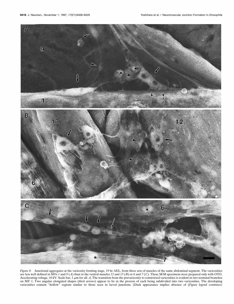
Figure 8. Junctional aggregates at the varicosity forming stage, 19 hr AEL, from three sets of muscles of the same abdominal segment. The varicositiesare less well defined in MFs 1 and 9 (A) than in the ventral muscles 12 and 13 (B) or 6 and 7 (C). These SEM specimens were prepared only with OTO.Accelerating voltage, 10 kV. Scale bar, 1 mm for all. A, The transition from the prevaricosity to constricted varicosities is evident in two terminal brancheson MF 1. Two angular elongated shapes (thick arrows) appear to be in the process of each being subdivided into two varicosities. The developingvaricosities contain “hollow” regions similar to those seen in larval junctions. (Dark appearance implies absence of (Figure legend continues)
8418 J. Neurosci., November 1, 1997, 17(21):8408–8426 Yoshihara et al. • Neuromuscular Junction Formation in Drosophila

We examined a series of embryos for evidence that the multipleinnervation of MFs 1 and 9 was present by the late embryo stage.In thin sections, at 15.25 hr AEL, growth cone-like nerve struc-tures (vacuolated unspecialized cytoplasm, ruffled profiles) werefound over many of the body wall muscles in rather loose associ-ations. At the NEP, two or three slightly enlarged axonal profileswere superimposed. Distinguishing cytoplasmic specializationswere lacking, and processes spreading away from this point werenot tightly adherent to the muscle.
By 16 hr AEL, serial thin sections of the ISN over MF 2 showedfour axons that would all be destined for MFs 1 and 9 at thatpoint. This would represent the full complement of the innerva-tion for the two muscles if none were secondary branches. Inembryos older than 15 hr AEL, most body wall muscles, includingMFs 1 and 9, were contacted by two or more axons, and theseaxon terminals were structurally distinguishable from each otherby 16–17 hr AEL. Adjacent terminals on the same MF differed invesicle type and cytoplasmic inclusions and were derived fromseparate axonal branches. Terminals with the vesicle compositionand morphology of Type I axons were present, as well as thosecontaining irregularly shaped dense-cored vesicles characteristicof Type II terminals. Consequently, we conclude that by the endof the growth cone period, multiple and possibly all axons arerepresented in the junctional aggregate as growth cones or asdeveloping terminal structures.
Prevaricosity stageAt 16.5 hr AEL, the axons and planar central regions of thegrowth cones suddenly began to enlarge and increase in thicknessat the site of the future NEP for each MF (Figs. 1A, 3, 4D).These enlarged regions reached up to 5 mm in length and 1–3 mmin thickness and might be subdivided into two or more “hollow-appearing” structures. The hollow appearance arises from the useof a labeling antibody that is recognizing membrane-bound anti-gens, leaving the center of the terminal unstained. We refer to theenlarged structures as “prevaricosities.” Their shapes were asvariable as those of the growth cones. Some were long andellipsoid, whereas others were tubular or polygonal with distinctangles (Figs. 3, 4D, 12B). Overall their outline and positionseemed to reflect that of the original growth cones before thethickening occurred. By 17 hr AEL, almost all terminals had oneor more prevaricosities, and several might be grouped at the NEPto form a quite thick structure (Fig. 4E). Sometimes an apparentventral-to-dorsal gradient in degree of development could berecognized within a single abdominal segment (Fig. 1) (see com-parison with MFs 6 and 7 described below).
SEM images of the junctional aggregates from animals 16–18.5hr AEL also showed a complicated structure, with layered inter-twined processes radiating outward from the NEP (Fig. 5). Evi-dently part of the increase in thickness in the confocal images wasattributable to axonal processes lying one on top of another. Inaddition, individual processes often had an enlarged region (;1mm in cross section) at the NEP, and which then divided into
smoothly tapered small diameter branches (0.2 mm) that contin-ued across the surface of the fiber (Figs. 1, 3). The enlargedregions seemed to correspond to the prevaricosities seen confo-cally and described in the previous paragraph, and the small-diameter branches seemed to be residual filopodia.
To elucidate this complicated structure, serial sections weretaken through parts of the NEPs and junctional aggregates ofMFs 1 and 9 from two embryos within the prevaricosity period.Sections from the oldest, 17 hr AEL are illustrated in Figure6A–C. The following features were seen in both animals. (1) Twoor more axons formed enlarged bulbous regions as they ap-proached the NEP of each MF. These axons could be distin-guished from each other throughout the series on the basis oftheir cytoplasmic inclusions and type of vesicles, but they couldnot always be assigned to a particular mature type such as Is or Ib(Fig. 6A). (2) At the NEPs of both MF 1 and MF 9, one of theenlarged axons typically formed a broad uninterrupted contactwith the MF. This contact region housed 10 or more immaturesynaptic specializations. The vesicle type and dense body struc-ture were consistent with its being a Type I terminal. (3) Theenlarged region of the second axon, which had larger and moreirregularly shaped clear-cored vesicles as well as dense-coredvesicles, (Type II?) typically lay on top of the first axon at theNEP. Four examples were encountered in which it formed asynapse-like structure in apposition to the first axon (Fig. 6B).This second axon was not seen to form synapses in apposition tothe MF within the available sample of sections. (4) At the NEP ofMF 9, a third axon was typically found outside the second. Itbifurcated and one branch was directed toward MF 1, whereas asecond was traced to the surface of MF 9. It appeared to be fromthe other Type I axon. (5) Processes with the morphologicalcharacteristics of growth cones were also seen in the vicinity ofMFs 1 and 9 at this time. They were not successfully traced to aspecific parent axon, so their origin is uncertain.
We conclude that axons with phenotypically differentiatedcharacteristics are forming terminals by the prevaricosity stage.Their swelling and superimposition results in the dramatic in-crease in thickness of the junctional aggregate seen at this time.The transition from the growth cone stage to the prevaricosity ispresented diagrammatically in Figure 7A–D.
To assess the time course of prevaricosity formation, the thick-ness of the overall structure was assessed using z-axis measure-ments of the confocal images. The growth cone was essentially aplanar structure, whereas .60% of the prevaricosities, or the jointstructures formed by layers of prevaricosities, growth cones, andfilopodia, had a thickness .2 mm measured in the vicinity of theNEP. Using this dimension as an arbitrary cutoff point betweenthe growth cone stage and the prevaricosity stage, we counted thenumber of segments that had terminals or layers of terminals .2mm in thickness at the NEP of MFs 1 and 9, from 15 to 17 hr AEL(Fig. 7E). The abrupt increase in the numbers of thick structuresfrom 16 to 17 hr AEL is evident.
4
osmiophilic structures such as organelles or synaptic vesicles.) In addition to major divisions, each terminal branch of an apparent Type Ib axon continuesto extend thin filopodial or sprout-like processes to the MF. The strong adhesive nature of the nerve–muscle contacts (thin arrows) is evident becauseof the tension placed on the ISN. Type II axons form varicosities within the nerve by this time (arrowheads). The third axon, presumably Is, formsbranches on the lateral side of the MF that are not seen here. Spherical, granular-appearing cells, which may be the persistent twist cells (asterisks), areoften seen at the NEP of MF 1. B, On MFs 12 and 13 it is possible to recognize several different terminal types by 19 hr AEL. Two or three large TypeIb varicosities are found on each fiber (thick arrows) as well as varicosities of other types (thin arrow and arrowheads). C, On MFs 6 and 7, large and smallvaricosities (thick and thin arrows) have formed in the region of adherence between the two fibers. Other approximately spherical structures (stars) withinthe MF can be distinguished from varicosities on the basis of their focal plane and absence of connecting neurites.
Yoshihara et al. • Neuromuscular Junction Formation in Drosophila J. Neurosci., November 1, 1997, 17(21):8408–8426 8419

Figure 9. Junctional aggregates on MFs 1 and 9 at 19 hr AEL. A, B, C, Serial sections 231, 242, and 254, respectively. The embryo was dissected flatfor fixation, so the tension on the ISN has pulled the NEP on MF 1 toward MF 2. Scale bars, 0.5 mm. A, On MF 9 (mf 9), axon 1 has formed a sphericalvaricosity that is almost completely wrapped by two other terminals (2, 3). In this plane of section, axon 2 has formed a synapse in apposition to axon1, and axon 3 has formed electron-dense specializations in apposition to a thin arm of the MF. The vesicle sizes in axon 1 were more uniform and smallerthan those seen in axon 2. Axon 2 had numerous dense-cored vesicles elsewhere in the series. A growth cone was part of the aggregate in another plane(not shown). At this level, numerous small tangled profiles were seen on MF 1; they included branches from axon 2 just seen on MF 9, and from a growthcone-like structure as well as an axon that formed Type I varicosities in apposition to the MF. B, Axon 1 on MF 9 (mf 9) (Figure legend continues)
8420 J. Neurosci., November 1, 1997, 17(21):8408–8426 Yoshihara et al. • Neuromuscular Junction Formation in Drosophila

Varicosity stageAfter 17 hr AEL, another structural transition was observed. Thelarge prevaricosities began to appear constricted into severalsmaller swollen regions. By 19 hr AEL, some of these constricted
regions had the spherical appearance of the individual varicositiesor boutons as seen in first instar and older larvae. (Hatchingoccurred at 21 hr AEL at 25°C under these culture conditions.)Between 19 hr AEL and hatching, some of the enlarged structuresdeveloped a series of dark-appearing central regions in SEM,reflecting an absence of osmiophilic structures in the centers ofthe terminals (Fig. 8). Others continued to look like tubes sepa-rated by constrictions, or they formed other irregular shapes. Thesize of a single large approximately ovoid varicosity, derived froma prevaricosity near the NEP, was ;2 mm in width and 3–4 mmin length, which is within the range of the Type I boutons in thirdinstar larvae (Atwood et al., 1993). Subjectively, there againappeared to be a small ventral-to-dorsal gradient in the develop-ment times of the varicosities (Fig. 8), but this was not quantified.
The NEP retained a complex form during the varicositystage. Multiple layers of axon and terminal branches were stillsuperimposed (Fig. 9). Most of the long filopodia have beenretracted by this time, but shorter filopodia, sprouts, and sidebranches continued to arise from both the varicosities and theneurites between varicosities (Figs. 4 F, 8). The average lengthsof filopodia gradually decreased (Fig. 7F ). At 13 hr AEL, someterminals had filopodia .10 mm in length, and there was agreat variation in their lengths. In later stages, the distributionof lengths shifted to an average of ,5 mm. From 19 hr AELthrough hatching, slender filopodia-like structures were stillpresent in the distal parts of the terminals (Figs. 4 F, G, 10 A).With confocal or SEM microscopy on fixed tissue, it is notpossible to determine whether these should be consideredfunctionally as filopodia or neurites. They presumably remainto add new varicosities as growth of the MF continues. Com-monly two or more terminal branches, including those havingvaricosities, remained intimately intertwined for some dis-tance as they crossed the surface of the MF (Fig. 10 B andTEM sections not illustrated). This configuration differs fromthat illustrated for third instar larvae (Atwood et al., 1993; Jiaet al., 1993), in which terminals are separated from each otherby a region of subsynaptic reticulum at the very least. Somevery slender “stick-like” terminal structures were seen alongwith the varicosities forming after 19 hr (Fig. 4 F). Theselacked obvious varicosities and may be the early form of TypeII terminals. In TEM, Type II terminals were clearly identifiedby their vesicle composition and morphology on MFs 1 and 9by 16 hr AEL.
Development of the terminals innervating MFs 6 and 7We compared the developmental time course of the terminalsinnervating MFs 6 and 7 with that observed for MFs 1 and 9,looking for both quantitative differences that might arise from thegreater distance that the ISN axons have to grow or qualitativedifferences that might arise from properties of the different axonsthat innervate these muscles. The terminals on MFs 6 and 7developed in a manner similar to those of MFs 1 and 9 (Fig. 11).During the early stages, the growth cone or growth cones spreadalong the cleft between the two fibers (Fig. 11A), where theyformed a planar structure that was generally perpendicular to theplane being viewed, and which was often directly superimposed
Figure 10. Varicosity formation, MF 9. At 19 hr AEL, the more distalregions (A) of nerve terminals have irregular sprout-like shapes; varicosi-ties are formed in the neurites closest to the NEP ( B). In both regions theprocesses from one or more different axons are typically intimately inter-twined, with membrane-to-membrane contact (verified by TEM views; seeFig. 9A,B), which is maintained over long distances as seen here. This isdifferent from the mature larval form in which Types Ib and Is are at leastseparated from each other by layers of subsynaptic reticulum. The basallamina was removed by treatment with 25% KOH for 2 min at 60°C afterfixation, followed by osmium-thiocarbohydrazide. Scale bars, 0.5 mm.
4
formed a total of eight presynaptic active zones of the multi-branched type in apposition with the MF in this and subsequent sections through thisvaricosity (0.7 3 1.0 3 1.7 mm), and three more in a second smaller one. The varicosity (axon 4) shown on MF 1 (mf 1) was 1.7 3 1.0 3 1.1 mm andhoused six active zones; in this grazing section their multipronged nature can be seen. C, At the edge of the NEP, a single relatively unspecialized profileof axon 4 continues across the surface of MF 1.
Yoshihara et al. • Neuromuscular Junction Formation in Drosophila J. Neurosci., November 1, 1997, 17(21):8408–8426 8421

on the terminals innervating MFs 14–1 and 14–2. Subsequently,portions of the growth cones swelled to form prevaricosities (Fig.11B,C). After this period, the prevaricosities were constricted toform a series of more typical varicosities (Fig. 11D). Overall,these processes appeared to be quite similar to those observed onMFs 1 and 9.
The time course of development for MFs 6 and 7, however,seemed to be slightly earlier than for MFs 1 and 9. This wasobserved subjectively in segments in which MFs 6 and 7 hadalready formed several distinct varicosities or prevaricosities,whereas the dorsal muscles in the same segment had not reachedthe comparable stage (Fig. 8). Objectively, prevaricosity forma-tion was detectable in MFs 6 and 7 at 16 hr AEL, using theminimum dimension of 2 mm in thickness (width in the x-y plane)as a criterion (Fig. 7E). This was about 0.5 hr earlier than for MFs1 and 9.
The time of varicosity or bouton formation that we are report-ing is later than that reported by Broadie and Bate (1993a), butthis relates partly to differences in the application of the term“bouton” to enlargements of the developing terminal. In addition,
we found it important to examine junctions in three dimensions toexclude the terminals of MFs 14–1 and 14–2 in quantitativeanalyses, and because occasionally single confocal images thatappeared to show a varicosity were misleading.
An artifactual swelling was seen at the NEP on occasion. It wasobserved in our specimens on the side of the animal on which thelateral incision was made, cutting the ISN roughly at the level ofMF 4. This balloon-like structure could be distinguished from theprevaricosity because it had a nearly perfectly spherical shape,and its membrane often had a much weaker density of anti-HRPlabel, suggesting that it was swollen and stretched. In addition, adirect comparison between the cut side and the contralateralsegment was quite striking.
Functional significance of the prevaricosityThe transition from growth cone to the varicosity stage wasexamined for maturity of functioning organelles. The develop-ment of a synapse-specific structure, namely a high density ofsynaptic vesicles, was examined with an antibody to synaptotag-min. Synaptotagmin is a membrane protein of synaptic vesicles
Figure 11. Prevaricosity formation was compared inthe terminals innervating MFs 6 and 7. A, At 15 hrAEL, the stereo pair illustrates growth cone lamellaeand filopodia, which are spreading both upward overthe surface of MF 6 (m.f.6 ) as well as along the cleftbetween MFs 6 and 7 (m.f.7 ). In the cleft, part of thejunction appears as a vertical plate-like structure, withsubdivisions that extend directly into and out of theplane of optical section. In the x-z image (bottom lef t inA), the upwardly directed filopodia and the verticalextent of the growth cone are illustrated. The locationof the x-z section, in the center of the cleft betweenMFs 6 and 7, is indicated by the pair of arrows. It is notpossible in the confocal images to determine whetherapparent subdivisions of the growth cone are derivedfrom different axons. B, Beginning of prevaricosity for-mation (16 hr AEL). In this stereo pair the image of theinnervation of MFs 6 and 7 is superimposed on that ofsubsequent branches of SNb (asterisks) that innervateMFs 14–1 (m.f.14–1) and 14–2 (m.f.14–2) (Bate, 1990).A tubular prevaricosity belonging to MFs 6 and 7 isindicated by an arrowhead. The cylindrical nature of theprevaricosity is shown more clearly in a single x-zsection, bottom lef t (B), and an x-y section, bottom right(B), (arrowheads). Thinner terminals with small vari-cose regions (thick arrows) may lie on top of this pre-varicosity; in addition, however, small “spots” of in-creased fluorescence are seen where small processesemerge from the prevaricosity perpendicular to theplane of optical section. The pairs of thin arrows indi-cate the planes of section of the x-z and x-y images. C,In this junctional aggregate on MFs 6 and 7 (17 hrAEL), two large prevaricosities are seen (arrowheads).Additional thinner terminal branches with small swell-ings (arrow) also appear to be present and are closelyintertwined with the prevaricosities. It is possible thatthese are terminal branches from the second axon,which is known to be present at this time from physio-logical findings (Broadie and Bate, 1993). Note, how-ever, that the bases of filopodia also give rise to smalldots of increased fluorescence, which at first glance mayappear to be small varicosities (above lower arrowhead).D, Hatching, 21 hr AEL. Six large varicosities arepresent in the cleft between MFs 6 and 7 (arrowheads),as well as some smaller ones. Note that the varicositiesare smaller than the enlarged swellings shown in C.Asterisk indicates the 14–1, 14–2 junctions. The stereopairs are helpful to exclude their varicosities from anycounts of the varicosities of the 6–7 junction. Scale bar(shown in D): 5 mm for all panels.
8422 J. Neurosci., November 1, 1997, 17(21):8408–8426 Yoshihara et al. • Neuromuscular Junction Formation in Drosophila

and has been detected in Drosophila by 15 hr AEL in the flatgrowth cone (Littleton et al., 1993, 1995), as well as in laterinstars.
We examined the development of immunoreactivity to synap-totagmin from 15 hr AEL to hatching. Immunoreactivity was lowduring the flat growth cone stage but increased greatly in MFs 1and 9 at 16.5 hr AEL (Fig. 12B). This increase was synchronouswith the formation of the prevaricosity. The synaptotagmin label-ing in the prevaricosity appeared in distinct patches locatedwithin the enlarged regions of the terminal near the musclemembrane. The discrete patches persisted into the stage of var-icosity formation, with the density of each patch, as well as theirnumber and size, increasing (Fig. 12C). The development of theaxonal or terminal swelling at the prevaricosity is therefore pre-sumably accompanied by an influx and a clustering of synapticvesicles near developing active zones within the region. Thisinterpretation is supported by TEM images that show clusters ofvesicles, which increase in number and cluster dimensions fromthe prevaricosity period onward (Figs. 6, 9). The relative timecourses of development of synaptotagmin immunoreactivity andprevaricosity formation, as well as other developmental features,are illustrated in Figure 13.
DISCUSSIONIn growth cones, the axon terminal seems to be designed forelongation, exploration, target selection, and establishment of anadhesive association with a target. In the maturing presynapticterminals on the body wall muscles of Drosophila, the transitionfrom the growth cone stage to the varicose larval terminal ismarked by an interim “prevaricosity” stage. The prevaricosities
are larger than the varicosities so that, at least during embryonicdevelopment, varicosity formation results from constriction andsubdivision of a larger structure rather than swelling of a smallerone. The prevaricosity stage begins between 15.5 and 16.5 hr
Figure 12. Development of anti-synaptotagmin immuno-reactivity at synaptic terminals innervating MFs 1 and 9.The surface membranes of presynaptic nerve terminalswere labeled with fluorescein-conjugated secondary anti-bodies to anti-HRP ( green), and the synaptic vesicles werelabeled with Cy-3-tagged antibodies to synaptotagmin (red).Each set is a stereo pair. A, There is little immunoreactivityto synaptotagmin (15 hr AEL), and the growth cones of thejunctional aggregates are planar. B, Prevaricosity stage (16.5hr AEL). Synaptotagmin immunoreactivity is clustered intodiscrete patches (arrowheads) within the prevaricosities. C,Hatching (21 hr AEL). Synaptotagmin immunoreactivity isdenser, it is accumulated into larger patches, and the dis-tribution of patches is restricted primarily to the outermargins of the varicosities (arrowheads).
Figure 13. The approximate time course of the stages of development ofthe presynaptic terminals of MFs 1 and 9 at 25°C. Several aspects ofdevelopment were explored. Bars represent the duration of each stage.The schematic drawings illustrate the changes in shape undergone by theterminals during this period. Hatched regions of bars indicate time whencharacteristic was the most obvious; dotted regions indicate time periodwhen it was observed only occasionally, or less clearly.
Yoshihara et al. • Neuromuscular Junction Formation in Drosophila J. Neurosci., November 1, 1997, 17(21):8408–8426 8423

AEL, depending on the specific MF. The prevaricosity stage isrecognizable not only by an increase in the thickness of terminalsat the NEP, but also by an influx of synaptotagmin-immunoreactive materials and the formation of synaptic special-izations in them.
Labeling with anti-HRP or even antibodies specific to sets ofmotor neurons such as the antibody to Fas II (Schuster et al.,1996a) cannot accurately determine when the junctional aggre-gates include growth cones from all the different neurons thatwill come to innervate a particular muscle as opposed toseveral branches from a “pioneer” neuron. In MFs 6 and 7 atthe time of prevaricosity formation, evoked junctional currents(Broadie and Bate, 1993) indicated that two separate excita-tory axons were present as early as 16 hr AEL. Two axons werefound in the cleft between MFs 6 and 7 at ;15 hr AEL, andultrastructural evidence for synapse formation was seen by 16hr AEL, which would be consistent with axons from both RP3and 6/7b, the total mature innervation, being present by then(Schuster et al., 1996a). Our ultrastructural observations ofMFs 1 and 9 similarly indicate that multiple axons are presentin the nerve supplying MFs 1 and 9 by 16 hr AEL, and that thissupply includes both Type I and Type II axons. It is still notknown, however, the degree to which their arrival times maybe staggered between 13 and 16 hr AEL.
In both 17 and 19 hr embryos we found several examples ofputative nerve–nerve synapses, with presynaptic specializa-tions, during the formation of the junctional aggregate at theNEP. They were consistently found at the NEP of MF 9(sample of three animals) between the second profile and thefirst, with the first being in immediate contact with the MF andhaving the characteristics of a Type I terminal. The secondaxon sometimes contained dense-cored vesicles of the typeoccurring in the Type II axons or occasionally in Type Is(CVo) (Jia et al., 1993). The nature and function of thesenerve–nerve synapses is unknown. It is possible that theyrepresent a transient developmental phenomenon, that one ofthe three axons innervating MFs 1 and 9 is inhibitory innature, or that the Type II terminals may on occasion formrelease sites in apposition to Type I terminals. Type II vari-cosities otherwise did not form morphologically discerniblerelease sites in direct apposition to the sarcolemma in any ofthe animals sectioned so far. Prokop et al. (1996) recentlyreported ;3% neuro-neural synaptic contacts in a randomsample of unidentified terminals in wild-type animals, whichwould be consistent with this type of synapse being foundwithin other junctional aggregates in addition to those on MFs1 and 9.
The description and time course of formation of varicosities inMFs 1 and 9 differ somewhat from that provided by Broadie andBate (1993) in their study of MFs 6 and 7. This difference arisesfrom two factors. First, we find that there are systematic differ-ences in the rate at which junctions appear to form on the moredorsal muscles innervated by the ISN versus those on the ventralmuscles innervated by branches of the SN. The developmentalsteps followed by MFs 1 and 9 seem to lag behind those of MFs6 and 7 by ;0.5 hr. Second, we are using the term “varicosity” or“bouton” differently. We apply the term only to the 1–2 mmswellings that form at the base of the junction from the prevari-cosity and house both clusters of vesicles and presynaptic densebodies. The varicosities that they report at 14.5 hr AEL are verysmall swellings, ;0.2 mm in diameter (about the size of foursynaptic vesicles), occurring along filopodia. Our observations
would suggest that these small swellings are not necessarily thesites of the first boutons. Such small varicosities seen alongfilopodia or distal terminal branches may house releasable vesi-cles; however, in thin sections to date no presynaptic dense bodieshave been found in them.
The mechanisms underlying determination of terminal shapeand varicosity formation are largely unknown. The developmentof a normal number of varicosities during larval life is perturbedboth by mutations that result in abnormal amounts of synaptictransmission and by mutations that affect adhesion molecules.These two factors appear to interact in complex ways. Doublemutants having increased neuronal excitability (ether-a-go-go,Shaker), and thus increased neuromuscular activity, also formmore varicosities by third instar (Budnik et al., 1990). Thesemutants also produce less of the adhesion molecule Fas II at theirneuromuscular junctions (Schuster et al., 1996b). Direct involve-ment of Fas II in varicosity formation was demonstrated byexamining mutants that have reduced levels of Fas II and byexamining hyperexcitable eag Sh mutants with Fas II levels ge-netically maintained at normal levels in certain terminals (Schus-ter et al., 1996a,b).
These relationships are not straightforward: a 90% reduction inFas II protein can give rise to terminals with fewer varicosities, buta 50% reduction gives rise to terminals with more varicosities(Schuster et al., 1996a). These direct and indirect effects ofsynaptic activity on varicosity formation occurred largely afterhatching; differences in embryonic varicosity formation in themutants were not noted, with even a mutant lacking Fas IIdeveloping some embryonic varicosities (Schuster et al., 1996a),so multiple mechanisms are likely to be involved in determiningterminal shape.
Several factors may be involved in prevaricosity development.The first may be the manufacture or transport of synapse-specificcomponents. The timing of prevaricosity formation coincides witha sudden increase in synaptotagmin immunolocalization withinthe terminal. If synaptic vesicles and other synaptic componentsare being transported down the axon to the terminal at the sametime at which adhesive contacts are being made at the nervemuscle interface, swelling to form the prevaricosity might result.If so, then terminals innervating the ventral MFs 6 and 7 mayswell in advance of those on the more dorsal muscles because ofthe shorter distance such synaptic components would have totravel along their respective axons.
Second, changes in the various types of cytoskeletal elements arelikely to be involved. The decrease in filopodial length may involveloss or remodeling of actin bundles and change in microtubuledistribution in the growth cone; the cytoplasm from retractedfilopodia would then contribute to the enlarging prevaricosity.Precisely organized microtubules and intermediate filamentswould subsequently be necessary for the formation of the elongatedvaricose shape and more permanent adhesions with the MF.
A third factor might be the interactions between the MF andthe terminal itself. By the prevaricosity stage, some of the filop-odia and other parts of the developing terminal lie beneath thebasal lamina of the MF and are beginning to settle into depres-sions in the muscle surface. The MF responds locally to thepresence of the nerve terminal by ruffling and forming processesthat wrap it (Fig. 8 and additional unpublished observations).Such morphogenetic nerve–muscle interactions may also be in-volved in forming the constrictions.
Finally, Fas II, in addition to affecting varicosity formation, alsoaffects axonal fasciculation (Lin et al., 1994). The degree to which
8424 J. Neurosci., November 1, 1997, 17(21):8408–8426 Yoshihara et al. • Neuromuscular Junction Formation in Drosophila

terminals in the embryo remain in intimate contact with eachother during the early stages of junction formation, somethingthat only gradually changes during the larval instars, was striking.Perhaps varicosity formation relies on modifying terminal–termi-nal interactions as well as nerve–muscle interactions.
The first pre- and postsynaptic specializations form in theprevaricosity along a broad contact region at the NEP. Becauseelectrical events seem to be important in establishing andstrengthening peripheral synapses (Colman et al., 1997), thiswould also be consistent with a hypothesis that both initialadhesive interactions and initial synaptic transmission help todetermine final target specificity as well as terminalmorphology.
There are relatively few descriptions of motor nerve terminalmaturation in vertebrates or other invertebrates. Lupa and Hall(1989) report a distinctive stage (III) in which an oval accumula-tion of synaptic vesicle proteins was seen in an enlargement of theterminals of immature rodent neuromuscular junctions, with adistinctive complementary distribution of neurofilament proteins.This step seems to be comparable to the prevaricosity stage seenin Drosophila and may therefore represent an interesting stage inthe maturation of all terminals.
REFERENCESAnderson MD, Halpern ME, Keshishian H (1988) Identification of the
neuropeptide transmitter proctolin in Drosophila larvae: characteriza-tion of muscle fiber-specific neuromuscular endings. J Neurosci8:242–255.
Ashburner M (1989) Drosophila: a laboratory manual. Cold Spring Har-bor, NY: Cold Spring Harbor Laboratory.
Atwood HL, Wojtowicz JM (1986) Short-term and long-term plasticityand physiological differentiation of crustacean motor synapses. Int RevNeurobiol 28:275–362.
Atwood HL, Govind CK, Wu C-F (1993) Differential ultrastructure ofsynaptic terminals on ventral longitudinal abdominal muscles in Dro-sophila larvae. J Neurobiol 24:1008–1024.
Bate M (1990) The embryonic development of larval muscles in Dro-sophila. Development 110:791–804.
Broadie KS, Bate M (1993) Development of the embryonic neuromus-cular synapse of Drosophila melanogaster. J Neurosci 13:144–166.
Broadie K, Prokop A, Bellen HJ, O’Kane CJ, Schulze KL, Sweeney ST(1995) Syntaxin and synaptobrevin function downstream of vesicledocking in Drosophila. Neuron 15:663–673.
Budnik V, Zhong Y, Wu C-F (1990) Morphological plasticity of motoraxons in Drosophila mutants with altered excitability. J Neurosci10:3754–3768.
Burns ME, Augustine GJ (1995) Synaptic structure and function: dy-namic organization yields architectural precision. Cell 83:187–194.
Campos-Ortega JA, Hartenstein V (1985) The embryonic developmentof Drosophila melanogaster. Berlin: Springer.
Cantera R, Nassel DR (1992) Segmental peptidergic innervation of ab-dominal targets in larval and adult dipteran insects revealed with anantiserum against leucokinin I. Cell Tissue Res 269:459–471.
Colman H, Nabekura J, Lichtman JW (1997) Alterations in synapticstrength preceding axon withdrawal. Science 275:356–361.
Crossley AC (1978) The morphology and development of the Drosophilamuscular system. In: The genetics and biology of Drosophila, Vol 2b(Ashburner M, Wright TRF, eds), pp 449–560. New York: Academic.
Goodman CS, Bastiani MJ, Doe CQ, du Lac S, Helfand SL, Kuwada JY,Thomas JB (1984) Cell recognition during neuronal development. Sci-ence 225:1271–1279.
Gorczyca M, Augart C, Budnik V (1993) Insulin-like receptor andinsulin-like peptide are localized at neuromuscular junctions in Dro-sophila. J Neurosci 13:3692–3704.
Hall ZW, Sanes JR (1993) Synaptic structure and development: theneuromuscular junction. Cell 72/Neuron 10[Suppl]:99–121.
Halpern ME, Chiba A, Johansen J, Keshishian H (1991) Growth conebehavior underlying the development of stereotypic synaptic connec-tions in Drosophila embryos. J Neurosci 11:3227–3238.
Harrison RG (1910) The outgrowth of the nerve fiber as a mode ofprotoplasmic movement. J Exp Zool 9:787–846.
Hertweck H (1931) Anatomie und Variabilitat des Nervansystems undder Sinnesorgane von Drosophila melanogaster (Meigen). Z Wiss Zool139:559–663.
Hoyle G (1955) The anatomy and innervation of locust skeletal muscle.Proc R Soc Lond [Biol] 143:281–292.
Jan LY, Jan YN (1976) L-glutamate as an excitatory transmitter at theDrosophila larval neuromuscular junction. J Physiol (Lond)262:215–236.
Jan LY, Jan YN (1982) Antibodies to horseradish peroxidase as specificneuronal markers in Drosophila and grasshopper embryos. Proc NatlAcad Sci USA 72:2700–2704.
Jia X, Gorczyca M, Budnik V (1993) Ultrastructure of neuromuscularjunctions in Drosophila: comparison of wild type and mutants withincreased excitability. J Neurobiol 24:1025–1044.
Johansen J, Halpern ME, Johansen KM, Keshishian H (1989a) Stereo-typic morphology of glutamatergic synapses on identified muscle cellsof Drosophila larvae. J Neurosci 9:710–725.
Johansen J, Halpern ME, Keshishian H (1989b) Axonal guidance andthe development of muscle fiber-specific innervation in Drosophilaembryos. J Neurosci 9:4318–4332.
Katz F, Moats W, Jan YN (1988) A carbohydrate epitope expresseduniquely on the cell surface of Drosophila neurons is altered by themutant nac (neurally altered carbohydrate). EMBO J 7:3471–3477.
Keshishian H, Chiba A, Chang TN, Halfon MS, Harkins EW, Jarecki J,Wang L, Anderson MD, Cash S, Halpern ME, Johansen J (1993)Cellular mechanisms governing synaptic development in Drosophilamelanogaster. J Neurobiol 24:757–787.
Kelley RD, Dekker RAF, Bluemink JG (1973) Ligand-mediated os-mium binding: its application in coating biological specimens for scan-ning electron microscopy. J Ultrastruct Res 45:254–258.
Kidokoro Y, Nishikawa K (1994) Miniature endplate currents at thenewly formed neuromuscular junction in Drosophila embryos and lar-vae. Neurosci Res 19:143–154.
Kurdyak P, Atwood HL, Stewart BA, Wu C-F (1994) Differential phys-iology and morphology of motor axons to ventral longitudinal musclesin larval Drosophila. J Comp Neurol 350:463–472.
Lin DM, Fetter RD, Kopczynski C, Grenningloh G, Goodman CS (1994)Genetic analysis of Fasciclin II in Drosophila: defasciculation, refascicu-lation, and altered fasciculation. Neuron 13:1055–1069.
Littleton JT, Bellen HJ, Perin MS (1993) Expression of synaptotagminin Drosophila reveals transport and localization of synaptic vesicles tothe synapse. Development 118:1077–1088.
Littleton JT, Upton L, Kania A (1995) Immunocytological analysis ofaxonal outgrowth in synaptotagmin mutations. J Neurochem 65:32–40.
Lupa MT, Hall ZW (1989) Progressive restriction of synaptic vesicleprotein to the nerve terminal during development of the neuromuscularjunction. J Neurosci 9:3937–3945.
Monastirioti M, Gorczyca M, Rapus J, Eckhart M, White K, Budnik V(1995) Octopamine immunoreactivity in the fruit fly Drosophila mela-nogaster. J Comp Neurol 356:275–287.
Nishikawa K, Kidokoro Y (1995) Junctional and extrajunctional gluta-mate receptor channels in Drosophila embryos and larvae. J Neurosci15:7905–7915.
Prokop A, Landgraf M, Rushton E, Broadie K, Bate M (1996) Presyn-aptic development at the Drosophila neuromuscular junction: assemblyand localization of presynaptic active zones. Neuron 17:617–626.
Ramon y Cajal S (1890) A quelle epoque apparaissent les expansions descellules nerveuses de la moelle epiniere du poulet? Anat Ang5:609–613.
Rheuben MB, Reese TS (1978) Three-dimensional structure and mem-brane specializations of moth excitatory neuromuscular synapse. JUltrastruct Res 65:95–111.
Schaner PJ, Rheuben MB (1985) Scanning and freeze-fracture study oflarval nerves and neuromuscular junctions in Manduca sexta. J Neuro-biol 16:83–96.
Schuster CM, Davis GW, Fetter RD, Goodman CS (1996a) Geneticdissection of structural and functional components of synaptic plastic-ity. I. Fasciclin II controls synaptic stabilization and growth. Neuron17:641–654.
Schuster CM, Davis GW, Fetter RD, Goodman CS (1996b) Geneticdissection of structural and functional components of synaptic plasticity.II. Fasciclin II controls presynaptic structural plasticity. Neuron17:655–667.
Yoshihara et al. • Neuromuscular Junction Formation in Drosophila J. Neurosci., November 1, 1997, 17(21):8408–8426 8425

Sink H, Whitington PM (1991) Location and connectivity of abdominalmotoneurons in the embryo and larva of Drosophila melanogaster.J Neurobiol 22:298–311.
Snow PM, Patel NH, Harrelson AL, Goodman CS (1987) Neural-specific carbohydrate moiety shared by many surface glycoproteins inDrosophila and grasshopper embryos. J Neurosci 7:4137–4144.
Sonea IM, Rheuben MB (1992) Degenerative changes in the function ofneuromuscular junctions of Manduca sexta during metamorphosis. JExp Biol 167:61–89.
Stewart BA, Atwood HL, Renger JJ, Wang J, Wu C-F (1994) Improvedstability of Drosophila larval neuromuscular preparations inhaemolymph-like physiological solutions. J Comp Physiol [A]175:179–181.
Stewart BA, Schuster CM, Goodman, CS, Atwood HL (1996) Ho-
meostasis of synaptic transmission in Drosophila with genetically al-.tered nerve terminal morphology. J Neurosci 16:3877–3886.
Usherwood PNR (1974) Nerve-muscle transmission. In: Insect neurobi-ology (Treherne JE, ed), pp 245–305. Amsterdam: North Holland.
Van Vactor D, Sink H, Fambrough D, Tsoo R, Goodman CS (1993)Genes that control neuromuscular specificity in Drosophila. Cell73:1137–1153.
Wood JI, Klomparens KL (1993) Characterization of agarose as anencapsulation medium for particulate specimens for transmission elec-tron microscopy. Microsc Res Tech 25:267–275.
Zhong Y, Budnik V, Wu C-F (1992) Synaptic plasticity in Drosophilamemory and hyperexcitable mutants: role of cAMP cascade. J Neurosci12:644–651.
8426 J. Neurosci., November 1, 1997, 17(21):8408–8426 Yoshihara et al. • Neuromuscular Junction Formation in Drosophila