treating chronic osteomyelitis in a sickle cell child ... · case report we present an interesting...
TRANSCRIPT
International Journal of Case Reports and Images, Vol. 10, 2019. ISSN: 0976-3198
Int J Case Rep Images 2019;10:101052Z01SB2019. www.ijcasereportsandimages.com
Basheer et al. 1
CASE REPORT PEER REVIEWED | OPEN ACCESS
Treating chronic osteomyelitis in a sickle cell child using multimodal approach: A novel case in a 14-year-old boy
Sadaf Mohammed Basheer, Ali Redha Karashi, Mohammed Abdul Basith
ABSTRACT
Introduction: Sickle cell hemoglobinopathy is a frequent cause of morbidity mostly in terms of vaso-occlusive crises and osteomyelitis, treatment of which is demanding as there is no set protocol to date designed for this subset of patients. Case Report: We report a case of a 14-year-old sickle cell boy who presented to us with chronic osteomyelitis involving the distal aspect of right radius and in whom the infection was successfully eradicated using multimodal treatment regimen which included a course of intravenous (IV) antibiotics along with surgical debridement and implantation of local antibiotic delivery system followed by appropriate course of oral antibiotics. Conclusion: Complete eradication of chronic osteomyelitis infection in a sickle cell patient represents challenge due to the pathophysiology of the disease which is significantly different in the patients. To our knowledge, this is the first reported case of such a combination therapy in pediatric sickle cell population in particular the usage of local antibiotic therapy in the form of cement beads which resulted in successful resolution of the osteomyelitis infection. Though this case alone
Sadaf Mohammed Basheer1, Ali Redha Karashi2, Mohammed Abdul Basith3
Affiliations: 1MBBS, BSc, Resident Orthopedics, Salmaniya Medical Complex, Bahrain; 2MUDr, ARAB (Ortho), Head of the Department Orthopaedics, Peadatric Orthopaedic Con-sultant, Salmaniya Medical Complex, Bahrain; 3MBBS, MS (Orthopedics), Chief Resident Orthopedics, Salmaniya Medi-cal Complex, Bahrain.Corresponding Author: Dr. Sadaf Mohammed Basheer, MBBS, BSc, Resident Orthopedics, Salmaniya Medical Complex, Bahrain; Email: [email protected]
Received: 15 August 2019Accepted: 02 September 2019Published: 23 September 2019
does not provide sufficient proof but it does validate the concept of usage of multimodal therapy for treatment of similar infections in sickle cell population for further analytical studies.
Keywords: Chronic osteomyelitis, Multimodal therapy, Pediatrics, Sickle cell
How to cite this article
Basheer SM, Karashi AR, Basith MA. Treating chronic osteomyelitis in a sickle cell child using multimodal approach: A novel case in a 14-year-old boy. Int J Case Rep Images 2019;10:101052Z01SB2019.
Article ID: 101052Z01SB2019
*********
doi: 10.5348/101052Z01SB2019CR
INTRODUCTION
Although the incidence of sickle cell disease is constantly decreasing due to an effective screening program in Bahrain, sickle cell disease remains the most common hemoglobinopathy affecting the inherited population of Bahrain with a reported incidence of 0.4% [1]. Involvement of the musculoskeletal system is the most common clinical manifestation of sickle cell disease and accounts for a large portion of morbidity caused by the disease itself [2]. The most frequent cause of hospital admissions in sickle cell patients are vaso-occlusive crises followed closely by osteomyelitis [3]. However, the distinction between the two may present a diagnostic dilemma due to the considerable similarities in the clinical presentation of the two entities and the limited value of conventional radiographs in diagnosing osteomyelitis [4, 5]. Treatment of chronic osteomyelitis in a sickle cell patient can be challenging due to complexity
International Journal of Case Reports and Images, Vol. 10, 2019. ISSN: 0976-3198
Int J Case Rep Images 2019;10:101052Z01SB2019. www.ijcasereportsandimages.com
Basheer et al. 2
of the disease pathology including susceptibility of sickle cell patients to infections, presence of infarcted bone, and impaired complement system [2]. There is reduced immunity due to splenectomy in the patients leading to reduced host response and opsonization. Our case involves multimodal treatment regimen in which the patient was treated with a course of IV antibiotics along with surgical debridement and implantation of local antibiotic delivery system followed by appropriate course of oral antibiotics, through this combined method we were able to achieve complete eradication of the infection. To our knowledge, there has been no prior reported cases of usage of such multimodal therapy in pediatric sickle cell population in particular the usage of local antibiotic therapy in the form of cement beads.
CASE REPORT
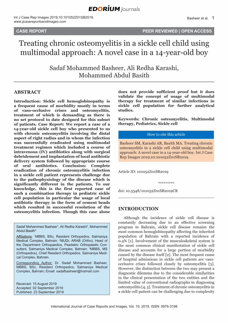
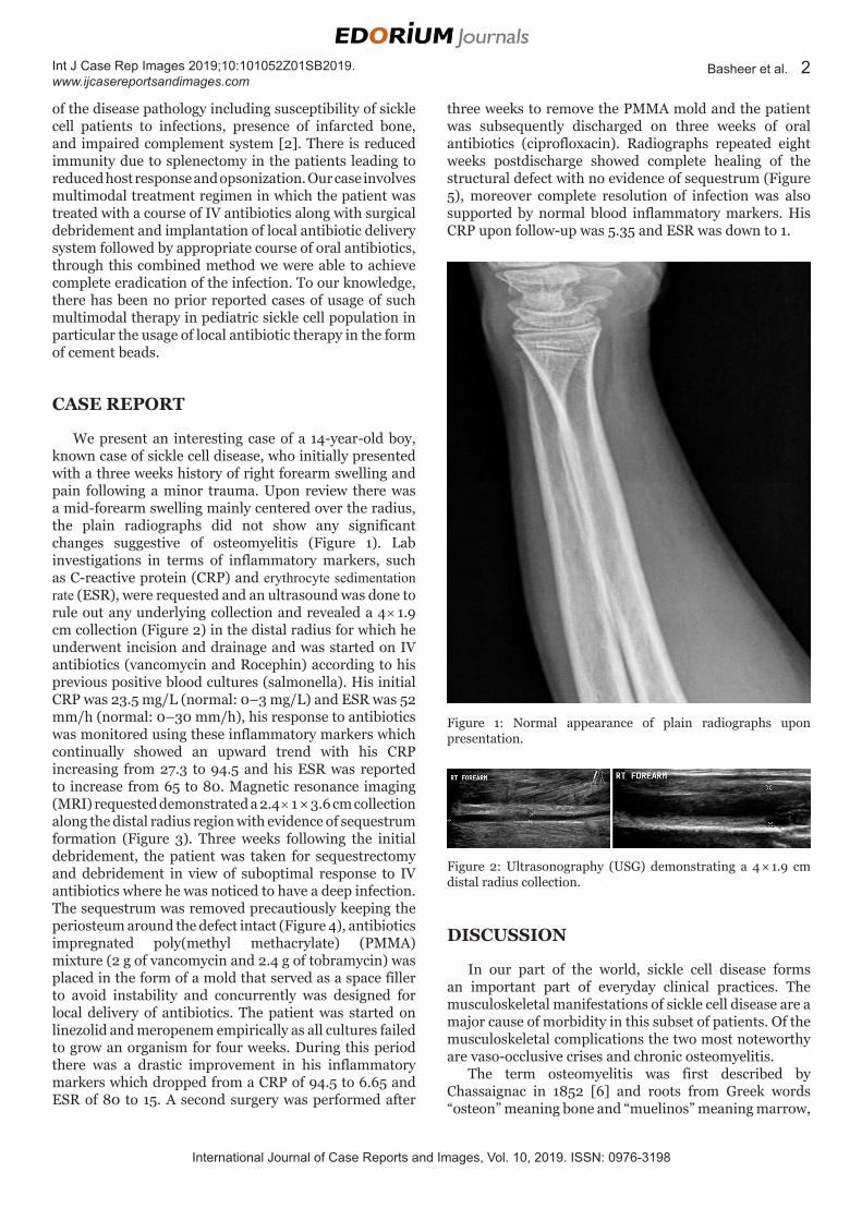
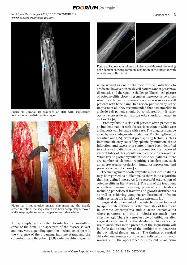
We present an interesting case of a 14-year-old boy, known case of sickle cell disease, who initially presented with a three weeks history of right forearm swelling and pain following a minor trauma. Upon review there was a mid-forearm swelling mainly centered over the radius, the plain radiographs did not show any significant changes suggestive of osteomyelitis (Figure 1). Lab investigations in terms of inflammatory markers, such as C-reactive protein (CRP) and erythrocyte sedimentation rate (ESR), were requested and an ultrasound was done to rule out any underlying collection and revealed a 4 × 1.9 cm collection (Figure 2) in the distal radius for which he underwent incision and drainage and was started on IV antibiotics (vancomycin and Rocephin) according to his previous positive blood cultures (salmonella). His initial CRP was 23.5 mg/L (normal: 0–3 mg/L) and ESR was 52 mm/h (normal: 0–30 mm/h), his response to antibiotics was monitored using these inflammatory markers which continually showed an upward trend with his CRP increasing from 27.3 to 94.5 and his ESR was reported to increase from 65 to 80. Magnetic resonance imaging (MRI) requested demonstrated a 2.4 × 1 × 3.6 cm collection along the distal radius region with evidence of sequestrum formation (Figure 3). Three weeks following the initial debridement, the patient was taken for sequestrectomy and debridement in view of suboptimal response to IV antibiotics where he was noticed to have a deep infection. The sequestrum was removed precautiously keeping the periosteum around the defect intact (Figure 4), antibiotics impregnated poly(methyl methacrylate) (PMMA) mixture (2 g of vancomycin and 2.4 g of tobramycin) was placed in the form of a mold that served as a space filler to avoid instability and concurrently was designed for local delivery of antibiotics. The patient was started on linezolid and meropenem empirically as all cultures failed to grow an organism for four weeks. During this period there was a drastic improvement in his inflammatory markers which dropped from a CRP of 94.5 to 6.65 and ESR of 80 to 15. A second surgery was performed after
three weeks to remove the PMMA mold and the patient was subsequently discharged on three weeks of oral antibiotics (ciprofloxacin). Radiographs repeated eight weeks postdischarge showed complete healing of the structural defect with no evidence of sequestrum (Figure 5), moreover complete resolution of infection was also supported by normal blood inflammatory markers. His CRP upon follow-up was 5.35 and ESR was down to 1.
Figure 1: Normal appearance of plain radiographs upon presentation.
Figure 2: Ultrasonography (USG) demonstrating a 4 × 1.9 cm distal radius collection.
DISCUSSION
In our part of the world, sickle cell disease forms an important part of everyday clinical practices. The musculoskeletal manifestations of sickle cell disease are a major cause of morbidity in this subset of patients. Of the musculoskeletal complications the two most noteworthy are vaso-occlusive crises and chronic osteomyelitis.
The term osteomyelitis was first described by Chassaignac in 1852 [6] and roots from Greek words “osteon” meaning bone and “muelinos” meaning marrow,
International Journal of Case Reports and Images, Vol. 10, 2019. ISSN: 0976-3198
Int J Case Rep Images 2019;10:101052Z01SB2019. www.ijcasereportsandimages.com
Basheer et al. 3
it may simply be translated to infection off medullary canal of the bone. The spectrum of the disease is vast and may vary depending upon the mechanism of spread, the virulence of the organism, immune status, and the comorbidities of the patient [7, 8]. Osteomyelitis in general
is considered as one of the most difficult infections to eradicate, however, in sickle cell patients and it presents a diagnostic and therapeutic challenge. The clinical picture of osteomyelitis closely resembles vaso-occlusive crises which is a far more presentation scenario in sickle cell patients with bone pains. In a review published by Jeane Baptisste et al., they recommended that osteomyelitis in a sickle cell patient should be considered only if vaso-occlusive crises do not subside with standard therapy in 1–2 weeks [9].
Osteomyelitis in sickle cell patients often presents in an indolent manner with abscess formation in which case a diagnosis can be made with ease. The diagnosis can be aided by various diagnostic modalities, MRI being the most sensitive one [10]. Several predisposing factors, such as immunodeficiency caused by splenic dysfunction, tissue infarction, and excess iron content, have been identified in sickle cell patients which account for the increased susceptibility of this population to chronic osteomyelitis. While treating osteomyelitis in sickle cell patients, there are number of elements requiring consideration, such as microvascular occlusion, immunosuppression, and presence of necrotic bone [2].
The management of osteomyelitis in sickle cell patients may be regarded as a dilemma as there is no algorithm that has defined measures for successful eradication of osteomyelitis in literature [11]. The aim of the treatment is centered around avoiding potential complications including pathological fracture and growth disturbances as well as achieving complete eradication of infection while restoring the function of the extremity [12].
Surgical debridement of the infected bone followed by appropriate antibiotics is the main stay of treatment in chronic osteomyelitis unlike acute osteomyelitis where parenteral and oral antibiotics are much more effective [13]. There is a greater role of antibiotics after surgical debridement of the devitalized tissues as the use of antibiotics in the presence of the sequestrum will be futile due to inability of the antibiotics to penetrate the devitalized tissues [12, 14]. The timings of surgical debridement remain controversial with many surgeons waiting until the appearance of sufficient involucrum
Figure 3: Coronal T2 sequence of MRI with sequestrum formation in the distal radius region.
Figure 4: Intraoperative images demonstrating the deeply seated infection, the sequestrum has been completely removed while keeping the surrounding periosteum sleeve intact.
Figure 5: Radiographs taken at a follow-up eight weeks following debridement showing complete resolution of the infection with remodeling of the defect.

International Journal of Case Reports and Images, Vol. 10, 2019. ISSN: 0976-3198
Int J Case Rep Images 2019;10:101052Z01SB2019. www.ijcasereportsandimages.com
Basheer et al. 4
to perform a sequestromy, however, there is sufficient evidence of performing a sequestromy if an involucrum has not appeared radiographically within three months of presentation [2, 15, 16].
The importance of preserving the periosteum has been previously described in an article by Bertram et al. [16] in which they stressed the role of an intact periosteum in generating an involucrum and creating skeletal stability. A subset of patients in their series were able to generate almost an entire diaphysis despite having massive skeletal defects which is very similar in our case. Postsequestromy the index case had a considerable bony defect in the distal radius which completely resolved within three months of debridement.
Following surgical debridement and sequestromy, the infection present within the dead bone is converted to an infection in well-vascularized tissue where antibiotics can penetrate readily [17]. Despite the prolonged use of intravenous antibiotics, a significant relapse rate has been found in chronic osteomyelitis [6, 8] more so in such difficult to treat cases, as ours, therefore it becomes vital to supplement the parental and oral antibiotics regimen with local mode of antibiotics delivery to produce complete eradication of the infection [6, 18, 19]. This in situ implantation works by locally eliminating the bacteria and reducing the dead space. There are several delivery vehicles used including both biodegradable and nonbiodegradable systems. Many commercially available preparations have shown promising results but they are expensive and not readily available in our settings. In the index case we used commonly available PMMA mixture which was combined with vancomycin and tobramycin both of which are known to maintain their efficacy when used along with PMMA. The elution kinetics of such a mixture of PMMA and antibiotics is biphasic with an increased bioavailability early on followed by a sustained release phase [20]. The antibiotics diffuse out of the PMMA carrier in a passive manner and therefore vary depending on the type of antibiotics, class of antibiotics, type of cement, and method of mixing [20].
Although there have been numerous advantages reported of local delivery of antibiotics in osteomyelitis, their application still remains controversial [19, 21] in particular in pediatric population where no published data is available for assessing the efficacy of such a treatment option. The major disadvantage of such a delivery method is a second surgery to remove the beads [21]. Managing resistant cases of chronic osteomyelitis with local antibiotic delivery method has the advantage of maintain a high concentration of antibiotics at the infection site while avoiding the systemic toxicity that may be associated with such a high concentration of antibiotics especially in pediatric population.
In the index case, we believe that the most important factor that played a crucial role in complete eradication of the infection was early sequestromy and the use of local antibiotic therapy, the utilization of which in pediatric population has not been attempted previously.
CONCLUSION
Chronic osteomyelitis is a dreaded complication of sickle cell disease which is associated with significant morbidity and a high recurrence rate. The gold standard for treatment remains surgical debridement and the use of parental antibiotics. The recurrence of osteomyelitis can be effectively reduced by local antibiotic impregnated delivery system, the most commonly used system has been the PMMA which can be molded into beads, blocks, or bars. This approach provides a promising adjunct to treating osteomyelitis especially in difficult to treat and resistant cases even in pediatric population. Another important consideration is the potential of the periosteum to regenerate and restore the continuity of the bone which would otherwise require complex reconstructive procedures.
REFERENCES
1. Al Arrayed S, Al Hajeri A. Public awareness of sickle cell disease in Bahrain. Ann Saudi Med 2010;30(4):284–8.
2. Almedia A, Roberts I. Bone involvement in sickle cell disease. Br J Haematol 2005;129(4):482–90.
3. Nwadiaro Hc, Ugwu BT, Legbo JN. Chronic osteomyelitis in patients with sickle cell disease. East Afr Ned J 2000;77(1):23–6.
4. Barrett-Connor E. Bacterial infection and sickle cell anemia. An analysis of 250 infections in 166 patients and a review of the literature. Medicine (Baltimore) 1971;50(2):97–112.
5. Bennett OM. Salmonella osteomyelitis and the hand-foot syndrome in sickle cell diease. J Pediatr Orthop 1992;12(4):534–8.
6. Gomes D, Pereira M, Bettencourt AF. Osteomyelitis: An overview of antimicrobial therapy. Braz J Pharm Sci 2013;49(1):13–27.
7. Tice AD, Hoaglund PA, Shoultz DA. Risk factors and treatment outcomes in osteomyelitis. J Antimicrob Chemother 2003;51(5):1261–8.
8. Tice AD, Hoaglund PA, Shoultz DA. Outcomes of osteomyelitis among patients treated with outpatient parentral antimicrobial therapy. Am J Med 2003;114(9):723–8.
9. Jean-Baptise G, De Ceulaer K. Osteoarticular disorders of haematological origin. Baillieres Best Pract Res Clin Rheumatol 2000;14(2):307-23.
10. Lonergan GJ, Cline DB, Abbondanzo SL. Sickle cell anemia. Radiographics 2001;21(4):971–94.
11. Sadat-Ali M, al-Umran K, al-Habdan I, al-Mulhim F. Ultrasonography: can it differentiate between vasoocclusive crisis and acute osteomyelitis in sickle cell disease? J Pediatr Orthop 1998;18(4):552–4.
12. Olaniyi JA, Akinwunmi AO. Polyostotic osteomyelitis in a sickle cell anemia patient. Ann Trop Pathol 2017;8(1):54–7.
13. Jones HW, Beckles VL, Akinola B, Steenson AJ, Harrison WJ. Chronic haematogenous osteomyelitis in children: An unsolved problem. J Bone Joint Surg Br 2011;93(8):1005–10.

International Journal of Case Reports and Images, Vol. 10, 2019. ISSN: 0976-3198
Int J Case Rep Images 2019;10:101052Z01SB2019. www.ijcasereportsandimages.com
Basheer et al. 5
14. Meier DE, Tarpley JL, Olaolorum DA, et al. Haematogenous osteomyelitis in the developing world: A practical approach to classification and treatment with limited resources. Contemporary Orthopaedics 1993;26:495–505.
15. Spiegel DA, Penny JN. Chronic osteomyelitis in children. Tech Orthop 2005;20:142–52.
16. Bertram FW, Harrison J, Freeman R. The role of the periosteum in healing a large structural defect following sequestrectomy. Trop Doct 2011;41(1):54–60.
17. Ciampolini J, Harding KG. Pathophysiology of chronic bacterial osteomyelitis. why do antibiotics fail so often? Postgrad Med J 2000;79(898):479–83.
18. Nair MB, Kretlow JD, Mikos AG, Kasper FK. Infection and tissues engineering in segmental bone defects – a mini review. Curr Opin Biotechnol 2011;22(5):721–5.
19. Azi ML, Junior MK, Martinez R, Paccola CAJ. Bone cement and gentamicin in the treatment of bone infection. Background and in vitro study. Acta Ortop Bras 2010;18(1):31–4.
20. Wentao Z, Lei G, Liu Y, Wang W, Song T, Fan J. Approach to osteomyelitis treatment with antibiotic loaded PMMA. Microb Pathog 2017;102:42–44.
21. Gogia JS, Meehan JP, Di Cesare PE, Jamali AA. Local antibiotic therapy in osteomyelitis. Semin Plast Surg 2009;23(2):100–7.
*********
Author ContributionsSadaf Mohammed Basheer – Conception of the work, Design of the work, Acquisition of data, Analysis of data, Drafting the work, Final approval of the version to be published, Agree to be accountable for all aspects of the work in ensuring that questions related to the accuracy or integrity of any part of the work are appropriately investigated and resolved
Ali Redha Karashi – Conception of the work, Analysis of data, Interpretation of data, Revising the work critically for important intellectual content, Final approval of the
version to be published, Agree to be accountable for all aspects of the work in ensuring that questions related to the accuracy or integrity of any part of the work are appropriately investigated and resolved
Mohammed Abdul Basith – Conception of the work, Analysis of data, Revising the work critically for important intellectual content, Final approval of the version to be published, Agree to be accountable for all aspects of the work in ensuring that questions related to the accuracy or integrity of any part of the work are appropriately investigated and resolved
Guarantor of SubmissionThe corresponding author is the guarantor of submission.
Source of SupportNone.
Consent StatementWritten informed consent was obtained from the patient for publication of this article.
Conflict of InterestAuthors declare no conflict of interest.
Data AvailabilityAll relevant data are within the paper and its Supporting Information files.
Copyright© 2019 Sadaf Mohammed Basheer et al. This article is distributed under the terms of Creative Commons Attribution License which permits unrestricted use, distribution and reproduction in any medium provided the original author(s) and original publisher are properly credited. Please see the copyright policy on the journal website for more information.
Access full text article onother devices
Access PDF of article onother devices