trends in cell biology - … · • genetic engineering of foods • biotechnology • organ growth...
TRANSCRIPT
TRENDS IN CELL BIOLOGY
ABS-931
3(3-0)

Course Contents:
• Introduction
• Cell Organization
• Cell Architecture
• Membrane Structure and Function
• Bio Transport
• Vesicular Transport
• Transport Signals
• Nuclear Transport
• Bio Energetic
• Mitochondrial Energy Conversion
• Chloroplast Energy Conversion
• Cytoskeleton
• Cell Shape
• Cell Contractility
• Cell to cell Communication
• Electrochemical Signaling
• Synaptic and Sensory Transduction
• Biochemical Signaling
• Receptor Ligand Interactions
• Second Messengers
• Signaling Cascades
• Cell Cycle and Apoptosis
• Phases of Cell Cycle and Cell Division
• Regulation of Cell Growth and Death
• Specialized Cell Systems

Recommended Books:
• The Cell by Bruce Albert and Dennis Bray, 4th Ed. Garland
Publishing Inc, New York and London.
• Biochemistry by Victor L. Davidson, Donald B. Sittman. 3rd Ed.
1993, Harwal Pub Co.
• Cell and Molecular Biology by Gerald Karp. 1996, John Willey
and Sons, Inc. London
• Gene VIII By Lewin Benjamin Eds 2004. Oxford University
press, Inc, New york.
• Molecular Biology of the Gene by Watson, J. D., T. A. Baker, S.
P. Bell, A. Gann, M. Levine, and R. Losick, 5th Ed. 2003. New
York, Benjamin Cummings ISBN 0-8053-4635-X

Policies
First 1 hr Test 17.5 %
Second 1 hr Test 17.5 %
Assignments (3-6) 5-10 %
Quizzes (3-6) 5-10 %
Terminal Exam (3 hrs) 50 %
• Please turn off your cell phones during class

What is cell Biology?

What is cell Biology?
Divisions in the biological sciences are based on
degrees of “complexity”

Biochemistry & Biophysics: study of
the structures and behaviors of
molecules

Microbiology: study of prokaryotic cells
and viruses
Cell Biology: study of the structure
and function of eukaryotic cells
Developmental Biology: study of how
communities of cells form tissues, organs,
and build an organism
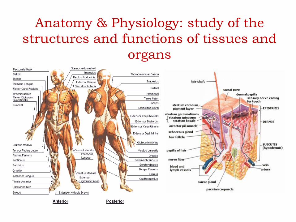
Anatomy & Physiology: study of the
structures and functions of tissues and
organs

Zoology & Plant Biology: study of the
organisms
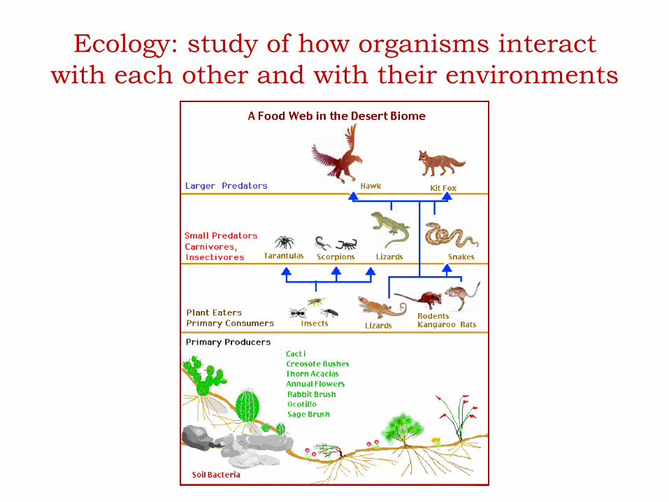
Ecology: study of how organisms interact
with each other and with their environments

Levels of Biological Complexity
1. Biochemistry & Biophysics
2. Microbiology
3. Cell Biology
4. Developmental Biology
5. Anatomy & Physiology
6. Zoology & Plant Biology
7. Ecology

Understanding cell biology is important to
understand the basis for disease
• Hypercholesterolemia (defective uptake of lipoproteins)
• Cystic fibrosis (misfolding of key protein)
• Hypertension (defective cell-cell adhesion in the kidney)
• Congenital heart defects (errors in cell migration during development)
• Muscular dystrophy (defective attachment of the plasma membrane to
the cytoskeleton)
• Lysosomal storage disease (defective intracellular transport of enzymes)
• Food-borne illness (Salmonella, E. coli)
• Cancer (errors in cell division, migration, cell polarity, growth, etc)
• Ageing
• All disease states are caused at the cellular level
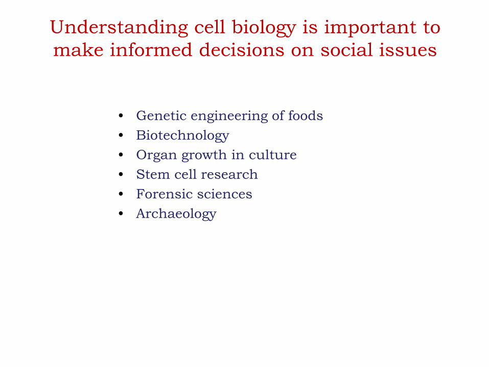
Understanding cell biology is important to
make informed decisions on social issues
• Genetic engineering of foods
• Biotechnology
• Organ growth in culture
• Stem cell research
• Forensic sciences
• Archaeology

The Dynamic CellBasic unit of organization or
structure of all living matter
CELL BIOLOGY

Galileo Galilei (Early Seventeenth century) used lenses
Beginning of the study of cells as the basis of life
Robert Hooke (Middle of Seventeenth century) microscopic
examination of sliced cork
Used Latin word cella (small room)
Anton van Leeuwenhoek (Late Seventeenth century)
Improved lenses, Improved magnification
Robert Brown (1831) observed nucleus as opaque spot
Methias Schleiden (1838)
Theodore Schwann (1839)Cell Theory
A brief History
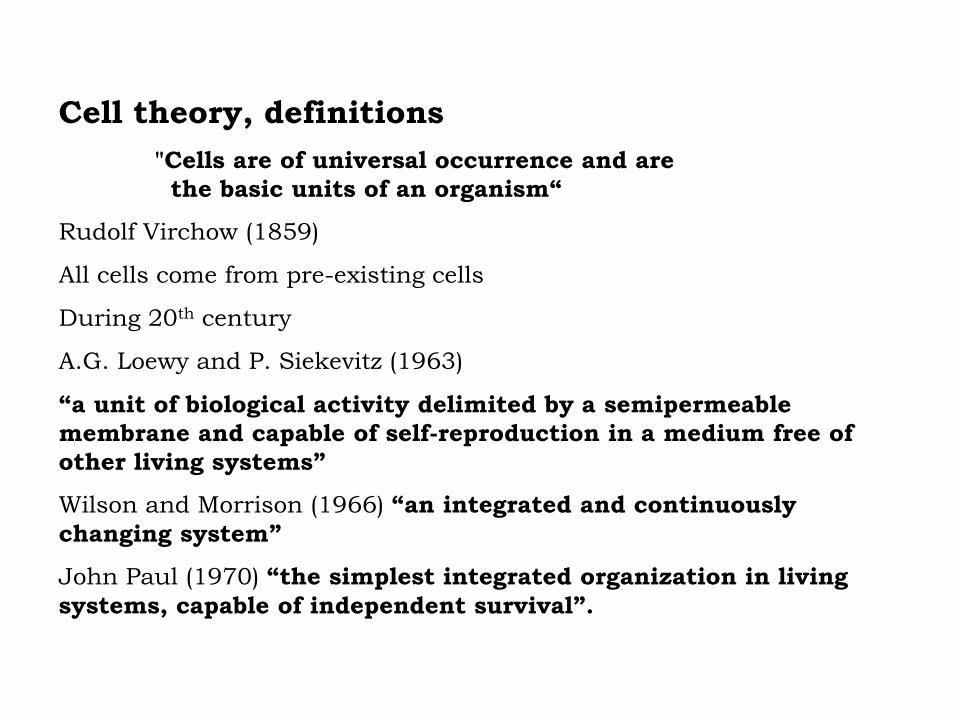
Cell theory, definitions
"Cells are of universal occurrence and are
the basic units of an organism“
Rudolf Virchow (1859)
All cells come from pre-existing cells
During 20th century
A.G. Loewy and P. Siekevitz (1963)
“a unit of biological activity delimited by a semipermeable
membrane and capable of self-reproduction in a medium free of
other living systems”
Wilson and Morrison (1966) “an integrated and continuously
changing system”
John Paul (1970) “the simplest integrated organization in living
systems, capable of independent survival”.

Principles of Cell Theory
• All living things are made of cells
• Smallest living unit of structure and function of all
organisms is the cell
• All cells arise from preexisting cells
(this principle discarded the idea of
spontaneous generation)

Cell Size

Cells Have Large Surface
Area-to-Volume Ratio
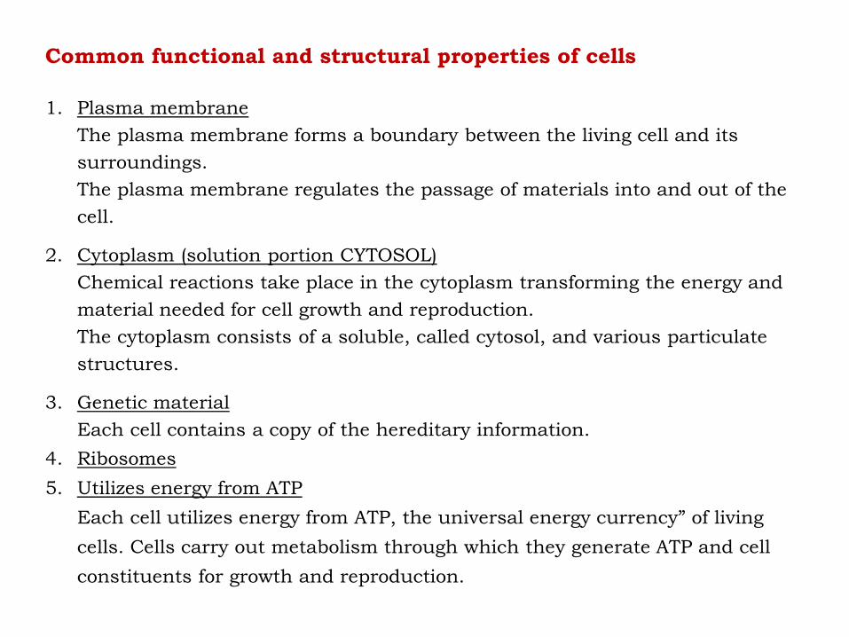
Common functional and structural properties of cells
1. Plasma membrane
The plasma membrane forms a boundary between the living cell and its
surroundings.
The plasma membrane regulates the passage of materials into and out of the
cell.
2. Cytoplasm (solution portion CYTOSOL)
Chemical reactions take place in the cytoplasm transforming the energy and
material needed for cell growth and reproduction.
The cytoplasm consists of a soluble, called cytosol, and various particulate
structures.
3. Genetic material
Each cell contains a copy of the hereditary information.
4. Ribosomes
5. Utilizes energy from ATP
Each cell utilizes energy from ATP, the universal energy currency” of living
cells. Cells carry out metabolism through which they generate ATP and cell
constituents for growth and reproduction.

Cell Types
• Prokaryotic
• Eukaryotic

Prokaryotic cell
Eukaryotic cell
Cell Organization

Prokaryotic cells• First cell type on earth
• Single cell organisms
• Two main types: bacteria and archaea
• Relatively simple structure
• No membrane bound nucleus
• Nucleoid = region of DNA concentration
• Organelles not bound by membranes

Eukaryotic cells • Single cell or multicellular organisms
• Plants and animals
• Structurally more complex: organelles,
cytoskeleton
• Nucleus bound by membrane
• Include fungi, protists, plant, and animal
cells
• Possess many organelles

Representative Animal Cell

Representative Plant Cell

Organelles
• Cellular machinery
• Two general kinds
– Derived from membranes
– Bacteria-like organelles

Bacteria-Like Organelles
• Derived from symbiotic bacteria
• Ancient association
• Endosymbiotic theory
– Evolution of modern cells from cells & symbiotic
bacteria
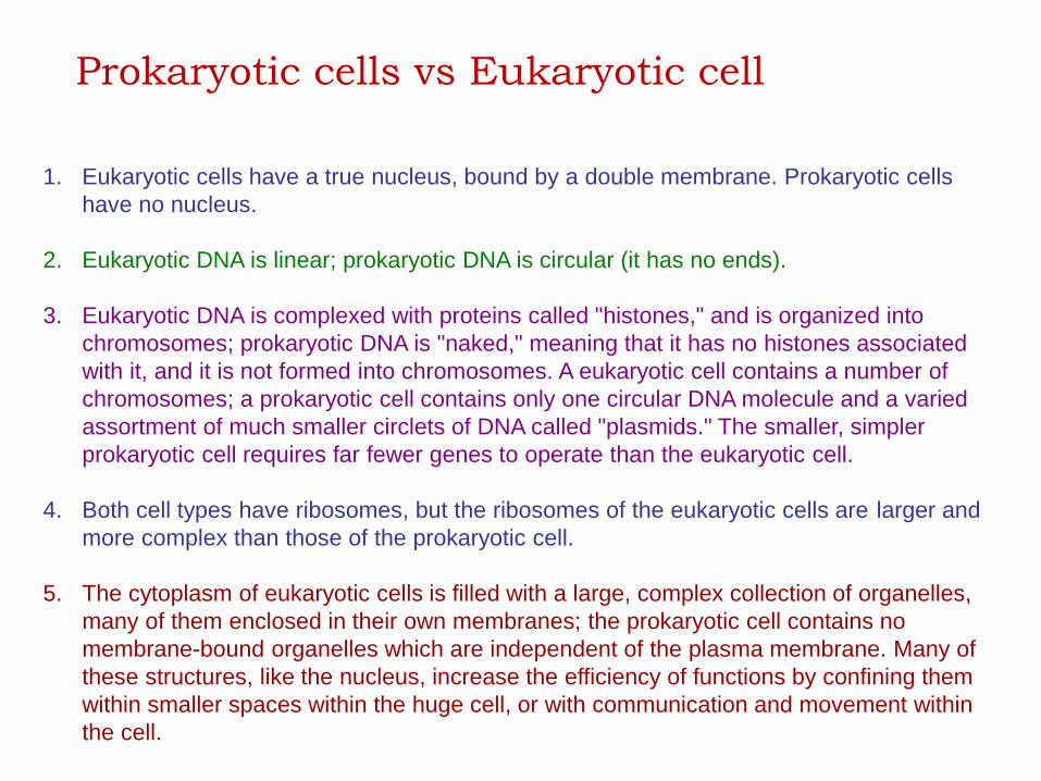
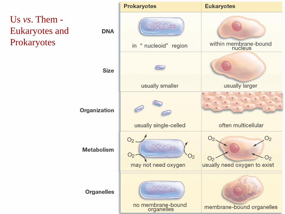
Prokaryotic cells vs Eukaryotic cell
1. Eukaryotic cells have a true nucleus, bound by a double membrane. Prokaryotic cells
have no nucleus.
2. Eukaryotic DNA is linear; prokaryotic DNA is circular (it has no ends).
3. Eukaryotic DNA is complexed with proteins called "histones," and is organized into
chromosomes; prokaryotic DNA is "naked," meaning that it has no histones associated
with it, and it is not formed into chromosomes. A eukaryotic cell contains a number of
chromosomes; a prokaryotic cell contains only one circular DNA molecule and a varied
assortment of much smaller circlets of DNA called "plasmids." The smaller, simpler
prokaryotic cell requires far fewer genes to operate than the eukaryotic cell.
4. Both cell types have ribosomes, but the ribosomes of the eukaryotic cells are larger and
more complex than those of the prokaryotic cell.
5. The cytoplasm of eukaryotic cells is filled with a large, complex collection of organelles,
many of them enclosed in their own membranes; the prokaryotic cell contains no
membrane-bound organelles which are independent of the plasma membrane. Many of
these structures, like the nucleus, increase the efficiency of functions by confining them
within smaller spaces within the huge cell, or with communication and movement within
the cell.

Comparison of features of prokaryotic and eukaryotic cells
Prokaryotes Eukaryotes
Typical organisms Bacteria, Archaea Protists, Fungi, Plants and Animals
Typical size ~ 1-10 µm ~ 10-100 µm (Sperm cells are smaller)
Type of nucleus nucleoid region real nucleus with double membrane
DNA circular (usually)linear molecules (chromosomes) with histone
proteins
RNA-/protein-
synthesiscoupled in cytoplasm
RNA-synthesis inside the nucleus
protein synthesis in cytoplasm
Ribosomes 50S+30S 60S+40S
Cytoplasmatic
structurevery few structures
highly structured by endomembranes and a
cytoskeleton
Cell movement flagella made of flagellin flagella and cilia made of tubulin, lamellipodia
Mitochondria Noneone to several thousand (though some lack
mitochondria)
Chloroplasts None in algae and plants
Organization usually single cellssingle cells, colonies, higher multicellular
organisms with specialized cells
Cell division Binary fission Mitosis (fission or budding), Meiosis
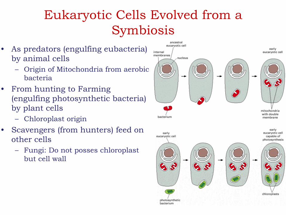
Eukaryotic Cells Evolved from a
Symbiosis
• As predators (engulfing eubacteria)
by animal cells
– Origin of Mitochondria from aerobic
bacteria
• From hunting to Farming
(engulfing photosynthetic bacteria)
by plant cells
– Chloroplast origin
• Scavengers (from hunters) feed on
other cells
– Fungi: Do not posses chloroplast
but cell wall
Us vs. Them -
Eukaryotes and
Prokaryotes
Internal Organization of the cell

Common functional and structural
properties of cells
1. Plasma membrane
2. Cytoplasm (solution portion CYTOSOL)
3. Genetic material
4. Ribosomes
5. Utilizes energy from ATP

Major Divisions of the Eukaryotic Cell

The Plasma Membrane – Gateway to the Cell
Plasma Membrane
• Crucial to the life of the cell,
– encloses the cell,
– defines its boundaries, and
– maintains the essential differences between the cytosol and the
extracellular environment.
• Inside eukaryotic cells, the membranes of the endoplasmic
reticulum, Golgi apparatus, mitochondria, and other membrane-
enclosed organelles maintain the characteristic differences
between the contents of each organelle and the cytosol.
• Ion gradients across membranes, established by the activities of
specialized membrane proteins, can be used
– to synthesize ATP,
– to drive the transmembrane movement of selected solutes, or,
– in nerve and muscle cells, to produce and transmit electrical signals.

• In all cells, the plasma membrane also contains proteins that
act as sensors of external signals, allowing the cell to
change its behavior in response to environmental cues; these
protein sensors, or receptors, transfer information rather
than ions or molecules across the membrane.
• Despite their differing functions, all biological membranes
have a common general structure: each is a very thin film of
lipid and protein molecules, held together mainly by
noncovalent interactions.
• Cell membranes are dynamic, fluid structures, and most of
their molecules are able to move about in the plane of the
membrane.

• The lipid molecules are arranged as a continuous double
layer about 5 nm thick.
• This lipid bilayer provides the basic fluid structure of the
membrane and serves as a relatively impermeable barrier to
the passage of most water-soluble molecules.
• Protein molecules that span the lipid bilayer mediate nearly
all of the other functions of the membrane, transporting
specific molecules across it, for example, or catalyzing
membrane-associated reactions, such as ATP synthesis.

• In the plasma membrane, some proteins serve as structural
links that connect the cytoskeleton through the lipid bilayer
to either the extracellular matrix or an adjacent cell, while
others serve as receptors to detect and transduce chemical
signals in the cell's environment.
• It takes many different membrane proteins to enable a cell to
function and interact with its environment.
• In fact, it is estimated that about 30% of the proteins that are
encoded in an animal cell's genome are membrane proteins.

Membrane functions
1. Compartmentalization
1. Allows specialized activities to proceed without
external interference
2. Enables cellular activities to be regulated
independently
3. Prevents mixing of various contents
2. Providing a selectively permeable membrane
1. Prevents free interchange o material to-and-fro
2. Provides means of communication between spaces
3. PM ensures the entry of appropriate substance into cytoplasm and
inappropriate substance are kept out (selectively permeable)
3. Transporting solutes
1. Physical transport of substances into and out of cell
2. Accumulation of sugars and amino acids necessary to fuel its metabolism
and build its macromolecules

4. Responding to external signals
1. Transfer of information from one side to other through ligand receptor interaction (signal transduction)
5. Intracellular interactions
1. PM mediates interaction between the cells
2. Recognition, adherence and exchange of material and information between cells
6. Scaffolding of biochemical activities
1. Provide framework for effective interaction of components
2. Localization of enzymatic machinery
7. Energy transduction
1. Energy conversion of
1. Sunlight to chemical energy contained in carbohydrates (photosynthesis)
2. Transfer of chemical energy from carbohydrates and fats into ATP (in mitochondria and chloroplast)
2. Sites of energy storage to run cellular activities

Small molecules and larger hydrophobic molecules move through.
Ions, hydrophilic molecules larger than water, and large molecules such as
proteins do not move through the membrane on their own.
The physical properties of phospholipids account for membrane
assembly and many of its properties.
The Plasma Membrane is Semipermeable

History of structure of cell membrane
a) Davidson-Daniellimodel (1954)
b) The Fluid Mosaic model (Singer & Nicolson, 1972)
c) Current representation of Plasma Membrane

Membrane Components

Plasma membrane
• Cytosolic face (internal face)
• Exoplasmic face (external face)
• What about organelles?
– Lysosome -
– Mitochondria –
– Chloroplast –
– Nucleus –
– Vacuole –

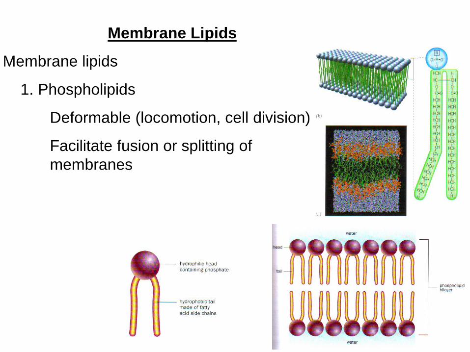
Membrane Lipids
• Amphipathic Molecules, most of which spontaneously form
bilayers
• Lipid that is, fatty molecules constitute about 50% of the mass
of most animal cell membranes, nearly all of the remainder
being protein.
• There are approximately 5 × 10⁶ lipid molecules in a 1 mm × 1
mm area of lipid bilayer, or about 109 lipid molecules in the
plasma membrane of a small animal cell.
• All of the lipid molecules in cell membranes are amphipathic (or
amphiphilic) that is, they have a hydrophilic ("water-loving") or
polar end and a hydrophobic ("water-fearing") or nonpolar end.

• It is the shape and amphipathic nature of the lipid molecules that
cause them to form bilayers spontaneously in aqueous
environments.
• Hydrophilic molecules dissolve readily in water because they contain
charged groups or uncharged polar groups that can form either
favorable electrostatic interactions or hydrogen bonds with water
molecules.
• Hydrophobic molecules, by contrast, are insoluble in water because
all, or almost all, of their atoms are uncharged and nonpolar and
therefore cannot form energetically favorable interactions with water
molecules.

• If dispersed in water, they force the adjacent water molecules
to reorganize into ice like cages that surround the
hydrophobic molecule.
• Because these cage structures are more ordered than the
surrounding water, their formation increases the free energy.
• This free energy cost is minimized, however, if the
hydrophobic molecules (or the hydrophobic portions of
amphipathic molecules) cluster together so that the smallest
number of water molecules is affected.

Membrane Lipids
• A typical biomembrane is assembled from
– Phosphoglycerides,
– Sphingolipids, and
– Steroids.
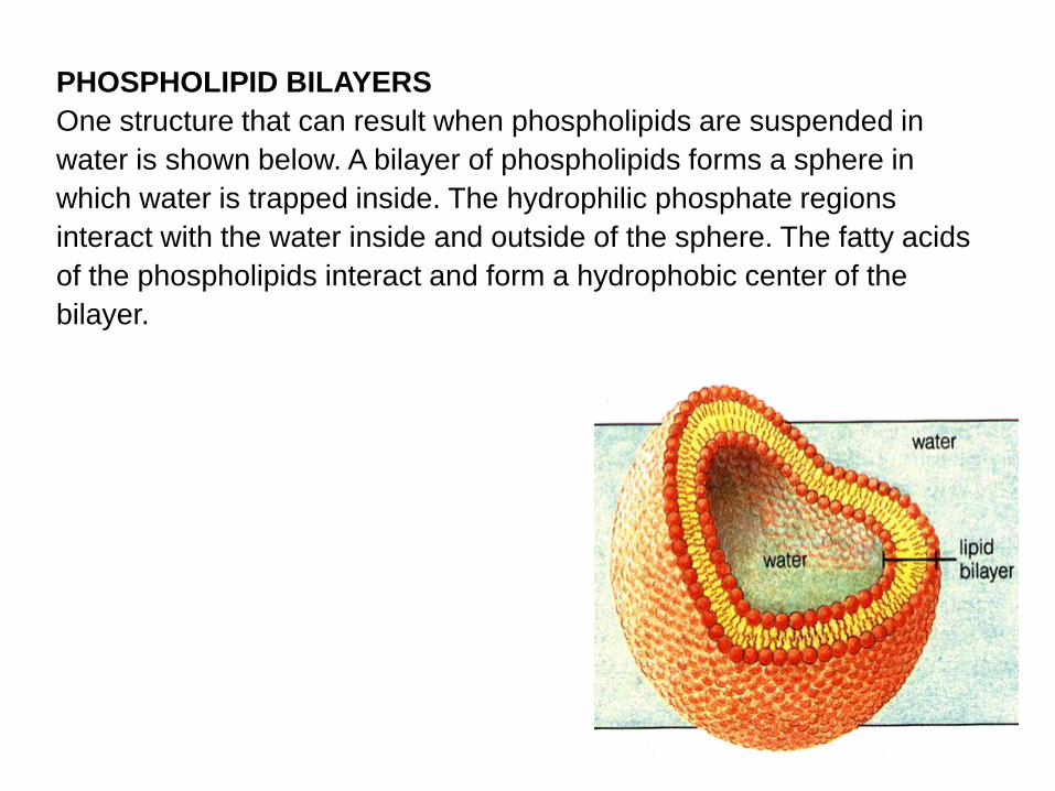
PHOSPHOLIPID BILAYERS
One structure that can result when phospholipids are suspended in
water is shown below. A bilayer of phospholipids forms a sphere in
which water is trapped inside. The hydrophilic phosphate regions
interact with the water inside and outside of the sphere. The fatty acids
of the phospholipids interact and form a hydrophobic center of the
bilayer.

• Phosphoglycerides, the most abundant class of lipids in
most membranes, are derivatives of glycerol 3-phosphate
• A typical phosphoglyceride molecule consists of a
hydrophobic tail composed of
– two fatty acyl chains esterified to the two hydroxyl groups in
glycerol phosphate and
– a polar head group attached to the phosphate group.
• The two fatty acyl chains may differ in the number of carbons
that they contain (commonly 16 or 18) and their degree of
saturation (0, 1, or 2 double bonds).

• Classified according to the nature of its head group.
• In phosphatidylcholines, the most abundant phospholipids in the
plasma membrane, the head group consists of choline, a positively
charged alcohol, esterified to the negatively charged phosphate.
• In other phosphoglycerides, an OH-containing molecule such as
ethanolamine, serine, and the sugar derivative inositol is linked to
the phosphate group.
• The plasmalogens are a group of phosphoglycerides that contain
one fatty acyl chain, attached to glycerol by an ester linkage, and
one long hydrocarbon chain, attached to glycerol by an ether linkage
(COOOC).
• These molecules constitute about 20 percent of the total
phosphoglyceride content in humans. Their abundance varies
among tissues and species but is especially high in human brain
and heart tissue.
Classification of Phosphoglycerides
Membrane Lipids
Membrane lipids
1. Phospholipids
Deformable (locomotion, cell division)
Facilitate fusion or splitting of
membranes

• The parts of a phospholipid molecule –
phosphatidylcholine

• A small tear in the bilayer creates a free edge with water; because
this is energetically unfavorable, the lipids spontaneously rearrange
to eliminate the free edge. (In eukaryotic plasma membranes, larger
tears are repaired by the fusion of intracellular vesicles.)
• The prohibition against free edges has a profound consequence: the
only way for a bilayer to avoid having edges is by closing in on itself
and forming a sealed compartment.
• This remarkable behavior, fundamental to the creation of a living
cell, follows directly from the shape and amphipathic nature of the
phospholipid molecule.

Phospholipid motility (The types of
movements possible for phospholipid molecules
in lipid bilayer)

Fluidity of membrane
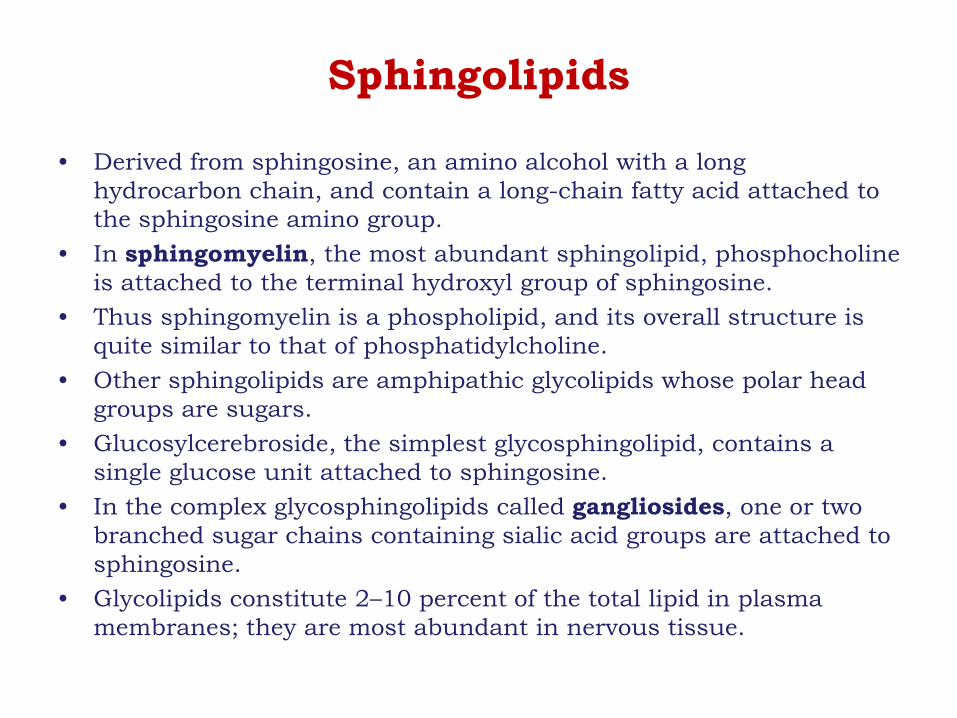
Sphingolipids
• Derived from sphingosine, an amino alcohol with a long
hydrocarbon chain, and contain a long-chain fatty acid attached to
the sphingosine amino group.
• In sphingomyelin, the most abundant sphingolipid, phosphocholine
is attached to the terminal hydroxyl group of sphingosine.
• Thus sphingomyelin is a phospholipid, and its overall structure is
quite similar to that of phosphatidylcholine.
• Other sphingolipids are amphipathic glycolipids whose polar head
groups are sugars.
• Glucosylcerebroside, the simplest glycosphingolipid, contains a
single glucose unit attached to sphingosine.
• In the complex glycosphingolipids called gangliosides, one or two
branched sugar chains containing sialic acid groups are attached to
sphingosine.
• Glycolipids constitute 2–10 percent of the total lipid in plasma
membranes; they are most abundant in nervous tissue.

Cholesterol
• Cholesterol and its derivatives constitute the third important
class of membrane lipids, the steroids.
• The basic structure of steroids is a four-ring hydrocarbon.
• Although cholesterol is almost entirely hydrocarbon in
composition, it is amphipathic because its hydroxyl group
can interact with water.
• Cholesterol is especially abundant in the plasma membranes
of mammalian cells but is absent from most prokaryotic cells.

Cholesterol
A) Formula B) Schematic drawing C) Space-filling model

Cholesterol in a lipid bilayer
a) In one layer of lipid bilayer
b) In lipid bilayer
(a)
(b)

• As much as 30–50 percent of the lipids in plant plasma
membranes consist of certain steroids unique to plants.
• At neutral pH, some phosphoglycerides (e.g.,
phosphatidylcholine and phosphatidyl ethanolamine) carry
no net electric charge, whereas others (e.g.,
phosphatidylinositol and phosphatidylserine) carry a single
net negative charge.
• Nonetheless, the polar head groups in all phospholipids can
pack together into the characteristic bilayer structure.
• Sphingomyelins are similar in shape to phosphoglycerides
and can form mixed bilayers with them.
• Cholesterol and other steroids are too hydrophobic to form a
bilayer structure unless they are mixed with phospholipids.

Three classes of membrane lipids
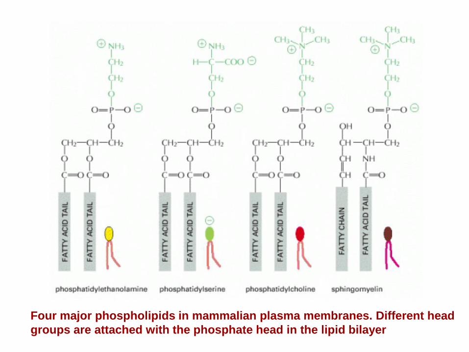
Four major phospholipids in mammalian plasma membranes. Different head
groups are attached with the phosphate head in the lipid bilayer

The asymmetrical distribution of phospholipids and glycolipids in the
lipid bilayer of human red blood cells.

Membranes are Asymmetric
Lateral Asymmetry of Lipids:
Lipids can cluster in the plane of the membrane - they are not
uniformly distributed
Transverse asymmetry of lipids
In most cell membranes, the composition of the outer monolayer is
quite different from that of the inner monolayer

Lipid Composition Influences the
Physical Properties of Membranes
A typical cell contains myriad types of membranes, each with
unique properties bestowed by its particular mix of lipids and
proteins.
Several phenomena contribute to these differences.
For instance, differences between membranes in the endoplasmic
reticulum (ER) and the Golgi are largely explained by the fact that
phospholipids are synthesized in the ER, whereas
sphingolipids are synthesized in the Golgi.
The proportion of sphingomyelin as a percentage of total membrane lipid
phosphorus is about six times as high in Golgi membranes as it is in ER
membranes.
In other cases, the translocation of membranes from one cellular
compartment to another can selectively enrich membranes in
certain lipids.
Differences in lipid composition may also correspond to
specialization of membrane function.
For example, the plasma membrane of absorptive epithelial
cells lining the intestine exhibits two distinct regions:
apical surface faces the lumen of the gut and is exposed to widely
varying external conditions;
basolateral surface interacts with other epithelial cells and with
underlying extracellular structures.
In these polarized cells, the ratio of sphingolipid to
phosphoglyceride to cholesterol in the basolateral membrane
is 0.5:1.5:1, roughly equivalent to that in the plasma
membrane of a typical unpolarized cell subjected to mild
stress.
In contrast, the apical membrane of intestinal cells, which is
subjected to considerable stress, exhibits a 1:1:1 ratio of
these lipids.
The relatively high concentration of sphingolipid in this
membrane may increase its stability

Membranes contain
specialized lipids and
proteins
Proteins 30-70% Phospholipids 7-40% Sterols 0-25% Specialized membranes
More than 90% Rhodopsin in
photoreceptor disc membrane
Protein rich mitochondrial
membranes
Transport optimized Red Blood Cell
membrane