trends - physics
TRANSCRIPT
Physics 4, 4 (2011)
Trends
Does cell biology need physicists?
Charles W. WolgemuthUniversity of Connecticut Health Center, Department of Cell Biology and Center for Cell Analysis and Modeling, Farmington,CT 06030-3505, USA
Published January 10, 2011
Allured by the chic perception and higher funding levels of disease-oriented research, many physicists havemigrated to cell biology. Does physics really play a dominant role, or is cellular physiology slave to genetics andchemistry?
Subject Areas: Biological Physics, Interdisciplinary Physics
Introduction
Cells are the fundamental units of life. At a basic level,a cell’s primary functions are to grow, replicate, and di-vide. Survival, at least for a sufficient length of time,is also extremely important in order to achieve theseother functions. However, as humans, we want more.We don’t only want our cells to provide us with a suffi-cient length of life that we can reproduce; we would alsolike to guarantee ourselves a long and healthy existence.These desires, though, are all too often thwarted by dis-ease. Though diseases can be caused by a number ofdifferent factors, such as molecular toxins, acquired orinherited genetic defects, viruses, and bacteria, whichall act at different scales (molecular, cellular, and evenmulticellular), in most cases, disease represents a cellu-lar level process; i.e., most diseases are, at their heart,a disruption of cellular function, which then ultimatelyproduces organ or organism disability or failure.
Since humans would prefer to live a disease-free ex-istence, it is not surprising that biological and health-related research is highly respected or that the NationalInstitutes of Health have deeper pockets than the Na-tional Science Foundation. In addition, recent techno-logical advances have allowed us to look at moleculesand cells in much more detail. We are now able toresolve and quantify, at subcellular and even molec-ular levels, the spatiotemporal dynamics of moleculesand processes inside cells. Therefore molecular andcellular biology have become more amenable to a re-search paradigm that melds experimental and theoret-ical investigations, and, more specifically, research thatis geared toward an accurate description of how thingsmove in space and time. It is, therefore, not surprisingthat physicists would be attracted to cell biological re-search.
But the past has shown that cell biologists are ex-tremely capable of making great progress without muchneed for physicists (other than needing physicists andengineers to develop many of the technologies that theyuse). Do the new data and new technological capabili-ties require a physicist’s viewpoint to analyze the mech-
anisms of the cell? Is physics of use to cell biology?
The elephants in the room: genetics,proteomics, and systems biology
It is hard to overestimate the role that genetics hasplayed in biology over the last 30 years. Molecular andcellular biology, to a very large extent, are about proteinsand their interactions. The ability to perturb a specificprotein inside a cell and then observe the consequenceshas led to an amazing number of advances in our under-standing of the roles of certain proteins in cellular func-tion. It would almost seem that if we could just com-pile a list of all the proteins inside the cell (a field calledproteomics) and determine their reactions and reactionrates, we would understand how a cell works. Withhigh-throughput methods, we are now able to quicklymeasure DNA transcripts and protein levels and cor-relate these with observed cell characteristics (“pheno-types”) [1]. We have amassed a lot of data, and the morewe put together, the more complex and harder to inter-pret the data becomes.
For a cell with a fairly small genome, such as the yeastSaccharomyces cerevisiae, which can produce between 104
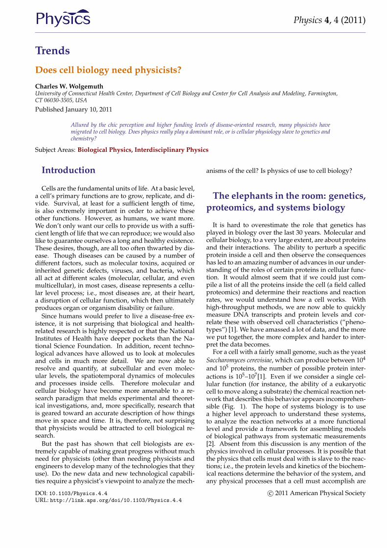
and 105 proteins, the number of possible protein inter-actions is 105–107[1]. Even if we consider a single cel-lular function (for instance, the ability of a eukaryoticcell to move along a substrate) the chemical reaction net-work that describes this behavior appears incomprehen-sible (Fig. 1). The hope of systems biology is to usea higher level approach to understand these systems,to analyze the reaction networks at a more functionallevel and provide a framework for assembling modelsof biological pathways from systematic measurements[2]. Absent from this discussion is any mention of thephysics involved in cellular processes. It is possible thatthe physics that cells must deal with is slave to the reac-tions; i.e., the protein levels and kinetics of the biochem-ical reactions determine the behavior of the system, andany physical processes that a cell must accomplish are
DOI: 10.1103/Physics.4.4URL: http://link.aps.org/doi/10.1103/Physics.4.4
c© 2011 American Physical Society
Physics 4, 4 (2011)
FIG. 1: The complexity of cellular biology. (a) A subset ofthe chemical reactions that drive eukaryotic cell crawling. Inbrief, cells sense the environment through membrane boundproteins. Activation of these receptors leads to activation ofa number of other proteins that promote the polymerizationof actin. The biochemical reactions that govern the dynam-ics of actin are included. These chemical reactions producecell motility. (b)–(d) Time series of a cancer cell (HT1080 fi-brosarcoma cell) moving through a collagen I matrix. Thereare two hour intervals between each frame. (Images courtesyof D. Wirtz, Johns Hopkins University.) (Credit: Carin Cain)
purely consequences of the biochemistry. Or, could it bethat cellular biology cannot be fully understood withoutphysics?
Some examples of successful physi-cal biology
The successes of genetics and biochemistry in describ-ing cellular function have overshadowed an importantpoint: Cells are not isolated bags of proteins. The insideof a cell has structure, and this structure is not static. Inaddition, cells must live in and interact with the envi-ronment, which is often unpredictable and not alwaysfavorable. In order to grow, move, and survive, cellsmust be able to produce force. That is, physics matters,
at least at some level.
Molecular motors
Inside cells there are proteins that convert chemicalenergy into useful work. For example, kinesins anddyneins haul cargo around the cell. Myosin moleculescan bind to actin filaments and exert forces, which ishow our muscles work. Other molecules rotate, such asthe protein that creates the molecules that are the primefuel of our cells. The general method by which thesemolecular motors operate is through a Brownian ratchetmechanism [3]. However, whereas the classic Brown-ian ratchet does not actually work, molecular motorsharness molecular binding energies to satisfy the sec-ond law of thermodynamics. Typically, binding of anion or molecule (such as ATP) to the motor leads to aconformational change in the protein. This conforma-tional change can act like a power-stroke in the motor.Hydrolysis of ATP or release of the bound ion then re-turns the motor to its original state, thereby completinga cycle (or, in the case of rotational motors, a bindingand release event typically only produces a substep of acomplete rotation).
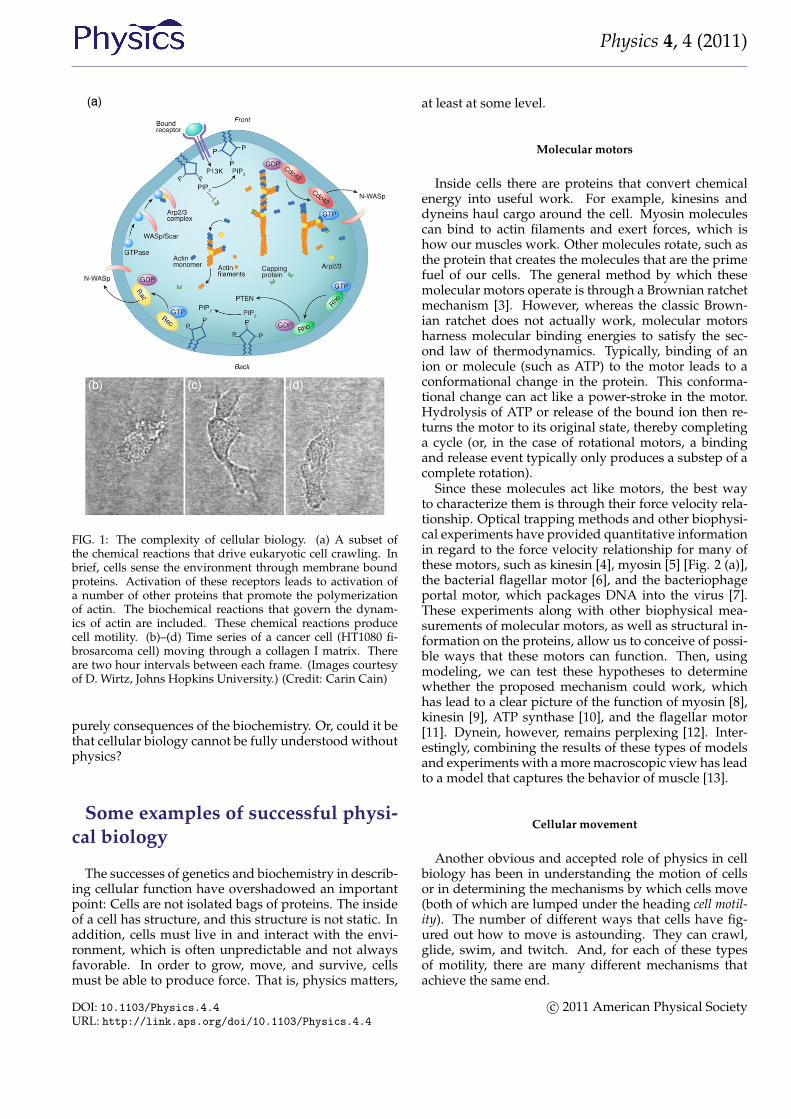
Since these molecules act like motors, the best wayto characterize them is through their force velocity rela-tionship. Optical trapping methods and other biophysi-cal experiments have provided quantitative informationin regard to the force velocity relationship for many ofthese motors, such as kinesin [4], myosin [5] [Fig. 2 (a)],the bacterial flagellar motor [6], and the bacteriophageportal motor, which packages DNA into the virus [7].These experiments along with other biophysical mea-surements of molecular motors, as well as structural in-formation on the proteins, allow us to conceive of possi-ble ways that these motors can function. Then, usingmodeling, we can test these hypotheses to determinewhether the proposed mechanism could work, whichhas lead to a clear picture of the function of myosin [8],kinesin [9], ATP synthase [10], and the flagellar motor[11]. Dynein, however, remains perplexing [12]. Inter-estingly, combining the results of these types of modelsand experiments with a more macroscopic view has leadto a model that captures the behavior of muscle [13].
Cellular movement
Another obvious and accepted role of physics in cellbiology has been in understanding the motion of cellsor in determining the mechanisms by which cells move(both of which are lumped under the heading cell motil-ity). The number of different ways that cells have fig-ured out how to move is astounding. They can crawl,glide, swim, and twitch. And, for each of these typesof motility, there are many different mechanisms thatachieve the same end.
DOI: 10.1103/Physics.4.4URL: http://link.aps.org/doi/10.1103/Physics.4.4
c© 2011 American Physical Society
Physics 4, 4 (2011)
FIG. 2: Some examples where physics has shed light on biol-ogy. (a) Physics-based simulations of the walking of myosin VIpredicted that the molecule would produce both hand-over-hand and inchworm type movements [8], which was later con-firmed with single molecule experiments [57]. (Image courtesyof S. X. Sun, Johns Hopkins University.) (b) A crawling cell isdriven primarily by the dynamics of its actin cytoskeleton, anetwork of filaments that polymerize at the leading edge ofthe cell. Force balance on the crawling cell comes from mem-brane forces (tension and bending), a polymerization forces,and contractile forces generated inside the cell by myosin mo-tors and other unknown mechanisms. Here we depict a fishkeratocyte, which crawls at a roughly constant speed V, whilemaintaining a steady cell shape. The cell body is shown ingray. (c) Stochastic simulations of microtubules determinedsome of the constraints for the accurate and efficient captur-ing of chromosomes during the formation of the mitotic spin-dle. (Image courtesy of A. Mogilner, University of California,Davis.) (Credit: Carin Cain)
Many animal cells, including metastatic cancer cells,white blood cells, and the skin cells that assist withhealing wounds, crawl. The general process has beenshown to require polymerization of filamentous actin atthe front of the cell, adhesion to the surrounding envi-ronment, and a contractile mechanism that produces theforce required to haul the rest of the cell forward. Physi-cists have generated and are still producing an informa-tive description of all three of these processes. For ex-ample, the actin polymerization that produces the forcethat pushes out the front of the cell is another Brown-ian ratchet mechanism [14]. Adhesion to the substrate
is mediated by proteins that span the cell membraneand can bind (indirectly) to actin. While these inter-actions have often been treated as rigid links betweenthe cytoskeleton and the environment, theoretical con-siderations of the biochemistry and force dependenceof adhesion proteins actually suggest that the resultingadhesive force behaves more like a viscous drag forcethan like a rigid attachment [15, 16]. Originally, thecontraction that drives the rear of the cell was believedto come from acto-myosin, as in muscle [17]. How-ever, myosin is most likely not the only mechanism atplay. Other mechanisms that have been proposed in-clude chemically-induced contraction and unbundlingor depolymerization of the cytoskeleton [18]. It is nowpossible to test theoretically the behavior of these mech-anisms using whole cell simulations. By comparing thepredictions of models such as these to experiments, itmay soon be possible to determine how cells pull them-selves forward.
How cells swim
In most instances, swimming is driven by the activemotion of filamentary objects. Bacteria and Archaebac-teria typically rotate long, thin, helical filaments usinga molecular rotary motor that is driven by ionic flux.The motor is attached at one end of the filament andanchored into the inner membrane of the cell. Eukary-otic swimmers, on the other hand, utilize filaments thatundulate or rotate, driven by dynein motors and aredistributed along the length of the filament. G. I. Tay-lor and Edward Purcell were the first to consider thefluid mechanical principles that allow microorganismsto move through viscous fluids. Motivated by theirwork, Howard Berg was one of the pioneers of quan-titative biophysical investigations of the swimming ofbacteria and showed that Escherichia coli swims by aseries of runs and tumbles, allowing bacteria to use arandom walk to move through the environment, whileothers were showing the role of the bacterial flagel-lum in motility [6]. Similarly, Charles Brokaw and co-workers were carrying out quantitative investigations ofthe swimming of mammalian sperm [19]. The field ofmicroorganism swimming has remained an active fieldfor physical biologists. In recent years, work has fo-cused on a number of interesting topics, such as theoptimization of different low Reynolds number swim-ming strokes [20–22] and the effects of swimming nearsurfaces [23–25]. Chlamydomonas was recently shownto use a run and tumble swimming mechanism thatmay be driven by the noisy oscillations of its flagella[26, 27]. Two areas that are beginning to receive a lot ofattention are collective swimming [28] and swimmingin non-Newtonian environments [29–33]. The formerdescribes the seemingly organized or sometimes tur-bulent behavior that naturally arises when groups ofcells are swimming near one another. But of all the
DOI: 10.1103/Physics.4.4URL: http://link.aps.org/doi/10.1103/Physics.4.4
c© 2011 American Physical Society
Physics 4, 4 (2011)
recent work on swimming, the behavior of swimmersin non-Newtonian environments may be the most rele-vant for human health and disease. For example, spermcells must navigate through viscoelastic cervical fluid[34, 35], and invading bacteria must maneuver throughthe collagen filament networks of our skin, penetratethrough layers of cells [36], and move through the mu-cus in our stomachs [37].
Cell growth and division
To divide, cells must separate the copies of their DNAand pinch the mother cell into two daughters. The pro-cess of separating the chromosomes in eukaryotes isdriven by a spindle of microtubules. Though a wealthof experimental data exists on this process, an under-standing of the self-assembly and mechanics of the pro-cess is lacking. Stochastic simulations have recently re-vealed some of the possible processes involved in theformation of the spindle [38] [Fig. 2 (c)], and a mechani-cal model has shown the force balance involved in de-termining spindle shape [39]. How bacteria partitionthe copies of the genome, though, is less clear [40]. Toconstrict the cell during division, bacteria and eukary-otic cells use a dynamic ring of proteins. In eukaryotes,the constriction of the ring is believed to be driven bymyosin, and consequently, not much modeling of thecytokinetic ring has been done. In bacteria, however,it appears that a motor protein like myosin may not beinvolved, and physicists have considered other mecha-nisms, such as condensation of the ring proteins [41] andsubunit conformational changes [42], as driving forcesfor constriction. Until recently, little attention was paidto the physical mechanisms by which cells grow. Withthe discovery that bacterial shape is tightly controlled,physicists have begun to think about what physics gov-erns growth and cell shape [43–45]. This field remainsin its infancy but is likely to show significant progress inthe near future.
How cells interact with the environment
The interaction of cells with the environment is onemore area where physics is bound to play a signifi-cant role in cell biology. Many crawling cells can senseand respond to the stiffness of the surrounding environ-ment, which is known as mechanosensing. It was notedover 25 years ago that fibroblasts that were plated onglass were more spread and less elongated than fibrob-lasts grown in three-dimensional collagen matrices, andcells that were grown on square adhesive islands haveactin filament bundles that lie along the diagonals of thesquare cell. More recently, it has been observed that cellproliferation can be affected by substrate stiffness [46–49]. Cell motility is also affected by substrate stiffness.For example, fibroblasts migrate more slowly on stiff
substrates than they do on soft ones [50]; however, di-rected motility is more persistent on stiff substrates thanon soft ones [51]. Even more surprising, when fibrob-lasts encounter a boundary between a hard substrateand a soft one, they behave differently depending onwhich side of the boundary they started on [51]. Cellson the soft side of the boundary will move into the hardregion; whereas cells that are on the hard side of theboundary will either move along the boundary or crawlaway from it. Furthermore, cells can actually adjust thestiffness of their cytoskeleton in order to try to match thesurrounding environment [52]. These abilities are pre-sumed to play a role in how cells respond when they arein different parts of the body, as tissue stiffness varies indifferent parts of the body [53]. It is interesting to spec-ulate that organism development may be more drivenby mechanical cues than by diffusing chemicals (whichis what has been previously proposed). How cells areable to sense and respond to the environment is stillunknown [54]; however, a recent theoretical model thatcouples protein kinetics with applied force may explainsome of the process [16].
Potential pitfalls for physicists inbiology
Fifteen years ago, around the time that I began work-ing in biophysics, there were very few collaborationsbetween physicists and cell biologists, especially if thephysicists were theorists. Theory was, and still is toa good degree, a word that should be avoided in thepresence of biologists. Those of us who use math andcomputers to try to understand how cells work tend tocall ourselves modelers instead of theorists. My suspicionis that many of the first physicists and mathematicianswho tried to develop models for how biology works at-tempted to be too abstract or too general. As physicistswe like to try to find universal laws, and though thereare undoubtedly general principles at play in cell biol-ogy, it is likely that there are no real universal rules. Evo-lution need not find only one way to do something butmore often probably finds many. Rather than search outgeneralities, we will serve biology better if we deal withspecifics. As Aharon Katchalsky, who is largely creditedwith bringing nonequilibrium thermodynamics to biol-ogy, purportedly said, “It is easier to make a theory ofeverything than a theory of something.”
In recent years, physicists have done a much betterjob at addressing specific problems in biology. However,there still remains a divide between the two communi-ties. Indeed, good physical biology that comes out ofthe physics community often goes unnoticed or is un-der appreciated. The burden falls on us to properly con-vey our work so as to be accessible to biologists. Weneed to make conscious efforts at communication anddissemination of our results. Two useful approaches to-
DOI: 10.1103/Physics.4.4URL: http://link.aps.org/doi/10.1103/Physics.4.4
c© 2011 American Physical Society

Physics 4, 4 (2011)
ward this end are to publish in broader audience jour-nals that reach both communities, and for papers thatcontain theoretical analyses to provide a qualitative de-scription of the modeling in the main text, while leav-ing the more mathematical details for the appendicesor supplemental material (for further discussion of thistopic, see Ref. [55]). It is also of prime importance tomaintain and to forge new connections between physi-cists and biologists.
There is one other concern that I harbor; I worry aboutthe misconceived equating of successful computer sim-ulations and understanding. Over the past 30 years,the computational power that one has at one’s fingertipshas increased by orders of magnitude. We are now ableto simulate hundreds of coupled ODEs describing largebiochemical networks. We can solve these in two andthree dimensions with arbitrary transport mechanisms.We can also reproduce in silico the stochastic dynam-ics of thousands of interacting proteins. We get closerand closer to having the ability to simulate, molecule bymolecule, a reasonable fraction of a cell. But then I re-flect on the modest advances that these investigationshave made in our understanding of how cells work. Itseems that Turing may have moved us farther forwardwith his analytic analysis of reaction-diffusion systemsthan we have moved since. It would almost seem thatthere is little or no correlation between computer powerand true scientific advancement.
Visions of the future
Though I do not know in which directions biophysicswill head, my current impression is that physicists willhave the most success by trying to provide a simplerview of the astounding complexity that we see in cellu-lar biology. In the end, the massive interconnected bio-chemical networks have developed to achieve a count-able number of functions, and on top of this, some of thecomplexity is redundant, a means for self-preservationin the face of mutation and a harsh environment. Ittherefore may be useful to focus our attention at thelevel of the functions rather than at the level of the pro-teins. Reductionism is not always useful. Physicistshave done very well with determining what details areimportant and which aren’t.
There are at least two means by which this can bedone. The first is to examine a high-level behavior andextrapolate general principles. Take, for example, theclassic story of Newton, whereby the law of gravity wasintuited by the falling of an apple. Whether the storyis true or not, Newton was able to determine generallaws for understanding macroscopic behavior (as longas the macroscopic object is not moving too fast). Thedetails of quantum mechanics and the intermolecularforces between atoms do not really matter for describingthe flight of the baseball on its route from the pitcher’s
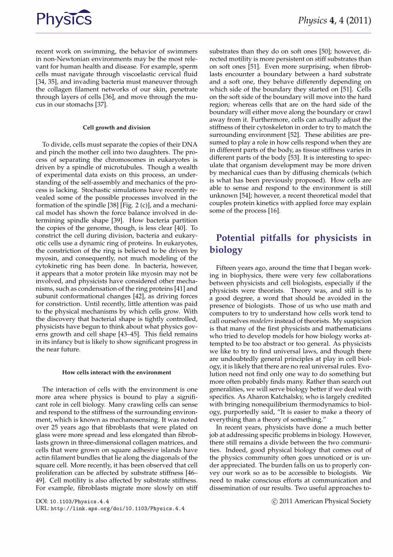
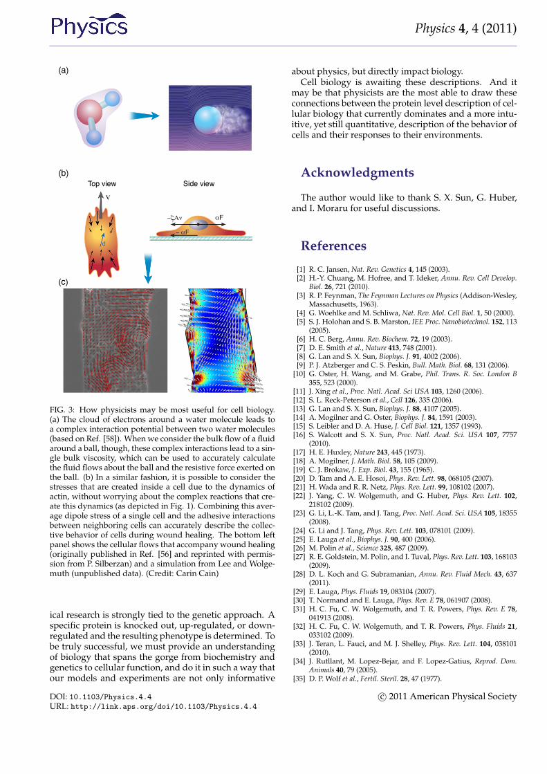
hand to the awaiting bat; the interaction between theball and the air matter much more. And now considerthis mass of air that surrounds the traveling ball [Fig.3 (a)]. Once again, the details of the molecular interac-tions, or even the identities of the molecular constituentsof the air, do not matter so much. Between statistical me-chanics and fluid mechanics, we can gain a much bet-ter description of the bulk behavior of the air than wecould if we considered the air at a more fundamentallevel. Indeed, at the level of a fluid, the molecular de-tails average together into a much smaller set of bulkmaterial parameters, such as the viscosity or the coeffi-cient of thermal expansion. It is these bulk parametersthat determine the course of the flying baseball.
As an example, Pilhwa Lee and I have recently beenworking on a model to describe the physics that isinvolved in wound healing. When an organism iswounded, epithelial cells crawl to fill in the woundedarea. An experimental method for exploring this pro-cess is to grow a monolayer of epithelial cells on a sub-strate and then to “wound” the layer using a scalpel orsome other object to scrape away a region of cells. PascalSilberzan and co-workers have shown that the motionof the cells during wound healing is not trivial and in-volves long-range order and complex cellular flows [56].Based on these observations and an analogy betweencrawling cells and the collective swimming of bacte-ria, we proposed a model that captures many featuresthat are observed in wound healing assays. We suggestthat two dominant physical attributes are responsiblefor most of the processes involved in wound healing:(i) the dipole nature of the stress distribution of a crawl-ing cell and (ii) cell-cell adhesions. This model absorbsall of the complex biochemistry and actin dynamics in-side a cell into two parameters that describe the stressthat a cell exerts on its surroundings, and cell-cell ad-hesion dynamics can be shown to lead to visco-elasticcouplings between cells [Fig. 3 (b)]. Therefore, wheremany groups have focused extensively on the complexbiochemical interactions inside the cell, at a functionallevel (i.e., healing of a wound), many of the moleculardetails may only act to regulate a few bulk physical pa-rameters.
However, for biology, and especially medicine, it willnot suffice to just develop nonreductionist theories ofcellular function; it will also be necessary to compute theeffective parameters of the theories in terms of the actualmolecular level interactions. (Yet another task for whichphysicists are well suited.) The current paradigm of dis-ease treatment is molecule based. We develop drugsthat interact with or replace the proteins that our bodiesare or aren’t making. We seek out poisons that specifi-cally target cancer cells. Alzheimer’s patients are treatedwith molecules that prevent the breakdown of acetyl-choline, a chemical implicated in learning and memory.And, some diseases, such as cystic fibrosis, can evenbe treated by replacing defective genes in an individualwith a functional copy of the gene. In addition, biolog-
DOI: 10.1103/Physics.4.4URL: http://link.aps.org/doi/10.1103/Physics.4.4
c© 2011 American Physical Society
Physics 4, 4 (2011)
FIG. 3: How physicists may be most useful for cell biology.(a) The cloud of electrons around a water molecule leads toa complex interaction potential between two water molecules(based on Ref. [58]). When we consider the bulk flow of a fluidaround a ball, though, these complex interactions lead to a sin-gle bulk viscosity, which can be used to accurately calculatethe fluid flows about the ball and the resistive force exerted onthe ball. (b) In a similar fashion, it is possible to consider thestresses that are created inside a cell due to the dynamics ofactin, without worrying about the complex reactions that cre-ate this dynamics (as depicted in Fig. 1). Combining this aver-age dipole stress of a single cell and the adhesive interactionsbetween neighboring cells can accurately describe the collec-tive behavior of cells during wound healing. The bottom leftpanel shows the cellular flows that accompany wound healing(originally published in Ref. [56] and reprinted with permis-sion from P. Silberzan) and a simulation from Lee and Wolge-muth (unpublished data). (Credit: Carin Cain)
ical research is strongly tied to the genetic approach. Aspecific protein is knocked out, up-regulated, or down-regulated and the resulting phenotype is determined. Tobe truly successful, we must provide an understandingof biology that spans the gorge from biochemistry andgenetics to cellular function, and do it in such a way thatour models and experiments are not only informative
about physics, but directly impact biology.Cell biology is awaiting these descriptions. And it
may be that physicists are the most able to draw theseconnections between the protein level description of cel-lular biology that currently dominates and a more intu-itive, yet still quantitative, description of the behavior ofcells and their responses to their environments.
Acknowledgments
The author would like to thank S. X. Sun, G. Huber,and I. Moraru for useful discussions.
References
[1] R. C. Jansen, Nat. Rev. Genetics 4, 145 (2003).[2] H.-Y. Chuang, M. Hofree, and T. Ideker, Annu. Rev. Cell Develop.
Biol. 26, 721 (2010).[3] R. P. Feynman, The Feynman Lectures on Physics (Addison-Wesley,
Massachusetts, 1963).[4] G. Woehlke and M. Schliwa, Nat. Rev. Mol. Cell Biol. 1, 50 (2000).[5] S. J. Holohan and S. B. Marston, IEE Proc. Nanobiotechnol. 152, 113
(2005).[6] H. C. Berg, Annu. Rev. Biochem. 72, 19 (2003).[7] D. E. Smith et al., Nature 413, 748 (2001).[8] G. Lan and S. X. Sun, Biophys. J. 91, 4002 (2006).[9] P. J. Atzberger and C. S. Peskin, Bull. Math. Biol. 68, 131 (2006).
[10] G. Oster, H. Wang, and M. Grabe, Phil. Trans. R. Soc. London B355, 523 (2000).
[11] J. Xing et al., Proc. Natl. Acad. Sci USA 103, 1260 (2006).[12] S. L. Reck-Peterson et al., Cell 126, 335 (2006).[13] G. Lan and S. X. Sun, Biophys. J. 88, 4107 (2005).[14] A. Mogilner and G. Oster, Biophys. J. 84, 1591 (2003).[15] S. Leibler and D. A. Huse, J. Cell Biol. 121, 1357 (1993).[16] S. Walcott and S. X. Sun, Proc. Natl. Acad. Sci. USA 107, 7757
(2010).[17] H. E. Huxley, Nature 243, 445 (1973).[18] A. Mogilner, J. Math. Biol. 58, 105 (2009).[19] C. J. Brokaw, J. Exp. Biol. 43, 155 (1965).[20] D. Tam and A. E. Hosoi, Phys. Rev. Lett. 98, 068105 (2007).[21] H. Wada and R. R. Netz, Phys. Rev. Lett. 99, 108102 (2007).[22] J. Yang, C. W. Wolgemuth, and G. Huber, Phys. Rev. Lett. 102,
218102 (2009).[23] G. Li, L.-K. Tam, and J. Tang, Proc. Natl. Acad. Sci. USA 105, 18355
(2008).[24] G. Li and J. Tang, Phys. Rev. Lett. 103, 078101 (2009).[25] E. Lauga et al., Biophys. J. 90, 400 (2006).[26] M. Polin et al., Science 325, 487 (2009).[27] R. E. Goldstein, M. Polin, and I. Tuval, Phys. Rev. Lett. 103, 168103
(2009).[28] D. L. Koch and G. Subramanian, Annu. Rev. Fluid Mech. 43, 637
(2011).[29] E. Lauga, Phys. Fluids 19, 083104 (2007).[30] T. Normand and E. Lauga, Phys. Rev. E 78, 061907 (2008).[31] H. C. Fu, C. W. Wolgemuth, and T. R. Powers, Phys. Rev. E 78,
041913 (2008).[32] H. C. Fu, C. W. Wolgemuth, and T. R. Powers, Phys. Fluids 21,
033102 (2009).[33] J. Teran, L. Fauci, and M. J. Shelley, Phys. Rev. Lett. 104, 038101
(2010).[34] J. Rutllant, M. Lopez-Bejar, and F. Lopez-Gatius, Reprod. Dom.
Animals 40, 79 (2005).[35] D. P. Wolf et al., Fertil. Steril. 28, 47 (1977).
DOI: 10.1103/Physics.4.4URL: http://link.aps.org/doi/10.1103/Physics.4.4
c© 2011 American Physical Society
Physics 4, 4 (2011)
[36] T. J. Moriarty et al., PLoS Pathogens 4, e1000090 (2008).[37] J. P. Celli et al., Proc. Natl. Acad. Sci. USA 106, 14321 (2009).[38] R. Paul et al., Proc. Natl. Acad. Sci. USA 106, 15708 (2009).[39] B. Rubinstein et al., Phys. Biol. 6, 016005 (2009).[40] T. Kruse and K. Gerdes, Trends Cell Biol. 15, 343 (2005).[41] G. Lan et al., Proc. Natl. Acad. Sci. USA 106, 121 (2009).[42] J. F. Allard and E. N. Cytrynbaum, Proc. Natl. Acad. Sci .USA 106,
145 (2009).[43] H. Y. Jiang and S. X. Sun, Phys. Rev. Lett. 105, 028101 (2010).[44] R. Mukhopadhyay and N. S. Wingreen, Phys. Rev. E 80, 062901
(2009).[45] O. Sliusarenko et al., Proc. Natl. Acad. Sci. USA 107, 10086 (2010).[46] E. Hadjipanayi, V. Mudera, and R. A. Brown, J. Tissue Eng. Regen.
Med. 3, 77 (2008).[47] J. P. Winer et al., Tissue Eng. Part A 15, 147 (2009).[48] H. B. Wang, M. Dembo, and Y. L. Wang, Am. Physiol. Cell Physiol.
279, C1345 (2000).[49] S. X. Hsiong et al., J. Biomed. Mater. Res. A 85, 145 (2008).[50] R. J. Pelham and Y. Wang, Proc. Natl. Acad. Sci. USA 94, 13661
(1997).[51] C. M. Lo et al., Biophys. J. 79, 144 (2000).[52] F. J. Byfield et al., Biophys. J. 96, 5095 (2009).[53] D. E. Discher, P. A. Janmey, and Y.-L. Wang, Science 310, 1139
(2005).[54] P. A. Janmey et al., Cell Motil. Cytoskel. 66, 597 (2009).[55] D. G. Drubin and G. Oster, Mol. Biol. Cell 21, 2099 (2010).[56] M. Poujade et al., Proc. Natl. Acad. Sci. USA 104, 15988 (2007).[57] S. Nishikawa et al., Cell 142, 879 (2010).[58] S. M. Kathmann, I. F. W. Kuo, and C. J. Mundy, J. Am. Chem. Soc.
130, 16556 (2008).
About the Author
Charles W. WolgemuthCharles Wolgemuth received his Ph.D. from the University of Arizona in 2000. After abrief stint at the University of California, Berkeley, working with George Oster, he took afaculty position in the Department of Cell Biology and the Richard D. Berlin Center for CellAnalysis and Modeling at the University of Connecticut Health Center, where he has beensince. He is currently the Director for the Cell Analysis and Modeling graduate programand an Editorial Board Member for the Biophysical Journal. His research investigates themechanisms that cells use to move, grow, and to create and maintain their shapes.
DOI: 10.1103/Physics.4.4URL: http://link.aps.org/doi/10.1103/Physics.4.4
c© 2011 American Physical Society