tumor necrosis factor ligand superfamily: involvement in the
TRANSCRIPT

REVIEW ARTICLE
Tumor Necrosis Factor Ligand Superfamily: Involvement in the Pathology of Malignant Lymphomas
By Hans-Jurgen Gruss and Steven K. Dower
P HYSIOLOGIC and pathologic activities of cytokines are mediated by binding to cell surface receptors (R). Se-
quence analysis of cytokine receptors defines several sub- families of membrane proteins with specific homology of functional domains. Receptor subfamilies of related proteins form (1) the Ig superfamily (eg, interleukin-l receptors [IL- lRs], fibroblast growth factor (FGF) Rs, platelet-derived growth factor (PDGF) Rs, c-kit, c-fms, flt-3) characterized by varying numbers of Ig-like repeats in the extracellular domain; (2) the hematopoietin (cytokine) receptor superfam- ily (eg, erythropoietin receptor [EPOR], growth hormone receptor [GHR], prolactin (PRL) Rs, mpl, ciliary neutrophic factor (CNTF) Rs, leukemia-inhibitory factor receptor [LIFR] gp130, IL-2R p and y chain, IL-3R, L 4 R , IL-SR, ILdR, IL-7R, IL-9R, granulocyte colony-stimulating factor receptor [G-CSFR], and granulocyte-macrophage-CSFR [GM-CSFR]) with conserved cysteine residues and the char- acteristic WSXWS motif; and (3) the tumor necrosis factor (TNF)/nerve growth factor (NGF) receptor superfamily based on cysteine-rich repeats in the extracellular region."' Further, receptors can be functionally, based on signaling, subdivided by common receptor subunits involved in form- ing several multimeric receptor complexes such as the gpl30-associated proteins for ILdR, LIFR, OncoMR, CNTFR, and IL-11R; common P-chain-associated mole- cules for IL-3R, IL-SR, and GM-CSFR complex; and com- mon y-chain-associated members (IL-2R, I L 4 R , L 7 R ,
The TNFNGF receptor superfamily contains at present 10 different membrane proteins and several viral open reading frames encoding TNFR-related molecules. The p75 low-af- finity NGF receptor was the first cloned receptor of this fa mil^.^ TNF was originally discovered by its antitumor ac- tivity in mice.' Subsequently, cloning of two specific recep- tors for TNF showed that there were related to the NGFR.9- I ' In recent years a new type-I-transmembrane TNF/NGF receptor superfamily has been e~tablished.'~~.~.' The TNF/ NGF receptor superfamily includes the p75 NGFR,7 p60 TNFR-I?-" p80 TNFR-11,'' TNFR-RPlTNFR-111," CD27,I3 CD30,14 CD40," 4-1BB,I6 0X4O,l7 and FAS/APO-1.".19 In addition, several viral open reading frames encoding soluble TNFRs have been identified, such as SFV-T2 in Shope fi- broma virus2' and Va53 or SaIF19R in Vaccinia virus.z'3zz These receptor superfamily is characterized by multiple cys-
IL-9R, IL-l3R, and IL-15R).6
From the Department of Biochemistry, Immunex Research and
Submitted November 3, 1994; accepted February 28, 1995. Address reprint requests to Hans-Jiirgen Gruss, MD, Freie Uni-
versity Berlin, UKRV-RRK, Department of Medical Oncology and Applied Molecular Biology, Lindenberger Weg 80, D-13122 Berlin, Germany.
Development COT, Seattle, WA.
0 1995 by The American Society of Hematology. 0006-4971/95/8512-W41$3.00/0
3378
teine-rich domains in the extracellular (amino-terminal) do- main, which have been shown to be involved in ligand bind-
extracellular region between the human family members are in the range of 25% to 30%.5,26 The NGFR, TNFR-I, TNFR- 11, and FAS/APO-1 have a broad tissue distribution, whereas the others (CD27, CD30, CD40, 4-1BB, and 0x40) are mainly restricted to cells of the lymphoidhematopoietic sys-
Ligands for these receptors have been identified and be- long to two recently formed cytokine superfamilies. The neurotrophins (NT; NGF ligand superfamily) are basic, NGF-like dimeric soluble molecules and include NGF, BDNF, NT-3, NT-4, and NT-5.27,28 The ligands of the TNF ligand superfamily are acidic, TNF-like molecules with ap- proximately 20% sequence homology in the extracellular domains (range, 12% to 36%) and exist mainly as mem- brane-bound forms; the biologically active form is a trimeric/ multimeric complex. To this group belong TNF,29-32 LTcx,"~.~~ LTP,3s CD27L,36 CD30L,37 CD40L:842 4-1BBL,'",44 0X40L,45,46 and FASL.47-50 Soluble forms of the TNF ligand superfamily have only been identified so far for TNF, LTcx,
of cell proliferation, activation, and differentiation, including control of cell survival or death by apoptosis or cytotoxic- it^.^,'
Malignant lymphomas are a heterogenous group of lymphoid tumors that mainly arise from the lymphoreticular system and are grouped, based on morphologic criteria, into two large categories, Hodgkin's disease (HD) and non- Hodgkin lymphomas (NHL).53 Furthermore, lymphomas are grouped into three major phenotype categories: B cell, T cell, and HD. The origin of the lymphoma cells in NHLs is most commonly from B cells (90%) and less frequently from T cells (lo%), with massive clonal expansion of the malig- nant cell population. In contrast, HD is defined by common clinical and pathologic The diagnosis of HD is typically based on a disrupted lymph node architecture and the presence of the malignant mononucleated Hodgkin and multinucleated Reed-Stemberg (H-RS) cells embedded in an abundance of normal, reactive cells (eg, lymphocytes, histiocytes, eosinophils, plasma cells, and stromal cells) without malignant tran~formation.'~ HD contains only a low proportion of the neoplastic H-RS cells, accounting for usu- ally less than 1% to 2% of the total tumor cell ma~s .~~ . " -~" The etiology of HD and the origin of the H-RS cells remains unclear with almost every cell type having been described as a normal ~ounterpart.~'"j~ Primary and cultured H-RS cells express a heterogenous panel of cytokines and cytokine re- ceptors that correlate with the typical clinical and pathologic presentation of HD case^.^^.^ Cytokines and a cell contact- dependent activation network are critical elements in the pathology of HD.
This review will summarize recent data on the functional
ing.2,10.23-25 The average homology in the cysteine-rich
and FASL.29,47,51,52 These proteins are involved in regulation
Blood, Vol 85, No 12 (June 15). 1995: pp 3378-3404
For personal use only.on January 10, 2019. by guest www.bloodjournal.orgFrom

TNF LIGAND FAMILY AND MALIGNANT LYMPHOMAS 3379
role and pathobiologic involvement of the TNF/NGF recep- tor and TNF ligand superfamilies for the pathology of malig- nant lymphomas.
STRUCTURAL FEATURES OF TNF RECEPTOR AND LIGAND SUPERFAMILIES
The TNF receptor superfamily consists, at present, of 10 mammalian In addition, several viral open reading frames (ORFs) have been identified that encode ac- quired soluble TNF receptors (eg, SFV-T2, va53, SaIF 19R, MYX-T2, G4R, and crmB).10*20-22.66-70 The TNF ligand super- family contains nine members, ie, the counterparts of the receptors.* The ligand for the low-affinity NFG receptor is structurally unrelated to the TNF superfamily of ligand^.'^
The crystal structures of TNFRI (p60), TNF, and LTa have been s01ved~”~~ and the mechanism of receptor-ligand interaction was i l l ~ s t r a t e d . ~ ~ . ~ ~ The ligand is a trir~~er.~’.~’ The receptor extracellular ligand binding region is a rod-like structure in which the four cysteine-rich 40 residue repeats each folds tightly together and forms intimate longitudinal contacts with its neighbors. The complex contains one LTaI TNF homotrimer and three receptor chains.73 The receptors bind in three grooves in the ligand trimer formed by the subunit interfaces; thus each receptor makes contact with two subunits. The structure predicts that the binding of ligand will crosslink three receptors together into a cluster. Recent reports suggest that, for LTa, this model may be an oversim- plification. A second form of lymphotoxin (lymphotoxin-P [LTP] or p33) has been identified35.75.76 that unlike LTa, appears to be membrane anchored. The TNFR-related pro- tein (TNFRF’) has been shown to be a specific receptor for LTP and hence is TNFRIII.77 In addition, it appears that LTa and LTP, when expressed in the same cell, can form heterotrimeric complexes.76 Because the sites for receptor binding lie at the subunit interfaces, such heterotrimers can contain three different sites (a2, P2, and ap) in various combinations depending on subunit stiochiometry. In which way such heterotrimeric ligands interact with cells express- ing combinations of TNFRI, TNFRII, and TNFRIII needs to be clarified.
The overall structures of two TNFR-like viral gene prod- ucts, the TNF receptors and the ligands, are shown in Fig 1 . In addition, Tables 1 and 2 summarize the sizes of the proteins as lengths of the various sequence segments and chromosomal location. Sequence alignments of the extracel- lular regions of the receptors show that they are distantly related (25% to 30% in general; summarized in Table 3). However when sequence conservation patterns are exam- ined, two features are immediately apparent?.” First, there is a characteristic pattern present in all the sequences, with the majority of conserved positions containing an unusually large number of cysteine residues. Second, all of the proteins are composed of several repeats of a core domain of 30 to 40 residue^.^.'^.^' The TNFRI crystal structure shows that this domain is composed of three elongated strands of residues held together by a twisted ladder of three disulphide bonds73; the strands that form the core of the structure contain approx- imately 25 residues, of which 6 are cysteines. The strands are joined by loops of less-conserved structure. Thus, the
three-dimensional structure shows that the cysteine-rich re- peats in the sequence present the charcteristic structural do- main in this family. TNFRI contains four such domains stacked longitudinally to form a bent rod. This rod structure thus forms an extended ligand binding unit, with the ligand contact side chains being located in domains 2 and 3. The largest variation in structure between the family members is found in the region between the C-terminus of the membrane proximal domain and the transmembrane region. This seg- ment varies from 8 residues in FASI8 to 70 residues in CD27I3 and is presumed to form a variable spacer between the ligand binding unit and the membrane. CD30 has an extracellular region in which the cysteine-rich repeat unit has been d~p1icated.l~ The observation that the CD30a and CD30b regions are far more closely related (50%; see Table 3) than any of the other family members are, suggests that this structure has arisen from tandem duplication event more recent than those that gave rise to the various family mem- bers from a common ancestor. CD30a is as related to the rest of the family as any of the other members, but CD30b, the membrane proximal region, is the most divergent se- quence of the family (Table 3). It seems likely therefore that it has evolved rapidly away from the rest of the family, may well have lost ligand binding activity, and serves the purpose of an extended spacer.
It is striking that this family of molecules shows a rela- tively low level of sequence conservation despite in all likeli- hood sharing a common fundamental structure. This finding suggests that the sequences have diverged rapidly. In support of this notion, comparisons of the sequences of the same member from different species show unusually low levels of conservation. This finding can be seen from two examples included in Table 3, ie, OX40 and CD40. In both cases the human and murine forms are only approximately 60% identical. For example, by contrast, the murine and human insulin receptor precursors are 95% identical when compar- ing the entire 1370 residues sequences. The selection pres- sure driving rapid divergence of the TNF receptor family members may well arise from the subversion of these sys- tems by pathogens. Thus, a number of TNFR viral ORFs (SFV-T2, va53, SaIF 19R, MYX-T2, G4R, and crmB) have been detected by sequence homology and found to be soluble TNF binding proteins capable of blocking TNF action.20.22369 These viral genes were presumably acquired from a host species by recombination and confer a selective advantage for the viruses by attenuating host immune and inflammatory responses. As a corollary of this argument, one would expect that evolution of a mutated ligand-receptor pair that was no longer inhibited by the viral gene product would be advanta- geous to the host.
Comparison of the cytoplasmic sequences of the receptors shows these to be considerably more diverse than the extra- cellular regions. Indeed, these differ markedly not only in sequence but also in size (Tables 2 and 3), and multiple sequence alignments show no evidence of any underlying shared structure running through the family. One striking comparison is that between murine and human CD40; whereas the membrane proximal 34 residues are 78% identi- cal, the murine molecule possesses an additional 27 residues
For personal use only.on January 10, 2019. by guest www.bloodjournal.orgFrom

3380
ox40
NGFR
41BB
CD27
CD30
CD40
TNFRIl
PV-ASJR
PV-TZ
II TNFRIII
TNFRI
I
B f" - 1
FAS [
100 amino - acids
GRUSS AND DOWER
0 x 4 0 ~
4IBBL
CD271.
- CDML
cD4n1.
LTu
TNF
LTa LTp 2
" FASL
Fig 1. The TNF receptor and ligand superfamilies. Schematic presentation of the 10 members of the TNF receptor superfamily and two examples of viral ORFs (PV-A53R and PV-T2) encoding soluble TNFR homologues (left panel). The TNF receptor family members are character- ized by variable numbers of cysteine-rich repeats in the extracellular domain. The homologous domains are shown as sequested open boxes, with the cysteine residues indicated by lines. TNFRlll is identical to the TNFR-RP (related protein). In addition, the nine members of the TNF ligand superfamily are shown in the right panel. The extracellular homologous C-terminal regions are indicated by open boxes and the nonhomologous sequences by lines. LT is shown in both the homotrimeric, secreted LTrr form and the heterotrimeric, membrane-anchored LTqLTp complex. Pro-TNF and FASL can be proteolytically cleaved for the release of active soluble forms. NGF is a basic, soluble dimeric molecule and the prototype of the NGF ligand superfamily. NGF has no homology to the TNF ligands and is therefore not included.
at the C-terminus. There are some elements shared between subsets of family members, thus TNFRI and FAS share the so-called "death d~main",~"'" but this sequence element is missing in the other two TNF receptors (TNFRII and TNFRPITNFRIII).'".'~.~~ Whether this degree of diversity is the consequence of rapid accumulations of mutations in this region of the genes for these molecules or of recombination events is not clear. However, it seems reasonable to suppose that there is unlikely to be any common immediate-early signalling event triggered through all these receptors. Indeed comparison of the sequences of this group of molecules as a whole with the databases fails to identify any motifs that resemble other families of signalling molecules, thus offer- ing no clues as to what the immediate early events for any of these molecules might be. Relatively little general infor- mation is yet available about the signal pathways to which the more recently characterized family members (CD40, CD30, 4-1BB, CD27, 0x40, and FAS) might couple, but a diverse set of signalling pathways seems likely for the diver- gent biologic responses. Most data for signalling through TNF receptors have come from studies using the TNFRI (p60) and TNFRII (p80) system and have been reviewed in detail recently."
The extracellular regions of TNF ligand family, like the receptors, are highly diverse in sequence, with identity levels
in the region of 20% for different family members. The murine and human forms of the same molecule are somewhat more conserved than the corresponding receptors, being ap- proximately 80% identical (Table 3C). However, sequence alignments show that as with the receptors there is a charac- teristic pattern of sequence conservation, there being 9 short regions of conserved sequence distributed along the length of the molecules (Fig 2). Superposition of the sequences on the three-dimensional structures of TNF and LTa shows that these regions correspond to the strands that form the core of the protein. The residues that lie on the loops joining the strands show no detectable conservation. Thus the ligand family has diverged as rapidly as the receptors. Several of the ligand superfamily members have moderate sized cyto- plasmic regions, and at least some are capable, when engaged by their receptors, of delivering signals (eg, CD27L, CD40L, and 4-1BBL)."" The signal pathway is presently unknown.
CHARACTERISTIC BIOLOGIC PROPERTIES OF TNF LIGAND SUPERFAMILY MEMBERS
TNF and LTa, products of activated macrophages and T cells can kill some transformed cell lines, mediate cell activa- tion and proliferation, and are functionally linked as primary mediators of immune regulation and inflammatory re- s p o n ~ e . ~ ~ ~ ~ ~ TNF has a pathogenic involvement, eg, in septic
For personal use only.on January 10, 2019. by guest www.bloodjournal.orgFrom

TNF LIGAND FAMILY AND MALIGNANT LYMPHOMAS 3381
Table 1. Molecular Characteristics for the Human TNF Receptor and Ligand Superfamily Members
Molecule Molecular Weight Length Chromosomal
(kD) (amino acids) Location ~ ~ ~~
TNFR family CD27 CD30 CD40 4.1 BB* OX40 FAS/APO-1 TNFRl (p601 TNFRll (p80) TNFRlll (TNFR-RP) NGFR p75
TNFL family CD27L CD3OL CD40L 4.1.BBL* OX40L FASL TNF LTa LT4
45-55 120 50 35 50 45 60 80
-75
75
50 26-40
33 50
26-28 40
17 (S) and 26 (m)l 25 33
242 578 258 234 250 320 435 440 414
402
193 234 261 309 183 281 233 205 244
12~13 1 p36 2Oqll-13 1 p36 1 p36 10q24.1 12~13 1 p36 12~13
17~121-22
19~13.3 9q33 Xq26-27 19~13.3 lq25 lq25 6 (MHCIS 6 (MHC) 6 (MHC)
* 4-168 and 4-1BBL represent the murine proteins. t Soluble (S) and membrane-bound (m) forms of TNF. * TNF, LTa. and LT@ chromosomal mapping as a cluster within the
location for the major histocompatibility complex (MHC).
shock, some autoimmune disorders, malignancies, and graft- versus-host disease.'
The nine TNF-related cytokines show distinctive but over- lapping cellular responses for developmental and regulatory networks involving cells of the lymphoid, hematopoietic, and other lineages, such as stromal cells and neuronal cells.4,' At least some of the nine TNF ligand superfamily members (eg, TNF, LTa, LTP, and CD40L) form trimeric proteins
members exert their biologic activity by causing receptor multimerization at the cell surface.73 As mentioned above, the LT0 homotrimer is the only entirely secreted protein and the TNF homotrimer is active after proteolytic release form the cell s ~ r f a c e . ~ ~ , ' ~ . ~ ~ The biologically relevant forms of the other family members are membrane-bound type I1 glyco-
CD30L, CD40L, 4-lBBL, and OX40L have not been re- ported. The exception is FASL, which exists in the predicted membrane-bound form but also as a soluble shed form in COS cell supernatants with presently unknown biologic rele- vance."
Most of the TNF receptor superfamily members exist also in a soluble form, released by proteolytic cleveage (eg, TNFR p60, TNFRp80, CD27, CD30, CD40, and FAS) or through alternative splicing (eg, 4-1BB).43,91"03 Although the cytoplasmic domains of most TNF receptor superfamily members are divergent from each other, several biologic functions, such as cytotoxic signals, induction of prolifera- tion and differentiation, and cellular activation, are shared
(see above).35.71.72,87-89 In general, the TNF ligand superfamily
proteins.3S-38.43.4S47,90 Natural soluble forms for CD27L,
between two or more ligands.' Biologic activities related to T-cell-mediated immunity are a unique feature for all members of the TNF ligand All ligands and receptors, without exception, are expressed on activated T cells (Table 4). Purified human T cells and T- cell clones show enhanced proliferation when stimulated with any recombinant TNF family ligand or crosslinked with antireceptor antibodies in the presence of anti-CD3 or other mitogens, such as phytohemagglutinin (PHA), phorbol my- ristate acetate (PMA), or Possible autocrine T-cell activation and growth control might be a common feature of this protein family. The induction of each ligand expression shows unique kinetics consistent with different roles for each of these ligands in the T-cell activa- tion."' For example, the induction of CD30L surface expres- sion on activated T cells is slower in comparison to other TNF ligands such as TNF, CD27L, CD40L, and 4-1BBL (maximal expression, 24 hours v 6 hours, respectively). B- cell proliferation and Ig secretion is induced by at least TNF, LTa, and CD40L. Further, several members participate in T- cell-dependent help for B cells, which are known to express TNFR-I, TNFR-11, CD27, CD30, CD40, FAS, and 4-1BB (Table 4).'06,'07 TNF, LTa, and CD4OL are mitogenic to B ce~~s~38.108-i i0 TNF, CD30L, and 4-1BBL are also abundantly expressed by activated macrophage^.^^,^^.^^^'^ Signals gener- ated by TNF superfamily ligands in target cells are produc- tively coordinated with accessory molecule expression (eg, LFA-1, ICAM-1, and B7 For example, TNF, LTa, CD30L, and CD40L are capable of inducing cellular aggregation and upregulation of LFA- 1 (CD 1 la)/ICAM- 1
Table 2. Structural Characteristics of TNF Receptor and Ligand Superfamily
No. of Amino Acid Residues
Extracellular Extracellular Transmembrane Cytoplasmic Domains
Receptors CD27 175 21 46 2.5 CD30 360 27 21 1 5.5 CD40 175 21 62 4 FAS 156 20 144 3 OX40 188 26 36 3.5 TNFRl 190 25 220 4 TNFRll 240 27 173 4 TNFRlll 20 1 26 187 4 4-1 BB 159 30 45 3.5 NGFR 225 23 154 4
Ligands CD27L 165 16 12 CD30L 172 26 36 CD40L 216 23 22 LTa 170 24 11 LT4 197 31 16 OX40L 139 21 23 TNF 176 28 29 4-1BBL 206 21 82 FASL 179 27 75
With the exception of 4-168 and 4-1BBL. all sequences used in the analysis were those of human proteins.
For personal use only.on January 10, 2019. by guest www.bloodjournal.orgFrom

3382 GRUSS AND DOWER
Table 3. Sequence Relationships Between Membsn of the TNF Receptor Superfamily and Between Members of the TNF Ligand Superfamily
A. Receptor Extracellular Regions MuOX40 HuNGFR Mu4-1BB HuCD27 HuCD3Oa HuCD30b HuCD40 MuCD40 HuTRll HuTR-RP HuTRl HuFAS
HuOX40 63.8 37.5 27.0 28.0 24.2 20.7 33.0 30.2 35.3 28.0 20.9 26.2 MuOX40 - 31.6 26.3 29.3 26.5 18.9 32.0 30.3 34.3 32.1 26.8 26.7 HuNGFR - - 25.3 25.8 25.6 25.8 25.6 28.4 26.4 26.3 34.4 23.3 Mu4-1BB - - - 26.4 16.8 16.9 30.8 32.6 31.7 29.5 17.3 27.5 HuCD27 - - - 19.5 22.5 22.8 25.7 25.5 23.3 21.3 25.7 HuCD30a - - - 50.9 24.2 25.2 28.7 29.3 29.1 23.1 HuCD3Ob - - - 19.1 21.0 24.4 18.6 18.7 16.0 HuCD40 - - - - - - - 59.3 37.0 35.3 26.1 31.1
MuCD40 - - - 32.7 30.1 27.2 35.3 HuTNFRll - - - 32.0 23.3 25.2 HuTNFR-RP - - - - - - - 27.0 33.8
HuTNFRl - - - 28.9
-
- - - - -
- - - - - - - - - - - - - -
- - - - - - - -
B. Receptor Cytoplasmic Regions MuOX40 HuNGFR
HuOX40 61.1 22.2 MuOX40 - 24.2 HuNGFR - Mu4”lBB
- -
HuCD27 HuCD3O HuCD40 MuCD40 HuTNFRll
- - - - - - - - - -
HuTNFR-RP
- -
HuTNFRl
- - -
Mu4-1BB HuCD27 MuCD40 HuCD40
16.1 14.3 27.6 21.6 18.6 33.8 -
HuCD30
30.5 11.4 18.8 19.5 28.9
HuTRll HuTR-RP
18.3 27.0 11.7 33.3 21.4 15.5 17.1 24.3 11.9 20.9 24.7 27.2 23.3 23.7 20.8 20.7 - 20.1
HuTRl HuFAS
14.3 17.4 30.3 8.8 20.7 15.2 12.2 7.5 21.7 18.6 18.6 9.7 21.0 25.0 15.9 11.4 16.1 15.6 21.4 14.5
23.9 -
18.9 26.7 18.2 -
13.9 23.3 19.6 28.5
21.6 14.3 16.9 19.0 23.4 15.6 77.9
C. Ligand Extracellular Regions Mu4-1BBL HuCD27L HuCDBOL HuCD4OL MuCD4OL HuTNF HuLTu HuLTP HuFASL MuFASL
HuOX40L Mu4-1BBL HuCD27L HuCD3OL HuCD40L MuCD40L HuTNF H U LTa HuLTP HuFASL
20.9 23.8 20.7 -
20.3 20.1 27.9 23.0
19.3 20.2 20.9 21.3 75.2 -
15.5 20.0 17.8 23.5 23.2 19.9 23.4 23.1 23.1 22.1 22.1 21.4 25.5 16.3 20.4 22.1 26.0 21.7 24.4 24.9 30.1 21.7 23.1 23.7 - 33.5 26.0 26.4 - - 30.9 29.6
26.4 - - - - - - -
22.1 24.5 20.3 24.7 24.6 24.0 31.7 28.2 31.7 80.7
The sequences were compared in individual pairs using BESTFIT. For the receptors, the regions compared were those defined in Table 1. For the ligands, the regions compared were the entire region C-terminal to the membrane spanning sequence. The values given are percent amino acid identity, allowing gaps to be placed to optimize the fit. The extracellular region of CD30 contains the same concensus sequences as the other receptors, but has been duplicated (see Fig 1) to be able to compare it directly with the other members; each of the duplicated regions was treated separately. CD30a is residues 23-190 of CD30 (numbered from the initiator Met) and CD3Ob is 191-379.
Abbreviations: Hu, human; Mu, murine; TRII, TNFR II ( ~ 8 0 ) ; TRI, TNFR l (p60); TR-RP, TNFR-RP (related protein).
(CD54) e~pression.”””~ CD30L and CD40L share the abil- ity to induce B7-1 and B7-2 expression, part of the strongest known T-cell costimulatory pathway.””’z0 In general, all TNF ligand superfamily members, including FASL and CD40L, are essential for T-cell costimulation and activation. It is of special interest that signals, at least through CD27L, CD40L, and 4-1BBL, can provide costimulation for acti- vated peripheral blood (PB) T cell^.*^^*^ Further studies need to be performed to see if other TNF ligand superfamily members are able to transduce a costimulatory signal.
The ability to induce cell death (necrosis andor apoptosis) is another unique feature of this family and is
presently established for TNF, LTa, CD30L, 4-1BBL, and
broadly on both myeloid, lymphoid, and stromal FAS mmoclonal antibodies (MoAbs) and FASL induce apop- totic (programmed) cell death and FASFASL interaction ap- pears to be involved in T-cell repertoire formation, including positive or negative selection, suggesting a role of the FAS- FASL interaction in peripheral T-cell tolerance.”3126136 Interest- ingly, the cytoplasmic domains of the 60-kD TNFR and the FAS antigen contains a 65 amino acid “death domain,” which is critical for signal transduction of the cytotoxic e f fec t~ .7~ . ’~~ Both receptors still use at least partially distinct s i e g path-
FASL.30,33,37.”.47.’2~.~~2 FAS and TNFRs expression is found
For personal use only.on January 10, 2019. by guest www.bloodjournal.orgFrom

TNF LIGAND FAMILY AND MALIGNANT LYMPHOMAS 3383
ways involved in apoptosis.80 The cytoplasmic domains of the p60 and p80 TNFRs are unrelated and the signaling of the p80 TNFR for cell death and in mediating TNF responses in general
Essential roles of several members of the TNF receptor or ligand family have been confirmed by naturally occuring or induced mutants that abolish the functional expression of the individual receptorfligand protein. Naturally occuring inactivating mutations of the FAS antigen (lpr mouse) and the FASL (gld mouse) cause both similar lymphoprolifera- tive diseases with lymphadenopathy and autoimmune dis- ease, suggesting a failure of the immune system to eliminate autoreactive T CD40 and CD4OL knock-out mice confirm that mutations of CD40L cause X-linked im- munodeficiency, with high levels of IgM and low levels of IgG (block for Ig isotype ~witching). '~~' '~' Hyper-IgM pa- tients show normal numbers and biologic function of B cells, but failure of T-cell-dependent B cell help because of non- functional CD40L.39*'52"55 Experimental deletion of the 60- kD TNFR gene in mice causes immunodeficiency with se- verely impaired clearance of bacterial pathogens and rapid death caused by infection, but resistance to the lethal effect of lipopolysaccharides (LPS).'56*'57 Lack of 80-kD TNFR showed only a minimal phenotype with modest resistance to the lethal effect of TNF. In addition, functional ablation of TNF and LTa by overexpression of a neutralizing TNF inhibitory fusion protein (60-kD TNFR extracellular domain fused to mouse IgG heavy chain) in mice show pronounced LPS and TNF resistance with comparable phenotypic effect seen for the homozygous deletion of the 60-kD TNFR
in a distinctive phenotype, characterized by the absence of structured lymph nodes and disordered splenic architec- ture.Iw In summary, several members of the TNF ligand and receptor superfamilies play crucial roles for lymphoid and thymic development, T-cell-mediated immune responses, T-cell-dependent help for B cells, and humoral B-cell activ- ity. The detailed interactive network for the immune re- sponse and lymphoid differentiation mediated by the TNF- like ligands needs further evaluation.
re,ns c0n~veTs~~,78 .79 .93 .138-147
gene.158*'59 Furthermore, the deletion of the LTa gene results
CD27L AND CD70 ARE IDENTICAL MOLECULES AND ARE EXPRESSED ON DIFFERENT LYMPHOMAS
The CD70 antigen was originally identified by the Ki-24 MoAb.I6' CD70 is expressed on many peripheral T- and B- cell lymphomas (50% to 70% of cases positive) with fre- quent CD70 positivity observed within the cytoplasm.'62 The strongest expression is found on H-RS cells of HD (96% to 100% of cases positive) followed by large-cell NHLs (60% to 80% of cases positive). CD70 expression was not found on lymphoma cells derived from precursor T and B cells, such as lymphoblastic lymphoma or acute lymphoblastic leu- kemia (ALL). Permanent cell lines showed high CD70 ex- pression only in those cell lines related to activated cells (eg, antigens, mitogens, and viral-transformed cells), but not in those resembling precursor T or B cells. In most cases, ex- pression of the CD70 molecule is associated with the expres- sion of other activation antigens, particularly CD25 and CD30.I6* However, 20% of B-cell NHLs and 5% of T-cell
NHLs expressed only CD70 antigen on the lymphoma cells.'62
For the immune system, CD70 is absent from resting lym- phocytes, but can be induced after activation.'" CD70 anti- gen is detectable on PHA-stimulated T and B lymphocytes after 24 hours and peaked at 96 hours, with 70% of stimu- lated peripheral blood B cells and 25% of T cells expressing the antigen.I6' CD70 expression could not be detected on resting or IFN-y-treated monocytes, neutrophils, or den- dritic cells. Recently, the CD70 gene was cloned and found to be identical to the cloned ligand for CD27 (CD27L).36.83*90 CD27 is expressed by medullary thymocytes, most periph- eral blood T cells, a subset of mature B cells, and NK cell^.'^^"^' CD27 expression on T cells is associated with the helper phenotype (naive T cells with CD45RAf), whereas most memory T cells (CD45RA-,CD45ROf) lack CD27.98,165.'7' Activation of T cells results in upregulation of CD27 expression as cell surface molecule but also in the release of a soluble 28- to 32-kD form of CD27
can serve as a marker for the immune activation in V ~ V O . ' ~ , ' ~ ~
The distribution and regulation pattern of CD27 for T cells supports a CD27 function for more naivehnprimed T cells than completely differentiated effector T cell^.^^,'^^.'^'
The biologic functions of CD27L include a costimulatory signal for T-cell proliferation, generation of cytotoxic T cells, and enhanced cytokine secretion,36 but its functional relevance for thymocytes and B cells remains to be eluci- dated. It is of interest that only CD27+ B cells can be induced to secrete Ig in vitro after stimulation with mitogens or CD27L.164.'65*'72 In addition, CD27L antibodies or CD27 block allogenic B-cell-mediated stimulation of T-cell prolif-
CD27LJCD70 molecule is expressed on most lymphocytes in occassional tonsil germinal centers, low number of lym- phocytes in the paracortical areas of tonsils, lymphoblasts in the skin and gut, and thymic epithelial cells, but not on cortical and medullary thymocyte^.'^^,'^' Preliminary func- tional data suggest that the CD27-CD27UCD70 interaction is part of the network involved in regulation of T-cell activa- tion during antigen-specific immune responses, generation of memory T-cell populations, and expansion of cytotoxic T cells.'74
A relatively high percentage of T- and B-cell NHLs but also HD express CD27UCD70; this expression is character- istically high on H-RS cell^.'^^.'^^ In addition, most HD- derived cell lines express CD27L surface molecules but not the counterstructure CD2763 (H.J.G., unpublished observa- tion). B-cell NHL cell lines can express both the CD27 and CD27LKD70 molecule, but its functional relevance for the growth control of these cells has not been established. CD27 expression was found in 50% of B-cell leukemias and 71% of B-cell CD27 was present on malignant B cells corresponding to early stages of antigen-independent B-cell maturation. Pro-B-cell ALLs were CD27-, but 30% of pre- B-cell ALLs were positive. Mature B-cell ALLs had a high level of expression of CD27 and chronic lymphocytic leuke- mia (CLL), prolymphocytic leukemias, and some hairy cell leukemias (HCL) were moderate to strong CD27+. In addi-
(sCD27).98-101,165.171 Th e proteolytic shed sCD27 molecule
eration~90,164,165.172,173 Immunohistologic studies show that the
For personal use only.on January 10, 2019. by guest www.bloodjournal.orgFrom

3384 GRUSS AND DOWER
Fig 2. TNF ligand superfamily alignment and predicted tertiary structure based on TNF. Sequence homology is mtricted to the C-terminal region of TNF family members (left panel). Colored bars on the top line (SECOND) Indicate @-strands of TNF tertiary structure. Conserved amino acid motifs are shown by the line CONCNS. Amino acids are color-coded green, hydrophobic; blue, basic; red, acidic; yellow, cysteine. (Right panel) Crystal structure of TNF with @-strands colored according to the corresponding sequence homology. Gray strands indicate nonhomologous amino acid. Hu and H, human; m, murine; 1, ligand; tnf, tumor nmoris factor; it, lymphatoxin; cd, CD (elustar designation).
tion, most low-grade diffuse and follicular lymphomas (85% of cases positive) and intermediate- and high-grade lympho- mas (62% of cases positive with variable expression levels) expressed CD27 on the malignant B-cell population. My- eloma cells lacked expression of CD27. Furthermore, sCD27 was elevated in the serum of patients with B-cell malignan- cies and the highest levels were observed in patients with CLL and low-grade NHLs. The sCD27 serum levels showed a strong correlation to the tumor load, indicating sCD27 s e m levels as a useful disease marker in patients with B- cell malignan~ies.'~~
Taken together, CD27 and CD27UCD70 seem to be ex- pressed with high frequency by the malignant cells of differ- ent entities of lymphomas and may serve as markers for tumor burden and disease activity, but any functional correla- tion to defined pathophysiologic presentation has not been established (Fig 3).
CD30 AN HD-ASSOCIATED ANTIGEN AND ITS LIGAND
HD-derived cell lines were used to develop MoAbs that could be used to visualize H-RS cells in tissue sections. Ki-
1, the first CD30 MoAb, was raised against the HD-derived cell line L-428 and described to react uniquely with primary and cultured H-RS cells.'" A small lymphoid cell population in reactive tonsils was also stained and postulated to be the precursor cells for H-RS cells.'78 Subsequent studies clearly showed that the CD30 MoAb (Ki-l) was neither cell-lineage restricted nor specific for H-RS cells.'79 Over the years, mul- tiple MoAbs against the CD30 antigen (eg, Ber-H2, HeFi, HRS-l, HRS-2, HRS-3, M44, M67, and C10) have been generated with better immunochemical properties than Ki-
CD30 MoAbs detect a phosphorylated 120-kD membrane glycoprotein and its nonphosphorylated 84-kD precursor protein.'",'8s The cloning of the CD30 antigen has suggested that it might act as a cyt~kine.receptor.'~~'~
In addition to the CD30 staining for a small cell population in the parafollicular area of hyperplastic lymph nodes and tonsils, most blasts appearing during infectious mononu- cleosis are positive for CD30 expre~sion.'~~~'~'~'~~.'~~ CD30 expression has also been detected on a subset of mitogen- or antigen-activated PBTs, Epstein-barr virus (EBV)-
For personal use only.on January 10, 2019. by guest www.bloodjournal.orgFrom

TNF LIGAND FAMILY AND MALIGNANT LYMPHOMAS 3385
Table 4. Role of the TNF Ligand Superfamily Members for T- and B-Cell Activation Involved in the Immune Response
Function CD27L CDBOL CD4OL 4.l.BBL OX40L FASL
Expressing cells Responding cells Signaling through ligand T-cell costimulation B-cell proliferation Enhanced cytokine secretion lg secretion Upregulation of cell surface antigen expression Upregulation of costimulatory molecules Aggregationladhesion Apoptosis/necrosis/AlCD
TI, B*, Mf T", Mf*, G T**, B*, NK T*, Bt. NK
+ ? + +
- + +
- + + + +
+ +§
r* T*.S,Mf* B*t T* t T* B, EP, Mf*, T* T*, Mf T' Tt, B, Mf. G, S
+ + ? 7 + + + + + + + + + + + + + + + -11 + +
Abbreviations: B, B cells; BA, basophils; EN, endothelial cells; EP, epithelial cells; G, granulocytes; M, mast cells; Mf, monocyteslmacrophages;
Strong expression after activation. t Induction of expression after viral transformation (eg, HTLV-1, HTLV-2, EBV, and HIV). * CD27 expression is found mainly for naive > memory T cells, but FAS expression for memory > naive T-cell populatons. § Cytolytic and cytostatic effect on LCAL-derived tumor cell lines. 11 Rescue of germinal center B cells from undergoing spontaneous apoptosis.
NK, natural killer cells; S, stromal cells/fibroblasts; T, T cells; AICD, activation-induced cell death.
transformed B cells, and human T-lymphotropic virus types I and I1 (HTLV-I and -11) infected lymphocytes or cell
among T-cell clones, CD30 is mainly expressed and released as CD30 from CD4' and CD8' T-cell clones producing TH-2 cytokines such as IL-4 and IL-5.'9"'93 Activated tonsil B cells have been reported to be CD30+.'78.'8' Similarly, human NK cell clones express the CD30 surface protein.'" Furthermore, the expression of CD30 was seen by late-stage differentiated PB macrophages.'95 These data are controver- sial because other groups could not detect CD30 protein or mRNA expression by activated monocytes or macro-
CD30 protein and mRNA expression are found in all HD- derived cell lines, with the exception of the myelomonocytic cell line HD-My2 and cell line SUP-HDl.14~h2.h"'22~'% The
~ ~ n e s ~ 1 7 " 1 8 0 - i " . l R . l ~ 9 . 1 ~ In addition, it has been shown that,
phages.63.122.17R.181
CD30 antigen is seen on the majority of H-RS cells of most HD cases, with the exception of the lymphocyte-predomi- nant (LP) subform.6'.63 Overall, 84% to 91 % of the lympho- cyte-depleted (LD), mixed cellularity (MC), and nodular sclerosing (NS) HD cases, but only 32% of LP HD cases express CD30 on the diagnostic primary H-RS
CD30 expression is not restricted to the malignant cells of HD because CD30 MoAbs also identify a new entity of NHLs with anaplastic morphology (CD30' anaplastic large-cell lymphomas [ALCL]).'7R In addition to H-RS cells of HD and the subgroup of CD30' ALCLs, CD30 is also expressed to variable extents on several histologic subtypes of other NHL, such as cutaneous T-cell lymphoma, nodular small cleaved-cell lymphoma, lymphocytic lym- phoma, peripheral T-cell lymphoma, Lennert's lymphoma, immunoblastic lymphoma, adult T-cell leukemidlymphoma
c e ~ ~ s ~ 1 7 7 . 1 7 K . l R l . 1 9 7 - l ~
Fig 3. Expression and potential functional involvement of CD27L for HD and NHL. (A) For HD, CD27L (CD701 expression is found for most H-RS cells and surrounding B and T cells. CD27L might be criti- cally involved in hyperreactive bystander cell reac- tion. (B) NHL cases show frequent coexpression of CD27 and CD27L. CD27L could be an autocrine andl or paracrine growth factor for lymphoma cells.
A. HD
'1 3 @ 1. Activation of bystander cells,
particularly CD4+ T cells
2. Involvement in typical, hyperreactive bystander cell reaction
6. NHL
3 1.
2.
3.
4.
Activation of lymphoid cells
T cell proliferation and cytokine release
Generation of cytotoxic T cells
Possible (?) autocrine and/or paracrine growth factor for
U lymphoma cells
For personal use only.on January 10, 2019. by guest www.bloodjournal.orgFrom

3386 GRUSS AND DOWER
(ATLL), T-acute lymphoblastic leukemia (T-ALL), and centroblastic/centrocytic follicular lyrnph~rna.'~~.'~~~~~-~'~ Taken together, around 10% of NHLs are positive for CD30 on their malignant lymphoma cells (32% of T-cell NHLs and 4% of B-cell NHLS).~~.'" CD30 expression in T-cell NHLs of different subtpes is restricted to the large-cell vari- ants.186 The functional relevance of CD30 expression for most entities of NHLs remains presently unclear. The associ- ation between CD30 expression and lymphoid malignancies has proven to be a useful pathologic and clinical marker for the identification of malignant cells within lymphoid tissues, particularly lymph nodes. However, expression of CD30 has also been reported on some embryonal carcinomas, nonem- bryonal carcinomas, malignant melanomas, mesenchymal tumors, some myeloid cell lines, and decidual The CD30 antigen is suitable for immuno-imaging using immunoscintigraphy with radioiodine-labeled antibodiesIs2 and immunotherapy using immunotoxins (ricin-A or saporin conjugated MoAbs) in HD patients.'86.214.21s
The 85-kD CD30 molecule is detectable in the serum under restricted conditions?2 Detectable high sCD30 serum levels were found in the majority of HD cases at diagnosis, more often in patients with advanced disease, bulky tumor, andor presence of constitutional B symptom^.^'^-^'^ Elevated sCD30 serum levels correlate with the clinical presentation, such as stage, presence of B symptoms, and tumor burden. The sCD30 in the serum of HD patients derives most likely from the CD30+ H-RS cells and is associated with the extent of neoplastic infiltration in HD-involved areas.177.'78,2i7,218 Most cases with LD HD had positive serum levels, but LP HD patients were frequently sCD30-.2203221 Soluble CD30 was not detected in any HD patients in complete remission. The levels of C D 3 0 represent an independent prognostic factor with high sCD30 levels being associated with reduced disease-free sur~ival.2'~ The detection of sCD30 seems to be a useful "tumor marker" for CD30+ HD patients based on its correlation to disease activity and presence of H-RS cells.
Recently, the ligand for the CD30 antigen has been cloned and biologically characteri~ed.~~ The presence of the CD30 counterstructure (CD30L) confirms the presumed cytokine receptor function of CD30. The CD30L is a 26- to 40-kD type I1 membrane glycoprotein, mainly expressed on acti- vated T cells and monocytes/macrophages but also on granu- locytes and some Burkitt's lymphoma cell line^."^'**,^^^
Nonpathologic, CD30 expression is largely restricted to antigen-activated T cells and is not detectable before complete and functional antigen receptor rearrange-
ogen- or antigen-induced CD30 expression on CD45RO' PBT cells.'9o In contrast, CD30L expression i s broader and has been found on CD4' and CD8+ activated T cells with all CD45 i s o f ~ r m s . ~ ~ , ' ~ ~ CD30L costimulates T-cell prolifera- tiod7 and enhances induction of activation antigens, such as ICAM-1, on activated T cells.Io5 In addition, CD30L upregu- lates cytokine secretion of activated PBTs such as IL-2, IFN- y , and TNF but not IL-4.Io5 CD30+ T-cell clones (CD4+ and CD8+) produce preferentially TH-2 cytokines (eg, IL-4 and IL-5), but peripheral blood T cells stimulated through CD30 release mainly T,-l cytokines (eg, IL-2 and I F N -
ments~10S.178.180,181.184.189.190 IL-2 is further able to enhance mit-
y)~lo5,191,1Y2,223 Further studies are needed to determine whether the heterogenous population of CD30' T cells con- tains T cells that are capable of developing into both TH-l and TH-2 phenotypes or whether clonal CD30' T cells are exclusively from the T,-2 subtype. In general, CD30L is a part of the cascade involved in antigen-induced T-cell activa- tion and proliferation and might play a pathogenic role in several immunologic diseases associated with TH-2 cytokine pattern (eg, systemic lupus erythematosus, atopic disorders, Omenn's syndrome, and human immunodeficiency virus [HIV] infe~tion).'~'
The analysis of 105 continous human leukemia-lymphoma cell lines for CD30L showed a restricted expression pattern for 6 of the 26 B-cell-lineage tumor cell lines (4 CD30- Burkitt lymphoma [BL] cell lines, 1 CD30- BL-like ALL, and 1 NHL).222 All HD-derived cell lines were CD30L mRNA and surface protein expression negative.I2' Recombi- nant CD30L was mitogenic for the "T-cell-like" HD-de- rived cell lines HDLM-2 and L-540 and the T-ALL cell line KE-37 but not for the "B-cell-like'' HD-derived cell lines KM-H2 and L-428 or CD30+ BL cell lines.I2' In addition CD30L is capable of enhancing IL-6, TNF, and LTa secre- tion for HD-derived cell lines and upregulates surface ex- pression of ICAM-1 and B7 family members in a similar fashion as seen for CD40L (see below).Il7 CD30L seems to be another paracrine-acting molecule involved in the deregu- lated cytokine and activation cascade, characteristic for HD.64 CD30L could be produced by H-RS cell surrounding bystander cells, such as activated CD4+ T cells and activated macrophages and granulocytes (Fig 4). CD30L could modify cytokine secretion of H-RS cells and enhance proliferation and activation of H-RS cells, including upregulation of cell contact-dependent signals. The overexpression of the cyto- kine receptor CD30 on most H-RS cells appears to be an important clinical, biologic, and pathologic marker for HD (Fig 4). Further understanding of the CD30-CD30L interac- tion for the oncogenesis of HD will be hopefully generated from studies analyzing CD30L expression in primary HD cases and in vivo models, such as the HD-SCID mice system.
CD3OL TRANSDUCES ANTIPROLIFERATIVE SIGNALS TO CD30+ ALCLs
CD30' ALCLs (approximately 10% of all NHLs) express characteristically high amounts of CD30 on the surface of their clustered malignant lymphoma cells with either T, B, or 0 phenotype.I7* CD30+ ALCLs are characterized by the presence of large, pleomorphic tumor cells; expression of lymphocyte activation antigens (eg, CD25, CD30, CD71, and MHC class 11); a frequent nonrandom chromosomal ab- normality (t:2; 5); and frequent extranodal disease affecting the skin, lung, gastrointestinal tract, soft tissue, and
CD30' ALCL can be confused with HD be- cause of the presence of occasional H-RS-like cells and the overlapping immunophenotype of the tumor ceUs.'78.'",22A-227.231 In practice, H-RS cells in HD are frequently CD15+, CD30', CD45-, EMA-, in contrast to ALCL tumor cells with CD15-, CD30+, CD45+, EMA+ immunophenotype.2322"1 HD is in 50% to 60% of cases associated with EBV.63 The association of EBV with CD30' ALCLs remains controver-
bone. 178,224-230
For personal use only.on January 10, 2019. by guest www.bloodjournal.orgFrom

TNF LIGAND FAMILY AND MALIGNANT LYMPHOMAS 3381
3
1. T cell activation with increased antigen expression, cytokine secretion and proliferation
2. Possible mitogenic growth factor (paracrine) for H-RS cels
3. Stimulation of H-RS cells with enhanced cytokine production and activation of antigen expression
4. Involvement in hyperreactive CD4+ T cell response with activation and rosetting
Fig 4. Schematic presentation for the possible functional role of CD3OL for the pathology of HD. CD3OL expression is found as membrane- bound protein on activated T cells, monocyteslmacrophages, and granulocytes (cell types all involved in hyperreactive bystander cell reaction). H-RS cells are CD3OL-. CD30, as an HD-associated antigen, is expressed by most primary H-RS cells (lymphocyte-predominant subform is an exception). The CD30-CD30L interaction between CD30’ H-RS cells and CD30L’ bystander cells could be part of the deregulated cell-cell interaction, including growth stimulation, upregulation of activation antigens, and enhancement of cytokine release.
sial (range, 0% to 67% of cases positive), but most data would support the lack of a strong relationship between CD30’ ALCLs and EBV.”4-”7 These findings suggest that the presence of CD30 is not a simple relation to EBV infec- tion in malignant lymphomas.
Several EBV- ALCL-derived cell lines show strong CD30 expression and could be biologic targets for CD30L. In con- trast to the HD-derived cell lines, CD30 mediates reduction of proliferation and cell growth arrest (antiproliferative ef- fect) for most of the CD30’ ALCL cell lines.”’ The antipro- liferative effect of CD30L includes a cytolytic effect and a cytostatic component with cell cycle arrest.’” The rnecha- nism for the cytolytic effect is presently unlear, but seems to be FAS-independent and not associated with apoptotic DNA fragmentation. Further investigations have to clarify
the interaction between tumor growth of the CD30’ anaplas- tic lymphoma cells, tumor progressionhegression. and CD30L expression in ALCL-involved tissues (Fig S).
It is of interest, that the t:2;S translocation is a common chromosomal finding for CD30’ ALCLS.”’.’~” This re- arrangement fuses the nucleolar phosphoprotein nucleophos- min (NPM) gene on chromosome 5q3S to the novel tyrosine kinase gene ALK (anaplastic lymphoma kinase) on chromo- some 2~23.’~’ The association of the overexpression of the truncated ALK-fusion protein with the malignant transfor- mation in the CD30’ ALCLs is presently unclear, but an 80-kD protein tyrosine kinase (identical to ALK) has been suggested to physically interact with CD30.13’ It is of interest that only a fraction of CD30’ ALCL ( 1 2% to 40%) cases show the rearrangement of the NPM gene and that at least
Fig 5. Pathologic role of CD30 and CD3OL for NHLs. NHL cells [L) express either CD30 (A) or CD3OL [B). CD30 expression of NHL cells could be involved in cellular activation, such as release of cytokines or enhanced expression of activation antigens, and in growth control. A subgroup of ALCL cells use CD30 to mediate an antiproliferative effect. The main source of CD30’ cells is activated T cells. CD3OL’ lymphoma cells might induce T-cell activation and
1. Possible involvement in growth control of lymphoma cells (e.g., antiproliferative effect on LCAL tumor cells) 3
2. Activation of lymphoma cells with enhanced cytokine secretion and activation including antigen expression
B. CD3OL’ NHL
m 3 1. T cell activation with proliferation,
cytokine secretion and surface antigen expression
2. Induction of cellular immune response
cellular immune responses. I
For personal use only.on January 10, 2019. by guest www.bloodjournal.orgFrom

3388 GRUSS AND DOWER
a fraction of HD cases may contain the NPWALK fusion transcripts (3 studies using reverse transcriptase-polymerase chain reaction [RT-PCR]: 0 of 40, 2 of 9, and 9 of 13 HD cases rearranged).”242 Further studies have to show whether HD cases with rearranged NPM gene represent ALCLs that mimic HD or whether it presents a separate HD subform closely related to ALCL. In addition, CD30f ALCLs present frequently with abnormal c-myc gene products (6 of 18 cases [33%]), but the functional significance of this is ~nc lea r .~ ’ Taken together, recently several molecular alterations for CD30+ ALCLs have been identified. Further studies will hopefully connect these molecular abnormalities with the functional role of CD30-CD30L interaction for oncogenesis and pathogenesis of ALCLs. In addition, the prognostic, bio- logic, and functional role of CD30 expression of the malig- nant cells of other NHLs needs further evaluation.
CD40L SHARES COMMON BIOLOGIC ACTIVITIES WITH CD3OL FOR HD
CD40 is a 50-kD glycoprotein and is expressed on a vari- ety of cell types, including normal, virally transformed and malignant B cells (see below for more details), but also monocytes, activated T cells, follicular dentritic cells, inter- digitating reticulum cells, thymic epithelium, and some epi- thelial carcinoma^.^^*^^'.^^^ Recently, the murine and hu- man CD40L have been characterized and cloned?8” The CD4OL is a 33- to 39-kD type I1 membrane glycoprotein and is expressed primarily on the surface of activated CD4+ T cells but also on some CD8’ T cells, mast and stromal cell lines, and basophil^.^*"^*^ Studies using MoAbs to CD40 or recombinant CD40L have shown diverse biologic activities as a result of signaling through CD40.65,249 These activities include the proliferation of B cells and induction of Ig secre- tion in the presence of other cytokine~?~’ Furthermore, CD4OL-CD40 interactions mediate rescue of germinal center centrocytes from apoptosis.251s252 CD40 exposure of thymic epithelial cells in the presence of IFN-y and IL-1 induced GM-CSF release.M Signals through CD40 upregulate the expression of LFA-1, B7 ligands, ICA“1, and CD23 with involvement in both homotypic and heterotypic cell adhesion
In addition, primary PB monocytes express low amounts of CD40 and cytokines, such as GM-CSF, IL-3, or IFN-y, upregulate this CD40 surface expre~sion.~’~ CD40L induces/enhances cytokine se- cretion of PB monocytes (eg, IL-6, IL-8, and TNF) and potent tumoricidal activity of monocytes.256 Recently, it was shown that CD4OL also costimulates T-cell proliferation and enhances expression of CD25 and CD4OL itself.” Further- more, CD40L can transduce signals by its own.” Taken together, CD40L may play a complex role in the immune response by functionally interacting with CD40 expressed on the surface of B cells, monocytes, T cells, and some epithelial cells.
A series of HD-derived cell lines (exception the myelomo- nocytic HD-MyZ cell line) express at the mRNA and protein level not only CD30, but also CD40.’22~’%~25’”58 On the other hand, these cell lines are negative for CD3OL and CD4OL mRNA and protein expre~sion.’~’*’~~ Expression of CD40 by H-RS cells has been initially described as an indication for
and costimu~ation~l13.115.”6.118-120,253-~55
Table 5. CDSOL and CD40L Share Common Biologic Activities on Cultured H-RS Cells
Function CDBOL CD4OL ~
Mitogenic activity + -
Enhanced clonogenic colony formation + + Induction of cytokine secretion + + Induction of ICAM-l surface expression + + Increased soluble ICAM-1 concentration + + Aggregatiodadhesion + + Upregulation of costimulatory molecules
(eg, B7 ligands) + + Downmodulation of CD30 surface expression + + Shedding of CD30 + + Shedding of C040 + +(V
a follicular dendritic cell origin of H-RS cells.259 Recently, three studies have been reported on high level CD40 expres- sion of primary H-RS ~ e 1 1 s . ” ~ ~ ’ ~ ~ ~ ’ ~ A total number of 156 HD cases have been investigated and primary H-RS cells in 145 HD cases express CD40 (95% of cases positive). HD- involved tissues with all four histologic subtypes expressed abundant amounts of CD40 by H-RS cells independent of CD30 expression and irrespective of their antigenic immuno- phenotype. Further studies have to investigate the relation- ship between the expression of cytokines (eg, IL-4 and IFN- y ) andlor EBV proteins (eg, LMP-1) known to upregulate CD40 expression of H-RS cells and the deregulated, strong CD40 expression of H-RS cells. Primary H-RS cells did not express CD40L, but scattered lymphoid cells in disease- involved areas of HD were CD40L+.’” It is of interest that at least in part the in vitro rosetting of C M L + (activated), CD4+ T cells to cultured CD40’ H-RS cells is mediated through the CD4O/CD40L adhesion pathway.’”
Functional analysis of the CD40’ cultured H-RS cells showed a 50% increase of colony formation using a soft agar system:57 but CD40L had no mitogenic activity on these CD40’ HD-derived cell lines?5* Recombinant CD4OL induced IL-8 secretion and enhanced IL-6, TNF, and LTa from cultured H-RS cell lines?58 In addition, CD40L en- hanced the expression of activation and adhesion molecules, such as ICA“1, B7-1, and B7-2, all of which are overex- pressed on primary H-RS cells.L’7~258~26L-2” Furthermore, CD40L induced a 40% to 60% reduction of the expression of the HD-associated CD30 antigen with an increase of sCD30 levels.258
Taken together, CD30L and CD40L share pleiotropic bio- logic activities on H-RS cells such as inductiodelevation of cytokine secretion and adhesiodactivation surface molecule expression (Table 5 and Figs 4 and 6). The CD30-CD30L and CD40-CD40L interactions might be critical elements in the unbalanced cytokine network and cell contact-dependent activation cascade typical for HD.M
CD40 EXPRESSION AND MALIGNANT B-CELL NEOPLASIAS
CD40 is found on B cells at most stages of differentiation (with the exception of plasma cells), malignant B cells such as lymphomas and leukemias, and virally transformed B
For personal use only.on January 10, 2019. by guest www.bloodjournal.orgFrom

TNF LIGAND FAMILY AND MALIGNANT LYMPHOMAS
Fig 6. CD40 expression of H- RS cells. In addition to B cells, T cells, and monocytes, most H-RS cells express high levels of CD40 on their surface. CD40L expres- sion is absent from H-RS cells, but is found with higher fre- quencyon some sunounding by- stander cells. CD40L might be in- volved in B-cell activation, costimulation of T cells, and stimulation of monocyteslmac- rophages, as well as in p m of the typical features of HD such as cellular adhesion and deregu- lated cytokine secretion.
t
ce~~s.2~.l~2'.2~-246.253.256.267.2bR In general, CD40 surface expres- sion is upregulated after activation, but downregulated on differentiation to plasma cells.'"2 Antigen-specific activation of B cells requires a two-step signaling pathway with initial antigen binding, processing, and presentation with MHC class I1 on B cells, followed by recognition of the antigen by helper T cells with activation and expression of costimu- latory signals for B cells (T-cell-dependent B-cell help).'" Collaboration between antigen-presenting B cells and acti- vated T cells is mediated both by soluble proteins (cytokines) and cell-cell contact-dependent membrane-bound (receptor- ligand for cytokines or activation antigens) intera~ti0n.l'~ A combination of these signals directs a B-cell response with proliferation, antibody production, and isotype switching.
CD40 expression on B cells is of crucial importance for B-cell function.269 Ligation of CD40 with MoAb induced proliferation of anti-IgM cross-linked or IL-4-stimulated B ce11s2"~2s'~270272; secretion of IgE, IgG, or IgM in the presence
centrocytes from a p o p t ~ s i s ~ " . ~ ~ ~ ; activation of homotypic and heterotypic adhesi~n"' ." ' .~~"~~~; and induction of bcl-2 expression.277 CD40 expression on B cells is enhanced by IL-4, IgM, CD20 MoAb, PMA, or IFN-y.'5.26.270 A S oluble form of CD40 has been detected in the supernatant of mito- gen-activated primary B cells and EBV-transformed B-cell Iines."*
The cloned CD40L is mainly expressed on activated CD4' T Induction of CD40L expression on activated T cells is rapid (maximum, 8 to 10 hours) and is tightly regulated (baseline level after 24 hours of As predicted, recombinant CD40L stimulates B-cell prolifer- ation in the absence of costimuli and in combination with cytokines (eg, IL-4 for IgE and IgG4; L 2 and IL-10 for IgA, IgGl.3, and IgM) stimulates secretion andor isotype switching of B cells.h5.249 TGF-P inhibits CD40L-mediated secretion of Igs.250
CD40L gene defects have been identified as the causative factor for the inherited condition of a severe immunodefi- ciency known as X-linked hyper-IgM syndrome, character- ized by elevated levels of serum IgM and low or nonexistent levels of IgG, IgA, and IgE; generalized failure to form
of various Cytokines2SS.273-27h. , rescue of germinal center
3389
1. Proliferation and activation of B cells, including lg secretion
2. Induction of cytokine secretin and tumoricidal activity of monocytes
3. Costimulation of T cell proliferation 3 and activation antigen expression
4. Upregulation of homo- and heterotypic cell adhesion and activation for normal lymphoid cells and H-RS cells
5. Part of the deregulated cytokine network present around H-RS cells
germinal centers; and increased susceptability to opportunis- CD40L is a major pathway in-
volved in T-cell-dependent help for B cells. Taken together, these studies confirm the essential role of CD40 for Ig heavy chain switching and the production of all Ig isotypes other than IgM.
Predicted on the pan-B-cell reactivity of CD40 for lymphoid organs,'h2~2".245~2s3~267~268 several studies indicate that most or virtually all B-cell CLLs (80% to 90% of cases) and B-cell NHLs (90% to 100% of cases) express
ALLs and 90% of HCL express CD40.245 Notably, the CD40 antigen is restricted to the B-lineage lymphoid cells, because none of the T-lineage ALLs, T-lineage CLLs, or T-lineage NHLs (exception ALCLs) stained with CD40 M0Abs.2~' In contrast, one study reported only 3 of 23 NHLs (12 B-cell NHLs and 11 T-cell NHLs) expressed CD40, including 2 B-cell NHLs and 1 T-cell lymphoblastic lymphoma?a These conflicting data may be explained by different sensitivities (affinity andor off-rate) of the CD40 MoAbs used or the detection system for low (normal) level CD40 expression. CLL B cells and B cells from NHLs activated through CD40 show enhanced DNA synthesis after stimulation with B-cell trophic factors?' Of 29 CD30' ALCL cases investigated, 13 expressed CD40 (45% of cases), but CD40 expression was seen for cases with B-cell, T-cell, or null pheno- type.2s7.258 Large numbers of tumor cells were labeled and displayed moderate to strong ~taining?'~.~'~ These findings further support a pathologic association between CD30' ALCLs and HD, as suggested by the unique high-level ex- pression of CD30, HD-lectin, IL-9, and c-kit for these two lymphoma en ti tie^.'^^.^^^.^^ The presence of the ALCL-asso- ciated translocation t:2;5 with rearrangement of the NPM gene in at least some HD cases further indicates a biologic relationship between ALCL and some HD cases, but the relationship for a common pathogenesis needs to be identi-
Analysis of CD40L presence in vivo confirms restricted expression to small mononuclear cells in lymphoid tissue but not other tissues, such as muscle, brain, kidney, intestine, ovary, uterus, testes, skin, lung, or liver.2R' CD40L expres-
tic infection.39.153-155.27R-2R0
~~40.162.244.245 In addition, 30% of biphenotypic or B-lineage
fied.240-242
For personal use only.on January 10, 2019. by guest www.bloodjournal.orgFrom
3390 GRUSS AND DOWER
B. T Cell NHL
1. B cell proliferation and induction
3 of Ig secretion
2. Activation of monocytes and T cells
3. Activation, growth control and differentiation of neoplastic B cell NHLs (LB )
1. Abnormal activation of B cells with proliferation and lg secretion
3 2. Stimulation of monocytes and T cells, as part of an antitumor immune response
3. Possible growth advantage of CD40L positive T cell NHLs (?)
Fig 7. Schematic presenta- tion of CD40-CD40L interaction for different NHLs. CD40 expres- sion is found not only on B cells at most stages of differentiation and virally transformed B cells but also on most malignant B cells (LB). (A) For CD40' B-cell NHLs, CD40L expressed by acti- vated surrounding T cells might stimulate B cells, T cells, and monocyteslmacrophages and, in addition, may also be involved in activation, growth control, and differentiation of L' cells. On the other hand (B), CD40L' T-cell NHLs (LT) might interact with surrounding CD40* immune cells and supporting cellular and humoral immune responses.
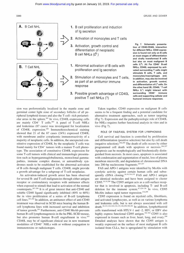
sion was preferrentially localized in the mantle zone and germinal center light zone of secondary follicles of all pe- ripheral lymphoid tissues and also the T-cell-rich periarteri- olar areas in the spleen.'" In vivo, CD40L-expressing cells are mainly CD4' T cells.'" A panel of T-cell NHLs and leukemias (87 cases) was investigated for localization of CD40L expression.'" Immunohistochemical staining showed that 21 of the 87 cases (24%) expressed CD40L with membrane and/or cytoplasmic immunoreactivity of a majority of neoplastic cells. In addition, the unexpected con- stitutive expression of CD40L by the neoplastic T cells was found mainly for CD4' tumors with a mature T-cell pheno- type. The association of constitutive CD40L expression for some T-cell tumors with clinical and immunologic presenta- tion such as hypergammaglobulinemia, monoclonal gamma- pathies, immune complex disease, or autoantibody syn- dromes needs to be established for the abnormal activation of B cells through malignant T cells. CD40L might provide a growth advantage for a subgroup of T-cell neoplasias.
An activation-induced growth arrest has been observed for several B- and T-cell malignancies through either antigen receptor or costimulatory receptors with antitumor effects when exposed to stimuli that lead to activation of the normal counterpart^."^^^^ I t is of great interest that antiCD40 and soluble CD40 ligand significantly inhibited (40% to 60%) the in vitro proliferation of a series of B-cell lymphoma cell line^.'^^^'‘" In addition, an antitumor effect of anti-CD40 treatment was observed in SCID mice bearing the human B- cell lymphoma lines with increased survival and inhibition of tumor growth.'"' Furthermore, anti-CD40 MoAb prevents human B-cell lymphomagenesis in the hu PBL-SCID mouse, but also promotes human B-cell engraftment in vivo.'93 CD40L may be of significant clinical use in new treatment modalities of CD40' NHLs with or without conjugation to immunotoxins or radioisotopes.
Taken together, CD40 expression on malignant B cells seems to be a frequent finding and a potential candidate for alternative treatment approaches, such as tumor targeting (Fig 7). Expression and the pathophysiologic role of CD40L for NHLs requires further functional analysis in vitro and in vivo (Fig 7).
ROLE OF FAS/FASL SYSTEM FOR LYMPHOMAS
Cell survival and function is controlled by proliferation and differentiation (positive selection) but also by cell death (negative selection).'y4.'y5 The death of cells occurs by either programmed cell death with apoptosis or n e c r o s i ~ . ~ ~ ~ ~ ' ~ ' Apoptosis can be morphologically and biochemically distin- guished from necrosis. In most cases, apoptosis is associated with condensation and segmentation of nuclei, loss of plasma membran microvilli, and degradation of chromosomal DNA into 200-bp nucleosome fragment^.'^'"^'
FAS and APO-l antigens were identified by MoAbs with cytolytic activity against certain human cells and subse- quently cDNA ~ l o n " g . ~ ' ~ ' ~ ~ ' ' ~ ~ " ~ FAS and APO-I antigen are identical molecules and have been assigned to cluster CD95.'X~1y~2y' The CD95 antigen acts as a cell-surface recep- tor that is involved in apoptosis, including T- and B-cell deletion for the immune s y ~ t e m ? ~ . ' ~ ~ - ' ~ ~ In vivo, CD95 MoAbs induce rapid tumor r eg re~s ion . '~~ . '~
CD95 expression is found on myeloid cells, fibroblasts, and activated lymphocytes, as well as on various lymphoma and leukemia cells, but is not always associated with cell
cells transformed with HTLV-l and -2, HIV, or EBV also highly express functional CD95 antigen.3""3N CD95 is also expressed in tissues such as liver, heart, lung, and ovary.'9' Detailed analyses have shown that the CD95 antigen is weakly expressed on the surface of most malignant B cells isolated from CLLs, but is upregulated by stimulation with
~ e a ~ ~ ~ ~ ~ . l ~ ~ . l ~ 4 . ~ ~ 8 . 1 ~ ~ . ~ ~ S . 1 4 K . 2 Y K , ~ W . ~ f l l In addition, lymphoblastoid
For personal use only.on January 10, 2019. by guest www.bloodjournal.orgFrom

TNF LIGAND FAMILY AND MALIGNANT LYMPHOMAS 3391
Staphylococcus aureus Cowan I (SAC) or IL-2 and showed CD95-mediated a p o p t o ~ i s . ~ ~ ~ In contrast, HCL B cells ex- pressed CD95 at moderate levels.305 The induction of CD95- mediated apoptosis was correlated in some instances with bcl-2 downreg~la t ion .~~~ The bcl-2 expression is correlated with inhibition of apoptosis3" and deregulation of bcl-2 ex- pression might be part of the pathogenesis of lymphomas (eg, follicular lymphomas) and leukemias (eg, B-cell CLL).307 In addition, mediastinal B-cell lymphomas coexpress, de- pending on their differentiation stage, CD95 and CD54.308 Interestingly, B cells from one CLL cases did not show bcl- 2 downregulation after stimulation with SAC and IL-2, and CD95 MoAb induced a proliferative Further, one case of B-cell lymphoma stimulated with IL-14 also showed significant growth enhancement with CD95 MoAb treat- ment.3w Similarly, fresh PBTs showed a costimulatory re- sponse in the presence of CD95 MoAbs or sCD95L, but chronic activated PBTs or T-cell clones underwent
CD95 expression of Burkitt's lymphoma (BL) cell lines is associated with a lymphoblastoid p h e n ~ t y p e . ~ ~ EBV- and EBV+ BL cell lines with type I phenotype (CD10+, CD21-, CD23-, CD30-, CD39-, CD70-, CD777 corresponding to primary BL tumor cells have no detectable CD95 surface expression. Accordingly, primary BL tumor cells (3 cases) were also CD95-. In contrast, EBV+ BL cell lines with type I11 lymphoblastoid phenotype (CD10-, CD21+, CD23+, CD30+, CD39+, CD70+, CD77-) as well as normal lym- phoblastoid B-cell lines expressed the CD95 antigen at high density, but 6 of 7 CD95+ BL cell lines were not sensitive to CD95-mediated killing. In addition, only one of eight B- cell NHLs and one of two T-cell NHLs expressed low levels of CD95, which was upregulated by IL-14.3w The expression of the CD95 antigen has been reported for the malignant cells of 83% (10 of 12 patients) of follicular lymphoma cases and 56% (18 of 32 patients) of diffuse lymphoma cases.313 Subgrouping into different histiologic subgroups showed similar distribution for all In addition, all adult T-cell leukemia cases (n = 12) were CD95+ and underwent apoptosis by adding CD95 M o A ~ s . ~ ' ~ The detailed functional role of CD95 for cell survival and tumor growth is presently not well understood. Recently, the cognate for CD95 (CD95LZASL) has been cloned and characterized as a 31- kD type I1 transmembrane protein with 25% to 30% homol- ogy to other members of the TNF ligand ~uperfamily.~~ Re- combinant CD95L exists also in a soluble form, with similar biologic activities seen for the CD95 MoAbs or membrane- bound CD95L.4' The physiologic presence and role of the sCD95L remain to be determined.
CD95 and CD95L expression and function has not been well investigated for HD, but the HD-derived cell lines HDLM-2, K"H2, L-428, and L-540 express CD95 and show apoptosis after treatment with CD95 MoAbs or soluble CD95L (H.J.G., manuscript in preparation).
In general, CD95 shares a dual role with the ability to mediate stimulatory or inhibitory/cytotoxic signals de- pending on the target cells or activation stage. The CD%/ CD95L-mediated T-cell cytotoxicity could play a major role in controlling the immune response of peripheral lympho-
~~ptoS~S~123,128,3M).302,310~312
cytes and might be involved in T-cell t ~ l e r a n c e . ~ ~ ~ . ~ ' ~ In gen- eral, the cloning of the CD95L will further improve our understanding of the mechanisms of apoptosis and the func- tional relevance of CD95(FAS)-CDML(FASL) interaction for malignancies, particular lymphoid tumors, such as a vari- ety of lymphomas and leukemias with B- and T-cell pheno- type.
4-1BW4-IBBL INTERACTION AND THE PATHOGENESIS OF LYMPHOMAS
4-1BB was identified and cloned from activated T cells (activation induced cDNA clone).10~16~"~317*318 The 33-kD 4- 1BB molecule is expressed on activated T cells (CD4+ and CD8') and t h y m o ~ y t e s . ~ ~ , ~ ' ' . ~ ~ ~ 4-1BB antibodies have co- stimulatory activity for T-cell pr~liferation.~'~ Initially, it was reported that extracellular matrix proteins bind 4-1BB, but the functional relevance remains unclear.320 Subsequently, murine and human 4-1BB ligands were identified and ex- pression c l0ned .4~~~ Expression of 4-IBBL was found for activated T cells, stromal cells, activated macrophages, EBV-transformed B cells, some tumor and leukemia cell lines, and a variety of tissues such as brain, placenta, lung, skeletal muscle, and l ~ i d n e y . ~ ~ , " , ~ 4-IBBL costimulates T- cell and thymocyte proliferation, but other biologic activities for the immune system and hematopoesis that are indicated by the wide distribution pattern of 4-1BB and 4-IBBL need to be identified. It is of additional interest that signals through 4-1BB enhance activation-induced cell death (AICD) of T cells." 4-IBBL is able to function as a signal transducing molecule.84 4-1BB and 4-1BBL are expressed on activated T cells and could play an autocrine regulatory role in T-cell intera~tion."".~ Primary lymphomas have not been ana- lyzed for 4-1BB and 4-IBBL expression, but a series of HD- derived cell lines express, in addition to the TNFRs, CD30, CD40, and FAS also 4-1BB (H.J.G., manuscript in prepara- tion). The functional and pathologic relevance needs to be examined.
THE OX40 MOLECULE AND HD
The OX40 molecule was originally described as a cell surface antigen on activated rat T cells32'; subsequently, the genes encoding rat, mouse, and human OX40 have been
to be restricted to activated CD4+ T ce11s.322.323 The human OX40 molecule is identical to the ACT35 antigen, described as strictly activation-associated antigen.162.324 No expression was found for resting peripheral blood lymphocytes, periph- eral blood B cells, and thymocytes. In lymphoid tissue, huOX40 expression was seen for scattered cells in the inter- follicular zone, the follicular mantle zone, and the germinal centers.16' Tissue macrophages were more weakly positive. For HD, only a few cases showed OX40 expression of H- RS cells, but T cells surrounding H-RS cells in a rosette fashion were strongly positive.162 Recently, murine and hu- man OX40L have been cloned from the murine lymphoma cell line S49.1 or the activated B-lymphoblastoid cell line MSAB, respectively, and the human homoloque identified to be gp34, a protein expressed on HTLV-1 -infected human
Cloned.45.46,322-324 Expression of OX40 was reported initially
For personal use only.on January 10, 2019. by guest www.bloodjournal.orgFrom

3392 GRUSS AND DOWER
leukemic T ce11s?5,46*325.326 OX40L expression is, as the OX40 receptor, selectively induced on activated CD4+ and CD8’ T cells, but not on B ~e l l s .4~ The OX40L is also expressed on HTLV-1 -transformed cell lines, stimulated B- lymphoblastoid cell lines, and THP-1 cells.& AS predicted, human OX4OL costimulates T-cell proliferation and cyto- kine production (eg, JL-2 and IL-4) as part of the regulatory cascade for immune r e s o n s e ~ . ~ ~ ~ ~ A possible correlation be- tween the OX4O/OX4OL system and virally induced patho- genesis needs further evaluation. Detailed expression and functional analyses have to be performed to understand the role of OX4O-OX4OL for the pathogenesis and/or tumorigen- esis of lymphomas, particularly in the context of viral trans- formation (eg, EBV, HTLV, and HIV).
TNF AND LT EXPRESSION IN HD WITH UNCLEAR BIOLOGIC RELEVANCE
TNF was originally defined by its antitumor activity but is also a major mediator of inflammation and cellular immune response.8 TNF was found to be cytotoxic to a number of transformed cell lines in TNF induces cachexia in LPS-treated mice with profound effects on general cellular metabolism and development of weight loss, fever, acute phase reaction, infection, or neoplasia.328 TNF enhances the proliferation of T cells, modulates T-cell receptor expression, enhances NK cell activity, and regulates human B-cell func- tion. TNF also has marked effects on neutrophils, eosinophil recruitment, monocyte/macrophage activation, fibroblast growth stimulation, and endothelial celllleukocyte interac- t i o n ~ . ~ ~ ~ TNF is produced by many cell types, including monocytes/macrophages, lymphocytes, and fibroblasts.’ Ac- tivated macrophages have the highest TNF prod~ction.’~’
LT is a cytokine structurally related to TNF with approxi- mately 50% sequence homology, the same chromosomal lo- calization, and trimer structure.8 LT is synthesized primarily by T cells, although some EBV-transformed B-cell lines and tonsil B cells produce it.8 LT and TNF have similar but not identical inflammatory and immunomodulatory activities8 LT is often less potent than TNF. Initially, LT (TNF-P) was cloned as a soluble ~ y t o k i n e . ~ ~ ~ Surface LT does not result from the presence of the transmembrane region but, rather, was found associated with a 33-kD integral membrane glyco- pr0tein.7~1~~ The cloned gene encoding this second protein in the surface LT complex was found to be a new member of the TNF ligand ~uperfamily.~~ Recently, the LT complex units have been renamed as LTa (TNF-PLT) and LT0 (p33), being synthesized in soluble form as a LTa homotri- mer or as a membrane-anchored heterotrimeric complex composed of LTa and LTP units (eg, P2a1).35 Generic li- gand-receptor interaction analysis predicts that the hetero- trimeric LT0 complex would produce a functionally inactive receptor-binding ~ r o t e i n . ~ ~ . ~ ~ The immunologic function and biologic role of LTP is presently not well understood.
Receptors for TNF and LT are expressed at low levels on most tissues and various cell type^.^,^^' Three distinct recep- tors have been shown to bind TNF, LTa, and LTp.9-”s77 The p60 TNFR type I and p80 TNFR type I1 cDNAs encode distinct proteins with 20% homology in the ligand-binding domain (extracellular domain); both receptors bind TNF and
LTa with similar affinities?-” The heterotrimer LT@ binds to the recently identified TNFR-RP surface protein (TNFR- III).’2*77 Expression of TNFRs on human PBT cells is activa- tion dependent.332 TNFR expression is upregulated by agents such as L-2, IFN-a, CAMP, and different hormones but is downregulated by IL-1, PMA, glucocorticoids, and LPS.8 Soluble forms of TNFR-I and TNFR-I1 have been identified in the serum of normal persons and tumor patient^.^'*^^.^'*^' It is of particularly interest that several viral ORFs such as SFV-T2, MYX-T2, G4R, crrnB, Va53, and SaIF19R, encode soluble homologues of the TNFR proteins with a possible role in viral host response.i0,20-22.333.334 These viral ORFs show a novel mechanism of viral subversion of the host immune re~ponse.6~
Expression of TNF and LTa protein and mRNA has been reported for a series of HD-derived cell lines.’%.z58.335-338 Sim- ilarly, TNF expression of H-RS cells was shown in primary tissue from HD patients.’75.265.336’3’7.339’”0 Using immunohis- tochemistry and/or in situ hybridization, 56 of 104 cases investigated (54% positive cases) showed strong cytoplasmic TNF signals for H-RS cells. In addition, immunoreactivity for TNF was observed for some macrophages. Furthermore, abundant LTa expression was found on primary H-RS cells for 47 of 59 HD cases analyzed (80% of cases p o s i t i ~ e ) . ’ ~ ~ . ~ ~ ~ In comparison with TNF, the LTa-specific mRNA signals were usually of higher intensity and present in larger propor- tions of H-RS cells.175 TNF and LTa gene transcripts were also seen in some lymphoid cells.
Most HD-derived cell lines express the p60 and p80 TNFRs on their surface or at the mRNA The presence of TNFR-RP protein on cultured or primary H-RS cells has not been investigated. Limited data are available for the TNFR expression in HD cases. One study reported the expression of p60 TNFR in 1 of 4 (25%) and p80 TNFR in 2 of 4 (50%) HD cases analyzed using immunohistochem- i ~ t r y . ~ ~ ’ Further detailed expression studies for primary HD material are required to fully understand the involvement of TNF and LTa for the pathogenesis of HD, including growth control for H-RS cells.
These different studies indicate that TNF and LTa expres- sion of the malignant H-RS cells in HD-involved tissue is a frequent finding and could have a central functional rele- vance for oncogenesis and pathogenesis of HD (Fig 8). High- level expression of LTa for cultured and primary H-RS cells is a striking feature for HD, with unknown biologic correla- tion.M Primary and cultured H-RS cells express not only TNF and LTa but also the TNFRs, suggesting a possible autocrine growth loop for H-RS cells (Fig 8). TNF and LTa lack mitogenic activity on cultured H-RS cells and a pre- sumed autocrine growth loop, based on coexpression of cor- responding ligands and receptors, has so far not been shown.M2 It is of interest, that recombinant CD30L and CD40L induce the secretion of TNF and LTa.”7*258
A number of typical pathologic and clinical features of HD are consistent with characteristics of a tumor of cytokine- producing cells, including occurence of B symptoms, sclero- sis, eosinophilia, acute-phase responses, T-cell rosetting and activation, impaired immune responses, and generalized it~hing.’~ Furthermore, cytokines produced by H-RS cells
For personal use only.on January 10, 2019. by guest www.bloodjournal.orgFrom

TNF LIGAND FAMILY AND MALIGNANT LYMPHOMAS 3393
Fig 8. TNF and LTa involve- ment in the interaction between H-RS cells and surrounding by- stander cells. TNF (A) is pro- duced by many cell types, such as monocyteslmacrophages, lym- phocytes, and stroma cells. Ex- pression of TNF and TNFRs is a common finding for H-RS cells. TNF involvement in paracrine andlor autocrine growth stimu- lation of H-RS cells remains un- clear. TNF could also be critical for accumulationlactivation of bystander cells and one media- tor for systemic B symptoms. Similarly, LTa (B) is frequently overexpressed by H-RS cells in HD. In contrast to TNF, LTa ex- pression is mainly restricted to lymphoid cells. The functional role of LTu for HD is presently unclear.
B. LT-a 4 S t r S a m -
might interact with surrounding bystander cells, particularly T cells; conversely, H-RS cells might respond to cytokines produced by surrounding normal reactive bystander cells. TNF and LTa are part of these deregulated cytokine network involved in HD. TNF and LTa could be involved in causing fever, weight loss, and night sweats, as cytokines involved in the development of the constitutional B symptoms.'28 TNF is also mitogenic for fibroblasts and induces collagen synthe- sis with a potential role in formation of sclero~is.~'' Elevation of, eg, fibrinogen or prostaglandin serum levels, frequently seen in HD, could be associated with TNF se~retion.~~.'~' It is of interest that HD patients have elevated TNF serum levels (47 of 76 HD patients [62%]); the extent of increase correlated with disease stage and the presence or absence of B symptoms.'" Similarly, the mean serum levels of the solu- ble p60 TNFR were significantly higher in HD patients than in healthy The degree of increase correlated with TNF serum levels, disease stage, and disease activity (pres- ence of B symptoms). Furthermore, increased soluble p60 TNFR serum levels of HD patients in remission could be involved in the cellular immune defect characteristic for HD
might support the escape of H-RS cells in the predisposing immunocompromised host from the tumor-suppressive ef- fects of TNF and LTa and also cellular antitumor immune response by blocking appropriate T-cell activation and func- tion. In addition, systemic TNF might be causative with other immune modulators, such as IL- 1 and IL-6, in develop- ment of B symptoms and/or metabolic wasting (Fig 8).
patients."s.34" The elevated soluble TNFR concentration
TNF AND TNFR INTERACTION FOR NHLS
Immunohistochemical studies of nonmalignant, reactive cells showed that TNF-positive cells were rarely detected in lymph nodes with activation of the B-cell compartment but were frequently detected in sections from patients with dif-
1. Enhancement of T cell proliferation, antigen expression, and cytokine production
2. Activation of granulocyte and eosinophil recruitment
3 3. Stimulation of monocyteslmacrophages
4. Fibroblast (stroma) growth stimulation with enhanced collagen formation
5. Involvement in control of H-RS cell growth (paracrine and/or autocrine loops), cytokine secretion and activation
6. "Central" cytokine for typical clinical and pathological presentations of HD, including acute phase response and B-symptoms
fuse or mixed lymphadenitis with expansion of the T-cell-
were associated with the presence of macrophages. The anal- ysis of 20 NHL cases showed only 4 of 16 B-cell NHL cases showed weak scattered TNF-positive cells.'"' In the four T- cell NHLs, TNF was not detectable on the neoplastic lymphoma cells but was detectable on macrophages in T- cell paracortical areas.2h5 In contrast to the high frequency of TNF and, particularly, the LTa expression of the neoplas- tic H-RS cells in HD, the neoplastic lymphoma cells of NHLs seems only rarely to produce TNF and LTa as an indicator of their malignant transformation. Similarly, RNA prepared from nonmalignant. reactive lymph nodes con- firmed low expression of TNF and LTa..'" In contrast, mod- erate to abundant levels of TNF mRNA were detected in 12 of 35 NHL specimens (33% of cases positive; 9 low-grade and 3 high-grade NHLs).'~' In contrast, in situ hybridization with IL-6, TNF, and LTa probes of eight lymphoplasmacytic lymphomas lacked IL-6, TNF, and LTa expre~sion."~ Vari- able amounts of increased LTa mRNA were detected in 19 of 35 NHL specimens (54% of cases positive).'" Ten of the NHL cases coexpressed TNF and LTa mRNA?' Interest- ingly, 8 of 12 lymphoma patients (67% of cases) with the presence of systemic B symptoms had high TNF mRNA levels and 11 of these 12 lymphoma cases (92% of cases) had also abundant LTa mRNA expression."' High TNF and LTa mRNA expression correlated significantly with the presence of systemic B symptoms. Similarly, murine and human NHL cell lines express either constitutively TNF and LTa mRNA and protein or can be Further de- tailed studies are needed to confirm these initial results and to demonstrate the localization of TNFLTa-expressing cells.
So far, only one study analyzed p60 and p80 TNFR ex- pression systematically by using immunohistochemistry for normal tissues and NHL specimens.."' For the normal
dependent areas 265.347.348 . . As presumed, strong TNF signals
For personal use only.on January 10, 2019. by guest www.bloodjournal.orgFrom

3394 GRUSS AND DOWER
lymphoid tissue sections, p60 TNFR expression was re- stricted to dentritic reticulum cells of germinal centers, but p80 TNFR was found on a major cell population of interdig- itating cells and activated lymphocytes in the interfollicular T-cell areas. The p60 and p80 TNFR expression sites are different and could allow different biologic function of TNF and LTa with either paracrine or autocrine signaling path- ways. In reactive lymph nodes, the number of p80 TNFR- positive cells was increased in the T-cell areas.34' The analy- sis of a limited number of B- and T-cell NHL sections (n = 30) showed that mainly lymphoma cells with a high-grade malignant phenotype (6 of 14 B-cell NHLs and 6 of 8 T-cell NHLs) expressed p80 but lacked p60 TNFR expre~sion.~~'
TNF and LTa have important roles in normal B-cell acti- vation, growth, and differentiati~n.'~~~'~~,~~~ TNF is capable of inducing proliferation of the TNFR-positive neoplastic B cells from CLL patients.35' Very recently, it was shown that lymphoblastoid B-cell lines and BL cell lines with a lym- phoblastoid phenotype (CD10-, CD77-, CD23+, CD40') use LTa as an autocrine growth factor and act mainly through the p60 TNFR.352 Overall, the expression and func- tional relevance of the cytokines TNF and LTa for NHLs remain unclear. Malignant B cells from CLL patients use TNF as a growth factor, but similar data are presently not reported for NHLs. Further studies have to confirm the ex- pression not only of TNF and LTa but also of the two TNFRs for different NHL entities as well as the potential biologic relevance for growth control, differentiation, and activation of NHL cells.
SUMMARY
The TNF receptor superfamily members are all type I membrane glycoproteins with typical homology in the extra- cellular domain of variable numbers of cysteine-rich repeats (overall homologies, 25% to 30%). In contrast, the TNF ligand superfamily members (with the exception of LTa) are type I1 membrane glycoproteins with homology to TNF in the extracellular domain (overall homologies, 20%). TNF and LTa are trimeric proteins and are composed of P-strands forming a P-jellyroll. The homology of the &strand regions for the TNF ligand superfamily members suggest a similar ,&sandwich structure and possible trimeric or multimeric complex formation for most or all members. A genetic link- age, as evidence for evolutionary relatedness, is found by chromosomal cluster of TNFR p80, CD30, 4-1BB, and OX40 for lp36; TNFR p60, TNFR-RP, and CD27 for 12~13 ; TNF, LTa, and LTP for 6 (MHC locus); CD27L and 4- lBBL for 19~13 ; and FASL and OX40L for lq25.
Of the TNF ligand superfamily, TNF, LTa, and LTP and their receptors (TNFR p60, TNFR p80, and TNFR-RP) inter- act in a complex fashion of cross-binding. However, the other family members presently have a one ligandone recep- tor binding principle (CD27/CD27L, CD30/CD30L, CD40/ CD40L, 4-1BB/4-1BBL, OX4O/gp34, and FASIFASL). In general, the members of the TNF ligand superfamily mediate interaction between different hematopoietic cells, such as T cell/B cell, T cell/monocyte, and T c e l n cell. Signals can be transduced not only through the receptors but also through at least some of the ligands. The transduced signals can be
stimulatory or inhibitory depending on the target cell or the activation state. Taken together, TNF superfamily ligands show for the immune response an involvement in the induc- tion of cytokine secretion and the upregulation of adhesion molecules, activation antigens, and costimulatory proteins, all known to amplify stimulatory and regulatory signals. On the other hand, differences in the distribution, kinetics of induction, and requirements for induction support a defined role for each of the ligands for T-cell-mediated immune responses. The shedding of members of the TNF receptor superfamily could limit the signals mediated by the corre- sponding ligands as a functional regulatory mechanism. In- duction of cytotoxic cell death, observed for TNF, LTa, CD30L, CD95L, and 4-1BBL, is another common functional feature of this cytokine family. Further studies have to iden- tify unique versus redundant biologic and physiologic func- tions for each of the TNF superfamily ligands.
Primary H-RS cells can express TNF, LTa, and CD27L but not CD30L and CD40L, in addition to IL-la, IL-5, IL- 6, IL-9, and M-CSF. In addition, H-RS cells express high copy numbers of several cytokine receptors such as IL-2R (p55, p75, and p64 subunits), IL-6R, M-CSFR (c-fms), SCFR (c-kit), CD30, CD40, and T N F R s . ~ Cytokines pro- duced by H-RS cells might support the growth of tumor cells (autocrine growth loop) and/or interact with surrounding re- active bystander cells, particularly T cells. Conversely, H-RS cells might respond to cytokines produced by surrounding reactive normal cells (paracrine growth loop). The different interactions between H-RS cells and surrounding normal, reactive bystander cells, such as lymphocytes, plasma cells, histiocytes, neutrophils, eosinophils, and stromal cells, is characteristic for HD. The expression and biologic effects of a panel of cytokines and their counterpart receptors seem to be involved in the pathobiologic interaction between H- RS cells and particularly lymphocytes, mainly CD4' T cells. Detailed analyses have to verify the predicted biologic activi- ties of TNF, LTa, CD27L, CD30L, CD40L, 4-1BBL, gp34/ OX40L, and FASL for the H-RS celVT-cell interactions with impact on tumor growth and pathogenesis of HD. Cytokines and cytokine receptors, including TNF/TNFRs, CD301 CD30L, and CD40/CD40L, are clearly critical elements in the pathology of HD and are part of the deregulated network of interactive signals between H-RS cells and surrounding bystander cells with membrane-associated and cytokine-me- diated events. HD is a tumor of cytokine-producing cells that is causative for several characteristic clinical and pathologic presentation of HD.
The functional role of cytokines for the pathogenesis of NHLs is presently unclear. Malignant NHL cells express, depending on their immunophenotype, several TNF receptor and ligand superfamily members. B-cell NHLs are fre- quently CD27/CD27L, CD30 or CD30L. CD40, and TNFRs/ TNF positive, but T-cell NHLs have expression of CD30, CD40L, and TNFRs/TNF. Further functional analysis will increase our understanding of the involvement of TNF super- family ligands in the pathogenesis of NHLs.
Several TNFR superfamily members could be candidates for novel treatment protocols. Recombinant CD30L and CD40L could be by itself antitumorigenic for CD30' ALCLs
For personal use only.on January 10, 2019. by guest www.bloodjournal.orgFrom

TNF LIGAND FAMILY AND MALIGNANT LYMPHOMAS 3395
and CD40+ B-cell NHLs, respectively. Furthermore, CD30 and CD40 might be used for tumor targeting after conjuga- tion with radioisotypes or cytostatic drugs for CD30+ and/ or CD40+ HD and NHLs.
REFERENCES 1. Williams AF, Barclay AN: The immunoglobulin superfamily-
domains for cell surface recognition. Annu Rev Immunol 6:381, 1988
2. Mallett S, Barclay AN: A new superfamily of cell surface proteins related to the nerve growth factor receptor. Immunol Today 12:220, 199 1
3. Cosman D: The hematopoietin receptor superfamily. Cytokine 5:95, 1993
4. Armitage RJ: Tumor necrosis factor receptor superfamily members and their ligands. Curr Opin Immunol 6:407, 1994
5. Smith CA, Farrah T, Goodwin RG: The TNF receptor super- family of cellular and viral proteins: activation, costimulation, and death. Cell 75:959, 1994
6. Kishimoto T, Taga T, Akira S: Cytokine signal transduction. Cell 76:253, 1994
7. Johnson D, Lanahan A, Buck CR, Sehgal A, Morgan C, Mercer E, Bothwell M, Chao M: Expression and structure of the human NGF receptor. Cell 47:545, 1986
8. Beutler B: Tumor Necrosis Factors: The Molecules and Their Emerging Role in Medicine. New York, NY, Raven, 1992
9. Loetscher H, Pan Y-CE, Lahm H-W, Gentz R, Brockhaus M, Tabuchi H, Lesslauer W: Molecular cloning and expression of the human 55 kd tumor necrosis factor receptor. Cell 61:351, 1990
10. Smith CA, Davis T, Anderson D, Solam L, Beckmann MP, Jerzy R, Dower SK, Cosman D, Goodwin RG: A receptor for tumor necrosis factor defines an unusual family of cellular and viral pro- teins. Science 248:1019, 1990
11. Schall TJ, Lewis M, Koller KJ, Lee A, Rice GC, Wong GH, Gatanaga T, Granger CA, Lentz R, Raab H, Kohr KJ, Goeddel DV: Molecular cloning and expression of a receptor for human tumor necrosis factor. Cell 61:361, 1990
12. Baens M, Chaffanet M, Cassiman J-3, van den Berghe H, Marynen P: Construction and evaluation of a hncDNA library of human 12p transcribed sequences derived from a somatic cell hybrid. Genomics 16:214, 1993
13. Camerini D, Walz G, Loenen WAM, Borst J, Seed B: The T cell activation antigen CD27 is a member of the nerve growth factor/ tumor necrosis factor receptor gene family. J Immunol 147:3165, 1991
14. Durkop H, Latza U, Hummel M, Eitelbach F, Seed B, Stein H: Molecular cloning and expression of a new member of the nerve growth factor receptor family that is characteristic for Hodgkin’s disease. Cell 68:421, 1992
15. Stamenkovic I, Clark EA, Seed B: A B-lymphocyte activation molecule related to the nerve growth factor receptor and induced by cytokines in carcinomas. EMBO J 8:1403, 1989
16. Kwon BS, Weissman SM: cDNA sequences of two inducible T-cell genes. Proc Natl Acad Sci USA 86:1963, 1989
17. Mallet S, Fossum S, Barclay AN: Characterization of the MRC OX40 antigen of activated CD4 positive T lymphocytes-A molecule related to nerve growth factor receptor. EMBO J 9:1063, 1990
18. Itoh N, Yonehara S, Ishii A, Yonehara M, Mizushima S-I, Sameshima M, Hase A, Set0 Y, Nagata S: The polypeptide encoded by the cDNA for human cell surface antigen Fas can mediate apoptosis. Cell 66:233, 1991
19. Oehm A, Behrmann I, Falk W, Pawlita M, Maier G, Klas C, Li-Weber M, Richards S, Dhein J, Trauth BC, Posting1 H, Krammer,
PH: Purification and molecular cloning of the APO-1 cell surface antigen, a member of the tumor necrosis factorherve growth factor receptor superfamily. Sequence identity with the Fas antigen. J Biol Chem 267:10709, 1992
20. Smith CA, Davis T, Wignall JM, Din WS, Farrah T, Upton C, McFadden G, Goodwin RG: T2 open reading frame from the shope fibroma virus encodes a soluble form of the TNF receptor. Biochem Biophys Res Commun 176:335, 1991
21. Howard ST, Chan YS, Smith GL: Vaccinia virus homologues of the shope fibroma virus inverted terminal repeat proteins and a discontinuous ORF related to the tumor necrosis factor receptor family. Virology 180:633, 1991
22. Upton C, Macen JL, Schreiber M, McFadden G: Myxoma virus expresses a secreted protein with homology to the tumor necro- sis factor receptor gene family that contributes to viral virulence. Virology 184:370, 1991
23. Welcher AA, Bitler CM, Radeke MJ, Shooter EM: Nerve growth factor binding domain of the nerve growth factor receptor. Proc Natl Acad Sci USA 88:159, 1991
24. Baldwin AN, Bitler CM, Welcher AA, Shooter EM: Studies on the structure and binding properties of the cysteine-rich domain of rat low affinity nerve growth factor receptor (p75NGFR). J Biol Chem 267:8352, 1992
25. Marsters SA, Frutkin AD, Simpson NJ, Fendly BM, Ashke- nazi A: Identification of cysteine-rich domains of the type 1 tumor necrosis factor receptor involved in ligand binding. J Biol Chem 267:5747, 1992
26. Banchereau J, Bazan F, Blanchard D, Briere F, Galizzi J-P, van Kooten C, Liu YJ, Rousset F, Saeland S: The CD40 antigen and its ligand. Annu Rev Immunol 12:881, 1994
27. Bradshaw RA, Blundell TL, Lapatto R, McDonald NQ, Mur- ray-Rust J: Nerve growth factor revisited. Trends Biochem Sci 18:48, 1993
28. Eide FF, Lowenstein DH, Reichardt L F Neurotrophins and their receptors-Current concepts and implications for neurologic disease. Exp Neurol 121:200, 1993
29. Pennica D, Nedwin GE, Hayflick JS, Seeburg PH, Derynck R, Palladino MA, Kohr WJ, Agganval BB, Goeddel DV: Human tumour necrosis factor: Precursor structure, expression and homol- ogy to lymphotoxin. Nature 312:724, 1984
30. Old LJ: Tumor necrosis factor (TNF). Science 230:630, 1985 31. Wang AM, Creasey AA, Ladner MB, Lin LS, Strickler J,
Van Arsdell JN, Yamamoto R, Mark DF: Molecular cloning of the complementary DNA for human tumor necrosis factor. Science 228:149, 1985
32. Beutler B, Cerami A: Cachectin and tumour necrosis factor as two sides of the same biological coin. Nature 320:584, 1986
33. Gray PW, Aggarwal BB, Benton CV, Bringman TS, Henzel WJ, Jarrett JA, Leung DW, Moffat B, Ng P, Svedersky LP, Palladino MA, Nedwin GE: Cloning and expression of cDNA for human lymphotoxin, a lymphokine with tumour necrosis activity. Nature 312:721, 1984
34. Aggarwal BB, Henzel WJ, Moffat B, Kohr WJ, Harkins RN: Primary structure of human lymphotoxin derived from 1788 lym- phoblastoid cell line. J Biol Chem 260:2334, 1985
35. Browning JL, Ngam-ek A, Lawton P, DeMarinis J, Tizard R, Chow EP, Hession C, O’Brien-Greco B, Foley SF, Ware C F Lymphotoxin p, a novel member of the TNF family that forms a heteromeric complex with lymphotoxin on the cell surface. Cell 72:847, 1993
36. Goodwin RG, Alderson MR, Smith CA, Armitage RJ, Van- denBos T, Jerzy R, Tough T W , Schoenbom MA, Davis-Smith T, Hennen K, Falk B, Cosman D, Baker E, Sutherland GR, Grabstein KH, Farrah T, Gin JG, Beckmann MP: Molecular and biological
For personal use only.on January 10, 2019. by guest www.bloodjournal.orgFrom

3396 GRUSS AND DOWER
characterization of a ligand for CD27 defines a new family of cyto- kines with homology to tumor necrosis factor. Cell 73:447, 1993
37. Smith CA, Gruss H-J, Davis T, Anderson D, Farrah T, Baker E, Sutherland GR, Brannan CI, Copeland NG, Jenkins NA, Grabstein KH, Gliniak B, McAlister IB, Fanslow W, Alderson M, Falk B, Gimpel S, Gillis S, Din WS, Goodwin RG, Armitage W: CD30 antigen, a marker for Hodgkin’s lymphoma, is a receptor whose ligand defines an emerging family of cytokines with homology to TNF. Cell 73:1349, 1993
38. Armitage W, Fanslow WC, Strockbine L, Sat0 TA, Clifford KN, Macduff BM, Anderson DM, Gimpel SD, Davis-Smith T, Mali- szewski CR, Clark EA, Smith CA, Grabstein KH, Cosman D, Spriggs MK: Molecular and biological characterization of a murine ligand for CD40. Nature 357:80, 1992
39. Graf D, Korthauer U, Mages HW, Senger G, Kroczek RA: Cloning of TRAP, a ligand for CD40 on human T cells. Eur J Immunol 22:3191, 1992
40. Spriggs MK, Armitage RJ, Strockbine L, Clifford KN, Mac- duff BM, Sato TA, Maliszewski CR, Fanslow WC: Recombinant human CD40 ligand stimulates B cell proliferation and immunoglob- ulin E secretion. J Exp Med 176:1543, 1992
41. Hollenbaugh D, Grosmaire LS, Kullas CD, Chalupny NJ, Braesch-Andersen S, Noelle RJ, Stamenkovic I, Ledbetter JA, Aruffo A: The human T cell antigen gp39, a member of the TNF gene family, is a ligand for the CD40 receptor: Expression of a soluble form of gp39 with B cell co-stimulatory activity. EMBO J 11:4313, 1992
42. Gauchat JF, Aubry JP, Mazzei G, Life P, Jomotte T, Elson G, Bonnefoy JY: Human CD40-ligand: Molecular cloning, cellular distribution and regulation of expression by factors controlling IgE production. FEBS Lett 315:259, 1993
43. Goodwin RG, Din WS, Davis-Smith T, Anderson DM, Gim- pel SD, Sat0 TA, Maliszewski CR, Brannan CI, Copeland NG, Jenkins NA, Farrah T, Armitage RJ, Fanslow WC, Smith CA: Mo- lecular cloning of a ligand for the inducible T-cell gene 4-1BB: A member of an emerging family of cytokines with homology to tumor necrosis factor. Eur J Immunol 23:2631, 1993
44. Alderson MR, Smith CA, Tough TW, Davis-Smith T, Armi- tage RJ, Falk B, Roux E, Baker E, Sutherland GR, Din WS, Goodwin RG: Molecular and biological characterization of human 4-1BB and its ligand. Eur J Immunol 24:2219, 1994
45. Baum PR, Gayle RB 111, Ramsdell F, Srinivasan S, Sorensen RA, Watson ML, Seldin MF, Baker E, Sutherland GR, Clifford KN, Alderson MR, Goodwin RG, Fanslow WC: Molecular characteriza- tion of murine and human OX4O/OX40 ligand systems: Identification of a human OX40 ligand as the HTLV-l regulated protein gp34. EMBO J 13:3992, 1994
46. Godfrey WR, Fagnoni FF, Hxara MA, Buck D, Engleman EG: Identification of a human OX-40 ligand, a costimulator of CD4+ T cells with homology to tumor necrosis factor. J Exp Med 180757, 1994
47. Suda T, Takahashi T, Golstein P, Nagata S: Molecular cloning and expression of the Fas ligand, a novel member of the tumor necrosis factor family. Cell 75:1169, 1993
48. Alderson MR, Tough TW, Davis-Smith T, Braddy S, Falk B, Schooley KA, Goodwin RG, Smith CA, Ramsdell F, Lynch DH: Fas ligand mediates activation-induced cell death in human T lym- phocytes. J Exp Med 181:71, 1995
49. Lynch DH, Watson ML, Alderson MR, Baum PR, Miller RE, Tough T, Gibson M, Davis-Smith T, Smith CA, Hunter K, Bhat D, Din W, Goodwin RG, Seldin MF: The mouse Fas-ligand gene is mutated in gld mice and is part of a TNF family gene cluster. Immunity 1:13l, 1994
50. Takahashi T, Tanaka M, Brannan CI, Jenkins NA, Copeland NG, Suda T, Nagata S: Generalized lymphoproliferative disease in
mice, caused by a point mutation in the Fas ligand. Cell 76:969, 1994
51. Kriegler M, Perez C, DeFay K, Albert I, Lu SD: A novel form of TNFkachectin is a cell surface cytotoxic transmembrane protein: Ramifications for the complex physiology of TNF. Cell 53:45, 1988
52. Mohler KM, Sleath PR, Fitzner JN, Cerretti DP, Alderson M, Derwar SS, Torrance DS, Own-Evans C, Greenstreet T, Weera- warna K, Kronheim SR, Petersen M, Gerhart M, Kozlosky CJ, March CJ, Black RA: Protection against a lethal dose of endotoxin by an inhibitor of tumour necrosis factor processing. Nature 370218, 1994
53. Harris NL, Jaffe ES, Stein H, Banks PM, Chan JKC, Cleary ML, Delsol G, De Worlf-Peeters C, Falini B, Gatter KC, Grogan TM, Isaacson PG, Knowles DM, Mason DY, Muller-Hermelink H-K, Pileri SA, Piris MA, Ralfkiaer E, Wamke RA: A revised European-American classification of lymphoid neoplasms: A pro- posal from the International Lymphoma Study Group. Blood 84:1361, 1994
54. Kaplan HS: Hodgkin’s Disease. Boston, MA, Harvard, 1980 55. Kaufman D, Longo DL: Hodgkin’s disease. Crit Rev Oncol
Hematol 13:135, 1992 56. Haluska FG, Brufsky AM, Canellos G P The cellular biology
of the Reed-Stemberg cell. Blood 84:1005, 1994 57. Lukes R, Butler J, Hicks E Natural history of Hodgkm’s
disease as related to its pathological picture. Cancer 19:317, 1966 58. Hansen NE, Kale H: The elusive Hodgkin cell. Scand J
Haematol 26:353, 1981 59. Jones DD: The histogenesis of the Reed-Stemberg cell and
its mononuclear counterparts. J Path01 151:191, 1987 60. Jaffe ES: The elusive Reed-Stemberg cell. N Engl J Med
320529, 1989 61. Drexler HG: Recent results on the biology of Hodgkin and
Reed-Sternberg cells. I. Biopsy material. Leuk Lymphoma 8:283, 1992
62. Drexler HG: Recent results on the biology of Hodgkin and Reed-Stemberg cells. 11. Continuous cell lines. Leuk Lymphoma 9:1, 1993
63. Herbst H, Stein H, Niedobitek G: Epstein-Barr virus and CD30+ malignant lymphomas. Crit Rev Oncog 4:191, 1993
64. Gruss H-J, Herrmann F, Drexler HG: Hodgkin’s disease: A cytokine producing tumor. Crit Rev Oncog 5:23, 1995
65. Spriggs MK, Fanslow WC, Armitage W, Belmont J: The biology of the human ligand for CD40. J Clin Immunol 13:373, 1993
66. Farrah T, Smith CA: Emerging cytokine family. Nature 358:26, 1992
67. Gooding LR: Virus proteins that counteract host immune de- fenses. Cell 715, 1992
68. Howard OMZ, Clouse KA, Smith C, Goodwin RG, Farrar WL: Soluble tumor necrosis factor receptor: Inhibition of human immunodeficiency virus activation. Proc Natl Acad Sci USA 90:2335, 1993
69. Hu F-Q, Smith CA, Pickup DJ: Cowpox virus contains two copies of an early gene encoding a soluble secreted form of the type I1 TNF receptor. Virology 204:343, 1994
70. Smith GL: Vaccinia virus glycoproteins and immune evasion. J Gen Virol 74:1725, 1993
7 1. Jones EY, Stuart DI, Walker N P The three-dimensional struc- ture of tumor necrosis factor. h o g Clin Biol Res 349:321, 1990
72. Eck MJ, Ultsch M, Rinderknecht E, de Vos AM, Sprang SR: The structure of human lymphotoxin (tumor necrosis factor-0) at 1.9-A resolution. J Biol Chem 267:2119, 1992
73. Banner DW, D’Arcy A, Janes W, Gentz R, Schoenfeld H-J, Broger C, Loetscher H, Lesslauer W: Crystal structure of the soluble
For personal use only.on January 10, 2019. by guest www.bloodjournal.orgFrom

TNF LIGAND FAMILY AND MALIGNANT LYMPHOMAS 3397
human 55 kd TNF receptor-human TNFP complex: Implications for TNF receptor activation. Cell 73:431, 1993
74. D'Arcy A, Banner DW, Janes W, Winkler FK, Loetscher H, Schonfeld HJ, Zulauf M, Gentz R, Lesslauer W: Crystallization and preliminary crystallographic analysis of a TNF-beta-55 kDa TNF receptor complex. J Mol Biol 229555, 1993
75. Browning JL, Androlewicz MJ, Ware C F Lymphotoxin and an associated 33-kDa glycoprotein are expressed on the surface of an activated human T cell hybridoma. J Immunol 147:1230, 1991
76. Androlewicz MJ, Browning JL, Ware C F Lymphotoxin is expressed as a heteromeric complex with a distinct 33-kDa glycopro- tein on the surface of an activated human T cell hybridoma. J Biol Chem 267:2542, 1992
77. Crowe PD, VanArsdale TL, Walter BN, Ware CF, Hession C, Ehrenfels B, Browning JL, Din WS, Goodwin RG, Smith CA: A lymphotoxin-P-specific receptor. Science 264:707, 1994
78. Tartaglia LA, Ayres TM, Wong GHW, Goeddel DV: A novel domain within the 55 kd TNF receptor signals cell death. Cell 74:845, 1993
79. Tartaglia LA, Rothe M, Hu Y-F, Goeddel DV: Tumor necro- sis factor's cytotoxic activity is signaled by the p55 TNF receptor. Cell 73:213, 1993
80. Wong GHW, Goeddel DV: Fas antigen and p55 TNF receptor signal apoptosis through distinct pathways. J Immunol 152:1751, 1994
81. Heller RA, Kronke M Tumor necrosis factor receptor-medi- ated signaling pathways, J Cell Biol 1265, 1994
82. Cayabyab M, Phillips JH, Lanier LL: CD40 preferentially costimulates activation of CD4' lymphocytes. J Immunol 152:1523, 1994
83. Bowman MR, Crimmins MAV, Yetz-Aldape J, Kriz R, Kel- leher K, Herrmann S : The cloning of CD70 and its identification as the ligand for CD27. J Immunol 152:1756, 1994
84. Pollok KE, Kim YJ, Hurtado J, Zhou Z, Kim KK, Kwon BS: 4-1BB T-cell antigen binds to mature B cells and macrophages, and costimulates anti-p-primed splenic B cells. Eur J Immunol 24:367, 1994
85. Beutler B, Cerami A: Tumor necrosis, cachexia, shock, and inflammation: A common mediator. Annu Rev Biochem 57505, 1988
86. Beutler B, Cerami A: The biology of cachectin/TNF-A pri- mary mediator of the host response. Annu Rev Immunol7:625, 1989
87. Smith RA, Baglioni C: The active form of tumor necrosis factor is a trimer. J Biol Chem 262:6951, 1987
88. E".k MJ, Sprang SR: The structure of tumor necrosis factor- a at 2.6 A resolution. Implications for receptor binding. J Biol Chem 264:17595, 1989
89. Peitsch MC, Jongeneel CV: A 3-D model for the CD40 ligand predicts that it is a compact trimer similar to the tumor necrosis factors. Int Immunol 5:233, 1993
90. Hintzen RQ, Lens SMA, Koopman G, Pals ST, Spits H, Van Lier RAW: CD70 represents the human ligand for CD27. Int Immu- no1 6:477, 1994
91. Engelmann H, Aderka D, Rubinstein M, Rotman D, Wallach D: A tumor necrosis factor-binding protein purified to homogeneity from human urine protects cells from tumor necrosis factor toxicity. J Biol Chem 264:11974, 1989
92. Josimovic-Alasevic 0, Wrkop H, Schwarting R, Backe E, Stein H, Diamantstein T Ki-l (CD30) antigen is released by Ki-l- positive tumor cells in vitro and in vivo. I. Partial characterization of soluble Ki-l antigen and detection of the antigen in cell culture Supernatants and in serum by an enzyme-linked immunosorbent
93. Engelmann H, Novick D, Wallach D: Two tumor necrosis factor binding proteins purified from human urine. Evidence for
assay. Eur J Immunol 19:157, 1989
immunological cross reactivity with cell surface tumor necrosis fac- tor receptors. J Biol Chem 265:1531, 1990
94. Kohno T, Brewer MT, Baker SL, Schwartz PE, King MW, Hale KK, Squires CH, Thompson RC, Vannice JL: A second tumor necrosis factor receptor gene product can shed a naturally occurring tumor necrosis factor inhibitor. Proc Natl Acad Sci USA 87:8331, 1990
95. Lantz M, Gullberg U, Nilsson E, Olsson I: Characterization in vitro of a human tumor necrosis factor-binding protein. A soluble form of a tumor necrosis factor receptor. J Clin Invest 86: 1396, 1990
96. Porteu F, Nathan C: Shedding of tumor necrosis factor recep- tors by activated human neutrophils. J Exp Med 172599, 1990
97. Aderka D, Englemann H, Hornik V, Skomick Y, Levo Y, Wallach D, Kushtai G: Increased serum levels of soluble receptors for tumor necrosis factor in cancer patients. Cancer Res 515602, 1991
98. de Jong R, Loenen WAM, Brouwer M, van Emmerik L, de Vries EFR, Borst J, Van Lier RAW: Regulation of expression of CD27, a T cell-specific member of a novel family of membrane receptors. J Immunol 146:2488, 1991
99. Hintzen RQ, de Jong R, Hack CE, Chamuleau M, de Vries EFR, ten Berge U M , Borst J, van Lier RAN: A soluble form of the human T cell differentiation antigen CD27 is released after triggering of the TCWCD3 complex. J Immunol 147:29, 1991
100. Hintzen RQ, van Lier RA, Kuijpers KC, Baars PA, Schaas- berg W, Lucas C J , Polman CH: Elevated levels of a soluble form of the T cell activation antigen CD27 in cerebrospinal fluid of multiple sclerosis patients. J Neuroimmunol 35211, 1991
101. Loenen WA, De Vries E, Gravestein LA, Hintzen RQ, Van Lier RA, Borst J: The CD27 membrane receptor, a lymphocyte- specific member of the nerve growth factor receptor family, gives rise to a soluble form by protein processing that does not involve receptor endocytosis. Eur J Immunol 22:447, 1992
102. van Kooten C, Gaillard C, Galizzi J-P, Hermann P, Fossiez F, Banchereau J, Blanchard D: B cells regulate expression of CD40 ligand on activated T cells by lowering the mRNA level and through the release of soluble CD40. Eur J Immunol 24:787, 1994
103. Cheng J, Zhou T, Liu C, Shapiro JP, Brauer MJ, Kiefer MC, Barr PJ, Mountz JD: Protection from Fas-mediated apoptosis by a soluble form of the Fas molecule. Science 263:1759, 1994
104. Armitage RI, Tough TW, Macduff BM, Fanslow WC, Spriggs MK, Ramsdell F, Alderson MR: CD40 ligand is a T cell growth factor. Eur J Immunol 23:2326, 1993
105. Gruss W, Hirschstein D, Alderson MA, Armitage RI: Regu- lation of CD30 ligand expression on peripheral blood T cells: Func- tional involvement in T-T cell interaction. J Immunol (in press)
106. Parker DC: T cell-dependent B cell activation. Annu Rev Immunol 1 1 :33 1, 1993
107. Clark EA, Ledbetter JA: How B and T cells talk to each other. Nature 367:425, 1994
108. Kehrl JH, Alvarez-Mon M, Delsing GA, Fauci AS: Lympho- toxin is an important T cell-derived growth factor for human B cells. Science 238:1144, 1987
109. Aversa G, Punnonen J, de Vries JE: The 26-kD transmem- brane form of tumor necrosis factor a on activated CD4+ T cell clones provides a costimulatory signal for human B cell activation. J Exp Med 177:1575, 1993
110. Boussiotis VA, Nadler LM, Strominger JL, Goldfeld AE: Tumor necrosis factor alpha is an autocrine growth factor for normal human B cells. Proc Natl Acad Sci USA 91:7007, 1994
l 1 l . Marlin SD, Springer TA: Purified intercellular adhesion mol- ecule-l (ICAM-l) is a ligand for lymphocyte function-associated antigen 1 (LFA-l). Cell 51:813, 1987
112. Linsley PS, Clark EA, Ledbetter JA: T-cell antigen CD28
For personal use only.on January 10, 2019. by guest www.bloodjournal.orgFrom
3398 GRUSS AND DOWER
mediates adhesion with B cells by interacting with activation antigen B7/BB-l. h o c Natl Acad Sci USA 87:5031, 1990
113. Barrett TB, Shu G, Clark EA: CD40 signaling activates CDllaICD18 (LFA-l)-mediated adhesion in B cells. J Immunol 146:1722, 1991
114. Rothlein R, Czajkowski M, O’Neill MM, Marlin SD, Mai- nolfi E, Merluzzi VJ: Induction of intercellular adhesion molecule l on primary and continuous cell lines by proinflammatory cytokines. Regulation by pharmacologic agents and neutralizing antibodies. J Immunol 141:1665, 1988
115. Bjorck P, Elenstrom-Magnusson C, RosCen A, Severinson E, Paulie S : CD23 and CD21 function as adhesion molecules in homotypic aggregation of human B lymphocytes. Eur J Immunol 23:1771, 1993
116. Clark EA, Lane PJL: Regulation of human B-cell activation and adhesion. Annu Rev Immunol 9:97, 1991
117. Gruss HJ, Braddy S, Ulrich D, Armitage RJ, Dower SK: Recombinant CD30 ligand and CD40 ligand share common biologi- cal activities on Hodgkin and Reed-Stemberg cells. Eur J Immunol (in press)
11 8. Ranheim EA, Kipps TJ: Activated T cells induce expression of B7/BB 1 on normal or leukemic B cells through a CD40-dependent signal. J Exp Med 177:925, 1993
119. Kennedy MK, Mohler KM, Shanebeck KD, Baum PR, Picha KS, Otten-Evans CA, Janeway CA Jr, Grabstein KH: Induction of B cell costimulatory function by recombinant murine CD40 ligand. Eur J Immunol 24: 1 16, 1994
120. Yellin MJ, Sinning J, Covey LR, Sherman W, Lee JJ, Glick- man-Nir E, Sippel KC, Rogers J, Cleary AM, Parker M, Chess L, Lederman S: T lymphocyte T cell-B cell-activating molecule/CD40- L molecules induce normal B cells or chronic lymphocytic leukemia B cells to express CD80 (B7/BB-1) and enhance their costimulatory activity. J Immunol 153:666, 1994
121. Liu CC, Detmers PA, Jiang SB, Young JD: Identification and characterization of a membrane-bound cytotoxin of murine cytolytic lymphocytes that is related to tumor necrosis factorkachectin. Proc Natl Acad Sci USA 86:3286, 1989
122. Gruss H-J, Boiani N. Williams DE, Armitage RJ, Smith CA, Goodwin RG: Pleiotropic effects of the CD30 ligand on CD30- expressing cells and lymphoma cell lines. Blood 83:2045, 1994
123. Miyawaki T, Uehara T, Nibu R, Tsuji T, Yachie A, Yone- hara S, Taniguchi N: Differential expression of apoptosis-related Fas antigen on lymphocyte subpopulations in human peripheral blood. J Immunol 149:3753, 1992
124. Iwai K, Miyawaki T, Takizawa T, Konno A, Ohta K, Yachie A, Saki H, Taniguchi N: Differential expression of bcl-2 and suscep- tibility to anti-Fas-medi.ated cell death in peripheral blood lympho- cytes, monocytes, and neutrophils. Blood 84:1201, 1994
125. Owen-Schaub LB, Radinsky R, Kruzel E, Berry K, Yone- hara S : Anti-Fas on nonhematopietic tumors: Levels of FaslAPO-l and bcl-2 are not predictive of biological responsiveness. Cancer Res 54:1580, 1994
126. Gillette-Ferguson I, Sidman CL: A specific intercellular pathway of apoptotic cell death is defective in the mature peripheral T cells of autoimmune lpr and gld mice. Eur J Immunol 24:1181, 1994
127. Kabelitz D, Pohl T, Pechhold K: Activation-induced cell death (apoptosis) of mature peripheral T lymphocytes. Immunol To- day 14:338, 1993
128. Alderson MR, Armitage RJ, Maraskovsky E, Tough TW, Roux E, Schooley K, Ramsdell F, Lynch DH: Fas transduces activa- tion signals in normal human T lymphocytes. J Exp Med 178:2231, 1993
129. Russell JH, Wang R: Autoimmune gld mutation uncouples
suicide and cytolundproliferation pathways in activated mature T cells. Eur J Immunol 23:2379, 1993
130. Russell JH, Rush B, Weaver C, Wang R: Mature T cells of autoimmune /prflpr mice have a defect in antigen-stimulated suicide. Proc Natl Acad Sci USA 90:4409, 1993
131. Bossu P, Singer GG, Andres P, Ettinger R, Marshak- Rothstein A, Abbas A K Mature CD4+ T lymphocytes from MRL/ lpr mice are resistant to receptor-mediated tolerance and apoptosis. J Immunol 151:7233, 1993
132. Krammer PH, Behrmann I, Daniel P, Dhein J, Debatin KM: Regulation of apoptosis in the immune system. Curr Opin Immunol 6:279, 1994
133. Singer GG, Abbas AK: The Fas antigen is involved in pe- ripheral but not thymic deletion of T lymphocytes in T cell receptor transgenic mice. Immunity 1:365, 1994
134. Trauth BC, Klas C, Peters AUT, Matzku S, Moller P, Falk W, Debatin K-M, Krammer PH: Monoclonal antibody-mediated tu- mor regression by induction of apoptosis. Science 245:301, 1989
135. Yonehara S, Ishii A, Yonehara M: A cell-killing monoclonal antibody (anti-Fas) to a cell surface antigen co-downregulated with the receptor of tumor necrosis factor. J Exp Med 169:1747, 1989
136. Green DR. Scott DW: Activation-induced apoptosis in lym- phocytes. Curr Opin Immunol 6:476, 1994
137. Itoh N, Nagata S : A novel protein domain required for apoptosis. Mutational analysis of human Fas antigen. J Biol Chem 268:10932, 1993
138. Grell M, Scheurich P, Meager A, Pfizenmaier K: TR60 and TR80 tumor necrosis factor (TNF)-receptors can independently me- diate cytolysis. Lymphokine Cytokine Res 12: 143, 1993
139. Grell M, Zimmermann G, Hulser D, Pfizenmaier K, Scheu- rich P TNF receptors TR60 and TR80 can mediate apoptosis via induction of distinct signal pathways. J Immunol 153:1963, 1994
140. Heller RA, Song K, Fan N, Chang DJ: The p70 tumor necro- sis factor receptor mediates cytotoxicity. Cell 70:47, 1992
141. Thoma B, Grell M, Pfizenmaier K, Scheurich P Identifica- tion of a 60-kD tumor necrosis factor (TNF) receptor as the major signal transducing component in TNF responses. J Exp Med 172:1019, 1990
142. Tartaglia LA, Weber RF, Figari IS, Reynolds C, Palladino M Jr, Goeddel DV: The two different receptors for tumor necrosis factor mediate distinct cellular responses. Proc Natl Acad Sci USA 88:9292, 1991
143. Engelmann H, Holtmann H, Brakebusch C, Avni YS, Sarov I, Nophar Y, Hadas E, Leitner 0, Wallach D: Antibodies to a soluble form of a tumor necrosis factor (TNF) receptor have TNF-like activ- ity. J Biol Chem 265:14497, 1990
144. Tartaglia LA, Goeddel DV, Reynolds C, Figari IS, Weber RF, Fendly BM, Palladino MA Jr: Stimulation of human T-cell proliferation by specific activation of the 75-kDa tumor necrosis factor receptor. J Immunol 151:4637, 1993
145. Lewis M, Tartaglia LA, Lee A, Bennett GL, Rice GC, Wong GH, Chen EY, Goeddel DV: Cloning and expression of cDNAs for two distinct murine tumor necrosis factor receptors demonstrate one receptor is species specific. Proc Natl Acad Sci USA 88:2830, 1991
146. Shalaby MR, Sundan A, Loetscher H, Brockhaus M, Les- slauer W, Espevik T: Binding and regulation of cellular functions by monoclonal antibodies against human tumor necrosis factor re- ceptors. J Exp Med 172:1517, 1990
147. Rothe M, Wong SC, Henzel WJ, Goeddel DV: A novel family of putative signal transducers associated with the cytoplasmic domain of the 75 kDa tumor necrosis factor receptor. Cell 78:681, 1994
148. Watanabe-Fukunaga R, Brannan CI, Copeland NG, Jenkins NA, Nagata S: Lymphoproliferation disorder in mice explained by defects in Fas antigen that mediates apoptosis. Nature 356:314, 1992
For personal use only.on January 10, 2019. by guest www.bloodjournal.orgFrom

TNF LIGAND FAMILY AND MALIGNANT LYMPHOMAS 3399
149. Kawabe T, Naka T, Yoshida K, Tanaka T, Fujiwara H, Suematsu S, Yoshida N, Kishimoto T, Kikutani H: The immune responses in CD40-deficient mice: Impaired immunoglobulin class switching and germinal center formation. Immunity 1:167, 1994
150. Renshaw B, Fanslow WC 111, Armitage RJ, Campbell KA, Liggitt D, Wright B, Davison B, Maliszewski CR: Humoral immune responses in CD40 ligand deficient mice. J Exp Med 180: 1889, 1994
15 1. Xu J, Foy TM, Laman JD, Elliott EA, Dunn JJ, Waldschmidt TJ, Elsemore J, Noelle RJ, Flavell RA: Mice deficient for the CD40 ligand. Immunity 1:423, 1994
152. Allen RC, Armitage RJ, Conley ME, Rosenblatt H, Jenkins NA, Copeland NG, Bedell MA, Edelhoff S, Disteche CM, Simo- neaux DK, Fanslow WC, Belmont J, Spriggs MK: CD40 ligand gene defects responsible for X-linked hyper-IgM syndrome. Science 259:990, 1993
153. Aruffo A, Farrington M, Hollenbaugh D, Li X, Milatovich A, Nonoyama S, Bajorath J, Grosmaire LS, Stenkamp R, Neubauer M, Roberts RL, Noelle RJ, Ledbetter JA, Francke U, Ochs HD: The CD40 ligand, gp39, is defective in activated T cells from patients with X-linked hyper-IgM syndrome. Cell 72:291, 1993
154. Korthauer U, Graf D, Mages H W , Brikre F, Padayachee M, Malcolm S, Ugazio AG, Notarangelo LD, Levinsky RJ, Kroczek RA: Defective expression of T-cell CD40 ligand causes X-linked immunodeficiency with hyper IgM. Nature 361539, 1993
155. DiSanto JP, Bonnefoy JY, Gauchat JF, Fischer A, de Saint B a d e G: CD40 ligand mutations in X-linked immunodeficiency with hyper-IgM. Nature 361541, 1993
156. Pfeffer K, Matsuyama T, Kiindig TM, Wakeham A, Kishi- hara K, Shahinian A, Wiegmann K, Ohashi PS, Kronke M, Mak TW: Mice deficient for the 55 kd tumor necrosis factor receptor are resistant to endotoxic shock, yet succumb to L. monocytogenes infection. Cell 73:457, 1993
157. Rothe J, Lesslauer W, Lotscher H, Lang Y, Koebel P, Kont- gen F, Althage A, Zinkernagel R, Steinmetz M, Bluethmann H: Mice lacking the tumor necrosis factor receptor 1 are resistant to TNF- mediated toxicity but highly susceptible to infection by Listeria mo- nocytogenes. Nature 364:798, 1993
158. Peppel K, Poltorak A, Melhado I, Jirik F, Beutler B: Expres- sion of a TNF inhibitor in transgenic mice. J Immunol 1515699, 1993
159. Kolls J, Peppel K, Silva M, Beutler B: Prolonged and effec- tive blockade of tumor necrosis factor activity through adenovirus- mediated gene transfer. Proc Natl Acad Sci USA 91:215, 1994
160. De Togni P, Goellner J, Ruddle NH, Streeter PR, Fick A, Mariathasan S, Smith SC, Carlson R, Shornick LP, Strauss-Schoen- berger J, Russell JH, K m R, Chaplin DD: Abnormal development of peripheral lymphoid organs in mice deficient in lymphotoxin. Science 264:703, 1994
161. McMichael AJ: Leukocyte Typing 111: White Cell Differen- tiation Antigens. Oxford, UK, Oxford, 1986
162. Knapp W, Dorken B, Rieber EP, Stein H, Gilks WR, Schmidt RE, Kr. von dem Borne AEG: Leukocyte Typing IV: White Cell Differentiation Antigens. Oxford, UK, Oxford, 1989
163. Maurer D, Holter W, Majdic 0, Fischer GF, Knapp W: CD27 expression by a distinct subpopulation of human B lympho- cytes. Eur J Immunol 20:2679, 1990
164. Maurer D, Fischer GF, Fae I, Majdic 0, Stuhlmeier K, von Jeney N, Holter W, Knapp W: IgM and IgG but not cytokine secre- tion is restricted to the CD27+ B lymphocyte subset. J Immunol 148:3700, 1992
165. Sugita K, Robertson MJ; Torimoto Y, Ritz J, Schlossman SF, Morimoto C: Participation of the CD27 antigen in the regulation of IL-2-activated human natural killer cells. J Immunol 149:1199, 1992
166. van Lier RAW, Borst J, Vroom TM, Klein H, Van Mourik
P, Zeijlemaker WP, Melief CJM: Tissue distribution and biochemical and functional properties of Tp55 (CD27), a novel T cell differentia- tion antigen. J Immunol 139:1589, 1987
167. van Lier RAW, Pool MO, Kabel P, Mous S, Terpstra F, De Rie MA, Melief CJ, Miedema F Anti-CD27 monoclonal antibodies identify two functionally distinct subpopulations. within the CD4+ T cell subset. Eur J Immunol 18531 1, 1988
168. Bigler RD, Bushkin Y, Chiorazzi N: S152 (CD27). A modu- lating disulfide-linked T cell activation antigen. J Immunol 141:21, 1988
169. Sugita K, Torimoto Y, Nojima Y, Daley JF, Schlossman SF, Morimoto C: The 1A4 molecule (CD27) is involved in T cell activation. J Immunol 147:1477, 1991
170. Martorell J, Rojo I, Vilella R, Martinez-Caceres E, Vives J: CD27 induction on thymocytes. J Immunol 145:1356, 1990
171. Hintzen RQ, de Jong R, Lens SMA, Brouwer M, Baars P, van Lier RAW: Regulation of CD27 expression on subsets of mature T-lymphocytes. J Immunol 151:2426, 1993
172. Agematsu K, Kobata T, Sugita K, Freeman GJ, Beckmann MP, Schlossman SF, Morimoto C: Role of CD27 in T cell immune response. Analysis by recombinant soluble CD27. J Immunol 153:1421, 1994
173. Hintzen RQ, Lens SM, Beckmann MP, Goodwin RG, Lynch D, van Lier RAW: Characterization of the human CD27 ligand, a novel member of the TNF gene family. J Immunol 152:1762, 1994
174. Hintzen RQ, de Jong R, Lens SMA, van Lier RAW: CD27: Marker and mediator of T-cell activation? Immunol Today 15:307, 1994
175. Foss HD, Herbst H, Oelmann E, Samol J, Grebe M, Blan- kenstein T, Matthes J, Qin ZH, Falini B, Pileri S, Diamantstein T, Stein H: Lymphotoxin, tumor necrosis factor and interleukin-6 gene transcripts are present in Hodgkin and Reed-Stemberg cells of most Hodgkin’s disease cases. Br J Haematol 84:627, 1993
176. van Oers MH, Pals ST, Evers LM, van der Schoot CE, Koopman G, Bonfrer JM, Hintzen RQ, von dem Borne AE, van Lier RA: Expression and release of CD27 in human B-cell malignan- cies. Blood 82:3430, 1993
177. Schwab U, Stein H, Gerdes J, Lemke H, Kirchner H, Schaadt M, Diehl V: Production of a monoclonal antibody specific for Hodg- kin and Stemberg-Reed cells of Hodgkin’s disease and a subset of normal lymphoid cells. Nature 299:65, 1982
178. Stein H, Mason DY, Gerdes J, O’Connor N, Wainscoat J, Pallesen G, Gatter K, Falini B, Delsol G, Lemke H, Schwarting R, Lennert K: The expression of the Hodgkin’s disease associated anti- gen Ki-l in reactive and neoplastic lymphoid tissue: Evidence that Reed-Stemberg cells and histiocytic malignancies are derived from activated lymphoid cells. Blood 66:848, 1985
179. Drexler HG, Jones DB, Diehl V, Minowada J: Is the Hodg- kin cell a T- or B-lymphocyte? Recent evidence from geno- and immunophenotypic analysis and in-vitro cell lines. Hematol Oncol 7:95, 1989
180. Hecht ‘IT, Longo DL, Cossman J, Bolen JB, Hsu SM, Israel M, Fisher RI: Production and characterization of a monoclonal anti- body that binds Reed-Sternberg cells. J Immunol 134:4231, 1985
181. Schwarting R, Gerdes J, Diirkop H, Falini B, Pileri S, Stein H: BER-HZ: A new anti-Ki-l (CD30) monoclonal antibody directed at a formol-resistant epitope. Blood 74: 1678, I989
182. Pfreundschuh M, Mommertz E, Meissner M, Feller AC, Hassa R, Krueger GR, Diehl V: Hodgkin and Reed-Stemberg cell associated monoclonal antibodies HRS-l and HRS-2 react with acti- vated cells of lymphoid and monocytoid origin. Anticancer Res 8:217, 1988
183. Bowen MA, Olsen KJ, Cheng L, Avila D, Podack ER: Func- tional effects of CD30 on a large granular lymphoma cell line, YT.
For personal use only.on January 10, 2019. by guest www.bloodjournal.orgFrom

3400 GRUSS AND DOWER
Inhibition of cytotoxicity, regulation of CD28 and IL-2R, and induc- tion of homotypic aggregation. J Immunol 151:5896, 1993
184. Froese P, Lemke H, Gerdes J, Havsteen B, Schwarting R, Hansen H, Stein H: Biochemical characterization and biosynthesis of the Ki-l antigen in Hodgkin-derived and virus-transformed human B and T lymphoid cell lines. J Immunol 139:2081, 1987
185. Nawrocki JF, Kirsten ES, Fisher RI: Biochemical and struc- tural properties of a Hodgkin’s disease-related membrane protein. J Immunol 141:672, 1988
186. Falini B, Pileri S, Pizzolo G, Diirkop H, Flenghi L, Stirpe F, Martelli MF, Stein H: CD30 (Ki-l) molecule: A new cytokine receptor of the tumor necrosis factor receptor superfamily as a tool for diagnosis and immunotherapy. Blood 85:1, 1995
187. Niedobitek G, Hamilton-Dutoit S, Herbst H, Finn T, Vetner M, Pallesen G, Stein H: Identification of Epstein-Barr virus-infected cells in tonsils of acute infectious mononucleosis by in situ hybrid- ization. Hum Pathol 20:796, 1989
188. Abbondanzo SL, Sat0 N, Straus SE, Jaffe ES: Acute infec- tious mononucleosis. CD30 (Ki-l) antigen expression and histologic correlations. Am J Clin Pathol 93:698, 1990
189. Andreesen R, Osterholz J, Lohr GW, Bross U: A Hodgkin cell-specific antigen is expressed on a subset of auto- and alloacti- vated T (helper) lymphoblasts. Blood 63:1299, 1984
190. Ellis TM, Simms PE, Slivnick DJ, Jack H-M, Fisher RI: CD30 is a signal-transducing molecule that defines a subset of human activated CD45RO+ T cells. J Immunol 151:2380, 1993
191. Del Prete GF, De Carli M, Almerigogna F, Daniel CK, D’Elios MM, Zancuoghi G, Pizzolo G, Romagnani S: Prefemential expression of CD30 by human CD4+ T cells producing Th2-type cytokines. FASEB J 9:81, 1995
192. Manetti R, Annunziato F, Biagiotti R, Giudizi MG, Piccinni M-P, Giannarini L, Sampognaro S, Parronchi P, Vinante F, Pizzolo G , Maggi E, Romagnani S: CD30 expression by CD8+ T cells producing type 2 helper cytokines. Evidence for large numbers of CD8+CD30+ T cell clones in human immunodeficiency virus infec- tion. J Exp Med 180:2407, 1994
193. Del Prete G , Maggi E, Pizzolo G, Romagnani S: CD30, Th2 cytokines and HIV infection: A complex and fascinating link. Immunol Today 16:76, 1995
194. Cambiaggi A, Cantoni C, Marciano S, De Toter0 D, Pileri S, Tazzari PL, Stein H, Ferrini S: Cultured human NK cells express the Ki-lKD30 antigen. Br J Haematol 85:270, 1993
195. Andreesen R, Brugger W, LOh GW, Bross U: Human mac- rophages can express the Hodgkin’s cell-associated antigen Ki-l (CD30). Am J Pathol 134:187, 1989
196. Bargou RC, Mapara MY, Zugck C, Daniel PT, Pawlita M, Dahner H, Dtirken B: Characterization of a novel Hodgkin cell line, HD-MyZ, with myelomonocytic features mimicking Hodgkin’s disease in severe combined immunodeficient mice. J Exp Med 177:1257, 1993
197. Miettinen M: CD30 distribution. Immunohistochemical study on formaldehyde-fixed, paraffin-embedded Hodgkin’s and non-Hodgkin’s lymphomas. Arch Path01 Lab Med 116:1197, 1992
198. Pallesen G: The diagnostic significance of the CD30 (Ki-l) antigen. Histopathology 16:409, 1990
199. Agnarsson BA, Kadin M E The immunopbenotype of Reed- Stemberg cells. A study of 50 cases of Hodgkin’s disease using fixed frozen tissues. Cancer 63:2083, 1989
200. Eckert F, Schmid U, Kaudewitz P, Burg G , Braun-Falco 0: Follicular lymphoid hyperplasia of the skin with high content of Ki- 1 positive lymphocytes. Am J Dermatopathol 11:345, 1989
201. Maeda K, Takahashi M: Characterization of skin infiltrating cells in adult T-cell leukaemidlymphoma (ATLL): Clinical, histo- logical and immunohistochemical studies on eight cases. Br J Der- matol 121:603, 1989
202. Hall PA, d’Ardenne AJ, Stansfeld AG: Paraffin section im- munohistochemistry. 11. Hodgkin’s disease and large cell anaplastic (Kil) lymphoma. Histopathology 13:161, 1988
203. Penny RJ, Blaustein JC, Longtine JA, Pinkus GS: Ki-l- positive large cell lymphomas, a heterogenous group of neoplasms. Morphologic, immunophenotypic, genotypic, and clinical features of 24 cases. Cancer 68:362, 1991
204. Bums BF, Dardick I: Ki-l -positive non-Hodgkin’s lympho- mas. An immunophenotypic, ultrastructural, and morphometric study. Am J Clin Pathol 93:327, 1990
205. Piris M, Brown DC, Gatter KC, Mason DY: CD30 expres- sion in non-Hodgkin’s lymphoma. Histopathol 17:211, 1990
206. Piris M, Gatter KC, Mason DY: CD30 expression in follicu- lar lymphoma. Histopathology 18:25, 199 1
207. Gianotti R, Alessi E, Cavicchini S, Berti E: Primary cutane- ous pleomorphic T-cell lymphoma expressing CD30 antigen. Am J Dermatopathol 13:503, 1991
208. de Bruin PC, Beljaards RC, van Heerde P, Van Der Valk P, Noorduyn LA, Van Krieken JH, Kluin-Nelemans JC, Willemze R, Meijer CJ: Differences in clinical behaviour and immunophenotype between primary cutaneous and primary nodal anaplastic large cell lymphoma of T-cell or null cell phenotype. Histopathology 23: 127, 1993
209. de Bruin PC, Noorduyn AL. van der Valk P, van Heerde P, van Diest PJ, van de Sandt MM, Ossenkoppele GJ, Meijer CJ: Noncutaneous T-cell lymphomas. Recognition of a lymphoma type (large cell anaplastic) with a relatively favorable prognosis. Cancer 71:2604, 1993
210. Noorduyn LA, de Bruin PC, van Heerde P, van de Sandt MM, Ossenkoppele GJ, Meijer CJ: Relation of CD30 expression to survival and morphology in large cell B cell lymphomas. J Clin Pathol 47:33, 1994
21 1. Pallesen G , Hamilton-Dutoit SJ: Ki-l (CD30) antigen is regularly expressed by tumor cells of embryonal carcinoma. Am J Pathol 133446, 1988
212. Mechtersheimer G , Moller P: Expression of Ki-l antigen (CD30) in mesenchymal tumors. Cancer 66: 1732, 1990
213. It0 K, Watanabe T, Horie R, Shiota M, Kawamura S, Mori S: High expression of the CD30 molecule in human decidual cells. Am J Pathol 145:276, 1994
214. Engert A, Burrows F, Jung W, Tazzari PL, Stein H, Pfreundschuh M, Diehl V, Thorpe P: Evaluation of ricin A chain- containing immunotoxins directed against the CD30 antigen as po- tential reagents for the treatment of Hodgkin’s disease. Cancer Res 50:84, 1990
215. Falini B, Bolognesi A, Flenghi L, Tazzari PL, Broe MK, Stein H, Durkop H, Aversa F, Corneli P, Pizzolo G, Barbabietola G, Sabattini E, Pileri S, Martelli MF, Stirpe F Response of refractory Hodgkin’s disease to monoclonal anti-CD30 immunotoxin. Lancet 339:1195, 1992
216. Pfreundschuh M, Pohl C, Berenbeck C, Schroeder J, Jung W, Schmits R, Tschiersch A, Diehl V, Gause A: Detection of a soluble form of the CD30 antigen in sera of patients with lymphoma, adult T-cell leukemia and infectious mononucleosis. Int J Cancer 45:869, 1990
2 17. Pizzolo G , Vinante F, Chilosi M, Dallenbach F, Josimovic- Alasevic 0, Diamantstein T, Stein H: Serum levels of soluble CD30 molecule (Ki-l antigen) in Hodgkin’s disease: Relationship with disease activity and clinical stage. Br J Haematol 75:282, 1990
218. Gause A, Pohl C, Tschiersch A, Da Costa L, Jung W, Diehl V, Hasenclever D, Pfreundschuh M: Clinical significance of soluble CD30 antigen in the sera of patients with untreated Hodgkin’s dis- ease. Blood 77:1983, 1991
219. Nadali G , Vinante F, Ambrosetti A, Todeschini G , Veneri D, Zanotti R, Meneghini V, Ricetti MM, Benedetti F, Vassanelli A,
For personal use only.on January 10, 2019. by guest www.bloodjournal.orgFrom

TNF LIGAND FAMILY AND MALIGNANT LYMPHOMAS 340 1
Perona G, Chilosi M, Menestrina F, Fiacchini M, Stein H, Pizzolo G: Serum levels of soluble CD30 are elevated in the majority of untreated patients with Hodgkm’s disease and correlate with clinical features and prognosis. J Clin Oncol 12:793, 1994
220. Gause A, Jung W, Keymis S , Schobert I, Scholz R, Schmits R, Diehl V, Pohl C, Hasenclever D, Pfreundschuh M: The clinical significance of cytokines and soluble forms of membrane-derived activation antigens in the serum of patients with Hodgkin’s disease. Leuk Lymphoma 7:439, 1992
221. Gause A, Jung W, Schmits R, Tschiersch A, Scholz R, Pohl C, Hasenclever D, Diehl V, F’freundschuh M: Soluble CD8, CD25 and CD30 antigens as prognostic markers in patients with untreated Hodgkin’s lymphoma. Ann Oncol 4:49, 1992
222. GNSS H-J, DaSilva N. Hu Z-B, Uphoff CC, Goodwin RG, Drexler HG: Expression and regulation of CD30 ligand and CD30 in human leukemia-lymphoma cell lines. Leukemia 8:2083, 1994
223. Alzona M, Jack H-M, Fisher R, Ellis TM: CD30 defines a subset of activated human T cells that produce IFN--y and IL-5 and exhibit enhanced B cell helper activity. J Immunol 153:2861, 1994
224, Kadin M E , Sako D, Berliner N, Franklin W, Woda B, Boro- witz M, Ireland K, Schweid A, Herzog P, Lange B: Childhood Ki- 1 lymphoma presenting with skin lesions and peripheral lymphade- nopathy. Blood 68:1042, 1986
225. Agnarsson BA, Kadin ME: Ki-l positive large cell lymphoma. A morphologic and immunologic study of 19 cases. Am J Surg Pathol 12:264, 1988
226. Delsol G , AI Saati T, Gatter KC, Gerdes J, Schwarting R, Caveriviere P, Rigal-Huguet F, Robert A, Stein H, Mason DY: Coex- pression of epithelial membrane antigen (EMA), Ki-l , and interleu- kin-2 receptor by anaplastic large cell lymphomas. Diagnostic value in so-called malignant histiocytosis. Am J Pathol 13059, 1988
227. Kinney MC, Glick AD, Stein H, Collins RD: Comparison of anaplastic large cell Ki-l lymphomas and microvillous lympho- mas in their immunologic and ultrastructural features. Am J Surg Pathol 14:1047, 1990
228. Mason DY, Bastard C, Rimokh R, Dastugue N, Huret JL, Kristoffersson U, Magaud JP, Nezelof C, Tilly H, Vannier JP, Hemet J, Warnke R: CD30-positive large cell lymphomas (‘E-l lymphoma’) are associated with a chromosomal translocation involv- ing 5435. Br J Haematol 74:161, 1990
229. Chott A, Kaserer K, Augustin I, Vesely M, Heinz R, Oeh- linger W, Hanak H, Radaszkiewicz T Ki-l-positive large cell lymphoma. A clinicopathologic study of 41 cases. Am J Surg Pathol 14:439, 1990
230. Le Beau MM, Bitter MA, Larson RA, Doane LA, Ellis ED, Franklin WA, Rubin CM, Kadin ME, Vardiman J W : The t(2;5)(p23;q35): A recumng chromosomal abnormality in Ki-l- positive anaplastic large cell lymphoma. Leukemia 3:866, 1989
231. Strickler JG, Michie SA, Warnke RA, Dorfman RF: The “syncytial variant” of nodular sclerosing Hodgkin’s disease. Am J Surg Pathol 10:470, 1986
232. Kadin ME: Primary Ki-l-positive anaplastic large-cell lymphoma: A distinct clinicopathologic entity. Ann Oncol 5:25, 1994
233. Kadin ME: Ki-UCD30’ (anaplastic) large-cell lymphoma: Maturation of a clinicopathologic entity with prospects of effective therapy. J Clin Oncol 12:884, 1994
234. Anagnostopoulos I, Herbst H, Niedobitek G, Stein H: Dem- onstration of monoclonal EBV genomes in Hodgkin’s disease and Ki- 1 -positive anaplastic large cell lymphoma by combined Southern blot and in situ hybridization. Blood 74:810, 1989
235. Herbst H, Dallenbach F, Hummel M, Niedobitek G, Finn T, Young LS, Rowe M, Muller-Lantzsch N, Stein H: Epstein-Barr virus DNA and latent gene products in Ki-l (CD30)-positive ana- plastic large cell lymphomas. Blood 78:2666, 1991
236. Ross CW, Schlegelmilch JA, Grogan TM, Weiss LM, Schnitzer B, Hanson CA: Detection of Epstein-Barr virus genome in Ki-l (CD30)-positive, large-cell anaplastic lymphomas using the polymerase chain reaction. Am J Pathol 141:457, 1992
237. Lopategui JR. Gaffey MJ, Chan JKC, Frierson HF Jr, Sun L-H, Bellafiore FJ, Chang KL, Weiss LW: Infrequent association of Epstein-Barr virus with CD30-positive anaplastic large cell lympho- mas from American and Asian patients. Am J Surg Pathol 19:42, 1995
238. Moms SW, Kirstein M N , Valentine MB, D i m e r KG, Sha- piro DN, Saltman DL, Look A T Fusion of a kinase gene, ALK, to a nucleolar protein gene, NPM, in non-Hodgkin’s lymphoma. Science 263:1281, 1994
239. Shiota M, Fujimoto J, Semba T, Satoh H, Yamamoto T, Mori S : Hyperphosphorylation of a novel 80 kDa protein-tyrosine kinase similar to Ltk in a human Ki-l lymphoma cell line, AMS3. Oncogene 9:1567, 1994
240. Ladanyi M, Cavalchire G, Moms SW, Downing J, Filippa DA: Reverse transcriptase polymerase chain reaction for the Ki-l anaplastic large cell lymphoma-associated t(2;5) translocation in Hodgkin’s disease. Am J Pathol 145:1296, 1994
241. Orscheschek K, Men H, Hell J, Binder T, Bartels H, Feller AC: Large-cell anaplastic lymphoma-specific translocation (t:2;5) (p23;q35) in Hodgkin’s disease: Indication of a common pathogene- sis? Lancet 345:87, 1995
242. Bullrich F, Moms SW, Hummel M, Pileri S , Stein H, Croce CM: Nucleophosmin (NPM) gene rearrangements in Ki-l-positive lymphomas. Cancer Res 54:2873, 1994
243. Inghirami G, Macri L, Cesarman E, Chadbum A, B o n g J, Knowles DM: Molecular characterization of CD30’ anaplastic large- cell lymphoma: High frequency of c-myc protooncogene activation. Blood 83:3581, 1994
244. Ledbetter JA, Shu G, Gallagher M, Clark EA: Augmentation of normal and malignant B cell proliferation by monoclonal antibody to the B cell-specific antigen BP50 (CDdO). J Immunol 138:788, 1987
245. Uckun FM, Gajl-Peczalska K, Myers DE, Jaszcz W, Haissig S , Ledbetter JA: Temporal association of CD40 antigen expression with discrete stages of human B-cell ontogeny and the efficacy of anti-CD40 immunotoxins against clonogenic B-lineage acute lym- phoblastic leukemia as well as B-lineage non-Hodgkin’s lymphoma cells. Blood 76:2449, 1990
246. Galy AHM, Spits H: CD40 is functionally expressed on human thymic epithelial cells. J Immunol 149:775, 1992
247. Gauchat J-F, Henchoz S , Mazzei G, Aubry J-P, Brunner T, Blasey H, Life P, Talabot D, Flores-Romo L, Thompson J, Kishi K, Buttefield J, Dahinden C, Bonnefoy J-Y: Induction of human IgE synthesis in B cells by mast cells and basophils. Nature 365:340, 1993
248. Lane P, Brocker T, Hubele S , Padovan E, Lanzavecchia A, McConnell F Soluble CD40 ligand can replace the normal T cell- derived CD40 ligand signal to B cells in T cell-dependent activation. J Exp Med 177:1209, 1993
249. Armitage RJ, Maliszewski CR. Alderson MR. Grabstein KH, Spriggs MK, Fanslow WC: CD4OL: A multi-functional ligand. Semin Immunol 5:401, 1993
250. Armitage RJ, Macduff BM, Spriggs MK, Fanslow WC: Hu- man B cell proliferation and Ig secretion induced by recombinant CD40 ligand are modulated by soluble cytokines. J Immunol 150:3671, 1993
251. Liu Y-J, Joshua DE, Williams GT, Smith CA, Gordon J, MacLennon ICM: Mechanism of antigen-driven selection in germi- nal centres. Nature 342:929, 1989
252. Holder MJ, Wang H, Milner AE, Casamayor M, Armitage R, Spriggs MK, Fanslow WC, MacLennan ICM, Gregory CD, Gor-
For personal use only.on January 10, 2019. by guest www.bloodjournal.orgFrom

3402 GRUSS AND DOWER
don J: Suppression of apoptosis in normal and neoplastic human B lymphocytes by CD40 ligand is independent of Bcl-2 induction. Eur J Immunol 23:2368, 1993
253. Clark EA, Ledbetter JA: Activation of human B cells medi- ated through two distinct cell surface differentiation antigens, Bp35 and Bp50. Proc Natl Acad Sci USA 83:4494, 1986
254. Gordon J, Millsum M, Guy GR, Ledbetter JA: Resting B lymphocytes can be triggered directly through the CDw40 (Bp50) antigen. J Immunol 140:1425, 1988
255. Jabara HH, Fu SM, Geha RS, Vercelli D: CD40 and IgE Synergism between anti-CD40 monoclonal antibody and interleukin 4 in the induction of IgE synthesis by highly purified human B cells. J Exp Med 172:1861, 1990
256. Alderson MR, Armitage RJ, Tough T W , Strockbine L, Fan- slow WC, Spriggs MK CD40 expression by human monocytes: Regulation by cytokines and activation of monocytes by the ligand for CD40. J Exp Med 178569, 1993
257. Carbone A, Gloghini A, Gattei V, Aldinucci D, Deagan M, De Paoli P, Zagonel V, Pinto A: Expression of functional CD40 antigen on Reed-Stemberg cells and Hodgkin’s disease cell lines. Blood 85:780, 1995
258. Gruss H-J, Hirschstein D, Wright B, Ulrich D, Caligiuri MA, Strockbine L, Armitage RI, Dower S K Expression and function of CD40 on Hodgkin and Reed-Stemberg cells and the possible rele- vance for Hodgkin’s disease. Blood W2305, 1994
259. Kennedy IC, Hart DN, Colls BM, Nimmo JC, Willis DA, Angus HB: Nodular sclerosing, mixed cellularity and lymphocyte- depleted variants of Hodgkin’s disease are probable dendritic cell malignancies. Clin Exp Immunol 76:324, 1989
260. O’Grady JT, Stewart S , Lowrey J, Howie SE, Krajewski AS: CD40 expression in Hodgkin’s disease. Am J Pathol 144:21, 1994
261. Pizzolo G, Vinante F, Nadali G, Ricetti MM, Morosato L, Marmcchella R, Vincenzi C, Semenzato G , Chilosi M: ICAM-l tissue overexpression associated with increased serum levels of its soluble form in Hodgkin’s disease. Br J Haematol W161, 1993
262. Delabie J, Ceuppens JL, Vandenberghe P, de Boer M, Coorevits L, De Wolf-Peeters C: The B7/BB1 antigen is expressed by Reed-Stemberg cells of Hodgkin’s disease and contributes to the stimulating capacity of Hodgkin’s diseasederived cell lines. Blood 82:2845, 1993
263. Munro JM, Freedman AS, Aster JC, Gribben JG, Lee NC, Rhynhart KK, Banchereau J, Nadler LM: In vivo expression of the B7 costimulatory molecule by subsets of antigen-presenting cells and malignant cells of Hodgkin’s disease. Blood 83:793, 1994
264. Gruss HJ, Dtillcen G, Brach MA, Mertelsmann R, Hemnann F Serum levels of circulating ICA“1 are increased in Hodgkin’s disease. Leukemia 7:1245, 1993
265. Ruco LP, Pomponi D, Pigott R, Stoppacciaro A, Monardo F, Uccini S , Boraschi D, Tagliabue A, Santoni A, Dejana E, Manto- vani A, B a n i CD: Cytokine production (IL-1 alpha, IL-1 beta, and TNF alpha) and endothelial cell activation (ELAM-l and HLA-DR) in reactive lymphadenitis, Hodgkin’s disease, and in non-Hodgkin’s lymphomas. An immunocytochemical study. Am J Path01 137:1163, 1990
266. Ruco LP, Pomponi D, Pigott R, Gearing A J , Baiocchini A, Baroni C D Expression and cell distribution of the intercellular adhesion molecule, vascular cell adhesion molecule, endothelial leu- kocyte adhesion molecule, and endothelial cell adhesion molecule (CD31) in reactive human lymph nodes and in Hodgkin’s disease. Am J Pathol 140:1337, 1992
267. Paulie S , Ehlin-Henriksson B, Mellstedt H, Koho H, Ben- Aissa H, Perlmann P A p50 surface antigen restricted to human urinary bladder carcinomas and B lymphocytes. Cancer Immunol Immunother 2023, 1985
268. Clark EA: C M A cytokine receptor in search of a ligand. Tissue Antigens 36:33, 1990
269. Uckun FM: Regulation of human B-cell ontogeny. Blood 76:1908, 1990
270. Valle A, Zuber CE, Defrance T, Djossou 0, DeRie M, Banchereau J: Activation of human B lymphocytes through CD40 and interleukin 4. Eur J Immunol 19:1463, 1989
271. Banchereau J, de Paoli P, Val16 A, Garcia E, Rousset F Long-term human B cell lines dependent on interleukin-4 and anti- body to CD40. Science 251:70, 1991
272. Banchereau J, Rousset F Growing human B lymphocytes in the CD40 system. Nature 353:678, 1991
273. Rousset F, Garcia E, Banchereau J: Cytokine-induced prolif- eration and immunoglobulin production of human B lymphocytes triggered through their CD40 antigen. J Exp Med 173:705, 1991
274. Rousset F, Garcia E, Defrance T, Wronne C, Vezzio N, Hsu D-H, Kastelein R, Moore KW, Banchereau J: Interleukin 10 is a potent growth and differentiation factor for activated human B lym- phocytes. Proc Natl Acad Sci USA 89:1890, 1992
275. Zhang K, Clark EA, Saxon A: CD40 stimulation provides and IFN-pindependent and IL-4-dependent differentiation directly to human B cells for IgE production. J Immunol 146:1836, 1991
276. Gascan H, Gauchat J-F, Aversa G, van Vlasselaer P, de Vries JE: Anti-CD40 monoclonal antibodies or CD4+ T cell clones and IL-4 induce IgG4 and IgE switching in purified human B cells via different signalling pathways. J Immunol 147:8, 1991
277. Liu YJ, Mason DY, Johnson GD, Abbot S , Gregory CD, Hardie DL, Gordon J, Mac Lennan IC: Germinal center cells express bcl-2 protein after activation by signals which prevent their entry into apoptosis. Eur J Immunol 21:1905, 1991
278. Conley ME: Molecular approaches to analysis of X-linked immunodeficiencies. Annu Rev Immunol 10:215, 1992
279. Notarangelo LD, Duse M, Ugazio AG: Immunodeficiency with hyper-IgM (HIM). Immunodefic Rev 3:101, 1992
280. Allen RC, Armitage RI, Conley ME, Rosenblatt H, Jenkins NA, Copeland NG, Bedell MA, Edelhoff S , Disteche CM, Simo- neaux DK, Fanslow WC, Belmont J, Spriggs MK: CD40 ligand gene defects responsible for X-linked hyper-IgM syndrome. Science 259:990, 1993
281. Fluckiger AC, Rossi JF, Bussel A, Bryon P, Banchereau J, Defrance T: Responsiveness of chronic lymphocytic leukemia B cells activated via surface Igs or CD40 to B-cell tropic factors. Blood 80:3173, 1992
282. Delabie J, Shipman R, Bruggen J, De Strooper B, van Leu- ven F, Tarcsay L, Cerletti N. mink K, Diehl V, Bilbe G, De Wolf- Peeters C: Expression of the novel intermediate filament-associated protein restin in Hodglan’s disease and anaplastic large-cell lymphoma. Blood 80:2891, 1992
283. Men H, Houssiau FA, Orscheschek K, Renauld JC, Fliedner A, Herin M, Noel H, Kadin M, Mueller-Hermelink HK, Van Snick J, Feller AC: Interleukin-9 expression in human malignant lymphomas: Unique association with Hodglun’s disease and large cell anaplastic lymphoma. Blood 78:1311, 1991
284. Pinto A, Gloghini A, Gattei V, Aldinucci D, Zagonel V, Carbone A: Expression of the c-kit receptor in human lymphomas is restricted to Hodgkin’s disease and CD30+ anaplastic large cell lymphomas. Blood 83:785, 1994
285. Lederman S , Yellin MJ, Inmami G , Lee JJ, Knowles DM, Chess L: Molecular interactions mediating T-B lymphocyte collabo- ration in human lymphoid follicles. J Immunol 149:3817, 1992
286. Inghirami G, Lederman S , Yellin MJ, Chadburn A, Chess L, Knowles DM: Phenotypic and functional characterization of T- BAM (CD40 ligand) T-cell non-Hodgkin’s lymphoma. Blood 84866, 1994
287. Ashwell JD, Longo DL, Bridges SH: T-cell tumor elimina-
For personal use only.on January 10, 2019. by guest www.bloodjournal.orgFrom

TNF LIGAND FAMILY AND MALIGNANT LYMPHOMAS 3403
tion as a result of T-cell receptor-mediated activation. Science 237:61, 1987
288. Bridges SH, Kruisbeek A M , Longo DL: Selective in vivo antitumor effects of monoclonal anti-I-A antibody on B cell lymphoma. J Immunol 139:4242, 1987
289. Page DM, DeFranco AL: Role of phosphoinositide-derived second messengers in mediating anti-IgM-induced growth arrest of WEHI-231 B lymphoma cells. J Immunol 140:3717, 1988
290. Beckwith M, Longo DL, OConnell CD, Moratz CM, Urba WJ: Phorbol ester-induced, cell-cycle-specific, growth inhibition of human B-lymphoma cell lines. J Natl Cancer Inst 82:501, 1990
291. Funakoshi S , Longo DL, Beckwith M, Conley DK, Tsarfaty G , Tsarfaty I, Armitage RI, Fanslow WC, Spriggs MK, Murphy WJ: Inhibition of human B-cell lymphoma growth by CD40 stimulation. Blood 83:2787, 1994
292. Sumimoto S-I, Heike T, Kanazashi S-I, Shintaku N, Jung E-Y, Hata D, Katamura K, Mayumi M: Involvement of LFA-l/ intracellular adhesion molecule-l -dependent cell adhesion in CD40- mediated inhibition of human B lymphoma cell death induced by surface IgM crosslinking. J Immunol 153:2488, 1994
293. Murphy WJ, Funakoshi S , Beckwith M, Conley DK, Armi- tage RJ, Longo DL: Antibodies to CD40 promote normal human B- cell engraftment and inhibit B cell lymphomagenesis in vivo. Roc Natl Acad Sci USA (in press)
294. Ellis RE, Yuan JY, Horvitz HR: Mechanisms and functions of cell death. Annu Rev Cell Biol 7:663, 1991
295. Raff MC: Social controls on cell survival and cell death. Nature 356:397, 1992
296. Wyllie AH, Kerr JF, Cume AR: Cell death: The significance of apoptosis. Int Rev Cytol 68:251, 1980
297. Walker NI, Harmon BV, Gobe GC, Kerr J F Patterns of cell death. Methods Achiev Exp Path01 13:18, 1988
298. Watanabe-Fukunaga R, Brannan CI, Itoh N, Yonehara S , Copeland NG, Jenkins NA, Nagata S : The cDNA structure, expres- sion, and chromosomal assignment of the mouse Fas antigen. J Immunol 148:1274, 1992
299. Ogasawara J, Watanabe-Fukunaga R, Adachi M, Matsuzawa A, Kasugai T, Kitamura Y, Itoh N, Suda T, Nagata S : Lethal effect of the anti-Fas antibody in mice. Nature 3645306, 1993
300. Owen-Schaub LB, Yonehara S , Crump WL 111, Grimm EA: DNA fragmentation and cell death is selectively triggered in acti- vated human lymphocytes by Fas antigen engagement. Cell Immunol 140:197, 1992
301. Daniel PT, Krammer PH: Activation induces sensitivity to- ward APO-I (CD95)-mediated apoptosis in human B cells. J Immu- no1 1525624, 1994
302. Debatin KM, Goldmann CK, Bamford R, Waldmann TA, Krammer P H Monoclonal-antibody-mediated apoptosis in adult T- cell leukaemia. Lancet 335:497, 1990
303. Kobayashi N, Hamamoto Y, Yamamoto N, Ishii A, Yone- hara M, Yonehara S : Anti-Fas monoclonal antibody is cytocidal to human immunodeficiency virus-infected cells without augmenting viral replication. Proc Natl Acad Sci USA 87:9620, 1990 304. Falk “I, Trauth BC, Debatin KM, Klas C, Gregory CD,
Rickinson AB, Calender A, Lenoir GM, Ellwart J W , Krammer PH, Bornkamm GW: Expression of the APO-1 antigen in Burkitt lymphoma cell lines correlates with a shift towards a lymphoblastoid phenotype. Blood 79:3300, 1992
305. Mapara MY, Bargou R, Zugck C, Dohner H, Ustaoglu F, Jonker RR, Krammer PH, Dorken B: APO-l mediated apoptosis or proliferation in human chronic B lymphocytic leukemia: Correlation with bcl-2 oncogene expression. Eur J Immunol 23:702, 1993
306. Hockenbery D, Nunez G, Milliman C, Schreiber RD. Kors- meyer SJ: Bcl-2 is an inner mitochondrial membrane protein that blocks programmed cell death. Nature 348:334, 1990
307. Reed JC: Bcl-2 and the regulation of programmed cell death. J Cell Biol 124:1, 1994
308. Moller P, Henne C, Leithauser F, Eichelmann A, Schmidt A, Bruderlein S , Dhein J, Krammer PH: Coregulation of the APO- 1 antigen with intercellular adhesion molecule-l (CD54) in tonsillar B cells and coordinate expression in follicular center B cells and in follicle center and mediastinal B-cell lymphomas. Blood 81:2067, 1993
309. Owen-Schaub LB, Meterissian S , Ford RI: FadAPO-l ex- pression and function on malignant cells of hematologic and nonhe- matologic origin. J Immunother 14234, 1993
310. Klas C, Debatin K-M, Jonker RR, Krammer PH: Activation interferes with the APO-I pathway in mature human T cells. Int Immunol 5:625, 1993
3 11. Rouvier E, Luciani M-F, Golstein P Fas involvement in Ca’+-independent T cell-mediated cytotoxicity. J Exp Med 177:195, 1993
312. Alderson MR, Tough T W , Braddy S , Davis-Smith T, Roux E, Schooley K, Miller RE, Lynch DH: Regulation of apoptosis and T cell activation by Fas-specific mAb. Int Immunol6:1799, 1994
313. Kondo E, Yoshino T, Yamadori I, Matsuo Y, Kawasaki N, Minowada J, Akagi T: Expression of Bcl-2 and Fas antigen in non- Hodgkin’s lymphomas. Am J Path01 145:330, 1994
314. Kotani T, Aratake Y, Kondo S , Tamura K, Ohtaki S : Expres- sion of functional Fas antigen on adult T-cell leukemia. Leuk Res 18:305, 1994
315. Vignaux F, Gulstein P Fas-based lymphocyte-mediated cy- totoxicity against syngeneic activated lymphocytes: A regulatory pathway: Eur J Immunol 24:923, 1994
316. Crispe IN: Fatal interactions: Fas-induced apoptosis of ma- ture T cells. Immunity 1:347, 1994
317. Kwon BS, Kim CS, Prystowsky MB, Lancki DW, Sabath DE, Pan JL, Weissman SM: Isolation and initial characterization of multiple species of T-lymphocyte subset cDNA clones. Proc Natl Acad Sci USA 84:2896, 1987
318. Schwarz H, Tuckwell J, Lotz M: A receptor induced by lymphocyte activation (1Ia)”A new member of the human nerve- growth-factor tumor-necrosis-factor receptor family. Gene 134:295, 1993
319. Pollok KE, Kim Y-J, Zhou 2, Hurtado J, Kim KK, Pickard RT, Kwon BS: Inducible T cell antigen 4-1BB. Analysis of expres- sion and function. J Immunol 150:771, 1993
320. Chalupny NJ, Peach R, Hollenbaugh D, Ledbetter JA, Farr AG, Aruffo A: T-cell activation molecule 4-1BB binds to extracellu- lar matrix proteins. Roc Natl Acad Sci USA 89:10360, 1992
321. Paterson DJ, Jefferies WA, Green J R , Brandon MR, Cor- thesy P, Puklavec M, Williams AF: Antigens of activated rat T lymphocytes including a molecule of 50,000 M, detected only on CD4 positive T blasts. Mol Immunol 24:1281, 1987
322. Mallett S , Fossum S , Barclay AN: Characterization of the MRC OX40 antigen of activated CD4 positive T lymphocytes-A molecule related to nerve growth factor receptor. EMBO J 9:1063, 1990
323. Calderhead DM, Buhlmann JE, van den Eertwegh AJM, Claassen E, Noelle RI, Fell H P Cloning of mouse OX40 A T cell activation marker that may mediate T-B cell interactions. J Immunol 151:5261, 1993
324. Latza U, Diirkop H, Schnittger S , Ringeling J, Eitelbach F, Hummel M, Fonatsch C, Stein H: The human OX40 homolog: cDNA structure, expression and chromosomal assignment of the ACT35 antigen. Eur J Immunol 24:677, 1994
325. Tozawa H, Andoh S , Takayama Y, Tanaka Y, Lee B, Naka- mura H, Hayami M, Hinuma Y: Species-dependent antigenicity of the 34-kDa glycoprotein found on the membrane of various primate lymphocytes transformed by human T-cell leukemia virus type-I
For personal use only.on January 10, 2019. by guest www.bloodjournal.orgFrom

3404 GRUSS AND DOWER
(HTLV-I) and simian T-cell leukemia virus (STLV-I). Int J Cancer 41:231, 1988
326. Miura S, Ohtani K, Numata N. Niki M, Ohbo K, h a Y, Gojobori T, Tanaka Y, Tozawa H, Nakamura M, Sugamura K: Mo- lecular cloning and characterization of a novel glycoprotein, gp34, that is specifically induced by the human T-cell leukemia virus type 1 transactivator p40’””. Mol Cell Biol 11:1313, 1991
327. Ruggiero V, Latham K, Baglioni C: Cytostatic and cytotoxic activity of tumor necrosis factor on human cancer cells. J Immunol 138:2711, 1987
328. Beutler B, Greenwald D, Hulmes JD, Chang M, Pan YC, Mathison J, Ulevitch R, Cerami A: Identity of tumour necrosis factor and the macrophage-secreted factor cachectin. Nature 316552, 1985
329. Bendtzen K: Interleukin 1, interleukin 6 and tumor necrosis factor in infection, inflammation and immunity. Immunol Lett 19:183, 1988
330. Paul NL, Ruddle NH: Lymphotoxin. Annu Rev Immunol 6:407, 1988
331. Tracey KJ, Cerami A: Tumor necrosis factor-A pleiotropic cytokine and therapeutic target. Annu Rev Med 45:491, 1994
332. Scheurich P, Thoma B, Ucer U, Pfizenmaier K: Immunoreg- ulatory activity of recombinant human tumor necrosis factor (TNF)- alpha: Induction of TNF receptors on human T cells and TNF-alpha- mediated enhancement of T cell responses. J Immunol 138:1786, 1987
333. Shchelkunov SN, Blinov VM, Sandakhchiev LS: Genes of variola and vaccinia viruses necessary to overcome the host protec- tive mechanisms. FEBS Lett 319:80, 1993
334. Hu FQ, Pickup DJ: Transcription of the terminal loop region of vaccinia virus DNA is initiated from the telomere sequences directing DNA resolution. Virology 181:716, 1991
335. Gruss H-J, Brach MA, Drexler HG, Bonifer R, Mertelsmann RH, Henmann F: Expression of cytokine genes, cytokine receptor genes, and transcription factors in cultured Hodgkin and Reed-Stern- berg cells. Cancer Res 52:3353, 1992
336. Hsu PL, Hsu SM: Production of tumor necrosis factor-alpha and lymphotoxin by cells of Hodgkin’s neoplastic cell lines HDLM- 1 and K”H2. Am J Pathol 135:735, 1989
337. Kretschmer C, Jones DB, Morrison K, Schluter C, Feist W, Ulmer AJ, Arnoldi J, Matthes J, Diamantstein T, Flad HD, Gerdes J: Tumor necrosis factor alpha and lymphotoxin production in Hodg- kin’s disease. Am J Pathol 137:341, 1990
338. Klein S, JUcker M, Diehl V, Tesch H: Production of multiple cytokines by Hodgkin’s disease derived cell lines. Hematol Oncol 10:319, 1992
339. Sappino AP, Seelentag W, Pelte MF, Albert0 P, Vassalli P: Tumor necrosis factorkachectin and lymphotoxin gene expression in lymph nodes from lymphoma patients. Blood 75:958, 1990
340. Xerri L, Birg F, Guigou V, Bouabdallah R, Poizot-Martin I, Hassoun J: In situ expression of the IL- 1 -alpha and TNF-alpha genes by Reed-Stemberg cells in Hodgkin’s disease. Int J Cancer 50:689, 1992
341, Ryffel B, Brockhaus M, Durmuller U, Gudat F: Tumor ne- crosis factor receptors in lymphoid tissues and lymphomas. Source and site of action of tumor necrosis factor alpha. Am J Pathol 139:7, 1991
342. Gruss HJ, Brach MA, Hemnann F Involvement of cytokines in Hodgkin’s disease, in Abraham NG, Konwalinka G, Marks P, Sachs L, Tauasser M (eds): Molecular Biology of Hematopoiesis, v01 2. Andover, UK, Intercept Ltd, 1992, p 217
343. Dayer JM, Beutler B, Cerami A: Cachectidtumor necrosis factor stimulates collagenase and prostaglandin E2 production by human synovial cells and dermal fibroblasts. J Exp Med 162:2163, 1985
344. Gruss HJ, Dolken G, Brach MA, Mertelsmann R, Henmann F: The significance of serum levels of soluble 60kDa receptors for tumor necrosis factor in patients with Hodgkin’s disease. Leukemia 7:1339, 1993
345. Slivnick DJ, Nawrocki IF, Fisher RI: Immunology and cellu- lar biology of Hodgkin’s disease. Hematol Oncol Clin North Am 3:205, 1989
346. Slivnick DJ, Ellis TM, Nawrocki JF, Fisher RI: The impact of Hodgkin’s disease on the immune system. Semin Oncol 17:673, 1990
347. Ruco LP, Stoppacciaro A, Pomponi D, Boraschi D, Santoni A, Tagliabue A, Uccini S, Baroni CD: Immunoreactivity for IL-1 beta and TNF alpha in human lymphoid and nonlymphoid tissues. Am J Path01 135:889, 1989
348. McCall JL, Yun K, Funamoto S, Parry BR: In vivo immuno- histochemical identification of tumor necrosis factorkachectin in human lymphoid tissue. Am J Pathol 135:421, 1989
349. Kronke M, Hensel G, Schluter C, Scheurich P, Schutze S , Pfizenmaier K Tumor necrosis factor and lymphotoxin gene expres- sion in human tumor cell lines. Cancer Res 485417, 1988
350. Kehrl JH, Miller A, Fauci AS: Effect of tumor necrosis factor (Y on mitogen-activated human B cells. J Exp Med 166:786, 1987
351. Digel W, Stefanic M, Schoniger W, Buck C, Raghavachar A, Frickhofen N, Heimpel H, Porzsolt F: Tumor necrosis factor induces proliferation of neoplastic B cells from chronic lymphocytic leukemia. Blood 73:1242, 1989
352. Gibbons DL, Rowe M, Cope AP, Feldmann M, Brennan FM: Lymphotoxin acts as an autocrine growth factor for Epstein- Barr virus-transformed B cells and differential Burkitt lymphoma cell lines. Eur J Immunol 24: 1879, 1994
For personal use only.on January 10, 2019. by guest www.bloodjournal.orgFrom

1995 85: 3378-3404
HJ Gruss and SK Dower pathology of malignant lymphomasTumor necrosis factor ligand superfamily: involvement in the
http://www.bloodjournal.org/content/85/12/3378.full.htmlUpdated information and services can be found at:
Articles on similar topics can be found in the following Blood collections
http://www.bloodjournal.org/site/misc/rights.xhtml#repub_requestsInformation about reproducing this article in parts or in its entirety may be found online at:
http://www.bloodjournal.org/site/misc/rights.xhtml#reprintsInformation about ordering reprints may be found online at:
http://www.bloodjournal.org/site/subscriptions/index.xhtmlInformation about subscriptions and ASH membership may be found online at:
Copyright 2011 by The American Society of Hematology; all rights reserved.Society of Hematology, 2021 L St, NW, Suite 900, Washington DC 20036.Blood (print ISSN 0006-4971, online ISSN 1528-0020), is published weekly by the American
For personal use only.on January 10, 2019. by guest www.bloodjournal.orgFrom