tumor-suppressor functions of the tp53...
TRANSCRIPT
Tumor-Suppressor Functions of theTP53 Pathway
Brandon J. Aubrey,1,2,3 Andreas Strasser,1,2 and Gemma L. Kelly1,2
1The Walter and Eliza Hall Institute of Medical Research, Parkville, Victoria 3052, Australia2Department of Medical Biology, University of Melbourne, Parkville, Victoria 3050, Australia3Department of Clinical Haematology and Bone Marrow Transplant Service, The Royal MelbourneHospital, Parkville, Victoria 3050, Australia
Correspondence: [email protected]; [email protected]
The fundamental biological importance of the Tp53 gene family is highlighted by its evolu-tionary conservation for more than one billion years dating back to the earliest multicellularorganisms. The TP53 protein provides essential functions in the cellular response to diversestresses and safeguards maintenance of genomic integrity, and this is manifest in its criticalrole in tumor suppression. The importance of Tp53 in tumor prevention is exemplified inhuman cancer where it is the most frequently detected genetic alteration. This is confirmed inanimal models, in which a defective Tp53 gene leads inexorably to cancer development,whereas reinstatement of TP53 function results in regression of established tumors that hadbeen initiated by loss of TP53. Remarkably, despite extensive investigation, the specificmechanisms by which TP53 acts as a tumor suppressor are yet to be fully defined. Wereview the history and current standing of efforts to understand these mechanisms andhow they complement each other in tumor suppression.
The TP53 protein is a critical tumor suppres-sor that plays a fundamental and multifa-
ceted role in the development of cancer andcancer therapy. Despite more than 30 years ofvigorous research and an expansive body ofliterature, the precise molecular mechanismunderlying TP53’s tumor-suppressor functionhas not been defined and remains the focus ofactive investigation. Understanding the tumor-suppressor function of the Tp53 gene will notonly have profound importance to the under-standing of cancer biology but will likely havean impact on cancer therapy and preventionthrough improved exploitation of wild-type
Tp53 functions as well as gained insight intospecific vulnerabilities imposed on tumors byloss of TP53 function. The TP53 protein exertseffector functions that impact on virtually all ofthe hallmark features of cancer (Hanahan andWeinberg 2011); however, it is still not clearwhich of these functions is essential to its po-tent tumor-suppressor function and how thesefunctions interact. Indeed, it is becoming in-creasingly apparent that multiple pathways arelikely to collaborate in exerting this tumor-sup-pression function and that the TP53 protein hascontext-specific roles. Here we discuss the func-tioning of the TP53 protein as a tumor suppres-
Editors: Guillermina Lozano and Arnold J. Levine
Additional Perspectives on The p53 Protein available at www.perspectivesinmedicine.org
Copyright # 2016 Cold Spring Harbor Laboratory Press; all rights reserved; doi: 10.1101/cshperspect.a026062
Cite this article as Cold Spring Harb Perspect Med 2016;6:a026062
1
ww
w.p
ersp
ecti
vesi
nm
edic
ine.
org
Press on December 30, 2019 - Published by Cold Spring Harbor Laboratoryhttp://perspectivesinmedicine.cshlp.org/Downloaded from

sor and review efforts to understand the under-lying mechanisms.
THE TUMOR-SUPPRESSOR TP53 PROTEIN
The TP53 protein was first discovered in 1979through its association with simian virus 40(SV-40) large T antigen in virally transformedcancer cells (DeLeo et al. 1979; Lane and Craw-ford 1979; Linzer and Levine 1979). For the firstdecade following its discovery, the TP53 pro-tein was considered to be encoded by a proto-oncogene because of its effect on increasing cellgrowth and survival when forcibly expressedin cell lines. It is now known that this initialresearch describing TP53 function was inadver-tently performed on mutant Tp53 genes ratherthan the wild-type form (Levine and Oren2009). The realization of its role as a tumorsuppressor came from a number of importantobservations. In 1989, the Tp53 gene was iden-tified as the target of the frequently re-occurring17p chromosomal deletion observed in humancolorectal carcinoma (Baker et al. 1989) with.50% of these tumors harboring missense mu-tations in the remaining Tp53 allele. The highfrequency of Tp53 inactivation strongly suggest-ed its tumor-suppressor function. Moreover, inthe same year, it was shown that enforced ex-pression of the wild-type TP53 protein couldblock oncogene-mediated transformation ofprimary rat embryonic fibroblasts in culture(Eliyahu et al. 1989; Finlay et al. 1989).
The role of the TP53 protein in tumor sup-pression has been experimentally proven andfurther examined using mouse models generat-ed by gene targeting. Confirming the tumor-suppressor function of the Tp53 gene, Tp53knockout (Tp532/2) mice and mice with loss-of-function mutations in Tp53 develop sponta-neous tumors with 100% incidence by 9 mo ofage (Donehower et al. 1992; Jacks et al. 1994;Lang et al. 2004; Olive et al. 2004). Interestingly,the genetic background influences the tumorspectrum: Tp532/2 mice on a C57BL/6 back-ground mostly develop thymic lymphoma,whereas sarcomas, hemangiomas, B-cell lym-phomas, and breast cancers can arise on 129SV,BALB/c, or mixed genetic backgrounds (Harvey
et al. 1993a; Jacks et al. 1994; Nacht et al.1996). The Tp532/2 mice also have an increasedsusceptibility to carcinogen and g-irradiation-induced tumor development (Harvey et al.1993b; Kemp et al. 1994), consistent with thecritical role of the TP53 protein in the cellularresponse to DNA damage. Inactivation of theTP53 pathway can also markedly accelerate on-cogene-driven tumor development (Eischenet al. 1999; Schmitt et al. 1999; Michalak et al.2009). In addition to preventing spontaneoustumor formation, the TP53 protein exerts astrong tumor-suppressive effect in establishedTP53-deficient tumors. Inducible restorationof the wild-type TP53 protein in establishedtumors that had been elicited by loss of TP53function leads to tumor regression and pro-longed survival of tumor-burdened mice (Mar-tins et al. 2006; Ventura et al. 2007; Xue et al.2007). Interestingly, functional TP53 restorationin such tumors in vivo shows dramatic contextdependence, with induction of apoptosis inlymphomas but cellular senescence in sarcomas.This may relate to the type of transformedcells or the nature of the oncogenic lesions thatdrove their transformation (in addition to lossof TP53) (Junttila et al. 2010). Regardless, thesestudies affirmed the TP53 tumor-suppressorfunction in vivo.
The importance of the Tp53 gene as a tu-mor suppressor is highlighted in human cancerwhere it is the most commonly mutated gene,with mutations found in a broad variety of can-cer types (Vogelstein et al. 2000; Petitjean et al.2007). Furthermore, in cancers in which theTp53 gene remains intact, TP53 function is of-ten impaired, for example, by interference fromviral proteins or up-regulation of negative regu-lators, such as the E3 ubiquitin ligase, MDM2(called HDM2 in humans) (Vogelstein et al.2000). Thus, most human cancers contain a ge-netic or epigenetic alteration that impairs theTP53 pathway.
The requirement for normal TP53 functionin tumor suppression is evident in families withthe Li–Fraumeni syndrome, which are proneto spontaneous tumor formation (Li and Frau-meni 1969) owing to the inheritance of a germ-line loss-of-function mutation in one Tp53
B.J. Aubrey et al.
2 Cite this article as Cold Spring Harb Perspect Med 2016;6:a026062
ww
w.p
ersp
ecti
vesi
nm
edic
ine.
org
Press on December 30, 2019 - Published by Cold Spring Harbor Laboratoryhttp://perspectivesinmedicine.cshlp.org/Downloaded from

allele (Malkin et al. 1990; Srivastava et al. 1990).Li–Fraumeni syndrome patients typically de-velop cancer before the age of 45 yr, whichmost often presents as a soft tissue or bony sar-coma, breast cancer, brain tumor, adrenal cor-tical carcinoma, or leukemia. However, withlarger epidemiological studies, it is now appar-ent that affected families may have a muchbroader range of malignancies and age of onset,with rare individuals even remaining tumor freeand experiencing longevity, highlighting thecomplexity of the TP53 tumor-suppressor net-work (Kamihara et al. 2014). In an informativeexample, a cluster of cases of childhood adrenalcortical carcinoma observed in Southern Brazil(Ribeiro et al. 2001; Achatz et al. 2007) led to thediscovery of a mutation, R337H, that results inpH-dependent instability of the TP53 tetramer(DiGiammarino et al. 2002) and tissue-restrict-ed tumor development. The study of humandisease continues to provide important insightinto the function of the TP53 protein.
The accumulated knowledge of the TP53tumor-suppressor function from more than30 yr of research has culminated in its exploi-tation for the treatment of human cancer. Tar-geted therapies aimed at specifically increasing,or restoring, TP53 function have proven effec-tive in eliciting tumor regression in preclinicalmodels, for example, by using small moleculeinhibitors that block the E3 ubiquitin ligase,MDM2 (HDM2), which is the major negativeregulator of TP53 (Vassilev 2005; Brown et al.2009).
REQUIREMENTS FOR TP53-MEDIATEDTUMOR SUPPRESSION
Detection and Response to Oncogenic Stress
The TP53 tumor suppressor can be activated bydiverse cellular stresses, including oncogene ex-pression, DNA damage, hypoxia, metabolic dys-function, and replicative stress, following whichit implements appropriate responses to opposecancer initiation. Activation of the TP53 proteinmay result in a variety of cellular responses, in-cluding apoptosis, cell senescence, cell-cyclearrest, DNA repair, metabolic adaptations, and
changes to cellular characteristics, such as differ-entiation state. The fate of the cell followingTP53 activation is determined by the type, du-ration, and amplitude of the stress signal as wellas the context in which it occurs, such as the celltype. The outcome is modulated by the interplaywith other signaling pathways that are active. Inaddition, the TP53 protein exerts substantialcontrol over cellular homeostasis in the steadystate, even before “activation” by stress signals.Control of TP53 activity is achieved through anelaborate system of posttranslational modifica-tions, including phosphorylation, acetylation,and ubiquitination, which impact TP53 proteinbinding to specific sites in the DNA, proteinturnover, and interaction with other proteinsthat affect TP53 protein transcriptional func-tion (Kruse and Gu 2009). Furthermore, theremay be an additional role for the regulation ofTP53 protein activity according to the levels andsites of Tp53 gene expression. The TP53 protein,therefore, lies at the convergence of a diverserange of signaling processes that communicatethe cell state (Fig. 1). These signals are then in-tegrated to elicit a protective TP53-mediated re-sponse; the dynamic regulation and activationof TP53 protein function is critical for effectivetumor suppression.
Tumor Suppression and TranscriptionalRegulation
Following activation, the TP53 protein func-tions predominantly as a transcription factor(Riley et al. 2008). The TP53 protein forms ahomotetramer (Friedman et al. 1993) that bindsto specific Tp53 response elements in genomicDNA (el-Deiry et al. 1992; Cho et al. 1994) todirect the transcription of a large number ofprotein-coding genes (Riley et al. 2008). Therequirement for TP53 transcriptional activityin tumor suppression has been examined by sys-tematically mutating the transactivation do-mains of the TP53 protein, rendering it eitherpartially or wholly transcriptionally defective(Brady et al. 2011; Jiang et al. 2011). Impor-tantly, mutations resulting in complete loss ofTP53 transcriptional activity ablate its ability toprevent tumor formation, supporting the con-
Tumor-Suppressor Functions of TP53 Pathway
Cite this article as Cold Spring Harb Perspect Med 2016;6:a026062 3
ww
w.p
ersp
ecti
vesi
nm
edic
ine.
org
Press on December 30, 2019 - Published by Cold Spring Harbor Laboratoryhttp://perspectivesinmedicine.cshlp.org/Downloaded from

cept that transcriptional regulation is centralto the tumor-suppressor function. Nontran-scriptional functions for the TP53 protein havebeen proposed; however, their biological impor-tance remains uncertain (Vousden and Lane2007). The majority of evidence suggests thatTP53-mediated tumor suppression is governedby transcriptional regulation; therefore, un-derstanding the critical TP53 gene targets andmechanisms of their transcriptional regulationis a key objective.
TP53-mediated transcriptional regulationvaries according to the type of stress stimulusand type of cell, so that appropriate correctiveprocesses can be implemented. For example,minor DNA damage may institute cell-cyclearrest and activate DNA-repair mechanisms,whereas stronger TP53-activating signals in-duce senescence or apoptosis. Accordingly, theTP53 transcriptional response varies dependingon the nature of the activating signal and thetype of cell; the detailed mechanisms underly-
Stress-induced TP53 activation
Mdm2
TP53
Ub
Ub
Transcriptionalrepression
ATMATRCHK1 CHK2P14/ARF
(P19/ARF)
Hdm2(Mdm2)Pirh2MdmXCop-1
TP53
Tumor suppression
Oncogeneexpression
Metabolicdysfunction
Ub
UbTP53
Proteosomaldegradation
A
B
TP53
Ac
Ac P
P Context-specifictranscriptional
activation
Hdm2 (Mdm2) CDNA damage
Nutrient deprivationOxidative stress
??
Basal TP53 activity
Figure 1. Appropriate activation and feedback control of TP53 activity is critical to effective tumor suppression.(A) In the absence of a TP53-activating signal, TP53 protein levels are maintained at low levels in most cell typesby the E3 ubiquitin ligase, MDM2 (HDM2), which ubiquitinates (Ub) TP53 and targets it for degradation by theproteosome. (B) The TP53 protein may also control gene expression in the absence of an activating stimulus, forexample, by transcriptional repression. The extent to which basal TP53 activities contribute to the tumor-suppressor function is not known. (C) Stress stimuli, such as oncogene expression, DNA damage, and metabolicdysfunction, rapidly lead to TP53 protein accumulation and activation; this is in part owing to inhibition ofMDM2 (HDM2), thus preventing TP53 ubiquitination and proteosomal degradation. Following activation, theTP53 protein acts as a sequence-specific transcription factor directing the expression of a large number of targetgenes, which are considered the primary determinants of the tumor-suppressor response. The specific mode ofthe TP53 response is influenced by extensive posttranslational modification, including acetylation (Ac) andphosphorylation (P).
B.J. Aubrey et al.
4 Cite this article as Cold Spring Harb Perspect Med 2016;6:a026062
ww
w.p
ersp
ecti
vesi
nm
edic
ine.
org
Press on December 30, 2019 - Published by Cold Spring Harbor Laboratoryhttp://perspectivesinmedicine.cshlp.org/Downloaded from
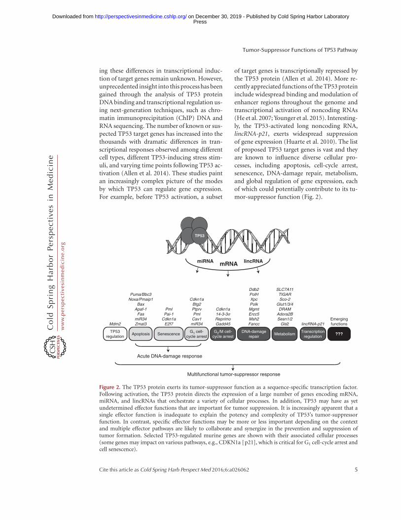
ing these differences in transcriptional induc-tion of target genes remain unknown. However,unprecedented insight into this process has beengained through the analysis of TP53 proteinDNA binding and transcriptional regulation us-ing next-generation techniques, such as chro-matin immunoprecipitation (ChIP) DNA andRNA sequencing. The number of known or sus-pected TP53 target genes has increased into thethousands with dramatic differences in tran-scriptional responses observed among differentcell types, different TP53-inducing stress stim-uli, and varying time points following TP53 ac-tivation (Allen et al. 2014). These studies paintan increasingly complex picture of the modesby which TP53 can regulate gene expression.For example, before TP53 activation, a subset
of target genes is transcriptionally repressed bythe TP53 protein (Allen et al. 2014). More re-cently appreciated functions of the TP53 proteininclude widespread binding and modulation ofenhancer regions throughout the genome andtranscriptional activation of noncoding RNAs(He et al. 2007; Younger et al. 2015). Interesting-ly, the TP53-activated long noncoding RNA,lincRNA-p21, exerts widespread suppressionof gene expression (Huarte et al. 2010). The listof proposed TP53 target genes is vast and theyare known to influence diverse cellular pro-cesses, including apoptosis, cell-cycle arrest,senescence, DNA-damage repair, metabolism,and global regulation of gene expression, eachof which could potentially contribute to its tu-mor-suppressor function (Fig. 2).
Apoptosis SenescenceG1 cell-
cycle arrestTP53
regulation
PmlPai-1
Cdkn1aE2f7
Puma/Bbc3Noxa/Pmaip1
BaxApaf-1
FasmiR34Zmat3
Cdkn1aBtg2PtprvPml
Cav1miR34Mdm2
MetabolismDNA-damage
repair
SLC7A11TIGARSco-2
Glut1/3/4DRAM
Adora2BSesn1/2
Gls2
Ddb2PolHXpcPolkMgmtErcc5Msh2Fancc
???
Acute DNA-damage response
Multifunctional tumor-suppressor response
mRNAmiRNA lincRNA
G2/M cell-cycle arrest
Emergingfunctions
TP53
lincRNA-p21
Transcriptionregulation
Cdkn1a14-3-3σReprimoGadd45
Figure 2. The TP53 protein exerts its tumor-suppressor function as a sequence-specific transcription factor.Following activation, the TP53 protein directs the expression of a large number of genes encoding mRNA,miRNA, and lincRNAs that orchestrate a variety of cellular processes. In addition, TP53 may have as yetundetermined effector functions that are important for tumor suppression. It is increasingly apparent that asingle effector function is inadequate to explain the potency and complexity of TP53’s tumor-suppressorfunction. In contrast, specific effector functions may be more or less important depending on the contextand multiple effector pathways are likely to collaborate and synergize in the prevention and suppression oftumor formation. Selected TP53-regulated murine genes are shown with their associated cellular processes(some genes may impact on various pathways, e.g., CDKN1a [p21], which is critical for G1 cell-cycle arrest andcell senescence).
Tumor-Suppressor Functions of TP53 Pathway
Cite this article as Cold Spring Harb Perspect Med 2016;6:a026062 5
ww
w.p
ersp
ecti
vesi
nm
edic
ine.
org
Press on December 30, 2019 - Published by Cold Spring Harbor Laboratoryhttp://perspectivesinmedicine.cshlp.org/Downloaded from

Insight into the critical TP53 transcriptionaltargets has been gained from genetic mousemodels expressing transcriptionally defectivemutant TP53 proteins. Interestingly, a partiallytransactivation defective TP53 protein, denotedTP5325,26 (Brady et al. 2011), can only activatea limited number of TP53 target genes and isunable to induce apoptosis, cell-cycle arrest,or senescence, yet it retains the ability to sup-press tumor formation. Complementary find-ings have been observed in a different mousemodel, in which key lysine residues of theTP53 protein, which are modified by acetyla-tion during posttranslational activation, havebeen mutated to arginine, denoted TP533KR
(Li et al. 2012). Similar to the TP5325,26 mutant,the mutant TP533KR protein is unable to activatetarget genes that mediate apoptosis, cell-cyclearrest, and cell senescence, yet it still retains theability to suppress tumor development. Exami-nation of these mutant strains of mice revealedpreserved regulation of several TP53 responsegenes involved in DNA-damage repair and me-tabolism, implicating a potentially critical rolefor these processes in tumor suppression. Atpresent, the search for the critical TP53 tumor-suppressor transcriptional targets is underwayin earnest.
KEY EFFECTOR FUNCTIONS FORTUMOR SUPPRESSION
Apoptosis
Apoptosis was one of the earliest identifiedcomponents of the TP53-mediated tumor-sup-pressor response (Yonish-Rouach et al. 1991).Induction of apoptosis is among the most ex-tensively studied cellular processes activated bythe TP53 protein and has been the focus ofmuch of the investigation into its tumor-sup-pressor effect. Impaired apoptosis is a cardinalfeature of malignancy and genetic alterationsthat result in evasion from apoptotic celldeath markedly accelerate tumor development(Vaux et al. 1988; Strasser et al. 1990; Czabotaret al. 2014). Multiple TP53 target genes havebeen implicated in TP53-mediated inductionof apoptosis: Puma, Noxa, Bax, Apaf1, Fas,Tnfrsf10B/DR5, miR34, TP53AIP1, Pidd, Pig3,
Zmat3, and Siva. Among these target genes,Puma, Noxa, Bax, and Apaf-1 play critical rolesin the intrinsic (also called BCL-2-regulated ormitochondrial) apoptotic pathway (Youle andStrasser 2008; Strasser et al. 2011), whereas Fasand Tnfrsf10B/DR5 encode for members ofthe tumor necrosis factor receptor (TNFR) fam-ily (FAS/APO-1/CD95 and TRAIL-R/DR5)that can trigger the death receptor (also calledextrinsic) apoptotic pathway (Strasser et al.2009). Of all these TP53 target genes, only theproapoptotic BH3-only BCL-2 family mem-bers PUMA and NOXA have been validated bystudies of gene-targeted mice to be essential forTP53-mediated apoptosis (Jeffers et al. 2003;Shibue et al. 2003; Villunger et al. 2003; Micha-lak et al. 2008). Although the Bax and Apaf-1genes are also direct TP53 targets (Riley et al.2008), TP53-deficient cells still express these ef-fectors of apoptosis. Therefore, their inductionlikely serves to amplify TP53-mediated apopto-sis signaling. The miR34 family of microRNAs isa TP53 target (He et al. 2007) predicted to exertbroad antioncogenic effects through the post-transcriptional regulation of a variety of genesthat not only sensitize to apoptosis, for example,by down-regulation of BCL-2 (Bommer et al.2007), but also through regulation of cell-cycleprogression and differentiation. Surprisingly,however, mice that are deficient of all miR34family members are not susceptible to sponta-neous or oncogene-induced tumor develop-ment (Concepcion et al. 2012). Importantly,the death receptor apoptotic pathway is dispen-sable for TP53-induced apoptosis (Newton andStrasser 2000). However, TP53-mediated in-duction of Fas and Tnfrsf10B/DR5 expressionmay serve to sensitize stressed cells to the deathreceptor ligands, FASL and TRAIL, and it hasbeen proposed that this could be exploited forcancer therapy (Ashkenazi 2008).
The role of TP53-mediated apoptosis inpreventing oncogene-driven cancer develop-ment has been defined using the Em-Myc trans-genic mouse model (Adams et al. 1985). Herethe immunoglobulin heavy chain gene enhanc-er (Em) has been juxtaposed to the c-Myc onco-gene, resulting in deregulated c-MYC expressionand, consequently, the rapid development of
B.J. Aubrey et al.
6 Cite this article as Cold Spring Harb Perspect Med 2016;6:a026062
ww
w.p
ersp
ecti
vesi
nm
edic
ine.
org
Press on December 30, 2019 - Published by Cold Spring Harbor Laboratoryhttp://perspectivesinmedicine.cshlp.org/Downloaded from

pre-B and B-cell lymphomas. In the Em-Mycmouse model, spontaneous inactivation of theTP53 pathway, most frequently through muta-tions in Tp53 itself, is seen in �20% of lympho-mas (Eischen et al. 1999; Schmitt et al. 1999;Michalak et al. 2009), indicative of its criticalrole in this setting. Accordingly, inactivation ofTP53 function markedly accelerates MYC-driv-en lymphoma development (Schmitt et al.1999; Michalak et al. 2009). Strikingly, completedeletion of the Tp53 gene using CRISPR/Cas9targeting in Em-Myc hematopoietic stem/pro-genitor cells (HSPC) results in the rapid devel-opment of lymphoma with a median latency ofonly 29 d (Aubrey et al. 2015) as compared withnontargeted Em-Myc HSPC that give rise tolymphoma with a mean latency of .110 d.The specific requirement for individual TP53apoptotic transcriptional targets in the tumor-suppressor function has been dissected usingthe Em-Myc mouse model where loss of BAX(Eischen et al. 2001; Dansen et al. 2006),PUMA, and NOXA (Hemann et al. 2004; Mi-chalak et al. 2009) can each accelerate lympho-ma development, although not to the same ex-tent as complete loss of TP53 function. Thissuggests critical roles for additional pathwaysin tumor suppression during MYC-driven lym-phoma development.
Interestingly, mice engineered to harboronly specific gene knockout of the TP53 apo-ptotic targets Puma (Jeffers et al. 2003), Noxa(Villunger et al. 2003), Bax (Knudson et al.2001), or even combined loss of Puma/Noxa/p21 (Valente et al. 2013) do not display a pro-pensity for tumor formation. Thus, in the ab-sence of constitutive oncogenic stress, the com-bined knockout of the major TP53-dependentmediators of apoptosis (PUMA and NOXA)and the major mediator of G1/S cell-cycle arrestand cell senescence (p21) does not recapitulatethe tumor predisposition observed in Tp532/2
mice. Apoptosis clearly plays a critical role intumor suppression; however, additional path-ways must be disabled to fully recapitulate theeffect from complete loss of TP53. Furthermore,these studies show that the animal model inwhich TP53 functions is examined will likelyinfluence the experimental findings. Although
PUMA, NOXA, and BAX are important medi-ators of TP53-induced apoptosis, there may beadditional proapoptotic effector mechanismsthat have yet to be fully defined. For example,the proapoptotic BH3-only protein BIM maybe induced indirectly by TP53 and contributeto the killing of tumor cells by DNA-damage-inducing chemotherapeutic drugs (Happo et al.2010). Importantly, there is substantial overlapbetween the regulation of apoptotic cell deathand other pathways, such as DNA-damage re-pair and metabolism; thus, the role of apoptosisin tumor suppression may be intertwined withother TP53-dependent effectors.
Cell-Cycle Regulation and DNA-Damage Repair
Cancer is a disease that results from the progres-sive acquisition and accumulation of geneticmutations (Hanahan and Weinberg 2011), andthe TP53 protein, as the “guardian of the ge-nome” (Lane 1992), has a salient role in main-taining genomic integrity and opposing thisprocess. The TP53 protein plays an intimaterole in the cellular response to DNA damage.It is critical to both the acute phase responseinvolving cell-cycle arrest, senescence, and apo-ptosis as well as long-term surveillance mecha-nisms for maintaining multiple DNA-damage-repair mechanisms, such as nucleotide excisionrepair, base excision repair, and nonhomolo-gous end-joining (Sengupta and Harris 2005).
In many cells, the initial TP53-mediated re-sponse to acute DNA damage is the induction oftransient G1 cell-cycle arrest, which allows timefor the detection and repair of DNA damagebefore replication of the genome in S phaseand subsequent cell division. The TP53 proteinalso exerts checkpoint control during the G2/Mtransition, at which time DNA replication hasalready occurred and cells prepare to undergomitotic cell division (Taylor and Stark 2001), atime when failed detection and repair of dam-aged DNA may be most catastrophic (e.g., re-sulting in aneuploidy). Both cell-cycle check-points are critical to maintaining genomicintegrity and the requirement for TP53-medi-ated cell-cycle arrest in maintaining genomic
Tumor-Suppressor Functions of TP53 Pathway
Cite this article as Cold Spring Harb Perspect Med 2016;6:a026062 7
ww
w.p
ersp
ecti
vesi
nm
edic
ine.
org
Press on December 30, 2019 - Published by Cold Spring Harbor Laboratoryhttp://perspectivesinmedicine.cshlp.org/Downloaded from

stability has been shown experimentally (Bar-boza et al. 2006). The key mediator of TP53-induced G1 cell-cycle arrest is thought tobe CDKN1a (p21), as shown by cells fromCdkn1a2/2 mice that show impaired G1 cell-cycle arrest in response to DNA damage andTP53 activation (El-Deiryet al. 1993; Brugarolaset al. 1995; Deng et al. 1995). Given the integralrole of cell-cycle arrest in DNA repair, CDKN1ahas been proposed to contribute to TP53-medi-ated tumor suppression. However, mice thatlack CDKN1a (p21) are not prone to spontane-ous tumor formation (Deng et al. 1995; Valenteet al. 2013). Additional TP53 transcriptionaltargets also contribute to G1 phase cell-cyclearrest including, but not limited to, the promye-locytic gene (Pml), protein tyrosine phospha-tase receptor type-V gene (Ptprv), Caveolin-1(Cav1) (Galbiati et al. 2001), and Btg2 (Rouaultet al. 1996). In addition, other TP53 targets spe-cifically instigate cell-cycle arrest at the G2/Mcheckpoint, including the growth-arrest andDNA-damage-inducible 45 a gene (Gadd45a),Reprimo, and the 14-3-3s protein (Taylor andStark 2001). The roles of GADD45a, PTPRV,PML, and CAV1 have been examined indi-vidually through the generation of knockoutmice but none of these animals spontaneouslydevelop cancer (Hollander et al. 1999; Razani etal. 2001; Rego et al. 2001; Doumont et al. 2005).However, similar to other candidate TP53 tu-mor-suppressor transcriptional targets, theirdeficiency can accelerate tumor formation un-der conditions of specific oncogenic stress (Ca-pozza et al. 2003; Tront et al. 2010).
TP53-induced cell-cycle arrest is thoughtnecessary to allow for appropriate DNA-repairprocesses to occur (Barboza et al. 2006). Pre-cancerous lesions are characterized by the ac-cumulation of DNA damage and consequentactivation of the TP53-mediated DNA-damageresponse (Bartkova et al. 2005; Gorgoulis et al.2005). The acquisition of mutations duringtumor initiation occurs through a variety ofmechanisms including mutations and epigenet-ic modifications followed by propagation ofthese alterations owing to defective DNA-dam-age repair mechanisms. The TP53 protein playspivotal roles in all of these processes and, in
keeping with the importance of TP53 in main-taining genomic stability, cells from Tp532/2
mice as well as TP53-defective human cancersare characterized by widespread genomic alter-ations. In line with this, TP53 has a large num-ber of direct transcriptional targets that mediateDNA-repair pathways, including Polk, Mgmt,Fancc, Ercc5, Xpc, Ddb2, Gadd45a, Msh2, andPolH (Allen et al. 2014; Bieging et al. 2014). Thecentral role of genomic instability during theevolution of thymic lymphomas in Tp532/2
mice has been directly observed over time,where defective DNA repair results in a veryhigh rate of gene copy number variations, in-cluding chromotrypsis-like events, which drivethe accumulation of the cooperating genetic le-sions required for malignant transformation(Dudgeon et al. 2014). This supports the notionthat genomic instability is a key driver of cancerdevelopment in the absence of the TP53 protein.
In certain cell types, activation of the TP53protein can result in the induction of apoptosisresulting in the elimination of irreversibly dam-aged cells. However, the acute DNA-damage re-sponse has been largely excluded from a role inthe TP53 tumor-suppressor function throughmultiple lines of investigation (Christophorouet al. 2006; Brady et al. 2011; Li et al. 2012).The role of the acute DNA-damage response intumor suppression was evaluated using timedrestoration of TP53 protein in Tp532/2 micefollowing g-irradiation to induce thymic lym-phoma formation. Remarkably, transient TP53restoration during the acute DNA-damage re-sponse did not produce a tumor-suppressoreffect (Christophorou et al. 2006). In contrast,transient restoration of TP53 function that wasdelayed until after the acute DNA-damage re-sponse had ended, at a time when there wasno discernable cell-cycle arrest or apoptosis,was sufficient for tumor suppression. Further-more, the delayed tumor-suppressor functionobserved is dependent on p19/ARF, implicatingoncogene-mediated TP53 activation in nascentneoplastic cells in this response. This is furtherconfirmed through studies of transcriptionallydefective and acetylation defective mutant TP53proteins that are unable to activate the acuteDNA-damage response yet retain potent tu-
B.J. Aubrey et al.
8 Cite this article as Cold Spring Harb Perspect Med 2016;6:a026062
ww
w.p
ersp
ecti
vesi
nm
edic
ine.
org
Press on December 30, 2019 - Published by Cold Spring Harbor Laboratoryhttp://perspectivesinmedicine.cshlp.org/Downloaded from

mor-suppressor function (Brady et al. 2011;Li et al. 2012). Therefore, the acute pathologi-cal DNA-damage response appears to be dis-pensable for tumor suppression, a remarkablefinding that also has major implications for can-cer therapy.
The consequences of oncogene overexpres-sion are twofold in the setting of DNA damageand TP53-mediated tumor suppression. First,acquired mutations may result in the activationof oncogenes, and cells expressing oncogenescan be selected for through enhanced prolifera-tion and cell survival. Second, chronic oncogeneactivation drives abnormal cell growth, therebyincreasing the risk of acquiring additional DNAlesions that may activate further oncogenes orinactivate tumor-suppressor genes (Halazonetiset al. 2008). TP53 may be purposed to eliminateor growth-arrest cells marked by oncogene over-expression, which is intimately connected withDNA damage, deregulated cell proliferation,and metabolic deregulation.
Senescence
Induction of cell senescence was first shownto play a critical role in TP53-mediated tumorsuppression in a mouse model of erythroleuke-mia (Metz et al. 1995). Moreover, restoration ofTP53 function in established solid-organ tu-mors (driven by loss of TP53) in vivo leads tothe induction of cellular senescence in associa-tion with tumor regression (Ventura et al. 2007;Xue et al. 2007). Cellular senescence is a distinctcell state involving permanent cell-cycle arrestof cells that remain viable and metabolically ac-tive, which is characterized by a discrete tran-scriptional profile (Shay and Roninson 2004).The TP53 protein controls cellular senescenceby activating a number of transcriptional targetsthat include Cdkn1a, Pml, Pai1, and E2f7 (Pear-son et al. 2000; Kortlever et al. 2006; Aksoy et al.2012), some of which (e.g., Cdkn1a) are nota-ble for additional function in cell-cycle regula-tion. Senescence is often associated with, andis thought to suppress, premalignant lesionspreventing their progression to overt malignan-cy (Collado et al. 2005; Mooi and Peeper 2006).TP53-mediated induction of cell senescence
was shown to be critical to preventing malignanttransformation in a mouse model of BRAF-driven pulmonary adenoma (Dankort et al.2007). Moreover, in a study examining the func-tional interdependence of defects in PTEN andTP53 in the development of prostate carcinoma,TP53-mediated senescence was required to pre-vent cancer development in the setting of PTENdeletion (Chen et al. 2005). It remains to beexamined whether complete loss of all targetgenes implicated in TP53-mediated senescencecan recapitulate the spontaneous tumor devel-opment seen in Tp532/2 mice and whether thestrong association between senescence and tu-mor suppression is causal or whether this is anassociation with other TP53-mediated effects.
Metabolism
The rapid proliferation of cells, anabolic growth,and metabolic stress that typifies neoplasticdisease requires substantial metabolic repro-gramming (Hanahan and Weinberg 2011). Fur-thermore, metabolic deregulation not only im-pacts on energy production and cell growth butalso influences additional processes importantto sustained cancer growth, such as macromo-lecular biosynthesis, epigenetic regulation, andantioxidant pathways (Cairns et al. 2011; Wardand Thompson 2012). The TP53 protein is acritical regulator of cellular metabolism andmany of the aforementioned processes affectedby metabolic stress (Berkers et al. 2013).
The best-characterized description of can-cer-associated metabolic reprogramming is theWarburg effect, whereby glucose is predomi-nantly metabolized by glycolysis rather than ox-idative phosphorylation, as normally occurs un-der aerobic conditions (Warburg 1956). TP53activation stimulates oxidative phosphorylationand inhibits glycolysis, both of which opposethe Warburg effect. The TP53 protein can regu-late the expression of several glucose transport-ers, including GLUT1, GLUT3, and GLUT4(Schwartzenberg-Bar-Yoseph et al. 2004; Ka-wauchi et al. 2008), which diminishes glycolysisthrough impaired glucose uptake. TP53 alsotransactivates the TP53-induced glycolysis andapoptosis regulator gene (TIGAR), which en-
Tumor-Suppressor Functions of TP53 Pathway
Cite this article as Cold Spring Harb Perspect Med 2016;6:a026062 9
ww
w.p
ersp
ecti
vesi
nm
edic
ine.
org
Press on December 30, 2019 - Published by Cold Spring Harbor Laboratoryhttp://perspectivesinmedicine.cshlp.org/Downloaded from

codes a fructose phosphatase enzyme that in-hibits glycolysis and increases production ofNADPH (Bensaad et al. 2006). NADPH is im-portant for the scavenging of reactive oxygenspecies (ROS), and this antioxidant effect ofTIGAR confers a prosurvival function in thesetting of ROS-mediated cell death. However,TIGAR knockout mice do not show spontane-ous tumor formation (Cheung et al. 2013) and,in some contexts, TIGAR deficiency actuallyimpedes tumor development (Bensaad et al.2006). TP53 also directly stimulates mitochon-drial oxidative phosphorylation through tran-scriptional activation of the gene-encoding syn-thesis of cytochrome c oxidase 2 (Sco2) (Matobaet al. 2006). Interestingly, dysregulated oxidativephosphorylation has been observed in cells frompatients with Li–Fraumeni syndrome, whichis attributed partly to altered Sco-2 expression(Wang et al. 2013).
The strict regulation of cellular antioxidantmechanisms is critical to maintaining intra-cellular signaling pathways and avoiding ROS-associated toxicity. Therefore, dysregulationof these processes can contribute to cancer de-velopment (Finkel 2003). Genes encoding en-zymes with antioxidant functions, includingGls2, Sestrin 1, and Sestrin 2, have been identi-fied as TP53 transcriptional targets (Budanovet al. 2004; Hu et al. 2010), defining a mecha-nism by which TP53 can regulate oxidant sig-naling and manage oxidative stress. Interest-ingly, treatment of Tp53 knockout mice withantioxidant therapy was reported to delay theonset of tumor formation, implicating a role forlimiting the ROS accumulation in TP53-medi-ated tumor suppression (Sablina et al. 2005).
Cancer-associated metabolic stress andhypoxia can activate distinct cell death pathwaysthrough a variety of mechanisms, including theintrinsic apoptotic pathway (Czabotar et al.2014). TP53 can modulate metabolic stress-induced cell death through a number of mech-anisms. For example, the TP53 protein drivesexpression of the ADORA2B gene, which candetect nutrient availability and sensitize cellsto PUMA-mediated apoptotic cell death (Longet al. 2013). In addition, a recently describedform of iron-dependent, nonapoptotic cell
death, denoted ferroptosis, is initiated underconditions of metabolic stress and accumulatedROS (Dixon et al. 2012). It has been proposedthat TP53 mediates tumor suppression throughtranscriptional repression of the SLC7A11 gene,which encodes a cystine/glutamate antiporterthat diminishes cellular predisposition to fer-roptosis (Jiang et al. 2015).
Finally, autophagy is another TP53-regu-lated metabolic process that may contribute totumor suppression (Maiuri et al. 2010; Kenzel-mann Broz et al. 2013). Autophagy enables cellsto adapt and survive in conditions of limitingnutrient availability by recycling intracellularcontents, such as damaged proteins and or-ganelles for the purpose of liberating energyand metabolites to maintain cellular integrity(Mathew and White 2011). Autophagy may fur-ther impact on tumor suppression by affectingapoptotic pathways and genomic stability. TP53regulates autophagyat multiple levels (Feng et al.2005) by transactivating a large number of genes(Kenzelmann Broz et al. 2013), including thegenes encoding damage-associated autophagymediator (DRAM) (Crighton et al. 2006) andULK1 (Gao et al. 2011). Interestingly, siRNA-mediated knockdown of DRAM was shownto reduce TP53-dependent apoptosis (Crightonet al. 2006).
EMERGING TP53 FUNCTIONSAND TUMOR SUPPRESSION
New components of the TP53 response contin-ue to emerge and many of these have been im-plicated in tumor suppression (Bieging et al.2014; Hager and Gu 2014). These include rolesfor TP53 in stem-cell function, differentiation,cellular invasion, and metastasis, as well as reg-ulation of the immune response and the tumormicroenvironment. For example, in a mousemodel of hepatocellular carcinoma, TP53 wasshown to influence the microenvironment andimmune response via a non-tumor-cell-auton-omous mechanism, which impacted the rate oftumor expansion and aggressiveness (Lujambioet al. 2013). These emerging functions haveas yet undetermined roles in tumor suppres-sion; however, they highlight the increasingly
B.J. Aubrey et al.
10 Cite this article as Cold Spring Harb Perspect Med 2016;6:a026062
ww
w.p
ersp
ecti
vesi
nm
edic
ine.
org
Press on December 30, 2019 - Published by Cold Spring Harbor Laboratoryhttp://perspectivesinmedicine.cshlp.org/Downloaded from
complex picture of the role of TP53 in cancerbiology.
In addition to newly identified functionsfor the TP53 protein, novel approaches to un-derstanding the mechanisms of tumor suppres-sion are emerging. Cell competition has recentlybeen established as a bona fide mechanism fortumor suppression (Martins et al. 2014) when itwas shown that disruption of normal cell com-petition in the thymus leads to the formation ofacute T-lymphoblastic leukemia. The conceptof cell competition provides a view of the overallfitness of cells as they compete within a largerpopulation of cells, accounting for various cel-lular attributes as well as context-dependentfactors. In studies of hematopoietic progenitorcells, the TP53 protein was shown to mediatecell competition (Bondar and Medzhitov 2010),raising the possibility that cell competitionmay be an important framework within whichto approach the question of TP53-mediated tu-mor suppression.
DISTINCTIONS BETWEEN HUMAN CANCERAND MOUSE MODELS
It is important to recognize a number of keydistinctions between findings from mousemodels and human disease. In human cancer,inactivation of the TP53 gene almost always oc-curs by acquisition of a missense mutation rath-er than deletion of the TP53 gene. Furthermore,these mutations frequently result in a singleamino acid substitution and the production ofa stable, overexpressed TP53 protein that canactively contribute to tumor development andgrowth over and above the consequences of los-ing wild-type TP53 function alone (Freed-Pas-tor and Prives 2012). In addition, the initialacquisition of Tp53 mutations is usually fol-lowed by loss of heterozygosity, which typicallyinvolves large deletions of the short arm of chro-mosome 17 (17p). The large chromosomal de-letion results in the loss of several additionalgenes and this raises the possibility that coop-erating lesions on 17p may contribute to humancancers. For example, a gene encoding a com-ponent of the RNA polymerase II complex,POLR2A, is almost always codeleted in human
cancers with the TP53 gene, and this has func-tional impact on the resulting tumor (Liu et al.2015). These are important features of humancancer that are inextricable from the questionof understanding how loss or mutation of TP53leads to the development of cancer.
CONCLUDING REMARKS
The tumor-suppressor function of the TP53protein is likely to be mediated through a num-ber of collaborating effector functions ratherthan through a single pathway or single tran-scriptional target. Understanding how thesemechanisms work together will require creativeapproaches to investigation that take into ac-count the combinatorial nature and complexityof the TP53 response as well as considerationof its functioning in normal cellular processes.It is an intriguing question as to whether theprimary purpose of the TP53 protein is tumorsuppression or whether this is a secondary man-ifestation of its many important roles in normalbiology. For example, the importance of theTP53 protein in maintaining genomic stabilityextends beyond the prevention of cancer tobeing a basic requirement for sustainable lifethat ensures an organism’s genetic material istransmitted faithfully to subsequent generations(Jackson and Bartek 2009; Kerr et al. 2012) andspeaks to the evolutionary conservation of theTp53 family of genes from the earliest multicel-lular organisms to humans (Belyi et al. 2010;Lane et al. 2010). Another important consider-ation is that TP53 contributes to normal cellularprocesses that may also be important in estab-lished tumors. For example, TP53 facilitatesadaptation to some forms of metabolic stress,such as serine deprivation, whereby Tp532/2
cells are actually disadvantaged (Maddockset al. 2013). As such, the complete loss of TP53function during cancer initiation may not beentirely advantageous and such vulnerabilitiesmay be exploited for treatment of TP53-defi-cient cancers. Understanding the role of TP53in tumor suppression remains one of the mostexciting and important biological questionsthat promises exciting advances for the next30 years of TP53 research.
Tumor-Suppressor Functions of TP53 Pathway
Cite this article as Cold Spring Harb Perspect Med 2016;6:a026062 11
ww
w.p
ersp
ecti
vesi
nm
edic
ine.
org
Press on December 30, 2019 - Published by Cold Spring Harbor Laboratoryhttp://perspectivesinmedicine.cshlp.org/Downloaded from

ACKNOWLEDGMENTS
The authors thank Drs. Marco J. Herold, DanielH.D. Gray, Jerry M. Adams, Suzanne Cory, andAna Janic for insightful discussions and advice,and Cameron Wells and Rachel Bucknall forassistance with graphic design. This work issupported by a Leukaemia Foundation NationalResearch Program Clinical PhD Scholarship(B.J.A.), a Kay Kendall Leukaemia Fund In-termediate Fellowship (KKL331 to G.L.K.),a National Health and Medical Research Coun-cil, Australia, Program Grant 1016701 (A.S.)and Fellowship 1020363 (A.S.), Project Grant1086291 from the National Health and Medi-cal Research Council, Australia (G.L.K.), a Can-cer Council Victoria Grant-in-Aid 1086157(G.L.K), and a Leukemia and LymphomaSociety SCOR Grant 7001-13 (A.S.). Thiswork is made possible through Victorian StateGovernment Operational Infrastructure Sup-port and Australian Government NationalHealth and Medical Research Council Indepen-dent Research Institutes Infrastructure SupportScheme.
REFERENCES
Achatz MI, Olivier M, Le Calvez F, Martel-Planche G, LopesA, Rossi BM, Ashton-Prolla P, Giugliani R, Palmero EI,Vargas FR, et al. 2007. The TP53 mutation, R337H, isassociated with Li–Fraumeni and Li–Fraumeni-like syn-dromes in Brazilian families. Cancer Lett 245: 96–102.
Adams JM, Harris AW, Pinkert CA, Corcoran LM, AlexanderWS, Cory S, Palmiter RD, Brinster RL. 1985. The c-myconcogene driven by immunoglobulin enhancers induceslymphoid malignancy in transgenic mice. Nature 318:533–538.
Aksoy O, Chicas A, Zeng T, Zhao Z, McCurrach M, Wang X,Lowe SW. 2012. The atypical E2F family member E2F7couples the p53 and RB pathways during cellular senes-cence. Genes Dev 26: 1546–1557.
Allen MA, Andrysik Z, Dengler VL, Mellert HS, GuarnieriA, Freeman JA, Sullivan KD, Galbraith MD, Luo X, KrausWL, et al. 2014. Global analysis of p53-regulated tran-scription identifies its direct targets and unexpected reg-ulatory mechanisms. eLife 3: e02200.
Ashkenazi A. 2008. Directing cancer cells to self-destructwith pro-apoptotic receptor agonists. Nat Rev Drug Dis-cov 7: 1001–1012.
Aubrey BJ, Kelly GL, Kueh AJ, Brennan MS, O’Connor L,Milla L, Wilcox S, Tai L, Strasser A, Herold MJ. 2015. Aninducible lentiviral guide RNA platform enables the iden-tification of tumor-essential genes and tumor-promotingmutations in vivo. Cell Rep 10: 1422–1432.
Baker SJ, Fearon ER, Nigro JM, Hamilton SR, Preisinger AC,Jessup JM, vanTuinen P, Ledbetter DH, Barker DF, Naka-mura Y, et al. 1989. Chromosome 17 deletions and p53gene mutations in colorectal carcinomas. Science 244:217–221.
Barboza JA, Liu G, Ju Z, El-Naggar AK, Lozano G. 2006. p21delays tumor onset by preservation of chromosomalstability. Proc Natl Acad Sci 103: 19842–19847.
Bartkova J, Horejsi Z, Koed K, Kramer A, Tort F, Zieger K,Guldberg P, Sehested M, Nesland JM, Lukas C, et al. 2005.DNA damage response as a candidate anti-cancer barrierin early human tumorigenesis. Nature 434: 864–870.
Belyi VA, Ak P, Markert E, Wang H, Hu W, Puzio-KuterA, Levine AJ. 2010. The origins and evolution of thep53 family of genes. Cold Spring Harb Perspect Biol 2:a001198.
Bensaad K, Tsuruta A, Selak MA, Vidal MN, Nakano K,Bartrons R, Gottlieb E, Vousden KH. 2006. TIGAR,a p53-inducible regulator of glycolysis and apoptosis.Cell 126: 107–120.
Berkers CR, Maddocks OD, Cheung EC, Mor I, VousdenKH. 2013. Metabolic regulation by p53 family members.Cell Metab 18: 617–633.
Bieging KT, Mello SS, Attardi LD. 2014. Unravelling mech-anisms of p53-mediated tumour suppression. Nat RevCancer 14: 359–370.
Bommer GT, Gerin I, Feng Y, Kaczorowski AJ, Kuick R, LoveRE, Zhai Y, Giordano TJ, Qin ZS, Moore BB, et al. 2007.p53-mediated activation of miRNA34 candidate tumor-suppressor genes. Curr Biol 17: 1298–1307.
Bondar T, Medzhitov R. 2010. p53-mediated hematopoieticstem and progenitor cell competition. Cell Stem Cell 6:309–322.
Brady CA, Jiang D, Mello SS, Johnson TM, Jarvis LA, KozakMM, Kenzelmann Broz D, Basak S, Park EJ, McLaughlinME, et al. 2011. Distinct p53 transcriptional programsdictate acute DNA-damage responses and tumor sup-pression. Cell 145: 571–583.
Brown CJ, Lain S, Verma CS, Fersht AR, Lane DP. 2009.Awakening guardian angels: Drugging the p53 pathway.Nat Rev Cancer 9: 862–873.
Brugarolas J, Chandrasekaran C, Gordon JI, Beach D, JacksT, Hannon GJ. 1995. Radiation-induced cell cycle arrestcompromised by p21 deficiency. Nature 377: 552–557.
Budanov AV, Sablina AA, Feinstein E, Koonin EV, ChumakovPM. 2004. Regeneration of peroxiredoxins by p53-regu-lated sestrins, homologs of bacterial AhpD. Science 304:596–600.
Cairns RA, Harris IS, Mak TW. 2011. Regulation of cancercell metabolism. Nat Rev Cancer 11: 85–95.
Capozza F, Williams TM, Schubert W, McClain S, Bouzah-zah B, Sotgia F, Lisanti MP. 2003. Absence of caveolin-1sensitizes mouse skin to carcinogen-induced epidermalhyperplasia and tumor formation. Am J Pathol 162:2029–2039.
Chen Z, Trotman LC, Shaffer D, Lin HK, Dotan ZA, Niki M,Koutcher JA, Scher HI, Ludwig T, Gerald W, et al. 2005.Crucial role of p53-dependent cellular senescence in sup-pression of Pten-deficient tumorigenesis. Nature 436:725–730.
B.J. Aubrey et al.
12 Cite this article as Cold Spring Harb Perspect Med 2016;6:a026062
ww
w.p
ersp
ecti
vesi
nm
edic
ine.
org
Press on December 30, 2019 - Published by Cold Spring Harbor Laboratoryhttp://perspectivesinmedicine.cshlp.org/Downloaded from

Cheung EC, Athineos D, Lee P, Ridgway RA, Lambie W,Nixon C, Strathdee D, Blyth K, Sansom OJ, VousdenKH. 2013. TIGAR is required for efficient intestinal re-generation and tumorigenesis. Dev Cell 25: 463–477.
Cho Y, Gorina S, Jeffrey PD, Pavletich NP. 1994. Crystalstructure of a p53 tumor suppressor–DNA complex: Un-derstanding tumorigenic mutations. Science 265: 346–355.
Christophorou MA, Ringshausen I, Finch AJ, Swigart LB,Evan GI. 2006. The pathological response to DNA dam-age does not contribute to p53-mediated tumour sup-pression. Nature 443: 214–217.
Collado M, Gil J, Efeyan A, Guerra C, Schuhmacher AJ,Barradas M, Benguria A, Zaballos A, Flores JM, BarbacidM, et al. 2005. Tumour biology: Senescence in premalig-nant tumours. Nature 436: 642.
Concepcion CP, Han YC, Mu P, Bonetti C, Yao E, D’AndreaA, Vidigal JA, Maughan WP, Ogrodowski P, Ventura A.2012. Intact p53-dependent responses in miR-34-defi-cient mice. PLoS Genet 8: e1002797.
Crighton D, Wilkinson S, O’Prey J, Syed N, Smith P, Harri-son PR, Gasco M, Garrone O, Crook T, Ryan KM. 2006.DRAM, a p53-induced modulator of autophagy, is crit-ical for apoptosis. Cell 126: 121–134.
Czabotar PE, Lessene G, Strasser A, Adams JM. 2014. Con-trol of apoptosis by the BCL-2 protein family: Implica-tions for physiology and therapy. Nat Rev Mol Cell Biol15: 49–63.
Dankort D, Filenova E, Collado M, Serrano M, Jones K,McMahon M. 2007. A new mouse model to explore theinitiation, progression, and therapy of BRAFV600E-in-duced lung tumors. Genes Dev 21: 379–384.
Dansen TB, Whitfield J, Rostker F, Brown-Swigart L, EvanGI. 2006. Specific requirement for bax, not bak, in MYC-induced apoptosis and tumor suppression in vivo. J BiolChem 281: 10890–10895.
DeLeo AB, Jay G, Appella E, Dubois GC, Law LW, Old LJ.1979. Detection of a transformation-related antigen inchemically induced sarcomas and other transformed cellsof the mouse. Proc Natl Acad Sci 76: 2420–2424.
Deng C, Zhang P, Harper JW, Elledge SJ, Leder P. 1995. Micelacking p21CIP1/WAF1 undergo normal development,but are defective in G1 checkpoint control. Cell 82:675–684.
DiGiammarino EL, Lee AS, Cadwell C, Zhang W, Bothner B,Ribeiro RC, Zambetti G, Kriwacki RW. 2002. A novelmechanism of tumorigenesis involving pH-dependentdestabilization of a mutant p53 tetramer. Nat StructBiol 9: 12–16.
Dixon SJ, Lemberg KM, Lamprecht MR, Skouta R, ZaitsevEM, Gleason CE, Patel DN, Bauer AJ, Cantley AM, YangWS, et al. 2012. Ferroptosis: An iron-dependent form ofnonapoptotic cell death. Cell 149: 1060–1072.
Donehower LA, Harvey M, Slagle BL, McArthur MJ, Mont-gomery CAJ, Butel JS, Bradley A. 1992. Mice deficient forp53 are developmentally normal but are susceptible tospontaneous tumours. Nature 356: 215–221.
Doumont G, Martoriati A, Beekman C, Bogaerts S, Mee PJ,Bureau F, Colombo E, Alcalay M, Bellefroid E, Marchesi F,et al. 2005. G1 checkpoint failure and increased tumorsusceptibility in mice lacking the novel p53 target Ptprv.EMBO J 24: 3093–3103.
Dudgeon C, Chan C, Kang W, Sun Y, Emerson R, Robins H,Levine AJ. 2014. The evolution of thymic lymphomas inp53 knockout mice. Genes Dev 28: 2613–2620.
Eischen CM, Weber JD, Roussel MF, Sherr CJ, Cleveland JL.1999. Disruption of the ARF-Mdm2-p53 tumor suppres-sor pathway in Myc-induced lymphomagenesis. GenesDev 13: 2658–2669.
Eischen CM, Roussel MF, Korsmeyer SJ, Cleveland JL. 2001.Bax loss impairs Myc-induced apoptosis and circum-vents the selection of p53 mutations during Myc-medi-ated lymphomagenesis. Mol Cell Biol 21: 7653–7662.
el-Deiry WS, Kern SE, Pietenpol JA, Kinzler KW, VogelsteinB. 1992. Definition of a consensus binding site for p53.Nat Genet 1: 45–49.
el-Deiry WS, Tokino T, Velculescu VE, Levy DB, Parsons R,Trent JM, Lin D, Mercer WE, Kinzler KW, Vogelstein B.1993. WAF1, a potential mediator of p53 tumor suppres-sion. Cell 75: 817–825.
Eliyahu D, Michalovitz D, Eliyahu S, Pinhasi-Kimhi O, OrenM. 1989. Wild-type p53 can inhibit oncogene-mediatedfocus formation. Proc Natl Acad Sci 86: 8763–8767.
Feng Z, Zhang H, Levine AJ, Jin S. 2005. The coordinateregulation of the p53 and mTOR pathways in cells. ProcNatl Acad Sci 102: 8204–8209.
Finkel T. 2003. Oxidant signals and oxidative stress. CurrOpin Cell Biol 15: 247–254.
Finlay CA, Hinds PW, Levine AJ. 1989. The p53 proto-on-cogene can act as a suppressor of transformation. Cell57: 1083–1093.
Freed-Pastor WA, Prives C. 2012. Mutant p53: One name,many proteins. Genes Dev 26: 1268–1286.
Friedman PN, Chen X, Bargonetti J, Prives C. 1993. The p53protein is an unusually shaped tetramer that binds di-rectly to DNA. Proc Natl Acad Sci 90: 3319–3323.
Galbiati F, Volonte D, Liu J, Capozza F, Frank PG, Zhu L,Pestell RG, Lisanti MP. 2001. Caveolin-1 expression neg-atively regulates cell cycle progression by inducing G0/G1
arrest via a p53/p21(WAF1/Cip1)-dependent mecha-nism. Mol Biol Cell 12: 2229–2244.
Gao W, Shen Z, Shang L, Wang X. 2011. Upregulation ofhuman autophagy-initiation kinase ULK1 by tumor sup-pressor p53 contributes to DNA-damage-induced celldeath. Cell Death Differ 18: 1598–1607.
Gorgoulis VG, Vassiliou LV, Karakaidos P, Zacharatos P, Kot-sinas A, Liloglou T, Venere M, Ditullio RA Jr, KastrinakisNG, Levy B, et al. 2005. Activation of the DNA damagecheckpoint and genomic instability in human precancer-ous lesions. Nature 434: 907–913.
Hager KM, Gu W. 2014. Understanding the non-canonicalpathways involved in p53-mediated tumor suppression.Carcinogenesis 35: 740–746.
Halazonetis TD, Gorgoulis VG, Bartek J. 2008. An onco-gene-induced DNA damage model for cancer develop-ment. Science 319: 1352–1355.
Hanahan D, Weinberg RA. 2011. Hallmarks of cancer: Thenext generation. Cell 144: 646–674.
Happo L, Cragg MS, Phipson B, Haga JM, Jansen ES, HeroldMJ, Dewson G, Michalak EM, Vandenberg CJ, SmythGK, et al. 2010. Maximal killing of lymphoma cells byDNA-damage inducing therapy requires not only the p53
Tumor-Suppressor Functions of TP53 Pathway
Cite this article as Cold Spring Harb Perspect Med 2016;6:a026062 13
ww
w.p
ersp
ecti
vesi
nm
edic
ine.
org
Press on December 30, 2019 - Published by Cold Spring Harbor Laboratoryhttp://perspectivesinmedicine.cshlp.org/Downloaded from

targets Puma and Noxa but also Bim. Blood 116: 5256–5267.
Harvey M, McArthur MJ, Montgomery CA Jr, Bradley A,Donehower LA. 1993a. Genetic background alters thespectrum of tumors that develop in p53-deficient mice.FASEB J 7: 938–943.
Harvey M, McArthur MJ, Montgomery CAJ, Butel JS, Brad-ley A, Donehower LA. 1993b. Spontaneous and carcino-gen-induced tumorigenesis in p53-deficient mice. NatGenet 5: 225–229.
He L, He X, Lim LP, de Stanchina E, Xuan Z, Liang Y, Xue W,Zender L, Magnus J, Ridzon D, et al. 2007. A microRNAcomponent of the p53 tumour suppressor network. Na-ture 447: 1130–1134.
Hemann MT, Zilfou JT, Zhao Z, Burgess DJ, Hannon GJ,Lowe SW. 2004. Suppression of tumorigenesis by the p53target PUMA. Proc Natl Acad Sci 101: 9333–9338.
Hollander MC, Sheikh MS, Bulavin DV, Lundgren K, Au-geri-Henmueller L, Shehee R, Molinaro TA, Kim KE,Tolosa E, Ashwell JD, et al. 1999. Genomic instability inGadd45a-deficient mice. Nat Genet 23: 176–184.
Hu W, Zhang C, Wu R, Sun Y, Levine A, Feng Z. 2010.Glutaminase 2, a novel p53 target gene regulating energymetabolism and antioxidant function. Proc Natl Acad Sci107: 7455–7460.
Huarte M, Guttman M, Feldser D, Garber M, Koziol MJ,Kenzelmann-Broz D, Khalil AM, Zuk O, Amit I, RabaniM, et al. 2010. A large intergenic noncoding RNA in-duced by p53 mediates global gene repression in thep53 response. Cell 142: 409–419.
Jacks T, Remington L, Williams BO, Schmitt EM, HalachmiS, Bronson RT, Weinberg RA. 1994. Tumor spectrumanalysis in p53-mutant mice. Curr Biol 4: 1–7.
Jackson SP, Bartek J. 2009. The DNA-damage response inhuman biology and disease. Nature 461: 1071–1078.
Jeffers JR, Parganas E, Lee Y, Yang C, Wang J, Brennan J,MacLean KH, Han J, Chittenden T, Ihle JN, et al. 2003.Puma is an essential mediator of p53-dependent and-independent apoptotic pathways. Cancer Cell 4: 321–328.
Jiang D, Brady CA, Johnson TM, Lee EY, Park EJ, Scott MP,Attardi LD. 2011. Full p53 transcriptional activation po-tential is dispensable for tumor suppression in diverselineages. Proc Natl Acad Sci 108: 17123–17128.
Jiang L, Kon N, Li T, Wang SJ, Su T, Hibshoosh H, Baer R, GuW. 2015. Ferroptosis as a p53-mediated activity duringtumour suppression. Nature 520: 57–62.
Junttila MR, Karnezis AN, Garcia D, Madriles F, KortleverRM, Rostker F, Brown Swigart L, Pham DM, Seo Y, EvanGI, et al. 2010. Selective activation of p53-mediated tu-mour suppression in high-grade tumours. Nature 468:567–571.
Kamihara J, Rana HQ, Garber JE. 2014. Germline TP53mutations and the changing landscape of Li–Fraumenisyndrome. Hum Mutat 35: 654–662.
Kawauchi K, Araki K, Tobiume K, Tanaka N. 2008. p53regulates glucose metabolism through an IKK-NF-kBpathway and inhibits cell transformation. Nat Cell Biol10: 611–618.
Kemp CJ, Wheldon T, Balmain A. 1994. p53-deficient miceare extremely susceptible to radiation-induced tumori-genesis. Nat Genet 8: 66–69.
Kenzelmann Broz D, Spano Mello S, Bieging KT, Jiang D,Dusek RL, Brady CA, Sidow A, Attardi LD. 2013. Globalgenomic profiling reveals an extensive p53-regulatedautophagy program contributing to key p53 responses.Genes Dev 27: 1016–1031.
Kerr JB, Hutt KJ, Michalak EM, Cook M, VandenbergCJ, Liew SH, Bouillet P, Mills A, Scott CL, FindlayJK, et al. 2012. DNA-damage-induced primordial follicleoocyte apoptosis and loss of fertility require TAp63-mediated induction of Puma and Noxa. Mol Cell 48:343–352.
Knudson CM, Johnson GM, Lin Y, Korsmeyer SJ. 2001. Baxaccelerates tumorigenesis in p53-deficient mice. CancerRes 61: 659–665.
Kortlever RM, Higgins PJ, Bernards R. 2006. Plasminogenactivator inhibitor-1 is a critical downstream target ofp53 in the induction of replicative senescence. Nat CellBiol 8: 877–884.
Kruse JP, Gu W. 2009. Modes of p53 regulation. Cell 137:609–622.
Lane DP. 1992. p53, guardian of the genome. Nature 358:15–16.
Lane DP, Crawford LV. 1979. T antigen is bound to a hostprotein in SV40-transformed cells. Nature 278: 261–263.
Lane DP, Cheok CF, Brown C, Madhumalar A, Ghadessy FJ,Verma C. 2010. Mdm2 and p53 are highly conservedfrom placozoans to man. Cell Cycle 9: 540–547.
Lang GA, Iwakuma T, Suh YA, Liu G, Rao VA, Parant JM,Valentin-Vega YA, Terzian T, Caldwell LC, Strong LC,et al. 2004. Gain of function of a p53 hot spot mutationin a mouse model of Li–Fraumeni syndrome. Cell 119:861–872.
Levine AJ, Oren M. 2009. The first 30 years of p53: Growingever more complex. Nat Rev Cancer 9: 749–758.
Li FP, Fraumeni JF Jr. 1969. Soft-tissue sarcomas, breastcancer, and other neoplasms. A familial syndrome? AnnIntern Med 71: 747–752.
Li T, Kon N, Jiang L, Tan M, Ludwig T, Zhao Y, Baer R, Gu W.2012. Tumor suppression in the absence of p53-mediatedcell-cycle arrest, apoptosis, and senescence. Cell 149:1269–1283.
Linzer DI, Levine AJ. 1979. Characterization of a 54K daltoncellular SV40 tumor antigen present in SV40-trans-formed cells and uninfected embryonal carcinoma cells.Cell 17: 43–52.
Liu Y, Zhang X, Han C, Wan G, Huang X, Ivan C, Jiang D,Rodriguez-Aguayo C, Lopez-Berestein G, Rao PH, et al.2015. TP53 loss creates therapeutic vulnerability in colo-rectal cancer. Nature 520: 697–701.
Long JS, Crighton D, O’Prey J, Mackay G, Zheng L, PalmerTM, Gottlieb E, Ryan KM. 2013. Extracellular adenosinesensing—A metabolic cell death priming mechanismdownstream of p53. Mol Cell 50: 394–406.
Lujambio A, Akkari L, Simon J, Grace D, Tschaharganeh DF,Bolden JE, Zhao Z, Thapar V, Joyce JA, Krizhanovsky V,et al. 2013. Non-cell-autonomous tumor suppression byp53. Cell 153: 449–460.
B.J. Aubrey et al.
14 Cite this article as Cold Spring Harb Perspect Med 2016;6:a026062
ww
w.p
ersp
ecti
vesi
nm
edic
ine.
org
Press on December 30, 2019 - Published by Cold Spring Harbor Laboratoryhttp://perspectivesinmedicine.cshlp.org/Downloaded from

Maddocks OD, Berkers CR, Mason SM, Zheng L, Blyth K,Gottlieb E, Vousden KH. 2013. Serine starvation inducesstress and p53-dependent metabolic remodelling in can-cer cells. Nature 493: 542–546.
Maiuri MC, Galluzzi L, Morselli E, Kepp O, Malik SA, Kroe-mer G. 2010. Autophagy regulation by p53. Curr OpinCell Biol 22: 181–185.
Malkin D, Li FP, Strong LC, Fraumeni JFJ, Nelson CE, KimDH, Kassel J, Gryka MA, Bischoff FZ, Tainsky MA, et al.1990. Germ line p53 mutations in a familial syndrome ofbreast cancer, sarcomas, and other neoplasms. Science250: 1233–1238.
Martins CP, Brown-Swigart L, Evan GI. 2006. Modeling thetherapeutic efficacy of p53 restoration in tumors. Cell127: 1323–1334.
Martins VC, Busch K, Juraeva D, Blum C, Ludwig C, RascheV, Lasitschka F, Mastitsky SE, Brors B, Hielscher T, et al.2014. Cell competition is a tumour suppressor mecha-nism in the thymus. Nature 509: 465–470.
Mathew R, White E. 2011. Autophagy, stress, and cancermetabolism: What doesn’t kill you makes you stronger.Cold Spring Harb Symp Quant Biol 76: 389–396.
Matoba S, Kang JG, Patino WD, Wragg A, Boehm M, Gav-rilova O, Hurley PJ, Bunz F, Hwang PM. 2006. p53 regu-lates mitochondrial respiration. Science 312: 1650–1653.
Metz T, Harris AW, Adams JM. 1995. Absence of p53 allowsdirect immortalization of hematopoietic cells by the mycand raf oncogenes. Cell 82: 29–36.
Michalak EM, Villunger A, Adams JM, Strasser A. 2008. Inseveral cell types the tumour suppressor p53 inducesapoptosis largely via Puma but Noxa can contribute.Cell Death Differ 15: 1019–1029.
Michalak EM, Jansen ES, Happo L, Cragg MS, Tai L, SmythGK, Strasser A, Adams JM, Scott CL. 2009. Puma and toa lesser extent Noxa are suppressors of Myc-induced lym-phomagenesis. Cell Death Differ 16: 684–696.
Mooi WJ, Peeper DS. 2006. Oncogene-induced cell senes-cence—Halting on the road to cancer. N Engl J Med 355:1037–1046.
Nacht M, Strasser A, Chan YR, Harris AW, Schlissel M,Bronson RT, Jacks T. 1996. Mutations in the p53 andSCID genes cooperate in tumorigenesis. Genes Dev 10:2055–2066.
Newton K, Strasser A. 2000. Ionizing radiation and chemo-therapeutic drugs induce apoptosis in lymphocytes in theabsence of fas or FADD/MORT1 signaling: Implicationsfor cancer therapy. J Exp Med 191: 195–200.
Olive KP, Tuveson DA, Ruhe ZC, Yin B, Willis NA, BronsonRT, Crowley D, Jacks T. 2004. Mutant p53 gain of functionin two mouse models of Li–Fraumeni syndrome. Cell119: 847–860.
Pearson M, Carbone R, Sebastiani C, Cioce M, Fagioli M,Saito S, Higashimoto Y, Appella E, Minucci S, PandolfiPP, et al. 2000. PML regulates p53 acetylation and prema-ture senescence induced by oncogenic Ras. Nature 406:207–210.
Petitjean A, Mathe E, Kato S, Ishioka C, Tavtigian SV, Hai-naut P, Olivier M. 2007. Impact of mutant p53 functionalproperties on TP53 mutation patterns and tumor phe-notype: Lessons from recent developments in the IARCTP53 database. Hum Mutat 28: 622–629.
Razani B, Engelman JA, Wang XB, Schubert W, Zhang XL,Marks CB, Macaluso F, Russell RG, Li M, Pestell RG, et al.2001. Caveolin-1 null mice are viable but show evidenceof hyperproliferative and vascular abnormalities. J BiolChem 276: 38121–38138.
Rego EM, Wang ZG, Peruzzi D, He LZ, Cordon-Cardo C,Pandolfi PP. 2001. Role of promyelocytic leukemia (PML)protein in tumor suppression. J Exp Med 193: 521–529.
Ribeiro RC, Sandrini F, Figueiredo B, Zambetti GP, Michal-kiewicz E, Lafferty AR, DeLacerda L, Rabin M, Cadwell C,Sampaio G, et al. 2001. An inherited p53 mutation thatcontributes in a tissue-specific manner to pediatric adre-nal cortical carcinoma. Proc Natl Acad Sci 98: 9330–9335.
Riley T, Sontag E, Chen P, Levine A. 2008. Transcriptionalcontrol of human p53-regulated genes. Nat Rev Mol CellBiol 9: 402–412.
Rouault JP, Falette N, Guehenneux F, Guillot C, Rimokh R,Wang Q, Berthet C, Moyret-Lalle C, Savatier P, Pain B,et al. 1996. Identification of BTG2, an antiproliferativep53-dependent component of the DNA damage cellularresponse pathway. Nat Genet 14: 482–486.
Sablina AA, Budanov AV, Ilyinskaya GV, Agapova LS, Krav-chenko JE, Chumakov PM. 2005. The antioxidant func-tion of the p53 tumor suppressor. Nat Med 11: 1306–1313.
Schmitt CA, McCurrach ME, de Stanchina E, Wallace-Bro-deur RR, Lowe SW. 1999. INK4a/ARF mutations accel-erate lymphomagenesis and promote chemoresistance bydisabling p53. Genes Dev 13: 2670–2677.
Schwartzenberg-Bar-Yoseph F, Armoni M, Karnieli E. 2004.The tumor suppressor p53 down-regulates glucose trans-porters GLUT1 and GLUT4 gene expression. Cancer Res64: 2627–2633.
Sengupta S, Harris CC. 2005. p53: Traffic cop at the cross-roads of DNA repair and recombination. Nat Rev Mol CellBiol 6: 44–55.
Shay JW, Roninson IB. 2004. Hallmarks of senescence incarcinogenesis and cancer therapy. Oncogene 23: 2919–2933.
Shibue T, Takeda K, Oda E, Tanaka H, Murasawa H, TakaokaA, Morishita Y, Akira S, Taniguchi T, Tanaka N. 2003.Integral role of Noxa in p53-mediated apoptotic re-sponse. Gene Dev 17: 2233–2238.
Srivastava S, Zou ZQ, Pirollo K, Plattner W, Chang EH.1990. Germ-line transmission of a mutated p53 gene ina cancer-prone family with Li–Fraumeni syndrome. Na-ture 348: 747–749.
Strasser A, Harris AW, Bath ML, Cory S. 1990. Novel prim-itive lymphoid tumours induced in transgenic mice bycooperation between myc and bcl-2. Nature 348: 331–333.
Strasser A, Jost PJ, Nagata S. 2009. The many roles of FASreceptor signaling in the immune system. Immunity 30:180–192.
Strasser A, Cory S, Adams JM. 2011. Deciphering the rulesof programmed cell death to improve therapy of cancerand other diseases. EMBO J 30: 3667–3683.
Taylor WR, Stark GR. 2001. Regulation of the G2/M tran-sition by p53. Oncogene 20: 1803–1815.
Tront JS, Huang Y, Fornace AJ Jr, Hoffman B, LiebermannDA. 2010. Gadd45a functions as a promoter or suppres-
Tumor-Suppressor Functions of TP53 Pathway
Cite this article as Cold Spring Harb Perspect Med 2016;6:a026062 15
ww
w.p
ersp
ecti
vesi
nm
edic
ine.
org
Press on December 30, 2019 - Published by Cold Spring Harbor Laboratoryhttp://perspectivesinmedicine.cshlp.org/Downloaded from
sor of breast cancer dependent on the oncogenic stress.Cancer Res 70: 9671–9681.
Valente LJ, Gray DH, Michalak EM, Pinon-Hofbauer J, EgleA, Scott CL, Janic A, Strasser A. 2013. p53 efficientlysuppresses tumor development in the complete absenceof its cell-cycle inhibitory and proapoptotic effectorsp21, Puma, and Noxa. Cell Rep 3: 1339–1345.
Vassilev LT. 2005. p53 activation by small molecules: Appli-cation in oncology. J Med Chem 48: 4491–4499.
Vaux DL, Cory S, Adams JM. 1988. Bcl-2 gene promoteshaemopoietic cell survival and cooperates with c-mycto immortalize pre-B cells. Nature 335: 440–442.
Ventura A, Kirsch DG, McLaughlin ME, Tuveson DA,Grimm J, Lintault L, Newman J, Reczek EE, WeisslederR, Jacks T. 2007. Restoration of p53 function leads totumour regression in vivo. Nature 445: 661–665.
Villunger A, Michalak EM, Coultas L, Mullauer F, Bock G,Ausserlechner MJ, Adams JM, Strasser A. 2003. p53- anddrug-induced apoptotic responses mediated by BH3-only proteins puma and noxa. Science 302: 1036–1038.
Vogelstein B, Lane D, Levine AJ. 2000. Surfing the p53 net-work. Nature 408: 307–310.
Vousden KH, Lane DP. 2007. p53 in health and disease. NatRev Mol Cell Biol 8: 275–283.
Wang PY, Ma W, Park JY, Celi FS, Arena R, Choi JW, Ali QA,Tripodi DJ, Zhuang J, Lago CU, et al. 2013. Increasedoxidative metabolism in the Li–Fraumeni syndrome. NEngl J Med 368: 1027–1032.
Warburg O. 1956. On the origin of cancer cells. Science 123:309–314.
Ward PS, Thompson CB. 2012. Metabolic reprogramming:A cancer hallmark even Warburg did not anticipate. Can-cer Cell 21: 297–308.
Xue W, Zender L, Miething C, Dickins RA, Hernando E,Krizhanovsky V, Cordon-Cardo C, Lowe SW. 2007. Sen-escence and tumour clearance is triggered by p53 resto-ration in murine liver carcinomas. Nature 445: 656–660.
Yonish-Rouach E, Resnitzky D, Lotem J, Sachs L, Kimchi A,Oren M. 1991. Wild-type p53 induces apoptosis of my-eloid leukaemic cells that is inhibited by interleukin-6.Nature 352: 345–347.
Youle RJ, Strasser A. 2008. The BCL-2 protein family: Op-posing activities that mediate cell death. Nat Rev Mol CellBiol 9: 47–59.
Younger ST, Kenzelmann-Broz D, Jung H, Attardi LD, RinnJL. 2015. Integrative genomic analysis reveals widespreadenhancer regulation by p53 in response to DNA damage.Nucleic Acids Res 43: 4447–4462.
B.J. Aubrey et al.
16 Cite this article as Cold Spring Harb Perspect Med 2016;6:a026062
ww
w.p
ersp
ecti
vesi
nm
edic
ine.
org
Press on December 30, 2019 - Published by Cold Spring Harbor Laboratoryhttp://perspectivesinmedicine.cshlp.org/Downloaded from

2016; doi: 10.1101/cshperspect.a026062Cold Spring Harb Perspect Med Brandon J. Aubrey, Andreas Strasser and Gemma L. Kelly Tumor-Suppressor Functions of the TP53 Pathway
Subject Collection The p53 Protein
ChallengesandInteraction for New Cancer Therapy: Progress
Protein−p53 Protein−Targeting the MDM2
al.Shaomeng Wang, Yujun Zhao, Angelo Aguilar, et
Cancer by p53 Downstream of TelomeresControl of Cellular Aging, Tissue Function, and
Caitlin M. Roake and Steven E. Artandi
ProteinsStructural Evolution and Dynamics of the p53
Francesca Bernassola, et al.Giovanni Chillemi, Sebastian Kehrloesser,
SyndromeFraumeni− Mutations and the LiTP53Inherited
Tanya Guha and David Malkin
Exploiting the p53 Pathway for TherapyChit Fang Cheok and David Philip Lane Lymphoblastic Leukemia
Mutations in Hypodiploid AcuteTP53
Evan Q. Comeaux and Charles G. Mullighan
Protein: Cellular SenescenceThe Regulation of Cellular Functions by the p53
and James G. JacksonCrystal A. Tonnessen-Murray, Guillermina Lozano
Cancer-Related Mutant Forms of p53Transcriptional Regulation by Wild-Type and
Neil T. Pfister and Carol Prives
The Transactivation Domains of the p53 ProteinNitin Raj and Laura D. Attardi Population
The Inherited p53 Mutation in the Brazilian
Maria Isabel Achatz and Gerard P. Zambetti
p53 Pathway−MDM2−The Evolution of the Ribosomal Protein
ZhangChad Deisenroth, Derek A. Franklin and Yanping
Mutations in Breast and Ovarian CancerTP53
Børresen-DaleLaxmi Silwal-Pandit, Anita Langerød and Anne-Lise
Sequencing Mutations in the Era of GenomeTP53Somatic
Pierre Hainaut and Gerd P. PfeiferInflammationp53 and the Carcinogenicity of Chronic
Andrei V. Gudkov and Elena A. KomarovaThe Paradox of p53: What, How, and Why?
Yael Aylon and Moshe OrenDevelopment and Cancer Stem-Cell FormationNourishes the Vicious Cycle of Tumor Oncogenic Mutant p53 Gain of Function
Yoav Shetzer, Alina Molchadsky and Varda Rotter
http://perspectivesinmedicine.cshlp.org/cgi/collection/ For additional articles in this collection, see
Copyright © 2016 Cold Spring Harbor Laboratory Press; all rights reserved
Press on December 30, 2019 - Published by Cold Spring Harbor Laboratoryhttp://perspectivesinmedicine.cshlp.org/Downloaded from