turner's hypoplasia and non-vitality: a case report of - medind
TRANSCRIPT
Contemporary Clinical Dentistry | Oct-Dec 2010 | Vol 1| Issue 4251
Turner’s hypoplasia and non-vitality: A case report of sequelae in permanent toothP. R. GEETHA PRIYA, JOHN B. JOHN, INDUMATHI ELANGO
Abstract
Hypoplasia is the result of disruption in the process of enamel matrix formation, which in turn causes defect in quality and thickness of enamel. Four cases of Turner’s hypoplastic teeth with a previous history of trauma/infection in their primary predecessors at the age of 2-3 years have been reported. These hypoplastic teeth had turned non-vital without any carious insult, cavitation or further trauma. This article thereby stresses the importance of early detection of enamel hypoplasia and proper management at the earliest possible stage to enable an effi cient prevention from clinically non-evident microbial invasion in the dentinal tubules and concomitant pulp pathosis.
Keywords: Enamel hypoplasia, infection, non-vital, trauma, Turner’s hypoplasia
Department of Pediatric and Preventive dentistry, KSR College of Dental Sciences and Research Institute, Trichengode, India
Correspondence: Dr. Geetha Priya, New No. 4, Basmathi complex, 1st fl oor, Balaji Nagar, Avarampalayam road, Opp. Ramakrishna Hospital, Siddhapudur, Coimbatore - 641044, Tamil Nadu, India. E-mail: [email protected]
Introduction
Hypoplasia is defined as a quantitative defect of enamel visually and is histomorphologically identified as an external defect involving the surface of the enamel and associated with reduced thickness of enamel.[1] The cervical and the incisal borders of the defect have a rounded appearance due to the prisms in the non-affected enamel being bent, which may be attributed to a change in the prism direction. The macro and microscopical appearances suggest that only some specific ameloblasts have ceased to form enamel, whereas others are partly or completely able to fulfil their task.[2]
Unlike other abnormalities which affect a vast number of teeth, Turner’s hypoplasia usually affects only one tooth in the mouth and it is referred to as a Turner’s tooth.
If Turner’s hypoplasia is found on a canine or a premolar, the most likely cause is an infection that was present when the
primary tooth was still in the mouth. Most likely, the primary tooth was heavily decayed and an area of inflamed tissues around the root of the tooth affected the development of the permanent tooth. The appearance of the abnormality will depend on the severity and longevity of the infection.
If Turner’s hypoplasia is found in the anterior area of the mouth, the most likely cause is a traumatic injury to a primary tooth. The traumatized tooth, which is usually a maxillary central incisor, is pushed into the developing tooth underneath it and consequently affects the formation of enamel. Because of the location of the permanent tooth’s developing tooth bud in relation to the primary tooth, the most likely affected area on the permanent tooth is the facial surface.[3] White or yellow discoloration may accompany Turner’s hypoplasia.
Hypoplasia was categorized into the following types by Silberman et al.[4]
Type I hypoplasia: Enamel discoloration due to hypoplasia
Type II hypoplasia: Abnormal coalescence due to hypoplasia
Type III hypoplasia: Some parts of enamel missing due to hypoplasia
Type IV hypoplasia: A combination of previous three types of hypoplasia.
Both dentitions could be affected by enamel hypoplasia; however, the incidence is more severe in permanent dentition. The characteristics of clinical enamel hypoplasia include unfavorable esthetics, higher dentin sensitivity, malocclusion and dental caries susceptibility.[5] The treatment challenge in this type of injury is to promote a complete oral rehabilitation in both esthetics and function. We have come
Access this article onlineQuick Response Code:
Website:www.contempclindent.org
DOI: 10.4103/0976-237X.76395
[Downloaded free from http://www.contempclindent.org on Thursday, July 18, 2013, IP: 164.100.31.82] || Click here to download free Android application for this journal
Contemporary Clinical Dentistry | Oct-Dec 2010 | Vol 1| Issue 4 252
Priya et al.: Sequelae of enamel hypoplasia in permanent teeth
across few cases of unattended hypoplastic teeth which had turned non-vital without any carious insult or trauma.
Case Reports
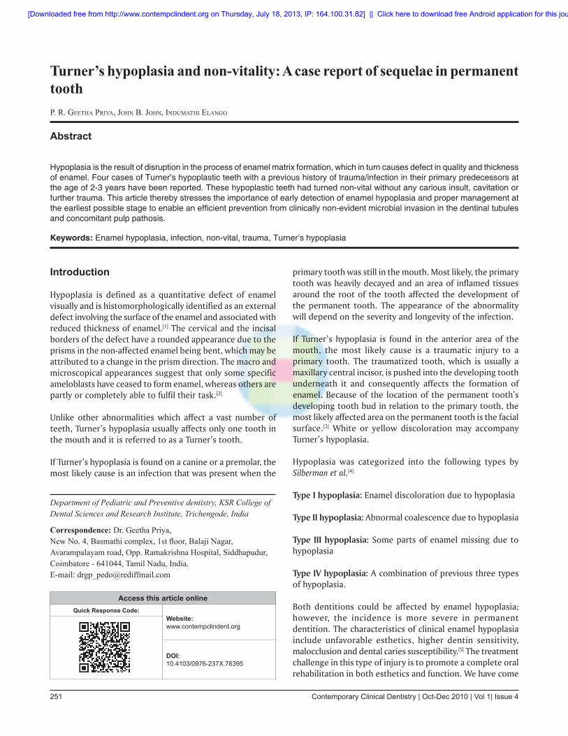
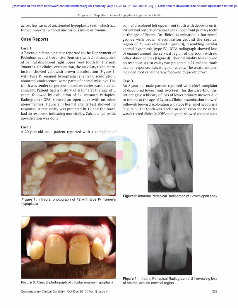
Case 1A 7-year-old female patient reported to the Department of Pedodontics and Preventive Dentistry with chief complaint of painful discolored right upper front tooth for the past 2months. On clinical examination, the maxillary right lateral incisor showed yellowish brown discoloration [Figure 1] with type IV enamel hypoplasia (enamel discoloration, abnormal coalescence, some parts of enamel missing). The tooth was tender on percussion and no caries was detected clinically. Patient had a history of trauma at the age of 3 years, followed by exfoliation of 52. Intraoral Periapical Radiograph (IOPA) showed an open apex with no other abnormalities [Figure 2]. Thermal vitality test showed no response. A test cavity was prepared in 12 and the tooth had no response, indicating non-vitality. Calcium hydroxide apexification was done.
Case 2A 20-year-old male patient reported with a complaint of
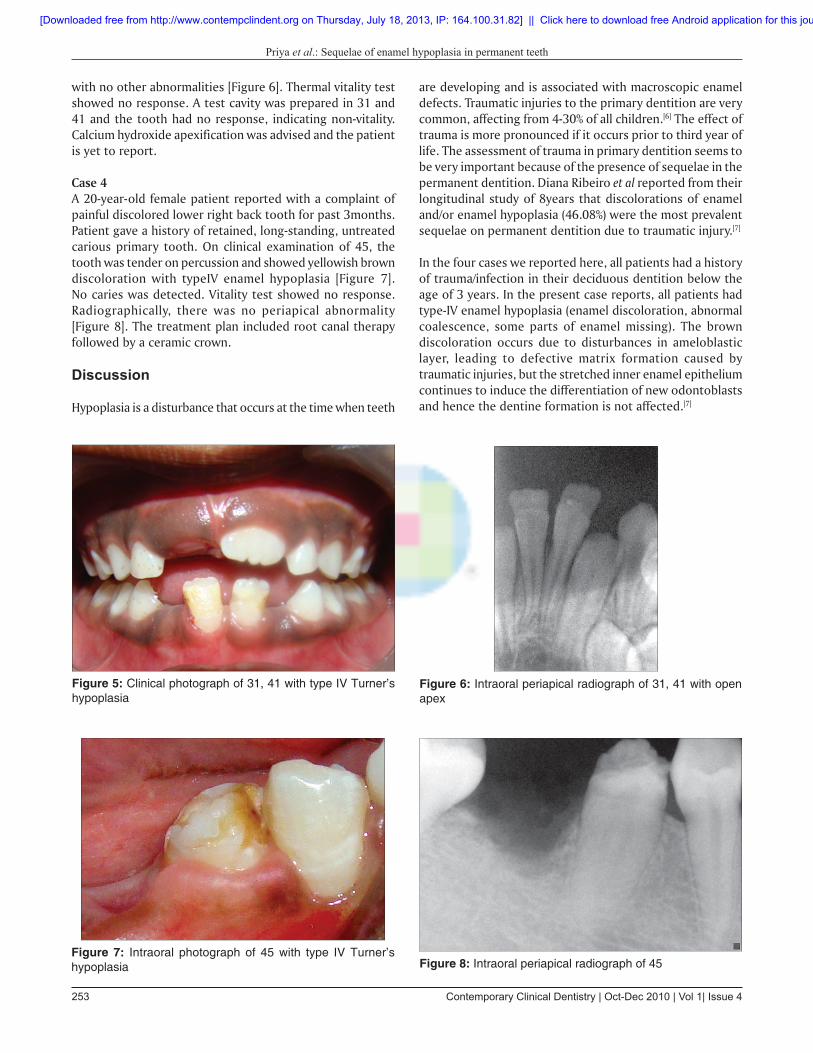
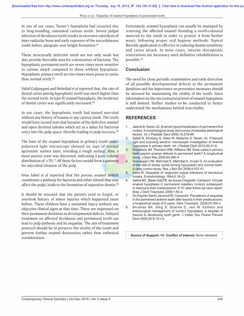
painful discolored left upper front tooth with deposits on it. Patient had history of trauma to his upper front primary teeth at the age of 2years. On clinical examination, a horizontal groove with brown discoloration around the cervical region of 21 was observed [Figure 3], resembling circular enamel hypoplasia (type IV). IOPA radiograph showed loss of enamel around the cervical region of the tooth with no other abnormalities [Figure 4]. Thermal vitality test showed no response. A test cavity was prepared in 21 and the tooth had no response, indicating non-vitality. The treatment plan included root canal therapy followed by jacket crown.
Case 3An 8-year-old male patient reported with chief complaint of discolored lower front two teeth for the past 4months. Patient gave a history of loss of lower primary incisors due to trauma at the age of 2years. Clinical examination showed yellowish brown discoloration with type IV enamel hypoplasia [Figure 5]. The tooth was tender on percussion and no caries was detected clinically. IOPA radiograph showed an open apex
Figure 1: Intraoral photograph of 12 with type IV Turner’s hypoplasia
Figure 3: Clinical photograph of circular enamel hypoplasia
Figure 2: Intraoral Periapical Radiograph of 12 with open apex
Figure 4: Intraoral Periapical Radiograph of 21 revealing loss of enamel around cervical region
[Downloaded free from http://www.contempclindent.org on Thursday, July 18, 2013, IP: 164.100.31.82] || Click here to download free Android application for this journal
Contemporary Clinical Dentistry | Oct-Dec 2010 | Vol 1| Issue 4253
Priya et al.: Sequelae of enamel hypoplasia in permanent teeth
with no other abnormalities [Figure 6]. Thermal vitality test showed no response. A test cavity was prepared in 31 and 41 and the tooth had no response, indicating non-vitality. Calcium hydroxide apexification was advised and the patient is yet to report.
Case 4A 20-year-old female patient reported with a complaint of painful discolored lower right back tooth for past 3months. Patient gave a history of retained, long-standing, untreated carious primary tooth. On clinical examination of 45, the tooth was tender on percussion and showed yellowish brown discoloration with typeIV enamel hypoplasia [Figure 7]. No caries was detected. Vitality test showed no response. Radiographically, there was no periapical abnormality [Figure 8]. The treatment plan included root canal therapy followed by a ceramic crown.
Discussion
Hypoplasia is a disturbance that occurs at the time when teeth
are developing and is associated with macroscopic enamel defects. Traumatic injuries to the primary dentition are very common, affecting from 4-30% of all children.[6] The effect of trauma is more pronounced if it occurs prior to third year of life. The assessment of trauma in primary dentition seems to be very important because of the presence of sequelae in the permanent dentition. Diana Ribeiro et al reported from their longitudinal study of 8years that discolorations of enamel and/or enamel hypoplasia (46.08%) were the most prevalent sequelae on permanent dentition due to traumatic injury.[7]
In the four cases we reported here, all patients had a history of trauma/infection in their deciduous dentition below the age of 3 years. In the present case reports, all patients had type-IV enamel hypoplasia (enamel discoloration, abnormal coalescence, some parts of enamel missing). The brown discoloration occurs due to disturbances in ameloblastic layer, leading to defective matrix formation caused by traumatic injuries, but the stretched inner enamel epithelium continues to induce the differentiation of new odontoblasts and hence the dentine formation is not affected.[7]
Figure 5: Clinical photograph of 31, 41 with type IV Turner’s hypoplasia
Figure 7: Intraoral photograph of 45 with type IV Turner’s hypoplasia
Figure 6: Intraoral periapical radiograph of 31, 41 with open apex
Figure 8: Intraoral periapical radiograph of 45
[Downloaded free from http://www.contempclindent.org on Thursday, July 18, 2013, IP: 164.100.31.82] || Click here to download free Android application for this journal
Contemporary Clinical Dentistry | Oct-Dec 2010 | Vol 1| Issue 4 254
Priya et al.: Sequelae of enamel hypoplasia in permanent teeth
Source of Support: Nil, Confl ict of Interest: None declared.
In one of our cases, Turner’s hypoplasia had occurred due to long-standing, untreated carious tooth. Severe pulpal infection of deciduous teeth results in excessive osteolysis of inter-radicular bone and early exposure of the succedaneous tooth before adequate root length formation.[5]
These structurally defective teeth are not only weak but also provide favorable area for colonization of bacteria. The hypoplastic permanent teeth are seven times more sensitive to carious attack compared to those without hypoplasia. Hypoplastic primary teeth are two times more prone to caries than normal teeth.[4]
Vahid Golpaygani and Mehrdad et al reported that, the rate of dental caries among hypoplastic teeth was much higher than the normal teeth. In type-IV enamel hypoplasia, the incidence of dental caries was significantly increased.[4]
In our cases, the hypoplastic tooth had turned non-vital without any history of trauma or any carious insult. The tooth would have turned non-vital because of the defective enamel and open dentinal tubules which act as a nidus for bacterial entry into the pulp space, thereby leading to pulp necrosis.[8]
The base of the enamel hypoplasia in primary tooth under polarized light microscope showed no sign of normal aprismatic surface layer, revealing a rough surface. Also, a more porous zone was discerned, indicating a pore volume distribution of <5%.[1] All these factors would form a gateway for microbial invasion of the pulp.
Nina Sabel et al reported that the porous enamel (which constitutes a pathway for bacteria and other stimuli that may affect the pulp), leads to the formation of reparative dentin.[2]
It should be stressed that the parents tend to forget, or overlook history of minor injuries which happened years before. These children have a sustained injury without any objective clinical signs at that time. These are expressed on their permanent dentition as developmental defects. Delayed treatment on affected deciduous and permanent teeth can lead to pulp pathosis and its sequelae. The aim of treatment protocol should be to preserve the vitality of the tooth and prevent further enamel destruction rather than esthetical consideration.
Fortunately, enamel hypoplasia can usually be managed by restoring the affected enamel (bonding a tooth-coloured material to the tooth in order to protect it from further wear), following proper oral hygiene methods. Topical fluoride application is effective in reducing dentin sensitivity and caries attack. In most cases, interim therapeutic restorations are necessary until definitive rehabilitation is possible.[6]
Conclusion
The need for close periodic examination and early detection of all possible developmental defects in the permanent dentition and the importance on preventive measures should be stressed for maintaining the vitality of the tooth. Since information on the microstructural level of enamel hypoplasia is still limited, further studies to be conducted to better understand the mechanisms behind non-vitality.
REFERENCES
1. Jalevik B, Noren JG. Enamel hypomineralization of permanent fi rst molars: A morphological study and survey of possible aetiological factors. Int J Paediatr Dent 2000;10:278-89.
2. Sabel N, Klinberg G, Dietz W, Nietsche S, Noren JG. Polarized light and scanning electron microscopic investigation of enamel hypoplasia in primary teeth. Int J Pediatr Dent 2010;20:31-6.
3. Broadbent JM, Thomson WM, Williams SM. Does caries in primary teeth predict enamel defects in permanent teeth? A longitudinal study. J.Dent Res 2005;84:260-4.
4. Golpaygani VM, Mehrdad K, Mehrdad A, Ansari G. An evaluation of the rate of dental caries among hypoplastic and normal teeth: A case control study. Res J Biol Sci 2009;4:537-41.
5. Kalra N. Sequelae of neglected pulpal infections of deciduous molars. Endodontology 1994;6:19-23.
6. Sellos MC, Bader SabTB, de Souza ChagasM, CamposV. Circular enamel hypoplasia in permanent maxillary incisors subsequent to trauma to their predecessors: A 10- year follow-up case report. Braz J Dent Traumatol 2009;1:50-3.
7. Do Espirito Santo JacomoDR, CamposV. Prevalence of sequelae in the permanent anterior teeth after trauma in their predecessors: a longitudinal study of 8 years. Dent Traumatol 2009;25:300-4.
8. Bhushan BA, Garg S, Sharma D, Jain M. Esthetic and endosurgical management of turner’s hypoplasia; a sequlae of trauma to developing tooth germ. J Indian Soc Pedod Prevent Dent 2008;26:S-121-4.
[Downloaded free from http://www.contempclindent.org on Thursday, July 18, 2013, IP: 164.100.31.82] || Click here to download free Android application for this journal