ultraviolet b irradiation induces expansion of intraepithelial tumor cells in a tissue model of...
TRANSCRIPT

ORIGINAL ARTICLESee related Commentary on page xiii
Ultraviolet B Irradiation Induces Expansion of IntraepithelialTumor Cells in a Tissue Model of Early Cancer Progression
AdarshV. Mudgil, Nadav Segal, Frank Andriani,Youai Wang, Norbert E. Fusenig,n and Jonathan A. GarlickOral Biology and Pathology, School of Dental Medicine, State University of NewYork at Stony Brook, USA, and nDivision of Di¡erentiation andCarcinogenesis, German Cancer Research Center, Heidelberg, Germany
Ultraviolet B irradiation is thought to enable skin can-cer progression as clones of genetically damaged keratin-ocytes escape apoptosis and expand at the expense ofadjacent normal cells. Mechanisms through which po-tentially malignant cells in human skin undergo clonalexpansion, however, are not well understood. The goalof this study was to characterize the role of ultraviolet Birradiation on the intraepithelial expansion of early stagehuman tumor cells in organotypic skin cultures. Toaccomplish this, we have studied the e¡ect of ultravioletB irradiation on organotypic cultures that were fabri-cated by mixing normal human keratinocytes withb-galactosidase-marked, intraepithelial tumor cells(HaCaT-ras, clone II-4), which bear mutations in bothp53 alleles and harbor an activated H-ras oncogene. Wefound that when organotypic mixtures were exposed toan ultraviolet B dose of 50 mJ per cm2, intraepithelialtumor cells underwent a signi¢cant degree of prolifera-tive expansion compared to nonirradiated cultures. To
understand this response, organotypic cultures of nor-mal keratinocytes were exposed to ultraviolet B andshowed a dose-dependent increase in numbers of sun-burn cells and TUNEL-positive cells although theirproliferation was suppressed. In contrast, neither theapoptotic nor the proliferative response of II-4 cellswas altered by ultraviolet B in organotypic cultures.The di¡erential response of these cell types suggestedthat II-4 cells were resistant to ultraviolet-B-induced al-terations, which allowed these intraepithelial tumorcells to gain a selective growth and survival advantagerelative to neighboring normal cells. These ¢ndingsdemonstrate that ultraviolet B exposure can induce theintraepithelial expansion of apoptosis-resistant, p53-mutant, and ras-activated keratinocytes, suggesting thatthis agent can act to promote the early stages of epithe-lial carcinogenesis. Key words: apoptosis/intraepithelial neo-plasia/organotypic culture/UVB irradiation. J Invest Dermatol121:191 ^197, 2003
Solar irradiation in the ultraviolet (UV) range is knownto be associated with the development of nonmelanomaskin cancers in humans (Setlow, 1974; Brash et al, 1996;Kraemer, 1997). Speci¢cally, irradiation in the UVA(315^400 nm) and UVB (280^315 nm) range has been
shown to induce morphologic, biochemical, and genetic damagein human skin keratinocytes, which leads to early neoplastic pro-gression in this tissue (Black et al, 1997). UVB causes speci¢c ge-netic alterations in oncogenes and tumor suppressor genes thatmay play a central role in the initiation and progression of skincancer (Brash et al, 1996; Black et al, 1997). For example, mutationof the ras oncogene has been found in 10%^40% of UV-asso-ciated skin cancers (Pierceall et al, 1991; 1992), but the role of thisgenetic alteration in early skin cancer progression remains un-clear. Another gene altered by UVB exposure is the p53 tumorsuppressor gene, which plays a central role in cell death, geneticinstability, and cancer susceptibility by mediating G1 arrest orprogrammed cell death in response to DNA damage. UVB irra-diation has been associated with mutations in the p53 gene that
are found in a large majority of cutaneous squamous cell and ba-sal cell carcinomas (Brash et al, 1996), a variety of premalignantlesions such as actinic keratosis, and normal, sun-damaged skin(Jonason et al, 1996). In recent years, it has been shown that p53-mutated keratinocytes are arranged in clonal patches in normalhuman skin and in nonmelanoma skin cancer and may involveas much as 4% of the epidermis (Jonason et al, 1996). As the in-duction of apoptosis by UVB radiation is an important mechan-ism for the elimination of sun-damaged keratinocytes (Brash,1997), the presence of p53 mutations may confer apoptosis resis-tance to these clonal patches of p53-mutant cells. It is theorizedthat further UVB exposure is more likely to induce cell death innormal cells that do not harbor p53 mutations, thereby allowingthe expansion of individual p53-mutant cells into a niche left bythe death of neighboring normal cells (Brash, 1997). This suggeststhat, in addition to its role as a mutagen, UVB irradiation can actas a tumor promoter by enabling the clonal expansion of p53-mutated cells.The ¢nding of such large numbers of cells with p53 mutations
suggests that normal skin harbors stable populations of mutantnoncancerous cells that may be predisposed to further neoplasticprogression upon subsequent UVB damage. Although there isevidence that UVB irradiation acts as a tumor promoter in mouseskin by enabling the clonal expansion of sun-damaged, p53-mu-tated cells, however, this has not been studied directly in humankeratinocytes due to a lack of tissue models in which human ker-atinocytes are present in such a dormant state.We have developed
Reprint requests to: Dr. Jonathan Garlick, Department of Oral Biologyand Pathology, School of Dental Medicine, SUNYat Stony Brook, StonyBrook, NY 11794-8702; Email: [email protected]: b-gal, b-galactosidase; NHK, normal human keratino-
cytes; LI, labeling index; SBC, sunburn cells; UV, ultraviolet.
Manuscript received January 7, 2002; revised June 3, 2002; accepted forpublication February 20, 2003
0022-202X/03/$15.00 . Copyright r 2003 by The Society for Investigative Dermatology, Inc.
191
a human tissue model through which the fate of small numbersof intraepithelial tumor cells, the ras-transfected HaCaT clone II-4, could be monitored during the premalignant stage of diseaseof strati¢ed epithelium (Javaherian et al, 1998; Karen et al, 1999;Vaccariello et al, 1999b). Using this tissue model, we previouslydemonstrated that neoplastic progression of these intraepithelialtumor cells was suppressed in vivo and in vitro by a mechanismmediated by cell^cell interactions (Javaherian et al, 1998).We sub-sequently found that this intraepithelial control of neoplastic pro-gression could be overcome by altering interactions betweentumor cells and adjacent normal keratinocytes (Karen et al, 1999).The goal of this study was to determine whether UVB irradia-
tion plays a role in the expansion of intraepithelial tumor cellsharboring p53 mutations and an activated H-ras oncogene, in anorganotypic model of early neoplastic progression in human stra-ti¢ed epithelium. We determined that biologically-meaningfulUVB exposure enabled the intraepithelial expansion of earlystage tumor cells by inducing a di¡erential apoptotic and pro-liferative response between these cells and adjacent normalkeratinocytes.
MATERIALS AND METHODS
Monolayer cell culture Normal human epidermal keratinocytes(NHK) were cultured from newborn foreskin by the method ofRheinwald and Green (1975) in keratinocyte medium described by Wuet al (1982). Cultures were established through trypsinization of foreskinfragments in 0.25% trypsin and were grown on 3T3 ¢broblasts irradiatedwith a g ray source of 2000 Ci (Cs-137, 100%¼ 1215 R per min) for 6.5min. 3T3 cells were maintained in Dulbecco’s modi¢ed Eagle’s medium(DMEM) containing 10% fetal bovine serum. The HaCaT-ras-II-4 (II-4)cell line (Boukamp et al, 1990) was grown in DMEM containing 5% fetalbovine serum. Human dermal ¢broblasts were derived from foreskins andgrown in medium containing DMEM and 10% fetal bovine serum.
Organotypic culture Early passage human dermal ¢broblasts wereadded to neutralized type I collagen (Organogenesis, Canton, MA) to a¢nal concentration of 2.5�104 cells per ml. Three milliliters of thismixture were added to each 35 mm well of a six-well plate and incubatedfor 4^6 d in medium containing DMEM and 10% fetal bovine serum untilthe collagen matrix showed no further shrinkage. At this time, a total of5�105 NHK, II-4 cells, or mixtures of these cell lines at a 12:1 ratio(NHK:II-4), were seeded on the contracted collagen gel. Cultures weremaintained submerged in low calcium epidermal growth medium for 2 d,submerged for 2 d in normal calcium epidermal growth medium, andraised to the air^liquid interface by feeding from below with normalcalcium corni¢cation medium for 3 d as previously published (Vaccarielloet al, 1999a). For proliferation assays, 10 mM bromodeoxyuridine (BrdU)(Sigma, St. Louis, MO) was added to organotypic cultures 8 h prior toharvesting.
Retroviral vectors and transduction of tumor cell lines II-4 tumorcells (HaCaT-ras-II-4) were transduced with the MFG-gal vector, which isa Moloney murine leukemia virus based vector that contains the gene forbacterial b-galactosidase (b-gal). Transduced keratinocytes were passaged atclonal density and clones were screened for persistence of transgeneexpression after 10 passages. No loss of transgene expression was observed.
UVB irradiation Irradiated cultures and their corresponding controlswere removed from media and placed into sterile six-well plates withoutmedia. UV was administered using four Westinghouse FS20 sunlampsemitting wavelengths of 290^400 nm with a peak wavelength at 335 nm(32.5 J per m2 per s), as determined through spectral radiometry aspreviously recommended (Gasparro and Brown, 2000). The dose rate wasdetermined using a spectroradiometer built by the Department of Biology,Brookhaven National Laboratories, using components manufactured byOriel and calibrated using a model 6315, 1000 w quartz tungsten halogenlamp. UVA and UVB were found to constitute 72% and 28% of the totalresulting spectrum, respectively. These were measured up to 400 nm anddid not include higher wavelengths. Although UVA output constituted aconsiderable portion of the incident spectrum, it has previously beenshown that the UVA dose required to induce epithelial damage is severalorders of magnitude greater for UVA than UVB in monolayer (Drobetskyet al, 1995) and organotypic (Bernerd and Asselineau, 1998) cultures. In this
light, the irradiation times and resultant dose of UVA used in ourexperiments were not of biologic signi¢cance. Organotypic cultures wereirradiated at doses between 0 and 50 mJ per cm2 as measured using theUVB and UVA outputs of a UVX Digital Dosimeter (UltravioletProducts, San Gabriel, CA) as a transfer standard from the spectralradiometer. UV dose was measured for each experiment and the UVBand UVA output of the lamps in watts and mJ per cm2 was calculated(UVB¼ 32.5 J per m2 per s and UVA¼ 30 J per m2 per s). UVCradiation was removed from the FS20 spectrum using a Kodacel acetate¢lter.
Immuno£uorescence Tissues were snap frozen in liquid nitrogen afterbeing immersed in a 2 M sucrose solution for 1 h, serial sectioned at 6 mm,and mounted onto gelatin�chrome alum coated slides. Sections wereincubated with rabbit polyclonal antiserum to bacterial b-gal (CortexPharmaceuticals, San Leandro, CA) and detected with Alexa 488-conjugated goat antirabbit IgG (Molecular Probes, Eugene, OR). Doublestain immuno£uorescence was performed by using a mouse monoclonalantibody to BrdU (Boehringer Mannheim, Indianapolis, IN), which wasdetected with Texas Red-conjugated horse antimouse IgG (VectorLaboratories) in combination with rabbit polyclonal antiserum to bacterialb-gal. Slides were coverslipped with Vectashield containing 1 mg per mlDAPI (Vector Laboratories). Fluorescence was visualized using a NikonOptiPhot microscope, and double exposure photomicroscopy wasperformed using £uorescein isothiocyanate (FITC) and Texas Redchannels. The mean percentage of b-gal-positive area before and afterirradiation was calculated in 10 serial sections using the UTHSCAImageTool (Version 1.27). Proliferation of NHK was measured as thepercentage of BrdU-positive basal NHK nuclei (LI, labeling index) andII-4 cell proliferation was calculated by counting basal and suprabasalBrdU-positive cells, as II-4 cells demonstrated proliferation throughoutthe tissue. Five serial sections from random areas in the specimen werestained. For routine light microscopy, tissues were ¢xed in 10% neutralbu¡ered formalin and embedded in para⁄n, and 4 mm sections werestained with hematoxylin and eosin.
Measurement of sunburn cells (SBC) and apoptosis The terminaldeoxynucleotidyl transferase mediated dUTP nick-end labeling (TUNEL)assay was used to identify apoptotic cells in organotypic cultures(Boehringer Mannheim, Indianapolis, IN). In order to prevent freezingdamage to tissues, which would lead to artifactual staining of nuclei,organotypic cultures were frozen at ^201C after being ¢xed in 4%paraformaldehyde. After sectioning at 6 mm, tissue sections were ¢xedwith 4% paraformaldehyde at room temperature for 30 min, rinsed inphosphate-bu¡ered saline, and incubated with the TUNEL reactionmixture for 1 h at 371C in the dark. Fluorescence was visualized using aNikon OptiPhot microscope. Identi¢cation of SBC was based on themorphologically-distinct appearance of these cells in hematoxylin andeosin stained tissues, i.e., a highly eosinophilic cytoplasm and pyknoticnuclei (Young, 1987). The percentage of TUNEL-positive cells and SBCwas calculated from the number of these cells divided by the totalnumber of cells counted in the entire epithelium in 10 tissue sections.Experiments were performed in duplicate.
RESULTS
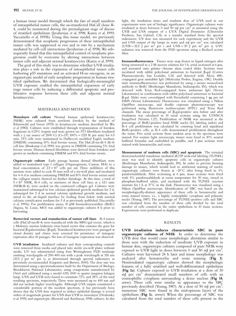
UVB irradiation induces characteristic SBC in pureorganotypic cultures of NHK In order to determine theUVB dose that would cause morphologic alterations similar tothose seen with the induction of moderate UVB exposure inhuman skin, organotypic cultures composed of pure NHKwereexposed to UVB light in doses between 0 and 50 mJ per cm2.Cultures were harvested 24 h later and tissue morphology wasanalyzed after hematoxylin and eosin staining (Fig 1).Nonirradiated organotypic cultures showed the morphologicfeatures of a fully strati¢ed and well-di¡erentiated epithelium(Fig 1a). Cultures exposed to UVB irradiation at a dose of 30mJ per cm2 demonstrated small numbers of cells with aneosinophillic cytoplasm surrounding a dense nucleus (Fig 1b,arrow). These cells were similar in appearance to the SBCpreviously described (Young, 1987). At a dose of 50 mJ per cm2,numerous SBC were found in the mid-spinous layers of theepithelium (Fig 1c, arrows). When the percentage of SBC wascalculated from the total number of these cells present in the
192 MUDGIL ETAL THE JOURNAL OF INVESTIGATIVE DERMATOLOGY

epithelium, it was found that 15% of the cells in this tissueappeared as SBC (Fig 2). UVB doses in excess of 50 mJ per cm2
were found to exert a toxic e¡ect as seen by a severely alteredtissue architecture and tissue necrosis (data not shown). As aresult, it was decided to irradiate mixed organotypic cultureswith a UVB dose of 50 mJ per cm2.
UVB induces a dose-dependent increase in the number ofSBC and apoptotic cells in pure organotypic cultures ofNHK but not in II-4 cells In order to compare the inductionof UV-associated cell death between NHK and II-4 cells, pureorganotypic cultures of these cells were irradiated at doses of 20,30, 40, and 50 mJ per cm2 and the extent of cellular damage wasmeasured by calculating the percentage of SBC and apoptoticnuclei (TUNEL assay) seen 24 h after irradiation (Fig 2). NoSBC were seen in nonirradiated cultures and TUNEL-positiveapoptotic nuclei were only observed in the uppermost layer ofthe epithelium, as directed by the normal terminaldi¡erentiation of this tissue (Fig 3a, arrow).With elevated UVBdoses, apoptotic nuclei increased in numbers (Fig 2) and wereseen in the deeper layers of this normal epithelium (Fig 3c, e,arrows), in a distribution that corresponded to the localization ofSBC (Fig 1). Roughly 2% of NHK exposed to 30 mJ per cm2
were TUNEL positive (Fig 2) and these cells were found in animmediately suprabasal postion (Fig 3c, arrow). NumerousTUNEL-positive cells, making up roughly 14% of the cells in
this tissue (Fig 2), were seen in the mid-spinous and basallayers of the epithelium upon irradiation at 50 mJ per cm2
(Fig 3e, arrows). Furthermore, this UVB dose induced theapoptosis of ¢broblasts found in the subepithelial connectivetissue. In general, induction of cell death demonstrated a directdependency on UVB dose, as numbers of TUNEL-positiveNHK and SBC increased with increasing UVB dosage (Fig 2).In contrast, irradiated pure organotypic cultures of II-4 cells didnot demonstrate TUNEL-positive cells at any UV dose (Fig 3b,d, f ) suggesting that p53-mutant II-4 cells were resistant toapoptosis.
UVB exposure enables intraepithelial expansion ofpreviously growth-suppressed II-4 tumor cells in organotypiccultures We next determined if the di¡erential induction ofcell death of NHK and II-4 cells in response to UVB irradiationwas associated with the clonal expansion of individual growth-suppressed II-4 cells that were grown in organotypic culture inthe context of a majority of NHK. Previous studies have shownthat these cells were growth suppressed and underwent terminaldi¡erentiation when cultured in the presence of NHK at mixingratios of 4:1 and 12:1 (NHK:II-4) (Vaccariello et al, 1999b).Whenmixed organotypic cultures (12:1, NHK:II-4) were not irradiated,cultures demonstrated small numbers of individual b-gal-positivecells in the mid- and upper spinous layers of the epithelium thatoccupied 2% of the tissue (Fig 4a, arrow). In contrast, mixedcultures irradiated with UVB at 50 mJ per cm2 demonstratedsigni¢cant expansion of II-4 cells as seen by the presence oflarge clusters of green b-gal-positive II-4 cells in the upperspinous layer of the epithelium (Fig 4b, thick arrow). Theseclusters had expanded to occupy roughly 28% of the tissuewhen the total area of green cells was determined as thepercentage from the entire tissue, showing that the number ofII-4 cells in an intraepithelial tumor cell cluster had increased.This showed that UVB irradiation could induce the
Figure1. UVB irradiation induces SBC in organotypic cultures ofnormal keratinocytes. Organotypic cultures, either not exposed toUVB irradiation (a) or exposed at 30 mJ per cm2 (b) or 50 mJ per cm2 (c),were studied 24 h postirradiation after hematoxylin and eosin staining.Nonirradiated cultures appeared as a well-strati¢ed epithelium, which de-monstrated normal tissue architecture including polarized basal cells andwell-organized spinous layer (a). Organtoypic cultures irradiated with 30mJ per cm2 demonstrated small numbers of keratinocytes, which had theappearance of SBC (b, arrow). Upon irradiation with 50 mJ per cm2, tissuearchitecture was signi¢cantly disrupted and large numbers of SBC wereseen in the mid-spinous layer of the epithelium (c, long arrows). Scale bar:30 mm.
Figure 2. UVB induces a dose-dependent increase in SBC and TU-NEL-positive cells in organotypic cultures of normal keratinocytes.Pure organotypic cultures of normal keratinocytes were irradiated withUVB doses between 20 and 50 mJ per cm2. Para⁄n-embedded tissues werestained with hematoxylin and eosin whereas frozen sections were stainedbyTUNEL assay to detect apoptotic nuclei. Numbers of SBC (^�^) andTUNEL-positive cells (&m&)were counted in 10 sections from two sepa-rate experiments and the number of positive cells in each tissue section wascalculated.
UVB AND PROGRESSION OF INTRAEPITHELIAL NEOPLASIA 193VOL. 121, NO. 1 JULY 2003

intraepithelial expansion of II-4 tumor cells and allowedthese cells to escape the growth control of adjacent NHK. Toprove that UVB irradiation had enabled the proliferation ofII-4 cells, irradiated and nonirradiated mixed organotypiccultures were double stained by immuno£uorescence forproliferation as detected by BrdU incorporation (red) and b-gal(green). Only irradiated mixed cultures demonstratedcolocalization of BrdU-positive nuclei and b-gal-positive cells
(Fig 4b, thin arrows). These ¢ndings demonstrated that UVB-induced expansion of II-4 cells was associated with the activeproliferation of II-4 cells.
UVB suppresses the proliferation of NHKwithout a¡ectingthe growth of II-4 cells In order to further understand theresponse of II-4 and NHK cells in mixed organotypic culture, thee¡ect of UVB irradiation on proliferation of pure organotypic
Figure 3. UVB induces TUNEL-positive apoptotic cells in organotypic cultures of normal keratinocytes without e¡ecting death of II-4cells. Pure organotypic cultures of NHK (a, c, e) and II-4 cells (b, d, f) were irradiated at either 30 mJ per cm2 (c, d) or 50 mJ per cm2 (e, f); control cultureswere not irradiated (a, b). Apoptotic cells in parafomaldehyde-¢xed and in situ TUNEL-stained sections were detected by their yellow-staining nuclei,which were seen in the surface layer of nonirradiated cultures of NHK (a, arrow). In addition to such apoptotic cells seen in the surface layer, small numbersof TUNEL-positive cells were seen in the mid-spinous layer of cultures irradiated at 30 mJ per cm2 (c, arrow) whereas large numbers of apoptotic cells wereseen in the lower layers of the epithelium upon irradiation with 50 mJ per cm2 (e, arrows). In contrast, no TUNEL-positive cells were seen at either UVBdose when cultures of II-4 cells were irradiated (b, d, f). Scale bar: 45 mm.
Figure 4. UVB induces clonal expansion and proliferation of II-4cells in mixed organotypic cultures. Double immuno£uorescence staindemonstrating superimposed £uorescent signals for b-gal (FITC channel,green) and BrdU (Texas Red channel, red). (a) Mixture of NHK:II-4 cells(12:1) demonstrating individual b-gal-positive cells in a suprabasal position(arrow) and red BrdU-positive nuclei limited to the normal basal keratino-cytes. Individual b-gal cells lack colocalization of b-gal and BrdU demon-strating that II-4 cells in the suprabasal position are growth suppressed. (b)Mixture of NHK:II-4 cells (12:1) exposed to 50 mJ per cm2 UVB irradiationshowing increased area of b-gal-positive II-4 cells (thick arrow). Cells in suchclusters show BrdU-positive nuclei (thin arrows), demonstrating that UVB ir-radiation induced the proliferation of II-4 cells that were previously growthsuppressed. Scale bar: 45 mm.
194 MUDGIL ETAL THE JOURNAL OF INVESTIGATIVE DERMATOLOGY
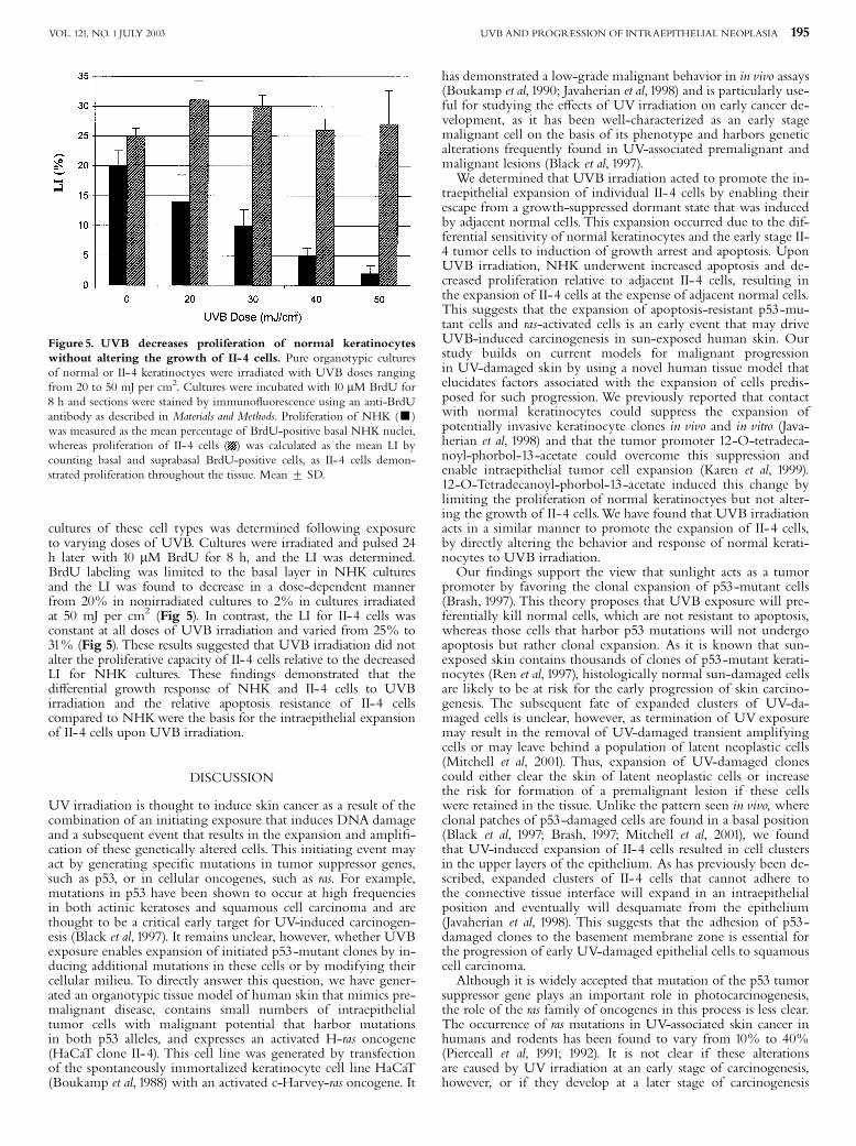
cultures of these cell types was determined following exposureto varying doses of UVB. Cultures were irradiated and pulsed 24h later with 10 mM BrdU for 8 h, and the LI was determined.BrdU labeling was limited to the basal layer in NHK culturesand the LI was found to decrease in a dose-dependent mannerfrom 20% in nonirradiated cultures to 2% in cultures irradiatedat 50 mJ per cm2 (Fig 5). In contrast, the LI for II-4 cells wasconstant at all doses of UVB irradiation and varied from 25% to31% (Fig 5). These results suggested that UVB irradiation did notalter the proliferative capacity of II-4 cells relative to the decreasedLI for NHK cultures. These ¢ndings demonstrated that thedi¡erential growth response of NHK and II-4 cells to UVBirradiation and the relative apoptosis resistance of II-4 cellscompared to NHKwere the basis for the intraepithelial expansionof II-4 cells upon UVB irradiation.
DISCUSSION
UV irradiation is thought to induce skin cancer as a result of thecombination of an initiating exposure that induces DNA damageand a subsequent event that results in the expansion and ampli¢-cation of these genetically altered cells. This initiating event mayact by generating speci¢c mutations in tumor suppressor genes,such as p53, or in cellular oncogenes, such as ras. For example,mutations in p53 have been shown to occur at high frequenciesin both actinic keratoses and squamous cell carcinoma and arethought to be a critical early target for UV-induced carcinogen-esis (Black et al, 1997). It remains unclear, however, whether UVBexposure enables expansion of initiated p53-mutant clones by in-ducing additional mutations in these cells or by modifying theircellular milieu. To directly answer this question, we have gener-ated an organotypic tissue model of human skin that mimics pre-malignant disease, contains small numbers of intraepithelialtumor cells with malignant potential that harbor mutationsin both p53 alleles, and expresses an activated H-ras oncogene(HaCaT clone II-4). This cell line was generated by transfectionof the spontaneously immortalized keratinocyte cell line HaCaT(Boukamp et al, 1988) with an activated c-Harvey-ras oncogene. It
has demonstrated a low-grade malignant behavior in in vivo assays(Boukamp et al, 1990; Javaherian et al, 1998) and is particularly use-ful for studying the e¡ects of UV irradiation on early cancer de-velopment, as it has been well-characterized as an early stagemalignant cell on the basis of its phenotype and harbors geneticalterations frequently found in UV-associated premalignant andmalignant lesions (Black et al, 1997).We determined that UVB irradiation acted to promote the in-
traepithelial expansion of individual II-4 cells by enabling theirescape from a growth-suppressed dormant state that was inducedby adjacent normal cells. This expansion occurred due to the dif-ferential sensitivity of normal keratinocytes and the early stage II-4 tumor cells to induction of growth arrest and apoptosis. UponUVB irradiation, NHK underwent increased apoptosis and de-creased proliferation relative to adjacent II-4 cells, resulting inthe expansion of II-4 cells at the expense of adjacent normal cells.This suggests that the expansion of apoptosis-resistant p53-mu-tant cells and ras-activated cells is an early event that may driveUVB-induced carcinogenesis in sun-exposed human skin. Ourstudy builds on current models for malignant progressionin UV-damaged skin by using a novel human tissue model thatelucidates factors associated with the expansion of cells predis-posed for such progression.We previously reported that contactwith normal keratinocytes could suppress the expansion ofpotentially invasive keratinocyte clones in vivo and in vitro (Java-herian et al, 1998) and that the tumor promoter 12-O-tetradeca-noyl-phorbol-13-acetate could overcome this suppression andenable intraepithelial tumor cell expansion (Karen et al, 1999).12-O-Tetradecanoyl-phorbol-13-acetate induced this change bylimiting the proliferation of normal keratinoctyes but not alter-ing the growth of II-4 cells.We have found that UVB irradiationacts in a similar manner to promote the expansion of II-4 cells,by directly altering the behavior and response of normal kerati-nocytes to UVB irradiation.Our ¢ndings support the view that sunlight acts as a tumor
promoter by favoring the clonal expansion of p53-mutant cells(Brash, 1997). This theory proposes that UVB exposure will pre-ferentially kill normal cells, which are not resistant to apoptosis,whereas those cells that harbor p53 mutations will not undergoapoptosis but rather clonal expansion. As it is known that sun-exposed skin contains thousands of clones of p53-mutant kerati-nocytes (Ren et al, 1997), histologically normal sun-damaged cellsare likely to be at risk for the early progression of skin carcino-genesis. The subsequent fate of expanded clusters of UV-da-maged cells is unclear, however, as termination of UV exposuremay result in the removal of UV-damaged transient amplifyingcells or may leave behind a population of latent neoplastic cells(Mitchell et al, 2001). Thus, expansion of UV-damaged clonescould either clear the skin of latent neoplastic cells or increasethe risk for formation of a premalignant lesion if these cellswere retained in the tissue. Unlike the pattern seen in vivo, whereclonal patches of p53-damaged cells are found in a basal position(Black et al, 1997; Brash, 1997; Mitchell et al, 2001), we foundthat UV-induced expansion of II-4 cells resulted in cell clustersin the upper layers of the epithelium. As has previously been de-scribed, expanded clusters of II-4 cells that cannot adhere tothe connective tissue interface will expand in an intraepithelialposition and eventually will desquamate from the epithelium(Javaherian et al, 1998). This suggests that the adhesion of p53-damaged clones to the basement membrane zone is essential forthe progression of early UV-damaged epithelial cells to squamouscell carcinoma.Although it is widely accepted that mutation of the p53 tumor
suppressor gene plays an important role in photocarcinogenesis,the role of the ras family of oncogenes in this process is less clear.The occurrence of ras mutations in UV-associated skin cancer inhumans and rodents has been found to vary from 10% to 40%(Pierceall et al, 1991; 1992). It is not clear if these alterationsare caused by UV irradiation at an early stage of carcinogenesis,however, or if they develop at a later stage of carcinogenesis
Figure 5. UVB decreases proliferation of normal keratinocyteswithout altering the growth of II-4 cells. Pure organotypic culturesof normal or II-4 keratinoctyes were irradiated with UVB doses rangingfrom 20 to 50 mJ per cm2. Cultures were incubated with 10 mM BrdU for8 h and sections were stained by immuno£uorescence using an anti-BrdUantibody as described in Materials and Methods. Proliferation of NHK (’)was measured as the mean percentage of BrdU-positive basal NHK nuclei,whereas proliferation of II-4 cells ( ) was calculated as the mean LI bycounting basal and suprabasal BrdU-positive cells, as II-4 cells demon-strated proliferation throughout the tissue. Mean 7 SD.
UVB AND PROGRESSION OF INTRAEPITHELIAL NEOPLASIA 195VOL. 121, NO. 1 JULY 2003

independent of UV damage. It appears that ras mutations are notof great signi¢cance in the induction of UV-induced skin cancerprogression and are more likely to occur in response to chemicalcarcinogenesis (Quintanilla et al, 1986). Recently, transfection ofHaCaT with an activated ras oncogene demonstrated augmenta-tion of cell growth and decreased di¡erentiation and apoptosiscompared to parental HaCaTcells (Delehedde et al, 2001). It there-fore appears that the presence of p53 mutation alone, as seen inHaCaT cells, is not su⁄cient to drive the proliferative growth ad-vantage seen in ras-transfected HaCaTcells. It is likely that the roleof rasmutation in II-4 cells, as well as in skin cancer progression invivo, lies in the activation of pathways through which cells losetheir growth regulation (Black et al, 1997). The ongoing prolifera-tion seen upon UV irradiation of II-4 cells in our tissue modelmay re£ect this abnormal growth potential, whereas altered apop-tosis may be a function of the p53 mutations present in these cells.Additionally, it is possible that other genetic alterations present inthe II-4 cells in£uence their observed behavior to UV irradiation.Karyotypic changes in the parental HaCaT cells (Boukamp et al,1997) and II-4 cells (Boukamp et al, 1995) have been extensivelystudied, but none of the alterations characterized has been corre-lated with a UVor stress sensitivity. It has been shown that HaCaTcells undergo apoptosis in response to UV irradiation whengrown in submerged monolayer cultures (Heinseleit et al, 1997).This study found that, despite its mutated status in HaCaT cells,p53 may help mediate this event as UV-induced apoptosis can bepartially blocked by suppressing p53 expression in these cells. Incontrast, we have found that II-4 cells are resistant to UV-inducedapoptosis, suggesting that both p53-dependent and p53-indepen-dent apoptotic pathways are not functioning after UV irradiationof these cells in organotypic culture.Previous models of chronic skin exposure to UV irradiation
have demonstrated that keratinocytes undergo a hyperplasticresponse when exposed to a one-time, acute UV dose adminis-tered after low-dose chronic irradiation (Mitchell et al, 2001).In contrast, as has previously been shown (Bernerd and Asseli-neau, 1997), we have found a decrease in the proliferation of nor-mal keratinocytes 24 h after a one-time exposure to UV.Compared to experiments designed to study skin carcinogenesisby chronic low-level UV exposure, our studies have been limitedto understanding the immediate tissue alterations induced byUVB that trigger clonal expansion of p53-mutant cells. Di¡er-ences in proliferative response between organotypic cultures andintact skin may be due to the basal level of proliferation in thesein vitro tissues, which are higher than those seen in their in vivocounterparts. As a result, the e¡ect of UV light in our tissuemodel may be to suppress an already hyperproliferative epithe-lium to a more basal level of proliferation. Although the elevatedproliferation of normal keratinocytes may enhance their UV sen-sitivity, it would be expected to do the same for adjacent II-4cells.As ethical reasons have limited the ability to directly study the
e¡ects of UVB irradiation in the skin of human volunteers, theuse of skin-like organotypic tissue models containing human ker-atinocytes o¡ers an attractive alternative for studying the e¡ectsof UVB irradiation on human skin. Furthermore, as there areimportant di¡erences in morphology, thickness, and UVB lightresponsiveness between human and murine skin (Reifenrath et al,1984), the ability to extrapolate mechanisms of carcinogenesisfrom one tissue to another is somewhat limited (Soballe et al,1996). Using organotypic cultures of human keratinocytes, wehave demonstrated that biologically relevant doses of UVB lightcan induce cellular damage that is similar to that seen in sun-ex-posed human skin. Organotypic cultures have previously beenused to study the e¡ects of UVB irradiation on release of cyto-kines (Nelson and Gay, 1993), melanin synthesis (Archambaultet al, 1995), DNA synthesis, and induction of apoptosis (Harrigerand Hull, 1994). Bernerd and Asselineau (1997) found that UVBexposure induced apoptosis, downregulation of markers of kera-tinocyte di¡erentiation, and an increase in proliferation several
days after UVB exposure, which subsequently restored normaltissue morphology.We have expanded on these previous studiesusing organotypic skin equivalents to study UVB e¡ects on hu-man tissues by constructing tissues that incorporate p53-damagedintraepithelial tumor cells into tissues with three-dimensional tis-sue architecture.In conclusion, UVB appears to enable clonal expansion of II-4
cells by selectively inducing apoptosis in normal keratinocytesand by decreasing the proliferative capacity of these cells. Thissuggests that UVB can act to promote the clonal expansion ofpotentially malignant cells by altering the behavior of normalkeratinocytes in the epithelium rather than by directly alteringthe phenotype of the potentially malignant cells. In this way,UVB may act to stimulate the early stages of neoplastic progres-sion in human strati¢ed epithelium by creating a microenviron-ment conducive for clonal expansion of previously suppressedpotentially malignant cells, thereby permitting UV-damagedclones to overcome the growth-suppressive e¡ects exerted bynormal cells. This suggests that clonal expansion and the earlieststages of UV-associated nonmelanoma skin cancer are driven bythe promoting e¡ects of UV irradiation and are not necessarilydue to the acquisition of additional mutations in previously sun-damaged skin.
We thank Dr. John Sutherland and JohnTrunk of Brookhaven National Laboratoriesfor assistance with standardization of spectral measurements of the UV light source,Dr. Richard Setlow for helpful discussions and assistance with dosimetric readings,Dr. Francis Gasparro for his assistance in characterizing our UV light source and forhelpful discussions, Ning Ling for technical assistance, and Dr. R. Mulligan for theMFG vector.Thanks to Karen Henrickson for preparation of illustrations.This workwas supported by a research grant from the Howard Hughes Medical Institute for Med-ical Students awarded to the medical student A. Mudgil and by the National Institutefor Dental and Craniofacial Research (DE011250-06).
REFERENCES
Archambault M, Yaar M, Gilchrest BA: Keratinocytes and ¢broblasts in a humanskin equivalent model enhance melanocyte survival and melanin synthesisafter ultraviolet irradiation. J Invest Dermatol 104:859^867, 1995
Bernerd F, Asselineau D: Successive alteration and recovery of epidermal di¡erentia-tion and morphogenesis after speci¢c UVB-damages in skin reconstructed invitro. Dev Biol 183:123^138, 1997
Bernerd F, Asselineau D: UVA exposure of human skin reconstructed in vitro inducesapoptosis of dermal ¢broblasts: subsequent connective tissue repair and impli-cations in photoaging. Cell Death Di¡erentiation 5:792^802, 1998
Black HS, deGruijl FR, Forbes PD, et al: Photocarcinogenesis: An overview. J Photo-chem Photobio B 40:29^47, 1997
Boukamp P, Petrussevka RT, Breitkreutz D, Hornung J, Markham A, Fusenig NE:Normal keratinization in a spontaneously immortalized aneuploid human ker-atinocyte cell line. J Cell Biol 106:761^771, 1988
Boukamp P, Stanbridge EJ,Yin-Foo D, Cerutti PA, Fusenig NE: c-Ha-ras oncogeneexpression in immortalized human keratinocytes (HaCaT) alters growth po-tential in vivo but lacks correlation with malignancy. Cancer Res 50:2840^2847,1990
Boukamp P, PeterW, Pascheberg U, Altmeier S, Fasching C, Stanbridge EJ, FusenigNE: Step-wise progression in human skin carcinogenesis in vitro involves mu-tational activation of p53,rasH oncogene activation and additional chromosomeloss. Oncogene 11:961^969, 1995
Boukamp P, Popp S, Altmeyer S, Hulsen A, Fasching C, Cremer T, Fusenig NE:Sustained nontumorigenic phenotype correlates with a largely stable chromo-some content during long-term culture of the human keratinocyte line Ha-CaT. Genes, Chromosomes Cancer 19:201^214, 1997
Brash DE: Sunlight and the onset of skin cancer.Trends Genet 13:410^414, 1997Brash DE, Ziegler A, Jonason AS, Simon JA, Kunala S, Le¡ell DJ: Sunlight and sun-
burn in human skin cancer: p53, apoptosis, and tumor promotion. J Invest Der-matol Symp Proc 1:136^142, 1996
Delhedde M, Song HC, Hamm R, Brisbay S, Ananthaswamy HN, Kripke M,McDonnell TJ: Impact of Bcl-2 and Ha Ras on keratinocytes in organotypicculture. J Invest Dermatol 116:366^373, 2001
Drobetsky EA,Turcotte J, Chateauneuf A: A role for ultraviolet A in solar mutagen-esis. Proc Natl Acad Sci USA 92:2350^2354, 1995
Gasparro F, Brown D: Photobiology 102. Sources and dosimetry ^ The proper useand measurement of ‘photons as a reagent’. J Invest Dermatol 114:613^615, 2000
196 MUDGIL ETAL THE JOURNAL OF INVESTIGATIVE DERMATOLOGY

Harriger MD, Hull BE: Characterization of ultraviolet radiation-induced damage tokeratinocytes in a skin equivalentin vitro. Arch Dermatol Res 286:319^324, 1994
Heinseleit U, Zhang J,Wanner R, Haase I, Kolde G, Rosenbach T: Role of p53 inUVB-induced apoptosis in human HaCaT keratinocytes. J Invest Dermatol109:722^727, 1997
Javaherian A,Vaccariello M, Fusenig NE, Garlick JA: Normal keratinocytes suppressearly stages of neoplastic progression in strati¢ed epithelium. Cancer Res58:2200^2208, 1998
Jonason AS, Kunala S, Price GJ, et al: Frequent clones of p53-mutated keratinocytesin normal human skin. Proc Natl Acad Sci USA 93:14025^14029, 1996
Karen J,Wang Y, Javaherian A,Vaccariello M, Fusenig NE, Garlick JA: 12-O-Tetra-decanoylphorbol-13-acetate induces clonal expansion of potentially malignantkeratinocytes in a tissue model of early neoplastic progression. Cancer Res59:474^481, 1999
Kraemer K: Sunlight and skin cancer: Another link revealed. Proc Natl Acad Sci94:11^14, 1997
Mitchell D,Volkmer B, Breitbart EW, Byrom M, Lowery M, Greinert R: Identi¢ca-tion of a non-dividing subpopulation of mouse and human epidermal cellsexhibiting high levels of persistent ultraviolet photodamage. J Invest Dermatol117:590^595, 2001
Nelson D, Gay RJ: E¡ects of UVB irradiation on a living skin equivalent. PhotochemPhotobiol 57:830^837, 1993
Pierceall WE, Goldberg LH, Tainsky MA, Mukhopadhyay T, Ananthaswamy HN:Ras gene mutation and ampli¢cation in human nonmelanoma skin cancers.Mol Carcinogenesis 4:196^202, 1991
Pierceall WE, Kripke ML, Ananthaswamy HN: N-ras mutation in ultraviolet radia-tion-induced murine skin cancers. Cancer Res 52:3946^3951, 1992
Quintanilla M, Brown K, Ramsden M, Blamain A: Carcinogen-speci¢c mutationand ampli¢cation of Ha-ras during mouse skin carcinogenesis. Nature 322:78^80, 1986
ReifenrathWG, Chellquist EM, Shipwash EA, JederbergWW, Krueger GG: Percu-taneous penetration in the hairless dog, weanling pig and grafted athymicnude mouse evaluation of models for predicting skin penetration in man. Br JDermatol 111 (Suppl. 27):123^135, 1984
Ren ZP, Ahmadian A, Ponten F, et al: Benign clonal keratinocyte patches with p53mutations show no genetic link to synchronous squamous cell precancer orcancer in human skin. AmJ Pathol 150:1791^1803, 1997
Rheinwald JG, Green H: Serial cultivation of strains of human epidermal keratino-cytes: The formation of keratinocyte colonies from single cells. Cell 6:331^344,1975
Setlow RB:The wavelengths in sunlight e¡ective in producing skin cancer: A theo-retical analysis. Proc Natl Acad Sci USA 71:3363^3366, 1974
Soballe PW, Montone KF, Satyamoorthy K, Nesbit M, Herlyn M: Carinogenesis inhuman skin grafted to SCID mice. Cancer Res 56:757^764, 1996
Vaccariello M, Javaherian A, Garlick JA: A skin substitute model for wound healing.In: Morgan JR, Yarmush ML (eds). Tissue Engineering Methods and Protocols.Totowa, NJ: Humana Press, 1999a; p 391^406
Vaccariello M, Javaherian A, Wang Y, Fusenig NE, Garlick JA: Cell interactionscontrol the fate of malignant keratinocytes in an organotypic model of earlyneoplasia. J Invest Dermatol 113:384^391, 1999b
WuY-J, Parker LM, Binder NE, Beckett MA, Sinard JH, Gri⁄ths CT, Reinwald JG:The mesothelial keratins: A new family of cytoskeletal proteins identi¢ed incultured mesothelial cells nonkeratinizing epithelia. Cell 31:693^703, 1982
Young AR:The sunburn cell. Photodermatol 4:127^134, 1987
UVB AND PROGRESSION OF INTRAEPITHELIAL NEOPLASIA 197VOL. 121, NO. 1 JULY 2003