understanding neonatal ventilation: strategies for ... · flow is displayed graphically on the...
TRANSCRIPT

N E O N A T A L N E T W O R K2 4 6 © 2 0 1 3 S p r i n g e r P u b l i s h i n g C o m p a n y J U L Y / A U G U S T 2 0 1 3 , V O L . 3 2 , N O . 4 http://dx.doi.org/10.1891/0730-0832.32.4.246
Accepted for publication March 2013.
Continuing Nursing Education (CNE) CreditA total of 3.6 contact hours may be earned as CNE credit for reading the articles in this issue identifi ed as CNE and for completing an online posttest and evaluation. To be successful the learner must obtain a grade of at least 80% on the test. Test expires three (3) years from publication date. Disclosure: The author/planning committee has no relevant fi nancial interest or affi liations with any commercial interests related to the subjects discussed within this article. No commercial support or sponsorship was provided for this educational activity. ANN/ANCC does not endorse any commercial products discussed/displayed in conjunction with this educational activity.The Academy of Neonatal Nursing is accredited as a provider of continuing nursing education by the American Nurses Credentialing Center’s Commission on Accreditation.Provider, Academy of Neonatal Nursing, approved by the California Board of Registered Nursing, Provider #CEP 6261; and Florida Board of Nursing, Provider #FBN 3218, content code 2505.
PROVIDING RESPIRATORY SUPPORT IN THE SICK OR PRETERM neonate is a signifi cant component of the care deliv-
ered in the neonatal unit. Many of the neonates admitted to neonatal care require some degree of mechanical ventila-tion. A core aim of neonatal ventilation is to achieve adequate gaseous exchange without any resultant lung injury or chronic lung disease (CLD), 1 a potential and signifi cant long-term effect of prolonged mechanical ven-tilation in the neonatal period. Understanding the complexi-ties of care given to any neonate requiring mechanical ventila-tion is essential to deliver safe and effective care. The range of modes and parameters in ventilation practice can pose a challenge for both the novice nurse and for those more experienced who require an update of knowledge. The deci-sion to use a specifi c type of strategy depends on a complex interplay of factors such as the nature and progression of the underlying condition, the state of the lungs, age, and ges-tation. The fi rst aim of this article is to provide the reader with an understanding of the range of strategies used to fully
support the neonate’s respiratory system in the intensive care unit. Secondly, the article will outline the factors that can guide and assist decision making for learners in this area of
practice. The reader is directed to many sources for further reading in this area that provide an overview of vent i lat ion modes and strategies in neonatal practice. 1–13
NEONATAL POSITIVE PRESSURE VENTILATION: OVERVIEW
Ventilation strategies can be viewed across a continuum of dependency starting with the neonate who requires oxygen only, through to the fully ven-tilated neonate requiring inten-
sive care. This article will focus on the latter area; that of positive pressure ventilation for the intensive care neonate specifi cally.
Positive pressure ventilation (sometimes referred to as mechanical, mandatory, or intermittent positive pressure ventilation [IPPV]) is a term that applies to the whole spec-trum of ventilation modes that deliver pressure according to
ABSTRACT
Neonatal ventilation is an integral component of care delivered in the neonatal unit. The aim of any ventilation strategy is to support the neonate’s respiratory system during compromise while limiting any long-term damage to the lungs. Understanding the principles behind neonatal ventilation is essential so that health professionals caring for sick neonates and families have the necessary knowledge to understand best practice. Given the range of existing ventilation modes and parameters available, these require explanation and clarifi cation in the context of current evidence. Many factors can infl uence clinical decision making on both an individual level and within the wider perspective of neonatal care.
Understanding Neonatal Ventilation: Strategies for Decision Making
in the NICU Julia Petty, BSc, MSc, PGCE, MAAP, RGN, RSCN

V O L . 3 2 , N O . 4 , J U L Y / A U G U S T 2 0 1 3 2 4 7N E O N A T A L N E T W O R K
the neonate attempting to breathe and the ventilator deliver-ing a mechanical breath.
Synchronized Intermittent Mandatory Ventilation (SIMV)SIMV delivers a predetermined number of breaths per
minute (BPM), but the breaths are triggered by detecting the neonate’s spontaneous breathing efforts and synchronizing the delivery of the ventilator breaths to match the neonate’s own breaths. 2–4,6,7,13 In SIMV, the neonate can take addi-tional spontaneous breaths between the ventilator-assisted breaths. SIMV can be used to wean the ventilator support and move toward extubation by reducing the preset rate and pressure over time. If a neonate has a high respiratory rate, it is challenging for him to fi t all his own breaths along with those set as backup into one minute, unless the inspiratory time (I T ) is minimal (less than 0.4 seconds; see later section). This mode is a widely used choice in neonatal practice. 5
Patient Trigger Ventilation (PTV) or “Assist Control” (A/C)
For this mode, each time the neonate starts to breathe, this triggers the ventilator to deliver a breath or assist the neonate’s breath at a set pressure and I T . Therefore, the rate delivered and recorded is determined by the neonate. If the neonate becomes apneic and does not trigger a breath, the ventilator will deliver the set backup rate, again with the predetermined pressure and I T . This mode can also be used to wean from ventilation support by reducing pressure only, because rate is controlled by the neonate. A meta-analysis 14 comprising 14 studies concluded that triggered ventilation leads to a shorter duration of ventilation overall as well as a reduction in air leaks compared with mandatory conven-tional ventilation. Another recent randomized, crossover trial of 26 stable preterm neonates with a mean gestational age of 27 weeks found that a reduced backup rate (30 BPM com-pared with 50 BPM) resulted in greater triggering of breaths and no discernible difference in cardiovascular stability. 15 Supporting a neonate’s own respiratory efforts should there-fore be encouraged by the use of triggered ventilation with an optimum backup rate while allowing him to take control of his own breathing in time.
Target Tidal Volume (TTV) or Volume Guarantee (VG)TTV or VG can be added to either SIMV, PTV, or A/C. A
desired tidal volume (V T ) is set by the operator and delivered by the ventilator using the lowest possible pressure necessary to reach the set volume. A further explanation of V T follows later in the article and within Table 1. TTV or VG ensures that the neonate receives an optimal V T but at minimal pres-sures to avoid the risk of barotrauma 8 and volutrauma to the lungs. It should be remembered that the measured peak inspiratory pressure (PIP) is likely to vary with each breath particularly as the lung compliance changes; in other words, how easy or not it is to expand the lung. For example, as the lung compliance worsens, the desired V T will be more
parameters set on a ventilator. It is used for full respiratory support in neonates who have undergone endotracheal intu-bation (Figure 1) and are unable to self-ventilate adequately and where noninvasive methods such as continuous posi-tive airway pressure (CPAP) are not suffi cient to maintain adequate respiratory function. Full ventilation includes fi rstly “conventional” modes that aim to mimic the normal respi-ratory cycle and are based on traditional pressure-limited, time-cycled ventilators. 11 More recently, “nonconventional” and newer modes of mechanical ventilation have been intro-duced, including pressure support, volume targeting, and high-frequency oscillation. 2 Adjunct therapies such as inhaled nitric oxide (NO) and extracorporeal membrane oxygenation (ECMO) that are used as “rescue” therapies for specifi c cases are beyond the scope of this article.
VENTILATOR MODES The terminology used to identify modes of ventilation
may differ between makes and models of different venti-lators. The reader should refer to Table 1 for explanations of ventilator terminology and relevant formulas referred to throughout this article. In addition, Case Studies 1 through 3 provide examples of ventilator modes and the rationale for selecting them based on the individual pathophysiology and assessment.
Continuous Mandatory Ventilation (CMV)This term refers to mandatory ventilation with a contin-
uous fl ow of gases, where the neonate can attempt to take spontaneous breaths between ventilator breaths. 9,10,12 With CMV, the ventilator will deliver a breath regardless of the neonate’s efforts, leading to the potential for asynchronous ventilation between the neonate and the ventilator. This mode is used for neonates who require maximum support in the presence of little or no spontaneous effort or where breathing should be minimal to avoid “asynchrony” between
FIGURE 1 ■ An intubated neonate receiving full ventilator support.
2 4 8 J U L Y / A U G U S T 2 0 1 3 , V O L . 3 2 , N O . 4N E O N A T A L N E T W O R K
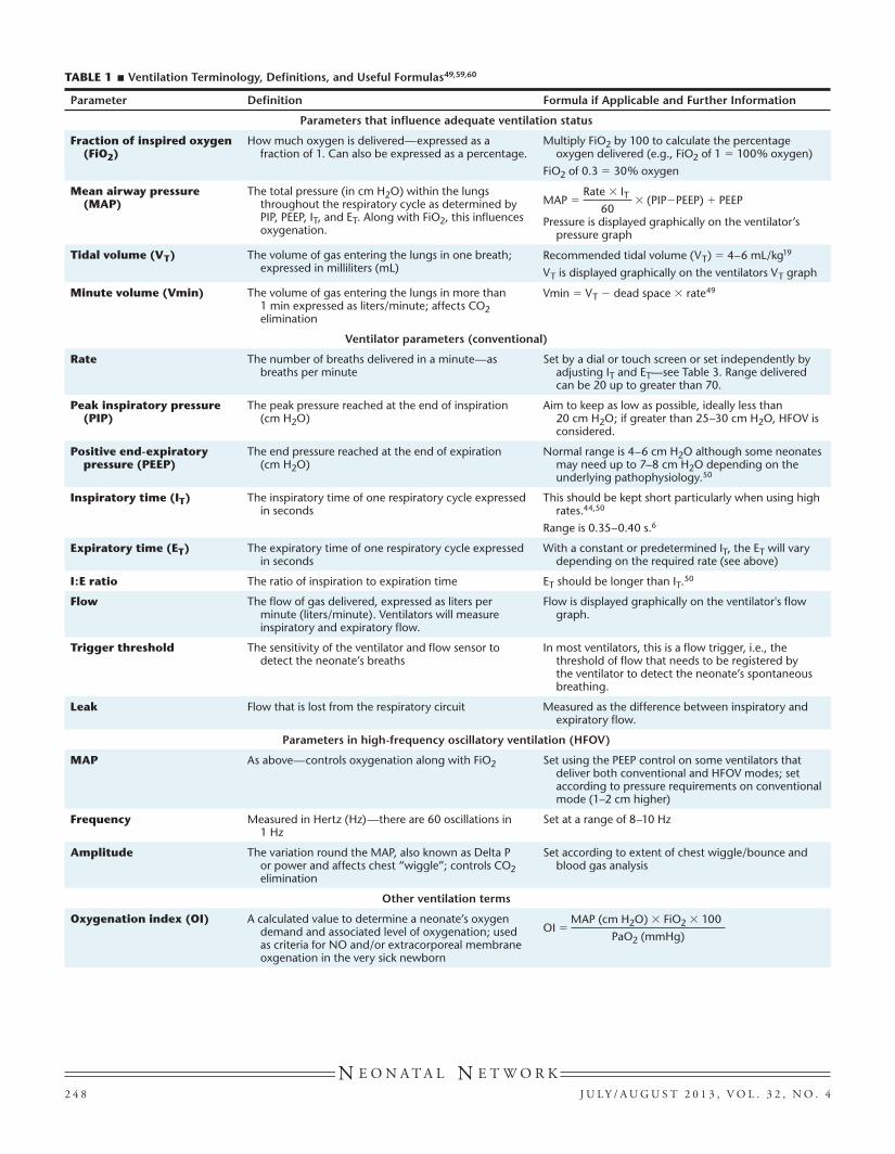
TABLE 1 ■ Ventilation Terminology, Definitions, and Useful Formulas49,59,60
Parameter Definition Formula if Applicable and Further Information
Parameters that influence adequate ventilation status
Fraction of inspired oxygen (FiO2)
How much oxygen is delivered—expressed as a fraction of 1. Can also be expressed as a percentage.
Multiply FiO2 by 100 to calculate the percentage oxygen delivered (e.g., FiO2 of 1 � 100% oxygen)
FiO2 of 0.3 � 30% oxygen
Mean airway pressure (MAP)
The total pressure (in cm H2O) within the lungs throughout the respiratory cycle as determined by PIP, PEEP, IT, and ET. Along with FiO2, this influences oxygenation.
MAP � Rate � IT
60 � (PIP�PEEP) � PEEP
Pressure is displayed graphically on the ventilator’s pressure graph
Tidal volume (VT) The volume of gas entering the lungs in one breath; expressed in milliliters (mL)
Recommended tidal volume (VT) � 4–6 mL/kg19
VT is displayed graphically on the ventilators VT graph
Minute volume (Vmin) The volume of gas entering the lungs in more than 1 min expressed as liters/minute; affects CO2 elimination
Vmin � VT � dead space � rate49
Ventilator parameters (conventional)
Rate The number of breaths delivered in a minute—as breaths per minute
Set by a dial or touch screen or set independently by adjusting IT and ET—see Table 3. Range delivered can be 20 up to greater than 70.
Peak inspiratory pressure (PIP)
The peak pressure reached at the end of inspiration (cm H2O)
Aim to keep as low as possible, ideally less than 20 cm H2O; if greater than 25–30 cm H2O, HFOV is considered.
Positive end-expiratory pressure (PEEP)
The end pressure reached at the end of expiration (cm H2O)
Normal range is 4–6 cm H2O although some neonates may need up to 7–8 cm H2O depending on the underlying pathophysiology.50
Inspiratory time (IT) The inspiratory time of one respiratory cycle expressed in seconds
This should be kept short particularly when using high rates.44,50
Range is 0.35–0.40 s.6
Expiratory time (ET) The expiratory time of one respiratory cycle expressed in seconds
With a constant or predetermined IT, the ET will vary depending on the required rate (see above)
I:E ratio The ratio of inspiration to expiration time ET should be longer than IT.50
Flow The flow of gas delivered, expressed as liters per minute (liters/minute). Ventilators will measure inspiratory and expiratory flow.
Flow is displayed graphically on the ventilator's flow graph.
Trigger threshold The sensitivity of the ventilator and flow sensor to detect the neonate’s breaths
In most ventilators, this is a flow trigger, i.e., the threshold of flow that needs to be registered by the ventilator to detect the neonate’s spontaneous breathing.
Leak Flow that is lost from the respiratory circuit Measured as the difference between inspiratory and expiratory flow.
Parameters in high-frequency oscillatory ventilation (HFOV)
MAP As above—controls oxygenation along with FiO2 Set using the PEEP control on some ventilators that deliver both conventional and HFOV modes; set according to pressure requirements on conventional mode (1–2 cm higher)
Frequency Measured in Hertz (Hz)—there are 60 oscillations in 1 Hz
Set at a range of 8–10 Hz
Amplitude The variation round the MAP, also known as Delta P or power and affects chest “wiggle”; controls CO2 elimination
Set according to extent of chest wiggle/bounce and blood gas analysis
Other ventilation terms
Oxygenation index (OI) A calculated value to determine a neonate’s oxygen demand and associated level of oxygenation; used as criteria for NO and/or extracorporeal membrane oxgenation in the very sick newborn
OI � MAP (cm H2O) � FiO2 � 100
PaO2 (mmHg)

V O L . 3 2 , N O . 4 , J U L Y / A U G U S T 2 0 1 3 2 4 9N E O N A T A L N E T W O R K
assists the infant’s own breath by pressurizing the breath to the set pressure support level. The fl ow termination sensitiv-ity is set so that the I T will terminate at a predetermined percentage of the peak fl ow. Full pressure support (PS) is a mode in its own right and may be useful for neonates who are weaning from their support, allowing them more control in line with their own breathing dynamics. 25 The main prin-ciples of PSV are summarized within the recent literature as a new and emerging mode. 3–6,10,25–28
PSV can also be used in conjunction with other modes by turning this on as an additional feature. 13 For example, synchronized intermittent mandatory ventilation with pres-sure support (SIMV with PS) added will ensure that every spontaneous breath is supported by the ventilator at a set percentage pressure relative to peak pressure set for the man-datory breaths on SIMV.
So, whereas with PS mode alone or PTV (A/C) with PS, all breaths are supported; in SIMV with PS, the neonates’ breaths only are supported. However, this imparts greater support for a neonate who perhaps will not be able to manage on SIMV alone and who requires additional support for his own breathing efforts. Adding in PSV with SIMV can be useful as a more gradual step down once the backup rate on SIMV starts to be reduced during weaning. Here, the neo-nate’s own breaths continue to be supported at a certain pres-sure until such time that he does not require this additional PS. It should be remembered that the measured V T will vary with each pressure-supported breath.
High-Frequency VentilationThis is a mode of ventilation that uses breath rates or
“frequencies” much greater than normal physiologic breath rates with a V T near anatomic dead space. One example is high-frequency jet ventilation (HFJV) that introduces small pulses of gas under pressure into the airway at a very fast rate
diffi cult to deliver at lower pressures, and so the maximum set PIP will be reached. Therefore, it is very important to set an appropriate maximum pressure limit should it become diffi cult to deliver the set V T in deteriorating lung condi-tions. Conversely, as lung compliance improves, it is easier for the desired volume to be delivered at lower pressures, and therefore the measured PIP will be lower, not reaching the maximum limit. When the PIP needed to generate the desired V T decreases, this signals improving lung conditions and readiness for weaning. The ability of VG to show changes in lung compliance is seen as one of the main benefi ts of this ventilation mode. 16,17 Further benefi ts stem from the ability to deliver a guaranteed and consistent V T at the lowest pressure which potentially reduces trauma to the lungs, 18 a feature not achievable by traditional pressure-limited time-cycled ventilation. 16
Based on a review of the literature, Brown and DiBlasi propose that the use of small V T in the range of 4–6 mL/kg is one of the key strategies for protecting the neonatal lung during mechanical ventilation. 19 Cheema and colleagues state that 4 mL/kg should be aimed for. 20 However, a higher V T than this may be required with conditions such as pneu-monia, bronchopulmonary dysplasia (BPD), or other lung pathology that results in increased resistance to airfl ow and the need for a greater volume to be delivered. 21 The reader should refer to three systematic reviews in this mode of ven-tilation for a full summary of the research in this area and the potential benefi ts of VG ventilation. 22–24 A comprehen-sive guide is also available on the practical application of this mode. 8
Pressure Support Ventilation (PSV)With PSV, the neonate’s breathing efforts are supported
with ventilator breaths set to a predetermined pressure. Pressure support alone does not supply a backup rate; it merely
TABLE 1 ■ Ventilation Terminology, Definitions, and Useful Formulas (continued)
Parameter Definition Formula if Applicable and Further Information
Functional residual capacity (FRC)
The volume of gas present in the lung alveoli at the end of passive expiration
FRC is reduced in conditions such as respiratory distress syndrome where there is poor lung compliance. A low FRC will affect optimum gaseous exchange.
Compliance The elasticity or distensibility of the respiratory system including the lungs and chest wall
Compliance � Volume/Pressure
The volume/pressure loop displayed on some ventilators represent this relationship graphically.
Resistance The capability of the airways and endotracheal tube to oppose airflow; expressed as the change in pressure per unit change in flow
Resistance � Pressure/Flow
Again, this is displayed graphically on some ventilators.
Pulmonary dynamics The real-time graphical representations of the neonate’s ventilation parameters
As stated above, graphs can be viewed within the graph section of the ventilator of pressure, VT, flow, compliance, and resistance. These can also be termed waveforms, loops, mechanics, and/or trending displays, all of which represent the neonate’s ventilation status in real-time.
Note: All measurements and graphical displays of parameters are dependent on the presence of a flow sensor. Absence of a flow sensor will mean the ventilator will still deliver breaths, but there will be no “measured” readings.
Abbreviations: I:E ratio � inspiratory to expiratory; NO � nitric oxide; PaO2 � partial pressure of oxygen in arterial blood; s � second.

2 5 0 J U L Y / A U G U S T 2 0 1 3 , V O L . 3 2 , N O . 4N E O N A T A L N E T W O R K
mode was originally used for short-term ventilation during airway surgery because of its capability to ventilate in the presence of air leaks. The short I T and small V T are thought to minimize fl ow through leaks within the lung fi elds, 31 for example, in a condition such as pulmonary interstitial emphy-sema (PIE). In addition, in meconium aspiration syndrome (MAS) where gas trapping may occur, passive exhalation of the jet helps CO 2 be removed without causing further trap-ping and preventing overexpansion of the lungs. Literature also indicates its use for other short-term conditions such as pulmonary hypertension of the newborn (PPHN) and during transport. 32–34 However, the practicalities of administration and the necessity for two machines have meant that other forms of high-frequency ventilation may be more suitable; hence, it is not as widely used.
More commonly used is high-frequency oscillatory ven-tilation (HFOV) where the pressure “oscillates” around a constant distending pressure that in effect is the same as posi-tive end-expiratory pressure (PEEP) and equivalent to mean airway pressure (MAP).
A further explanation of MAP will follow later and is detailed in Table 1. Thus, gas is pushed into the lung during inspiration and then pulled out during expiration. HFOV gen-erates very low V T that is generally less than the dead space of the lung. Figure 3 (A and B) depicts the waveforms for conven-tional versus high-frequency ventilation; the typical pressure graph for HFOV is therefore very different from what we see in conventional modes. Oscillation causes the chest to “wiggle” or vibrate. The increasing use of HFOV in neonates has been documented. 35 In neonatal practice, this is a mode used either
or frequency (4–11 Hertz [Hz]; see in the following text) for a brief duration (approximately 0.02 seconds), using very small V T of � 1 mL/kg, thus creating lower distal airway and alveolar pressures than those produced by a mechanical ven-tilator. Exhalation during HFJV is passive. It was thought this may reduce the severity of lung injury associated with mechanical ventilation. 29,30 The jet actively pulses gas into the neonate’s lungs which travels down the center of the tra-cheal tube. A CO 2 then spirals up and around that jet of gas and out of the expiratory circuit passively (Figure 2). This
FIGURE 2 ■ Pattern of gas flow in high-frequency jet ventilation.
Adapted by Solon JF, from: Rausch K via Ellis R, unpublished data, Milpitas, California. Reprinted by permission.
FIGURE 3 ■ Waveforms for (A) conventional ventilation and (B) high-frequency oscillatory ventilation (HFOV).
A B
From SLE. SLE5000 Neonatal Ventilator with High Frequency Oscillation, 2010. SLE Limited; Surrey UK. Reprinted with permission.

V O L . 3 2 , N O . 4 , J U L Y / A U G U S T 2 0 1 3 2 5 1N E O N A T A L N E T W O R K
E T adjusted until the desired rate is obtained. Information on setting a rate using this method can be seen in Table 3. E T should always be longer than I T .
Both the PIP (at the end of inspiration) and the PEEP (at the end of expiration) are set according to the needs of the neonate and condition. These are depicted in the pressure graph in Figure 3A (top image). Increasing PIP and PEEP will raise the MAP and improve oxygenation because, as for I T , they are integral components of the MAP formula. Conversely, reducing PIP and PEEP will lower MAP when oxygenation is adequate. Making changes to PIP and PEEP will also affect the V T for each breath and so also infl uence CO 2 elimination—this will be covered again later in the article. The side effects of high or low settings for PIP and PEEP should be kept in mind while setting these parame-ters. A high PIP particularly above 25 cm H 2 O can damage the delicate lung alveoli by barotrauma in association with shearing forces of mechanical ventilation. Raising the PIP also increases V T , and therefore a risk of volutrauma is also present. Conversely, a PIP that is too low may not be effective in achieving adequate chest expansion, leading to hypoven-tilation. A high PEEP can lead to an inadequate expiratory phase with poor emptying of gas and limiting CO 2 elimi-nation as the end pressure for each breath is not suffi cient to allow CO 2 removal from the respiratory dead space. Conversely, setting the PEEP too low may lead to alveolar collapse and diminished functional residual capacity (FRC). Therefore, optimum settings should be provided with sound rationale and evaluation tailored to the individual neonate.
Oxygen is set from a dial that blends air and oxygen, keeping this to the minimum possible because of the potential damag-ing effects of oxygen toxicity. Flow (in liters/minute) is set in some ventilators (to 8–10 liters/minute), whereas in others this is automatically delivered without needing to be set. A fl ow graph is depicted in Figure 3A (middle image). In addition, the trigger threshold should be set on the maximum sensitivity in neonates (i.e., the minimum effort for the neonate). For neo-natal fl ow triggering, it is ideal if a change in fl ow of approxi-mately 0.2–0.4 mL/kg is recognized by the ventilator as a spontaneous effort. If it is any higher than this, the neonate’s effort may not be strong enough to actually trigger the fl ow.
In addition, if PS is added to an existing mode, a percent-age of support is set; that is, any spontaneous breaths by the neonate will be supported by fl ow-cycled, pressure-limited breaths to the predetermined percentage of the set PIP. Flow termination sensitivity is also set—that is, when the pressure-supported breath fl ow is terminated. When using TTV or VG, the desired V T is set at approximately 4–6 mL/kg 19 or higher if the neonate’s condition necessitates this, as stated earlier. Refer to Case Studies 1 and 2 to see how PSV and VG are used with other existing modes.
Alarm limits should also be set; for example, high and low pressure, V T alarm, and high and low Vmin alarm thresholds are set. An apnea alarm is also set on some ventilators, often functional if the BPM is less than 20.
as a “rescue” therapy when conventional modes have been ineffective or, in some neonatal units, as a fi rst-line ventila-tion strategy. However, like HFJV, a link between HFOV and improved outcomes has not been demonstrated. 36–38
Proportional Assist Ventilation (PAV)This mode gives assistance that is proportional to the neo-
nate’s effort, whereby the applied pressure increases in pro-portion to the V T and fl ow generated by the neonate, with the frequency, timing, and rate of lung infl ation being con-trolled by the neonate. 39,40 However, this new mode is not frequently used at present compared with other modes dis-cussed thus far. In order for PAV to work effectively, there should be no leak, and a mature respiratory system should be in place; clearly, this is not the case in preterm neonates. 4
Neurally Adjusted Ventilatory Assist (NAVA)NAVA is another new mode of ventilation designed to
reduce the asynchrony that can exist between the ventilator and the neonate. Gas delivery from the ventilator is triggered, controlled, and cycled by a diaphragmatic electromyelo-gram (EMG) signal. The ventilator is aware of the change in EMG by the insertion of a specially designed nasogastric tube (NGT) with EMG electrodes that cross the diaphragm. Several preliminary studies in neonates have demonstrated that patient–ventilator synchrony is improved with the appli-cation of NAVA, 41–43 and this may be a strategy for future work and application.
UNDERSTANDING SETTINGS IN VENTILATION
In addition to understanding what each mode is and how it works, it is important for the neonatal nurse to also under-stand the settings on the ventilator (see Table 1). Nurses record settings hourly on an ongoing basis, and so having the knowledge behind what they mean is paramount.
Firstly, in CMV and SIMV, the desired number of BPM is set either by a BPM dial, or, in some ventilators, the rate is set by adjusting I T and expiratory times (E T ) separately (see in the following text).
A backup respiratory rate is set for some modes (e.g., PTV, A/C, and PS). As seen in Tables 1 and 2, rate along with the V T affects “minute volume” (Vmin) and so affects CO 2 clearance. Increasing Vmin improves CO 2 elimination, and decreasing it will lower this elimination.
The I T is usually set at no higher than 0.36–0.40 seconds in the preterm neonate particularly, recommended because of short physiologic time constants in neonates. 44 The I T may be slightly higher than this in older neonates or when the rate used is slow, but it is usually no higher than 0.50 seconds. Increasing I T will raise the MAP, thereby improving oxy-genation, while lowering the I T may lower partial pressure of oxygen in arterial blood (PaO 2 ) levels (see Table 1). Both I T and E T can be independently set on some ventilators to adjust and set the rate. In that case, I T is confi rmed and then

2 5 2 J U L Y / A U G U S T 2 0 1 3 , V O L . 3 2 , N O . 4N E O N A T A L N E T W O R K
TABLE 2 ■ Changing Ventilation—A General Guide10,12,49,50
Manipulating oxygenationMAP controls oxygenation, so oxygenation can be influenced by changing any of the variables that alter MAP (PIP, PEEP, IT, and ET).
Conventional Ventilation
Desired Outcome Aim and Possible Actions Evaluation
To increase oxygenation (increase MAP)
Increase FiO2 in increments of 5%–10%
Increase MAP by increasing PIP or PEEP in increments of 1–2 cm H2O
Increase IT but no higher than 0.4 seconds for preterm neonates
Consider adding PSV, if on SIMV
Consider starting HFOV if MAP and FiO2 significantly increase
Observe oxygen requirement, pulse oximetry, or transcutaneous oxygenation and PaO2 on blood gas analysis
Look for improvements in lung compliance; e.g., chest expansion
Observe pulmonary dynamics/graphs—e.g., volume/pressure loop and pressure graph
To decrease oxygenation when condition improves and/or during weaning (decrease MAP)
Aim to get FiO2 to an acceptable level
Reduce MAP by reducing PIP or PEEP (again, in small steps of 1–2 cm H2O at a time)
IT can also be reduced slightly, aiming for range 0.36–0.40 s for the preterm neonate
Stop PSV if this has been added to another mode
Change mode to a “trigger” mode that synchronizes spontaneous breaths and/or responds to a trigger threshold and neonate’s own breathing.
As above
Manipulating CO2 eliminationMinute volume (Vmin) controls CO2 elimination. CO2 levels will be influenced by any changing measure affecting Vmin; that is, manipulating the rate, VT, or
both will alter the Vmin.
To clear more CO2
CO2 elimination will be improved by any measure that increases respiratory Vmin (i.e., increasing the rate, VT, or both will increase the Vmin).
Increase rate (in increments of 5–10 BPM) to increase Vmin and remove more CO2.
OR increase PIP (in steps of 1–2 cm H2O) with caution, which will increase VT for each breath and increase Vmin.
Decrease PEEP; however, this may cause a reduction in oxygenation which needs to be observed. In addition, if CO2 is increased because of atelectasis, decreasing the PEEP may worsen the situation and increase CO2; again this needs to considered.
If on VG (TTV), increase set or desired VT.
Observe measured VT and Vmin on the ventilator
Check CO2 on blood gas analysis and/or Tc monitoring
To clear less CO2 when weaning—CO2 elimination will be reduced by any measure that decreases respiratory Vmin (i.e., decreasing the rate, VT, or both will decrease the Vmin).
Reduce rate in increments of 5–10 BPM
and/or reduce PIP (in steps of 1–2 cm H2O).
Consider reducing PEEP as the PIP is reduced.
Reduce set/desired VT if VG (TTV) is being used.
As above
High-Frequency Oscillatory Ventilation
Desired Outcome Aim and Possible Actions Evaluation
To increase oxygenation Increase FiO2 in increments of 5%–10%.
Increase MAP in increments of 1–2 cm H2O.
As for conventional ventilation. Ensure chest x-ray (CXR) is done after being put onto HFOV.
To decrease oxygenation when weaning
Aim to get FiO2 to an acceptable level.
Reduce MAP in increments of 1–2 cm H2O.
As for conventional ventilation
To clear more CO2 Increase amplitude (Delta P) in increments of 2–5 cm H2O according to blood gas (CO2) and chest wiggle
OR decrease frequency (Hz), allowing greater efficiency of oscillations to reach the peak and trough of the pressure wave.
As for conventional ventilation
Observe for adequate chest wiggle/bounce.
To clear less CO2 when weaning Reduce amplitude in increments of 1–2 cm H2O according to CO2 and chest wiggle.
As above
Suggested actions and changes should be based on assessment of the individual neonate.Abbreviations: ET � expiratory times; FiO2 � fractional concentration of oxygen; HFOV � high-frequency oscillatory ventilation; IT, � inspiratory time;
MAP � mean arterial pressure; PEEP � positive end-expiratory pressure; PIP � peak inspiratory pressure; PSV � pressure support ventilation; SIMV � synchronized intermittent mandatory ventilation; TTV � target tidal volume; VG � volume guarantee; VT � tidal volume.

V O L . 3 2 , N O . 4 , J U L Y / A U G U S T 2 0 1 3 2 5 3N E O N A T A L N E T W O R K
breaths will not be synchronized with the neonate’s respira-tory efforts.
The following measurements can be recorded by the neo-natal nurse at the bedside. The MAP is the total pressure within the lungs throughout the respiratory cycle as deter-mined by PIP, PEEP, I T , and E T . The MAP has a direct infl u-ence on oxygenation—for example, if you need to increase oxygenation, the MAP must be increased by manipulation (increasing) of one or more of the PIP, PEEP, and I T . If oxy-genation is adequate or high, then the values can be decreased to reduce MAP. Although MAP is a measured value in con-ventional modes, it can be manipulated by changing the parameters that determine MAP (see Table 1). Oscillation measurements comprise the total rate or frequency (BPM), V T , Vmin, leak, MAP, and oxygen.
The relationship between the I T and E T is expressed as the I:E (inspiratory to expiratory) ratio. For example, if the I T is 0.5 seconds and the rate is 60, the E T will also be 0.5 seconds. The I:E ratio will therefore be 1:1.0. With a lower I T of 0.4, a rate of 60 will mean an E T of 0.6 with an I:E ratio of 1:1.2 (see Table 3).
The ventilator will measure the total number of breaths detected and delivered by the ventilator (mechanical and patient triggered). The number of patient-triggered breaths is usually displayed separately and is an indication of neonatal respiratory effort.
Volume is also measured and gives us valuable information about the dynamics of ventilation. The V T is the volume of gas delivered to the lungs in one breath and is measured as the expired V in milliliters (mL).
Vmin (liter) is the accumulated expiratory V T over a one-minute period. V T multiplied by the rate gives the Vmin; this has a direct infl uence on CO 2 clearance in that increas-ing either the rate or V T (or both) will increase removal of CO 2 . However, this will not work if the CO 2 is high because of overdistension or impeded pulmonary venous return. Such actions could in these cases hamper CO 2 removal. In general, decreasing the rate, V T or both will slow CO 2 removal. Being mindful of the neonate’s underly-ing pathophysiology is essential in selecting optimal venti-lator settings. 45
The difference between the I:E fl ow expressed as a per-centage leak can indicate the need for endotracheal tube change. Further lung dynamics are also monitored by measuring the resistance; the total change in the applied pressure to the lung divided by the maximum fl ow into the lung (resistance to fl ow); and compliance, the ratio of the change in lung volume to the change in the applied pressures.
Graphical representations of the dynamics of the neonate’s breathing pattern can be seen on the ventilator display screen. Figure 3 (A and B) shows pressure graphs for both conven-tional and high-frequency ventilation.
A summary of all modes discussed including what is both set and measured can be seen in Table 4.
OSCILLATION SETTING For HFOV, the frequency of oscillations is expressed in
Hertz. There are 60 breaths in 1 Hz. This mode will deliver very small V T at very high rates, for example, 600 BPM. The MAP is set within this mode and manipulated to control oxygenation, and this is usually set above the MAP that was given for conventional modes (e.g., 2 cm H 2 O higher). Pressure amplitude (Delta P) is the “power” setting and determines the strength of the oscillations (and so the extent of “chest wiggle” in the neonate).
Increasing the Delta P will increase chest wiggle and increase the height of the pressure trace as displayed in Figure 3B. This controls the volume entering the lungs and so con-trols CO 2 elimination. Oxygen, high and low alarm pressure, V T alarm threshold, and Vmin high and low alarm thresh-olds are also set.
Humidifi cation is also an essential element of normal respira-tory function. Any mode , be it noninvasive or full ventilation, should deliver warm, humidifi ed gases by a humidifi er within the inspiratory limb of the ventilator circuit. A humidifi er should ensure an airway temperature of as close to 98.6°F (37°C) as possible.
UNDERSTANDING MEASUREMENTS IN VENTILATION
All measured readings of the dynamics of the neonate’s lungs are taken by the fl ow sensor situated on the connec-tion between the ventilation tubing and the endotracheal tube. This sensor is designed to measure certain param-eters, which are then displayed in various forms. The ven-tilator screen panel displays the measured and calculated parameters. It is important to calibrate the fl ow sensor prior to use (fl ow calibration) and to prevent damage or disrup-tion of the measuring capability because of excessive con-densation from the tubing. In the absence of a fl ow sensor, dynamic measurements are not possible. The ventilator will still be able to deliver the desired settings; however, the
TABLE 3 ■ Setting the Rate Using Inspiratory and Expiratory Times
Confirm desired rate
Divide this into 60
From this number, subtract the inspiratory time (IT)
This gives you the expiratory time (ET) that you need to set to get the desired rate
Example 1—You want a rate of 60 and IT of 0.4 second
60 divided by 60 � 1 second
1.0 minus 0.4 � 0.6 (set the ET at 0.6 second)
This will give you a rate of 60
Example 2—You want a rate of 40 and IT of 0.5 second
60 divided by 40 � 1.5 second
1.5 minus 0.5 � 1 second (set the ET at 1 second)
This will give you a rate of 40

2 5 4 J U L Y / A U G U S T 2 0 1 3 , V O L . 3 2 , N O . 4N E O N A T A L N E T W O R K
and found that the use and availability of modes are variable. Ninety-eight percent response to a structured questionnaire found that pressure-controlled ventilation was most fre-quently used as the primary form of ventilator support (69.8 percent) compared with volume targeted (24.5 percent) and HFOV (5.7 percent). Similarly, an international survey of 50 tertiary units in Australia, New Zealand, Sweden, Denmark, Finland, and Norway evaluating the use of volume-targeted ventilation again found that use was variable, 47 ranging from 40 to 60 percent of units. The most common reason cited for its use was the reduction in BPD. Overall, it is clear that no single ventilation mode or strategy has shown to have a truly signifi cant benefi t in terms of mortality or CLD. 19,48 Moreover, although there is a wealth of research on ventila-tion strategies, each individual neonatal unit will be unique
DECISION MAKING IN NEONATAL VENTILATION
Selecting or Switching Modes of Ventilation The decision to move a neonate from one strategy to another
is infl uenced by the severity and progression of the neonate’s underlying condition, lung pathology, and the response to ventilation changes and attempts at weaning. Gestation, birth weight, and age should also be considered. This emphasizes the importance of matching ventilation strategies to the under-lying pathophysiology 45 and individualized factors applicable to each neonate. The type or level of a specifi c unit in which a neonate is delivered or admitted also needs are considered as this will determine the level of support and strategies avail-able. A United Kingdom–wide survey of ventilation strate-gies within 54 tertiary-level neonatal units was undertaken 46
TABLE 4 ■ Summary of Settings and Measurements for Ventilation Modes6
Mode Settings Measurements
CMV Rate (BPM), PEEP, PIP, IT, oxygen, high and low alarm pressure thresholds, VT alarm threshold, Vmin high and low alarm thresholds
I:E ratio, ET measured, rate (BPM), VT, Vmin, leak, resistance, compliance, PIP/PEEP, and MAP. Recorded breath rate is what is set, not what the neonate does.
SIMV Rate (BPM), apnea time (if backup breath rate is less than 20 breaths) per minute or lower, PEEP, PIP, IT, oxygen, trigger threshold, high and low alarm pressure thresholds, VT alarm threshold, Vmin high and low alarm thresholds
IT measured, total rate (BPM), trigger (number of neonate’s triggered breaths), VT, Vmin, leak, resistance, compliance, PIP/PEEP, and MAP
PTV/ A/C Backup rate (BPM), apnea time (if backup breath rate is less than 20), PEEP, PIP, IT, oxygen, trigger threshold, high and lowalarm pressure thresholds, VT alarm threshold, Vmin high and low alarm thresholds
IT measured, total rate (BPM), trigger (number of neonate “triggered” breaths), VT, Vmin, leak, resistance, compliance, PIP/PEEP, and MAP
PSV Backup rate (BPM), apnea time (if backup breath rate less than 20), PEEP, PIP, maximum IT, oxygen, flow termination sensitivity, trigger threshold, high and low alarm pressure thresholds, VT alarm threshold, Vmin high and low alarm thresholds
IT measured, total rate (BPM), trigger (number of neonate’s triggered breaths), VT, Vmin, leak, resistance, compliance and PIP/PEEP, MAP, oxygen
SIMV � PS As for SIMV above but turn PS to “on” and set % of pressure support plus flow termination sensitivity
IT measured, total rate (BPM), trigger (number of neonate’s triggered breaths), VT, Vmin, leak, resistance, compliance, PIP/PEEP, MAP, and oxygen
VG/TTV As for these modes above but turn on TTV and set VT requiredfor each breath.
As for each mode above plus “measured” PIP (will vary)
HFJV Set short bursts with short IT, frequency, and flow See below as for HFOV
HFOV HFO rate (in Hz), mean pressure, pressure amplitude (or Delta P), oxygen, high and low alarm pressure thresholds, VT alarm threshold, and Vmin high and low alarm thresholds
Rate (BPM) total, VT, Vmin, leak, MAP and oxygen
CMV PLUS HFOV
BPM, IT, PIP, PEEP, HFO rate, HFO activity (oscillations in inspiratory and expiratory phases or expiratory phase only), pressure amplitude (Delta P), oxygen, high and low alarm pressure thresholds, VT alarm threshold, Vmin high and low alarm thresholds
I:E ratio, ET measured, rate (BPM) and HFO rate, PIP/PEEP, MAP, and oxygen
NAVA Trigger threshold to pick up electrical diaphragmatic activity. Adapt NAVA level to regulate pressure support.6
As for above
Note: Other modes of ventilation are less commonly cited in the literature and so are not included in this article; for example, volume-controlled ventilation, volume-limited ventilation, pressure-regulated volume control, volume-assured pressure support (VCV, VLV, PRVC, VAPS, respectively) and high-frequency flow interruption (HFFI); refer to key literature for more information.4,6
Abbreviations: A/C � assist control; BPM � breaths per minute; CMV � continuous mandatory ventilation; HFJV � high-frequency jet ventilation; HFO � high-frequency oscillation; HFOV � high-frequency oscillatory ventilation; I:E ratio � inspiratory and expiratory ratio; IT � inspiratory time; MAP � mean airway pressure; NAVA � neurally adjusted ventilatory assist; PEEP � positive end-expiratory pressure; PIP � peak inspiratory pressure; PS � pressure support; PSV � pressure support ventilation; PTV � patient trigger ventilation; SIMV � synchronized intermittent mandatory ventilation; TTV � target tidal volume; VG � volume guarantee; Vmin � minute volume; VT � tidal volume.

V O L . 3 2 , N O . 4 , J U L Y / A U G U S T 2 0 1 3 2 5 5N E O N A T A L N E T W O R K
the minimum requirement needed for adequate oxygen-ation of the neonate. The I T should be bought down to the minimum, to be as close as possible to the neonate's own physiologic time. SIMV has been found to be the most common mode in practice, and, therefore, it follows that this mode is most commonly used for weaning. 5 In this mode, rate, pressure, or both are reduced in increments depending on individual assessment of blood gas analysis and clinical picture. If PS is turned on, the percentage of pressure support is gradually reduced until this is eventu-ally switched to off, so that the neonate’s breaths are no longer supported. Some clinicians prefer to wean from PTV or A/C mode as this has been found to be associated with a shorter duration of weaning. 14 In this mode, pressure alone is reduced. If PSV is used, similarly the level of pressure support is reduced gradually. In A/C or PSV, because the neonate controls the ventilator rate, reducing this rate has no effect on delivered rate unless respiratory effort is poor or the backup rate is greater than the spontaneous breath-ing rate.
The VG mode of ventilation results in automatic weaning of the PIP as lung function improves. 3–5 The only param-eters that should be altered during weaning are the FiO 2 and the set V T . When the set V T is below the neonate’s sponta-neously generated V T , the PIP will be reduced. Extubation should be considered when the PIP reaches minimal levels. 7,52,53 Extubation is considered if the MAP is consis-tently � 8 to 10 cm H2O with set V T T 3.5 to 4.5 mL/kg with satisfactory blood gases. 8 Readiness for extubation can also be ascertained by assessing the neonate’s spontaneous breathing. 54
To serve as a guide, protocol-led care has been recom-mended. 11,55 Overall, however, there does not seem to be any one strategy that is recommended for weaning. 55–57 In rela-tion to any weaning strategy, key questions should be consid-ered as seen in Figure 4. It is always important to emphasize here that, whatever strategy is chosen, each change must have rationale. Moreover, the individualized evaluation of any change made is vital to know if this has been effective in the best interest of the neonate.
CASE STUDY 1: CMV, SIMV, SIMV PLUS PSV, AND PSV
Baby John was a preterm neonate born at 27 weeks ges-tation (birth weight 1 kg) by spontaneous vaginal delivery. The neonate’s mother received a course of antenatal steroids on admission to hospital; the aim was to enhance surfactant maturation and reduce the severity of respiratory distress syn-drome (RDS). The neonate was rigorous and active at birth, spontaneously breathing and required no resuscitation except for some stimulation and prevention of heat loss. He was placed onto binasal CPAP with a pressure of 5 cm H 2 O and 35 percent oxygen to maintain an oxygen saturation (SaO 2 ) target of 89–93 percent. An initial arterial blood gas (ABG) showed; pH 7.3, PaO 2 64 mmHg (8.5 kPa [kilopascal]), CO 2
in its application of research data, preferences, patient-specifi c population, and unit outcomes.
However, for any neonatal unit, accurate patient assess-ment is of vital importance for the decision-making process. This is a key element in coming to a diagnosis but also to gain a full picture of how compromised the respiratory system and other related systems are and which actions to take or whether to transfer appropriately if necessary.
Decision-Making Tools for Neonatal Ventilation Practice Various tools exist in neonatal ventilation practice to
guide and inform decision making in practice. However, they should never replace a holistic approach to care of the neonate and family. Nurses can be assisted in their practice by tools that can facilitate understanding of interventions their neo-nates undergo. Tools may provide clarifi cation and guidance at a novice level which may serve to facilitate safer practice (Tables 3–5); however, whichever tool is employed, assess-ment is central when deciding what modes and parameters are suitable for any specifi c neonate.
Making Changes to Ventilation Parameters Overall, changing ventilation parameters is based on
assessment in the fi rst instance of both blood gas analysis and clinical picture. Then, the initial question to ask is “What am I trying to achieve?”: Is it the need to change oxygen-ation, CO 2 or both? Other questions may be: “What is the target oxygen saturation? What does the neonate look like—for example, chest movement and synchrony? Do ventilation requirements need increasing or decreasing or remain static?” Balance is important, or, in other words, in line with protect-ing the lungs, there is a need to balance the ventilator set-tings. For example, if a neonate is in 100 percent oxygen but with low pressures settings, it may be preferable to reduce the FiO 2 but increase the pressures. Similarly, if the neonate is on high pressure settings but a low rate, it may be better to give a faster rate and lower pressures.
A full summary of the principles behind changing ventilation can be seen in Table 5. The clinical application of how changes are made in practice can be seen in Case Studies 1, 2, and 3.
One important change in ventilation practice is the weaning of support. In essence, weaning a neonate from a ventilator should be a central objective at the point of intubation and should be commenced as soon as possible in line with a protective lung strategy. However, it is reported that approximately 30 percent of intubated neonates may fail attempted extubation, 51 and so, bearing in mind the clinical implications of this, appropriate decision making for weaning must be employed. Strategies for weaning include any change to any parameter that facilitates the neonate taking control of his own ventilation. Weaning can be done on any mode mentioned thus far, with the excep-tion of CMV which should be changed to a synchronized or trigger mode as soon as possible. Oxygen should cer-tainly be weaned down within any mode of ventilation to

2 5 6 J U L Y / A U G U S T 2 0 1 3 , V O L . 3 2 , N O . 4N E O N A T A L N E T W O R K
TABLE 5 ■ Interpretation of Blood Gases in the Neonatal Unit
A. BLOOD GAS VALUES61–66
In relation to values, different sources may cite slight variations.
pH CO2 O2 Bicarbonate Base References
CORD (arterial) 7.25–7.28 48 mmHg 6.5 kPa
18–22.5 mmHg 2.4–3 kPa
n/a �4 35, 36
CORD (venous) 7.28–7.35 35–45 mmHg 5–6 kPa
27–38 mmHg 3.8–5 kPa
n/a �4 35, 36
NEONATAL (arterial)*
7.35–7.45 35–45 mmHg38 4.6–6.0 kPa
50–90 mmHg 7.0–12.0 kPa Term
50–80 mmHg 6.5–10.5 kPa Preterm
22–26 mEq/liter Term
20–24 mEq/liter Preterm38
Or 22–26 mmol39
�2 to �2 37, 38, 39
For “Uncompensated” gas (i.e., pH is abnormal)
Low pH and high CO2 � respiratory acidosis
Low pH and large base deficit/low bicarbonate � metabolic acidosis (Note: Mixed acidosis � low pH, high CO2 & large base deficit)
High pH and low CO2 � respiratory alkalosis
High pH and large base excess/high bicarbonate � metabolic alkalosis
For “compensated” gas (i.e., pH is normal, but other values are out of range)
pH CO2 Bicarbonate Problem
Low normal High High Compensated respiratory acidosis
High normal Low Low Compensated respiratory alkalosis
Low normal Low Low Compensated metabolic acidosis
High normal High High Compensated metabolic alkalosis
Permissive hypercapnia
To avoid overventilating the lungs, keep pH �7.25
Watch base deficit and keep between �4 and �450
*Capillary venous neonatal sampling can also be considered for all values (pH, CO2, base, and bicarbonate) except oxygenation status.
B. BLOOD GAS ALGORITHM65,66
1. Assess pH
(Is the pH normal? If not, is it acidotic or alkalotic?—see values)
2. Assess respiratory component
Is CO2 within normal range? See A, above.
3. Assess metabolic component
Is the bicarbonate within normal range and is there a large base deficit (a negative (�) number) or base excess (a positive (�) number)? See A, above.
4. Assess if compensation has occurred
(i.e., The pH has normalized, but the other values are out of normal range.)
5. Assess oxygenation (PaO2)
A low PaO2 can contribute to a metabolic acidosis by anaerobic respiration by cells and lactic acidosis accumulation.
Plus
Consider lactate levels
6. Interpret and make plan of action
7. Evaluate/Reassess
When to evaluate/recheck a blood gas depends on how abnormal the values are.50 If they are very abnormal OR a significant change has been made (e.g., surfactant, high-frequency oscillatory ventilation has been commenced), the blood gas may need rechecking within 30 minutes. If the neonate is stable, then it will need rechecking less frequently.
Decide on an individual basis following discussion with the clinical team.
Abbreviation: HFOV � high-frequency oscillatory ventilation.
V O L . 3 2 , N O . 4 , J U L Y / A U G U S T 2 0 1 3 2 5 7N E O N A T A L N E T W O R K
45 mmHg (6.0 kPa), Base � 2, and bicarbonate 22 mEq/liter (mmol/liter); all within satisfactory limits (Table 5).
However, six hours later, the neonate started to show signs of tachypnea, chest recession, and nasal fl aring with poor saturations and an increasing oxygen requirement. The ABG showed a respiratory acidosis pH 7.2, PaO 2 57 mmHg (7.6 kPa), CO 2 67 mmHg (8.9 kPa), Base � 3, and bicarbonate 21 mEq/liter (mmol/liter). Therefore, the CPAP pressure was increased to 7 cm H 2 O with a biphasic level added after the ABG an hour later failed to show any signifi cant improve-ment. This continued and along with both the clinical assess-ment and a CXR led to a diagnosis of RDS.
The decision was made to intubate and ventilate the neonate to provide respiratory support and to enable surfac-tant to be administered. Following intubation, Baby John made little spontaneous effort, and CMV was commenced (PIP 22, PEEP 5, I T 0.4, rate 60). The ABG improved an hour later: pH 7.31, PaO 2 60 mmHg (8.0 kPa), CO 2 42 mmHg (5.6 kPa), Base � 2, and bicarbonate 22 mEq/liter. After eight hours, the CXR appearance, lung expansion, and ABG values showed improvement. Baby John was also spontane-ously breathing but appeared to be in asynchrony with the ventilator breaths and exhibiting signs of discomfort. The mode was therefore switched to SIMV to allow the ventilator
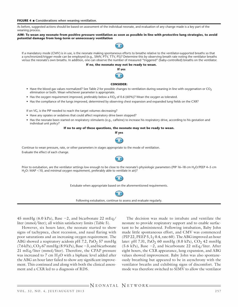
FIGURE 4 ■ Considerations when weaning ventilation.
As before, suggested actions should be based on assessment of the individual neonate, and evaluation of any change made is a key part of the weaning process.
AIM: To wean any neonate from positive pressure ventilation as soon as possible in line with protective lung strategies, to avoid potential damage from long-term or unnecessary ventilation
If a mandatory mode (CMV) is in use, is the neonate making spontaneous efforts to breathe relative to the ventilator-supported breaths so that a synchronized/trigger mode can be employed (e.g., SIMV, PTV, TTV, PS)? Determine this by observing breath rate noting the ventilator breaths versus the neonate's own breaths. In addition, one can observe the number of measured “triggered” (baby-controlled) breaths on the ventilator.
If no, the neonate may not be ready to wean.
If yes
CONSIDER:
• Have the blood gas values normalized? See Table 2 for possible changes to ventilation during weaning in line with oxygenation or CO2 elimination or both. Wean whichever parameter is appropriate.
• Has the oxygen requirement improved, preferably below a FiO2 of 0.6 (60%)? Wean the oxygen as tolerated.
• Has the compliance of the lungs improved, determined by observing chest expansion and expanded lung fields on the CXR?
If on VG, is the PIP needed to reach the target volumes decreasing?
• Have any opiates or sedatives that could affect respiratory drive been stopped?
• Has the neonate been started on respiratory stimulants (e.g., caffeine) to increase his respiratory drive, according to his gestation and individual unit policy?
If no to any of these questions, the neonate may not be ready to wean.
If yes
Continue to wean pressure, rate, or other parameters in stages appropriate to the mode of ventilation.
Evaluate the effect of each change.
Prior to extubation, are the ventilator settings low enough to be close to the neonate’s physiologic parameters (PIP 16–18 cm H2O/PEEP 4–5 cm H2O: MAP �10, and minimal oxygen requirement, preferably able to ventilate in air)?
Extubate when appropriate based on the aforementioned requirements.
Following extubation, continue to assess and evaluate regularly.

2 5 8 J U L Y / A U G U S T 2 0 1 3 , V O L . 3 2 , N O . 4N E O N A T A L N E T W O R K
ventilated on SIMV—PIP 23, PEEP 5, I T 0.36, and a rate of 50. The capillary blood gas showed a picture of permis-sive hypercapnia accepting a higher-than-normal CO 2 with a pH maintained higher than 7.25 and preventing overventilat-ing the premature lung: pH 7.28, PaO 2 37.5 mmHg (5 kPa; capillary), CO 2 66 mmHg (8.8 kPa), Base � 5, and bicarbon-ate 32 mEq/liter. In addition, this gas shows compensation had occurred.
However, it was not possible to wean any further than this because Baby Faye became unstable after any attempts to change the ventilator settings. The last attempt to wean down the pressures led to a drop on SaO 2 and a worsen-ing pH of 7.22, lower than the acceptable range. It was also noted that the measured V T was quite variable and often much lower than 4 mL/kg even when the PIP was increased. Therefore, the decision was made to switch on VG, and a target V T was set at 4 mL/kg (3.6 mL); the aim was to achieve a balance between ensuring delivery of an adequate volume while also being mindful of limiting the V T to prevent any further damage from volutrauma. This also allowed the lung volumes to be optimized but at the lowest possible pressures, thus reducing the incidence also of barotrauma.
On assessment, measured PIP was 18–20 cm H 2 O, lower than the previous settings on SIMV. However, the pH and PaCO 2 did improve to some degree and did not revert back to the previous acceptable values; pH hovered around 7.24–7.25. Therefore, to optimize the V T further, the target V T was increased to 5 mL/kg (4.5 mL). The measured PIP continued to remain acceptable, and blood gases then improved with greater clearance of CO 2 . pH was then stable at 7.25–7.28.
Over time, the previous situation continued, and Baby Faye was then able to be weaned with a gradual reduction of the ventilator rate in increments of 5 cm H 2 O.
After two weeks, the mode was changed to A/C (PTV) allowing her to control her own rate since she was now 6 weeks old. Overall, this case study shows how the complex-ity of the lung condition and the associated chronic changes in the preterm neonate necessitate signifi cant support over a lengthy period. However, to synchronize and tailor the ventilation to a neonate’s own efforts while optimizing V T , using a trigger or synchronized mode along with VG can allow this.
CASE STUDY 3: SIMV, HFOV, A/C (PTV) A term neonate (birth weight 4 kg) was born in poor con-
dition in thick meconium following a prolonged and diffi cult labor. The neonate, Baby Ahmed, required full resuscitation measures at birth and was intubated and ventilated in the delivery suite with a subsequent transfer to the neonatal unit. He was ventilated on CMV mode requiring PIP 26 cm H2O, PEEP 5 cm H2O, I T 0.4, and a rate of 45. The ABG showed a severe mixed acidosis: pH 7.1, PaO 2 37 mmHg (4.9 kPa), CO 2 70 mmHg (9.3 kPa), Base � 8, and bicarbonate 16 mEq/liter (mmol/liter). The PIP was therefore increased
breaths to be synchronized to his breaths. He remained stable on this mode through the night, enabling the pressures and I T to be reduced in increments to avoid any undue excessive expansion of the lungs. By the morning, the settings were PIP 20, PEEP 4, I T 0.35, and rate 55. The measured V T was adequate at 4–6 mL/kg; spontaneous breathing was approxi-mately one-third of the total breaths with synchrony evident on clinical assessment.
By the afternoon, two doses of surfactant had been admin-istered, and he was stable on SIMV; the rate was turned down to 40 as the CO 2 on the blood gas was satisfactory and Baby John was making good spontaneous efforts between the ven-tilator breaths. Later, the pressure settings were reduced to 18/4 cm H2O to limit the applied pressure to the lungs as much as possible. There was good chest expansion and sound clinical assessment at this time with adequate oxygenation (SaO 2 and PaO 2 ).
Later that day, however, a follow-up gas was not accept-able, with a developing mixed acidosis evident. Therefore, rather than increasing the SIMV rate, the decision was made to turn on PSV with SIMV to support Baby John’s breaths at 100 percent of the set PIP. In other words, the ventilator was set to deliver 40 breaths at 18/4 cm H2O with all Baby John’s own breaths supported at this pressure and I T . This was effective as the blood gases stabilized.
PSV allowed additional support for this neonate who appeared to be becoming tired on SIMV alone. Rather than increasing the settings, adding this additional mode allowed such support without having to increase pressures again. The oxygen requirement throughout the previous period had been between 45 and 55 percent, and, after eight hours on SIMV with PSV, the oxygen requirement was reduced and stable at 40–45 percent.
Over the course of the next two days, the PSV level was slowly reduced in increments to 50 percent of the set PIP; that is, the neonate’s breaths were supported to 50 percent of the PIP set in the ventilator. Assessment was satisfactory, and, when the neonate was fi ve days old, he was able to be weaned further from the ventilator settings. Because he was making a good spontaneous effort, the decision was made to wean on full PSV; this meant that Baby John controlled his own rate and I T, and the ventilator supported every triggered breath with pressures of 18/4 cm H2O. This pressure was slowly weaned down to 16/4 cm H2O with an oxygen requirement of 35–40 percent until he was ready to be extubated to CPAP at 6 days of age.
CASE STUDY 2: SIMV WITH VG AND A/C Baby Faye was born spontaneously at 25 weeks gestation
to an unbooked mother who presented in precipitous labor and did not receive any antenatal steroids. The neonate had then spent a diffi cult period on high ventilation require-ments leading to a diagnosis of CLD of prematurity with patchy appearance of PIE throughout both lungs. She was now 4 weeks old, weighing 900 g, and continued to be

V O L . 3 2 , N O . 4 , J U L Y / A U G U S T 2 0 1 3 2 5 9N E O N A T A L N E T W O R K
example, changes in CXR fi ndings, blood pressure, oxygen concentrations, and oxygen saturations. Current strategies must also have these dangers as a central consideration. As stated earlier, CLD or BPD is one of the most common long-term complications in very premature infants. 50,58 There has been a revolution in the therapies that are used, either to manage initial RDS with an aim to prevent CLD or to manage the established condition, and several devices and strategies have been developed to provide respiratory support with reduced risk of lung injuries. These protective lung strat-egies include the use of noninvasive ventilation modes such as CPAP and biphasic CPAP and minimizing both oxygen delivery along with pressure and volume from the point of birth and beyond. Brown and DiBlasi 19 state that the keys to protecting the neonatal lung during mechanical ventilation are to optimize lung volume, limit excessive expansion, apply appropriate end pressure, use shorter inspiratory times and smaller V T , and allow permissive hypercapnia appropriately. All these protective lung strategies are clearly seen within Case Studies 1, 2, and 3 highlighting, within each of the modes included, the differences in management and decision making between conditions but also the vital considerations around minimizing lung damage as much as possible.
CONCLUSION Technological advances have resulted in improvements in
ventilators and strategies that are more sophisticated to be in synchrony with the neonate’s own efforts, aiming to limit the damaging effects by pressure and/or volume trauma. Those caring for such sick and vulnerable neonates owe it to them and their families to have a full understanding of common but often-complex practices such as ventilation practice while at the same time working toward minimizing any resultant damage and respiratory morbidity. Knowledge of the modes and terms used in neonatal ventilation practice is valuable because this practice comprises a signifi cant proportion of care given to sick neonates and their families within the neo-natal unit. It has not been possible to discuss the care of neo-nates in relation to ventilation modes or to discuss the details of underlying neonatal physiology. Rather, the aim is for the reader to have a basic underlying foundation of knowledge in ventilation practice so she has the rationale for the care she learns about within the clinical area and also to gain a basis for further learning. The decision to place a neonate onto a specifi c type of ventilation strategy and/or to change a mode or parameter depends on a complex interplay of factors. It is vital that effective clinical decisions are based on valid, sound judgments that consider the neonate’s clinical assessment cues including sound evaluation on an ongoing basis. Finally, ventilation practice and decision making must be supported by best evidence-based rationale not only so that health professionals can learn and progress but also so that parents are given information about the treatments their neonates receive and the outcomes they might expect, a vital component of true family-centered neonatal care.
to raise the MAP, aiming to increase oxygenation. However, no change was observed to Baby Ahmed’s condition or oxy-genation, and the measured V T did not improve regardless of any change in PIP. All parameters continued to be increased until the settings were PIP 30, PEEP 6, I T 0.5, and a rate of 45. The required FiO 2 was 0.8 (80 percent) to maintain SaO 2 at a range of 95–100 percent for a term neonate. The measured MAP was 16 cm H2O, and V T was 12 ml (i.e., only 3 mL/kg). Therefore, the decision was made to switch Baby Ahmed to HFOV to enable a higher MAP while avoid-ing excessive lung expansion from any higher PIP and shear-ing forces of conventional ventilation at high pressures and I T . Settings were MAP 18 cm H 2 O, amplitude 40, frequency 10 Hz, FiO 2 0.75 (75 percent); set to maintain a MAP 2 cm H 2 O higher than the previous conventional ventilation and to observe adequate chest wiggle or bounce.
This continued for an eight-hour period during which the clinical picture and the blood gases started to improve. The CXR showed good lung expansion. The amplitude was able to be reduced as chest wiggle was pronounced and CO 2 started to decrease. Oxygenation was slower to improve, but, after a 24-hour period on MAP of 18, the FiO 2 could be reduced to 0.6 (60 percent). The PaO 2 then also started to increase in value and continued to do so over the next 24 hours. During this time, the MAP was slowly reduced by increments of 1–2 cm H 2 O until this reached 14 cm H 2 O. The oxygen requirement was now 50–60 percent.
Baby Ahmed started to attempt to take spontaneous breaths on Day 3 of life—therefore, the decision was made to switch from HFOV to a conventional but trigger (syn-chronized) mode of ventilation. A/C (PTV) was given as a weaning mode for Baby Ahmed who, from this point, started to clinically improve. The pressure was reduced in increments because it was futile to reduce the rate because he was breath-ing above the backup rate. On Day 5 of life, Baby Ahmed was able to be extubated onto a short period of nasal CPAP until his lungs has signifi cantly improved and all meconium had cleared. By Day 6, he was self-ventilating in air.
Current Strategies to Reduce Lung Injury The importance of a protective lung strategy has been high-
lighted throughout the article. It is worth reiterating this fact as an integral component of ventilation practice. Ultimately, we must achieve a balance between providing optimum respi-ratory status and support while avoiding overventilation or an unnecessary length of time on mechanical ventilation and the associated effects on the neonate. When deciding how to deliver optimum ventilator support to neonates, it is impor-tant to be aware of these potential negative effects of ventila-tion and place emphasis on preventing them. They include lung injury from barotrauma (pressure) and/or volutrauma (volume) leading to CLD, oxygen toxicity, hypotension, lung hyperinfl ation, air leak, and nosocomial respiratory infection. Bedside assessment and monitoring are the keys to ensur-ing any negative effect is identifi ed as soon as possible, for

2 6 0 J U L Y / A U G U S T 2 0 1 3 , V O L . 3 2 , N O . 4N E O N A T A L N E T W O R K
24. Wheeler K, Klingenberg C, Morley CJ, Davies PG. Volume-targeted versus pressure-limited ventilation for preterm infants: a systematic review and meta-analysis. Neonatology . 2011;100:219-227. http://dx.doi.org/10.1159/000326080.
25. Reyes ZC, Claure N, Tauscher MK, D’Ugard C, Vanbuskirk S, Bancalari E. Randomized, controlled trial comparing synchronized intermittent mandatory ventilation and synchronized intermittent mandatory ventilation plus pressure support in preterm infants. Pediatrics . 2006;118(4):1409-1417.
26. Sarkar S, Donn SM. In support of pressure support. Clin Perinatol . 2007;34(1):117-128, vii.
27. Gupta S, Sinha SK, Donn SM. The effect of two levels of pressure support ventilation on tidal volume delivery and minute ventilation in preterm infants. Arch Dis Child Fetal Neonatal Ed . 2009;94:F80-F83.
28. Patel DS, Rafferty GF, Lee S, Hannam S, Greenough A. Work of breathing during SIMV with and without pressure support. Arch Dis Child . 2009;94:434-436. http://dx.doi.org/10.1136/adc.2008.152926.
29. Bhuta T, Henderson-Smart DJ. Elective high frequency jet ventilation versus conventional ventilation for respiratory distress syndrome in preterm infants. Cochrane Database Syst Rev . 1998;(2):CD000328. http://dx.doi.org/10.1002/14651858.CD000328.
30. Joshi VH, Bhuta T. Rescue high frequency jet ventilation versus conventional ventilation for severe pulmonary dysfunction in preterm infants. Cochrane Database Syst Rev . 2006;(1):CD000437. http://dx.doi.org/10.1002/14651858.CD000437.pub2.
31. Jones A. High frequency jet ventilation. http://www.respiratorycare-online.com/lecture_series/jet_ventilation_mp3/HFJV_handout.pdf . Published January 14, 2011. Accessed June 13, 2013.
32. Coates EW, Klinepeter ME, O’Shea TM. Neonatal pulmonary hypertension treated with inhaled nitric oxide and high-frequency ventilation. J Perinatol . 2008;28(10):675-679.
33. Mainali ES, Greene C, Rozycki HJ, Gutcher GR. Safety and effi cacy of high-frequency jet ventilation in neonatal transport. J Perinatol . 2007;27(10):609-613.
34. Friedlich P, Subramanian N, Sebald M, Noori S, Seri I. Use of high-frequency jet ventilation in neonates with hypoxemia refractory to high frequency oscillatory ventilation. J Matern Fetal Neonatal Med . 2003;13(6):398-402.
35. Froese AB, Kinsella JP. High-frequency oscillatory ventilation: lessons from the neonatal/pediatric experience Crit Care Med . 2005;33(3)(suppl):S115-S121.
36. Cools F, Henderson-Smart DJ, Offringa M, Askie LM. Elective high frequency oscillatory ventilation versus conventional ventilation for acute pulmonary dysfunction in preterm infants. Cochrane Database Syst Rev . 2009;(3):CD000104. http://dx.doi.org/10.1002/14651858.CD000104.pub3.
37. Marlow N, Greenough A, Peacock JL, et al. Randomised trial of high frequency oscillatory ventilation or conventional ventilation in babies of gestational age 28 weeks or less: respiratory and neurological outcomes at 2 years. Arch Dis Child Fetal Neonatal Ed . 2006;91(5):F320-F326.
38. Henderson-Smart DJ, De Paoli AG, Clark RH, Bhuta T. High frequency oscillatory ventilation versus conventional ventilation for infants with severe pulmonary dysfunction born at or near term. Cochrane Database Syst Rev . 2009;(3):CD002974. http://dx.doi.org/10.1002/14651858.CD002974.pub2.
39. Schulze A, Rieger-Fackeldey E, Gerhardt T, Claure N, Everett R, Bancalari E. Randomized crossover comparison of proportional assist ventilation and patient-triggered ventilation in extremely low birthweight infants with evolving chronic lung disease. Neonatology . 2007;92:1-7.
40. Schulze A. Proportional assist ventilation. In: Donn SM, Sinha SK, eds. Manual of Neonatal Respiratory Care . 3rd ed. New York, NY: Springer Science � Business Media; 2012:291-299.
41. Spahija J, de Marchie M, Albert M, et al. Patient-ventilator interaction during pressure support ventilation and neurally adjusted ventilatory assist. Crit Care Med . 2010;38(2):518-526.
42. Breatnach C, Conlon NP, Stack M, Healy M, O’Hare BP. A prospective crossover comparison of neurally adjusted ventilatory assist and pressure-support ventilation in a pediatric and neonatal intensive care unit population. Pediatr Crit Care Med . 2010;11(1):7-11.
REFERENCES 1. Bellettato B, Carlo W, Rosenkrantz T, Carter BS, Windle ML. Assisted
ventilation of the newborn. 2011. http://emedicine.medscape.com/article/979268-overview. Accessed February 12, 2013.
2. Baert AL, Knauth M, Sartor K. Radiological Imaging of the Neonatal Chest . 2nd ed. New York, NY: Springer; 2008.
3. Hummler H, Schulze AA. New and alternative modes of mechanical ventilation in neonates. Semin Fetal Neonatal Med . 2009;14(1):42-48.
4. Broster SC, Ahluwalia JS. Overview of assisted ventilation of the newborn. Pediatr Child Health . 2009;19(12):537-543.
5. van Kaam AHA, Rimensberger PCA, Borensztajn DA, De Jaegere APA. Ventilation practices in the neonatal intensive care unit: a cross-sectional study. J Pediatr . 2010;157(5):767-771.
6. Habre WA. Neonatal ventilation. Best Pract Res Clin Anaesthesiol . 2010; 24(3):353-364.
7. Schulzke SM, Pillow J, Ewald B, Patole SK. Flow-cycled versus time-cycled synchronized ventilation for neonates. Cochrane Database Syst Rev . 2010;(7):CD008246. http://dx.doi.org/10.1002/14651858.CD008246.pub2
8. Klingenberg C, Wheeler KI, Davis PG, Morley CJ. A practical guide to neonatal volume guarantee ventilation. J Perinatol . 2011;31:575-585.
9. Mahmoud RA, Schmalisch G. Modern mechanical ventilation strategies in newborns: a review. Technol Health Care . 2011;19(5):307-318.
10. Goldsmith JP, Karotkin E. Assisted Ventilation of the Neonate . 5th ed. St. Louis, MO: Elsevier Sanders; 2011.
11. Sant’Anna GM, Keszler M. Developing a neonatal unit ventilation protocol for the preterm baby. Early Hum Dev . 2012;88(12):925-929.
12. Donn SM, Sinha SK. Manual of Neonatal Respiratory Care . 3rd ed. New York, NY: Springer Science � Business Media; 2012.
13. Claure N, Bancalari E. New modes of mechanical ventilation in the preterm newborn: evidence of benefi t. Arch Dis Child Fetal Neonatal Ed . 2007;92:F508-F512.
14. Greenough A, Dimitriou G, Prendergast M, Milner AD. Synchronized mechanical ventilation for respiratory support in newborn infants. Cochrane Database Syst Rev . 2008;(1):CD000456. http://dx.doi.org/10.1002/14651858.CD000456.pub3.
15. Wheeler K, Morley CJ, Hooper SB, Davis PG. Lower back-up rates improve ventilator triggering during assist-control ventilation: a randomized crossover trial. J Perinatol . 2012;32:111-116.
16. Grover A, Field D. Volume targeted ventilation in the neonate: time to change? Arch Dis Child Fetal Neonatal Ed . 2006;93:F7-F9.
17. McCallion N, Lau R, Morley CJ, Dargaville PA. Neonatal volume guarantee ventilation: effects of spontaneous breathing triggered and untriggered infl ations. Arch Dis Child Fetal Neonatal Ed . 2008;93:F36-F39.
18. Singh J, Sinha S. Volume-controlled ventilation in the newborn. Infant . 2006;2(6):229,230,232,233.
19. Brown MK, DiBlasi RM. Mechanical ventilation of the premature neonate. Respir Care. 2011;56(9):1298-1313.
20. Cheema IU, Sinha AK, Kempley ST, Ahluwalia JS. Impact of volume guarantee ventilation on arterial carbon dioxide tension in newborn infants: a randomized controlled trial. Early Hum Dev . 2007;83(3):183-189.
21. Patel DS, Sharma A, Prendergast M, Rafferty GF, Greenough A. Work of breathing and different levels of volume-targeted ventilation. Pediatrics . 2009;123(4):e679-e684.
22. McCallion N, Davis PG, Morley CJ. Volume-targeted versus pressure-limited ventilation in the neonate. Cochrane Database Syst Rev . 2005;(3):CD003666.
23. Wheeler K, Klingenberg C, McCallion N, Morley CJ, Davis PG. Volume-targeted versus pressure-limited ventilation in the neonate. Cochrane Database Syst Rev . 2010;(11):CD003666. http://dx.doi.org/10.1002/14651858.CD003666.pub3.
V O L . 3 2 , N O . 4 , J U L Y / A U G U S T 2 0 1 3 2 6 1N E O N A T A L N E T W O R K
65. Brown B, Eilemann B. Understanding blood gas interpretation. Newborn InfantNurs Rev . 2006;6(2):57-62.
66. Lynch F. Arterial blood gas analysis: implications for nursing. Paediatr Nurs . 2009;21(1):41-44.
About the Author Julia Petty, BSc, MSc, PGCE, MAAP, RGN, RSCN, is a senior
lecturer in Children’s Nursing at the University of Hertfordshire, UK and has worked in this fi eld of education for over 12 years. She has an interest in clinical teaching and the application of evidence-based guidelines to support practice as well the use of technology-enabled tools to support clinical knowledge. At the time of writing this article, Julia was teaching various neonatal courses to pre- and postregistration nurses from varying backgrounds.
For further information, please contact: Julia Petty, BSc, MSc, PGCE, MAAP, RGN, RSCN University of Hertfordshire United Kingdom E-mail: [email protected]
43. Alander M, Peltoniemi O, Pokka T, Kontiokari T. Comparison of pressure-,fl ow-, and NAVA-triggering in pediatric and neonatal ventilatory care. Pediatr Pulmonol . 2012;47(1):76-83.
44. Kamlin COF, Davis PG. Long versus short inspiratory times in neonates receiving mechanical ventilation. Cochrane Database Syst Rev . 2003;(4):CD004503. http://dx.doi.org/10.1002/14651858.CD004503.pub2.
45. Greenough A, Donn SM. Matching ventilatory support strategies to respiratorypathophysiology. Clin Perinatol . 2007;34(1): 35-53, v-vi.
46. Mallya P, Gupta S, Harikumar C, Sinha S. Neonatal ventilation: evidencebased or technological conundrum. Pediatr Res . 2011;70:529. http://dx.doi.org/10.1038/pr.2011.754.
47. Klingenberg C, Wheeler KI, Owen LS, Kaaresen PI, Davis PG. Aninternational survey of volume-targeted neonatal ventilation. Arch DisChild Fetal Neonatal Ed . 2011;96:F146-F148. http://dx.doi.org/10.1136/adc.2009.181081.
48. Tidy C. Bronchopulmonary dysplasia. http://www.patient.co.uk/doctor/Bronchopulmonary-Dysplasia.htm. Published November 19, 2010. Accessed June 13, 2013.
49. Chaban B. Invasive neonatal ventilation. http://pdfcast.org/download/neonatal-ventilation.pdf. Published May 12, 2011. Accessed June 13, 2013.
50. Auckland District Health Board. Newborn Services Clinical Guideline:Basic Principles and Guidelines for Conventional Ventilation. http://www.adhb.govt.nz/newborn/TeachingResources/Ventilation/VentilationBasics.htm. Published December, 2003. Accessed June 13, 2013.
51. Gizzi C, Moretti C, Agostino R. Weaning from mechanical ventilation. J Matern Fetal Neonatal Med . 2011;24(suppl 1):61-63.
52. Wheeler KI, Wong C, Morley CJ, Davis PG. Volume guarantee ventilation: how low should we go? J Paediatr Child Health . 2011;47(suppl 1):60-116.
53. Lista G, Castoldi F, Fontana P, et al. Lung infl ammation in preterm infants with respiratory distress syndrome: effects of ventilation with different tidal volumes. Pediatr Pulmonol . 2006;41:357-363.
54. Kamlin CO, Davis PG, Morley CJ. Predicting successful extubation of very low birthweight infants. Arch Dis Child Fetal Neonatal Ed . 2006;91:F180-F183.
55. Sant’Anna GM, Keszler M. Weaning infants from mechanical ventilation. Clin Perinatol . 2012;39(3):543-562. http://dx.doi.org/10.1016/j.clp.2012.06.003.
56. Bancalari E, Claure N.Weaning preterm infants from mechanical ventilation. Neonatology . 2008;94(3):197-202. http://dx.doi.org/10.1159/000143722.
57. Sinha SK, Donn SM. Weaning from assisted ventilation: art or science? Arch Dis Child Fetal Neonatal Ed . 2000;83:F64-F70.
58. Mulder EEM, Lopriore E, Rijken M, Walther FJ, te Pas AB. Changes inrespiratory support of preterm infants in the last decade: are we improving? Neonatology . 2012;101:247-253. http://dx.doi.org/10.1159/000334591.
59. Chang DW. Respiratory Care Calculations . 3rd ed. Albany, NY: Delmar; 2011.
60. Mathur NB, Seth A. Oxygen therapy. In: Gupte S, ed. Recent Advances inPediatrics. Special edition Volume 12-Neonatal Emergencies . Daryaganj, India:Jaypee Bros.; 2003.
61. Farmand M, Nash P. Blood gas analysis and the fundamentals of acid-basebalance. Neonatal Netw . 2009;28(2):125-128.
62. Pomerance J. Umbilical cord blood gas casebook. J Perinatol. 2002;22(6):504-505.
63. Armstrong L, Stenson BJ. Use of umbilical cord blood gas analysis in the assessment of the newborn. Arch Dis Child Fetal Neonatal Ed . 2007;92(6):F430-F434.
64. Hartley P. Neonatal blood gas interpretation. http://www.ceufast.com/courses/viewcourse.asp?id = 68#Normal_Values. Published 2007. AccessedFebruary 18, 2013.