understanding the role of rab gtpases in plant immunity · 2019. 5. 21. · this extends known...
TRANSCRIPT

Understanding the role of Rab GTPases in plant immunity
Thesis submitted to the University of East Anglia for the degree of Doctor of Philosophy
Jelle Ludolf Postma
The Sainsbury Laboratory
John Innes Centre
Norwich, UK
July 2018
© This copy of the thesis has been supplied on condition that anyone who consults it is understood to
recognize that its copyright rests with the author and that no quotation from the thesis, nor any
information derived there from, may be published without the author‘s prior, written consent

2
ABSTRACT
Plants employ cell-surface localized receptor-like kinase (RLK) and receptor-like protein (RLP)-type
pattern recognition receptors to monitor their surroundings for the presence of infectious pathogens.
RLPs interact with the SUPPRESSOR OF BIR1-1 (SOBIR1) RLK for stability and signaling, but it is
unclear how signaling is activated. Here, I reveal that the SERK3 RLK is recruited to the tomato Cf-4 RLP
upon activation by the Cladosporium fulvum effector Avr4, followed by Cf-4-SOBIR1 co-internalization
into late endosomes/multivesicular bodies (LE/MVBs). This extends known RLK-type early activation
mechanisms and subcellular localization changes to RLP-SOBIR1-based detection systems, pointing at a
widely generalisable mechanism for receptor turnover. Secretory and endocytic membrane trafficking
pathways underlie the potentiation and execution of defence. Membrane-associated Rab GTPases are
critical regulators of specific pathways, and accumulate at pathogen infection sites, yet their roles in
immunity are poorly understood. Here, proteomic analysis of the secretory ARA5/RABD2a complex upon
elicitation with bacterial flagellin revealed increased secretion of several uncharacterized RLKs and
modules of cell-surface based chemical defences. Comparison between proteomic changes on
ARA5/RABD2a and the endocytic ARA7/RABF2b points at flagellin-induced turnover of cell-surface
proteins that monitor cell wall integrity and function in processes involving reactive oxygen species.
ARA7/RABF2b concentrated under bacterial infection sites, and upon flagellin elicitation recruited the
atypical resistance proteins TIR-NBS3 (TN3) and HOMOLOG OF RPW8 4 (HR4). TN3 and HR4 were
required for immunity against non-adapted powdery mildew and could be monitoring Rab GTPase-
mediated endosomal activity in plant defence.

3
ACKNOWLEDGEMENTS
Firstly, Silke, I owe you an enormous thank you.
You offered me the choice of joining your lab, to continue and deliver on a promising lead, and to explore
new, Humboldt-style projects that turned out to be hugely interesting too. I am struck by the many
opportunities you gave me, which allowed me to develop myself as a scientist in the best possible
manner, and you were there when times were tough. Thanks!
I would like to sincerely thank my supervisory committee members for helping me navigate the scary but
thrilling world of science, by being involved and available when it mattered. Jonathan, Christine, Jan,
Hannah. This mind is eternally indebted to you.
Thank you Matthieu for your continued guidance at critical points during the project, I felt you connected
me to home.
I extend my thanks to the examiners, who spend valuable time on critically evaluating the academic end
product of the project. Thank you, profs. Birch, Downie.
Finally, I would like to acknowledge all former lab colleagues and office mates, and shoutout to my SR-lab
PhD buddies - the other two Js - Jenna and Janina. Cheers also to Sara, Will and Jess who overlapped
with me.

4
CONTENTS
ABSTRACT................................................................................................................................................... 2
ACKNOWLEDGEMENTS ............................................................................................................................. 3
CONTENTS................................................................................................................................................... 4
GLOSSARY................................................................................................................................................10
CHAPTER 1 - GENERAL INTRODUCTION .............................................................................................. 11
1.1 - Plants and microbes ................................................................................................................... 11
1.2 - Cell-surface based surveillance by pattern recognition receptors .............................................. 12
1.3 - Receptor-like kinases in immunity .............................................................................................. 13
1.4 - Receptor-like proteins in immunity .............................................................................................. 14
1.5 - Intracellular surveillance by NLRs .............................................................................................. 15
1.6 - RPW8-type resistance proteins .................................................................................................. 16
1.7 - Plant subcellular trafficking and Rab GTPases .......................................................................... 17
1.8 - Secretory and endocytic pathways ............................................................................................. 19
1.9 - Trafficking pathways share the trans-Golgi network/early endosome ........................................ 21
1.10 - Functions of the secretory pathway in immunity ....................................................................... 22
1.11 - Functions of the endocytic pathway in immunity ...................................................................... 24
1.12 - Connections between the endocytic pathway and cell-surface delivery .................................. 25
1.13 - Involvement of Rab GTPases in pathogen-targeted traffic ....................................................... 26
1.14 - Trafficking is subject to effector manipulation and host surveillance ........................................ 27
1.15 - Concluding remarks .................................................................................................................. 28
1.16 - Thesis aims ............................................................................................................................... 28
CHAPTER 2 - EXPERIMENTAL PROCEDURES ...................................................................................... 30
2.1 - Plant materials ............................................................................................................................ 30
2.1.1 - Plant lines used in this study ............................................................................................... 30
2.1.2 - Growth of Arabidopsis on soil .............................................................................................. 30
2.1.3 - Growth of Nicotiana benthamiana on soil ............................................................................ 31
2.1.4 - Growth of Arabidopsis in liquid media ................................................................................. 31
2.1.5 - Generation of Arabidopsis protoplasts ................................................................................. 31
2.1.6 - Crossing of Arabidopsis ....................................................................................................... 31
2.2 - Transformation and transfection systems ................................................................................... 32

5
2.2.1 - Generation of Arabidopsis transgenic plants ....................................................................... 32
2.2.2 - Transfection of Arabidopsis by particle bombardment ........................................................ 32
2.2.3 - Arabidopsis protoplast transfection ...................................................................................... 32
2.2.4 - Agrobacterium tumefaciens mediated transformation of N. benthamiana .......................... 33
2.2.5 - Virus-induced gene silencing in N. benthamiana ................................................................ 33
2.3 - DNA techniques and molecular cloning ...................................................................................... 34
2.3.1 - Primers used in this study .................................................................................................... 34
2.3.2 – Plasmids used in this study ................................................................................................. 34
2.3.3 - DNA extraction for Arabidopsis genotyping ......................................................................... 36
2.3.4 - Polymerase Chain Reaction for Arabidopsis genotyping .................................................... 36
2.3.5 - RNA extraction and cDNA-preparation ................................................................................ 36
2.3.6 - Cloning and colony PCR ...................................................................................................... 36
2.3.7 - Gateway entry cloning ......................................................................................................... 37
2.3.8 - Gateway binary vector generation ....................................................................................... 37
2.3.9 - Generation of protoplast expression vectors ....................................................................... 38
2.3.10 - DNA gel electrophoresis .................................................................................................... 38
2.3.11 - Escherichia coli transformation .......................................................................................... 38
2.3.12 - Agrobacterium tumefaciens transformation ....................................................................... 39
2.4 - Protein biochemistry ................................................................................................................... 39
2.4.1 - Protein purification from N. benthamiana ............................................................................ 39
2.4.2 - Co-immunoprecipitation from N. benthamiana .................................................................... 39
2.4.3 - Ligand treatment of Arabidopsis seedlings .......................................................................... 40
2.4.4 - Protein purification from Arabidopsis seedlings ................................................................... 40
2.4.5 - Immunoprecipitation from Arabidopsis seedlings ................................................................ 40
2.4.6 – Protein extraction and immunoprecipitation from Arabidopsis protoplasts ......................... 41
2.4.7 - SDS-PAGE/Western blot with pre-cast gradient and home made gels ............................... 41
2.4.8 - Antibody treatment of PVDF membranes ............................................................................ 42
2.4.9 - Conjugated antibody signal detection .................................................................................. 42
2.5 - Mass spectrometry ...................................................................................................................... 42
2.5.1 - Tryptic digest from gradient gel fragments .......................................................................... 42
2.5.2 - OrbiTrap Fusion run and spectral matching ........................................................................ 43
2.5.3 - Filtering criteria to define proteomes ................................................................................... 44
2.6 - Bioassays and bacteria ............................................................................................................... 45
2.6.1 - Bacterial strains used in this study ...................................................................................... 45
2.6.2 - Pseudomonas spray inoculation .......................................................................................... 45
6
2.6.3 - Pseudomonas inoculation by infiltration and induced resistance pretreatment .................. 46
2.6.4 - Quantifying bacterial proliferation in dilution series ............................................................. 46
2.6.5 - Hypersensitive response assays in N. benthamiana ........................................................... 46
2.7 - Light microscopy ......................................................................................................................... 47
2.7.1 - Confocal laser scanning microscopy ................................................................................... 47
2.7.2 – Fluorescent probes used in this study ................................................................................ 47
2.7.3 - Automated high throughput spinning disc microscopy ........................................................ 47
2.7.4 - Confocal microscopy in N. benthamiana tissues ................................................................. 48
2.7.5 - Confocal microscopy in Arabidopsis tissues........................................................................ 49
2.7.6 - Confocal imaging of focal accumulations ............................................................................ 49
CHAPTER 3 - AVR4 PROMOTES CF-4 RECEPTOR-LIKE PROTEIN ASSOCIATION WITH THE
BAK1/SERK3 RECEPTOR-LIKE KINASE TO INITIATE RECEPTOR ENDOCYTOSIS AND PLANT
IMMUNITY................................................................................................................................................... 51
RESULTS ................................................................................................................................................ 51
3.1 - CF-4 AND SOBIR1 CO-INTERNALIZE UPON CF-4 ACTIVATION ........................................... 51
3.1.1 - Cf-4 interacts with SOBIR1 at the plasma membrane ......................................................... 51
3.1.2 - Avr4 triggers endocytosis of the Cf-4-SOBIR1 complex ...................................................... 53
3.1.3 - Cf-4 endocytosis requires functional BAK1/SERK3 and SOBIR1 ....................................... 55
3.2 - CF RLPS RECRUIT SERK FAMILY RLKS UPON ACTIVATION .............................................. 59
3.2.1 - Cf-4 recruits BAK1/SERK3 and SERK1 upon activation ..................................................... 59
3.2.2 - Cf-4 and Cf-9 both recruit SERK members upon activation ................................................ 61
3.3 - SERK MEMBERS MEDIATE AVR4-TRIGGERED IMMUNITY .................................................. 61
3.3.1 - Cf-4 hypersensitive response requires SERK members ..................................................... 61
3.3.2 - Cf-4-mediated resistance in tomato requires SlSERK3 ....................................................... 63
DISCUSSION .......................................................................................................................................... 63
3.4 - Cf-4 and SOBIR1 work with SERKs to initiate immunity and receptor endocytosis ................... 63
3.5 - Ternary receptor complexes form ............................................................................................... 63
3.6 - Different SERKs may do the job ................................................................................................. 64
3.7 - Cf-4-SOBIR1 as a two-component PRR ..................................................................................... 65
3.8 - Shared mechanisms of receptor endocytosis ............................................................................. 65
3.9 - Cf-4 as a pattern recognition receptor ........................................................................................ 67
3.10 – RLP-SOBIR1 as a cell-surface sensor-helper platform ........................................................... 68
7
CHAPTER 4 - IMMUNE SIGNALING-INDUCED CHANGES IN THE LOCALIZATION OF
ARA7/RABF2B AND ITS CO-PURIFYING PROTEOME .......................................................................... 70
RESULTS ................................................................................................................................................ 70
4.1 – THE LOCALIZATION PATTERN OF ARA7/RABF2B CHANGES UPON IMMUNE STIMULUS
............................................................................................................................................................ 70
4.1.1 - ARA7 focally accumulates at the cell periphery upon MAMP and pathogen stimulus ........ 70
4.1.2 - ARA7 focal accumulations occur under bacterial infection sites ......................................... 71
4.1.3 - A panel of different bacteria and MAMPs trigger ARA7 focal accumulation ....................... 73
4.1.4 - ARA7 does not change in vesicle number upon immune stimulus (0-3 h) .......................... 77
4.2 - THE ARA7 CO-PURIFYING PROTEOME CHANGES UPON IMMUNE STIMULUS ................ 79
4.2.1 - Purifying ARA7 from seedlings under water and flagellin conditions .................................. 79
4.2.2 - The ARA7 co-purifying proteome contains expected TGN/EE and LE/MVB proteins......... 86
4.2.3 - The ARA7 co-purifying proteome changes upon 3-h flagellin treatment ............................. 90
4.3 – TIR-NBS 3 INTERACTS WITH ARA7 AND LOCALIZES TO THE NUCLEUS, CYTOSOL AND
MOBILE PUNCTAE ............................................................................................................................ 96
4.3.1 - Confirming the identification of TN3 in flagellin-treated ARA7 purifications ........................ 96
4.3.2 - TN3 interacts with ARA7 in a flg22 independent manner .................................................... 98
4.3.3 - TN3 interacts with ARA7 on ARA7 vesicles in bimolecular fluorescence complementation
experiments ................................................................................................................................... 100
4.3.4 - TN3 localizes to the nucleus, cytosol and mobile punctae in Arabidopsis leaves ............. 103
4.3.5 - The TN3 localization pattern does not change upon bacterial stimulus ............................ 106
4.4 – HOMOLOG OF RPW8 4 LOCALIZES TO MITOCHONDRIA .................................................. 108
4.4.1 - Confirming the identification of HR4 in flagellin-treated ARA7 purifications ...................... 108
4.4.2 - HR4 localizes to the periphery of mitochondria ................................................................. 110
4.5 - TN3 AND HR4 ARE REQUIRED FOR PLANT IMMUNITY ...................................................... 112
4.5.1 TN3 and HR4 are not required for basal and flagellin-induced antibacterial immunity ....... 112
4.5.2 - TN3 and HR4 are required for immunity against nonadapted powdery mildew ................ 114
4.5.3 - TN3 and HR4 are not required for resistance against the downy mildew Albugo candida 115
4.5.4 - Constitutive expresion of TN3-GFP and HR4-GFP does not provoke a cell death response
in Nicotiana species. ..................................................................................................................... 115
4.5.5 - Constitutively expressed TN3-GFP exhibits localization patterns non-responsive to flg22 in
Nicotiana benthamiana. ................................................................................................................ 117
DISCUSSION ........................................................................................................................................ 120
4.6 - ARA7 exhibits dynamic localization patterns ............................................................................ 120
4.7 - ARA7 purifications likely represent a Rab-associated complex ............................................... 122
4.8 - The ARA7 proteome contains defence-related proteins ........................................................... 125
4.9 - The atypical NLRs TN3 and HR4 mediate plant immunity ....................................................... 127
8
CHAPTER 5 - IMMUNE SIGNALING-INDUCED CHANGES IN THE LOCALIZATION OF THE
SECRETORY RAB GTPASE ARA5/RABD2A AND ITS CO-PURIFYING PROTEOME IN COMPARISON
TO THE ENDOCYTIC ARA7/RABF2B .................................................................................................... 132
RESULTS .............................................................................................................................................. 132
5.1 – THE LOCALIZATION PATTERN OF ARA5/RABD2A CHANGES UPON IMMUNE STIMULUS
.......................................................................................................................................................... 132
5.1.1 - ARA5 focally accumulates at the cell periphery upon pathogen stimulus ......................... 132
5.1.2 - ARA5 does not change in vesicle number upon immune stimulus (0-3 h) ........................ 135
5.2 - THE ARA5 CO-PURIFYING PROTEOME CHANGES UPON IMMUNE STIMULUS .............. 137
5.2.1 - Purifying ARA5 from seedlings under water and flg22 conditions ..................................... 137
5.2.2 - The ARA5 proteome contains known secretory pathway-associated proteins ................. 141
5.2.3 - Proteins that are abundant in ARA5 purifications which also occur in ARA7 .................... 143
5.2.4 - The ARA5 co-purifying proteome changes upon 3-h flg22 treatment ............................... 145
5.3 - THE ARA7 AND ARA5 CO-PURYFING PROTEOMES SHOW DIFFERENTIAL OVERLAP IN
RESTING AND IMMUNE ACTIVATED CONDITIONS ..................................................................... 149
5.3.1 - The shared ARA7 and ARA5 co-purifying proteome contains TGN/EE, cell wall, and
membrane trafficking-associated proteins .................................................................................... 149
5.3.2 - Trafficking regulators identified in the shared ARA7 and ARA5 co-purifying proteome .... 151
5.3.3 - Cell wall metabolism and biotic stress related proteins in the shared ARA7 and ARA5 co-
purifyng proteome ......................................................................................................................... 152
5.3.3 - The shared ARA7 and ARA5 co-purifying proteome changes upon 3-h flg22 stimulus .... 153
5.4 - ARA7 AND ARA5 LOCALIZE TO DISTINCT VESICLE POPULATIONS IN RESTING AND
IMMUNE ACTIVATED CONDITIONS ............................................................................................... 156
5.4.1 - ARA7 and ARA5 do not co-localize at resting state .......................................................... 156
5.4.2 - ARA7 and ARA5 do not co-localize upon 3 h flg22-treated conditions ............................. 156
DISCUSSION ........................................................................................................................................ 159
5.5 - ARA5 vesicles concentrate at the cell surface.......................................................................... 159
5.6 - The ARA5-specific proteome contains defence-related proteins and changes upon flg22
stimulus ............................................................................................................................................. 160
5.7 - The shared ARA7 and ARA5 proteome contains TGN/EE proteins ......................................... 162
5.8 - Upon flg22 treatment, the shared ARA7 and ARA5 proteome enriches with proteins functioning
at the cell surface .............................................................................................................................. 164
5.9 - Expanding on the method ......................................................................................................... 165

9
CHAPTER 6 - GENERAL DISCUSSION ................................................................................................. 167
SUPPLEMENTAL MATERIALS ............................................................................................................... 170
FIGURES .............................................................................................................................................. 170
TABLES................................................................................................................................................. 185
REFERENCES .......................................................................................................................................... 194

10
GLOSSARY
ARA7 Arabidopsis Rab GTPase 7
BAK1 BRI1-ASSOCIATED KINASE 1
BIFC Bimolecular fluorescence complementation
BIR1 BAK1-INTERACTING RECEPTOR-LIKE KINASE 1
Cf-4 Cladosporium fulvum resistance-4
CNL Coiled-coil NLR
DAMP Damage-associated molecular pattern
ER Endoplasmic reticulum
EV Extracellular vesicle
FLS2 FLAGELLIN-SENSITIVE 2
GO Gene ontology
GTP Guanosine tri-phosphate
HR Hypersensitive response
HR4 HOMOLOG OF RPW8.2 4
ILV Intraluminal vesicle
IP Immunoprecipitation
KD Kinase-dead
LE/MVB Late endosome/multivesicular body
LRR Leucine-rich repeat
MAMP Microbe-associated molecular pattern
MS Mass-spectrometry
NLR Nucleotide-binding leucine-rich repeat containing protein
PM Plasma membrane
PRR Pattern recognition receptor
RLK Receptor-like kinase
RLP Receptor-like protein
RPW8 RESISTANCE TO POWDERY MILDEW 8
SERK SOMATIC EMBRYOGENESIS RECEPTOR KINASE
SOBIR1 SUPPRESSOR OF BIR1-1
TGN/EE trans-Golgi network/early endosome
TN3 TIR-NBS 3
TNL Toll/interleukin-1 receptor NLR
TRV Tobacco rattle virus
YFP Yellow fluorescent protein

11
CHAPTER 1
GENERAL INTRODUCTION
This chapter contains edited portions from the following published works to which I contributed:
(1) “Avr4 promotes Cf-4 receptor-like protein association with the BAK1/SERK3 receptor-
like kinase to initiate receptor endocytosis and plant immunity”
Jelle Postma, Thomas W. H. Liebrand, Guozhi Bi, Alexandre Evrard, Ruby R. Bye, Malick
Mbengue, Hannah Kuhn, Matthieu H. A. J. Joosten, Silke Robatzek
New Phytologist (2016) doi: 10.1111/nph.13802
(2) “A Moving View: Subcellular Trafficking Processes in Pattern Recognition Receptor-
Triggered Plant Immunity”
Sara Ben Khaled, Jelle Postma, Silke Robatzek.
Annual Review of Phytopathology (2015) doi: 10.1146/annurev-phyto-080614-120347
1.1 - Plants and microbes
Plants exist in ecosystems shared with a diversity of other organisms, with which they
continuously interact (Austin and Ballaré, 2014). Microbes populate the phyllosphere and
rhizosphere, and can engage in mutualistic, commensal, or pathogenic interactions with the plant
host (Rosado et al., 2018). Most agronomically important microbial plant symbionts are found to
be filamentous fungi, oomycetes, or bacteria (Dean et al., 2012; Mansfield et al., 2012; Kamoun
et al., 2015). Besides engaging in crucial mutualistic symbioses with arbuscular mycorrhizal fungi
or rhizobacteria, plants are under constant threat of microbial pathogens which depend on plants
for access to nutrients in order to complete their life cycle, at the expense of plant biological
fitness. Plants are resistant to most pathogens, but can be infected by lineages that have adapted
to particular hosts through co-evolution, or by pathogens that have developed strategies to
circumvent defenses of a broad range of hosts (Garcia-Guzman and Morales, 2007).
Upon coming into physcial contact with the host epidermis, pathogens encounter pre-formed
structural defenses, such as the waxy cuticle and the cell wall, which consists of a rigid, highly
interlinked polysaccharide matrix (Serrano et al., 2014; Underwood, 2012). Spores of filamentous
fungal and oomycete pathogens germinate and produce an initial germ tube which may grow into

12
an appressorium, or may form hyphae that grow across the surface and invade natural openings
such as stomata or wounds. The appressorium is a structurally reinforced hyphal bulb, which
builds up to high fluid pressures and projects a penetration peg that breaches the host cell wall,
upon which an intimate interaction between fungal invasion structures and the host plasma
membrane (PM) is established (Szabo and Bushnell, 2001; Whisson et al., 2016). At this site, an
elaborate molecular exchange takes place, where pathogen effectors accumulate in the
extracellular space and translocate into the host cell to modify its physiology and suppress
defenses.
Foliar bacterial pathogens broadly seek to access the intracellular space of the host apoplast, and
can enter through natural openings such as stomata or wounds. Like filamentous pathogens,
bacteria concentrate their efforts on specific locations at the host cell surface. Instead of
breaching the cell wall and establishing close membrane contact, they extend needle-like
modified pili that reach the host cell membrane and pierce it, forming a channel through which
effector molecules are inserted into the host cytoplasm, where they modulate host processes in
order to facilitate bacterial infection (Melotto and Kunkel, 2013). Recent studies have shown that
the bacterial pathogens Pseudomonas syringae pv. tomato (Pto) DC3000 which is the causative
agent of bacterial speck disease, as well as Agrobacterium tumefaciens which causes crown gall
disease, translocate effectors into the host cell in a non-homogenous manner. Using small
fluorescent probes that allow tagged effector molecules to pass through secretion systems,
effectors were shown to accumulate in the host cytoplasm coinciding with bacterial positions at
the cell surface (Li et al., 2014; Li and Pan, 2017; Park et al., 2017).
1.2 - Cell-surface based surveillance by pattern recognition receptors
In addition to pre-formed structural defenses, the plasma membrane of host cells is decorated
with pattern recognition receptors (PRRs), that have specific binding affinity to a broad range of
extracellular microbe-associated molecular patterns (MAMPs) that signify the presence of ―non-
self‖ microbial intruders, or danger-associated molecular patterns (DAMPs) that are ―self‖ or
―modified-self‖ molecules which are formed extracellularly, or are secreted in response to stress
(Choi and Klessig, 2016; Monaghan and Zipfel, 2012). Most PRRs consist of transmembrane
(TM) proteins that present an extracellular leucine-rich repeat (LRR) containing domain that
confers recognition specificity, but can also contain extracellular carbohydrate-binding LysM or
lectin domains that mediate pattern detection instead (Boutrot and Zipfel, 2017).

13
1.3 - Receptor-like kinases in immunity
Broadly, two classes of PRRs are distinguished, based on the presence of an intracellular kinase
domain on receptor-like kinases (RLKs), or a short cytosolic non-kinase tail present in receptor-
like proteins (RLPs (Wu and Zhou, 2013). Known RLK-type PRRs include CHITIN-ELICITOR
RECEPTOR KINASE 1 (CERK1) and LYSM-CONTAINING RECEPTOR-LIKE KINASE 5 (LYK5),
which cooperatively bind the fungal MAMP chitin, LIPOOLIGOSACCHARIDE-SPECIFIC
REDUCED ELICITATION (LORE) mediating recognition of bacterial lipopolysaccharides,
FLAGELLIN-SENSING 2 (FLS2) that binds the bacterial flagellin-derived 22 amino acid epitope
flg22, and EF-TU RECEPTOR (EFR) that binds the bacterial Elongation Factor-Tu (EF-Tu)-
derived 18 amino acid epitope elf18 (Cao et al., 2014; Ranf et al., 2015; Boller and Felix, 2009).
In addition, RLK-type PRRs can function in perceiving DAMPs, such as the PEP1 RECEPTOR 1
and 2 (PEPR1, PEPR2) which bind the secreted peptide pep1, and WALL-ASSOCIATED
KINASE 1 (WAK1) which detects cell-wall derived oligogalacturonide DAMPs (Kohorn and
Kohorn, 2012; Tang and Zhou, 2016).
FLS2 serves as a model PRR, of which the events following flg22 perception and receptor
activation are well characterized. Upon binding flg22, FLS2 recruits the co-receptor
BRASSINOSTEROID INSENSITIVE 1 (BRI1)-ASSOCIATED RECEPTOR KINASE/SOMATIC
EMBRYOGENESIS RECEPTOR KINASE 3 (BAK1/SERK3) and engages in transphosphorylation
events. SERK-member engagement is shared with the developmental brassinosteroid receptor
BRI1, but results in different outputs (Roux et al., 2011; Schwessinger et al., 2011; Chinchilla et
al., 2009). The receptor-like cytoplasmic kinase (RLCK) BOTRYTIS-INDUCED KINASE (BIK1)
that interacts with FLS2 pre-activation, is phosphorylated and released from the receptor
complex, and subsequently phosphorylates the membrane-localized NADPH-oxidase
RESPIRATORY BURST OXIDASE HOMOLOGUE D (RBOHD) which results in the production of
extracellular reactive oxygen species (ROS; (Kadota et al., 2014). Additionally, FLS2 activation
results in the sequential phosphorylation of MITOGEN-ASSOCIATED PROTEIN KINASES
(MAPKs) in a MAPK cascade, that positively regulates the expression of defense-related genes
and confers resistance to pathogens (Rasmussen et al., 2012). Furthermore, FLS2 activation is
coupled to the production and downstream signaling of the phytohormones salicylic acid (SA) and
ethylene, which is a hallmark of defense activation in response to biotrophic pathogens (Zipfel et
al., 2004). FLS2 activation can be regulated at the receptor complex level by BAK1-
INTERACTING RECEPTOR-LIKE KINASE 2 and 3 (BIR2, BIR3), which bind to BAK1/SERK3
and inhibit its interaction with FLS2 (Halter et al., 2014; Imkampe et al., 2017). Additionally,
heterotrimeric G-proteins and protein phosphatases interact with BIK1 and control its
phosphorylation status (Liang et al., 2016).
14
1.4 - Receptor-like proteins in immunity
LRR-RLP-type PRRs perceive highly diverse extracellular patterns, including MAMPs that are not
associated with specialized virulence factors, such as Phytophthora elicitins detected by the
solanaceous ELICITIN RESPONSE (ELR), bacterial cold-shock protein epitope csp22 detected
by both Nicotiana benthamiana RECEPTOR-LIKE PROTEIN REQUIRED FOR CSP22
RESPONSIVENESS (CSPR) and Arabidopsis RLP23, and Cuscuta factor secreted by the
parasitic plant Cuscuta reflexa, which is detected by tomato CUSCUTA RECEPTOR 1 (CuRe1;
(Domazakis et al., 2018; Du et al., 2015; Saur et al., 2016; Hegenauer et al., 2016).
RLPs also perceive apoplastic enzymatic virulence factors such as the P. sojae glycosyl hydrolse
XEG1 detected by N. benthamiana Response to XEG1 (RXEG1), ethylene-inducing fungal
xylanases detected by tomato EIX-RESPONDING 2 (EIX2), and fungal endopolygalacturonases
detected by Arabidopsis RESPONSIVENESS TO BOTRYTIS POLYGALACTURONASES1
(RBPG1)/RLP42 (Wang et al., 2018; Ron, 2004). In addition, RLPs function in detecting pathogen
secreted effectors, such as tomato Ve1 that detects Verticillium dahliae Avirulence on Ve1 tomato
(Ave1), as well as RLPs from the well-studied model pathosystem Cladosporium fulvum/tomato,
with tomato Cf-2 that detects effects of the Avr2 effector on the apoplastic protease Required for
C. fulvum Resistance 3 (RCR3), Cf-9 that detects the Avr9 effector which requires the presence
of a yet-unidentified high-affinity binding site (HABS) at the cell surface, and Cf-4 that detects the
apoplastic effector Avr4 which protects fungal chitin against the activity of apoplastic chitinases
(Jonge et al., 2012; Dixon et al., 1996; Van der Hoorn, 2001; Takken et al., 1999).
RLPs interact with the RLK SUPPRESSOR OF BIR1-1 (SOBIR1), which is necessary for
accumulation and signaling of the majority of tested RLPs (Liebrand et al., 2014). SOBIR1 was
previously identified in a screen for suppressors of the spurious cell death phenotype in bir1-1
mutants, and was recently found to engage with BAK1/SERK3 to trigger cell death (Gao et al.,
2009; Domínguez-Ferreras et al., 2015). Based on biochemical studies, SOBIR1-BAK1/SERK3
heterodimerization and cell death activation can be inhibited by the RLK BIR1, which reminisces
of the role for BIR2 and BIR3 in preventing heterodimerization and signal activation of FLS2 with
BAK1/SERK3 (Liu et al., 2016).
Evidence is mounting that RLP-SOBIR1 pairs also depend on recruitment of SERK members in
order to initiate signaling. Genetically, SERK1 has previously been implicated in Cf-4 mediated
resistance, as well as SERK3 for Ve1 and RLP30 resistance (Fradin et al., 2011). Recent
biochemical studies show that RLP-SOBIR1 two-component-receptor modules that contain for
example ELR, RLP23, NBRXEG1, Cf-4 or Cf-9 physically recruit SERK members in order to
activate down stream signaling, converging the RLP pathway onto known RLK signal acivation
pathways at the PM (Chapter 3; (Du et al., 2015; Albert et al., 2015; Wang et al., 2018; Postma et

15
al., 2016). The outputs of RLP-based receptor systems are similar to known RLK outputs, but
often include the induction of a hypersensitive response (HR), which manifests as cell death in
expressing tissue. Additionally, MAPK activation, ethylene and SA production, as well as ROS
production are coupled to RLP activation (Stulemeijer et al., 2007; Thomas et al., 2000; Brading
et al., 2000).
1.5 - Intracellular surveillance by NLRs
In addition to cell-surface perception of extracellular MAMPs, plants employ intracellular detection
platforms that recognize the presence of pathogen-borne effectors. These consist of nucleotide-
binding (NB) domain and LRR-containing (NLR) proteins, which are among the most rapidly
evolving gene families in plants (Wu et al., 2017). Broadly, two classes of NLRs are defined, that
are distinguished based on the presence of an N-terminal TOLL/INTERLEUKIN1 RECEPTOR
(TIR) or coiled coil (CC) domain, grouping them in TIR-NLRs (TNLs) and CC-NLRs (CNLs) (El
Kasmi and Nishimura, 2016). They can directly bind effectors, or bind to effector targets
(guardees) and monitor their integrity, triggering immune signaling upon effector modification of
self molecules. In addition, they can contain non-canonical integrated domains that replicate
patterns found on bona fide effector targets. Thus, NLRs can probe for the presence of effectors
without physically associating to the effector target (Sarris et al., 2016; Ellis, 2016).
NLR activation is coupled with a conformational change from the ADP-bound ―off-state‖ to the
ATP-bound ―on-state‖, and can require homodimerization through TIR or CC domains. Activation
is further hallmarked by signaling through the major determinants ENHANCED DISEASE
SUSCEPTIBILITY 1 (EDS1), commonly associated with TNL signaling, and NON-RACE-
SPECIFIC DISEASE RESISTANCE 1 (NDR1), which is associated with CNL signaling. EDS1
furthermore interacts with and PHYTOALEXIN DEFICIENT 4 (PAD4) and SENESCENCE-
ASSOCIATED GENE 101 (SAG101), both necessary for defense activation which is coupled to
the production of SA (El Kasmi and Nishimura, 2016; Li et al., 2015). Often, NLR-activation
results in cell death, but this does not fully correlate with their capacity to mediate resistance
(Greenberg et al., 2000).
TNLs can be truncated, and exist as functional TIR, TIR-NBS (TN) or TIR-unknown (TX) proteins.
Arabidopsis encodes 21 TN genes, which are spread throughout the genome and cluster together
with genes encoding full-length NLRs, with which they are thought to function in heteromultimeric
complexes as a general mechanism (Nandety et al., 2013). TN proteins have the capacity to
induce cell death and resistance through canonical EDS1/PAD4 defense activation modules.
16
NLRs can function together in multimeric complexes, as exemplified by Arabidopsis RESISTANT
TO P. SYRINGAE 4 (RPS4) and RESISTANT TO RALSTONIA SOLANACEARUM 1 (RRS1),
which form heteromultimers that include NLR homodimers and EDS1 and PAD4 (Cesari et al.,
2014; Williams et al., 2014). More extremely, the recently emerging ―sensor-helper‖ model,
present in asterids, reveals the functional dependency of multiple sensor-NLRs, that detect
unrelated effectors, on a limited number of helper-NLRs. Sensor-NLR clades are highly
diversified, while helper-NLR clades are conserved, which points at an evolutionary flexible
system that allows for rapid adaptation to novel effectors while maintaining common signaling
output (Wu et al., 2017, 2018).
1.6 - RPW8-type resistance proteins
In Arabidopsis, such a network may be echoed. A small number of ACTIVATED DISEASE
RESISTANCE 1 (ADR1) family of CNLs is required for the defense activation of multiple sensor-
TNLs, that detect functionally unrelated effectors. ADR1 encodes an atypical CNL, in which the
CC-domain in its entirety is homologous to full-length RESISTANCE TO POWDERY MILDEW 8
(RPW8) (Bonardi et al., 2011; Collier et al., 2011; Dong et al., 2016). The RPW8 locus is present
in Arabidopsis ecotype Col-0 (Colombia), but was originally identified in ecotype Ms-0 (Moscow),
where it encodes the homologs RPW8.1 and RPW8.2, of which both confer broad-spectrum
resistance to adapted powdery mildews when expressed in Col-0 (Xiao et al., 2005). So-defined
CC-RPW8 (CCR) domains are additionally found in the family of tobacco helper-NLR N-
REQUIREMENT GENE 1 (NRG1), and overexpression of individual CCR-domains of ADR1-like
and NRG1-like proteins is associated with the induction of cell death (Collier et al., 2011; Peart et
al., 2005).
In Ms-0, RPW8.2 encodes a TM-CC protein, which is membrane associated, carried on secretory
vesicles to the haustoria of powdery mildews, and there promotes cell wall apposition, localized
ROS production, the initiation of cell death and associated resistance (Kim et al., 2014). RPW8.2
membrane targeting is required for its function in promoting cell death and in conferring post-
penetration resistance against all tested powdery mildew strains (Wang et al., 2013). Initiation of
RPW8-based defense signaling is dependent on EDS1, PAD4 and NDR1, and is dependent on
SA signaling (Xiao et al., 2005).
Col-0 encodes four genes on the RPW8 locus which consist of HOMOLOG OF RPW8.2 1
through 4 (HR1-4). HR3 is most similar to RPW8.2 and RPW8.1, and all RPW8 homologs in Col-
0 and Ms-0 are thought to derive from a HR3-like ancestral gene (Zhong and Cheng, 2016). Out
of HR1-4, it was recently found that upon p35S-driven overexpression in Arabidopsis, only HR3
triggered necrotic cell death at resting state, but HR1-3 conferred resistance to the adapted
powdery mildew Golovinomyces cichoracearum (Berkey et al., 2017). C-terminally fluorescently

17
tagged HR1 and HR3 localized to the PM, HR2 surrounded chloroplasts, and HR4 could not be
detected in Arabidopsis, but localized to punctae and the cytoplasm in N. benthamiana transient
expression. Upon G. cichoracearum infection, only HR3 accumulated under the attempted
penetration site, but at later time points, all homologs enriched around haustoria. Specifically,
HR4 was found to localize to punctate structures at G. cichoracearum haustoria in transiently
expressing N. benthamiana epidermal cells (Berkey et al., 2017).
1.7 - Plant subcellular trafficking and Rab GTPases
The ability for spatial reorganisation of cellular components provides an essential platform
through which to prepare and execute defences. Pre-formed cell walls are produced by cellulose
synthases, which are translocated from inside the cell to the cell surface where they deposit cell
wall polymers to build initial barriers (Kumar and Turner, 2015; Zhang et al., 2016). Upon infection
by pathogens that concentrate their efforts at small regions on the cell surface, induced structural
defenses such as the location-specific formation of callose-rich cell wall appositions in the form of
papillae, depend on the focal accumulation of callose synthases, which are retrieved from their
default location and concentrate at the pathogen contact site (Ellinger and Voigt, 2014).
Plant cells achieve spatial reorganisation through employing membrane trafficking (fig. 1.1).
Bounding membranes of, among others, the endoplasmic reticulum (ER), Golgi or PM can
produce membrane vesicles that carry membrane-associated and soluble cytosolic or lumenal
cargoes through the cytoplasm to deliver them at other subcellular locales. This process starts by
the induction of membrane curvature at the donor membrane through the recruitment of coat
proteins such as COPI functioning in the Golgi, and clathrin, which performs this function at the
PM. This is coupled to cargo sorting mediated by adaptor-proteins. Further membrane
invagination, up to the point where scission is induced by proteins such as dynamin, and
subsequent vesicle release into the cytoplasm. Upon trafficking through the cytosol, often
mediated by interaction with actin or microtubule filaments, tethering factors link vesicles to the
acceptor membrane, and upon closer approach, soluble N-ethylmaleimide-sensitive-factor
attachment receptors (SNAREs) present on both membranes form multimeric complexes, and
mediate membrane fusion which results in cargo delivery at the acceptor membrane (Bonifacino
and Glick, 2004).

18
Figure 1.1. Schematic overview of membrane trafficking mechanisms, and association with Rab GTPases. At the cytosolic
side of the donor membrane (top left), membrane invagination, recruitment of coat proteins and membrane scission generate a
vesicle. Trafficking regulators such as SNARE proteins and Rab GTPases are present on the vesicle (middle), which associates
with, and travels along the cytoskeleton. Rab GTPases cycle through a GDP-bound cytosolic state, and a GTP-bound membrane-
associated state. At the membrane, upon GTP hydrolysis by the Rab GTPase, Rab effectors are activated and perform diverse
downstream functions. Vesicles then tether and fuse with acceptor membranes, upon which their cargo is delivered (lower right).
Distinct subcellular trafficking pathways exist, which define specific routes between donor and
acceptor membranes. Pathway-specific membrane-associated proteins govern the biochemical
processes occurring on these membrane compartments, and can influence their fate. Among
these are small membrane-associated G-proteins belonging to the Rab GTPase family .
Arabidopsis encodes 57 Rab GTPases, which fall within 8 clades (RABA-H) that are grouped
based on homology to mammalian clades (Woollard and Moore, 2008). Rab GTPases are small,
lipid-modified molecular switches that shuttle between a membrane-associated, active GTP-
bound state, and a cytosolic, inactive GDP-bound state. Cytosolic Rab GTPases interact with
19
GDP-dissociation inhibitors (GDIs) which cover the lipid group, thus preventing membrane
association and nucleotide exchange (Saito and Ueda, 2009a). Rab GTPases engage with GDI-
dissociation factors (GDFs) which remove GDIs and allow membrane association. In Arabidopsis,
GDIs are represented by Prenylated Rab Acceptor (PRA)/Ypt-Interacting Proteins (Yip) (Alvim
Kamei et al., 2008). Membrane-associated, GDP-bound Rab GTPases interact with Rab Guanine
Exchange Factors (Rab GEFs), which promote GTP association. In their GTP-bound state, Rab
GTPases are considered active, and engage with a diversity of associated proteins which are
Rab effectors, and of which the activity can be regulated by their interacting Rab GTPases
(Grosshans et al., 2006). Activity is ceased upon GTP hydrolysis, promoted by Rab GTPase-
Activating Proteins (Rab GAPs), which leads to subsequent removal from the membrane (Saito
and Ueda, 2009a).
1.8 - Secretory and endocytic pathways
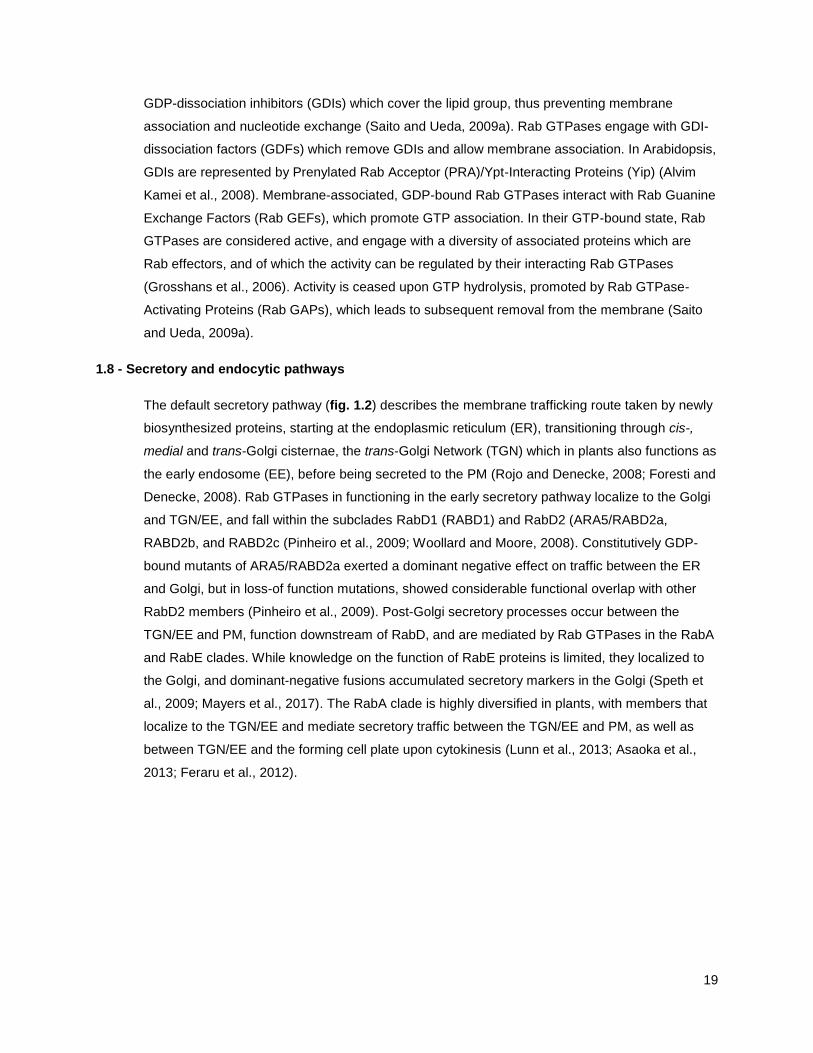
The default secretory pathway (fig. 1.2) describes the membrane trafficking route taken by newly
biosynthesized proteins, starting at the endoplasmic reticulum (ER), transitioning through cis-,
medial and trans-Golgi cisternae, the trans-Golgi Network (TGN) which in plants also functions as
the early endosome (EE), before being secreted to the PM (Rojo and Denecke, 2008; Foresti and
Denecke, 2008). Rab GTPases in functioning in the early secretory pathway localize to the Golgi
and TGN/EE, and fall within the subclades RabD1 (RABD1) and RabD2 (ARA5/RABD2a,
RABD2b, and RABD2c (Pinheiro et al., 2009; Woollard and Moore, 2008). Constitutively GDP-
bound mutants of ARA5/RABD2a exerted a dominant negative effect on traffic between the ER
and Golgi, but in loss-of function mutations, showed considerable functional overlap with other
RabD2 members (Pinheiro et al., 2009). Post-Golgi secretory processes occur between the
TGN/EE and PM, function downstream of RabD, and are mediated by Rab GTPases in the RabA
and RabE clades. While knowledge on the function of RabE proteins is limited, they localized to
the Golgi, and dominant-negative fusions accumulated secretory markers in the Golgi (Speth et
al., 2009; Mayers et al., 2017). The RabA clade is highly diversified in plants, with members that
localize to the TGN/EE and mediate secretory traffic between the TGN/EE and PM, as well as
between TGN/EE and the forming cell plate upon cytokinesis (Lunn et al., 2013; Asaoka et al.,
2013; Feraru et al., 2012).
20
Figure 1.2. Schematic overview of membrane trafficking pathways in plants. Newly synthesized secreted proteins originate at
the endoplasmatic reticulum (ER, lower left), and travel through the Golgi apparatus, encountering cis-, medial- and trans-Golgi
compartments successively. Mediated by RabD and RabA/E-type GTPases, they are transferred into the trans-Golgi network/early
endosome (TGN/EE, middle), from where they are delivered to the plasma membrane (PM) by secretory vesicles (SV). The
TGN/EE consists of a domain, or population, that is VAMP721 and RabA-positive and has secretory activity, as well as a domain or
population that is SYP43, SYP61 and VHA-a1 positive, which exhibits both endocytic and secretory activity. Retrieval of PM-
localized cargoes occurs through clathrin-dependent endocytosis (top right) by means of clathrin-coated vesicles (CCVs), or by
clathrin-independent endocytosis. Endocytosed cargoes traffic through the TGN/EE which, under control of the ESCRT complex,
generates late endosomes/multivesicular bodies (LE/MVB) containing cargo-carrying intraluminal vesicles (ILVs). LE/MVBs
associate with RabF and RabG-type GTPases, which govern its maturation and delivery to the vacuole (lower right), into which ILVs
and their cargoes are released. Arrows indicate trafficking directionality of secreted and endocytosed cargoes.
21
The endocytic pathway (fig. 1.2) originates at the PM, where T-PLATE/clathrin-dependent and
clathrin-independent endocytosis internalize membrane localized cargoes which then accumulate
in the TGN/EE (Paez Valencia et al., 2016). From here, cargoes can recycle back to the PM, or
be targeted to late endosomes (LE)/multivesicular bodies (MVBs), which are thought to mature
from the TGN/EE (Scheuring et al., 2011; Dettmer et al., 2006). LE/MVB maturation is coupled to
the formation of intraluminal vesicles (ILVs) through the action of multimeric ENDOSOMAL
SORTING COMPLEX REQUIRED FOR TRANSPORT (ESCRT) components, which are required
for the endosomal sorting of endocytic cargoes that are targeted to the vacuole for degradation
(Gao et al., 2017; Buono et al., 2017). Known Rab GTPases functioning along the endocytic
pathway belong to the RabF clade with the close homologs RHA1/RABF2a and ARA7/RABF2b
localizing to TGN/EE as well as LE/MVBs (Ueda et al., 2004), the plant-unique RabF protein
ARA6/RABF1 localizing to LE/MVBs (Ueda et al., 2001), and RabG proteins that localize to the
vacuolar membrane (Geldner et al., 2009). During endosomal maturation, LE/MVBs undergo
RabF to RabG conversion, through the action of the LE/MVB localized RabF effectors
SAND/MONENSIN SENSITIVITY 1 (SAND/MON1) and CALCIUM CAFFEINE ZINC
SENSITIVITY 1 (CCZ1), which are bona-fide ARA7 effectors and function in complex as the GEF
for RabG proteins (Cui et al., 2014a; Singh et al., 2014a). Interestingly, it has recently been
shown that the ER actively participates in facilitating endocytic processes. ER-PM-contact site-
localized VAMP-ASSOCIATED PROTEIN 27 (VAP27) proteins associated with clathrin and
endocytic membranes, and positively regulated endocytic activity (Stefano et al., 2018).
Furthermore, LE/MVBs physically associate with the ER, which influences their distribution and
streaming, with overexpression of ER-localized structural proteins that affect ER morphology also
leading to abberant localizations of LE/MVB-dependent endosomal cargoes (Stefano et al.,
2015).
1.9 - Trafficking pathways share the trans-Golgi network/early endosome
Both secretory and endocytic traffic pass through the TGN/EE, yet cargoes of these pathways
can be transported to their destinations in a pathway-specific manner. The TGN/EE is not one
homogenous compartment, but can be subdivided into separate populations based on multiple
criteria (Gendre et al., 2015). Firstly, TGN/EE compartments can mature from trans-Golgi
cisternae, which is supported by the observation of Golgi-associated (GA) and Golgi-independent
(GI) populations of TGN/EE, when marked by the TGN/EE-resident SNARE SYNTAXIN OF
PLANTS 43 (SYP43; (Uemura et al., 2014). In agreement with this, upon chemical disruption,
some TGN/EE compartments recover in a partially Golgi-independent manner, pointing at partialy
Golgi-independent origins, potentially of endocytic origin (Ito et al., 2017). Secondly, two
populations of TGN/EE can be distinguished, based on the presence of RABA2a, RABA1b and
VAMP721 and high amounts of clathrin, which mark a population with secretion and PM-

22
recycling, or the presence of SYP61, SYP43 and VHA-a1, and secretory vesicle clusters, which
mark a populations that is associated with both secretory and endocytic traffic (Gendre et al.,
2015; LaMontagne and Heese, 2017). The precise compartmentalization and dynamics of
TGN/EE are as of yet poorly understood, but recent advances in subcellular fractionation,
proteomics and lipidomics have begun to shed light on mechanisms of cargo sorting at this major
trafficking hub (Wattelet-Boyer et al., 2016).
1.10 - Functions of the secretory pathway in immunity
The secretory pathway plays an important role in delivering PRRs to the cell surface, potentiating
cells for the detection of extracellular patterns. Newly synthesized PRRs first localize to the ER,
where they are processed and folded into their correct structures. This requires the ER quality
control (ERQC) machinery that, when impaired, cause the accumulation of EFR in this organelle
(Farid et al., 2013; Häweker et al., 2010; Li et al., 2009; Lu et al., 2009; Nekrasov et al., 2009).
Consequently, loss-of-function mutants in ERQC components are insensitive to elf18 and show
enhanced susceptibility to bacterial infection (Häweker et al., 2010). PRRs carry a typical N-
terminal signal peptide that directs the receptors for ER export to enter the secretory pathway.
FLS2 associates with ER-resident RETICULON-LIKE PROTEINS GROUP B (RTNLB) 1 and 2,
which in loss-of-function mutants caused FLS2 accumulation at the ER (Lee et al., 2011).
Similarly, Cf-4 interacts with ERQC machinery, and upon genetic interference with the ERQC
components CALRETICULIN 3a (CRT3a) and HSP70-interacting BiP chaperones, Cf-4
accumulated, but was incorrectly glycosylated and affected in its capacity to mount HR upon Avr4
treatment (Liebrand et al., 2012).
A distinct member of the RabA clade, RABA1b has been implicated in transport of FLS2 to the
PM (Choi et al., 2013). Co-expression of dominant-negative (DN) RABA1b and FLS2 in N.
benthamiana significantly reduced FLS2 PM localization, while FLS2 accumulated in small
cytosolic vesicles. As DN-RABA1b expression caused morphological changes to SYP61-marked
TGN/EE, this suggests that RABA1b is involved in transport of FLS2 from the TGN/EE to the PM.
The secretory pathway also responds to activation of immunity. Upon treatment with the MAMPs
flg22 or fungal xylanase, the PM-localized secretory pathway SNARE proteins PEN1/SYP121,
SYP122 and SYP132 are rapidly phosphorylated (Kalde et al., 2007; Benschop et al., 2007).
Furthermore, treatment with the central defense hormone SA leads to transcriptional changes
that depend on the SA-receptor NON-EXPRESSOR OF PATHOGENESIS-RELATED (PR)
GENES 1 (NPR1), and include upregulation of PR-genes which encode for secreted antimicrobial
proteins (Wang et al., 2005). SA and NPR1-dependent transcriptional reprogramming also
includes the upregulation of ERQC components and secretory pathway genes that are required
for the secretion of PR proteins (Wang et al., 2005). SA-induced secretory trafficking also seems

23
to be engaged in PRR delivery to the PM. SA upregulates expression and PM-accumulation of
the PRRs FLS2, EFR and CERK1, as well as BAK1/SERK3 (Tateda et al., 2015). This is
dependent on the ER-localized ACCELERATED CELL DEATH 6 (ACD6), a membrane-
associated protein involved in positive feedback regulation with SA. ACD6 forms complexes with
FLS2 and ER-chaperones, possibly to facilitate the folding and delivery of FLS2 under SA-
inducing conditions (Zhang et al., 2014b).
A client of upregulated secretion upon SA-signaling is PR-1, which is a cysteine-rich protein that
accumulates in the extracellular space to high levels upon pathogen challenge (van Loon, 1975;
Van Loon and Van Strien, 1999). It exerts antimicrobial effects through binding and sequestering
sterols from pathogen membranes (Gamir et al., 2017). Upon fluorescent tagging, it localizes to
ER and Golgi, and is dependent on canonical secretory processes to be exported (Watanabe et
al., 2013; Pečenková et al., 2017). PR-1 secretion is negatively affected by the Golgi-resident
SNARE MEMBRIN-12 (MEMB12), which promotes retrograde Golgi-ER traffic, and is itself
negatively regulated by RNA-interference upon infection by Pto DC3000, supporting a role for the
Golgi in PR-1 secretion and immunity (Zhang et al., 2011b). Post-Golgi secretion of PR-1 requires
the TGN/EE localized ubiquitin E3 ligase KEEP ON GOING (KEG) which when mutated causes
PR-1 to accumulate in the vacuole (Gu and Innes, 2012), and requires the PM-localized SNARE
SYP132, which forms complexes with the TGN/EE-localized secretory SNAREs VESICLE-
ASSOCIATED MEMBRANE PROTEIN 721 (VAMP721) and VAMP722 that are involved in
TGN/EE-PM transport (Kalde et al., 2007).
In addition to potentiating the PM with detection capacity and facilitating the secretion of
antimicrobial proteins, the secretory pathway is involved in the execution of cell-surface based
immune responses. The PM-localized SNARE PEN1/SYP121 engages with VAMP721/722 and
the cytosolic SNARE SNAP33, which have resting state functions in default secretion, to promote
pre-penetration resistance and formation of the callose-rich papilla upon pathogen challenge
(Kwon et al., 2008; Assaad et al., 2004). Furthermore, upon powdery mildew attempted
penetration, formation of the papilla co-incides with focal accumulation of the callose synthase
POWDERY MILDEW RESISTANT 4 (PMR4)/GLUCAN SYNTHASE-LIKE 5 (GSL5), which is
necessary for pathogen-induced callose deposition (Ellinger et al., 2013). Interestingly,
overexpression of the secretory Rab GTPase RABA4c results in enhanced PMR4-dependent
callose deposition and resistance to the adapted powdery mildew Golovinomyces oronti (Ellinger
et al., 2014a). RABA4c and PMR4 interact in vivo, and because PMR4-dependent enhanced
callose deposition depends on the activation status of RABA4c, PMR4 is considered to be its Rab
effector. Upon infection with Pto DC3000, Golgi and PM-localized RABE1d focally accumulated at
the cell periphery, suggesting the plant engages in the cell-surface concentration of secretory
processes also in response to bacteria (Speth et al., 2009). Indeed, expression of a permanently

24
GTP-bound constitutive-active (CA) RABE1d conferred increased resistance against Pto
DC3000, coupled with enhanced secretion of PR-1.
1.11 - Functions of the endocytic pathway in immunity
Internalization and late endosomal sorting, which pass cargoes from the PM through the TGN/EE
and LE/MVB, also contribute to plant immunity. Mutants affected in genes coding for the
endocytic coat protein CLATHRIN HEAVY CHAIN (CHC) and dynamin-related proteins (DRPs),
which are homologs of mammalian dynamins that mediate vesicle scission, are impaired in
endocytic uptake, and show enhanced susceptibility to Pto DC3000 (Collings et al., 2008; Smith
et al., 2014; Mbengue et al., 2016). Similarly, infection success of Pto DC3000, as well as the
adapted oomycete pathogen Hyaloperonospora arabidopsidis (Hpa) is enhanced in vps28-2 and
vps37-1, which are loss-of function mutants in ESCRT subunits that mediate cargo sorting into
LE/MVBs (Lu et al., 2012; Spallek et al., 2013; Scheuring et al., 2011). Thus, endocytic uptake
and correct endosomal cargo sorting are required to mount a successful defense response.
Upon ligand stimulation, cell-surface lcoalized PRRs such as FLS2, EFR and PEPR1 translocate
into endosomes in a clathrin-dependent manner (Ortiz-Morea et al., 2016; Mbengue et al., 2016;
Robatzek et al., 2006). At resting state, FLS2 recycles between the PM and TGN/EE as
evidenced by chemical inhibition of TGN/EE-PM transport using Brefeldin-A (Beck et al., 2012b).
Upon activation, FLS2 is sorted into LE/MVBs in a BAK1/SERK3-dependent manner, as
evidenced by co-localization with FM4-64 and endosomal Rab GTPases ARA7 and ARA6 (Beck
et al., 2012b). Endosomal accumulation of FLS2 occurs in a transient manner, with a maximum of
FLS2 endosomes at ca. 1 h after activation, and is thought to underlie its vacuolar delivery and
subsequent degradation (Beck et al., 2012b; Choi et al., 2013).
Similarly, the tomato RLP EIX2 resides at the PM, and after activation with xylanase, it shows
increased localization at endosomes that are marked by the LE/MVB marker FYVE (Sharfman et
al., 2011). In agreement, chemical interference of late endosomal trafficking and disruption of the
actin cytoskeleton reduces EIX2 endocytosis (Bar and Avni, 2009a). EPS15 homology domain 2
(EHD2) has been implicated in decreasing the bundling of actin filaments and impairs
endocytosis of EIX2 upon overexpression (Bar and Avni, 2009a, 2009b). This correlates with a
reduction in xylanase-triggered HR, ethylene production and PR1 gene expression, and is
consistent with reduced xylanase-induced HR upon chemical disruption of actin (Bar and Avni,
2009a). Interestingly, although actin is required for endocytosis of activated FLS2, EHD2 does not
seem to affect flg22-induced PR1 gene expression, but can interfere with Cf-4 and Cf-9 mediated
HR (Bar and Avni, 2009b). However, whether EHD2 also inhibits Cf-4 endocytosis, and not FLS2
endocytosis, remains to be demonstrated. This could be related to EHD2 associating with RLPs
but not RLKs, as there is evidence that EHD2 interacts with SlEix2 through its coiled-coil domain.

25
1.12 - Connections between the endocytic pathway and cell-surface delivery
Initial contact between the pathogen and the plant cell is associated with the recruitment and
focal accumulation of diverse subcellular components. These responses are especially well-
studied in the context of infection by filamentous pathogens, and include the formation to a
callose-rich papilla (Voigt, 2014), local immobilization of mitochondria, peroxisomes, clustering of
ER and accumulation of endomembrane compartments of endocytic nature (Nielsen et al., 2012;
Griffis et al., 2014; Fuchs et al., 2015b; Nielsen et al., 2017b). Indeed, in electron microscopy
studies, LE/MVBs have been shown to fuse with the PM under pathogen contact sites, which is
thought to be coupled to the release of ILVs which are then considered extracellular vesicles
(EVs; (An et al., 2006).
In concert with this, upon attempted penetration, the plant upregulates the local production of
toxic metabolites, among which those derived from indole-3-glucosinolate precursors (Bednarek
et al., 2009; Clay et al., 2009). Enzymes functioning early in the pathway that converts indole-3-
glucosinolates such as those belonging to the CYTOCHROME P81 (CYP81) family have been
observed at the ER, while final conversion steps are mediated by myrosinases among which is
PENETRATION 2 (PEN2) that localizes to mitochondria and peroxisomes (Fuchs et al., 2015b).
The PM-localized ABC-transporter PEN3 focally accumulates at the site of attempted penetration,
and is thought to export the toxic metabolites thus produced, concentrating chemical defenses at
the biologically relevant location (Stein et al., 2006; Underwood and Somerville, 2008, 2013).
PEN3 focal accumulation is thought to be mediated by endocytic uptake from the PM, and fusion
of LE/MVBs at the cell surface in an unconventional secretion process, but conclusive evidence
for this is lacking (Underwood et al., 2017). In addition, the PM-localized SNARE PEN1/SYP121
focally accumulates, and is found in the extracellular space at fungal invasion sites, possibly
mediated by LE/MVB redirection as well (Assaad et al., 2004; Nielsen et al., 2012). Yet, LE/MVB-
dependent extracellular accumulation of PEN1 is not thought to be required for its biological
function. While a PEN1 function in regulating the dynamics of callose deposition, which is
underpinned by default secretory processes, has been demonstrated (Assaad et al., 2004), it can
form complexes with the LE/MVB-localized snare VAMP727, suggesting potential involvement in
unconventional secretion (Ebine et al., 2012).
PEN3 focal accumulation is insensitive to chemical disruption of TGN/EE-dependent recycling
and default secretory processes (Underwood and Somerville, 2013), providing further evidence
for its intermediary translocation into LE/MVBs before arriving at the PM. This echoes the
LE/MVB sorting of FLS2 upon activation, which also did not depend on TGN/EE-based secretion
or recycling as tested upon chemical disruption (Beck et al., 2012b). Interestingly, PEN3 focal
accumulation can be triggered in the absence of pathogens upon MAMP treatment with flg22 and
chitin, independent of protein biosynthesis (Underwood and Somerville, 2013). Using fluorescent

26
fusions, PEN3 can be observed to accumulate at endosomal compartments at time points
preceding its cell-surface accumulation (Underwood et al., 2017).
1.13 - Involvement of Rab GTPases in pathogen-targeted traffic
While both secretory and endocytic traffic can participate in the delivery of defence components
to the PM and extracellular space, the roles for Rab GTPases on these trafficking pathways are
poorly understood. Yet, Rab GTPases accumulate at the pathogen interface, suggesting their
involvement. ARA6-positive LE/MVBs accumulate under attempted penetration sites, and around
haustoria of the powdery mildew Blumeria graminis f.sp. hordei (Bgh; (Nielsen et al., 2012).
Interestingly, at resting state, ARA6 has been shown to induce SNARE-complex formation
between the LE/MVB-localized VAMP727 and PM-localized SYP121/PEN1, pointing at a non-
canonical role for this plant-unique Rab GTPase (Ebine et al., 2011b).
Upon penetration, haustoria of fungal G. orontii and the oomycete pathogen Phytophthora
infestans are surrounded by both ARA7 and ARA6, and LE/MVB-dependent cargoes such as
activated FLS2 accumulate in the extrahaustorial matrix of P. infestans (Inada et al., 2016;
Bozkurt et al., 2015). Furthermore, RABG3c localized around P. infestans haustoria (Bozkurt et
al., 2015). RabG clade proteins are normally localized to the vacuolar membrane, and are
involved in vacuolar delivery of RabF compartments (Geldner et al., 2009; Singh et al., 2014b).
Taken together, these data suggest that Rab-GTPase dependent cargo delivery occurs at
pathogen interfaces.
All RabF GTPases, RHA1, ARA7 and ARA6, share a common activator in the Rab GEF
VACUOLAR PROTEIN SORTING 9a (VPS9a; (Goh et al., 2007). ARA7 and ARA6 localize to the
host-derived extrahaustorial membrane (EHM) of the adapted mildew G. orontii, but VPS9a is
conspicuously excluded from this location (Inada et al., 2016). This suggests pathogen-mediated
manipulation of EHM composition, and could suggest active interference with Rab GTPase-
mediated processes through targeting their regulatory complex. Indeed, G. orontii effector
candidates have been predicted to interact with PRA1 Rab GTPase-regulatory proteins (Mukhtar
et al., 2011; Weßling et al., 2014). In studies using the nonadapted Bgh on Arabidopsis, VPS9a
mutants showed an increased penetration success, as well as decreased callose apposition at
pathogen interfaces (Nielsen et al., 2017b). It is therefore thought that VPS9a/RabF pathways
contribute to both pre- and postpenetration resistance to nonadapted pathogens.
LE/MVBs have been proposed to generate EVs through cell-surface delivery and secretion of
ILVs, based on electron microscopy studies (An et al., 2006). This hypothesis is further supported
by the observation that Pto DC3000-infection stimulates the biogenesis of LE/MVBs and the
occurrence of EV-like vesicles in the paramural space, which was dependent on regulators of
LE/MVB biosynthesis (Wang et al., 2014, 2015). Correspondingly, apoplastic purifications of

27
Arabidopsis contained EVs which share high proteomic identity to published LE/MVB proteomes
(Rutter and Innes, 2017b). Their occurrence is upregulated upon both SA treatment and infection
with Pto DC3000, and they carry biotic-stress related proteins such as PEN1/SYP121, PEN3 and
indole-3-glucosinolate metabolic enzymes (Rutter and Innes, 2017b). More recently, in studies
using the necrotrophic fungus Botrytis cinerea, ARA6 clusters were observed under attempted
penetration sites, coupled to the increased delivery of EVs that were shown to carry micro-RNAs
with fungal targets, pointing at a role for EVs in trans-kingdom delivery of plant-borne defense
components which thus accumulate inside the pathogen (Cai et al., 2018). While taken together,
the above observations point at critical roles for endosomal Rab GTPase-regulated trafficking
pathways in executing defense, it remains to be shown how changes in Rab GTPase activation
status and subsequent activation of their downstream Rab-effectors contribute to immunity.
1.14 - Trafficking is subject to effector manipulation and host surveillance
Because trafficking processes are of fundamental importance to the preparation and execution of
plant immunity, to pathogens that seek to circumvent host defenses they would make great
effector targets. Indeed, large scale yeast-2-hybrid (Y2H) screens that probed Arabidopsis host
protein interactions with a panel of G. orontii, Hpa and Pto DC3000 effectors have yielded
predicted interactions between pathogen effectors and trafficking regulators that include coat
proteins, Rab GTPase regulatory proteins, motor proteins and cytoskeletal elements in both
secretory and endocytic traffic (Mukhtar et al., 2011; Weßling et al., 2014). Furthermore,
individual effectors have been specifically shown to target trafficking components during infection,
such as Phytophthora AVR3a which targets dynamin-dependent endocytic processes and can
accumulate at host endosomes (Chaparro-Garcia et al., 2015; Engelhardt et al., 2012), AVR1
which targets secretory EXOCYST tethering factors (Du et al., 2015), and AvrBlb2, which inhibits
the secretion of host-borne proteases and thus confers increased virulence (Bozkurt et al., 2011).
Similarly, Pseudomonas effectors of unknown composition suppress PEN3 focal accumulation,
and HopM1 targets the TGN/EE localized trafficking regulator HOPM1 INTERACTOR
7/BREFELDIN-A VISUALISED ENDOCYTIC TRAFFICKING DEFECTIVE 1 (MIN7/BEN1),
suppresses secretion of PR-1 and callose deposition, and thus confers enhanced bacterial
virulence (Nomura et al., 2006, 2011).
Conversely, in plants, connections between trafficking processes and NLR-mediated immunity
exist. Interference with subcellular trafficking processes can result in cell-death associated
defences. Firstly, the pen1/syp121 syp122 double mutant of closely homologous SNAREs
exhibits a lesion-mimic phenotype with programmed cell death (Zhang et al., 2008). This can be
rescued by mutations in NLR genes. Secondly, the HR-like cell death in the accelerated cell
death 11 (acd11) mutant, and HR induced by NLR-mediated immunity, are suppressed by loss-
of-function in LAZARUS 1 (LAZ1), whose protein partially localizes to endosomes (Malinovsky et

28
al., 2010). Thirdly, NLR-associated immunity prevents degradation of MIN7/BEN1 by HopM1.
Finally, the secretory tethering factor EXO70B1, interacts with SNARE-protein SNAP33 and the
TN-type NLR TIR-NBS2 (TN2) in planta. Exo70b1 mutants exhibit a sponteneous HR-like cell
death, and an increased resistance to powdery mildews, both phenotypes fully dependent on TN2
(Zhao et al., 2015; Liu et al., 2017). This suggests that EXO70B1 or a dependent pathway is
guarded by TN2. These observations support the concept that molecular components that
regulate subcellular transport are monitored by NLRs and thus, when absent, trigger the induction
of strong defense responses. It is noteworthy that NLR signaling increases SA levels, and SA in
turn exerts regulation on secretory and endocytic trafficking processes (Du et al., 2013; Wang et
al., 2005).
1.15 - Concluding remarks
Taken together, plant subcellular trafficking is interlinked with the immune system, and
contributes to host defenses at multiple stages of pathogen infection. This includes building pre-
existing cell surface barriers, maintaining cell surface-based detection capacity, and employing
transport pathways to recruit defense components to regions of pathogen contact in a timely
manner. Pathogens deliver effectors to manipulate the underlying trafficking pathways, and plants
employ sensors to monitor their integrity. Rab GTPases localize and function on these pathways,
and are required for full immunity, yet their precise contributions to defense are poorly
understood, which warrants further investigation.
1.16 - Thesis aims
Signal initiation of RLK-type PRRs that detect MAMPs in Arabidopsis is well understood, and
involves ligand-induced heterodimerization with BAK1/SERK3, activation of the PM-localized
receptor complex, and subsequent ligand-induced internalization of the PRR which is then sorted
for degradation through RabF-positive compartments (Ben Khaled et al., 2015). Cf-4 represents
an RLP-type PRR that functions in effector-detection in tomato, and unlike RLK-type PRRs,
besides genetic evidence for a role of SERK1, no direct involvement in Cf-mediated immunity of
SERK members has been reported to date (Fradin et al., 2011). Instead, Cf-RLPs interact with
the RLK SOBIR1 for stability and signaling capacity, independent of the presence of their ligands
(Liebrand et al., 2013). C. fulvum infection is confined to the apoplast, and correspondingly,
secretes effectors that accumulate there (Joosten and de Wit, 1999; Stergiopoulos and de Wit,
2009). This suggests that Cf proteins function at the PM, but their exact subcellular localization
has remained unclear.

29
In order to elucidate the subcellular localization and signal activation mechanisms of SOBIR1-
dependent RLPs, I aimed to:
Reveal the subcellular localization of Cf-4
Reveal the subcellular localization of constitutively interacting Cf-4-SOBIR1 complexes
Probe for ligand-induced subcellular localization dynamics of Cf-4 upon activation by
Avr4
Investigate the requirements for SERK members in Cf-dependent receptor outputs and
immunity
Secondly, besides carrying ligand-activated PRRs, endosomal Rab GTPases and associated
compartments dynamically respond to pathogen infection and contribute to the delivery of
defense cargoes at pathogen contact sites (Cai et al., 2018; Nielsen et al., 2017b). Equally, Rab
GTPases along the secretory pathway have been shown to govern delivery and activation of cell-
surface localized defense cargoes (Ellinger et al., 2014b; Choi et al., 2013). Yet, most studies
have been performed in the context of infection by filamentous pathogens, and defence-induced
cargo changes along Rab GTPase mediated pathways, or changes in proteins associating with
the Rab GTPases themselves have not been mapped in a comprehensive way.
In order to gain insight into the dynamics of the secretory pathway marked by ARA5 and the
endocytic pathway marked by ARA7 upon bacterial infection, as well as into MAMP-induced
proteomic changes in the proteomes interacting with these Rab GTPases, I sought to:
Investigate localization changes of ARA7 and ARA5 upon bacterial infection
Reveal changes in ARA7 and ARA5-associated proteins after flg22-stimulus
Identify flg22-responsive ARA7-interacting proteins which are defence-related
Further characterize a subset of flg22-responsive ARA7 interacting defence-
related proteins for roles in immunity

30
CHAPTER 2
EXPERIMENTAL PROCEDURES
2.1 - Plant materials
2.1.1 - Plant lines used in this study
2.1.2 - Growth of Arabidopsis on soil
Arabidopsis thaliana seeds were germinated on soil (Levington F2 peat, 4 mm grit,
Exemptor insecticide) and grown under 80 % humidity and 20 °C. Seedlings were
transplanted to individual 4 cm x 4 cm pots. For plants used in experiments, a short-day
regime of 10 hours light / 14 hours dark was used. For propagation and crossings, a long-
day regime of 16 hours light / 8 hours dark was used. (Dettmer et al., 2006)
Species Genotype Source
Arabidopsis thaliana Col-0
Col-0 pUB::YFP-ARA5 Geldner et al., 2009
Col-0 pUB::YFP-ARA7 Geldner et al., 2009
Col-0 pUB::RFP-ARA7 Dettmer et al., 2006
Col-0
pUB::YFP-ARA5 x pUB::RFP-ARA7
Generated, cross
Col-0 fls2 Zipfel et al., 2004
Col-0 tn3 SALK_018440.56.00
Col-0 hr4 SALK_208828
Col-0 tn3 / pUB::TN3-GFP Generated, transformation
Col-0 hr4 / pUB::HR4-GFP Generated, transformation
Col-0 pUB::TN3-GFP Generated, transformation
Col-0 pUB::HR4-GFP Generated, transformation
Nicotiana tabacum Samsun
Nicotiana benthamiana Domin.
Domin. p35S::Cf-4 Liebrand et al., 2013
Domin. p35S::Cf-4-GFP Postma et al., 2016
Solanum lycopersicum Moneymaker Cf-0 Dixon et al., 1996
Moneymaker Cf-4 Wulff et al., 2004

31
2.1.3 - Growth of Nicotiana benthamiana on soil
Nicotiana benthamiana Domin. seeds were germinated on soil (Levington F2 peat, 4 mm
grit) and kept under plastic foil at high humidity for 1 week. Seedlings were transplanted
to individual 9 cm x 9 cm pots and grown under 80% humidity with a regime of 16 hours
light / 8 hours dark. Plants were used at 2 weeks for virus-induced gene silencing, or 4-5
weeks for all other purposes.
2.1.4 - Growth of Arabidopsis in liquid media
For gas sterilisation, ca. 50 mg of Arabidopsis seeds in a paper bag were placed in a
sealable chamber with a volume of ca. 10 L, alongside a beaker containing 200 mL
commercial thick bleach (Parozone) to which 8 mL hydrochloric acid was added. The
chamber was sealed and left in a fume hood overnight, after which the seed bag was
retrieved and placed in a laminar flow hood to dry.
Under laminar flow, seeds were sown in resealable conical flasks containing 200 mL
liquid MS medium supplemented with 1% (w/v) sucrose, without antibiotic selection.
Flasks with seeds were stored at 4 °C overnight for vernalization, and then placed in a
120 rpm shaker for an incubation time of 8 days, growing at 22 °C under a light regime of
16 hours light / 8 hours dark. (Dixon et al., 1996; Wulff et al., 2004; Zipfel et al., 2004)
2.1.5 - Generation of Arabidopsis protoplasts
Protoplasts were obtained from soil-grown plants at ca. 4-5 weeks of age using the tape
sandwich method based on Sheen et al., 2002 . Fully grown leaves were sandwiched in
between strips of masking tape, and the lower epidermis was removed by separating the
tape. Tape strips containing the leaves were then placed face-down into a petri dish
containing enzyme solution (1.5% cellulase (Onuzuka R10, Yakult BIC2224), 0.4%
macerozyme (R10, Apollo Scientific cat. BIM0481), 0.1% BSA (Sigma cat. A-6793), 0.4
M mannitol, 20 mM KCl, 20 mM MES pH 5.7, 10 mM CaCl2) and the digestion was
allowed to continue for 1.5 hours at room temperature. Protoplasts were recovered by
filtration through 40 mm nylon mesh, centrifugation at 100 x g for 2 min at 4 °C and
resuspension in W5 buffer (154 mM NaCl, 125 mM CaCl2, 5 mM KCl, 2 mM MES pH
5.7).
2.1.6 - Crossing of Arabidopsis
Arabidopsis plants were grown in long-day conditions for 5-8 weeks until inflorescences
started appearing. Mature flowers with fully developed anthers on donor inflorescences of
both parents were opened using tweezers. Closed buds on recipient inflorescences of

32
both parents were also opened using tweezers. Cross-pollination was achieved by
separating the donor flower from the inflorescence using tweezers, and physically
rubbing it onto the opened recipient flower for several seconds. Pollinated buds were
marked using colored tape and plants were returned to long-day conditions. Seeds were
harvested from pollinated siliques, and selected by germination on MS media with
appropriate antibiotics.
2.2 - Transformation and transfection systems
2.2.1 - Generation of Arabidopsis transgenic plants
Arabidopsis was transformed using the floral dipping method (Bent, 2006) using
Agrobacterium tumefaciens strain GV3101. Transformations were performed by Matthew
Smoker and Jodie Pike (The Sainsbury Laboratory, Norwich, UK). Transformants were
selected by germination on soil, growing for 1 week under short day conditions and spray
application of phosphinothricin (PPT)/BASTA herbicide (Melford cat. P0159). At 2 weeks
of age, survivors were transplanted into invidual 4 cm x 4 cm pots and recovered for 1
additional week. Confocal microscopy was used to confirm expression of fluorescent
proteins, and T1 plants were used for experiments and propagation.
2.2.2 - Transfection of Arabidopsis by particle bombardment
Adult leaf tissue was subjected to particle bombardment for transient expression of
fluorescent tagged proteins. Purified plasmid DNA (2 µg) was coated onto 125 µg gold
particles ( = 1.0 µm) using 1 M CaCl2 and 20 mM spermidine, and washed with 100 %
ethanol. Three separated Arabidopsis leaves were placed on solid Murashige and Skoog
(MS) medium adaxial side up, and petioles making contact with the medium. A
pressurized helium-based particle delivery system (Bio-Rad Biolistics PDS-1000) was
used to deliver gold particles into distal leaf tissue at optimized pressure (1100 or 900
psi) and height. Transfected leaves on MS were incubated on a bench top at room
temperature overnight, and a scalpel was used to prepare tissue for microscopic
analysis.
2.2.3 - Arabidopsis protoplast transfection
Protoplasts were transfected using poly-ethylene glycol (PEG). Briefly, 100-150 µg
plasmid DNA was added to a 2 mL suspension of ca. 5 x 10⁵ protoplasts mL
-1 in MMg
buffer (4 mM MES pH 5.7, 0.4 M mannitol, 15 mM MgCl2). Then, 2 mL PEG buffer (40%
v/v PEG4000 (Sigma cat. 81240), 0.4 M mannitol, 0.2 M CaCl2) was mixed in, and the
transfection was allowed to continue for 10 minutes at room temperature. After one wash

33
with excess W5 buffer, pellets were resuspended in 2 mL W5 buffer and kept at room
temperature overnight. On day 2, protoplasts were used for confocal microscopy or
excess liquid was removed and protoplasts were flash-frozen in liquid nitrogen in
preparation for protein extraction.
2.2.4 - Agrobacterium tumefaciens mediated transformation of N. benthamiana
A. tumefaciens strains GV3101 and GV3101 carrying the helper plasmid PMP90RK were
grown for 2-3 days under appropriate selection at 28 °C in 10 mL liquid L-medium
shaking at 120 rpm, or on solid L-medium 1 % agarose plates without shaking. Liquid
cultures were spun down at 3000 x g for 5 minutes, and pellets were resuspended in
dH2O. From plates, bacteria were harvested using a spatula and resuspended in distilled
water (dH2O). Bacteria were washed 1 x with dH2O and dilutions were adjusted to OD600
= 0.3 for each strain, in preparation for (co-)infiltration. Acetosyringone was added to a
final dilution of 250 µM, and bacterial suspensions were syringe-infiltrated in N.
benthamiana leaves.
2.2.5 - Virus-induced gene silencing in N. benthamiana
Tobacco rattle virus (TRV)-mediated VIGS in N. benthamiana was performed as follows.
A. tumefaciens GV3101 carrying TRV-RNA1 (pYL155) at OD600 = 0.4 and GV3101
carrying TRV-RNA2 (pYL279) containing the respective target sequence at OD600 = 0.2
were co-infiltrated in leaves of 2-week old N. benthamiana plants. TRV-RNA1 alone, or
TRV-RNA2::GUS (Tameling and Baulcombe, 2007) was used as a control. At 4-5 weeks
of age, plants were used for further analysis.

34
2.3 - DNA techniques and molecular cloning
2.3.1 - Primers used in this study
2.3.2 – Plasmids used in this study
(see next page)
Name Sequence nt Purpose
Cf-9-TOPO-F CACCATGGATTGTGTAAAACTTGTA 25 Cf-9 TOPO cloning
Cf-9-nostop-R ATATCTTTTCTTGTGCTTTTTC 22
BAK1-TOPO-F CACCATGGAACGAAGATTAATGATC 25 BAK1 TOPO cloning
BAK1-TOPO-R TTATCTTGGACCCGAGGGGTATTC 24
TN3-Ftopo CACCATGGCTGCATCTACTTCTTCT 25 AT1G66090 / TIR-NBS 3 cloning
TN3-Rnostop TACAAAATCAGCCAGAGACAC 21
HR4-Ftopo CACCATGCCGATTGCTGAGCTTGCT 25 AT3G50480 / HR4 cloning
HR4-Rnostop TTTATTATGCTTTTCAGATAT 21
FLOT1a-CDNA-F TTGGGCGAAAAGTCCAAATTAAATCAAA 28 FLOT1 cDNA-specific amplification
FLOT1a-CDNA-R CACAAGTAAACGACCAAACCATGCCACT 28
FLOT-Ftopo CACCATGTTCAAAGTTGCAAGAGCG 25 AT5G25250 / FLOT1 cloning
FLOT-Rstop TTAGCTGCGAGTCACTTGCTTC 22
PUB-GW-F TGATTACGAATTCGAGCTCGG 21 amplify cassette from pUB-DEST
PUB-GW-R GGCCCGACGTCGCATGCCTGC 21
F-LP GAAACTTCGATTGGGGAAGAC 23 SALK_018440.56.00.x (tn3) g.typing
F-RP GTGGCAAAGCTTATGGTGAAG 23
J-LP2 GGGATCGAATCGGTAAGCTC 20 SALK_208828 (hr4) g.typing
J-RP2 GGCTTAGATGATGGGCCTTA 20
YFPn-F GTGACCACCCTGGGCTACGG 20 sequencing pAM-PAT YFPc/n fusion
YFPc-F CATTTGGAGAAGGACCTCGAG 21
YFPn-R GGCGGACTTGAAGAAGTCGT 20
YFPc-R GTCACGAACTCCAGCAGGAC 20
M13-F GTAAAACGACGGCCAG 16 sequencing pENTR and pGEM-T
M13-R CAGGAAACAGCTATGAC 17 GFP-R CGAGGGTGGGCCAGGGCACGGG 22 sequencing binary vectors
RFP-R CAGCTTGATGTCGGTCTTGTAGGCG 25 sequencing binary vectors

35
Name Vector Origin
p35S:Cf-4-eGFP pBIN-KS Liebrand et al., 2012
p35S:SlSOBIR1-eGFP pBIN-KS Liebrand et al., 2013
p35S:SlSOBIR1-like-eGFP pBIN-KS Liebrand et al., 2013
p35S:AtSOBIR1-eGFP pBIN-KS Liebrand et al., 2013
p35S:SlSOBIR1-mCherry pAM-PAT Postma et al., 2016
p35S:AtSOBIR1-mCherry pAM-PAT Postma 2016 NPhyt
p35S:SlSOBIR1-HA pGWB14 Postma 2016 NPhyt
p35S:AtSOBIR1-10Myc pGWB20 Liebrand et al., 2013
p35S:AtSOBIR1-RD/N-10Myc pGWB20 Liebrand et al., 2013
p35S:SlSERK3-10Myc pGWB20 Postma et al., 2016
pFLS2:FLS2-GFP pCAMBIA2300 Robatezek et al., 2006
pUB:BAK1-KD pUB-GW Postma et al., 2016
pUB:TN3-eGFP pUB-GW-eGFP Present study
pUB:TN3-mRFP pUB-GW-mRFP Present study
pGEM-T-pUB:TN3-eGFP pGEM-T Easy Present study
pUB:HR4-eGFP pUB-GW-eGFP Present study
pUB:eGFP-FLOT1 pUB-eGFP-GW Present study
p35S:Avr4 pMOG1048 van der Hoorn et al. 2000
p35S:YFPc-ARA7/RABF2b pAM-PAT Present study
p35S:YFPn-ARA7/RABF2b pAM-PAT Present study
p35S:TN3-YFPc pAM-PAT Present study
p35S:TN3-YFPn pAM-PAT Present study
p35S:ARA6/RabF1-YFPc pAM-PAT Present study
p35S:YFPc-ARA5/RABD2a pAM-PAT Michaela Kopischke
p35S:YFPc-RABG3b pAM-PAT Michaela Kopischke
p35S:Cf-4-YFPc pAM-PAT Postma et al., 2016
p35S:Cf-4-YFPn pAM-PAT Postma et al., 2016
p35S:SlSOBIR1-YFPc pAM-PAT Postma et al., 2016
p35S:SlSOBIR1-YFPn pAM-PAT Postma et al., 2016
p35S:SlSOBIR1-like-YFPc pAM-PAT Postma et al., 2016
p35S:SlSOBIR1-like-YFPn pAM-PAT Postma et al., 2016
p35S:AtSOBIR1-YFPc pAM-PAT Postma et al., 2016
p35S:AtSOBIR1-YFPn pAM-PAT Postma et al., 2016
p35S:FLS2-YFPc pAM-PAT Frei dit Frey et al., 2012
p35S:FLS2-YFPn pAM-PAT Frei dit Frey et al., 2012
pUB:mCherry-ARA7/RabF2b pNIGEL Geldner et al., 2009
pUB:mCherry-MEMB12 pNIGEL Geldner et al., 2009
pUB:mCherry-ARA5/RABD2a pNIGEL Geldner et al., 2009
pUB:eYFP-ARA5/RABD2a pNIGEL Geldner et al., 2009
p35S:ARA6/RABF1-RFP pHTS13 Ueda et al., 2004
pUB:VHAa1-RFP pUBQ10 Dettmer et al., 2006
p35S:ACA8-mCherry Postma et al., 2016
Px-RK (peroxisome) pBIN20 Nelson et al., 2007
Mt-RK (mitochondrion) pBIN20 Nelson et al., 2007
RxD460V pB1 Bendahmane et al., 2002
BAX p30B-TMV Lacombe et al., 1999
TRV-RNA2:GUS pyl156-TRV2 Temeling et al., 2007
TRV-RNA2:Cf-4 pyl156-TRV2 Liebrand et al., 2013
TRV-RNA2:NbSOBIR1/NbSOBIR1-like pyl156-TRV2 Liebrand et al., 2012
TRV-RNA1 pyl156-TRV1 Liu et al., 2007
TRV-RNA2:NbSERK3a/b pyl156-TRV2 Heese et al., 2007

36
2.3.3 - DNA extraction for Arabidopsis genotyping
Genomic DNA (gDNA) extractions were performed using a rough preparation method.
From each plant at ca. 2-3 weeks of age, a leaf disc of ca. 0.25 cm2 was obtained using
cork borer. Tissue was submerged in 400 µL pre-heated extraction buffer (200 mM Tris
pH 7.5, 250 mM NaCl, 25 mM EDTA, 0.5% SDS) and incubated at 65 °C for 20-30
minutes. Debris was removed by centrifugation at 16.000 x g for 10 minutes, and
supernatant was transferred to a new tube. Equal volumes of pre-cooled isopropanol
were added and incubated for 5 minutes at room temperature. Precipitated gDNA was
pelleted by centrifugation at 16.000 x g for 15 minutes. gDNA was washed once with 70%
EtOH and resuspended in water.
2.3.4 - Polymerase Chain Reaction for Arabidopsis genotyping
Polymerase chain reactions (PCR) were performed on newly acquired SALK mutant lines
to test for the presence and homozygosity of T-DNA insertions. 20 µL Taq polymerase
reactions were set up, containing 2 µL of gDNA samples prepared as above. Per mutant
plant, two reactions were set up. Forward and reverse primers flanking the predicted T-
DNA insertion site were used to test for the presence of the wildtype sequence. To test
for the presence of the SALK pBIN-pROK2 T-DNA insertion, insertion-specific primer
LBb1.3 was used in combination with a forward or reverse flanking primer, depending on
the predicted orientation of the T-DNA insertion. PCRs were ran for 32 cycles with an
elongation time of 2 minutes, and 10 µL of the samples was used for ethidium bromide
agarose gel electrophoresis.
2.3.5 - RNA extraction and cDNA-preparation
Arabidopsis and N. benthamiana RNA was extracted using the RNeasy Plant Mini kit
(QiaGen cat. 74904) and eluted in 50 µL RNAse-free water. In both cases, ca. 1 cm2
tissue was used, and Arabidopsis was treated with flg22 by leaf infiltration 3 hours prior to
extraction. DNA was digested using a Turbo DNA-free kit (Ambion cat. AM1907), and
RNA concentrations were measured using a NanoDrop 8000 spectrophotometer
(ThermoFisher cat. ND-8000-GL) and up to 2 µg per reaction was used for cDNA
synthesis using SuperScript III reverse transcriptase (ThermoFisher cat. 18080093) in the
presence of RNAse Out (ThermoFisher cat. 10777019). cDNA was stored at -20 °C.
2.3.6 - Cloning and colony PCR
PCR was used to clone gene fragments using Phusion High Fidelity polymerase (New
England Biolabs cat. M0530S), or to test the presence or absence of sequences using

37
Taq polymerase produced in the lab by heterologous expression in E. coli. Reactions
contained 1x Phusion Buffer or Taq buffer (New England Biolabs cat. M0273S), 0.5 mM
dNTP, 5 U/µL Phusion polymerase, 0.1 volume of lab-produced Taq polymerase, 10 µM
primer DNA, and a variable concentration of input DNA. Reactions were performed in 200
µL PCR strips in a thermal cycler (G-Storm cat. GS4) with a default cycle number of 30
and annealing temperature of 56 °C, both subject to optimization for each experiment.
Cloning (Phusion) and genotyping (Taq) reactions were set up to run 5 initial cycles with
a lower annealing temperature of 50 °C. Colony PCRs (cPCR), on bacterial cells picked
from colonies with a pipette tip, were preceded by a 5 minutes 90 °C melting step to
release template DNA into the reaction.
2.3.7 - Gateway entry cloning
Desired gene fragments were amplified using Phusion PCR from either cDNA, existing
expression vectors, or bacterial cells carrying the template DNA. Forward primers were
extended with the CACC sequence at the 5‘ end to allow directional cloning. Reverse
primers omitted the stop codon when amplifying fragments for C-terminal fluorescent
protein tagging. 50 µL Phusion PCRs were performed and 20-30 µL of the product was
used for gel electrophoresis. Bands of the expected size were identified, excised using a
scalpel and purified using a NucleoSpin gel and PCR clean-up kit (Macherey Nagel cat.
740609.50).
1-4 µL of pure PCR product was mixed with 1 µL pENTR/d-TOPO vector (Invitrogen
K2400-20) and incubated for 1 hour at room temperature. The full ligation reaction was
added to 30 µL chemocompetent E. coli DH5α cells, and heat-shock transformation was
performed for 1 min at 42 °C. cPCR-identified positive colonies were grown in liquid
culture, plasmids were purified using a NucleoSpin Plasmid Miniprep kit (Macherey Nagel
cat. 740588.50) and sent for sequencing (GATC Biotech) with the universal M13F/R
primers. Detected sequences were compared to expected sequences using web-hosted
Clustal Omega Multiple Sequence Alignment (EMBL-EBI).
2.3.8 - Gateway binary vector generation
To transfer coding sequences from pENTR/d-TOPO into destination vectors, Gateway LR
reactions were performed with LR Clonase II Enzyme mix (ThermoFisher cat. 11791020).
Enzyme mix was thawed on ice from -80 °C, spun down briefly at 10.000 x g, and used in
a downscaled LR reaction with 50-100 ng entry vector and 200-300 ng binary destination
vector. The reaction was incubated overnight at room temperature, and transfected into
chemocompetent E. coli cells.

38
Coding sequences were transferred into binary destination vectors containing the
Arabidopsis ubiquitin-10 (UB) promoter (untagged, GFP, RFP; Grefen et al. 2010), pAM-
PAT-based vectors with the Cauliflower Mosaic Virus 35S promoter (mCherry;
unpublished, GenBank AY436765, provided by Ralph Panstruga), or pAM-PAT-based
35S vectors for bimolecular fluorescence complementation (BiFC, split-YFP) encoding
fusions with the YFP N- and C-termini (YFPn, YFPc; Bernoux et al. 2008).
2.3.9 - Generation of protoplast expression vectors
In order to generate short backbone expression vectors for use in protoplast transfection,
the pGEM-T Easy vector system (Promega cat. A1360) was used. pUB-driven binary
vectors containing the desired expression cassettes were used as a template. Vector
backbone-universal primers were designed to amplify the expression cassette including
promoter, coding sequence, fluorescent protein sequence and terminator. The PCR
products were ran on gel, and fragments of expected size were excised and purified. In
preparation for mounting into pGEM-T Easy, a single-nucleotide adenine extension was
added to the fragments in a 30 minute 80 °C reaction using Taq polymerase in the
presence of MgCl2 and dATP. 3 µL of the reaction product was supplemented with rapid
ligation buffer, pGEM-T Easy linearized vector (Promega) and T4 DNA ligase (New
England Biolabs cat. M0202S), and incubated for 1 hour at room temperature. The
resulting ligation was mixed with 30 µL chemocompetent E. coli DH5α cells for
transformation and selection. Colonies were selected based on IPTG/X-Gal blue-white
selection, and successful ligation was confirmed by sequencing with universal M13 F/R
primers.
2.3.10 - DNA gel electrophoresis
Gel electrophoresis was performed on 1 % agarose Tris/Borate/EDTA (TBE) gels.
Samples were mixed with 0.1 volume of loading dye (50% glycerol, bromophenol blue)
before loading 10 µL into gel slots, alongside a 2-log DNA size marker (New England
Biolabs cat. N3200S). Electrophoresis was ran at 100 V for 30 minutes or until desired
separation of ladder and/or fragments. Gel pictures were obtained using a Gel-Doc XR
with UV-illumination (Bio-Rad cat. 1708195) and printed for documentation.
2.3.11 - Escherichia coli transformation
DNA plasmids were introduced into chemocompetent Escherichia coli DH5α cells using
heat shock. Briefly, a 50 µL aliquot of cells was defrosted from -80 °C on ice and mixed
with 0.5 µL purified plasmid (50-100 ng) in water. After 20 minutes incubation on ice, cells
were subjected to heat shock at 42 °C for 1.5 minutes in a thermomixer (Eppendorf cat.

39
5355000038). Cells were cooled on ice for 1 minute, and 500 µL L-media was added,
and allowed to recover for at least 1 hour at 37 °C and 120 rpm shaking. 50 µL and 450
µL were plated on L-agar plates with appropriate selection. 1 day after transformation,
positive colonies were tested with cPCR.
2.3.12 - Agrobacterium tumefaciens transformation
Binary vectors were introduced into electrocompetent Agrobacterium tumefaciens
GV3101 cells carrying helper plasmids pMP90 or pMP90RK (for PAM-PAT-based vectors
and mCherry vector). 30 µL aliquots of cells were thawed on ice and mixed with 50-100
ng plasmid DNA. The mixture was transferred to a cuvette and transfected at 2.4 kV / 200
Ω using a Gene Pulser Xcell electroporation system (Bio-Rad cat. 1652666). Cells were
recovered in 500 µL L-media for at least 2 hours at 28 °C and 120 rpm shaking. 50 µL
and 450 µL were plated on L-agar plates with appropriate selection. 2-3 days after
transformation, positive colonies were tested with cPCR.
2.4 - Protein biochemistry
2.4.1 - Protein purification from N. benthamiana
Samples from fully expanded mature leaves of N. benthamiana plants were obtained
using a cork borer no. 3 ( = 1 cm) and added to tubes with 2 steel beads ( = 3.2 mm)
and crude total protein extraction buffer (40 mM TRIS pH 7.5, 6 % SDS, 50 mM DTT, 0.5
mM PMSF, 50 % glycerol, bromophenol blue, 1 % plant protease inhibitor cocktail (Sigma
cat. P9599)). Tissue was homogenized in a TissueLyser (MM200, Retsch) for 1 minute at
30 Hz. Tubes were centrifuged at 16000 x g for 20 minutes to pellet debris, and
supernatant was transferred to a new tube, heated at 70 °C for 10 minutes and used in
SDS-PAGE.
2.4.2 - Co-immunoprecipitation from N. benthamiana
Immunoprecipitation (IP) and co-IP experiments from N. benthamiana were performed
with GFP-trap beads (ChromoTek cat. GFP-Trap_A) or Myc-trap beads (ChromoTek cat.
Myc-trap_A). Briefly, tissue was flash-frozen in liquid nitrogen and homogenized with
steel beads in a TissueLyser. 0.5 g powder was measured in tubes and suspended in 2
mL IP-buffer B (50 mM Tris pH 7.5, 150 mM NaCl, 5 % glycerol, 1 mM EDTA, 1 %
IGEPAL/NP-40, 5 mM DTT, 1 % plant protease inhibitor cocktail (Sigma cat. P9599), 1 %
phosphatase inhibitor cocktail 2 (Sigma cat. P5726), 1% phosphatase inhibitor cocktail 3
(Sigma cat. P0044) and 0.5 mM PMSF). Samples were gently mixed, filtered through
miracloth (Merck Millipore cat. 475855), and centrifuged at 16000 x g for 30 min. 1.5 mL
40
cleared lysate was transferred to a Protein LoBind 1.5 mL tube and 20 µL IP bead slurry
was added. IP was performed at 4 °C for 2.5 h rotating, and beads were washed three
times with IP buffer B. Immunoprecipitated proteins were released in Laemmli buffer (50
mM Tris, 50% glycerol, 4% SDS, 1% plant protease inhibitor cocktail (Sigma cat. P9599),
1 M DTT, 0.5 mM PMSF, bromophenol blue) by heating for 10 min at 70 °C, and used for
SDS-PAGE.
2.4.3 - Ligand treatment of Arabidopsis seedlings
Arabidopsis thaliana seedlings were flask-grown in 200 mL liquid medium for 8 days as
described above, and treated with flg22 peptide or water control. For treatments, 1 mL of
liquid medium was removed from the flask and used to resuspend a 30 µl stock of 10 mM
flg22 peptide (aiming for a final treatment at a concentration of 1.5 µM) or 30 µL water as
a control, upon which the resulting mixture was returned to the flask. Flasks were swirled
by hand and placed in a vacuum chamber. Vacuum was created using a pump for 90
seconds, and released over the course of 3 minutes. Flasks were then returned to the
growth chamber to shake at 120 rpm for 3 hours. Liquid media was poured off, seedlings
were then rinsed with an excess of dH2O once and retrieved from the flask onto tissue
paper using a metal spoon. Seedlings were dabbed with tissue paper to remove excess
liquid, wrapped in aluminium foil, flash-frozen in liquid nitrogen and either used
immediately or stored at -80 °C for later use.
2.4.4 - Protein purification from Arabidopsis seedlings
Flash-frozen seedlings were ground into a powder by pestle and mortar, and 15 grams of
powder was mixed with 70 mL IP-buffer A (HEPES-NaOH 100 mM pH 7.5, 10 mM EDTA,
10 mM EGTA, 7.5 mM KCl, 17.5 % Sucrose, 0.01 % NP40/IGEPAL, 10 mM DTT, 1 %
plant protease inhibitor cocktail (Sigma cat. P9599)). The mixture was agitated for ca. 5-
10 minutes at 4 °C, filtered through miracloth, and distributed equally across two 50 mL
tubes (2 x ca. 35 mL). Filtrate was centrifuged at 5000 x g for 20 minutes at 4 °C to pellet
remaining debris, and the clear lysate was transferred to 2 clean 50 mL tubes.
2.4.5 - Immunoprecipitation from Arabidopsis seedlings
To a total of 70 mL clear lysate, after removing 500 µL to use as ‗input‘ sample, 60 µL of
GFP-Trap bead slurry was added. The precipitation was allowed to continue for 2.5 hours
under roll-shaking at 4 °C, and beads were recovered by centrifugation at 500 x g for 5
minutes. Beads were transferred to 1.5 mL Protein LoBind Tubes (Eppendorf cat.
0030108116) and sequentially washed with 5 x 1 mL IP buffer A, allowing equilibration of

41
3 minutes after each addition of fresh IP buffer A. Excess liquid was removed using a
needled syringe, and samples were stored at -80 °C for later use.
2.4.6 – Protein extraction and immunoprecipitation from Arabidopsis protoplasts
Protoplasts were transfected overnight and harvested by centrifugation, removal of
excess liquid and flash-freezing in liquid nitrogen. To a pellet of frozen protoplasts, 1.5
mL of pre-cooled complete IP buffer A (as used for Arabidopsis seedlings) was added,
and tubes were gently inverted several times at 4 °C until completely resuspended. The
homogenate was then centrifuged at 5000 x g for 20 minutes at 4 °C to pellet debris, and
1 mL of clear supernatant was transferred to a Protein LoBind tube. To the clear lysate,
20 µL of GFP-trap bead slurry was added, and the immunoprecipitation was performed
as described above for Arabidopsis seedlings.
2.4.7 - SDS-PAGE/Western blot with pre-cast gradient and home made gels
In preparation for SDS-PAGE, clear lysates from protein extractions were mixed with
equal volumes of 2x extraction buffer (100 mM Tris, 60% glycerol, 8 % SDS, 2 % plant
protease inhibitors (Sigma cat. P9599), 1M DTT, 2% PMSF, bromophenol blue). To
samples containing IP beads, 100 µL 1X Laemmli buffer was added. In both cases,
samples were heated at 70 °C for 10 minutes, centrifuged at 500 x g for 1 minute, and
loaded onto acrylamide gels.
For loading of IP samples used in mass spectrometry, 1 mm 4–20% acrylamide gradient
Mini-PROTEAN TGX Precast Protein Gels (BioRad) were used. For all other samples,
gels were prepared in two parts: stacking gel (4.5 % acrylamide, 0.15 M Tris, 0.1 % SDS,
0.5% APS, 0.5% TEMED) and running gel (10 % acrylamide, 0.35 M Tris, 0.1 % SDS,
0.5% APS, 0.5% TEMED). 20 µL of each sample was loaded onto 15-slots gels, 30-40
µL was loaded onto 10-slots gels.
Electrophoresis was performed at 80 V for 10 minutes, followed by 100-120 V until the
green front escaped the gel and molecluar weight markers (PageRuler Plus prestained
protein ladder, ThermoFisher cat. 26619) were sufficiently expanded. Proteins were then
transferred to a PVDF membrane using semi-dry transfer (TransBlot transfer cell, Bio-
Rad cat. 1703940) for 1 hour at 25 V, and blocked for at least 1 hour and up to 1 day
using Tris-buffered saline (TBS, 50 mM Tris-Cl, pH 7.5. 150 mM NaCl) containing 0.1 %
surfactant (Tween, polysorbate-20, Sigma cat. P1379) and 5% milk powder (Marvel UK,
dried skimmed milk) at 4 °C.
42
2.4.8 - Antibody treatment of PVDF membranes
The PVDF membrane containing proteins was rinsed once in TBS-Tween 0.1% (TBST).
Antibodies were premixed with TBST / 5% milk in the appropriate dilution factor, and
applied to the membrane in a square petri dish. Antibody treatments proceeded for at
least 1 hour subject to optimization. For primary/secondary antibody systems, the
membrane was washed 3 x 10 minutes in TBST between antibodies. In all cases, after
the final antibody treatment the membrane was washed 2 x 10 minutes with TBST and 1
x 10 minutes with TBS.
2.4.9 - Conjugated antibody signal detection
The membrane was transferred to a transparent plastic sheet and 500 µL substrate was
applied. For horse radish peroxidase (HRP)-conjugated antibodies, Pierce ECL Western
Blotting Substrate (ThermoFisher cat. 32106) or SuperSignal West Femto Maximum
Sensitivity Substrate (ThermoFisher cat. 34095) was used, covered with a second plastic
sheet, and incubated in the dark for 5 minutes. For alkaline-phosphatase (AP)-conjugated
antibodies, CDP-Star substrate (ThermoFisher cat. T2304) was diluted 1:100 in AP-buffer
(100 mM NaCl, 100 mM Tris, 50 mM MgCl2, 1% Tween).
Excess substrate was dripped off the membrane, which was then sandwiched between
clean plastic sheets, and signal was captured using light sensitive X-ray film (Fujifilm cat.
Super RX) by incubation for variable, optimized periods of time. Light sensitive films were
developed in a dark room, and digitized using an office USB scanner at 300 dots per
inch. Finally, total proteins were stained using Ponceau S (Ponceau S 0.1% w/v, acetic
acid 5%) and destained with dH2O, or stained with InstantBlue (Expedeon cat. ISB1L)
coommassie blue and destained in 20% methanol, 5 % acetic acid until bands of interest
were clearly discernible.
2.5 - Mass spectrometry
2.5.1 - Tryptic digest from gradient gel fragments
Protein samples obtained by immunoprecipitation from Arabidopsis seedlings were run
on pre-cast 4-20% gradient SDS-PAGE gels, allowing one free lane between samples to
prevent cross-contamination. Gels were then stained using InstantBlue (Expedeon)
Coomassie staining until a band at ca. 55 kDa, corresponding to the expected molecular
weight of YFP-ARA5 / YFP-ARA7, was visible, and then destained using dH2O. Using a
scalpel, each lane containing proteins was excised and further cut into five individual
samples: one narrow segment around the intense band at ca. 55 kDa, and four more
43
segments resulting from cutting the remaining gel material above and below ca. 55 kDa
in halves. The five samples were assigned to 1.5 mL Eppendorf Protein LoBind tubes,
then further mashed with a scalpel and stored at -20 °C or used immediately.
Gel samples were washed/destained twice with acetonitrile (AcN)/ammonium
bicarbonate (ABC), and dried with 100% AcN for ca. 15 minutes. Disulfide bonds were
reduced using 10 mM dithiotriol (DTT) in ABC for 30 min at 56 °C shaking. Cysteines
were alkalized using 55 mM chloroacetamide in ABC for 30 minutes in the dark. Samples
were washed (AcN/ABC) and dried (AcN), and 100 ng Trypsin (Promega) in 40 µL
AcN/ABC was added to initiate tryptic digest. After ca. 5 min, 60 µL dH2O was added to
fully submerge samples, and the reaction was incubated overnight at 37 °C. Peptides
were retrieved with three washes of 100 µL 5% formic acid / 50% AcN with 10 min
sonication, and transferred to a new Protein LoBind tube.
2.5.2 - OrbiTrap Fusion run and spectral matching
LC-MS/MS analysis was performed using a hybrid Orbitrap Fusion mass spectrometer,
fed by nanoflow UHPLC (ThermoScientific). Tryptic peptides were injected onto a reverse
phase trap column connected to an analytical column (Acclaim Pepmap 100,
ThermoScientific). They were eluted with a 9-50% gradient of acetonitrile across 50
minutes, followed by a 50-60% gradient over 30 min at a flow rate of 300 nL/min. MS
events consisted of an initial full scan using the Orbitrap analyser, followed by a collision-
induced dissociation and higher-energy collisional dissociation maximizing the chance to
acquire ions. Orbitrap Fusion software settings were as follows: resolution = 120000, m/z
range = 300-1800, maximal infusion time = 50 ms, and automatic gain control was set for
target 200000 ions. The precursor dissociation events were driven by the data dependent
algorithm function with dynamic exclusion 30 s after the collision is triggered. Only
precursor ions with positive charge states 2-7 and intensity threshold greater than 10000
were selected for fragmentation.
Peak lists in the form of Mascot generic files (mgf files) were prepared from raw data
using MS Convert (Proteowizard project) and sent to peptide match search on Mascot
server v2.4.1 using Mascot Daemon (Matrix Science Ltd.). Peak lists were searched
against protein databases including typical protomics contaminants such as keratin.
Tryptic peptides with up to two possible miscleavages and charge states +2 to +4 were
allowed in the search. Data were searched with a monoisotopic precursor and fragment
ion mass tolerance of 10 ppm and 0.6 Da respectively. Mascot results were combined in
Scaffold 4.4.0 (Proteome Software Inc.), filtered to retain proteins with PeptideProphet
lower limit of 95% confidence, and ProteinProphet lower limit of 99% confidence. At least

44
2 identified peptides per protein were required. Results were exported to MS Excel
(Microsoft Office).
2.5.3 - Filtering criteria to define proteomes
A Microsoft Excel table was exported from Scaffold 4 (version 4.3.4 20140611,
ProteomeSoftware), listing all Arabidopsis proteins and their detected number of spectra
in any replicate of any treatment or genotype (pUB::YFP-ARA5 (ARA5), pUB::YFP-ARA7
(ARA7), pUB::YFP (YFP); water, flg22; replicates 1,2,3), with a minimum ProteinProphet
confidence score of 99% and with 2 or more associated detected spectra.
A base proteome for ARA7 under water treatment was defined as follows. 1) Quality
control: a protein must be detected in >1 replicates of ARA7-water, and the total of
combined spectra found across all 3 replicates must be >5. 2) Comparison to YFP
control: The total of combined spectra of a protein in Y7-water must be >4 fold greater
than the total of combined spectra in YFP-water. A base proteome for ARA5-water was
defined in the same way.
A list of proteins upregulated in ARA7 purifications under flg22-treated conditions was
defined by applying the above quality control and YFP-control comparisons, now to
spectral counts in flg22-treated samples only. Then, for each protein, the total of
combined spectra in ARA7-flg22 must be >2 fold greater than the total in ARA7-water.
ARA5 was analyzed in the same way.
In order to allow the above calculations with total spectral values across all three
replicates that are 0, an arbitrary value of 0.000001 was added to all total spectral values.
Enrichment factors, eg. log10 (ARA7-water / YFP-water), were calculated to capture bait
specificity and fold change ratio between treatments.
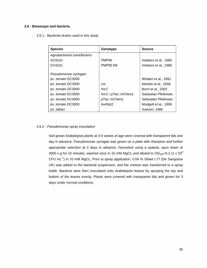
45
2.6 - Bioassays and bacteria
2.6.1 - Bacterial strains used in this study
(Whalen, 1991; Melotto et al., 2006; Boch et al., 2002; Gasson, 1980)
2.6.2 - Pseudomonas spray inoculation
Soil-grown Arabidopsis plants at 4-5 weeks of age were covered with transparent lids one
day in advance. Pseudomonas syringae was grown on a plate with rifampicin and further
appropriate selection at 2 days in advance, harvested using a spatula, spun down at
3000 x g for 10 minutes, washed once in 10 mM MgCl2 and diluted to OD600=0.2 (1 x 10⁸
CFU mL-1
) in 10 mM MgCl2. Prior to spray application, 0.04 % Silwet L77 (De Sangosse
UK) was added to the bacterial suspension, and the mixture was transferred to a spray
bottle. Bacteria were then inoculated onto Arabidopsis leaves by spraying the top and
bottom of the leaves evenly. Plants were covered with transparent lids and grown for 3
days under normal conditions.(Mudgett and Staskawicz, 1999; Holsters et al., 1980)
Species Genotype Source
Agrobacterium tumefaciens:
GV3101 PMP90 Holsters et al., 1980
GV3101 PMP90 RK Holsters et al., 1980
Pseudomonas syringae:
pv. tomato DC3000 Whalen et al., 1991
pv. tomato DC3000 cor Melotto et al., 2006
pv. tomato DC3000 hrcC Boch et al., 2002
pv. tomato DC3000 hrcC / pTac::mCherry Sebastian Pfeilmeier
pv. tomato DC3000 pTac::mCherry Sebastian Pfeilmeier
pv. tomato DC3000 AvrRpt2 Mudgett et al., 1999
pv. tabaci Gasson, 1980

46
2.6.3 - Pseudomonas inoculation by infiltration and induced resistance pretreatment
Soil-grown Arabidopsis plants at 4-5 weeks of age were covered with transparent lids one
day before inoculation. For induced resistance assays, on the same day three fully
expanded leaves per plant were marked and infiltrated with 10 µM flg22 peptide
(QRLSTGSRINSAKDDAAGLQIA, EzBiolabs) or a water control. Pseudomonas syringae
was pregrown on plate with rifampicin and appropriate selection 2 days before
inoculation, harvested using a spatula, washed once in water and diluted to OD600 =
0.0002 (1 x 10⁵ cfu mL
-1). Bacteria were then syringe-infiltrated into the abaxial side of
marked Arabidopsis leaves. Plants were covered with transparent lids and grown for 3
days under normal conditions.
2.6.4 - Quantifying bacterial proliferation in dilution series
Using a cork borer, ca. 0.75 cm2
tissue was harvested from three leaves per plant and
placed into 200 µL of MgCl2 10 mM together with two steel beads ( = 3.2 mm). Tissue
was homogenized at 30 Hz for 2 minutes using a TissueLyser (MM200, Retsch) and
transferred into 96-wells plates. A dilution series using MgCl2 10 mM was set up, and 10
µL of dilution factors 10-1
, 10-2
, 10-3
and 10-4
were plated on square L-media plates
supplemented with rifampicin and nystatin. After 24-48 hours of incubation at 28 °C,
colony numbers in all dilution spots were counted by hand and data was analyzed in
Microsoft Excel. Using data from the lowest dilution factor with individually discernible
colonies, the number of colony forming units (CFU) per cm2 of originally harvested leaf
tissue was calculated and averaged over the number of plants used. Means were
compared to each other using one-way ANOVA and labeled in significance grouping
using a post-hoc Tukey‘s HSD test.
2.6.5 - Hypersensitive response assays in N. benthamiana
Cladosporium fulvum Avr4 and Avr2 were heterologously produced in Pichia pastoris and
purified using the His-affinity tag (Dirk-Jan Valkenburg, Laboratory of Phytopathology,
Wageningen UR, The Netherlands). N. benthamiana plants were subjected to VIGS at 2
weeks of age, and were used for hypersensitive response assays at 4-5 weeks of age.
Purified effector proteins were infiltrated at 300 µM, or expressed through A. tumefaciens
mediated transformation alongside positive controls RxD460V (Bendahmane et al., 2002)
and BAX (Lacomme and Santa Cruz, 1999). HR phenotypes were recorded 3-6 days
after infiltration.
47
2.7 - Light microscopy
2.7.1 - Confocal laser scanning microscopy
Confocal laser scanning microscopy (CLSM) was performed using a Leica SP5 upright
microscope fitted with hybrid (HyD) detectors. Objectives used in this study are: 63 ×
1.20 NA water immersion; 40 × 0.85 NA air; 20 × NA 0.50 air. For multi channel image
acquisition, line sequential scanning and 3x line averaging was used. Z-stacks were
acquired over 10 µm, in steps of 1 µm. Excitation and detection wavelengths were
adjusted for each fluorophore.
The Leica Image Format (LIF) files were exported from the Leica Application Suite
Advanced Fluorescence (LAS-AF, version 1.6.3 build 1163, Leica), processed in FIJI
(FIJI Is Just ImageJ, versions from 2013-2018) and annotated in Microsoft Powerpoint.
2.7.2 – Fluorescent probes used in this study
2.7.3 - Automated high throughput spinning disc microscopy
Sample preparation and ligand treatment
Arabidopsis seedlings were grown on F2 soil for 14 days in a controlled
environment chamber (CentiForce Fitotron SGC 120) at 22 °C and a regime of
12 hours light / 12 hours dark. Cotyledons were picked using tweezers and
transferred adaxial side-up to the central 60 pins of a custom 96-pin rubber
stamp (made by Simon Foster) using high-vacuum silicone grease as an
adhesive (Sigma cat. Z273554-1EA).
An optical 96-wells plate (Greiner Bio-One cat. 655892) was prepared, adding
200 µL dH2O to 30 wells left of centre, and 200 µL flg22 (10 µM) to 30 wells right
of centre. The custom stamp with cotyledons was lowered into the optical plate,
incubated bench-top at room temperature and used for high throughput confocal
imaging at indicated time points (30, 90, 150 min).
Probe Excitation (nm) Detection (nm)
eGFP 488 495-540
eYFP 514 520-570
mRFP 561 570-630
mCherry 561 600-630
autofluorescence - 700-800

48
Laser and acquisition settings
The Opera High Content Screening System (PerkinElmer) with a 40 × 0.90 NA
water immersion objective was used for rapid image acquisition. For both GFP
and YFP imaging, a solid state 488 nm laser at 4000 µW was used for excitation,
and emission was captured between 502-577 nm. Images were captured with a
CCD camera and an exposure time of 40 ms. For each cotyledon, z-stacks of 21
images were acquired at 5 locations successively. Cotyledons were scanned
row-by-row, measuring 5 water treated wells and 5 flg22 treated wells in an
alternating manner. Total acquisition time per optical plate was ca. 30 minutes.
Post processing
For automated spot detection, the resulting FLEX image files were processed
using Single Channel Spot Detection v. 4.1 (by Ji Zhou, The Sainsbury
Laboratory, Norwich, UK) for Acapella Studio (PerkinElmer, version 2.2.2.7338).
Analysis was configured to require at least 70 % valid image area (less than 30
% dark area), a spot roundness of greater than 0.125 and a spot width-to-length
ratio of greater than 0.425. The resulting annotations were visually inspected by
taking an arbitrary sample of output images. Data was further processed in
Microsoft Excel.
2.7.4 - Confocal microscopy in N. benthamiana tissues
Magnifications and focus stacking
For GFP localizations, GFP-RFP/mCherry colocalizations, and for GFP vesicle
quantitation, the 63 × objective was used and z-stacks of 10 µm were obtained.
For BiFC/split-YFP of vesicle localized signals, the 63 × objective was used and
single images were obtained. For BiFC/split-YFP of plasma membrane localized
reconstituted signal, pictures were taken with the 40 × objective to capture
numerous cells per field of view.
Ligand treatments for receptor mediated endocytosis
100 µM of purified C. fulvum Avr4 or Avr2 dissolved in water were syringe-
infiltrated into fully expanded mature N. benthamiana leaves. After 1 hour, 1 cm
diameter leaf discs were obtained by cork borer and used for microscopic
analysis. Stable expressing plants were treated at 4-5 weeks of age, and
transiently expressing plants at 4-5 weeks of age were treated at 2 days after
agroinfiltration.

49
Vesicle quantification
Images were acquired using 63 × magnification. The resulting LIF-files were
processed using FIJI, and maximum projections of the combined GFP and
autofluorescence channels were automatically generated using a custom macro
(FileIO, by Ji Zhou). Spot quantification in z-projections was automated using
adaptive intensity and color thresholding in EndoQuant v. 1.075 (by Ji Zhou) for
Acapella Studio. The resulting annotations were visually inspected and data
were processed in Microsoft Excel.
2.7.5 - Confocal microscopy in Arabidopsis tissues
Transgenic Arabidopsis plants were observed using the 63 × objective. Seedling
cotyledons were picked using tweezers, and adult leaf tissue was obtained using
a biopsy punch ( = 2 mm). In both cases, abaxial sides were imaged.
Transiently expressing protoplasts were transferred in a volume of 1 µL onto a
microscope slide with reaction wells (Marienfeld cat. 1216521) and directly used
for imaging. For particle bombardment, transfected cells were located through
the binocular using a GFP filter cube at 20 × magnification, and images were
acquired at 63 × magnification.
2.7.6 - Confocal imaging of focal accumulations
Introduction of bacteria
Pto DC3000 or Pto DC3000 cor, carrying a genomic insertion with constitutive
promoter driven pTac::mCherry (generated by Sebastian Pfeilmeier) were grown
on plate at 28 °C for 2 days, and incubated on bench top at room temperature for
1 day. Bacteria were harvested using a spatula, washed once with dH2O, and
resuspended at OD600 = 0.2. Bacterial suspensions were syringe-infiltrated into
mature Arabidopsis leaves. Infiltrated plants were kept on bench top at room
temperature for 5-6 hours, and samples were taken with a biopsy punch.
Image acquisition
For detailed, qualitative imaging of abaxial mesophyll, the Leica SP5 confocal
microscope and 63 × magnification was used. Z-stacks were acquired starting
from the epidermal equatorial plane up to 10-20 µm into the tissue, in steps of 1
µm. For image acquisition in preparation for quantification, single images of the
first adaxial mesophyll layer at lower magnification of 20 × were acquired.

50
Quantification of focal accumulations with FIJI (script)
In order to quantify the number of focal accumulations per field of view, a custom
macro for FIJI was written that applies the following functions to a series of 8-bit
greyscale images: Gaussian blur (σ = 2 px), global threshold (grey value min. =
40, max. = 255), create mask, analyze particles (min. area = 20 px.), list number
of particles. The results were further processed in Microsoft Excel. Mean values
within experiments were statistically analysed using a Student‘s t-test comparing
treatment to water.

51
CHAPTER 3
AVR4 PROMOTES CF-4 RECEPTOR-LIKE PROTEIN ASSOCIATION WITH THE
BAK1/SERK3 RECEPTOR-LIKE KINASE TO INITIATE RECEPTOR ENDOCYTOSIS
AND PLANT IMMUNITY
This chapter consists of an edited and expanded version of the following publication:
“Avr4 promotes Cf-4 receptor-like protein association with the BAK1/SERK3 receptor-like
kinase to initiate receptor endocytosis and plant immunity”
Jelle Postma, Thomas W. H. Liebrand, Guozhi Bi, Alexandre Evrard, Ruby R. Bye, Malick
Mbengue, Hannah Kuhn, Matthieu H. A. J. Joosten, Silke Robatzek
New Phytologist (2016) vol. 210, issue 2, p. 627-42, doi: 10.1111/nph.13802
The majority of the data are based on experiments performed by myself. Specific experimental
contributions of co-authors are acknowledged in the main text as well as in figure legends.
RESULTS
3.1 - CF-4 AND SOBIR1 CO-INTERNALIZE UPON CF-4 ACTIVATION
3.1.1 - Cf-4 interacts with SOBIR1 at the plasma membrane
In order to study the dynamic subcellular localization of Cf-4 in antifungal immunity, I used the
fluorescently tagged Cf-4-GFP construct which triggers a hypersensitive response in N.
benthamiana upon perception of the Cladosporium fulvum effector Avr4 (fig. S1; Liebrand et al.,
2012). I transiently expressed Cf-4-GFP with the plasma membrane marker Autoinhibitory
Calcium ATPase 8 (ACA8, Frei dit Frey et al., 2012), and revealed by co-localization that Cf-4-
GFP is present at the plasma membrane (figs. 3.1, S2), which corresponds to its predicted site of
biological function in perceiving Avr4, which has been shown to be present in the apoplast
(Joosten et al., 1994; van den Burg et al., 2006). In addition, the constitutive Cf-4-interacting RLK
SlSOBIR1 and its homologs SlSOBIR1-like and AtSOBIR1 also localized to the plasma
membrane (figs. 3.1-2, S2-4), raising the possibility that Cf-4-SOBIR1 dimers are present at this
location. To test for presence of Cf-4-SOBIR1 dimers, I co-expressed bimolecular fluorescence
complementation (BiFC) fusion constructs of both, and revealed that Cf-4 and SOBIR1

52
reconstituted a signal at the PM as confirmed by colocalization with ACA8 (fig. 3.1). As a
negative control, I co-expressed BiFC fusion constructs of SlSOBIR1 and FLS2, and while both
proteins could be expressed, they did not reconstitute a signal (fig. S5), corresponding to
published data that SOBIR1 does not interact with RLK-type PRRs (Liebrand et al., 2013). Taken
together, I conclude that Cf-4 and SlSOBIR1 interact at the PM.
Fig. 3.1. Cf-4 and SlSOBIR1 are present at the plasma membrane. Single z-plane confocal micrographs show N. benthamiana
leaf epidermal cells transiently co-expressing the indicated Cf-4 and SlSOBIR1 fusion proteins and plasma membrane-localized
ACA8-mCherry. The first column of panels show GFP/YFP fluorescence, the second panels show mCherry fluorescence and the
third column of panels depict the overlay images of the two fluorescence signals shown in the first and second panels. Overlay
images indicate co-localization of the proteins fused to GFP or YFP and mCherry, as a yellow color is produced. Dashed squares in
these images are shown as detailed pictures (magnified in the fourth column of panels). White lines in the detailed pictures indicate
the regions of interest (ROIs) that correspond to the intensity profiles in the last column of panels. Intensity profiles indicate the grey-
value of pixels across the ROI in the green and red channels, on a scale of 1-300. Images were taken at three days post infiltration
(dpi) for Cf-4-GFP and SlSOBIR1-GFP and at two dpi for BiFC of Cf-4, C-terminally fused to the C-terminal half of YFP (YFPc), with
SlSOBIR1 C-terminally fused to the N-terminal half of YFP (YFPn); scale bars = 10 μm.

53
3.1.2 - Avr4 triggers endocytosis of the Cf-4-SOBIR1 complex
Previously, AtSOBIR1 has been shown to localize to FM4-64-positive vesicles, suggesting
endosomal localization (Leslie et al., 2010b). To further probe the identify of SOBIR1-positive
vesicles, I co-expressed the GFP-tagged tomato and Arabidopsis SOBIR1 homologs with a panel
of RFP-tagged known endomembrane markers, which localize to Golgi (MEMB12), TGN/EE
(VHA-a1), TGN/EE-LE/MVB (ARA7) and LE/MVB (ARA6). SOBIR1 punctae did not overlap with
MEMB12 or VHA-a1, but did overlap with ARA7 and ARA6, confirming constitutive SOBIR1
localization to endosomes (figs. 3.2, S3-4).
Endosomal localization of cell surface receptors can be triggered by ligand perception (Mbengue
et al., 2016; Ortiz-Morea et al., 2016; Beck et al., 2012b). Because Cf-4 interacted with SOBIR1
at the PM as shown in BiFC, and SOBIR1 constitutively localized to endosomes, I was prompted
to test whether Cf-4 could also localize to endosomes upon activation by Avr4. To address this, I
co-expressed Cf-4-GFP and SlSOBIR1-mCherry in N. benthamiana and infiltrated the purified C.
fulvum effector protein Avr4. I also infiltrated Avr2, which is not recognized by Cf-4, as a negative
control. In untreated leaves, Cf-4-GFP localized to the PM, while SlSOBIR1-mCherry localized to
the PM and endosomes (figs. 3.1, 3.3A). Interestingly, Avr4 treatment triggered Cf-4-GFP
localization to SlSOBIR1-mCherry-positive vesicles, pointing at ligand-induced endocytosis (figs.
3.3A, S6). I could confirm the endosomal nature of Avr4-induced Cf-4-GFP compartments by co-
localization with the LE/MVB-marker ARA6 (fig. 3.3A). Because Avr2 did not trigger
internalization of Cf-4-GFP (fig. 3.3A, S6), I conclude that Cf-4 endocytosis is ligand-specific and
dependent on receptor activation.
Because upon Avr4 treatment, Cf-4 and SOBIR1 localized at late endosomes, I hypothesized that
they may be internalized in complex with each other. To investigate this, I co-expressed Cf-4 and
SOBIR1 BiFC constructs, which reconstituted signal at the PM (fig. 3.1). Upon activation of Cf-4
by Avr4 treatment, but not upon Avr2 treatment, BiFC signal was also reconstituted at ARA6-
positive LE/MVBs (fig. 3.3B), confirming ligand-induced endocytosis of the Cf-4-SOBIR1
heterodimer, which corresponds to Avr4-induced endosomal colocalization of Cf-4 and SOBIR1
(fig. 3.3A). Ligand-induced internalization of receptor heterodimers has been shown before for
BRI1-BAK1/SERK3 in Arabidopsis protoplasts (Russinova, 2004). In contrast, upon co-
expression of BiFC fusion constructs of FLS2 which revealed localization of homodimers to the
PM, flg22-treatment could not induce internalization, suggesting the complex may be impaired in
recruiting the BAK1/SERK3 co-receptor, which is required for ligand-induced FLS2 endocytosis
(Chinchilla et al., 2007; Beck et al., 2012b).

54

55
Fig. 3.2. SlSOBIR1 constitutively localizes to endosomes. Maximum projections of confocal micrograph z-stacks show N.
benthamiana leaf epidermal cells transiently expressing SlSOBIR1-GFP or Cf-4-GFP (left panels), either without or with co-
expression of the indicated organelle markers fused to mCherry/RFP (middle, left panels). Co-localization in the overlay images is
depicted by the development of a yellow color (middle, right panels). Dashed squares in these images are shown as detail pictures
(magnified in the right panels). Arrowheads in the detail pictures point at mobile vesicles in the upper right panel and indicate
SlSOBIR1 localization at endosomes in the two lower right panels. Images were taken at three dpi; scale bars = 10 μm.
3.1.3 - Cf-4 endocytosis requires functional BAK1/SERK3 and SOBIR1
Ligand-induced activation and endocytosis of FLS2 depends on BAK1/SERK3 (Beck et al.,
2012b; Choi et al., 2013). Therefore, I hypothesized that ligand-induced endocytosis of Cf-4
shares the same requirement. To address this, in collaboration with Alexandre Evrard (The
Sainsbury Laboratory, Norwich, UK), I used tobacco rattle virus (TRV)-based virus-induced gene
silencing (VIGS) and knocked down the two endogenous tobacco BAK1/SERK3 homologs
NbSERK3a/b (Heese et al., 2007) and tested for the capacity for Avr4 to induce Cf-4-GFP
endocytosis. Interstingly, upon Avr4 treatment, Cf-4-GFP endocytosis was reduced in
TRV::NbSERK3a/b leaves as compared to TRV-control leaves (figs. 3.4, S7), while Cf-4-GFP
accumulation levels were unaffected by NbSERK3a/b silencing (fig. S7). Upon NbSERK3a/b
silencing, which did not affect the total number of ARA6 endosomes (fig. 3.4), transient co-
expression of Arabidopsis BAK1/SERK3, could restore the capacity for Cf-4-GFP to internalize
upon Avr4 treatment (figs. 3.4, S7). Cf-4-GFP internalization was dependent on the full
functionality of BAK1/SERK3, as co-expression with the kinase-dead AtBAK1-KD could not
restore Avr4-induced Cf-4-GFP accumulation at endosomes (figs. 3.4, S7). These data together
provide evidence that both RLK and RLP-type cell-surface immune receptors share the functional
requirement for the RLK BAK1/SERK3. Upon analysis by qRT-PCR, performed in collaboration
with Ruby Bye (Laboratory of Phytopathology, Wageningen UR, The Netherlands), we could
show that TRV::NbSERK3a/b silencing additionally co-silences a NbSERK1 homolog, which
indicates that Cf-4 endocytosis might additionally require SERK1.

56
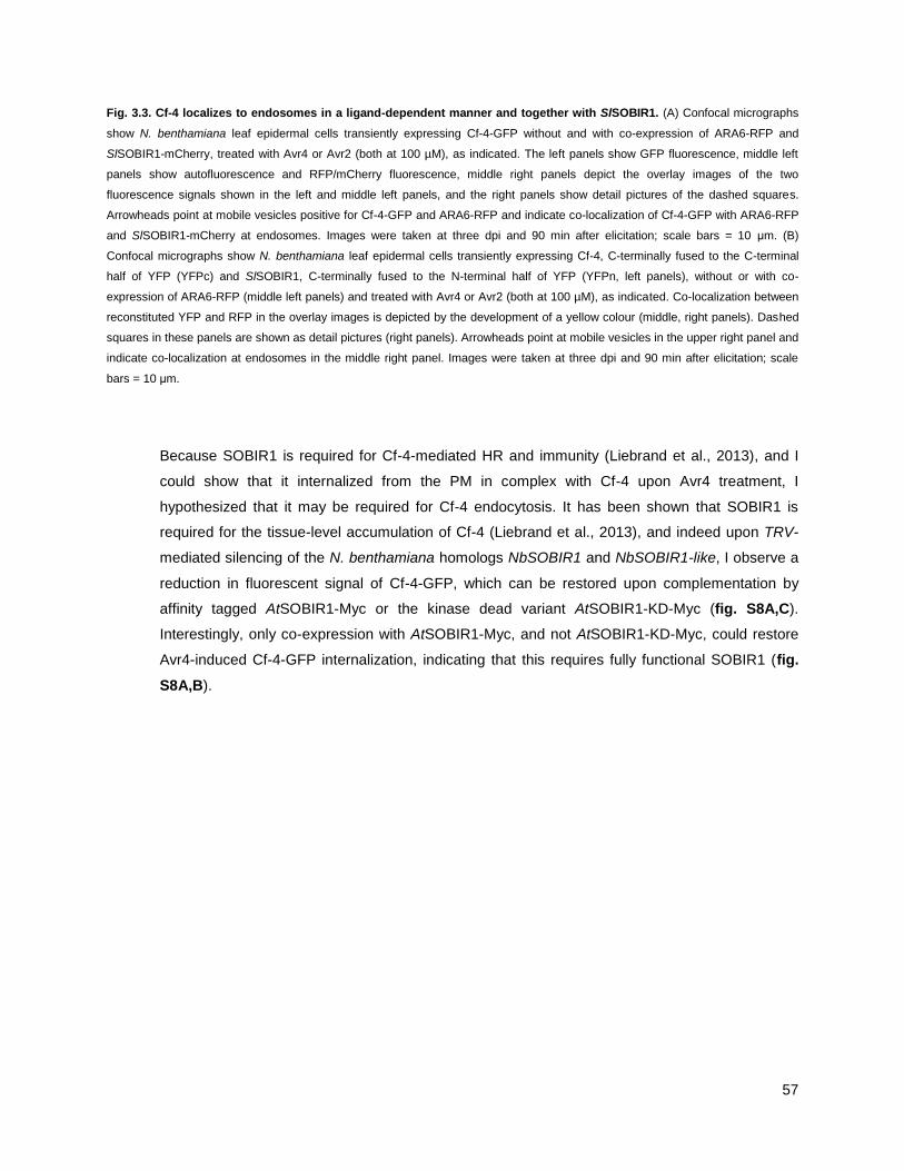
57
Fig. 3.3. Cf-4 localizes to endosomes in a ligand-dependent manner and together with SlSOBIR1. (A) Confocal micrographs
show N. benthamiana leaf epidermal cells transiently expressing Cf-4-GFP without and with co-expression of ARA6-RFP and
SlSOBIR1-mCherry, treated with Avr4 or Avr2 (both at 100 µM), as indicated. The left panels show GFP fluorescence, middle left
panels show autofluorescence and RFP/mCherry fluorescence, middle right panels depict the overlay images of the two
fluorescence signals shown in the left and middle left panels, and the right panels show detail pictures of the dashed squares.
Arrowheads point at mobile vesicles positive for Cf-4-GFP and ARA6-RFP and indicate co-localization of Cf-4-GFP with ARA6-RFP
and SlSOBIR1-mCherry at endosomes. Images were taken at three dpi and 90 min after elicitation; scale bars = 10 μm. (B)
Confocal micrographs show N. benthamiana leaf epidermal cells transiently expressing Cf-4, C-terminally fused to the C-terminal
half of YFP (YFPc) and SlSOBIR1, C-terminally fused to the N-terminal half of YFP (YFPn, left panels), without or with co-
expression of ARA6-RFP (middle left panels) and treated with Avr4 or Avr2 (both at 100 µM), as indicated. Co-localization between
reconstituted YFP and RFP in the overlay images is depicted by the development of a yellow colour (middle, right panels). Dashed
squares in these panels are shown as detail pictures (right panels). Arrowheads point at mobile vesicles in the upper right panel and
indicate co-localization at endosomes in the middle right panel. Images were taken at three dpi and 90 min after elicitation; scale
bars = 10 μm.
Because SOBIR1 is required for Cf-4-mediated HR and immunity (Liebrand et al., 2013), and I
could show that it internalized from the PM in complex with Cf-4 upon Avr4 treatment, I
hypothesized that it may be required for Cf-4 endocytosis. It has been shown that SOBIR1 is
required for the tissue-level accumulation of Cf-4 (Liebrand et al., 2013), and indeed upon TRV-
mediated silencing of the N. benthamiana homologs NbSOBIR1 and NbSOBIR1-like, I observe a
reduction in fluorescent signal of Cf-4-GFP, which can be restored upon complementation by
affinity tagged AtSOBIR1-Myc or the kinase dead variant AtSOBIR1-KD-Myc (fig. S8A,C).
Interestingly, only co-expression with AtSOBIR1-Myc, and not AtSOBIR1-KD-Myc, could restore
Avr4-induced Cf-4-GFP internalization, indicating that this requires fully functional SOBIR1 (fig.
S8A,B).

58

59
Fig. 3.4. Cf-4 endocytosis requires BAK1/SERK3. Leaves of Cf-4-GFP-N. benthamiana stable and wild type N. benthamiana
plants were TRV-silenced for NbSERK3a/b and GUS as a control for three weeks and subsequently used to transiently express
AtBAK1, AtBAK1-KD, and ARA6/RabF1-RFP as indicated for three days. Confocal micrographs show Cf-4-GFP localisation upon
treatment with Avr4 (100 µM, left panels) and ARA6/RabF1-RFP-labelled endosomes (left panels), and detail pictures from dashed
squares (middle panels). Arrowheads point at Cf-4-GFP- and ARA6/RabF1-RFP-positive vesicles. Quantification of Cf-4-GFP- and
ARA6/RabF1-RFP-positive vesicles was done with EndoQUANT (right panels, bars depict means ± 2 SE; n = 6; p < 0.05; statistical
significant differences are indicated by asterisks). Note that much fewer Cf-4-GFP-positive vesicles are observed in NbSERK3a/b-
silenced leaves compared to the TRV-silenced control. The amount of Cf-4-GFP-positive vesicles increased upon transient co-
expression of AtBAK1/SERK3 but not when its kinase-inactive variant AtBAK1-KD was co-expressed. No difference in the amount
of ARA6/RabF1-RFP-positive endosomes was observed when NbSERK3a/b was silenced. Images were taken 90 min after Avr4
elicitation; scale bars = 10 μm. See Fig. S.7 for transcript abundance of silenced genes and protein levels of Cf-4. The above
microscopy experiments were performed in collaboration with Alexandre Evrard.
3.2 - CF RLPS RECRUIT SERK FAMILY RLKS UPON ACTIVATION
3.2.1 - Cf-4 recruits BAK1/SERK3 and SERK1 upon activation
Well-studied RLK-type PRRs such as FLS2 and EFR recruit BAK1/SERK3 upon ligand
perception (Roux et al., 2011). Having established that both Cf-4 and FLS2 endocytosis share the
requirement for BAK1/SERK3 in endocytosis, I addressed a possible role for BAK1/SERK3 in Cf-
4-mediated immunity using biochemical interaction studies, performed in collaboration with
Thomas Liebrand and Guozhi Bi (Laboratory of Phytopathology, Wageningen UR, The
Netherlands). We performed transient co-expression of Cf-4-GFP, SlSOBIR1-HA and SlSERK3a-
Myc in N. benthamiana, treated the leaves with Avr4, Avr2 or flg22, and purified either Cf-4-GFP
or SlSERK3a-Myc using immunoprecipitation. In all conditions, SlSOBIR1-HA co-purified with Cf-
4-GFP as revealed by western blot (figs. 3.5, S9), corresponding to their known constitutive
interaction and the reconstitution of signal in BiFC experiments (fig. 1; Liebrand et al., 2013). In
addition, upon pulldown of SlSERK3a-Myc, SlSOBIR1-HA co-purified under all conditions, which
contrasts with the findings of Liebrand et al., 2013, who did not detect SlSERK3a-GFP in
SlSOBIR1-Myc purifications. This may be explained by the use of biochemical methods with
different sensitivities, but it is conceivable that SlSOBIR1 and SlSERK3a interact through another
RLP.
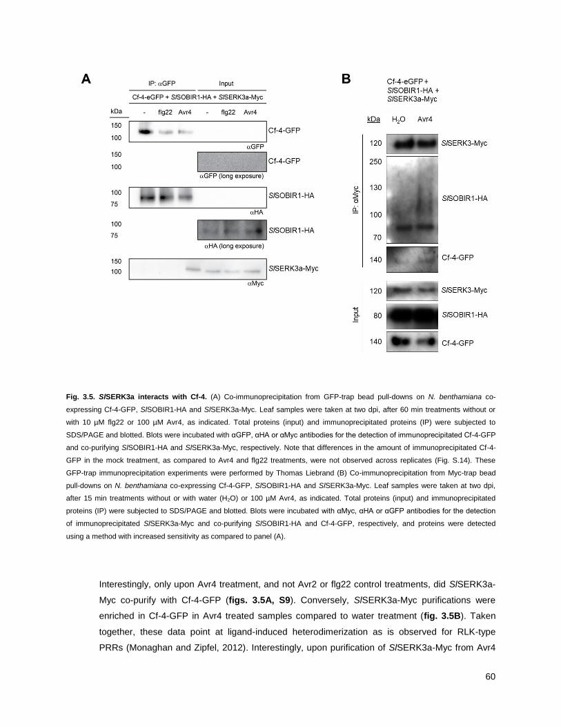
60
Fig. 3.5. SlSERK3a interacts with Cf-4. (A) Co-immunoprecipitation from GFP-trap bead pull-downs on N. benthamiana co-
expressing Cf-4-GFP, SlSOBIR1-HA and SlSERK3a-Myc. Leaf samples were taken at two dpi, after 60 min treatments without or
with 10 µM flg22 or 100 µM Avr4, as indicated. Total proteins (input) and immunoprecipitated proteins (IP) were subjected to
SDS/PAGE and blotted. Blots were incubated with αGFP, αHA or αMyc antibodies for the detection of immunoprecipitated Cf-4-GFP
and co-purifying SlSOBIR1-HA and SlSERK3a-Myc, respectively. Note that differences in the amount of immunoprecipitated Cf-4-
GFP in the mock treatment, as compared to Avr4 and flg22 treatments, were not observed across replicates (Fig. S.14). These
GFP-trap immunoprecipitation experiments were performed by Thomas Liebrand (B) Co-immunoprecipitation from Myc-trap bead
pull-downs on N. benthamiana co-expressing Cf-4-GFP, SlSOBIR1-HA and SlSERK3a-Myc. Leaf samples were taken at two dpi,
after 15 min treatments without or with water (H2O) or 100 µM Avr4, as indicated. Total proteins (input) and immunoprecipitated
proteins (IP) were subjected to SDS/PAGE and blotted. Blots were incubated with αMyc, αHA or αGFP antibodies for the detection
of immunoprecipitated SlSERK3a-Myc and co-purifying SlSOBIR1-HA and Cf-4-GFP, respectively, and proteins were detected
using a method with increased sensitivity as compared to panel (A).
Interestingly, only upon Avr4 treatment, and not Avr2 or flg22 control treatments, did SlSERK3a-
Myc co-purify with Cf-4-GFP (figs. 3.5A, S9). Conversely, SlSERK3a-Myc purifications were
enriched in Cf-4-GFP in Avr4 treated samples compared to water treatment (fig. 3.5B). Taken
together, these data point at ligand-induced heterodimerization as is observed for RLK-type
PRRs (Monaghan and Zipfel, 2012). Interestingly, upon purification of SlSERK3a-Myc from Avr4

61
treated samples, we observed a ―smear‖ pattern migrating at higher molecular weights of co-
purifying SlSOBIR1-HA, providing evidence of Avr4-induced posttranslational modifications on
the SlSERK3a-interacting pool of SlSOBIR1-HA.
3.2.2 - Cf-4 and Cf-9 both recruit SERK members upon activation
It has previously been shown that another SERK member, SlSERK1, is genetically required for
Cf-4 mediated resistance in tomato (Fradin et al., 2011). Therefore, we addressed whether Cf-4
similarly recruits SERK1 upon activation. Upon purification of Cf-4-GFP, we could reveal
SlSERK1-Myc already interacting under Avr2 control treated conditions, but observed a marked
enhancement of co-purifying SlSERK1-Myc in Avr4 treated leaves (fig. S9). While in this
expression system SlSERK1-Myc accumulated to higher levels than SlSERK3-Myc, possibly
explaining ectopic interactions with Cf-4-GFP, it is also possible that Cf-4 exists in a preformed
complex with SlSOBIR1 and SlSERK1, which upon activation of Cf-4 becomes stabilized and
recruits additional SlSERK3a.
Cf-4 is highly homologous to Cf-9, which detects C. fulvum Avr9 (Thomas et al., 1997). To
investigate whether SERK recruitment might be a more generalized mechanism by which RLPs
initiate immune signaling, we purified Cf-9-GFP from leaves additionally co-expressing
SlSOBIR1-HA and either SlSERK3a-Myc or SlSERK1-Myc, under Avr9 treatment or Avr4 control
treatments. As a result, we observed that similar to Cf-4, SlSERK3a-Myc and SlSERK1-Myc
strongly co-purified with Cf-9-GFP only upon activation by Avr9 (fig. S10). Taken together, these
data suggest that other SOBIR1-dependent RLPs may similarly recruit SERK family members to
initiate signaling, as has recently been shown for ELICITIN RESPONSE (ELR) and RLP23 (Albert
et al., 2015; Du et al., 2015).
3.3 - SERK MEMBERS MEDIATE AVR4-TRIGGERED IMMUNITY
3.3.1 - Cf-4 hypersensitive response requires SERK members
Upon activation by Avr4, Cf-4 triggers HR in N. benthmiana which could be suppressed upon
silencing of NbSOBIR1 and NbSOBIR1-like (fig. 3.6A; Liebrand et al., 2013). Because
SlSERK3a was recruited to Cf-4 upon activation by Avr4, and was required for Cf-4 endocytosis, I
hypothesized that NbSERK3a might also be involved in immune signaling of Cf-4. To address
this, in collaboration with Thomas Liebrand and Ruby Bye, we silenced NbSERK3a/b using VIGS,
and revealed that this significantly reduced Avr4-triggered HR in N. benthamiana stably
expressing Cf-4, whereas Avr4 could consistenly induce HR in TRV control inoculated Cf-4 plants
(figs. 3.6A, S7). The capacity for NbSERK3a/b-silenced plants to initiate HR was not generally
affected, as this was still induced by the autoactive NLR RxD460V and pro-apoptotic factor BCL2-
ASSOCIATED PROTEIN X (BAX; Lacomme and Santa Cruz, 1999; Bendahmane et al., 2002).
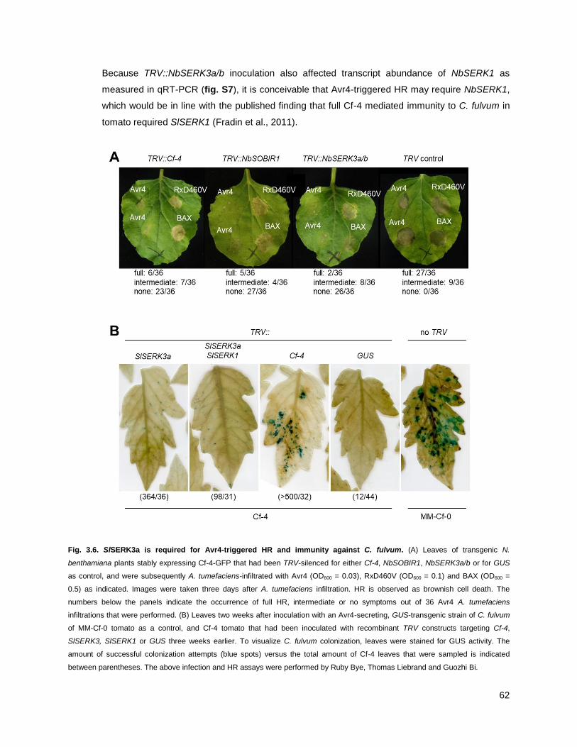
62
Because TRV::NbSERK3a/b inoculation also affected transcript abundance of NbSERK1 as
measured in qRT-PCR (fig. S7), it is conceivable that Avr4-triggered HR may require NbSERK1,
which would be in line with the published finding that full Cf-4 mediated immunity to C. fulvum in
tomato required SlSERK1 (Fradin et al., 2011).
Fig. 3.6. SlSERK3a is required for Avr4-triggered HR and immunity against C. fulvum. (A) Leaves of transgenic N.
benthamiana plants stably expressing Cf-4-GFP that had been TRV-silenced for either Cf-4, NbSOBIR1, NbSERK3a/b or for GUS
as control, and were subsequently A. tumefaciens-infiltrated with Avr4 (OD600 = 0.03), RxD460V (OD600 = 0.1) and BAX (OD600 =
0.5) as indicated. Images were taken three days after A. tumefaciens infiltration. HR is observed as brownish cell death. The
numbers below the panels indicate the occurrence of full HR, intermediate or no symptoms out of 36 Avr4 A. tumefaciens
infiltrations that were performed. (B) Leaves two weeks after inoculation with an Avr4-secreting, GUS-transgenic strain of C. fulvum
of MM-Cf-0 tomato as a control, and Cf-4 tomato that had been inoculated with recombinant TRV constructs targeting Cf-4,
SlSERK3, SlSERK1 or GUS three weeks earlier. To visualize C. fulvum colonization, leaves were stained for GUS activity. The
amount of successful colonization attempts (blue spots) versus the total amount of Cf-4 leaves that were sampled is indicated
between parentheses. The above infection and HR assays were performed by Ruby Bye, Thomas Liebrand and Guozhi Bi.

63
3.3.2 - Cf-4-mediated resistance in tomato requires SlSERK3
To investigate whether BAK1/SERK3 is required for Cf-4-mediated resistance in tomato,
collaborating with with Thomas Liebrand and Ruby Bye, we silenced SlSERK3 individually, or
SlSERK3a together with SlSERK1 using VIGS, and tested for colonization success of C. fulvum
carrying Avr4 and the GUS reporter gene. In both cases, C. fulvum colonized leaves more
strongly than in TRV control leaves, as evidenced by increased GUS staining and subsequent
quantification (fig. 3.6B). Because silencing of Cf-4 itself even further increased C. fulvum
colonization success (fig. 3.6B), it is possible that VIGS only partially knocks down the
expression of SlSERK3 and SlSERK1 allowing for some Cf-4 activation, and/or that there is
functional redundancy between thes two SERKs. These data together show that BAK1/SERK3 is
a key positive regulator of full Cf-4-mediated immunity upon Avr4 perception.
DISCUSSION
3.4 - Cf-4 and SOBIR1 work with SERKs to initiate immunity and receptor endocytosis
The Cf signaling pathway is essential for the immune response of tomato to C. fulvum
(Stergiopoulos and de Wit, 2009; Rivas and Thomas, 2005). A number of Cf signaling
components were identified through genetic and proteomic approaches, but the mechanism by
which Cf-4 initiates downstream signaling remained unclear (Liebrand et al., 2014). In the present
study, I show that both SERK1 and SERK3 are recruited to the Cf-4 receptor in an Avr4-
responsive manner, a mechanism that was additionally confirmed for Avr9-induced activation of
Cf-9.
Consistently, I show that Cf- 4 requires these RLKs for its function in order to initiate HR and
restrict fungal colonization as silencing of the respective genes in N. benthamiana and tomato
suppresses Avr4-induced HR symptom formation and facilitates fungal penetration, respectively.
My observations imply that Avr4 enhances the formation of a complex of Cf-4 and SlSERK1/3 to
induce Cf-4 signaling. Furthermore, I confirm that Cf-4 interacts with SOBIR1 at the plasma
membrane and, could demonstrate ligand-induced SERK3-dependent late endocytic trafficking of
the Cf-4 RLP together with SlSOBIR1 as a novel pathway in Cf-mediated downstream events.
3.5 - Ternary receptor complexes form
The finding that Cf-4 and SOBIR1 are present in complex and recruit additional RLKs to form
multimeric complexes upon activation is in agreement with existing data on RLP-SOBIR1
systems. Solanum microdontum (wild tomato) ELICITIN RESPONSE (ELR) is an RLP that
detects the MAMP elicitin, and recruits SERK3 upon activation, while ELR maintains interaction
64
with SOBIR1 independent of activation status (Domazakis et al., 2018; Du et al., 2015).
Furthermore, Arabidopsis RLP23, which activates upon binding of the MAMP Nep1-like protein
(NLP)-derived peptide nlp20, interacts with SOBIR1 independent of activation status, and recruits
BAK1/SERK3 upon activation (Albert et al., 2015). However, the above data, including from the
present study, were obtained from tissue-level biochemical analyses, and cannot fully exclude
that separate pair-wise interactions between RLP-SOBIR1 and RLP-SERK3 may exist. It would
be interesting to more precisely address this question through RLP-purification and subsequent
non-denaturing gel electrophoresis, or with in vivo fluorescence-based association assays.
3.6 - Different SERKs may do the job
In my experiments, Cf-4 associated with SERK1 before activation, and more strongly upon Avr4
treatment (fig. S9). In addition, SERK1 has been shown to be required for full Cf-4 immunity in
tomato (Fradin et al., 2011). The recruitment and requirement of multiple SERK members has
been shown in existing studies on RLPs dependent on SOBIR1. Arabidopsis RLP23 interacted
with SOBIR1 and SERK4 before activation, and recruited increased amounts of SERK1, SERK2,
SERK3 and SERK4 upon activation by nlp20 (Albert et al., 2015). Additionally, SERK members
showed functional redundancy in mediating RLP23 associated outputs (Albert et al., 2015). On
the other hand, Arabidopsis RESPONSIVENESS TO BOTRYTIS POLYGALACTURONASES 1
(RBPG1)/RLP42, which interacts with SOBIR1 and triggers HR upon detection of fungal
endopolygalacturonase, did not recruit SERK3 and did not depend on SERK3 for HR (Zhang et
al., 2014a).
The well-characterized RLK-type PRRs FLS2 and EFR also recruit multiple SERKs upon
activation, or interact with SERK members at steady state. FLS2 interacted with Arabidopsis
SERK1, SERK2, BAK1/SERK3 and BKK1/SERK4 upon expression in N. benthamiana before
activation, and upon activation FLS2 most strongly recruited BAK1/SERK3, but also recruited
additional SERK1 and SERK2 (Roux et al., 2011). Similarly, EFR recruited equal amounts
SERK1, SERK2, BAK1/SERK3 and BKK1/SERK4 upon activation (Roux et al., 2011), but for
most FLS2 and EFR outputs, BAK1/SERK3 is the major contributor (Schwessinger et al., 2011).
As observed for RLPs, there are SERK3-independent outputs, as evidenced by the capacity for
bak1 mutants to trigger elevated SA-levels and PEN3 cell-surface focal accumulation upon
activation of FLS2 by flg22 (Underwood and Somerville, 2013; Yamada et al., 2016).
These data taken together, for both RLP-SOBIR1 activation, as well as PRR-based cell-surface
immune activation, the requirements for specific SERK members can vary. Since I could restore
Avr4-induced Cf-4 endocytosis in NbSERK3a/b and partial NbSERK1-silenced plants through
expression of the singular Arabidopsis BAK1/SERK3, I hypothesize that this member is the major

65
determinant for Cf-4 triggered outputs, but its ability to complement Cf-4 HR and full tomato
immunity to C. fulvum remains to be demonstrated.
3.7 - Cf-4-SOBIR1 as a two-component PRR
The requirement of SERK members hints at clear similarities between the Cf-4/Cf-9 effector
receptors and FLS2 MAMP receptor pathways, further evidenced by overlapping transcriptional
reprogramming upon activating FLS2- and Cf-mediated immune responses (Navarro et al., 2004).
This overlap potentially involves the regulation of downstream responses through similar
components which, in addition to the SERK family members, include the receptor-like cytoplasmic
kinases BOTRYTIS- INDUCED KINASE 1 (BIK1), and AVRPPHB SUSCEPTIBLE 1-LIKE 1
(PBL1) kinases downstream of FLS2, and the AVR9/CF-9 INDUCED KINASE 1 (ACIK1)
regulating Cf-4 immunity, as well as the CAST AWAY kinase involved in SOBIR1/EVR signaling
(Burr et al., 2011; Monaghan and Zipfel, 2012; Liebrand et al., 2014).
Given these similarities, it is conceivable that the constitutive association between the RLP Cf-4
and the RLK SlSOBIR1 represents a PRR complex, in which the Cf-4 ectodomain mediates
specific ligand recognition and the SlSOBIR1 kinase domain is regarded as the signaling part.
This is in contrast to PRRs exemplified by FLS2, in which both functions are present within the
same molecule. Ligand-enhanced interaction of PRRs with SERK member RLKs is subsequently
required to trigger receptor endocytosis and further downstream signaling by both RLP- and RLK-
type PRRs (fig. 3.7).
3.8 - Shared mechanisms of receptor endocytosis
Receptor-mediated endocytosis is part of the eukaryotic immune response and in addition to
FLS2 and Cf-4, for example, is also found for the EFR and PEPR1 RLKs (Husebye et al., 2006;
Robatzek et al., 2006; Spallek et al., 2013; Mbengue et al., 2016; Ortiz-Morea et al., 2016). An
important role of receptor-mediated endocytosis is to control the abundance of receptor
(complexes) at the plasma membrane, a process that is well established in animals and involves
lysosomal/vacuolar degradation (Lemmon and Schlessinger, 2010). Activated FLS2 traffics into
the late endosomal pathway and is a cargo of multivesicular bodies localizing to the lumen of
these late endosomes for delivery to the vacuole (Beck et al., 2012a; Spallek et al., 2013;
Mbengue et al., 2016). This pathway could be responsible for the flg22-induced FLS2
degradation, because chemicals affecting endosomal trafficking inhibit the degradation of this
receptor (Smith et al., 2014). Our colocalization data strongly suggest that activated Cf-4 is also
targeted for vacuolar degradation through the late endosomal pathway, consistent with the
observation that Cf-4-GFP protein levels are reduced upon Avr4 elicitation (fig. S1; Choi et al.,
2013).

66
Fig. 3.7. Model of the Cf-4, SOBIR1 and SERK3 subcellular trafficking pathways. (A) Un-elicited: Following its folding and
maturation in the endoplasmic reticulum (ER), Cf-4 is predominantly trafficked to the plasma membrane, where it constitutively
interacts with SOBIR1, which itself is constitutively endocytosed. SOBIR1 can interact with BAK1/SERK3, a co-receptor known to
recycle between the plasma membrane and the trans-Golgi network/early endosome. (B) Elicited: Upon colonization of Cf-4 tomato
leaves, C. fulvum secretes Avr4 into the apoplast, which is recognized by Cf-4 and promotes interaction of Cf-4 with BAK1/SERK3.
This complex is required for Cf-4-mediated immunity (not shown) and endocytosis into ARA7- and ARA6-positive compartments of
the late endosomal pathway, destined for vacuolar degradation. Subcellular localization of markers (ACA8, MEMB12, VHA-a1,
ARA6 and ARA7) used in this study is depicted in brown lettering. TGN, trans-Golgi network; EE, early endosome; LE, late
endosome; MVB, multivesicular body.
Endosomal sorting for vacuolar degradation requires the transfer of ubiquitin to the plasma
membrane cargo, and subsequent deubiquitination of the cargo at multivesicular bodies (Beck et
al., 2012b). In line with this, the Pto DC3000 effector AvrPtoB ubiquitinates the kinase domain of
FLS2, promoting its degradation (Göhre et al., 2008), and upon activation by flg22, ubiquitin-
67
ligases PLANT U-BOX 12 (PUB12) and PUB13 ubiquitinate FLS2, resulting in its degradation (Lu
et al., 2011). Ubiquitination processes have also been implicated in Cf downstream signaling
pathways upon activation (Navarro et al., 2004). Is there any evidence for ubiquitination on RLP
receptor complexes? The ubiquitin ligase CYS, MET, PRO, AND GLY PROTEIN CMPG1 is
required for Cf-4 and Cf-9 HR (Gilroy et al., 2011). Phytophthora effector Avr3a targets CMPG1
and suppresses the immune response to elicitin, which is mediated by ELR, an RLP that interacts
with SOBIR1 and recruits SERK3 (Domazakis et al., 2018; Du et al., 2015). However, based on
localization and functional studies, CMPG1 is likely to function in the nucleus (Bos et al., 2010).
Tobacco Avr9/Cf-9-Rapidly-Elicited 189 (ACRE189)/ Avr9/Cf-9–INDUCED F-BOX1 (ACIF1) and
ACRE276/PUB17 are required for Cf-4 and Cf-9 mediated HR, but both most likely function in the
nucleus (van den Burg et al., 2008; He et al., 2015; Yang et al., 2006). Finally, Suppressor of G2
Allele of SKP1 (SGT1) which is homologous to ubiquitin-ligase complex interacting proteins in
yeast, is required for Cf-4 and Cf-9 HR, but also for intracellular NLR-mediated immunity, and
most likely functions further downstream of both types of immune receptors (Peart et al., 2002).
Taken together, these data point at positive roles for ubiquitination in Cf-triggered outputs. Unlike
the direct ubiquitination of the FLS2 cytosolic domain, the abovementioned Cf-associated
ubiquitination proteins are not thought to function at the PM themselves, but predominantly in the
nucleus or further downstream of initial Cf activation. That makes them unlikely to have a role in
direct modification of the ubiquitination status of the RLP-SOBIR1 receptor complexes. However,
I observe increased molecular weight ―smear‖ patterns in SOBIR1 co-purifying with SERK3 upon
activation of Cf-4 (fig. 3.5), suggesting posttranslational modifications of SOBIR1 in active
complexes. In agreement with this, the de-ubiquitinating tobacco enzyme UBIQUITIN-SPECIFIC
PROTEASE 12 (UBP12) modulates Cf-4 and Cf-9-mediated HR (Ewan et al., 2011), and the
Arabidopsis homolog AtUBP12 localizes to the nucleus and cytosol, which leaves the possibility
that it mediates cytosolic ubiquitination processes such as those on cell-surface receptors (Cui et
al., 2013).
3.9 - Cf-4 as a pattern recognition receptor
C. fulvum Avr4 is considered an effector, which makes Cf-4 a receptor functioning in effector-
triggered immunity (ETI), in line with its historic classification as an R-protein (Thomas et al.,
1997; Hammond-Kosack and Kanyuka, 2007). However, I could demonstrate that it shares
similar requirements for signal initiation at the cell surface with RLK-type PRRs such as FLS2,
which functions as MAMP detector and confers MAMP-triggered immunity. This points at early
convergence of ETI and PTI. Besides my data that suggest both perception systems at the cell
surface converge early on the shared module SERK3, other findings support the notion that Cf-4
can be considered a PRR.

68
Cf-4 not only recognizes the Avr4 protein secreted by C. fulvum, but is also activated by Avr4
orthologs produced by the banana pathogen Mycosphaerella fijiensis and the pine-infecting
fungus Dothistroma septosporum (Stergiopoulos et al., 2010; de Wit et al., 2012). CfAvr4, MfAvr4
and DsAvr4 bind to chitin and consequently protect the fungal cell wall against hydrolysis by
secreted host chitinases (Van den Burg et al., 2003; van den Burg et al., 2006; van Esse et al.,
2007; Stergiopoulos et al., 2010; Mesarich et al., 2016). It has been suggested that, reminiscent
of MAMP recognition, the chitin-binding motif of Avr4 is the pattern that is recognized through
interaction with the LRRs of Cf-4 (Thomma et al., 2011). However, natural allelic variants of C.
fulvum Avr4 that avoid detection by Cf-4 were mutated in cysteine residues that affect protein
folding and thus susceptibility to cleavage by apoplastic proteases (Van den Burg et al., 2003).
While the resulting lower accumulation of Avr4 can thus limit activation of Cf-4, it is remarkable
that detection avoidance by wild Avr4 alleles did not seem to occur through mutations in the chitin
binding effector domain.
Recently, a conserved Avr4 proline residue necessary for Cf-4 HR elicitation was identified within
all three abovementioned Avr4 homologs (P87 of C. fulvum Avr4), and is dispensable for chitin
binding but hypothesized to provide protein stability in the apoplast (Van Den Burg et al., 2004;
Mesarich et al., 2016). Conversely, on tomato pathogen Pseudocercospora fuligena Avr4, which
also binds chitin, protects against chitinases, and triggers Cf-4 HR, the chitin binding pocket is not
the pattern detected by Cf-4, as the mutations (N89A, D69A) that reduced chitin affinity still
triggered Cf-4 HR (Kohler et al., 2016). Furthermore, PfAvr4 mutations that did not alter chitin
binding, but showed a reduced capacity to trigger Cf-4 HR (W88A, N89A, D96A), were rendered
sensitive to proteolytic degradation in N. benthmiana apoplasts (Kohler et al., 2016).
Taken together, the above data support the hypothesis that Cf-4 detects a conserved fold on Avr4
orthologs across distantly related pathogens, which protects the effector from proteolytic
cleavage. The detection capacity of Cf-4 for such a conserved pattern provides further reason to
consider it a PRR. Similarly, the N. benthamiana SOBIR1-dependent RLP RESPONSE TO XEG1
(RXEG1) recognizes the apoplastic Phytophthora sojae virulence factor XEG1 independent of
XEG1 mutations or allelic variations which disrupt its virulence function, and the tomato RLP Eix2
recognizes fungal ETHYLENE-INDUCING XYLANASE (EIX) despite mutations that abolish its
enzymatic β-1-4-xylanase activity (Wang et al., 2018; Furman-Matarasso et al., 1999).
3.10 – RLP-SOBIR1 as a cell-surface sensor-helper platform
Cf RLPs are encoded by highly duplicated genes, generating allelic diversity thought to be driven
by host-pathogen co-evolution, especially at the LRR sensor domain. The Cf-4 gene itself is
neighboured by several homologs originating from duplication events, leading to novel
specialization such as detection capacity for variations of the same effector, such as Avr4E which

69
is detected by the Cf-4 homolog Hcr9-4E (Takken et al., 1999). Cf-9, also present on the same
locus, shares near full identity with Cf-4 in the cytosolic domain, but is divergent in its LRR
domains, suggesting diversifying selection to adapt to novel pathogen effectors such as Avr9
(Wulff et al., 2009). Similar clustering and duplication patterns are observed for Arabidopsis RLP
genes (Mondragon-Palomino and Gaut, 2005). Yet, Arabidopsis encodes only one SOBIR1, and
tomato encodes only SOBIR1 and SOBIR1-like, and current knowledge suggests RLPs depend
on SOBIR1 for stability and signaling capacity (Liebrand et al., 2013).
The current data on diverse RLP-type PRRs converging on SOBIR1 at the plasma membrane for
signal transduction upon activation bring to mind the recently emerging ―sensor-helper‖ model of
intracellular NLRs. There, many sensor-NLRs confer recognition specificity to novel patterns, and
converge upon a limited number of helper-NLRs for signal transduction and initiation of immunity
(Wu et al., 2017, 2018). Thus, the RLP-SOBIR1 system may provide a similar, but cell-surface
based sensor-helper platform, freeing up RLP-encoding genes to generate variation, but
maintaining common downstream components, allowing for flexible evolutionary adaptation to
changing invasion patterns in the apoplast.

70
CHAPTER 4
IMMUNE SIGNALING-INDUCED CHANGES IN THE LOCALIZATION OF
ARA7/RABF2B AND ITS CO-PURIFYING PROTEOME
RESULTS
4.1 – THE LOCALIZATION PATTERN OF ARA7/RABF2B CHANGES UPON IMMUNE STIMULUS
4.1.1 - ARA7 focally accumulates at the cell periphery upon MAMP and pathogen stimulus
Rab GTPases have previously been shown to accumulate at pathogen interfaces. Late
endosomal functioning Rab GTPases ARA7 and RABG3c accumulate around Phytophthora
infestans haustoria (Bozkurt et al., 2015), and ARA7 and ARA6 both accumulate around
haustoria of Golovinomyces orontii (Inada et al., 2016). Furthermore, ARA6-positive endosomes
accumulate around attempted penetration sites of Blumeria graminis f.sp. hordei (Nielsen et al.,
2012, 2017b). Finally, RABE1d focal accumulations were observed at the cell surface upon
bacterial infiltration (Speth et al., 2009). This prompted me to investigate whether ARA7 responds
in a similar way when challenged with bacterial presence.
I challenged plants expressing YFP-ARA7 under the ubiquitin-10 promoter (Geldner et al., 2009)
by injecting water, 10 µM flg22, or the compatible bacterial pathogen Pseudomonas syringae pv.
tomato DC3000 (Pto DC3000) at an OD600 of 0.2 and I observed the infiltrated tissue using
confocal microscopy at 6 h post-infiltration. Strikingly, both upon flg22 and Pto DC3000 infiltration,
ARA7 vesicles strongly concentrated at the cell periphery in both mesophyll and epidermal cells
(fig. 4.1). This response peaked between 6-7 h after both treatments, and subsided below
observable levels at >8 hpi. The ARA7 concentration at the cell periphery was also observed in
water treatments, but in a small minority of cells. The accumulations did not move throughout the
cell but remained in one location, and showed a high motility of vesicles within the cluster.
Multiple accumulations per cell were observed, but were always adjacent to the cell periphery.
Accumulations were observed at positions that did not correspond to the presence of bacteria at
the cell surface. This could be a result of the bacteria detaching from the cell surface, perhaps
upon sample preparation which involves infusing the apoplast with water, or because of the
unnaturally high bacterial titers infiltrated into the apoplast, excess free-floating MAMPs such as
flagellin are detected by the host cells at other positions. Taken together, results suggest that
ARA7 concentration at the cell surface is a previously unidentified part of the antibacterial
immune response.
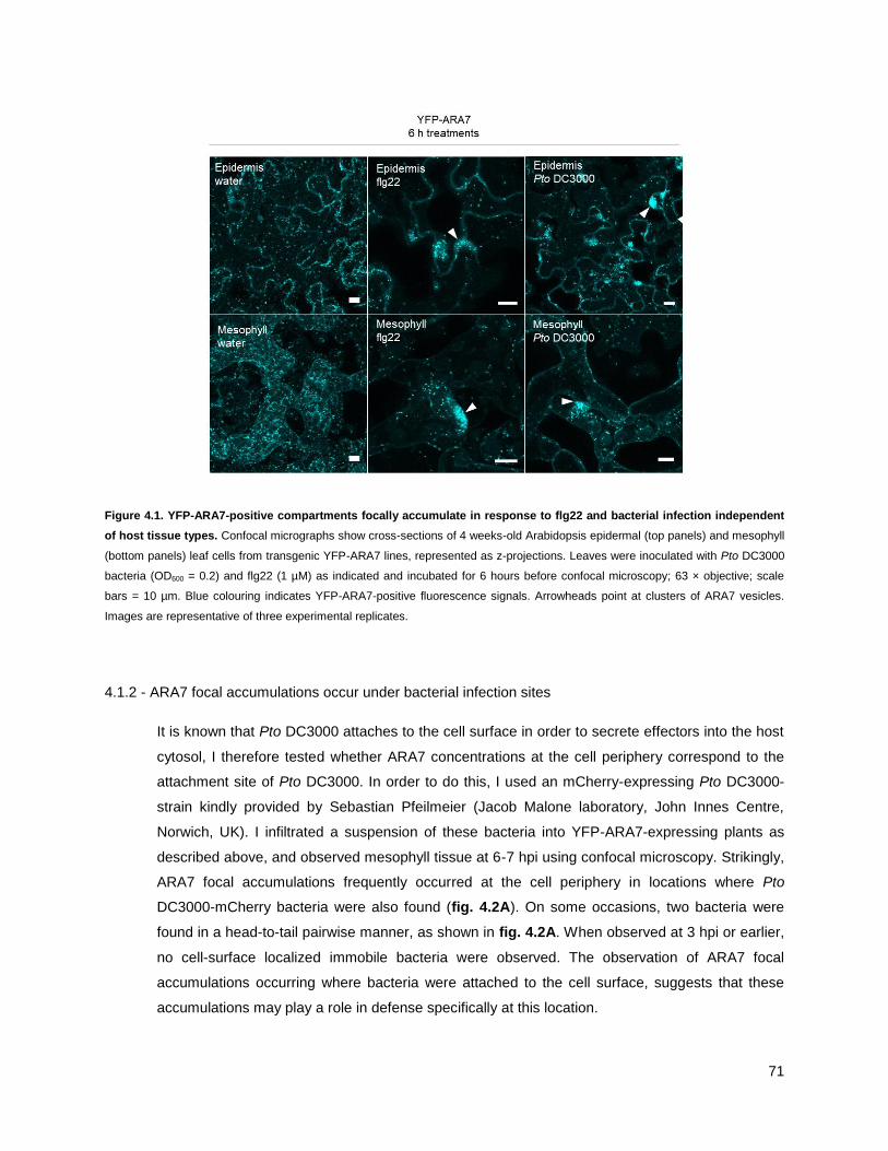
71
Figure 4.1. YFP-ARA7-positive compartments focally accumulate in response to flg22 and bacterial infection independent
of host tissue types. Confocal micrographs show cross-sections of 4 weeks-old Arabidopsis epidermal (top panels) and mesophyll
(bottom panels) leaf cells from transgenic YFP-ARA7 lines, represented as z-projections. Leaves were inoculated with Pto DC3000
bacteria (OD600 = 0.2) and flg22 (1 µM) as indicated and incubated for 6 hours before confocal microscopy; 63 × objective; scale
bars = 10 µm. Blue colouring indicates YFP-ARA7-positive fluorescence signals. Arrowheads point at clusters of ARA7 vesicles.
Images are representative of three experimental replicates.
4.1.2 - ARA7 focal accumulations occur under bacterial infection sites
It is known that Pto DC3000 attaches to the cell surface in order to secrete effectors into the host
cytosol, I therefore tested whether ARA7 concentrations at the cell periphery correspond to the
attachment site of Pto DC3000. In order to do this, I used an mCherry-expressing Pto DC3000-
strain kindly provided by Sebastian Pfeilmeier (Jacob Malone laboratory, John Innes Centre,
Norwich, UK). I infiltrated a suspension of these bacteria into YFP-ARA7-expressing plants as
described above, and observed mesophyll tissue at 6-7 hpi using confocal microscopy. Strikingly,
ARA7 focal accumulations frequently occurred at the cell periphery in locations where Pto
DC3000-mCherry bacteria were also found (fig. 4.2A). On some occasions, two bacteria were
found in a head-to-tail pairwise manner, as shown in fig. 4.2A. When observed at 3 hpi or earlier,
no cell-surface localized immobile bacteria were observed. The observation of ARA7 focal
accumulations occurring where bacteria were attached to the cell surface, suggests that these
accumulations may play a role in defense specifically at this location.

72
Figure 4.2. Focal accumulation of YFP-ARA7-positive compartments is targeted to bacterial infection sites. (A) Confocal
micrographs (z-projections) show YFP-ARA7 and Pto DC3000-mCherry in 4 wk-old Arabidopsis mesophyll tissue, 6 hours after
syringe infiltration of bacterial suspensions. Bacterial OD600 = 0.2 upon infiltration. Lower right panel shows bright field + Pto
DC3000-mCherry. 63 × objective; scale bars = 10 µm. (B) Confocal micrographs (single focal plane) show YFP-ARA7 and FM4-64
(1 µM) at 7 hours after infiltration of Pto DC3000 bacteria without mCherry fluorophores at OD600 = 0.2, and at 30 minutes after
infiltration of FM4-64. 63 × objective; scale bars = 10 µm. (C) Confocal micrographs (single focal plane) show YFP-ARA7 and FM4-
64 (1 µM) at 3 hours after infiltration of Pto DC3000 bacteria without mCherry fluorophores at OD600 = 0.2, and at 5 minutes after
infiltration of FM4-64. 63 × objective; scale bars = 10 µm.
In order to test whether the observed ARA7 concentrations at the cell periphery reflect a locally
increased endocytic activity, I performed uptake experiments with the lipophilic endocytic tracer
dye FM4-64. FM4-64 associates with the lipid bilayer at the PM, is internalized, and starts
accumulating at late endosomes when measured ca. 30 minutes after application but not at
earlier time points (Beck et al., 2012b). FM4-64 also associates with membranes of Pto DC3000

73
shortly after application, allowing for visualisation of bacterial cells (see fig. 4.2C). I applied FM4-
64 to leaves that exhibited ARA7 focal accumulation as confirmed using confocal microscopy at
6-7 h after infiltration of Pto DC3000, and then evaluated the co-localization of YFP-ARA7 and
FM4-64. Interestingly, only a small pool of YFP-ARA7-positive endosomes that were
concentrated at infection sites were stained by FM4-64 (fig. 4.2B). Bacterial cells could be
visualised by intense FM4-64 staining (fig. 4.2B). While it remains possible that local endocytic
activity was upregulated before application of FM4-64, the observation that few endosomes in
ARA7 concentrations were stained within 30 minutes of dye application would suggest that either
existing endosomes are concentrating, or endosomes resulting from increased endocytic activity
do not spread throughout the cell but remain concentrated.
4.1.3 - A panel of different bacteria and MAMPs trigger ARA7 focal accumulation
The pathogen-induced focal accumulation of RABE1d has been shown to increase upon
infiltration of Pto DC3000 AvrRpt2, which secretes the effector AvrRpt2 that triggers ETI (Speth et
al., 2009). Conversely, a role for Pto DC3000 effectors has been identified in suppressing the
occurrence of PEN3 cell-surface focal accumulations (Xin et al., 2013). To test whether disabled
or ETI-triggering Pseudomonas bacteria affected the ARA7 polarized immune response, I
infiltrated YFP-ARA7-expressing leaves with (i) Pto DC3000 COR-, which is deficient in the
secretion of Coronatine, an agent that induces stomatal opening and contributes to virulence in
the mesophyll; (ii) Pto DC3000 HrcC-, which does not form a type-3-secretion system to inject
effectors; (iii) Pto DC3000 AvrRpt2 which triggers ETI; and (iv) the non-adapted Pseudomonas
syringae pv. tabaci (P. s. pv. tabaci). In addition, I infiltrated mCherry-expressing strains of Pto
DC3000 and Pto DC3000 HrcC- to test whether the presence of the mCherry molecule affected
the ability to elicit focal accumulations. For comparison, I also infiltrated the MAMPs flg22 (10 µM)
and chitin (ca. 10 mg/mL).
I observed the tissue at 6-7 hpi using confocal microscopy, taking lower magnification pictures of
the mesophyll to capture numerous cells per field of view to allow for subsequent quantifications.
Interestingly, all tested stimuli, including a water control, triggered focal accumulations of ARA7 at
6-7 hpi to various extent (fig. 4.3). The response is observed at a lower magnification of 20 × as
strong YFP-ARA7 fluorescent maxima at the cell periphery of adaxial mesophyll cells. These
results show that bacterial stimulus can trigger ARA7 focal accumulation despite the bacteria
lacking Coronatine or HrcC, and independent of the presence or absence of a constitutively
expressed mCherry fluorophore. Furthermore, focal accumulations were also observed upon
infiltration with Pto DC3000 AvrRpt2, suggesting that the response occurs despite the activation
of ETI signaling. Finally, both flg22 and chitin treatments triggered ARA7 focal accumulation in
the mesophyll (fig. 4.3).

74
Figure 4.3. Focal accumulation of YFP-ARA7-positive compartments is conserved across different bacterial strains and
MAMP stimuli. Representative confocal micrographs (single focal planes) of acquisition series of YFP-ARA7 in 4 wk-old
Arabidopsis upper mesophyll tissue, 6-7 h after syringe infiltration of the indicated treatments. Upon application, bacterial OD600 =
0.2, flg22 = 1 µM and chitin = ca. 10 mg/mL. 63 × objective, scale bars = 100 µm.
75
Water treatments also induced ARA7 focal accumulation. Indeed, a ―flooding response‖ has been
described before, where re-injection of wildtype intracellular fluid extractions into the apoplast
leads to MAPK activation, indicative of stress signaling (Romeis et al., 2001). PEN1/SYP121 and
PEN3 accumulation at endosomes, thought to underlie their subsequent delivery at the cell
surface, was also triggered to a minor extent under water treatments (Underwood et al., 2017).
This raises the possibility that the clustering of endosomes, and resulting cargo accumulations to
the cell surface, are part of a general stress response but can be targeted to pathogen positions.
Because of this, I was interested in quantifying the response to see if any particular treatment
induced accumulations at a level significantly above that of water treatment. To do this, I
developed a short macro in the image analysis software FIJI (ImageJ) which is described in fig.
4.4A. Briefly, the software applies gaussian blur, intensity thresholding and area thresholding to
separate bright, relatively large spots from the background. Finally, the number of resulting spots
is extracted.
Analysing a series of images in this way revealed that flg22 treatment significantly and strongly
induced focal accumulation (69 ± 16.10 foci / field of view (FOV); fig. 4.4B,D) in comparison to
water (4.20 ± 2.22 foci / FOV). In a second replicate, flg22 also significantly induced focal
accumulation compared to water (fig. 4.4C,D). Pto DC3000, which was tested in one
experimental replicate, induced ARA7 focal accumulations significantly compared to water
control. Pto DC3000-mCherry triggered significantly more ARA7 focal accumulations compared to
water in only one experimental replicate (9.67 ± 3.28; fig. 4.4C,D). Similarly, Pto DC3000 HrcC-
(6.14 ± 0.88 vs. 0 ± 0; fig. 4.4C,D) and Pto AvrRpt2 (29.00 ± 5.51 vs. 4.20 ± 2.22; fig. 4.4B,D)
induced focal accumulations in only one experimental replicate. Interestingly, P. syringae pv.
tabaci did not significantly trigger more ARA7 focal accumulations than water in both
experimental replicates. P. s. pv. tabaci has been shown to elicit HR-like cell death in Arabidopsis
Col-0, which is abolished in mutants lacking flagellin (Ishiga et al., 2005). In contrast, Pto
DC3000-AvrRpt2 triggers NLR-mediated cell death, yet retained the capacity to elicit ARA7
concentrations beyond levels observed in water treatment. Knowing that P. s. pv. tabaci
successfully colonizes Arabidopsis Col-0 in the absence of flagellin, and without eliciting cell
death, it is conceivable that it secretes effectors that suppress trafficking responses in Col-0.
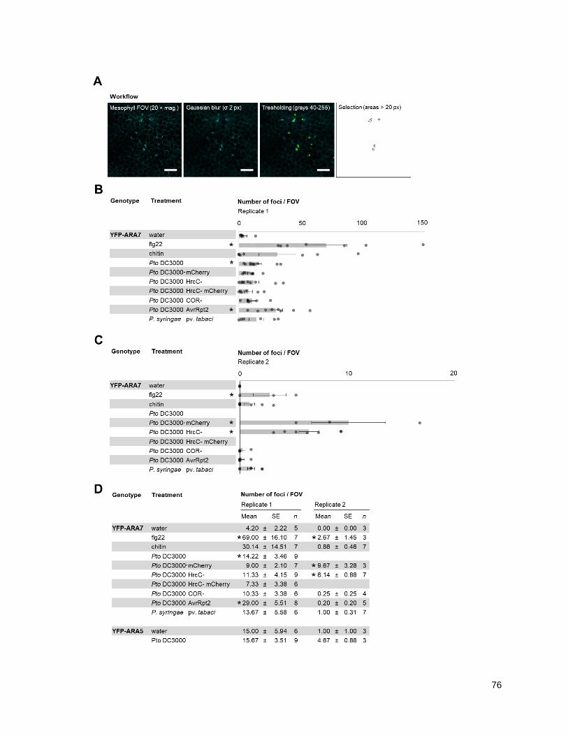
76

77
Figure 4.4. Quantitative analysis reveals significant differences in focal accumulation of YFP-ARA7-positive compartments.
(A) Workflow as encoded in a custom FIJI script, showing individual image modifications performed to obtain final number of focal
accumulations per image as used in B-D. 20 × objective, scale bars = 100 µm. (B,C) Graphical representations of number of focal
accumulations detected using the workflow in (A), showing individual measurements and mean (bars) ± SE (error bars) for indicated
treatments corresponding to replicates shown in (D). (D) Overview listing the means ± SE of the number of YFP-ARA7 focal
accumulations per field-of view, and n = number of images, for two replicates. Quantifications were performed in an automated way
using confocal micrographs acquired as in Figure 2.3 at 6 hours after syringe infiltration of indicated treatments.
As is evident from visual inspection of the results (eg. fig. 4.4A), the FIJI macro does not pick up
on all focal accumulations that would be identified by eye. There are focal accumulations of which
the fluorescent signal locally contrast with the surrounding environment, but on a global level fall
within the background. This is a limitation of using global filtering and thresholding to extract
features. Furthermore, the macro was developed and optimized by using water treated and flg22-
treated samples as the input, which might cause a bias toward being able to distinguish between
those treatments, although the expectation is that the response looks the same independent of
stimulus. Further optimization, employing local thresholding analysis, as well as using multiple
stimuli to calibrate the script, would likely yield further improvements.
4.1.4 - ARA7 does not change in vesicle number upon immune stimulus (0-3 h)
In order to test whether increased trafficking requirements could be reflected by an upregulation
in the number of ARA7 endosomes, I quantified them using high throughput confocal imaging of
cotyledons under flagellin elicitation. Cotyledons of the pUB::YFP-ARA7 genotype, alongside
pFLS2::FLS2-GFP as a positive control, were submerged in liquid Murashige and Skoog medium
(MS), or liquid MS + flg22 in a 96-wells optical plate.
Although no significant ARA7 focal accumulation was observed before ca. 6 h of flg22 or bacterial
treatments, immune-related trafficking events are induced, such as the ligand-induced
endocytosis of FLS2 which peaks between 1-1.5 h after addition of flg22 (Beck et al., 2012). In
addition, cell-surface cargoes involved in defence such as PEN1 and PEN3 can be observed to
accumulate at endomembrane compartments at 4 h after triggering immune signaling using flg22
(Underwood et al., 2017). I therefore examined the number of YFP-ARA7-positive endosomes. At
0.5, 1.5 and 2.5 h, measurements were started and performed across an acquisition window of
30 minutes, and the average numbers of detected endosomes for YFP-ARA7 and FLS2-GFP
under control and flg22 treatments were calculated. Interestingly, no statistically significant
differences in the number of YFP-ARA7 spots per field-of-view between control or flg22
treatments were detected in any time point (fig. 4.5). By contrast, the number of FLS2-GFP
endosomes was significantly increased at 2 and 3 h after flg22 treatment compared to control
treatment, with a maximum at 2 hpi, indicating that flg22 treatment could successfully induce
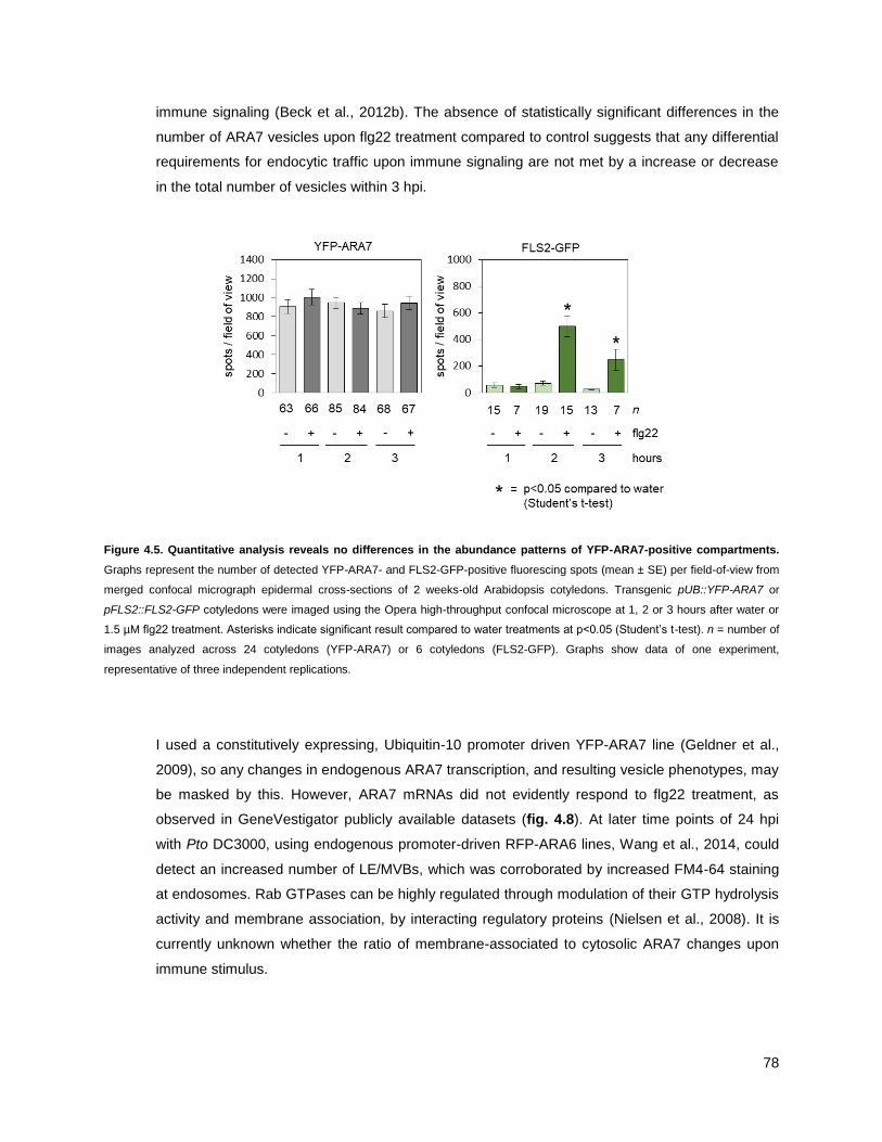
78
immune signaling (Beck et al., 2012b). The absence of statistically significant differences in the
number of ARA7 vesicles upon flg22 treatment compared to control suggests that any differential
requirements for endocytic traffic upon immune signaling are not met by a increase or decrease
in the total number of vesicles within 3 hpi.
Figure 4.5. Quantitative analysis reveals no differences in the abundance patterns of YFP-ARA7-positive compartments.
Graphs represent the number of detected YFP-ARA7- and FLS2-GFP-positive fluorescing spots (mean ± SE) per field-of-view from
merged confocal micrograph epidermal cross-sections of 2 weeks-old Arabidopsis cotyledons. Transgenic pUB::YFP-ARA7 or
pFLS2::FLS2-GFP cotyledons were imaged using the Opera high-throughput confocal microscope at 1, 2 or 3 hours after water or
1.5 µM flg22 treatment. Asterisks indicate significant result compared to water treatments at p<0.05 (Student‘s t-test). n = number of
images analyzed across 24 cotyledons (YFP-ARA7) or 6 cotyledons (FLS2-GFP). Graphs show data of one experiment,
representative of three independent replications.
I used a constitutively expressing, Ubiquitin-10 promoter driven YFP-ARA7 line (Geldner et al.,
2009), so any changes in endogenous ARA7 transcription, and resulting vesicle phenotypes, may
be masked by this. However, ARA7 mRNAs did not evidently respond to flg22 treatment, as
observed in GeneVestigator publicly available datasets (fig. 4.8). At later time points of 24 hpi
with Pto DC3000, using endogenous promoter-driven RFP-ARA6 lines, Wang et al., 2014, could
detect an increased number of LE/MVBs, which was corroborated by increased FM4-64 staining
at endosomes. Rab GTPases can be highly regulated through modulation of their GTP hydrolysis
activity and membrane association, by interacting regulatory proteins (Nielsen et al., 2008). It is
currently unknown whether the ratio of membrane-associated to cytosolic ARA7 changes upon
immune stimulus.

79
4.2 - THE ARA7 CO-PURIFYING PROTEOME CHANGES UPON IMMUNE STIMULUS
4.2.1 - Purifying ARA7 from seedlings under water and flagellin conditions
The polarized immune response of YFP-ARA7-positive vesicles suggests that ARA7 plays a role
in the cell-surface delivery of defense components. To investigate this, I set out to identify ARA7-
interacting proteins, and cargoes of ARA7 endosomes, with the goal to compare resting-state and
immune stimulated conditions. For this, I used a method of immunoprecipitation and mass
spectrometry from Arabidopsis seedlings that was previously developed in our laboratory and
shown to identify endosomal enriched proteins in a trafficking pathway-specific manner (Heard et
al., 2015). To allow for substantial transcriptional reprogramming upon flg22 treatment (Denoux et
al., 2008; Jiménez-Góngora et al., 2015), and anticipating to immunoprecipitate ARA7 from tissue
where endosomes are accumulating defence-related cargoes (Underwood et al., 2017) yet do not
markedly change in localization (fig. 4.5), I selected a time point of 3 h flg22 treatment.
I grew Arabidopsis seedlings of the genotypes pUB::YFP-ARA7 and pUB::YFP in liquid MS
medium for 8 days, and subjected both to 3 h of water or 1.5 µM flg22 treatment, starting the
timer after 1.5 minutes of vacuum application (fig. 4.6A). I included YFP as a control, as proteins
may unspecifically bind to the affinity beads, the bead-bound nanobody, or the YFP domain used
to purify ARA7. I then performed immunoprecipitation using YFP affinity beads, separated
proteins using 1D gel electrophoresis, and subjected the resulting samples to mass spectrometry
analysis performed by Jan Sklenar (Proteomics Support Team, The Sainsbury Laboratory,
Norwich, UK; fig. 4.6A). The above experiment was performed a total of three times
independently.
As a result, 882 proteins co-purified with YFP-ARA7 under water treatment, and 1101 proteins
co-purified with YFP under water treatment (fig. 4.6B). In order to determine a specific YFP-
ARA7 co-purifying proteome, I decided the total number of detected peptides for a given protein
in the YFP-ARA7 purification should be more than 4x greater than the total number of peptides in
the YFP purification. Using this criterion, 723 proteins in the YFP-ARA7 purification were deemed
unspecific, and 159 proteins remained, which I define as the YFP-ARA7 co-purifying proteome
(table 4.1).

80
Figure 4.6. Identification of proteins associated with YFP-ARA7-positive compartments in a flg22-responsive manner. (A)
Schematic overview shows experimental setup. Confocal micrographs show YFP fluorescent signal in z-projections of 8 days-old
Arabidopsis seedlings expressing pUB::YFP-ARA7 or pUB::YFP; scale bars = 10 µm. Seedlings were grown in liquid culture and
treated with 1.5 µM flg22 and water for 3 hours before extraction (text box). (B) Venn-diagram shows groups of proteins co-purifying
with the indicated bait proteins, as revealed by immunoprecipitation and mass spectrometry. Across three replicates, co-purifying
proteins with YFP-ARA7-positive compartments were compared to YFP co-purifying proteins. The YFP-ARA7 co-purifying proteome
was defined based on quality control criteria (present in >1 replicates, peptides = >5) and fold-change ratio of >4 in YFP-ARA7
compared to YFP. Details available in Table 4.1. (C) Venn-diagram shows YFP-ARA7 co-purifying proteins as compared with known
trafficking regulators (source: Heard et al. 2015). Abundant trafficking regulators co-purifying with YFP-ARA7 listed. (D) Venn-
diagram shows YFP-ARA7 co-purifying proteins without and with flg22 treatment, as compared with each other. Prominent proteins
associating with YFP-ARA7-positive compartments in a flg22-responsive manner are shown in the call-out. Details available in table
4.3.
81
Table 4.1: ARA7 co-purifying proteome. Listed here is the ARA7 co-purifying proteome under water treated conditions. 8-day old
seedlings of genotypes pUB::YFP-ARA7 and pUB::YFP were treated with water or 1.5 µM flg22 for three hours, and subjected to
immunoprecipitation using YFP affinity beads. Mass spectrometry was performed in collaboration with Jan Sklenar, Paul Derbyshire
and Frank Menke (The Sainsbury Laboratory, Norwich, UK). To determine co-purifying proteomes, Scaffold software was used to
retain proteins with a ProteinProphet score of 99% based on a minimum of 2 spectra with a PeptideProphet score of 95%. MS Excel
was then used to retain proteins with at least 5 spectra across all three replicates in the YFP-ARA7 water-treated samples, with an
occurence in more than 1 of these replicates, and an abundance increase of at least 4x compared to matching YFP controls.
(Columns 1-8) Observed in # of replicates, and Spectra in combined replicates: Numbers and associated green cell-shading
intensities indicate in how many experimental replicates the protein was detected, or how many spectra for that protein were
identified in all three replicates combined, separated for water or flagellin treatment. (Columns 9-10) Enrichment factors: For each
protein, the number of total spectra across three replicates of the green-labeled (rightmost) pulldown was divided by the number of
total spectra in red-labeled (left-most) pulldown. Log10 values that correspond to these ratios are displayed in this table. Green or
red bars are graphical representations of magnitude and direction of positive (green) or negative (red) values, centered at 0.
(Columns 11-12) Locus ID and associated names. Source: TAIR10, accessed through www.genevestigator.org. The table is sorted
for total spectral numbers in ―YFP-ARA7 water‖, in descending order.
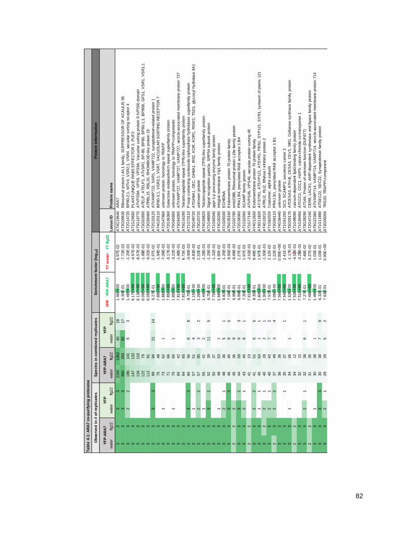
82
Ta
ble
4.1
: A
RA
7 c
o-p
uri
fyin
g p
rote
om
e
wate
rflg
22
wate
rflg
22
wate
rflg
22
wate
rflg
22
Lo
cu
s ID
Pro
tein
na
me
33
33
1160
1362
40
18
1.4
6E
+00
6.9
7E
-02
AT4G
19640
AR
A7
33
33
350
293
82
17
6.3
0E
-01
-7.7
2E
-02
AT3G
09630
Rib
osom
al pro
tein
L4/L
1 fam
ily,
SU
PP
RE
SS
OR
OF
AC
AU
LIS
56
33
22
186
141
63
1.4
9E
+00
-1.2
0E
-01
AT2G
14720
BP
80-2
;1,
MTV
4,
VS
R2;1
, V
SR
4,
vacuola
r sort
ing r
ecepto
r 4
33
147
132
8.1
7E
+00
-4.6
7E
-02
AT1G
24560
PLA
NT-U
NIQ
UE
RA
B5 E
FF
EC
TO
R 2
, P
UF
2
33
134
110
8.1
3E
+00
-8.5
7E
-02
AT3G
19770
ATV
PS
9A
, V
PS
9,
VP
S9A
, V
acuola
r sort
ing p
rote
in 9
(V
PS
9)
dom
ain
33
122
79
8.0
9E
+00
-1.8
9E
-01
AT3G
52850
ATE
LP
, A
TE
LP
1,
ATV
SR
1,
BP
-80,
BP
80,
BP
80-1
;1,
BP
80B
, G
FS
1,
VS
R1,
VS
R1;1
33
112
91
8.0
5E
+00
-9.0
2E
-02
AT3G
58460
ATR
BL15,
RB
L15,
RH
OM
BO
ID-lik
e p
rote
in 1
5
33
33
89
94
21
14
6.2
7E
-01
2.3
7E
-02
AT2G
33120
ATV
AM
P722,
SA
R1,
VA
MP
722,
synapto
bre
vin-r
ela
ted p
rote
in 1
33
75
48
7.8
8E
+00
-1.9
4E
-01
AT4G
20110
BP
80-3
;1,
VS
R3;1
, V
SR
7,
VA
CU
OLA
R S
OR
TIN
G R
EC
EP
TO
R 7
33
173
62
11.8
6E
+00
-7.0
9E
-02
AT2G
47960
unknow
n p
rote
in,
hom
olo
gy t
o T
RA
PP
33
71
66
7.8
5E
+00
-3.1
7E
-02
AT2G
36360
Gala
cto
se o
xid
ase/k
elc
h r
epeat
superfam
ily p
rote
in
33
170
65
11.8
5E
+00
-3.2
2E
-02
AT5G
65950
unknow
n p
rote
in,
hom
olo
gy t
o T
RA
PP
I-com
ponents
33
65
42
7.8
1E
+00
-1.9
0E
-01
AT3G
54300
ATV
AM
P727,
VA
MP
727,
VA
MP
727,
vesic
le-a
ssocia
ted m
em
bra
ne p
rote
in 7
27
33
64
65
7.8
1E
+00
6.7
3E
-03
AT5G
16280
Tetr
atr
icopeptide r
epeat
(TP
R)-
like s
uperfam
ily p
rote
in
33
33
60
56
88
8.7
5E
-01
-3.0
0E
-02
AT1G
73110
P-loop c
onta
inin
g n
ucle
osid
e t
riphosphate
hydro
lases s
uperfam
ily p
rote
in
33
157
51
41.1
5E
+00
-4.8
3E
-02
AT5G
49720
ATG
H9A
1,
DE
C,
GH
9A
1,
IRX2,
KO
R,
KO
R1,
RS
W2,
TS
D1,
gly
cosyl hydro
lase 9
A1
33
22
57
95
32
1.2
8E
+00
2.2
2E
-01
AT3G
20720
unknow
n p
rote
in
33
155
41
21.4
4E
+00
-1.2
8E
-01
AT4G
39820
Tetr
atr
icopeptide r
epeat
(TP
R)-
like s
uperfam
ily p
rote
in
33
32
52
39
11
56.7
5E
-01
-1.2
5E
-01
AT1G
48900
Sig
nal re
cognitio
n p
art
icle
, S
RP
54 s
ubunit p
rote
in
33
50
37
7.7
0E
+00
-1.3
1E
-01
AT1G
69340
appr-
1-p
pro
cessin
g e
nzym
e fam
ily p
rote
in
33
148
53
11.6
8E
+00
4.3
0E
-02
AT4G
30260
Inte
gra
l m
em
bra
ne Y
ip1 fam
ily p
rote
in
33
21
48
36
51
9.8
2E
-01
-1.2
5E
-01
AT1G
09330
EC
HID
NA
33
33
46
45
95
7.0
9E
-01
-9.5
5E
-03
AT5G
35160
Endom
em
bra
ne p
rote
in 7
0 p
rote
in fam
ily
22
245
36
96.9
9E
-01
-9.6
9E
-02
AT1G
02780
em
b2386,
Rib
osom
al pro
tein
L19e fam
ily p
rote
in
33
32
44
59
93
6.8
9E
-01
1.2
7E
-01
AT2G
38360
PR
A1.B
4,
pre
nyla
ted R
AB
accepto
r 1.B
4
33
21
43
45
81
7.3
0E
-01
1.9
7E
-02
AT1G
32050
SC
AM
P fam
ily p
rote
in
22
41
23
7.6
1E
+00
-2.5
1E
-01
AT1G
77140
ATV
PS
45,
VP
S45,
vacuola
r pro
tein
sort
ing 4
5
33
22
41
51
65
8.3
5E
-01
9.4
8E
-02
AT4G
12650
Endom
em
bra
ne p
rote
in 7
0 p
rote
in fam
ily
33
11
40
63
11
1.6
0E
+00
1.9
7E
-01
AT3G
11820
AT-S
YR
1,
ATS
YP
121,
ATS
YR
1,
PE
N1,
SY
P121,
SY
R1,
synta
xin
of pla
nts
121
32
21
40
28
21
1.3
0E
+00
-1.5
5E
-01
AT4G
19210
ATR
LI2
, R
LI2
, R
NA
se l inhib
itor
pro
tein
2
33
21
40
42
72
7.5
7E
-01
2.1
2E
-02
AT1G
62020
Coato
mer,
alp
ha s
ubunit
23
22
37
49
34
1.0
9E
+00
1.2
2E
-01
AT3G
56110
PR
A1.B
1,
pre
nyla
ted R
AB
accepto
r 1.B
1
33
36
36
7.5
6E
+00
0.0
0E
+00
AT4G
36440
unknow
n p
rote
in
33
135
37
17.5
4E
+00
2.4
1E
-02
AT1G
61250
SC
3,
SC
AM
P3,
secre
tory
carr
ier
3
33
134
26
11.5
3E
+00
-1.1
7E
-01
AT5G
05170
ATC
ES
A3,
ATH
-B,
CE
SA
3,
CE
V1,
IXR
1,
Cellu
lose s
ynth
ase fam
ily p
rote
in
22
34
12
7.5
3E
+00
-4.5
2E
-01
AT1G
48090
calc
ium
-dependent
lipid
-bin
din
g fam
ily p
rote
in
33
32
15
7.5
1E
+00
-3.2
9E
-01
AT1G
30450
ATC
CC
1,
CC
C1,
HA
P5,
cation-c
hlo
ride c
o-t
ransport
er
1
33
21
32
38
61
7.2
7E
-01
7.4
6E
-02
AT3G
28290
AT14A
, P
rote
in o
f unknow
n function (
DU
F677)
22
31
35
7.4
9E
+00
5.2
7E
-02
AT2G
47240
CE
R8,
LA
CS
1,
AM
P-d
ependent
synth
eta
se a
nd lig
ase fam
ily p
rote
in
33
130
38
11.4
8E
+00
1.0
3E
-01
AT5G
22360
ATV
AM
P714,
VA
MP
714,
VA
MP
714,
vesic
le-a
ssocia
ted m
em
bra
ne p
rote
in 7
14
33
33
30
38
79
6.3
2E
-01
1.0
3E
-01
AT1G
11890
ATS
EC
22,
SE
C22,
Synapto
bre
vin fam
ily p
rote
in
33
21
29
29
52
7.6
3E
-01
0.0
0E
+00
AT3G
05000
TR
S33,
TR
AP
PI-com
ponent
Ob
serv
ed
in
# o
f re
pli
ca
tes
Sp
ectr
a i
n c
om
bin
ed
re
pli
ca
tes
En
rich
me
nt
facto
r (l
og
10)
Pro
tein
in
form
ati
on
YF
P-A
RA
7Y
FP
YF
P-A
RA
7Y
FP
YF
P
YF
P-A
RA
7Y
7 w
ate
r
Y7 f
lg22

83
33
29
29
7.4
6E
+00
0.0
0E
+00
AT1G
08770
PR
A1.E
, pre
nyla
ted R
AB
accepto
r 1.E
33
227
25
57.3
2E
-01
-3.3
4E
-02
AT1G
02130
AR
A-5
, A
RA
5,
ATR
AB
1B
, A
TR
AB
D2A
, R
A-5
, R
AB
D2A
, R
AS
5
33
127
51
21.1
3E
+00
2.7
6E
-01
AT2G
36850
ATG
SL08,
ATG
SL8,
CH
OR
, G
SL08,
GS
L8,
glu
can s
ynth
ase-lik
e 8
22
27
20
7.4
3E
+00
-1.3
0E
-01
AT2G
26890
GR
V2,
KA
M2,
DN
AJ
heat
shock N
-term
inal dom
ain
-conta
inin
g p
rote
in
32
126
10
21.1
1E
+00
-4.1
5E
-01
AT5G
13560
unknow
n p
rote
in
33
32
25
20
45
7.9
6E
-01
-9.6
9E
-02
AT5G
42220
Ubiq
uitin
-lik
e s
uperfam
ily p
rote
in
33
225
25
39.2
1E
-01
0.0
0E
+00
AT5G
04830
Nucle
ar
transport
facto
r 2 (
NTF
2)
fam
ily p
rote
in
22
24
26
7.3
8E
+00
3.4
8E
-02
AT1G
59820
ALA
3,
am
inophospholip
id A
TP
ase 3
32
24
67.3
8E
+00
-6.0
2E
-01
AT2G
14740
ATV
SR
3,
BP
80-2
;2,
VS
R2;2
, V
SR
3,
VS
R3,
vaculo
lar
sort
ing r
ecepto
r 3
22
23
37
7.3
6E
+00
2.0
6E
-01
AT3G
05280
Inte
gra
l m
em
bra
ne Y
ip1 fam
ily p
rote
in
33
123
21
38.8
5E
-01
-3.9
5E
-02
AT5G
11040
AtT
RS
120,
TR
S120,
TR
S120
33
22
17
7.3
4E
+00
-1.1
2E
-01
AT5G
58030
TR
S31,
TR
AP
PI-com
ponent
32
122
15
11.3
4E
+00
-1.6
6E
-01
AT1G
28490
ATS
YP
61,
OS
M1,
SY
P61,
synta
xin
of pla
nts
61
33
21
26
7.3
2E
+00
9.2
8E
-02
AT1G
75850
VP
S35B
, V
PS
35 h
om
olo
g B
33
20
20
7.3
0E
+00
0.0
0E
+00
AT2G
40316
unknow
n p
rote
in
32
11
20
10
41
6.9
9E
-01
-3.0
1E
-01
AT3G
10350
P-loop c
onta
inin
g n
ucle
osid
e t
riphosphate
hydro
lases s
uperfam
ily p
rote
in
33
19
18
7.2
8E
+00
-2.3
5E
-02
AT5G
17230
PS
Y,
PH
YTO
EN
E S
YN
TH
AS
E
33
19
30
7.2
8E
+00
1.9
8E
-01
AT2G
22475
GE
M,
GR
AM
dom
ain
fam
ily p
rote
in
22
18
18
7.2
6E
+00
0.0
0E
+00
AT2G
17790
VP
S35A
, ZIP
3,
VP
S35 h
om
olo
g A
32
118
18
17.2
6E
+00
0.0
0E
+00
AT5G
20490
ATXIK
, XI-17,
XIK
, M
yosin
fam
ily p
rote
in w
ith D
il dom
ain
33
118
10
11.2
6E
+00
-2.5
5E
-01
AT2G
30880
Ple
ckstr
in h
om
olo
gy (
PH
) dom
ain
-conta
inin
g p
rote
in
33
13
18
16
46
6.5
3E
-01
-5.1
2E
-02
AT1G
54270
EIF
4A
-2,
eif4
a-2
33
17
28
7.2
3E
+00
2.1
7E
-01
AT3G
07160
ATG
SL10,
CA
LS
9,
gsl1
0,
GS
L10,
glu
can s
ynth
ase-lik
e 1
0
33
117
25
11.2
3E
+00
1.6
7E
-01
AT2G
28520
VH
A-A
1,
vacuola
r pro
ton A
TP
ase A
1
33
17
19
7.2
3E
+00
4.8
3E
-02
AT1G
26670
ATV
TI1
2,
VTI1
2,
VTI1
B,
Vesic
le t
ransport
v-S
NA
RE
fam
ily p
rote
in
22
23
17
15
33
7.5
3E
-01
-5.4
4E
-02
AT2G
43130
AR
A-4
, A
RA
4,
ATR
AB
11F
, A
TR
AB
A5C
, R
AB
A5C
33
16
16
7.2
0E
+00
0.0
0E
+00
AT5G
45130
ATR
AB
-F2A
, A
TR
AB
5A
, A
TR
AB
F2A
, R
AB
-F2A
, R
AB
5A
, R
AB
F2A
, R
HA
1
33
116
82
9.0
3E
-01
-3.0
1E
-01
AT4G
32285
EN
TH
/AN
TH
/VH
S s
uperfam
ily p
rote
in
23
15
19
7.1
8E
+00
1.0
3E
-01
AT5G
54440
AtT
RS
130,
CLU
B,
CLU
B
32
115
14
28.7
5E
-01
-3.0
0E
-02
AT4G
32410
AtC
ES
A1,
CE
SA
1,
RS
W1,
cellu
lose s
ynth
ase 1
32
15
57.1
8E
+00
-4.7
7E
-01
AT5G
42090
Lung s
eve
n t
ransm
em
bra
ne r
ecepto
r fa
mily
pro
tein
22
115
17
11.1
8E
+00
5.4
4E
-02
AT1G
52780
Pro
tein
of unknow
n function (
DU
F2921)
32
15
97.1
8E
+00
-2.2
2E
-01
AT5G
20350
TIP
1,
Ankyrin r
epeat
fam
ily p
rote
in w
ith D
HH
C z
inc fin
ger
dom
ain
33
15
67.1
8E
+00
-3.9
8E
-01
AT2G
03820
nonsense-m
edia
ted m
RN
A d
ecay N
MD
3 fam
ily p
rote
in
33
14
15
7.1
5E
+00
3.0
0E
-02
AT1G
31130
unknow
n p
rote
in
33
114
13
11.1
5E
+00
-3.2
2E
-02
AT3G
58170
ATB
ET11,
ATB
S14A
, B
ET11,
BS
14A
, B
ET1P
/SF
T1P
-lik
e p
rote
in 1
4A
22
214
14
28.4
5E
-01
0.0
0E
+00
AT1G
58684
Rib
osom
al pro
tein
S5 fam
ily p
rote
in
32
13
18
7.1
1E
+00
1.4
1E
-01
AT3G
17900
unknow
n p
rote
in
33
13
17
7.1
1E
+00
1.1
7E
-01
AT1G
27930
Pro
tein
of unknow
n function (
DU
F579)
33
13
28
7.1
1E
+00
3.3
3E
-01
AT3G
05500
Rubber
elo
ngation facto
r pro
tein
(R
EF
)
22
13
57.1
1E
+00
-4.1
5E
-01
AT5G
44240
ALA
2,
am
inophospholip
id A
TP
ase 2
33
113
17
11.1
1E
+00
1.1
7E
-01
AT3G
47810
ATV
PS
29,
MA
G1,
VP
S29,
Calc
ineurin-lik
e m
eta
llo-p
hosphoeste
rase s
uperfam
ily p
rote
in
33
113
12
11.1
1E
+00
-3.4
8E
-02
AT3G
60340
alp
ha/b
eta
-Hydro
lases s
uperfam
ily p
rote
in
33
213
10
28.1
3E
-01
-1.1
4E
-01
AT4G
35870
early-r
esponsiv
e t
o d
ehydra
tion s
tress p
rote
in (
ER
D4)
33
21
13
13
32
6.3
7E
-01
0.0
0E
+00
AT1G
50500
ATV
PS
53,
HIT
1,
VP
S53,
Mem
bra
ne t
raffi
ckin
g V
PS
53 fam
ily p
rote
in
21
21
13
73
26.3
7E
-01
-2.6
9E
-01
AT3G
54840
AR
A-6
, A
RA
6,
ATR
AB
5C
, A
TR
AB
F1,
RA
BF
1,
Ras-r
ela
ted s
mall
GTP
-bin
din
g fam
ily p
rote
in
22
12
19
7.0
8E
+00
2.0
0E
-01
AT4G
03550
ATG
SL05,
ATG
SL5,
GS
L05,
GS
L05,
GS
L5,
PM
R4,
glu
can s
ynth
ase-lik
e 5
33
12
22
7.0
8E
+00
2.6
3E
-01
AT4G
16120
CO
BL7,
SE
B1,
CO
BR
A-lik
e p
rote
in-7
pre
curs
or
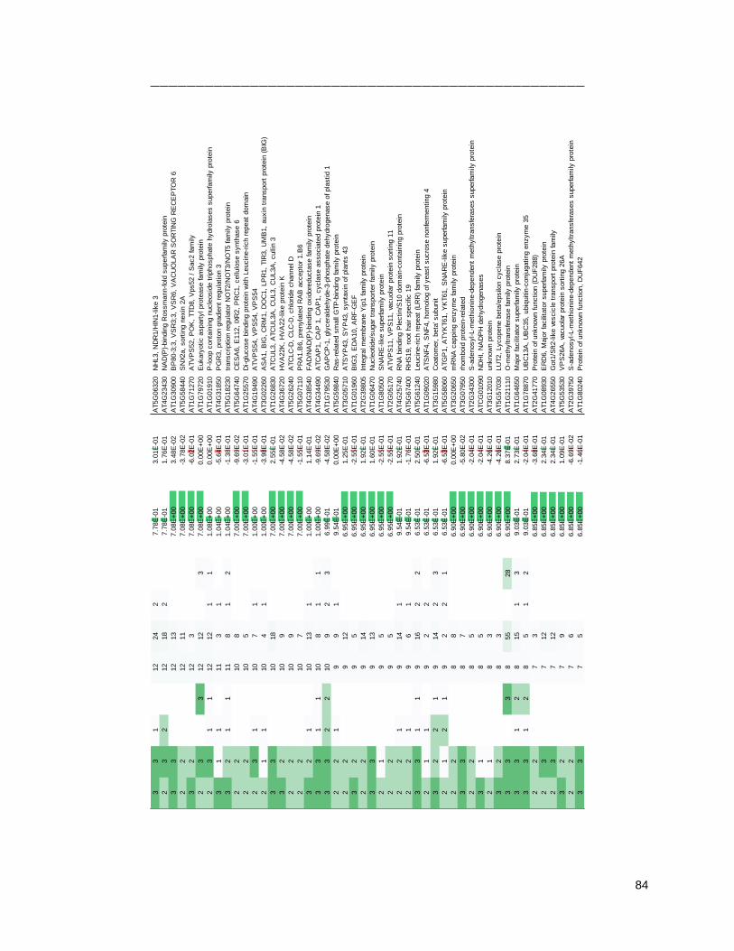
84
33
112
24
27.7
8E
-01
3.0
1E
-01
AT5G
06320
NH
L3,
ND
R1/H
IN1-lik
e 3
23
212
18
27.7
8E
-01
1.7
6E
-01
AT4G
23430
NA
D(P
)-bin
din
g R
ossm
ann-fold
superfam
ily p
rote
in
33
12
13
7.0
8E
+00
3.4
8E
-02
AT1G
30900
BP
80-3
;3,
VS
R3;3
, V
SR
6,
VA
CU
OLA
R S
OR
TIN
G R
EC
EP
TO
R 6
22
12
11
7.0
8E
+00
-3.7
8E
-02
AT5G
58440
SN
X2a,
sort
ing n
exin
2A
32
12
37.0
8E
+00
-6.0
2E
-01
AT1G
71270
ATV
PS
52,
PO
K,
TTD
8,
Vps52 /
Sac2 fam
ily
23
312
12
37.0
8E
+00
0.0
0E
+00
AT1G
79720
Eukary
otic a
spart
yl pro
tease fam
ily p
rote
in
23
11
12
12
11
1.0
8E
+00
0.0
0E
+00
AT1G
01910
P-loop c
onta
inin
g n
ucle
osid
e t
riphosphate
hydro
lases s
uperfam
ily p
rote
in
31
111
31
1.0
4E
+00
-5.6
4E
-01
AT4G
31850
PG
R3,
pro
ton g
radie
nt
regula
tion 3
32
11
11
81
21.0
4E
+00
-1.3
8E
-01
AT5G
18230
transcription r
egula
tor
NO
T2/N
OT3/N
OT5 fam
ily p
rote
in
22
10
87.0
0E
+00
-9.6
9E
-02
AT5G
64740
CE
SA
6,
E112,
IXR
2,
PR
C1,
cellu
lose s
ynth
ase 6
22
10
57.0
0E
+00
-3.0
1E
-01
AT1G
25570
Di-glu
cose b
indin
g p
rote
in w
ith L
eucin
e-r
ich r
epeat
dom
ain
23
110
71
1.0
0E
+00
-1.5
5E
-01
AT4G
19490
ATV
PS
54,
VP
S54,
VP
S54
21
110
41
1.0
0E
+00
-3.9
8E
-01
AT3G
02260
AS
A1,
BIG
, C
RM
1,
DO
C1,
LP
R1,
TIR
3,
UM
B1,
auxin
tra
nsport
pro
tein
(B
IG)
33
10
18
7.0
0E
+00
2.5
5E
-01
AT1G
26830
ATC
UL3,
ATC
UL3A
, C
UL3,
CU
L3A
, culli
n 3
32
10
97.0
0E
+00
-4.5
8E
-02
AT4G
36720
HV
A22K
, H
VA
22-lik
e p
rote
in K
22
10
97.0
0E
+00
-4.5
8E
-02
AT5G
26240
ATC
LC
-D,
CLC
-D,
chlo
ride c
hannel D
22
10
77.0
0E
+00
-1.5
5E
-01
AT5G
07110
PR
A1.B
6,
pre
nyla
ted R
AB
accepto
r 1.B
6
32
110
13
11.0
0E
+00
1.1
4E
-01
AT4G
38540
FA
D/N
AD
(P)-
bin
din
g o
xid
ore
ducta
se fam
ily p
rote
in
33
11
10
81
11.0
0E
+00
-9.6
9E
-02
AT4G
34490
ATC
AP
1,
CA
P 1
, C
AP
1,
cycla
se a
ssocia
ted p
rote
in 1
33
22
10
92
36.9
9E
-01
-4.5
8E
-02
AT1G
79530
GA
PC
P-1
, gly
cera
ldehyde-3
-phosphate
dehydro
genase o
f pla
stid 1
22
19
91
9.5
4E
-01
0.0
0E
+00
AT5G
59840
Ras-r
ela
ted s
mall
GTP
-bin
din
g fam
ily p
rote
in
22
912
6.9
5E
+00
1.2
5E
-01
AT3G
05710
ATS
YP
43,
SY
P43,
synta
xin
of pla
nts
43
32
95
6.9
5E
+00
-2.5
5E
-01
AT1G
01960
BIG
3,
ED
A10,
AR
F-G
EF
22
914
6.9
5E
+00
1.9
2E
-01
AT2G
39805
Inte
gra
l m
em
bra
ne Y
ip1 fam
ily p
rote
in
33
913
6.9
5E
+00
1.6
0E
-01
AT1G
06470
Nucle
otide/s
ugar
transport
er
fam
ily p
rote
in
21
95
6.9
5E
+00
-2.5
5E
-01
AT1G
80500
SN
AR
E-lik
e s
uperfam
ily p
rote
in
22
95
6.9
5E
+00
-2.5
5E
-01
AT2G
05170
ATV
PS
11,
VP
S11,
vacuola
r pro
tein
sort
ing 1
1
22
19
14
19.5
4E
-01
1.9
2E
-01
AT4G
25740
RN
A b
indin
g P
lectin/S
10 d
om
ain
-conta
inin
g p
rote
in
22
19
61
9.5
4E
-01
-1.7
6E
-01
AT5G
67400
RH
S19,
root
hair s
pecifi
c 1
9
33
11
916
22
6.5
3E
-01
2.5
0E
-01
AT5G
61240
Leucin
e-r
ich r
epeat
(LR
R)
fam
ily p
rote
in
21
19
22
6.5
3E
-01
-6.5
3E
-01
AT1G
09020
ATS
NF
4,
SN
F4,
hom
olo
g o
f yeast
sucro
se n
onfe
rmenting 4
32
21
914
23
6.5
3E
-01
1.9
2E
-01
AT3G
15980
Coato
mer,
beta
' subunit
21
21
92
21
6.5
3E
-01
-6.5
3E
-01
AT5G
58060
ATG
P1,
ATY
KT61,
YK
T61,
SN
AR
E-lik
e s
uperfam
ily p
rote
in
22
88
6.9
0E
+00
0.0
0E
+00
AT3G
20650
mR
NA
cappin
g e
nzym
e fam
ily p
rote
in
33
87
6.9
0E
+00
-5.8
0E
-02
AT3G
07950
rhom
boid
pro
tein
-rela
ted
22
85
6.9
0E
+00
-2.0
4E
-01
AT2G
34300
S-a
denosyl-L-m
eth
ionin
e-d
ependent
meth
yltra
nsfe
rases s
uperfam
ily p
rote
in
31
85
6.9
0E
+00
-2.0
4E
-01
ATC
G01090
ND
HI, N
AD
PH
dehydro
genases
21
83
6.9
0E
+00
-4.2
6E
-01
AT3G
12010
unknow
n p
rote
in
32
83
6.9
0E
+00
-4.2
6E
-01
AT5G
57030
LU
T2,
Lycopene b
eta
/epsilo
n c
ycla
se p
rote
in
33
38
55
28
6.9
0E
+00
8.3
7E
-01
AT1G
21110
O-m
eth
yltra
nsfe
rase fam
ily p
rote
in
33
12
815
13
9.0
3E
-01
2.7
3E
-01
AT1G
64650
Majo
r fa
cili
tato
r superfam
ily p
rote
in
33
12
85
12
9.0
3E
-01
-2.0
4E
-01
AT1G
78870
UB
C13A
, U
BC
35,
ubiq
uitin
-conju
gating e
nzym
e 3
5
22
73
6.8
5E
+00
-3.6
8E
-01
AT2G
41770
Pro
tein
of unknow
n function (
DU
F288)
23
712
6.8
5E
+00
2.3
4E
-01
AT1G
08930
ER
D6,
Majo
r fa
cili
tato
r superfam
ily p
rote
in
23
712
6.8
5E
+00
2.3
4E
-01
AT4G
26550
Got1
/Sft2-lik
e v
escic
le t
ransport
pro
tein
fam
ily
32
79
6.8
5E
+00
1.0
9E
-01
AT5G
53530
VP
S26A
, va
cuola
r pro
tein
sort
ing 2
6A
22
76
6.8
5E
+00
-6.6
9E
-02
AT2G
39750
S-a
denosyl-L-m
eth
ionin
e-d
ependent
meth
yltra
nsfe
rases s
uperfam
ily p
rote
in
33
75
6.8
5E
+00
-1.4
6E
-01
AT1G
80240
Pro
tein
of unknow
n function,
DU
F642

85
33
17
13
16.8
5E
+00
2.6
9E
-01
AT1G
74020
SS
2,
str
icto
sid
ine s
ynth
ase 2
22
17
91
8.4
5E
-01
1.0
9E
-01
AT1G
67330
Pro
tein
of unknow
n function (
DU
F579)
33
17
81
8.4
5E
-01
5.8
0E
-02
AT3G
28720
unknow
n p
rote
in
33
17
61
8.4
5E
-01
-6.6
9E
-02
AT3G
13720
PR
A1.F
3,
PR
A8,
PR
A1 (
Pre
nyla
ted r
ab a
ccepto
r) fam
ily p
rote
in
22
16
11
17.7
8E
-01
2.6
3E
-01
AT5G
57350
AH
A3,
ATA
HA
3,
HA
3,
H(+
)-A
TP
ase 3
32
63
6.7
8E
+00
-3.0
1E
-01
AT3G
16785
PLD
ZE
TA
1,
PLD
P1,
PLD
Z1,
PLD
ZE
TA
1,
phospholip
ase D
P1
23
611
6.7
8E
+00
2.6
3E
-01
AT1G
25420
Regula
tor
of V
ps4 a
ctivi
ty in t
he M
VB
path
way p
rote
in
23
610
6.7
8E
+00
2.2
2E
-01
AT1G
80360
Pyridoxal phosphate
(P
LP
)-dependent
transfe
rases s
uperfam
ily p
rote
in
22
68
6.7
8E
+00
1.2
5E
-01
AT3G
53520
ATU
XS
1,
UXS
1,
UD
P-g
lucuro
nic
acid
decarb
oxyla
se 1
32
67
6.7
8E
+00
6.6
9E
-02
AT1G
79050
recA
DN
A r
ecom
bin
ation fam
ily p
rote
in
22
66
6.7
8E
+00
0.0
0E
+00
AT1G
67430
Rib
osom
al pro
tein
L22p/L
17e fam
ily p
rote
in
32
66
6.7
8E
+00
0.0
0E
+00
AT1G
67560
PLA
T/L
H2 d
om
ain
-conta
inin
g lip
oxygenase fam
ily p
rote
in
33
65
6.7
8E
+00
-7.9
2E
-02
AT1G
68310
Pro
tein
of unknow
n function (
DU
F59)
22
65
6.7
8E
+00
-7.9
2E
-02
AT2G
35610
XE
G113,
xylo
glu
canase 1
13
23
64
6.7
8E
+00
-1.7
6E
-01
AT1G
02475
Poly
ketide c
ycla
se/d
ehydra
se a
nd lip
id t
ransport
superfam
ily p
rote
in
21
63
6.7
8E
+00
-3.0
1E
-01
AT2G
04280
unknow
n p
rote
in
23
16
41
6.7
8E
+00
-1.7
6E
-01
AT4G
24550
Cla
thrin a
dapto
r com
ple
xes m
ediu
m s
ubunit fam
ily p
rote
in
23
16
61
7.7
8E
-01
0.0
0E
+00
AT2G
40690
GLY
1,
SF
D1,
NA
D-d
ependent
gly
cero
l-3-p
hosphate
dehydro
genase fam
ily p
rote
in
21
16
41
7.7
8E
-01
-1.7
6E
-01
AT3G
15060
AtR
AB
A1g,
RA
BA
1g,
RA
B G
TP
ase h
om
olo
g A
1G
33
11
67
11
7.7
8E
-01
6.6
9E
-02
AT5G
45620
Pro
teasom
e c
om
ponent
(PC
I) d
om
ain
pro
tein
23
13
621
14
7.7
8E
-01
5.4
4E
-01
AT5G
16830
ATP
EP
12,
ATS
YP
21,
PE
P12,
PE
P12P
, S
YP
21,
synta
xin
of pla
nts
21
86
4.2.2 - The ARA7 co-purifying proteome contains expected TGN/EE and LE/MVB proteins
In order to gather further confidence that the so-defined YFP-ARA7 co-purifying proteome is
specific to ARA7, I filtered the proteome to retain only endomembrane trafficking regulators, by
using a comprehensive manually curated list of regulators from (Heard et al., 2015). 49 trafficking
regulators were found in the dataset (fig. 4.6C), which are listed in table 4.2. Besides ARA7 itself,
the most abundant proteins in this resulting list are TGN/EE and LE/MVB-associated proteins,
which correspond to the endomembrane compartments to which ARA7 localizes.
Vacular Sorting Receptors
I found proteins of the vacuolar sorting receptor (VSR) group, with VSR1/ELP/BP80,
VSR4/MTV4, and VSR7 at high abundance (table 4.2). VSRs are present in the endomembrane
system along the endocytic route. They localize to the PM, clathrin-coated vesicles and TGN/EE,
as well as LE/MVBs (Robinson and Neuhaus, 2016). VSR4 cycles between TGN/EE and PM
(Saint-Jean et al., 2010). VSR1/ELP/BP80 is strongly TGN/EE and MVB localized (Li et al.,
2002). After performing their vacuolar sorting function, which does not occur at the LE/MVB,
VSRs are themselves recycled to their original location by the membrane-associated retromer
complex (Künzl et al., 2016). The core retromer complex contains subunits VPS26, 29 and 35
(Zelazny et al., 2013), and is found in its entirety in my ARA7 purifications (table 4.2). This
complex has previously been demonstrated to be present at the LE/MVB (Heucken and Ivanov,
2017).
Table 4.2: Trafficking regulators in the ARA7 co-purifying proteome. Listed here is the ARA7 co-purifying proteome under
water treated conditions, copied from Table 4.1, and filtered to retain only endomembrane trafficking regulators as defined in Heard
et al. 2015.
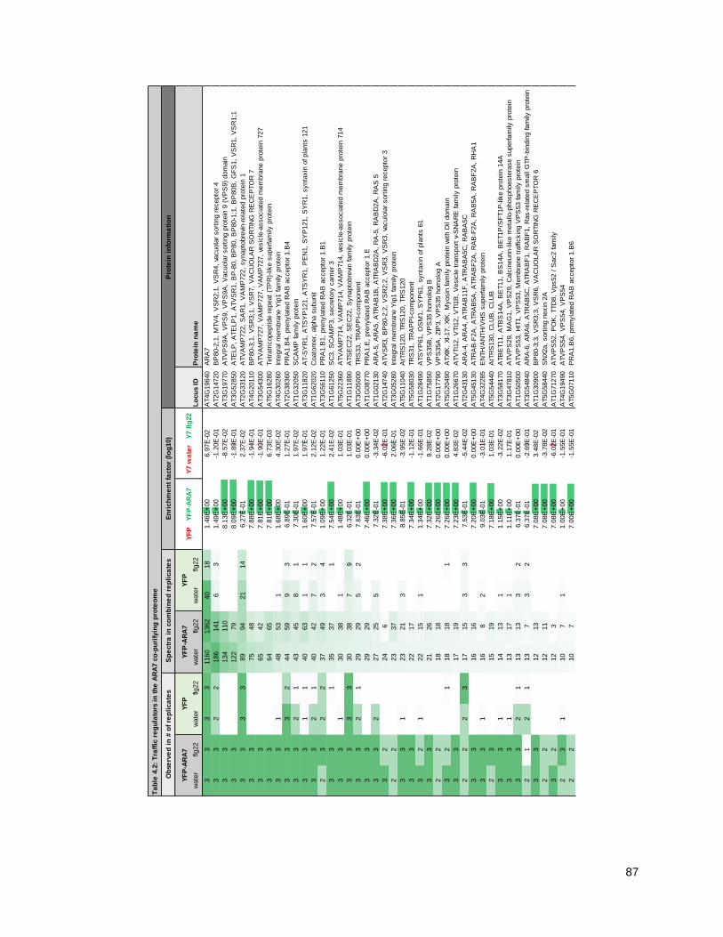
87
Ta
ble
4.2
: T
raff
ic r
eg
ula
tors
in
th
e A
RA
7 c
o-p
uri
fyin
g p
rote
om
e
wate
rflg
22
wate
rflg
22
wate
rflg
22
wate
rflg
22
Lo
cu
s ID
Pro
tein
na
me
33
33
1160
1362
40
18
1.4
6E
+00
6.9
7E
-02
AT4G
19640
AR
A7
33
22
186
141
63
1.4
9E
+00
-1.2
0E
-01
AT2G
14720
BP
80-2
;1,
MTV
4,
VS
R2;1
, V
SR
4,
vacuola
r sort
ing r
ecepto
r 4
33
134
110
8.1
3E
+00
-8.5
7E
-02
AT3G
19770
ATV
PS
9A
, V
PS
9,
VP
S9A
, V
acuola
r sort
ing p
rote
in 9
(V
PS
9)
dom
ain
33
122
79
8.0
9E
+00
-1.8
9E
-01
AT3G
52850
ATE
LP
, A
TE
LP
1,
ATV
SR
1,
BP
-80,
BP
80,
BP
80-1
;1,
BP
80B
, G
FS
1,
VS
R1,
VS
R1;1
33
33
89
94
21
14
6.2
7E
-01
2.3
7E
-02
AT2G
33120
ATV
AM
P722,
SA
R1,
VA
MP
722,
synapto
bre
vin-r
ela
ted p
rote
in 1
33
75
48
7.8
8E
+00
-1.9
4E
-01
AT4G
20110
BP
80-3
;1,
VS
R3;1
, V
SR
7,
VA
CU
OLA
R S
OR
TIN
G R
EC
EP
TO
R 7
33
65
42
7.8
1E
+00
-1.9
0E
-01
AT3G
54300
ATV
AM
P727,
VA
MP
727,
VA
MP
727,
vesic
le-a
ssocia
ted m
em
bra
ne p
rote
in 7
27
33
64
65
7.8
1E
+00
6.7
3E
-03
AT5G
16280
Tetr
atr
icopeptide r
epeat
(TP
R)-
like s
uperfam
ily p
rote
in
33
148
53
11.6
8E
+00
4.3
0E
-02
AT4G
30260
Inte
gra
l m
em
bra
ne Y
ip1 fam
ily p
rote
in
33
32
44
59
93
6.8
9E
-01
1.2
7E
-01
AT2G
38360
PR
A1.B
4,
pre
nyla
ted R
AB
accepto
r 1.B
4
33
21
43
45
81
7.3
0E
-01
1.9
7E
-02
AT1G
32050
SC
AM
P fam
ily p
rote
in
33
11
40
63
11
1.6
0E
+00
1.9
7E
-01
AT3G
11820
AT-S
YR
1,
ATS
YP
121,
ATS
YR
1,
PE
N1,
SY
P121,
SY
R1,
synta
xin
of pla
nts
121
33
21
40
42
72
7.5
7E
-01
2.1
2E
-02
AT1G
62020
Coato
mer,
alp
ha s
ubunit
23
22
37
49
34
1.0
9E
+00
1.2
2E
-01
AT3G
56110
PR
A1.B
1,
pre
nyla
ted R
AB
accepto
r 1.B
1
33
135
37
17.5
4E
+00
2.4
1E
-02
AT1G
61250
SC
3,
SC
AM
P3,
secre
tory
carr
ier
3
33
130
38
11.4
8E
+00
1.0
3E
-01
AT5G
22360
ATV
AM
P714,
VA
MP
714,
VA
MP
714,
vesic
le-a
ssocia
ted m
em
bra
ne p
rote
in 7
14
33
33
30
38
79
6.3
2E
-01
1.0
3E
-01
AT1G
11890
ATS
EC
22,
SE
C22,
Synapto
bre
vin fam
ily p
rote
in
33
21
29
29
52
7.6
3E
-01
0.0
0E
+00
AT3G
05000
TR
S33,
TR
AP
PI-com
ponent
33
29
29
7.4
6E
+00
0.0
0E
+00
AT1G
08770
PR
A1.E
, pre
nyla
ted R
AB
accepto
r 1.E
33
227
25
57.3
2E
-01
-3.3
4E
-02
AT1G
02130
AR
A-5
, A
RA
5,
ATR
AB
1B
, A
TR
AB
D2A
, R
A-5
, R
AB
D2A
, R
AS
5
32
24
67.3
8E
+00
-6.0
2E
-01
AT2G
14740
ATV
SR
3,
BP
80-2
;2,
VS
R2;2
, V
SR
3,
VS
R3,
vaculo
lar
sort
ing r
ecepto
r 3
22
23
37
7.3
6E
+00
2.0
6E
-01
AT3G
05280
Inte
gra
l m
em
bra
ne Y
ip1 fam
ily p
rote
in
33
123
21
38.8
5E
-01
-3.9
5E
-02
AT5G
11040
AtT
RS
120,
TR
S120,
TR
S120
33
22
17
7.3
4E
+00
-1.1
2E
-01
AT5G
58030
TR
S31,
TR
AP
PI-com
ponent
32
122
15
11.3
4E
+00
-1.6
6E
-01
AT1G
28490
ATS
YP
61,
OS
M1,
SY
P61,
synta
xin
of pla
nts
61
33
21
26
7.3
2E
+00
9.2
8E
-02
AT1G
75850
VP
S35B
, V
PS
35 h
om
olo
g B
22
18
18
7.2
6E
+00
0.0
0E
+00
AT2G
17790
VP
S35A
, ZIP
3,
VP
S35 h
om
olo
g A
32
118
18
17.2
6E
+00
0.0
0E
+00
AT5G
20490
ATXIK
, XI-17,
XIK
, M
yosin
fam
ily p
rote
in w
ith D
il dom
ain
33
17
19
7.2
3E
+00
4.8
3E
-02
AT1G
26670
ATV
TI1
2,
VTI1
2,
VTI1
B,
Vesic
le t
ransport
v-S
NA
RE
fam
ily p
rote
in
22
23
17
15
33
7.5
3E
-01
-5.4
4E
-02
AT2G
43130
AR
A-4
, A
RA
4,
ATR
AB
11F
, A
TR
AB
A5C
, R
AB
A5C
33
16
16
7.2
0E
+00
0.0
0E
+00
AT5G
45130
ATR
AB
-F2A
, A
TR
AB
5A
, A
TR
AB
F2A
, R
AB
-F2A
, R
AB
5A
, R
AB
F2A
, R
HA
1
33
116
82
9.0
3E
-01
-3.0
1E
-01
AT4G
32285
EN
TH
/AN
TH
/VH
S s
uperfam
ily p
rote
in
23
15
19
7.1
8E
+00
1.0
3E
-01
AT5G
54440
AtT
RS
130,
CLU
B,
CLU
B
33
114
13
11.1
5E
+00
-3.2
2E
-02
AT3G
58170
ATB
ET11,
ATB
S14A
, B
ET11,
BS
14A
, B
ET1P
/SF
T1P
-lik
e p
rote
in 1
4A
33
113
17
11.1
1E
+00
1.1
7E
-01
AT3G
47810
ATV
PS
29,
MA
G1,
VP
S29,
Calc
ineurin-lik
e m
eta
llo-p
hosphoeste
rase s
uperfam
ily p
rote
in
33
21
13
13
32
6.3
7E
-01
0.0
0E
+00
AT1G
50500
ATV
PS
53,
HIT
1,
VP
S53,
Mem
bra
ne t
raffi
ckin
g V
PS
53 fam
ily p
rote
in
21
21
13
73
26.3
7E
-01
-2.6
9E
-01
AT3G
54840
AR
A-6
, A
RA
6,
ATR
AB
5C
, A
TR
AB
F1,
RA
BF
1,
Ras-r
ela
ted s
mall
GTP
-bin
din
g fam
ily p
rote
in
33
12
13
7.0
8E
+00
3.4
8E
-02
AT1G
30900
BP
80-3
;3,
VS
R3;3
, V
SR
6,
VA
CU
OLA
R S
OR
TIN
G R
EC
EP
TO
R 6
22
12
11
7.0
8E
+00
-3.7
8E
-02
AT5G
58440
SN
X2a,
sort
ing n
exin
2A
32
12
37.0
8E
+00
-6.0
2E
-01
AT1G
71270
ATV
PS
52,
PO
K,
TTD
8,
Vps52 /
Sac2 fam
ily
23
110
71
1.0
0E
+00
-1.5
5E
-01
AT4G
19490
ATV
PS
54,
VP
S54,
VP
S54
22
10
77.0
0E
+00
-1.5
5E
-01
AT5G
07110
PR
A1.B
6,
pre
nyla
ted R
AB
accepto
r 1.B
6
Ob
serv
ed
in
# o
f re
pli
ca
tes
Sp
ectr
a i
n c
om
bin
ed
re
pli
ca
tes
En
rich
me
nt
facto
r (l
og
10)
Pro
tein
in
form
ati
on
YF
P-A
RA
7Y
FP
YF
P-A
RA
7Y
FP
YF
P
YF
P-A
RA
7Y
7 w
ate
r
Y7 f
lg22

88
22
912
6.9
5E
+00
1.2
5E
-01
AT3G
05710
ATS
YP
43,
SY
P43,
synta
xin
of pla
nts
43
32
95
6.9
5E
+00
-2.5
5E
-01
AT1G
01960
BIG
3,
ED
A10,
AR
F-G
EF
22
914
6.9
5E
+00
1.9
2E
-01
AT2G
39805
Inte
gra
l m
em
bra
ne Y
ip1 fam
ily p
rote
in
22
95
6.9
5E
+00
-2.5
5E
-01
AT2G
05170
ATV
PS
11,
VP
S11,
vacuola
r pro
tein
sort
ing 1
1
32
21
914
23
6.5
3E
-01
1.9
2E
-01
AT3G
15980
Coato
mer,
beta
' subunit
21
21
92
21
6.5
3E
-01
-6.5
3E
-01
AT5G
58060
ATG
P1,
ATY
KT61,
YK
T61,
SN
AR
E-lik
e s
uperfam
ily p
rote
in
32
79
6.8
5E
+00
1.0
9E
-01
AT5G
53530
VP
S26A
, va
cuola
r pro
tein
sort
ing 2
6A
33
17
61
8.4
5E
-01
-6.6
9E
-02
AT3G
13720
PR
A1.F
3,
PR
A8,
PR
A1 (
Pre
nyla
ted r
ab a
ccepto
r) fam
ily p
rote
in
21
16
41
7.7
8E
-01
-1.7
6E
-01
AT3G
15060
AtR
AB
A1g,
RA
BA
1g,
RA
B G
TP
ase h
om
olo
g A
1G
23
13
621
14
7.7
8E
-01
5.4
4E
-01
AT5G
16830
ATP
EP
12,
ATS
YP
21,
PE
P12,
PE
P12P
, S
YP
21,
synta
xin
of pla
nts
21

89
SNAREs
Soluble N-ethylmaleimide-sensitive factor Attachment Protein Receptors (SNAREs) are
endomembrane pathway-specific, predominantly transmembrane proteins with cytosolic coiled-
coil domains that mediate membrane fusion (Uemura et al., 2004; Lipka et al., 2007). In ARA7
purifications, I identified PEN1/SYP121 (table 4.2), which is a PM and TGN/EE-localized SNARE
that cycles between these compartments (Nielsen et al., 2012; Nielsen and Thordal-Christensen,
2012; Assaad et al., 2004). Furthermore, I identified the TGN/EE-localized SNAREs SYP43
(Uemura et al., 2012) and SYP61, which functions in complex with VTI12 (Drakakaki et al., 2012),
which itself is also identified in my ARA7 purifications (table 4.1). Further identified VPS45 is a
regulatory protein of this SNARE complex, which also localizes to the TGN/EE (Zouhar et al.,
2009). Curiously, the known SYP61 interaction partner SYP41 was not found (Zouhar et al.,
2009). SNAREs further along the endocytic route that were identified are VAMP722, which co-
localizes to the TGN/EE marker VHA-a1 which is also found in my dataset (tables 4.1, 4.2,
(Zhang et al., 2011a)), and finally VAMP727 which localizes to the LE/MVB (Ebine et al., 2008).
Rab-GTPase associated proteins
In addition to these membrane-associated proteins, Rab GTPase-associated proteins such as
VPS9a and PRA1 family proteins were identified. Importantly, VPS9a is the only gunanine
nucleotide exchange factor (GEF) that is known to interact with and facilitate GTP-binding of all
three Arabidopsis Rab5 proteins including ARA7 (Goh et al., 2007). PRA1 family proteins interact
with Rab GTPases and allow the Rab to bind to the membrane (Alvim Kamei et al., 2008; Gendre
et al., 2014). Most abundantly, I found PRA1.B4 and PRA1.B1, which were identified in previous
ARA7 purifications (Heard et al., 2015), upon which colocalization of PRA1.B1 with ARA7 in leaf
tissue was further confirmed (Heard et al., 2015). Finally, PUF2, mediates competition for
activation between ARA7 and ARA6 on the same endosome and localizes to the LE/MVB
compartment (Ito et al., 2018).
Novel proteins
Some proteins were found that are not known to localize to TGN/EE or LE/MVB compartments.
The most abundant protein in ARA7 purifications, besides ARA7 itself, is SUPPRESSOR OF
ACAULIS 56 (SAC56), which is an L4/L1 family integral subunit of ribosomes. Furthermore, both
SEC22 and coatomer proteins function at the interface between ER and Golgi, with a role for
SEC22 in anterograde traffic, and for a heptameric complex of coatomer subunits in retrograde
traffic (Chatre, 2005; Brandizzi and Barlowe, 2013). Finally, PRA1.B6 localizes to the Golgi (Jung
et al., 2011). Mostly, the above ER/Golgi associated proteins were found with low abundance.

90
4.2.3 - The ARA7 co-purifying proteome changes upon 3-h flagellin treatment
Having established that I can obtain a specific ARA7 proteome, I applied the technique to
flagellin-treated Arabidopsis seedlings. I treated seedlings with 1.5 µM flg22 for three h, allowing
for PTI-induced transcriptional upregulation and the accumulation of potential immune-related
cargoes in endosomes. Then, I defined an ARA7 co-purifying proteome in the same way as I did
for the above-described water treated conditions, and filtered the list to retain only proteins of
which the associated spectral counts were upregulated at least twice in the flg22-treated
conditions compared to water control. The flg22-upregulated ARA7 co-purifying proteome
contains 70 proteins, of which 13 were also found under water control treatment, and 57 that
were not. (fig. 4.6D, table 4.3).
GO-analysis shows enrichment for inorganic acid chemistry-related terms
In order to obtain a global picture of the biological processes in which the flg22-induced ARA7
interactors are involved, I performed a GO-term analysis querying for terms in the ―biological
process‖ category (table S2). Interestingly, there is a strong overrepresentation of metabolic
processes such as organic and carboxylic acid metabolism as well as drug/small molecule
metabolism, indicating that the purified compartment is chemically active, or contributes to
changed metabolic processes at its location. In correspondence with the GO-term enrichment for
general organic acid metabolism, among the flg22-induced ARA7 interactors (table 4.3) I find
Indole Glucosinolate O-Methyl Transferase 4 (IGMT4).
Table 4.3: ARA7 co-purifying proteins that are more abundant upon flagellin treatment. Listed here is the ARA7 co-purifying
proteome under flagellin treated conditions. 8-day old seedlings of genotypes pUB::YFP-ARA7 and pUB::YFP were treated with
water or 1.5 µM flg22 for three hours, and subjected to immunoprecipitation using YFP affinity beads. Mass spectrometry was
performed in collaboration with Jan Sklenar, Paul Derbyshire and Frank Menke (The Sainsbury Laboratory, Norwich, UK). To
determine co-purifying proteomes, Scaffold software was used to retain proteins with a ProteinProphet score of 99% based on a
minimum of 2 spectra with a PeptideProphet score of 95%. MS Excel was then used to retain proteins with at least 5 spectra across
all three replicates in the YFP-ARA7 flg22-treated samples, with an occurence in more than 1 of these replicates, an abundance
increase of at least 4x compared to matching YFP controls, and an abundance increase of at least 2x compared to matching water
treated controls. (Columns 1-8) Observed in # of replicates, and Spectra in combined replicates: Numbers and associated green
cell-shading intensities indicate in how many experimental replicates the protein was detected, or how many spectra for that protein
were identified in all three replicates combined, separated for water or flagellin treatment. (Columns 9-10) Enrichment factors: For
each protein, the number of total spectra across three replicates of the green-labeled (rightmost) pulldown was divided by the
number of total spectra in red-labeled (left-most) pulldown. Log10 values that correspond to these ratios are displayed in this table.
Green or red bars are graphical representations of magnitude and direction of positive (green) or negative (red) values, centered at
0. (Columns 11-12) Locus ID and associated names. Source: TAIR10, accessed through www.genevestigator.org. The table is
sorted for total spectral numbers in ―YFP-ARA7 flg22‖, in descending order.

91
Ta
ble
4.3
: A
RA
7 c
o-p
uri
fyin
g p
rote
ins
tha
t a
re m
ore
ab
un
da
nt
up
on
fla
ge
llin
tre
atm
en
t
wate
rflg
22
wate
rflg
22
wate
rflg
22
wate
rflg
22
Lo
cu
s ID
Pro
tein
na
me
336
0.0
0E
+00
7.5
6E
+00
AT5G
25250
FLO
TIL
LIN
1
33
23
15
36
45
8-4
.77E
-01
3.8
0E
-01
AT2G
30870
ATG
STF
10,
ATG
STF
4,
ER
D13,
GS
TF
10,
glu
tath
ione S
-tra
nsfe
rase P
HI 10
33
22
15
32
61
4-6
.09E
-01
3.2
9E
-01
AT5G
41670
6-p
hosphoglu
conate
dehydro
genase fam
ily p
rote
in
13
32
11
30
46
4-6
.21E
-01
4.3
6E
-01
AT5G
01410
ATP
DX1,
ATP
DX1.3
, P
DX1,
PD
X1.3
, R
SR
4,
Ald
ola
se-t
ype T
IM b
arr
el fa
mily
pro
tein
33
13
28
7.1
1E
+00
3.3
3E
-01
AT3G
05500
Rubber
elo
ngation facto
r pro
tein
(R
EF
)
326
0.0
0E
+00
7.4
1E
+00
AT1G
66090
TN
3,
ATTN
3,
TIR
-NB
S3
23
33
424
45
0.0
0E
+00
7.7
8E
-01
AT1G
21130
IGM
T4,
Indole
Glu
cosin
ola
te O
-Meth
yl Tra
nsfe
rase 4
31
221
44
-6.6
0E
+00
7.3
2E
+00
AT5G
06860
ATP
GIP
1,
PG
IP1,
poly
gala
ctu
ronase inhib
itin
g p
rote
in 1
23
13
621
14
7.7
8E
-01
5.4
4E
-01
AT5G
16830
ATP
EP
12,
ATS
YP
21,
PE
P12,
PE
P12P
, S
YP
21,
synta
xin
of pla
nts
21
22
21
719
27
2-5
.86E
-01
4.3
4E
-01
AT3G
53460
CP
29,
chlo
ropla
st
RN
A-b
indin
g p
rote
in 2
9
22
418
6.6
0E
+00
6.5
3E
-01
AT5G
03700
D-m
annose b
indin
g lectin p
rote
in w
ith A
pple
-lik
e c
arb
ohydra
te-b
indin
g d
om
ain
12
32
717
19
4-4
.34E
-01
3.8
5E
-01
AT2G
47000
AB
CB
4,
ATP
GP
4,
MD
R4,
PG
P4,
ATP
bin
din
g c
assett
e s
ubfa
mily
B4
12
116
6.0
0E
+00
1.2
0E
+00
AT4G
24160
Lip
id h
om
eosta
sis
alp
ha-b
eta
hydro
lase
22
14
16
16.0
2E
-01
6.0
2E
-01
AT1G
78240
OS
U1,
QU
A2,
TS
D2,
S-a
denosyl-L-m
eth
ionin
e-d
ependent
meth
yltra
nsfe
rase
33
17
16
66.6
9E
-02
3.5
9E
-01
AT5G
26710
Glu
tam
yl/glu
tam
inyl-tR
NA
synth
eta
se,
cla
ss Ic
33
22
715
32
3.6
8E
-01
3.3
1E
-01
AT5G
10730
NA
D(P
)-bin
din
g R
ossm
ann-fold
superfam
ily p
rote
in
23
21
615
50
1-9
.21E
-01
3.9
8E
-01
AT2G
42600
ATP
PC
2,
PP
C2,
phosphoenolp
yru
vate
carb
oxyla
se 2
13
314
6.4
8E
+00
6.6
9E
-01
AT5G
16550
unknow
n p
rote
in
13
11
114
11
1-1
.04E
+00
1.1
5E
+00
AT3G
16530
Legum
e lectin fam
ily p
rote
in
12
22
14
16
-9.0
3E
-01
8.4
5E
-01
AT4G
10480
Nascent
poly
peptide-a
ssocia
ted c
om
ple
x (
NA
C),
alp
ha s
ubunit fam
ily p
rote
in
33
22
613
92
-1.7
6E
-01
3.3
6E
-01
AT4G
12790
P-loop c
onta
inin
g n
ucle
osid
e t
riphosphate
hydro
lases s
uperfam
ily p
rote
in
22
26
13
33.0
1E
-01
3.3
6E
-01
AT2G
20120
CO
V1,
Pro
tein
of unknow
n function (
DU
F502)
23
15
13
6-7
.92E
-02
4.1
5E
-01
AT4G
23850
LA
CS
4,
AM
P-d
ependent
synth
eta
se a
nd lig
ase fam
ily p
rote
in
13
21
313
17
1-7
.53E
-01
6.3
7E
-01
AT5G
42790
AR
S5,
ATP
SM
30,
PA
F1,
pro
teasom
e a
lpha s
ubunit F
1
21
12
1-6
.00E
+00
7.0
8E
+00
AT4G
23180
CR
K10
23
25
12
12
-3.8
0E
-01
3.8
0E
-01
AT2G
34590
Tra
nsketo
lase fam
ily p
rote
in
311
0.0
0E
+00
7.0
4E
+00
AT1G
14540
PE
R4,
PE
RO
XID
AS
E 4
13
12
11
13.0
1E
-01
7.4
0E
-01
AT2G
04400
Ald
ola
se-t
ype T
IM b
arr
el fa
mily
pro
tein
12
11
10
10.0
0E
+00
1.0
0E
+00
AT1G
51800
IOS
1
12
13
10
6-3
.01E
-01
5.2
3E
-01
AT4G
08850
LR
R-R
LK
, M
IK2
13
210
6.3
0E
+00
6.9
9E
-01
AT1G
52200
PLA
C8 fam
ily p
rote
in
12
13
10
26.4
8E
+00
5.2
3E
-01
AT3G
09900
ATR
AB
8E
, A
TR
AB
E1E
, R
AB
E1e,
RA
B G
TP
ase h
om
olo
g E
1E
23
14
10
16.0
2E
-01
3.9
8E
-01
AT1G
55190
PR
A1.F
2,
PR
A7,
PR
A1 (
Pre
nyla
ted r
ab a
ccepto
r) fam
ily p
rote
in
12
13
10
30.0
0E
+00
5.2
3E
-01
AT2G
30200
cata
lytics;t
ransfe
rases;[
acyl-carr
ier-
pro
tein
] S
-malo
nyltra
nsfe
rases;b
indin
g
12
23
10
8-4
.26E
-01
5.2
3E
-01
AT5G
63570
GS
A1,
glu
tam
ate
-1-s
em
iald
ehyde-2
,1-a
min
om
uta
se
12
21
310
13
2-6
.37E
-01
5.2
3E
-01
AT2G
16600
RO
C3,
rota
mase C
YP
3
12
11
49
22
3.0
1E
-01
3.5
2E
-01
AT4G
18800
ATH
SG
BP
, A
TR
AB
11B
, A
TR
AB
A1D
, R
AB
A1d,
RA
B G
TP
ase h
om
olo
g A
1D
33
31
49
91
-3.5
2E
-01
3.5
2E
-01
AT5G
56030
AtH
sp90.2
, E
RD
8,
HS
P81-2
, H
SP
90.2
, heat
shock p
rote
in 8
1-2
22
39
6.4
8E
+00
4.7
7E
-01
AT2G
20840
Secre
tory
carr
ier
mem
bra
ne p
rote
in (
SC
AM
P)
fam
ily p
rote
in
12
14
91
6.0
2E
-01
3.5
2E
-01
AT5G
44790
HM
A7,
RA
N1,
responsiv
e-t
o-a
nta
gonis
t 1 /
copper-
transport
ing A
TP
ase (
RA
N1)
12
11
19
22
-3.0
1E
-01
9.5
4E
-01
AT1G
03220
Eukary
otic a
spart
yl pro
tease fam
ily p
rote
in
29
0.0
0E
+00
6.9
5E
+00
AT2G
39400
alp
ha/b
eta
-Hydro
lases s
uperfam
ily p
rote
in
Ob
serv
ed
in
# o
f re
pli
ca
tes
Sp
ectr
a i
n c
om
bin
ed
re
pli
ca
tes
En
rich
me
nt
facto
r (l
og
10)
Pro
tein
in
form
ati
on
YF
P-A
RA
7Y
FP
YF
P-A
RA
7Y
FP
YF
P
YF
P-A
RA
7Y
7 w
ate
r
Y7 f
lg22

92
32
92
0.0
0E
+00
6.9
5E
+00
AT2G
43620
Chitin
ase fam
ily p
rote
in
23
14
94
0.0
0E
+00
3.5
2E
-01
AT1G
55020
ATLO
X1,
LO
X1,
lipoxygenase 1
32
913
-7.1
1E
+00
6.9
5E
+00
AT2G
37040
ATP
AL1,
PA
L1,
PH
E a
mm
onia
lyase 1
12
31
19
21
2-1
.32E
+00
9.5
4E
-01
AT5G
43940
AD
H2,
ATG
SN
OR
1,
GS
NO
R,
HO
T5,
PA
R2,
zin
c-b
indin
g d
ehydro
genase fam
ily p
rote
in
13
38
6.4
8E
+00
4.2
6E
-01
AT1G
73030
CH
MP
1A
, V
PS
46.2
, S
NF
7 fam
ily p
rote
in
13
12
81
3.0
1E
-01
6.0
2E
-01
AT3G
17810
PY
D1,
pyrim
idin
e 1
12
12
82
0.0
0E
+00
6.0
2E
-01
AT2G
29720
CTF
2B
, F
AD
/NA
D(P
)-bin
din
g o
xid
ore
ducta
se fam
ily p
rote
in
23
13
818
-7.7
8E
-01
4.2
6E
-01
AT1G
09130
ATP
-dependent
casein
oly
tic (
Clp
) pro
tease/c
roto
nase fam
ily p
rote
in
37
0.0
0E
+00
6.8
5E
+00
AT3G
50480
HR
4,
hom
olo
g o
f R
PW
8 4
22
37
6.4
8E
+00
3.6
8E
-01
AT4G
20410
GA
MM
A-S
NA
P,
GS
NA
P,
gam
ma-s
olu
ble
NS
F a
ttachm
ent
pro
tein
23
12
71
3.0
1E
-01
5.4
4E
-01
AT2G
21600
ATR
ER
1B
, R
ER
1B
, endopla
sm
atic r
eticulu
m r
etr
ieva
l pro
tein
1B
13
12
71
3.0
1E
-01
5.4
4E
-01
AT3G
55430
O-G
lycosyl hydro
lases fam
ily 1
7 p
rote
in
12
13
73
0.0
0E
+00
3.6
8E
-01
AT3G
10380
ATS
EC
8,
SE
C8,
subunit o
f exocyst
com
ple
x 8
21
75
-6.7
0E
+00
6.8
5E
+00
AT4G
13780
meth
ionin
e--
tRN
A lig
ase,
puta
tive
/ m
eth
ionyl-tR
NA
synth
eta
se,
puta
tive
/ M
etR
S,
puta
tive
26
0.0
0E
+00
6.7
8E
+00
AT2G
19160
Core
-2/I-b
ranchin
g b
eta
-1,6
-N-a
cety
lglu
cosam
inyltra
nsfe
rase fam
ily p
rote
in
21
61
0.0
0E
+00
6.7
8E
+00
AT4G
08770
Pero
xid
ase s
uperfam
ily p
rote
in
12
11
61
0.0
0E
+00
7.7
8E
-01
AT3G
17410
Pro
tein
kin
ase s
uperfam
ily p
rote
in
12
16
6.0
0E
+00
7.7
8E
-01
AT3G
63250
ATH
MT-2
, H
MT-2
, H
MT2,
hom
ocyste
ine m
eth
yltra
nsfe
rase 2
12
26
6.3
0E
+00
4.7
7E
-01
AT3G
47550
RIN
G/F
YV
E/P
HD
zin
c fin
ger
superfam
ily p
rote
in
22
26
6.3
0E
+00
4.7
7E
-01
AT5G
02890
HXXXD
-type a
cyl-tr
ansfe
rase fam
ily p
rote
in
21
62
-6.3
0E
+00
6.7
8E
+00
AT5G
16300
Vps51/V
ps67 fam
ily (
com
ponents
of ve
sic
ula
r tr
ansport
) pro
tein
12
12
63
-1.7
6E
-01
4.7
7E
-01
AT4G
24840
CO
G2
12
12
63
-1.7
6E
-01
4.7
7E
-01
AT5G
17380
Thia
min
e p
yro
phosphate
dependent
pyru
vate
decarb
oxyla
se fam
ily p
rote
in
12
22
66
-4.7
7E
-01
4.7
7E
-01
AT4G
20830
OG
OX1,
OLIG
OG
ALA
CTU
RO
NID
E O
XID
AS
E 1
12
22
66
-4.7
7E
-01
4.7
7E
-01
AT5G
45280
Pectinacety
leste
rase fam
ily p
rote
in
22
67
-6.8
5E
+00
6.7
8E
+00
AT5G
16970
AE
R,
AT-A
ER
, alk
enal re
ducta
se
23
12
616
-9.0
3E
-01
4.7
7E
-01
AT3G
52150
RN
A-b
indin
g (
RR
M/R
BD
/RN
P m
otifs
) fa
mily
pro
tein
22
621
-7.3
2E
+00
6.7
8E
+00
AT1G
29880
gly
cyl-tR
NA
synth
eta
se /
gly
cin
e--
tRN
A lig
ase

93
Indole-3-glucosinolate pathway enzymes
IGMT4 is highly similar to its three homologs and clustered genes IGMT1-3 (Pfalz et al., 2016), of
which IGMT1 and IGMT2 have been characterized to function as final catalyzers in the indole-3-
glucosinolate based antimicrobial secretion pathway which involves PEN2 and PEN3 (Birkenbihl
et al., 2017). IGMT1 and IGMT2 convert indole-3-methylglucosinolates (I3M) into 4-
methodxyindole-3-methylglucosinolate (4MOI3M), in preparation for conversion into toxic end
products by the atypical myrosinase PEN2 (Pfalz et al., 2011; Birkenbihl et al., 2017; Xu et al.,
2016; Lu et al., 2015). Based on sequence homology, IGMT4 is likely to also be able to catalyze
the I3M – 4MOI3M reaction, but this has not been conclusively shown. End products of this
pathway, predominantly toxic indole-3-ylmethylamine (I3A) and indole-3-carboxylic acid (ICA)
accumulate in Pto DC3000-infected tissue (Stahl et al., 2016) and have been previously
described to accumulate and function in defense against filamentous fungi (Lipka et al., 2005;
Bednarek et al., 2010; Oa et al., 2010).
It must be noted that, PENETRATION 3/PLEIOTROPIC DRUG RESISTANCE 8 (PEN3/PDR8),
while not picked up by my filtering criteria, has the following spectral counts: YFP-ARA7 + water:
222, + flg22: 409, YFP + water: 84, + flg22: 37. It highly specifically associated with YFP-ARA7
under flg22 treated conditions, but only enriched with a factor of ca. 1.8 compared to water
treatment, which is below the minimum factor of 2 that is required to be considered flg22-
responsive under my filtering criteria. The spectral increase of PEN3 under flg22 conditions is
noteworthy in the context of IGMT4 and indole glucosinolate metabolism
PEN3 is a PM-localized transmembrane ATP-binding cassette transporter which is thought to
transport antimicrobial compounds generated by PEN2 (Stein et al., 2006). It has been shown to
focally accumulate at the cell surface under fungal attempted penetration sites, and upon
bacterial and flagellin or chitin MAMP challenge (Underwood and Somerville, 2008, 2013;
Underwood et al., 2017; Xin et al., 2013). Finding it specifically associating with ARA7 upon flg22-
treatment is interesting, because in microscopic analysis, PEN3 has been shown to accumulate
at endosomes when measured at 4 h after MAMP-stimulus, and is proposed to traffic through the
endocytic pathway to focal accumulations at the cell-surface (Underwood and Somerville, 2013;
Underwood et al., 2017). The above finding provides evidence for a role in ARA7 endosomes in
this redirection process.
The clathrin-independent endocytic protein FLOTILLIN
FLOTILLIN 1 (FLOT1) belongs to the family of membrane-bound flotillin/reggie-like proteins,
which in animals have been well characterized to associate with the cytosolic leaflet of lipid
bilayers, and function in bending membranes such as invagination of the plasma membrane in
clathrin-independent endocytic processes (Glebov et al., 2006; Frick et al., 2007; Langhorst et al.,
94
2007). Flotillins interact with receptors and can organise them into a signaling platform in distinct
lipid microenvironments (Otto and Nichols, 2011). In plants, FLOT1 has been described to be PM
microdomain-associated (Jarsch et al., 2014; Jarsch and Ott, 2011), present in addition on
endosomes, and functioning in clathrin-independent endocytosis (Li et al., 2012a). Furthermore,
in the context of bacterial symbiosis with legumes, cell-surface symbiosis-pattern recognizing
receptor LYK3 co-localizes with FLOT4, which is thought to reflect the assembly of a signaling
platform to initiate bacterial infection (Downie, 2014; Haney et al., 2011).
Arabidopsis FLOT1 was found to accumulate at late endosomes in an increased manner upon
flagellin immune stimulus (Yu et al., 2017). In animals, flotillins are considered good membrane
microdomain markers, present in lipid environments high in saturated sphingolipids and
cholesterol (Langhorst et al., 2007). Finding FLOT1 in ARA7 purifications upon flagellin treatment
points at the possibility for a change in lipid composition on TGN/EE or LE/MVB compartments
during immunity. Interestingly, in mammals, flotillins that are present at endosomes are
associated with MVB targeting to the plasma membrane (Meister and Tikkanen, 2014).
Receptors, oxidative stress, and the cell wall
Several receptor kinases (CRK10, IOS1, MIK2) were found to be upregulated in ARA7
purifications upon flg22 treatment. Cysteine-rich receptor-like kinase 10 (CRK10) belongs to the
RLK subfamily of cysteine-rich receptor-like kinases (CRKs), which have a wide range of
functionality in abiotic and biotic stresses, pathogen defense and cell death (Chen et al., 2003;
Ederli et al., 2011). A subset of CRKs, including CRK10, are implicated in regulating the
dynamics of MAMP-induced stomatal closure, an intermediate response that occurs at a time
scale of several hours post elicitation, potentially through sensing of ROS as a secondary
messenger (Bourdais et al., 2015; Kimura et al., 2017). Functionally distinct cell-surface receptors
undergo ligand-induced endocytosis, and accumulate in LE/MVBs upon perception of
extracellular patterns, a response generally observed to peak between 1-1.5 h after treatment
(Ben Khaled et al., 2015; Mbengue et al., 2016; Ortiz-morea et al., 2016). Although not
demonstrated for CRKs, finding CRK10 in ARA7 purifications at 3 h after perception of immune
stimulus could point at secondary messenger-induced endocytosis, potentially through perception
of extracellular reactive oxygen species. Finally, I find proteins that could function in the
protection against oxidative stress, such as two peroxidases (PEROXIDASE 4 (PER4) and
AT4G08770), and Glutathione Transferase 10 (GSTF10, (Sappl et al., 2009)).
IMPAIRED OOMYCETE SUSCEPTIBILITY 1 (IOS1) is a broad regulator of antifungal and
antibacterial cell-surface perception, and interacts with and regulates complex formation of
various other transmembrane receptors such as FLS2, EFR and CERK1 (Yeh et al., 2016).
MALE DISCOVERER 1-INTERACTING RECEPTOR LIKE KINASE 2 (MIK2) is a cell-wall

95
integrity sensing RLK with various functions in response to abiotic and biotic stress, including
antifungal defense (Van der Does et al., 2017). Finding these receptors at the endocytic pathway
suggests that they are undergoing increased turnover in response to a stimulus. Since ligand-
induced endocytosis of well-studied receptors peaks between 1-1.5 h (Ben Khaled et al., 2015), if
IOS1 and MIK2 also undergo ligand-induced endocytosis, it could be in response to a secondary
messenger in response to MAMP treatment. Considering the broad involvement of IOS1 in
receptor complexes, and the involvement of MIK2 in cell wall integrity sensing, potentially these
modules are engaged in response to cell wall modifications performed by the plant cell itself upon
immune stimulus. This could be coupled to the modification of cell-wall polymers which are
increasingly cross-linked in the presence of reactive oxygen species (Tenhaken, 2015).
Atypical immune receptors TN3 and HR4
Peptides of the two atypical intracellular immune receptors TIR-NBS3 (TN3) and Homolog of
RPW8-4 (HR4) were found in ARA7 purifications, specifically upon flg22 treatment (table 4.3).
TN3 is a truncated member of the NOD-like receptor (NLR) type, which typically contain a coiled-
coil (CC) or Toll-Interleukin Receptor (TIR) domain, a nucleotide binding site (NBS) and a leucine-
rich repeat domain (LRR). TN3 lacks the typical LRR domain, and has been previously shown to
be localized to the nucleo-cytosol, and to trigger a hypersensitive response when overexpressed
in N. benthamiana (Nandety et al., 2013).
HR4 is a short transmembrane (TM) CC-domain-containing atypical resistance protein. In
Arabidopsis ecotype Col-0, there are four homologs (HR1-4), named after the ortholog RPW8.2
which is found in ecotype Ms-0 (Xiao et al., 2001). RPW8.2 is the most extensively characterized
orthologue, and has been shown to be carried on VAMP721/VAMP722-positive vesicles to the
pathogen interface upon G. orontii infection, which requires a short targeting motif (Kim et al.,
2014). On-site, RPW8.2 promotes encasement formation, accumulation of ROS and can trigger a
hypersensitive response (Gao et al., 2009). Recently, it was shown that HR3, the closest RPW8.2
orthologue in Col-0, accumulates at the extrahaustorial interface upon infection with
Golovinomyces cichoracearum, and the same work showed fluorescently tagged HR4
accumulating there when transiently expressed in N. benthamiana (Berkey et al., 2017).
Because of its high specificity to ARA7 upon flagellin treatment (table 4.3), and the well described
direct roles for NLRs in plant immunity, I selected TN3 for further characterization. NLRs mediate
the recognition and signal transduction at the earliest events in the intracellular detection of
pathogen-effector mediated immunomodulatory effects, suggesting that these processes could
also be occurring near to ARA7, or an ARA7-involving process. Previously, TN2 has been shown
to interact biochemically with another trafficking regulator, the exocyst tethering complex subunit
Exo70B1, and was shown to be required for previously unexplained constitutive defense

96
responses in the exo70b1 mutant background (Zhao et al., 2015). In addition, RPW8-type
proteins have been shown to be central regulators of NLR-mediated defense, and exhibited
pathogen-targeting subcellular trafficking behavior, which was required for full immunity. Knowing
that ARA7 vesicles accumulate under bacterial pathogens, and ARA7 seems to recruit HR4 upon
MAMP-stimulus, I selected HR4 for further characterization.
4.3 – TIR-NBS 3 INTERACTS WITH ARA7 AND LOCALIZES TO THE NUCLEUS, CYTOSOL AND
MOBILE PUNCTAE
4.3.1 - Confirming the identification of TN3 in flagellin-treated ARA7 purifications
I obtained the amino acid sequences of the two most abundant TN3 peptides detected across
three replicates of YFP-ARA7 purifications upon flagellin treatment, and used Protein BLAST
(NCBI web interface) to match the peptides against the known Arabidopsis protein database
(TAIR10). As a result, the only protein with a 100% identity score to each peptide was TN3
(AT1G66090), with the exception of the peptide VMTIFYGVNPSDVRK, which also fully aligned
with a predicted protein encoded by a transposable element (OAP18409.1; fig. 4.7A).
Furthermore, I graphically mapped which peptides were found across all MS analyses on flg22-
induced ARA7-purifications, together with their position in the TN3 amino acid sequence (fig.
4.7B). Thus, I could confirm that both TIR and NBS domains of the same protein were detected in
all three replicates of the experiment (fig. 4.7B).
Next, based on data from the only publication characterizing TN3 to date (Nandety et al., 2013), I
obtained a list of all TIR-NBS proteins. The authors defined and phylogenetically aligned 21
Arabidopsis TIR-NBS genes alongside 30 TIR-X proteins; lacking NBS and LRR domains, but
including a variable C-terminal domain. I excerpted from their work the TN and TX genes most
closely related to TN3, re-aligned them using Clustal Omega (EMBL web suite), and reproduced
the encoded domain structure schematically (fig. 4.7C). The closest homolog of TN3 is the
pseudogenized TIR-X 4 (TX4, AT1G56470), encoding for parts of a TIR-only protein, and the
closest homolog of TN3 sharing the TIR-NBS domain organisation is TIR-NBS13 (TN13,
AT3G04210) with a genomic sequence identity of 50% and a protein sequence identity of 45%. In
the phylogenetic alignment of close TN3 homologs, I included more distantly related TN1
(genomic identity = 46%, protein identity = 28%) as an outgroup (fig. 4.7C).

97
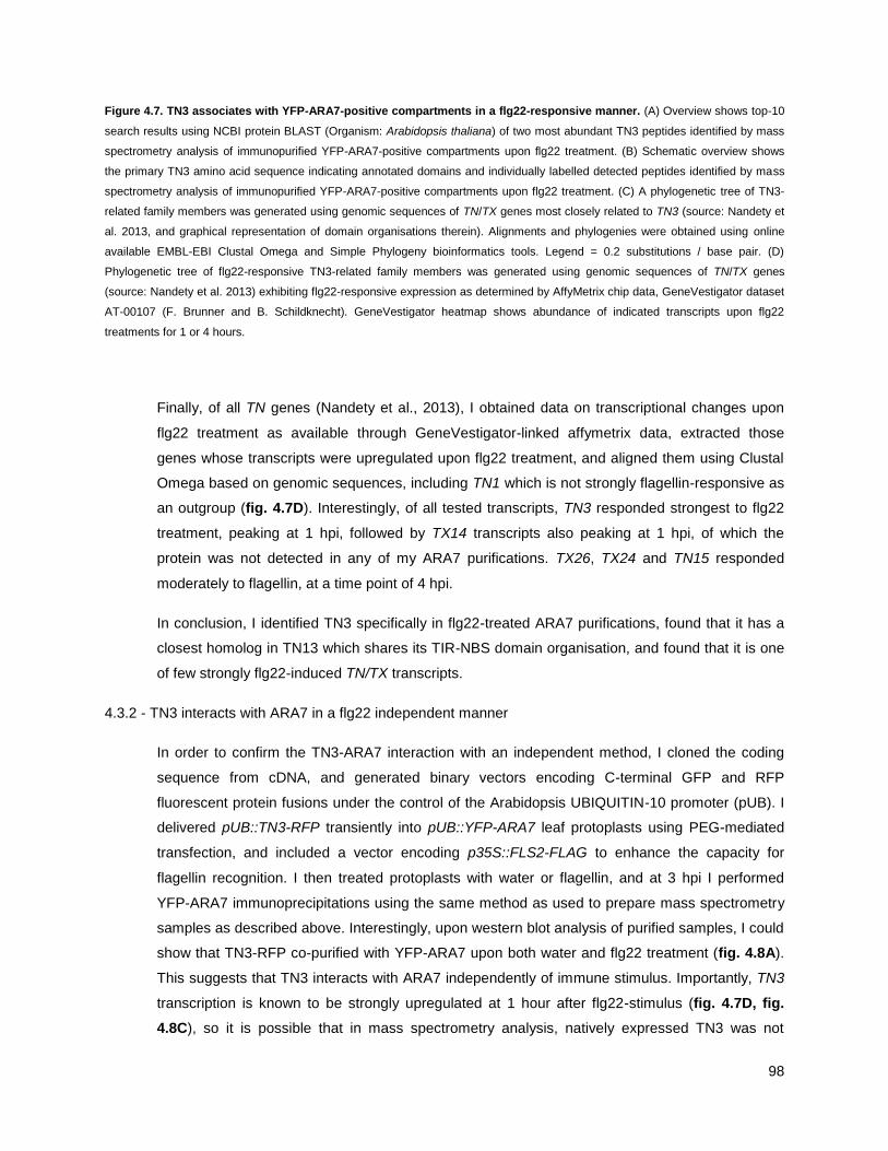
98
Figure 4.7. TN3 associates with YFP-ARA7-positive compartments in a flg22-responsive manner. (A) Overview shows top-10
search results using NCBI protein BLAST (Organism: Arabidopsis thaliana) of two most abundant TN3 peptides identified by mass
spectrometry analysis of immunopurified YFP-ARA7-positive compartments upon flg22 treatment. (B) Schematic overview shows
the primary TN3 amino acid sequence indicating annotated domains and individually labelled detected peptides identified by mass
spectrometry analysis of immunopurified YFP-ARA7-positive compartments upon flg22 treatment. (C) A phylogenetic tree of TN3-
related family members was generated using genomic sequences of TN/TX genes most closely related to TN3 (source: Nandety et
al. 2013, and graphical representation of domain organisations therein). Alignments and phylogenies were obtained using online
available EMBL-EBI Clustal Omega and Simple Phylogeny bioinformatics tools. Legend = 0.2 substitutions / base pair. (D)
Phylogenetic tree of flg22-responsive TN3-related family members was generated using genomic sequences of TN/TX genes
(source: Nandety et al. 2013) exhibiting flg22-responsive expression as determined by AffyMetrix chip data, GeneVestigator dataset
AT-00107 (F. Brunner and B. Schildknecht). GeneVestigator heatmap shows abundance of indicated transcripts upon flg22
treatments for 1 or 4 hours.
Finally, of all TN genes (Nandety et al., 2013), I obtained data on transcriptional changes upon
flg22 treatment as available through GeneVestigator-linked affymetrix data, extracted those
genes whose transcripts were upregulated upon flg22 treatment, and aligned them using Clustal
Omega based on genomic sequences, including TN1 which is not strongly flagellin-responsive as
an outgroup (fig. 4.7D). Interestingly, of all tested transcripts, TN3 responded strongest to flg22
treatment, peaking at 1 hpi, followed by TX14 transcripts also peaking at 1 hpi, of which the
protein was not detected in any of my ARA7 purifications. TX26, TX24 and TN15 responded
moderately to flagellin, at a time point of 4 hpi.
In conclusion, I identified TN3 specifically in flg22-treated ARA7 purifications, found that it has a
closest homolog in TN13 which shares its TIR-NBS domain organisation, and found that it is one
of few strongly flg22-induced TN/TX transcripts.
4.3.2 - TN3 interacts with ARA7 in a flg22 independent manner
In order to confirm the TN3-ARA7 interaction with an independent method, I cloned the coding
sequence from cDNA, and generated binary vectors encoding C-terminal GFP and RFP
fluorescent protein fusions under the control of the Arabidopsis UBIQUITIN-10 promoter (pUB). I
delivered pUB::TN3-RFP transiently into pUB::YFP-ARA7 leaf protoplasts using PEG-mediated
transfection, and included a vector encoding p35S::FLS2-FLAG to enhance the capacity for
flagellin recognition. I then treated protoplasts with water or flagellin, and at 3 hpi I performed
YFP-ARA7 immunoprecipitations using the same method as used to prepare mass spectrometry
samples as described above. Interestingly, upon western blot analysis of purified samples, I could
show that TN3-RFP co-purified with YFP-ARA7 upon both water and flg22 treatment (fig. 4.8A).
This suggests that TN3 interacts with ARA7 independently of immune stimulus. Importantly, TN3
transcription is known to be strongly upregulated at 1 hour after flg22-stimulus (fig. 4.7D, fig.
4.8C), so it is possible that in mass spectrometry analysis, natively expressed TN3 was not

99
detected in ARA7 purifications under water treatment because the gene was significanly less
expressed compared to flg22-treated conditions.
Confocal microscopy analysis of a sample of the protoplast samples revealed that TN3-RFP
localizes to the nucleus and the cytosol, whereas YFP-ARA7 localizes to the cytosol and mobile
vesicles (fig. 4.8B, upper panels). This localization pattern did not change at three h post flg22
treatment (fig. 2.8B, lower panels). I conclude that here, TN3 is nucleocytosolic, consistent with
data on Agrobacterium-mediated transiently overexpressed 35S::TN3-GFP in Nicotiana
benthamiana leaves (Nandety et al., 2013).
Figure 4.8. Constitutively expressed TN3-RFP associates with YFP-ARA7 independent of flg22 stimulation in Arabidopsis
protoplasts. (A) Immunoblot of anti-GFP-immunoprecipitated YFP-ARA7 reveals association with TN3-RFP. Protoplasts from 4
weeks-old leaves were transiently transfected with pUB::YFP-ARA7, pUB::TN3-mCherry and pUB::FLS2-FLAG, incubated for 16
hours for co-expression, and then subjected to flg22 treatment for 3 hours. YFP- and FLAG-tagged proteins were revealed using
anti-GFP-HRP and anti-FLAG-HRP conjugated antibodies, respectively. RFP-tagged TN3 and Bip2 were revealed using primary
anti-RFP and anti-Bip2 antibodies, then secondary alkaline phosphatase-conjugated antibodies. Ponceau staining was performed
on input membranes after antibody treatments for loading control. (B) Confocal micrographs of protoplasts as prepared in (A),
showing YFP signal (left panels), RFP signal (middle panels) and overlay (right panels) in z-projections, at 3 hours after indicated
treatments. 63 × objective, scale bars = 10 µm. (C) GeneVestigator heatmap shows relative abundance of indicated Arabidopsis
transcripts upon flagellin treatment of 1 or 4 hours (AffyMetrix chip data, GeneVestigator dataset AT-00107, F. Brunner and B.
Schildknecht)

100
The interaction of TN3 and ARA7 is now corroborated by two independent experimental methods,
and microscopic analysis suggests TN3 and ARA7 co-localize in the cytosol, and TN3 is not
enriched at vesicle structures. This could indicate that the interaction takes place in the cytosol
preferentially, or involves only a small subpool of TN3. However, constitutively driven expression
of TN3 may lead to ectopic localization of TN3 as compared to the endogenous situation, and it is
unknown whether TN3 has an interaction preference for subpools of ARA7.
4.3.3 - TN3 interacts with ARA7 on ARA7 vesicles in bimolecular fluorescence complementation
experiments
To further probe the interaction between TN3 and ARA7 with an independent method, and aiming
to investigate whether TN3 interacts specifically with ARA7 or could also interact with secretory
RABs (ARA5/RABD2a), homologous endocytic RABs (ARA6/RABF2a), or RABs further along the
endocytic pathway (RABG3b), I employed bimolecular fluorescence complementation (BiFC;
split-YFP). I generated binary vectors encoding TN3 C-terminal fusions with the YFP C- and N-
termini (p35S::TN3-YFPc, p35S::TN3-YFPn), and performed Agrobacterium-mediated transient
co-expressions with BiFC fusions of other RAB-GTPases in N. benthamiana leaves. Transient
expression of individual RAB-GTPases tagged with YFPc/n in the absence of the complementary
YFPn/c terminus does not result in fluorescent YFP signal (fig. 4.9A, top and left peripheral
panels).
Co-expression of TN3-YFPc and YFPn-ARA7, or the reverse combination of TN3-YFPn and
YFPc-ARA7 exhibited clear reconstitution of fluorescent signal was observed on punctae (fig.
4.9A,B). The fluorescent YFP signal co-localized with mCherry-ARA7-labeled endocytic vesicles,
but not with mCherry-ARA5 labeled secretory vesicles (fig. 4.9B). No evident fluorescent signal
was observed in the cytosol. Upon co-expression of TN3-YFPn and YFPc-ARA5, no significant
reconstitution of fluorescent signal was observed (fig. 4.9A). YFPc-ARA5 could be successfully
expressed in N. benthamiana leaves using this method, as evidenced by the reconstitution of
signal in co-expression with YFPn-ARA7 (fig. 4.9A). ARA5 and ARA7 are expected to occur in
proximity to one another, as both partially localize to the TGN/EE compartment.

101

102
Figure 4.9. Constitutively expressed TN3-RFP colocalizes and associates with YFP-ARA7-positive compartments in N.
benthamiana. (A) Bimolecular flurorescence complementation (BiFC). Confocal micrographs showing signal of reconstituted YFP
molecules in Nicotiana benthamiana adult leaves at 2 days after infiltration of Agrobacterium tumefaciens strain GV3101 carrying
the indicated bimolecular fluorescence complementation constructs. Combinations of infiltrated constructs are indicated in panels.
63 × objective, scale bars = 10 µm. (B) As in (A), but bimolecular complementations were co-infiltrated with A. tumefaciens carrying
indicated mCherry-tagged constructs. 63 × objective, scale bars = 10 µm.
Co-expression of TN3-YFPn with LE/MVB-localized RAB-GTPase ARA6-YFPc also reconstituted
strong fluorescent signal on punctae, suggesting association of TN3 and ARA6 on
endomembrane compartments (fig. 4.9A). Fluorescently tagged ARA6 and ARA7 were shown to
substantially overlap on the same compartment in earlier studies (Ueda et al., 2004), and indeed
upon co-expression of YFPn-ARA7 and ARA6-YFPc, fluorescent signal was observed on
punctae, which are likely LE/MVB compartments. Due to the substantial co-localization of ARA6
and ARA7 on endosomes, it is possible that ARA7-associated TN3-YFPn is present on LE/MVBs,
where as a consequence it is brought in close proximity to locally present ARA6-YFPc molecules,
leading to reconstitution of YFP signal.
TN3-YFPn in combination with tonoplast-localized RAB7-GTPase YFPc-RABG3b did not result in
a reconstituted signal (fig. 4.9A), together with the absence of fluorescent signal upon co-
expression of TN3-YFPn and YFPc-ARA5, suggesting that TN3 specifically associates with RAB-
GTPases at the LE/MVB, and not with secretory or vacuolar RAB-GTPases. Co-expression of
YFPn-ARA7 and YFPc-RABG3b also did not lead to substantial recovery of fluorescent signal.
This confirms specificity, since ARA7 induces its own dissociation and subsequent recruitment of
RAB7 proteins to compartments through interaction with, and activation of, intermediate
endosomal-localized regulators (Singh et al., 2014b; Cui et al., 2014b), and from localization
studies, ARA7 is present at LE/MVBs but not tonoplast. Another RAB7 member RABG3f was
shown to be present at the tonoplast but not LE/MVBs. Preliminary data suggests that RABG3b
localizes to the tonoplast (Kopischke et al., submitted).
Co-expresion of TN3-YFPn and TN3-YFPc resulted in the reconstitution of flurorescent signal in
the cytosol, and not the nucleus (fig. 4.9A), which is notably different from the nucleocytosolic
localization of TN3-RFP as observed in transient expression in Arabidopsis protoplasts (fig.
4.8A). It is possible that the reconstituted molecule TN3-YFP-TN3 with a predicted molecular
weight of 122 kDa exceeds the maximum size of passive diffusion through nuclear pores, or
alternatively, the reconstituted dimer could fail to recruit the necessary interaction partners for
active transport into the nucleus. It remains to be demonstrated whether TN3 molecules dimerize
in the absence of forced dimerization using YFP fragments.
103
In conclusion, using BiFC I could show that TN3 exists in close proximity to the LE/MVB-localized
RAB5-GTPases ARA7 and ARA6, but not with secretory ARA5 or late endocytic/tonoplastic
RAB7 member RABG3b. It is interesting to find the irreversibly bound ARA7-TN3 dimer at ARA7-
positive endosomes, because when separately tagged, TN3 did not enrich at vesicles but co-
localized with ARA7 predominantly in the cytosol. TN3 in combination with ARA6 reconstituted
YFP fluorescent signals, raising the hypothesis that TN3 may interact with other endosomal Rab
GTPases in addition to ARA7.
4.3.4 - TN3 localizes to the nucleus, cytosol and mobile punctae in Arabidopsis leaves
Transient expression by particle delivery
Next, I was interested in probing the localization of TN3 in intact Arabidopsis leaves. In a previous
study, novel mass-spectrometry-identified ARA7-interactors of the PRA1 family of RAB-regulator
proteins have been confirmed to co-localize with ARA7 on endosomes upon transient expression
using particle bombardment (Heard et al., 2015).
In order to achieve this, I generated constructs in the small pGEM-T vector backbone, using the
Ubiquitin-promoter-driven fluorescently tagged TN3-RFP expression cassette that I generated for
transient expression in Arabidopsis protoplasts and N. benthamiana leaves. I used gold-particle
based tissue bombardment on separated Arabidopsis leaves to deliver the construct into the
pUB::YFP-ARA7 background, and analysed cells using confocal microscopy one day after
particle delivery.
In successfully transformed cells, TN3-RFP exhibited a nucleocytosolic and punctate localization
pattern, where YFP-ARA7 located to endosomes and the cytosol (fig. 4.10A). TN3 puncate were
less numerous than ARA7 punctae (data not shown). The mobility of TN3 punctae becomes
evident in confocal micrographs where the path of mobile punctae corresponds to the direction of
confocal laser scanning acquisition, as shown in fig. 4.10A, right panels. Secondly, in time-
course acquisitions of cells expressing TN3-RFP, the mobility of both YFP-ARA7 vesicles and
TN3-RFP punctae can be observed as punctate signals observed at t = 0 s were not found in
later acquisitions at t = 5 s or 10 s (fig. 4.10B). TN3-RFP punctate signals did not overlap with
YFP-ARA7-positive vesicles, but TN3-RFP cytosolic signal overlapped with YFP-ARA7 cytosolic
signal.

104
Figure 4.10 Constitutively expressed TN3-GFP localizes to vesicles, the cytosol and the nucleus in transiently expressing
Arabidopsis leaf pavement cells. (A) Confocal micrographs of leaf epidermal cells of genotype pUB::YFP-ARA7 at ca. 16 h after
tissue-bombardment using gold particles coated with the binary vector carrying pUB::TN3-mRFP. Left panels: z-projection of co-
expressing cell. Right panels: single focal acquisition of co-expressing cell. Label and arrow indicate laser scanning direction during
image acquisition. (B) As in (A), but showing three time points of an acquisition time-series. Arrowheads in t = 0, 5, 10 s point at the
location of an RFP-positive puncta at t = 0 s. 63 × objective, scale bars = 10 µm.
Stable transgenic expression
In order to investigate TN3 localization patterns in non-transiently expressing tissue, in
collaboration with Matthew Smoker and Jodie Taylor (Tissue Culture & Transformation Support
Team, The Sainsbury Laboratory, Norwich, UK) I generated Arabidopsis lines expressing
pUB::TN3-GFP in a Col-0, pUB::RFP-ARA7 (source: Karin Schumacher, Heidelberg, Germany)
and tn3 (SALK_018440.56.00) mutant background. tn3 lines contain a T-DNA insertion in the
TN3 promoter, and homozygosity for the insertion was confirmed by genomic DNA extraction and
PCR using insert- and gene-specific primers.
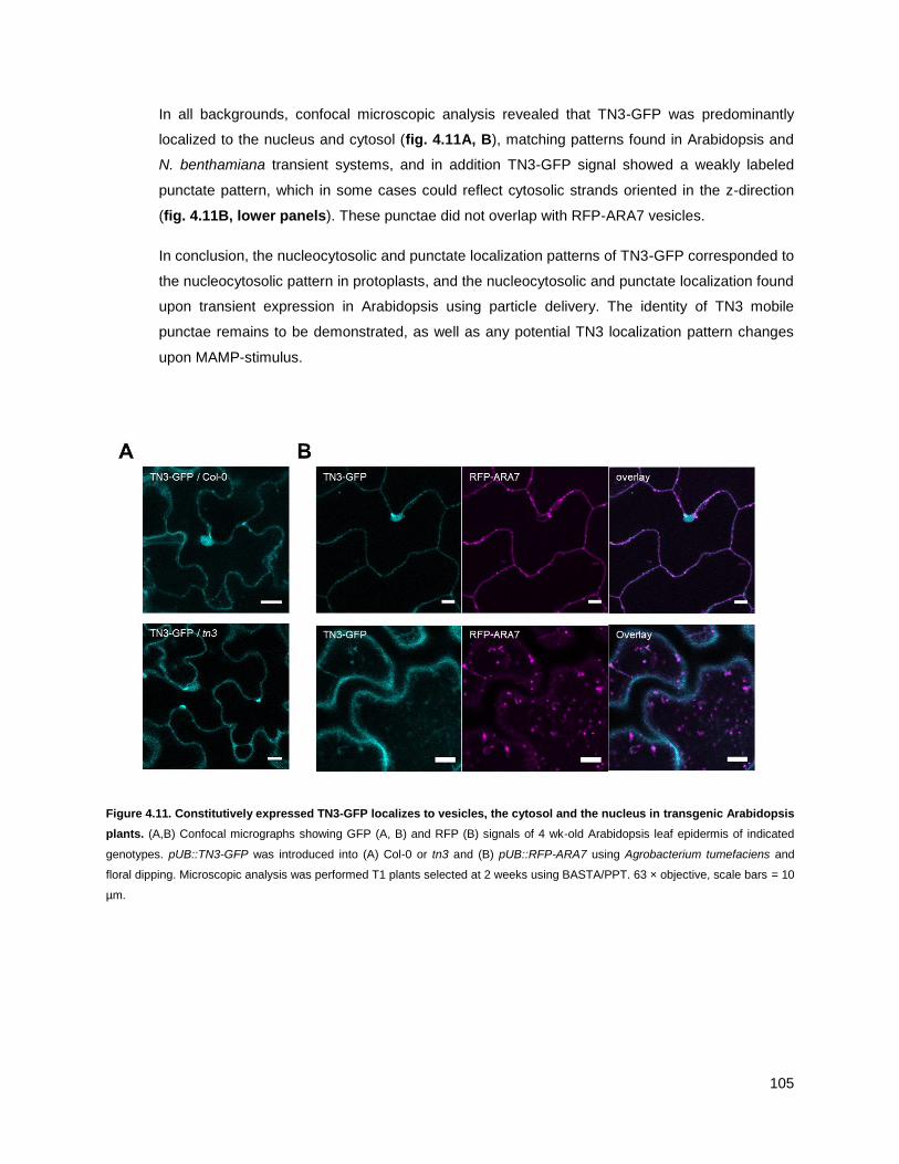
105
In all backgrounds, confocal microscopic analysis revealed that TN3-GFP was predominantly
localized to the nucleus and cytosol (fig. 4.11A, B), matching patterns found in Arabidopsis and
N. benthamiana transient systems, and in addition TN3-GFP signal showed a weakly labeled
punctate pattern, which in some cases could reflect cytosolic strands oriented in the z-direction
(fig. 4.11B, lower panels). These punctae did not overlap with RFP-ARA7 vesicles.
In conclusion, the nucleocytosolic and punctate localization patterns of TN3-GFP corresponded to
the nucleocytosolic pattern in protoplasts, and the nucleocytosolic and punctate localization found
upon transient expression in Arabidopsis using particle delivery. The identity of TN3 mobile
punctae remains to be demonstrated, as well as any potential TN3 localization pattern changes
upon MAMP-stimulus.
Figure 4.11. Constitutively expressed TN3-GFP localizes to vesicles, the cytosol and the nucleus in transgenic Arabidopsis
plants. (A,B) Confocal micrographs showing GFP (A, B) and RFP (B) signals of 4 wk-old Arabidopsis leaf epidermis of indicated
genotypes. pUB::TN3-GFP was introduced into (A) Col-0 or tn3 and (B) pUB::RFP-ARA7 using Agrobacterium tumefaciens and
floral dipping. Microscopic analysis was performed T1 plants selected at 2 weeks using BASTA/PPT. 63 × objective, scale bars = 10
µm.

106
4.3.5 - The TN3 localization pattern does not change upon bacterial stimulus
It is possible that TN3 may show the same pathogen-induced focal accumulation pattern as
ARA7 in the same time frame. To test this, I syringe-infiltrated Pto DC3000 wildtype, and Pto
DC3000-mCherry fluorescently tagged bacteria into fully developed leaves of 4-week old tn3 /
pUB::TN3-GFP Arabidopsis plants, and analysed cells using confocal microscopy.
Between 6-7 h after infiltration of Pto DC3000, the time point at which ARA7 showed focal
accumulations, I did not observe a change in the predominantly nucleocytosolic localization of
TN3-GFP (fig. 4.12A). While cell-periphery-localized structures were observed, these are nuclei,
as evidenced by the visible decrease in TN3-GFP-intensity in the nucleolus. To be able to
visualise bacterial contact sites, I infiltrated Pto DC3000-mCherry bacteria. However, when
observing multiple bacterial contact sites with immobilized Pto DC3000-mCherry bacteria, no
obvious localization pattern changes, or accumulations, of TN3-GFP were observed (fig. 4.12A,
lower panels, B). For nearly all bacterial contact sites that were analysed in earlier experiments,
YFP-ARA7 concentration was observed at infection sites.
I conclude that TN3 does not focally accumulate like ARA7 upon bacterial infection, but shows no
evident changes in its predominantly nucleocytosolic localization pattern. This raises further
questions on the nature of the interaction between TN3 and ARA7, because the predominant
localization of ARA7 at these time points is at clearly observable vesicle clusters. TN3 may be
involved in guarding many host processes, including those that are ARA7 dependent.

107

108
Figure 4.12. TN3-GFP does not show focal accumulation in response to bacterial infection. (A) Confocal micrographs
showing GFP (A, B) and autofluorescence (A) or mCherry signals (A,B) in leaf epidermal cells of 4 wk-old T1 Arabidopsis plants
expressing pUB::TN3-GFP in a tn3 background, 6 hours after syringe infiltration with wildtype Pto DC3000 bacteria (A, top panels)
or at 6 hours after syringe infiltration of Pto DC3000-mCherry (A, bottom panels; B, all panels). Arrowheads point at nuclei. 63 ×
objective, scale bars = 10 µm. (B) Additional observations of immobile Pto DC3000-mCherry bacteria at plant cell surfaces, as in (A)
lower panels.
4.4 – HOMOLOG OF RPW8 4 LOCALIZES TO MITOCHONDRIA
4.4.1 - Confirming the identification of HR4 in flagellin-treated ARA7 purifications
As a second candidate that was identified in ARA7 purifications upon flg22 treatment only, I
selected Homolog of RPW8 4 (HR4) for follow-up investigation. RPW8 homologs, and the well-
studied orthologue in Ms-0 RPW8.2, have been shown to exhibit accumulation around pathogen
interfaces of powdery mildews in Arabidopsis and N. benthamiana (Berkey et al., 2017; Wang et
al., 2013; Kim et al., 2014), and confer broad-spectrum resistance to infectious powdery mildews
(Xiao et al., 2001). It is possible that ARA7 could play a role in directing the localization of HR4,
potentially contributing to its functioning in immunity.
I obtained amino acid sequences of the two peptides identified in mass spectrometry, and used
Protein BLAST (NCBI web interface) to match the peptides against the known Arabidopsis protein
database (TAIR10). Both peptides returned predominantly 100 % sequence identity to HR4, with
the exception of peptide ―TMESISPVRDR‖, identified in two experimental replicates, which also
matched against the transcription factor bHLH147, but with only 63% coverage of the HR4
peptide sequence (fig. 4.13A). Next, I graphically mapped the identified HR4 peptides to its
amino acid sequence, with annotated transmembrane (TM) and coiled-coil (CC) domains. One
detected peptide covers the N-terminus of HR4, with a 4-amino acid overlap into the N-terminal
TM-domain (fig. 4.13B).
While the coiled-coil domain of HR4 was not detected in my experiments, two peptides were
found in separate experimental replicates that match between the TM and CC domains (fig.
4.13B). In addition to using SCAFFOLD PeptideProphet and ProteinProphet confidence
treshholds during mass-spectrometry data analysis in preparation for defining ARA7-co-purifying
proteomes, I also intended to confirm that the peptides I found for HR4 were specific to HR4, and
not to other HR family members. Indeed, the N-terminal peptide ―MPIAELAVIK‖ matched with a
highly HR4-specific inserted stretch of amino acids at the N-terminus, as can be observed when
aligning HR1-4 and plotting the location of this peptide in the multiple sequence alignment (fig.
S14). In addition, the second peptide ―TMESISPVRDR‖ fully matched with a sequence that aligns
with all HR homologs, but is specific to HR4 on amino acid level, and not to HR1,2 or 3 (fig. S14).

109
Figure 4.13. HR4 associates with YFP-ARA7-positive compartments in a flg22-responsive manner. (A) Overview shows top-
10 search results using NCBI protein BLAST (Organism: Arabidopsis thaliana) of two most abundant HR4 peptides identified in
mass spectrometry analysis of immunopurified YFP-ARA7 upon flg22 treatment. (B) Schematic overview shows the primary HR4
amino acid sequence indicating annotated domains and individually labelled detected peptides identified by mass spectrometry
analysis of immunopurified YFP-ARA7 upon flg22 treatment. (C) Phylogenetic tree of all four Homologs of RPW8.2 (HR1-4) was
generated using genomic sequences of all four HR genes (source: The Arabidopsis Information Resource (TAIR)-10). Alignments
and phylogenies were obtained using online available EMBL-EBI Clustal Omega and Simple Phylogeny bioinformatics tools. Legend
= 0.2 substitutions / base pair.

110
In a multiple sequence alignment based on genomic sequences of all four Col-0 Homolog of
RPW8 genes using Clustal Omega (EMBL web suite), HR4 aligns most closely to HR3 (fig.
4.13C). In a recent study that further analysed the function of HR1, HR2, HR3 and HR4 from Col-
0, HR3 was found to have the most pronounced effects on powdery mildew resistance (Berkey et
al., 2017). In addition, HR3 has been previously suggested to be most similar to the ancestral
gene that gave rise to RPW8.1 and RPW8.2 in Ms-0, as well as HR1-4 in Col-0 (Xiao et al.,
2005). Interestingly, HR3 was found a the papilla under Golovinomyces cichoracearum attempted
penetration sites in the leaf epidermis, in addition to subsequently formed encasements around
the haustorial neck (Berkey et al., 2017). In addition, upon transient expression in N.
benthamiana, HR4 was found to accumulate around G. cichoracearum encasements (Berkey et
al., 2017). In my studies, finding HR4 interacting with ARA7 upon MAMP-stimulus is consistent
with ARA7 accumulating around haustoria of Golovinomyces orontii and Phytophthora infestans
(Bozkurt et al., 2015; Inada et al., 2016), and specifically at encasements in the interaction with
Blumeria graminis f.sp. hordei (Nielsen et al., 2017b), but at this point it is unknown whether HR4
also accumulates at bacterial infection sites.
4.4.2 - HR4 localizes to the periphery of mitochondria
Next, I sought to reveal HR4 subcellular localization using fluorescent tagging and confocal
microscopy. To probe for colocalization with ARA7, and potential other subcellular localizations, I
generated binary expression vectors that encode pUB::HR4-GFP and pUB::HR4-RFP, which are
ubiquitin-promoter driven C-terminally GFP and RFP-tagged versions of HR4, based on its coding
sequence obtained through reverse-transcription from mRNA.
Upon transient Agrobacterium-mediated expression in N. benthamiana leaves, HR4-GFP
localized to punctae that did not overlap with co-expressed mCherry-ARA7 under untreated
conditions, or after syringe-infiltration of flg22 at 3 h, matching flg22 treatment time points at
which HR4 was originally found in mass spectrometry of ARA7 purifications (fig. 4.14A). In order
to probe the identity of these mobile HR4-GFP-positive punctae, I co-expressed HR4-GFP with
published mitochondrial (fig. 4.14B) and peroxisomal (fig. 4.14C) RFP-tagged markers (Nelson
et al., 2007) in N. benthamiana. HR4-GFP fully co-localized with the mitochondrial marker, and
upon closer inspection, can be seen to localize more strongly to the periphery of these
mitochondria (fig. 4.14B), pointing at a potential outer membrane association. HR4-GFP did not
colocalize with the peroxisomal marker (fig. 4.14C). Syringe infiltration of flg22 into HR4-GFP and
mitochondrial marker co-expressing N. benthamiana leaves did not change the co-localization
pattern of HR4 with mitochondria (fig. 4.14B, lower panels).
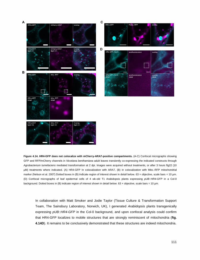
111
Figure 4.14. HR4-GFP does not colocalize with mCherry-ARA7-positive compartments. (A-C) Confocal micrographs showing
GFP and RFP/mCherry channels in Nicotiana benthamiana adult leaves transiently co-expressing the indicated constructs through
Agrobacterium tumefaciens mediated transformation at 2 dpi. Images were acquired without treatments, or after 3 hours flg22 (10
µM) treatments where indicated. (A) HR4-GFP In colocalization with ARA7, (B) in colocalization with Mito.-RFP mitochondrial
marker (Nelson et al. 2007) Dotted boxes in (B) indicate region of interest shown in detail below. 63 × objective, scale bars = 10 µm.
(D) Confocal micrographs of leaf epidermal cells of 4 wk-old T1 Arabidopsis plants expressing pUB::HR4-GFP in a Col-0
background. Dotted boxes in (B) indicate region of interest shown in detail below. 63 × objective, scale bars = 10 µm.
In collaboration with Matt Smoker and Jodie Taylor (Tissue Culture & Transformation Support
Team, The Sainsbury Laboratory, Norwich, UK), I generated Arabidopsis plants transgenically
expressing pUB::HR4-GFP in the Col-0 background, and upon confocal analysis could confirm
that HR4-GFP localizes to mobile structures that are strongly reminiscent of mitochondria (fig.
4.14D). It remains to be conclusively demonstrated that these structures are indeed mitochondria.

112
In conclusion, the evidence I gathered using fluorescent tagging and confocal microscopy
suggests that HR4 localizes to the periphery of mitochondria, and does not localize to ARA7-
positive vesicles. Since mitochondria have been described to accumulate and immobilize under
attempted penetration sites of powdery mildew (Fuchs et al., 2015a), it could be that the local
high abundance of mitochondria, in concert with local accumulation of ARA7, may lead to their
close proximity and potentially their association.
4.5 - TN3 AND HR4 ARE REQUIRED FOR PLANT IMMUNITY
4.5.1 TN3 and HR4 are not required for basal and flagellin-induced antibacterial immunity
Flagellin-induced resistance to Pto DC3000
TN3 and HR4 proteins have a role in immunity. I therefore tested the infection success of a panel
of bacterial, fungal and oomycete pathogens on Arabidopsis T-DNA insertion mutants or SALK
lines, generated by the Salk Institute for Biological Studies, affected in TN3 (tn3,
SALK_018440.56.00, insertion in promoter of AT1G66090) and HR4 (hr4, SALK_208828,
insertion in first intron of AT3G50480). The T-DNA insertion lines were confirmed for homozygous
insertion using PCR with insert- and gene-specific primers on genomic DNA extractions.
The association of native TN3 and HR4 with ARA7 was dependent on flg22 stimulation. I
therefore tested whether tn3 and hr4 are affected in their ability to induce resistance to syringe-
infiltrated Pto DC3000 bacteria upon 1-day pre-treatment with flg22. I performed the experiment
with water infiltration as a control, to also reveal potential basal resistance defects in either
mutant. Upon measuring in planta bacterial proliferation after three days of incubation in water
pre-treated plants, I observed no statistically significant differences between Col-0, fls2, tn3, or
hr4 (fig. 4.15A). The lack of difference in bacterial infection success between Col-0 and fls2 in
this type of assay has been described before (Nekrasov et al., 2009). Interestingly, flg22
treatment successfully induced bacterial resistance in Col-0, tn3 and hr4, indicating that neither
mutant is affected in mounting flg22-induced defenses. Mutant fls2 plants did not show
statistically significant differences in bacterial colonization between water or flg22-pretreatments
(fig. 4.15A, left panel).

113
Figure 4.15. TN3 and HR4 are required for plant immunity. (A) Bar graphs represent growth of Pto DC3000 and cor bacteria as
number of bacterial colony-forming units (CFU) per cm2. 4 weeks-old Arabidopsis leaves of Col-0, fls2, tn3 and hr4 genotypes.
Plants were infected with the indicated bacteria by syringe-infiltration without and with 1 µM flg22 induction 1 day prior bacterial
infection, or spray inoculation, and harvested at 3 dpi. Experiments show results (bars = mean, error bars = SE, n = 8 plants per
treatment) of one experiment, representative of a set of three replicates. (B) Bar graphs represent penetration success (% spores
forming cell-penetrating structures) of Golovinomyces orontii or Erisyphe pisi after spore application on the surface of 6 weeks-old
Arabidopsis leaves of the indicated genotypes. Coommassie staining and microscopic analysis to visualise fungal structures were
performed at 48 hpi. Experiments show combined data of four independent experimental replicates. Between 400-600 total spores
per treatment per experiment were examined. (A,B) Letters indicate significance grouping after one-way ANOVA analysis and post-
hoc Tukey significance testing (p < 0.05). (C) Macroscopic images show symptoms of disease progression on abaxial and adaxial
sides of 5 wk-old Arabidopsis leaves inoculated with Albugo candida Ex1 and imaged at 21 dpi. Images show symptoms
representative of ca. 10 plants per treatment, experiment was performed once. Infection assays in (B) were performed in
collaboration with Dr. Hannah Kuhn, laboratory of Prof. Ralph Panstruga, RWTH Aachen University, Aachen DE. Infection assays in
(C) were performed in collaboration with Baptiste Castel MSc., laboratory of Prof. Jonathan Jones, The Sainsbury Laboratory,
Norwich UK.

114
Resistance to Pto DC3000 cor
Work from our laboratory revealed that ara7 mutants are more susceptible to Pto DC3000 cor
bacteria (Bourdais et al., 2018, submitted). Because it is possible that TN3 and HR4 may function
in the same pathway as ARA7 during immune responses, I tested the infection success of Pto
DC3000 cor on tn3 and hr4.
I chose to perform infections for both syringe-infiltrated bacteria and surface-spray inoculated
bacteria. Upon syringe infiltration, I observe no statistical differences in bacterial proliferation of
Pto DC3000 cor between Col-0, fls2, tn3 and hr4 (fig. 4.15A, middle panel). Spray-inoculation
also did not reveal statistically significant differences when comparing Col-0 to tn3 and hr4, but
Pto DC3000 cor bacteria were more successful on fls2c, consistent with the inability to close
stomata in response to flg22 (Spallek et al., 2013; Bourdais et al. 2018., submitted).
I conclude that tn3 and hr4 are not impaired in basal or flg22-induced resistance against Pto
DC3000, or basal resistance against virulence-impaired Pto DC3000 cor bacteria. While Pto
DC3000 is an adapted pathogen of Arabidopsis, potential more subtle resistance effects of TN3
or HR4 that might have been masked by the action of coronatine at both stomatal or post-
stomatal defenses, are not revealed in this assay.
4.5.2 - TN3 and HR4 are required for immunity against nonadapted powdery mildew
Filamentous pathogens that project infection structures into plant cells elicit the delivery of host
membranes, including from endocytic origin, to extrahaustorial membranes. LE/MVBs fuse with
the EHM of G. orontii and deliver cell wall materials there (Micali et al., 2011), LE/MVB-
associated RABG3c targets P. infestans haustoria (Bozkurt et al., 2015), and the presence of
both secretory and endocytic vesicles around Hyaloperonospora arabidopsidis haustoria
suggests the direction of a variety of vesicle trafficking pathways to infection structures (Lu et al.,
2012). ARA7 itself accumulates at haustoria of powdery mildews, and is involved in resistance
against Bgh (Nielsen et al., 2017b; Inada et al., 2016). Homologs of RPW8 were shown to
function in antifungal resistance (Berkey et al., 2017), and NLRs in general can function in the
detection of fungal effectors (Stotz et al., 2014; Selin et al., 2016). For these reasons, I was
interested to test whether tn3 and hr4 may be affected in resistance against powdery mildew.
These experiments were performed in collaboration by Hannah Kuhn (RWTH Aachen University,
Aachen, Germany).
Upon application of spore suspensions to the leaf surface of Col-0, tn3 and hr4, and subsequent
quantification of the percentage of spores successfully forming secondary hyphae, we found that
compared to Col-0, tn3 and hr4 were equally susceptible to the adapted powdery mildew G.
orontii (fig. 4.15B, left panel). However, both tn3 and hr4 were more susceptible to the non-
115
adapted powdery mildew Erisyphe pisi (fig. 4.15B, middle and right panels). This prompted us
to test whether the phenotype could be reversed upon constitutive expression of TN3 and HR4 in
their respective mutant backgrounds. In addition to the tn3 / pUB::TN3-GFP plants used in
localization studies described elsewhere in this work, I generated hr4 / pUB::HR4-GFP lines in
collaboration with Matt Smoker and Jodie Taylor (Tissue Culture & Transformation Support
Team, The Sainsbury Laboratory, Norwich, UK). Upon testing for E. pisi resistance, we found that
both tn3 / pUB::TN3-GFP and hr4 / pUB::HR4-GFP complementation lines were significantly
more resistant than Col-0 or their respective mutant backgrounds (fig. 4.15B, right panels). This
indicates that the fluorescent fusion proteins TN3-GFP and HR4-GFP are functional.
4.5.3 - TN3 and HR4 are not required for resistance against the downy mildew Albugo candida Ex1
In collaboration with Baptiste Castel (Jonathan Jones group, The Sainsbury Laboratory, Norwich,
UK), we tested for altered resistance of tn3 and hr4 to the adapted oomycete pathogen Albugo
candida isolate Exeter1 (Prince et al., 2017). Upon inspection of disease symptoms 21 days after
spray inoculation of spore suspensions, we found that Col-0, tn3 and hr4 generally exhibited
similar levels of yellowing and A. candida sporulation, whereas susceptible genotype Col-0 / wrr4
(Borhan et al., 2008) showed higher levels of sporulation, and resistant Arbidopsis ecotype Oy-0
(Baptiste Castel, PhD thesis) showed no signs of sporulation or yellowing (fig. 4.15C).
Taken together, these results point at a role for both TN3 and HR4 in defence against
nonadapted powdery mildew. This result raises questions on the mechanisms by which both
proteins achieve this, of subject to further study.
4.5.4 - Constitutive expresion of TN3-GFP and HR4-GFP does not provoke a cell death response in
Nicotiana species.
TN3 has been previously described to trigger a hypersensitive response in N. benthamiana upon
35S-driven overexpression of untagged versions (Nandety et al., 2013), a response which was
exacerbated by co-infiltration of a flg22 solution. To test for potential autoactivity of TN3-GFP, I
used Agrobacterium-mediated transient expression in N. tabacum of pUB::TN3-GFP from the
binary vector used for the generation of Arabidopsis transgenic lines. I performed the assay
initially in N. tabacum due to its general predisposition to reveal NLR-mediated auto-HR in a more
evident manner than N. benthamiana (Amey Redkar, Jonathan Jones lab, personal
communication).
Upon transient expression of TN3-GFP in N. tabacum, and macroscopic observation at four days
post infiltration, no cell death symptoms were observed (fig. 4.16, top left panel). In order to
match the original finding of 35S::TN3-induced cell death (Nandety et al., 2013), I performed the
same analysis in N. benthamiana, which also did not trigger cell death (fig. 4.16, lower left

116
panel). The construct can be successfully transiently expressed using Agrobacterium, as evident
from further localization studies in N. benthamiana (fig. 4.17A,B,C).
Figure 4.16. Constitutive expression of TN3-GFP and HR4-GFP does not provoke a cell death response in Nicotiana
species. Representative macroscopic images show no obvious cell death symptoms upon constitutive expression of GFP-tagged
TN3 and HR4 in Nicotiana. Nicotiana tabacum or Nicotiana benthamiana leaf regions were transiently transformed with
Agrobacterium tumefaciens using the indicated combinations of constructs without and with infiltration of 10 µM flg22. Images were
taken at 4 dpi.

117
Since flg22 treatment was shown to exacerbate the cell death response of 35S::TN3 (Nandety et
al., 2013), I co-infiltrated Agrobacterium tumefaciens carrying pUB::TN3-GFP with a solution of
flg22, and as a control I co-infiltrated the bacterial Elongation Factor-Tu (EF-Tu) immunogenic
epitope elf18, which is not recognized by Nicotiana spp. which lack the cognate PRR EFR
(Lacombe et al., 2010). However, in neither case, cell death symptoms were observed (fig. 4.16,
top and bottom panels).
In parallel, I tested transient expression of pUB::HR4-GFP, which was functional in recovering
mildew resistance defects of the hr4 genotype, and reveal that in N. tabacum, no cell death
symptoms were observed, in untreated and flg22 or elf18 co-treated leaves (fig. 4.16, third row
panels). Flagellin can be perceived by N. tabacum, which endogenously expresses a homolog of
FLS2 (Wei et al., 2012).
Based on the biochemical interaction studies of ARA7 and TN3, I hypothesized that TN3 may
interact with, and potentially monitor the integrity of, ARA7 or a component of the ARA7 greater
complex. Therefore, co-expression with ARA7 may alter the capacity of TN3 to induce cell death
upon constitutive expression. To tes tthis, I performed Agrobacterium-mediated co-expression of
pUB::TN3::GFP with pUB::mCherry-ARA7 and as a control included pUB::mCherry-ARA5.
However, in neither N. benthamiana or N. tabacum this lead to an induction of cell death at 4 dpi
(fig. 4.16, middle and lower rows).
The above results suggest that transient ubiquitin-promoter-driven expression of fluorescently
tagged TN3 and HR4, which functioned in mildew resistance, does not trigger cell death
responses in Nicotiana spp., independent of MAMP-signaling. It is possible that the fluorescent
tag interferes with the induction of cell death by TN3-overexpression as was previously found for
35S::TN3 untagged overexpression in N. benthamiana (Nandety et al., 2013). In addition,
potentially the accumulation levels of TN3 when driven by the weaker ubiquitin promoter are
below the threshold needed to trigger cell death, which may not be the case upon 35S-driven
overexpression. One experimental replicate testing untagged pUB::TN3 expression in N. tabacum
did not result in cell death.
4.5.5 - Constitutively expressed TN3-GFP exhibits localization patterns non-responsive to flg22 in
Nicotiana benthamiana.
In order to verify whether ubiquitin-promoter-driven fluorescently tagged versions of TN3 and HR4
could be successfully expressed using Agrobacterium-mediated expression, I performed confocal
microscopy localization studies in N. benthamiana leaves using pUB::TN3-GFP, pUB::TN3-RFP
and HR4-GFP, and included co-expression of pUB::mCherry-ARA7 and pUB::mCherry-ARA5. I
found that TN3-GFP and TN3-RFP localized to the nucleus and cytosol (fig. 4.17A), largely
matching the localization pattern found in Arabidopsis leaves.

118

119
Figure 4.17. Constitutively expressed TN3-GFP exhibits localization patterns non-responsive to flg22 in Nicotiana
benthamiana. (A-C) Confocal micrographs of Nicotiana benthamiana adult leaves transiently co-expressing the indicated constructs
through Agrobacterium tumefaciens mediated transformation at 2 dpi. Images acquired after 3 hours flg22 (10 µM) treatments in
lower panels of (B). (A) Expressing single TN3-GFP or TN3-RFP constructs, (B) co-expressing TN3-GFP and mCherry-ARA7, (C)
co-expressing TN3-GFP and mCherry-ARA5, (D) co-expressing HR4-GFP and TN3-RFP. 63 × objective, scale bars = 10 µm.
Co-expression with fluorescently tagged ARA7 or ARA5 did not induce a localization change of
TN3-GFP, and overlapping signals were only found in the cytosol (fig. 4.17B). This pattern did
not change upon 3 h flg22 treatment (fig. 4.17B), timed to match the ligand treatments performed
in Arabidopsis seedlings in preparation for immunoprecipitation/mass-spectrometry experiments.
It is noteworthy that upon TN3-GFP co-expression with mCherry-ARA5, the fluorescent signals
overlapped in the cytosol (fig. 4.17C), but in BiFC/split-YFP experiments (fig. 4.9), and
immunoprecipitation/mass-spectrometry experiments described in chapter 3 of this thesis, ARA5
did not interact with TN3 upon untreated, water control or flagellin-induced conditions. This
provides further evidence that cytosolic TN3 does not unspecifically associate with Rab GTPases
due to possible ectopic localization. The localization pattern of TN3-GFP in N. benthamiana is
predominantly the same as found for Arabidopsis, with the exception that in Arabidopsis, a minor
amount of mobile TN3-GFP punctae were found.
Since both HR4 and TN3 interacted with ARA7 upon flg22 treatment as identified in mass
spectrometry experiments, I tested whether co-expression of fluorescently tagged TN3 and HR4
could influence their localizaton (fig. 4.17D), which was not the case. HR4-GFP still localized to
punctae likely to represent mitochondria, and TN3-RFP remained nucleocytosolic. In summary,
co-expression of functional TN3-GFP and HR4-GFP with tested RAB-GTPases, or with each
other, does not change the localization pattern of these proteins in N. benthamiana, independent
of flg22 treatment.

120
DISCUSSION
4.6 - ARA7 exhibits dynamic localization patterns
I found that, between 6-7 hours after infiltration of Pto DC3000 or the bacterial MAMP flg22,
ARA7-positive vesicles tend to cluster at the cell periphery. These clusters coincided with the
position of bacteria attached to the cell surface (fig. 4.1). The focal accumulations of ARA7 also
occurred to a lower extent in water treatments, but upon quantification using a FIJI macro, I could
show that in some experimental replicates, they could be triggered significantly more by Pto
DC3000 wildtype, or expressing mCherry, as well as by effector disarmed Pto DC3000 HrcC- and
the ETI-triggering Pto DC3000 AvrRpt2 (fig. 4.3-4). Qualitatively, it was striking that whenever a
cell-surface localized Pto DC3000-mCherry bacterium was observed, most often an ARA7 vesicle
cluster was found beneath it. There were many occasions of ARA7 vesicle clusters that did not
coincide with bacterial presence.
Notably, the response came up at ca. 6 hpi, and subsided quickly after ca. 7 hpi, with no obvious
localization changes before. Why not cluster immediately? While it is possible that concentration
of ARA7 at the cell periphery starts at earlier time points, I could not visualise this using confocal
microscopy before ca. 6 hpi, but found numerous accumulations rapidly appearing subsequently.
I hypothesize that at earlier time points, plants load endosomes with defense related cargoes,
await the position of pathogen attack, and then mass-deliver endosomal contents in a focused
manner, thus achieving high concentrations of defensive components delivered in the pathogen-
defined location. This is corroborated by the existing finding that the cell-surface defense module
PEN3 increased in abundance at endosomal compartments at 4 hours of flg22 treatment,
whereas PEN3 cell-surface focal accumulations were measurable at >6 hpi (Underwood and
Somerville, 2013).
It is known that default secretory Rab GTPases such as RabE1d cluster at the cell surface at
these time points, upon bacterial presence (Speth et al., 2009). Therefore, it is possible that,
coupled to an increased local secretory activity, endocytosis may need to be upregulated locally
to maintain membrane integrity. Using the lipophilic tracer dye FM4-64, I could show that the
majority of ARA7 vesicles present in clusters under bacteria are pre-existing, as the majority of
them are not marked with FM4-64 at 30 minutes of staining (fig. 4.2), whereas at this time point
the externally applied dye reaches LE/MVBs (Beck et al., 2012b). This indicates that most of the
ARA7 vesicles in the observed clusters do not derive from locally increased endocytic activity.
However, bacteria-induced changes along membrane trafficking pathways might alter the rate of
FM4-64 uptake and distribution throughout the endomembrane network, which I did not test at
earlier time points.
121
The phenomenon of pathogen-induced endosomal concentration at the cell surface has been well
described in response to attempted fungal penetration, where endosomal Rab GTPases are
thought to contribute to the formation of defensive structures, and the delivery of intraluminal
vesicles by fusing with the plasma membrane, which then become extracellular vesicles (EVs,
(Boevink, 2017)).
The endosomal ARA7 and vacuolar RABG3c accumulate around Phytophthora infestans
haustoria (Bozkurt et al., 2015), and ARA7 and ARA6 both accumulate around Golovinomyces
orontii haustoria (Inada et al., 2016). ARA6 endosomes also accumulate under attempted
penetration sites of Blumeria graminis f.sp. hordei (Nielsen et al., 2012, 2017a; An et al., 2006).
More recently, the LE/MVB localized ARA6 was further implicated in the delivery of endosomes
upon infection with Botrytis cinerea. LE/MVBs fused with the PM at host-pathogen contact sites
as evidenced from electron microscopy studies, and confocal analysis showed strong clustering
of ARA6 vesicles at this location (Cai et al., 2018). Similar to my FM4-64 analyses, the authors
use the endocytic tracer dye FM4-64 on B. cinerea infected tissue and show that upon 30
minutes staining, ARA6 vesicle clusters were not co-stained by FM4-64, providing evidence that
they are existing, relocalizing vesicles (Cai et al., 2018). Interestingly, the authors identify
Tetraspanin 8 (TET8) as a bona fide EV cargo, which I do not identify in any of my proteomic
analysis of ARA7, suggesting a more specialized role for ARA6 in this process. While I have not
demonstrated that bacterial-induced ARA7 concentration at the cell surface is coupled to their
fusion to the PM, the previously known accumulation around pathogen interfaces of RabF
GTPases and their incorporation to the EHM suggests that ARA7 can fuse and deliver contents at
the plasma membrane during immune responses. I suggest that this response is at least partially
conserved in response to host detection of bacteria at specific extracellular positions.
ARA7 has been implicated in the delivery of defence molecules to the plant-pathogen interface
before. GTP-locked ARA7-QL, which cannot complete its activation cycle and normally forms
enlarged ARA7-positive LE/MVBs (Jia et al., 2013), accumulated in the haustorial encasement in
response to the non-adapted pathogen Blumeria graminis f.sp. hordei (Bgh, (Nielsen et al.,
2017b)). Encasements are callose-rich structures that are formed secondary to the initial callose-
rich cell-surface papillae (Nielsen et al., 2017b). Interestingly, the authors conclude that while
ARA6 vesicles accumulate near Bgh infection structures (Nielsen et al., 2012), it is not ARA6, but
activation of ARA7 through its interacting ARF-GEF VPS9a thus confers pre-invasive immunity to
Bgh. Upon infection with the adapted fungal pathogen G. orontii, it has been shown that ARA7
does accumulate around the haustorium, but with the conspicuous absence of its activator
VPS9a, leading to the hypothesis that the EHM of adapted pathogens adopts a modified
endosomal identity, likely through the modulation of host processes by pathogen secreted

122
effectors (Inada et al., 2016). Possibly, the targeting of Rab regulatory proteins is guarded by
NLRs such as the ARA7-interacting TN3 described in this thesis.
There are existing data that support that upon bacterial infection, endosomes fuse in an
increased manner with the PM, thus releasing intraluminal vesicles which are then considered
EVs (Rutter and Innes, 2017). Pto infection promoted the amount of EVs secreted by
Arabidopsis, as revealed by quantifying membrane amounts in purifications of apoplastic fluid
(Rutter and Innes, 2017). In fact, the EV proteome derived from those purifications was most
similar to the published ARA7 proteome (Rutter and Innes, 2017; Heard et al., 2015). This
supports a role for ARA7 LE/MVBs in secreting EVs in response to pathogens. Based on my
data, I would suggest that this occurs in a targeted manner, thus I propose that EVs are secreted
from LE/MVBs that fuse to bacterial infection sites. However, Rutter and Innes (2017) found
Patellin1 to be a bona fide marker of EVs, which I do not find in my ARA7 purifications (tables
4.1, 4.3). In addition, they use PEN1 as an EV marker, which is found in my ARA7 purifications,
but does not evidently respond in quantity upon immune stimulus (table 4.1)
While the above studies point at different roles for both ARA6 and ARA7 in the delivery of
defence components to different pathogens, as of yet, it is not clear how they contribute to post-
stomatal antibacterial immunity. It would therefore be interesting to test also ARA6 for clustering
at the 6-7 hpi time point in response to bacterial infiltration, in combination with bacterial
apoplastic infection assays in both ara7 and ara6, to further probe their involvement.
4.7 - ARA7 purifications likely represent a Rab-associated complex
In order to reveal proteomic changes along the endocytic pathway upon immune stimulus at time
points preceding the clustering of ARA7 under bacterial infection sites, I purified ARA7 from 3-
hours water or flg22 treated seedlings, and performed mass spectrometry analysis (tables 4.1-3,
fig. 4.6). I used a previously published method, where the authors could successfully categorise
proteins in separate membrane trafficking pathways using purifications of a panel of membrane-
associated baits (Heard et al., 2015). However, my purifications lead to substantially different co-
purifying proteomes than were published, yet overlapping to some extent (fig. S.5). There are
several underlying causes that could lead to this. Firstly, for ARA7, I used YFP affinity purification,
whereas Heard et al. (2015) used RFP purifications. Secondly, a different model of mass
spectrometer was used for my experiments. Finally, the way in which I define my YFP-ARA7
proteome, comparing it to YFP purifications and requiring a 4-fold spectral count increase, differs
from the method used in Heard et al. (2015), who applied a statistical analysis method ―SAINT‖ in
order to separate the different endomembrane compartment proteomes based on confidence
scores for each protein (Choi et al., 2011). I inspected the ARA7 co-purifying proteomes that
resulted from my pulldown/mass-spec experiments and conclude that the majority of abundant

123
proteins correspond to the known subcellular localization and function of ARA7, making the co-
purifying proteome suitable for discovering novel interactors of the ARA7 complex upon flg22
treatment, which was my goal.
It is uncertain whether complete endomembrane compartments are purified using the method I
employ. Heard et al. (2015) point out that ca. 30% of cargoes identified in ARA7 purifications
contain transmembrane domains, suggesting that at least part of the vesicle membrane was co-
purified. However, my proteomes differed considerably (fig. S.5). When inspecting my ARA7 co-
purifying proteome (tables 4.1-2), it is evident that the majority of co-purifying proteins are known
to associate with membranes on the cytosolic side (SNAREs, coatomer, SCAMPs) or are Rab
GTPase-associated (PRA1 members, PUF2, VPS9a). However, I do find cargoes of the pathway
such as CESAs, glucan synthases and PEN3, but they all contain transmembrane domains. A
minority of proteins, such as CER8 and ubiquitin-like proteins do not contain transmembrane
domains. From these co-purifying proteins, I conclude that I purify the greater Rab GTPase-
interacting complex at minimum, and from TM-domain containing proteins, I speculate that at
least membrane fragments originating from TGN/EE-LE/MVB co-purified with ARA7. Taking all
evidence together, this suggests that I purified membrane-associated ARA7, because the
cytosolic phase interacts with a limited amount of proteins, mostly belonging to the Rab regulatory
complex (Saito and Ueda, 2009b), and co-purifying proteins that I found such as SNAREs are
membrane-integral and do not cycle between cytosolic and membrane-associated states.
A number of Golgi proteins passed my criteria of defining an ARA7 co-purifying proteome, as they
were associated with YFP-ARA7 with a factor of more than four-fold compared to YFP controls.
When comparing my ARA7 proteome to the published Golgi proteome obtained using the same
affinity purification method (Heard et al., 2015), I observe that ca. 7% (11 out of 159) ARA7
proteins were found in the Golgi proteome. This is comparable to the amount of Golgi proteins
that were found in the ARA7 proteome from the same publication, where ca. 10% (29 out of 280)
of ARA7 proteins were also identified in Golgi. Yet, my ARA7 ―Golgi contaminants‖ overlapped
with the published ARA7 ―Golgi contaminants‖ with only 4 out of 11 proteins. Overlap included
VAMP722, PRA1.B1 and two Endomembrane Protein 70 members.
It is possible that the purification method is inaccurate to some extent, and purifies unspecific
proteins or contaminants. However, they could also reflect constitutive biogenesis of the YFP-
ARA7 fusion protein, the transcription of which is driven by the constitutively active Ubiquitin-10
promoter (Geldner et al., 2009). Co-localization analysis of Golgi-TGN/EE-localized ARA5 with
ARA7 showed no punctate overlap (chapter 5, figure 5.5), so it is unlikely that a high abundance
of YFP-ARA7 is present at the Golgi. However, fluorescent signal can only occur when the newly
synthesized YFP chromophore is fully matured, so a subpool of ―dark‖ YFP-ARA7 may be
present at the Golgi. Alternatively, TGN/EE and Golgi may be linked to some extent, and difficult

124
to separate. Indeed, the TGN/EE partially originates from the Golgi (Ito et al., 2017), and there is
a closely Golgi-Associated (GA) pool of TGN/EE, separate from Golgi-Independent (GI) TGN/EE
(Uemura et al., 2014).
Strikingly, CONSERVED OLIGOMERIC GOLGI 2 (COG2, AT4G24840), a Golgi-functioning
tethering factor, which was low abundant, but upregulated in ARA7 upon flg22 treatments in my
purifications (table 4.3), was predicted to be the target of H. arabidopsidis HaRxL60, HaRxL73,
HaRxLL518 and ATR13, as well as G. orontii effector candidate OEC25 (Mukhtar et al., 2011;
Weßling et al., 2014). COG2 has been detected in a vacuolar proteomic study (Carter, 2004),
pointing at its likely trafficking from the Golgi to vacuole, which could occur through ARA7
TGN/EE and LE/MVBs. With COG known to function in intra-Golgi trafficking processes, ARA7 is
unlikely to be its biologically relevant localization, or the location at which effectors target it.
However, it would be interesting to confirm this using bimolecular fluorescence complementation
with predicted interacting effectors, pinpointing the location of their interaction in relation to ARA7.
In addition to some Golgi proteins, at higher abundances I find ER localized proteins such as
ribosomal proteins, ribosomal-associated signal-particle proteins, and SEC22 which functions in
ER-Golgi export (table 4.1). While ARA7 is a broad endocytic marker localizing to TGN/EE and
LE/MVB, it has not been described to localize to the ER. Potentially, above-described
biosynthesis of YFP-ARA7 could lead to some purification of ER proteins, but the high abundance
of the ribosomal protein SUPPRESSOR OF ACAULIS 56 gives pause. An explanation may be
found in the observations that LE/MVBs physically associate with ER, which facilitates endosomal
streaming, and that ER-localized VAP27 proteins physically participate in early clathrin-mediated
endocytosis as well as bridge the ER to TGN/EE, where ARA7 also localizes (Stefano et al.,
2018, 2015). Seeing the critical role for the ER in physically guiding endocytic processes involving
TGN/EE and LE/MVB, it would be interesting to see whether the described ER-localized modules
responsible for this are also involved in guiding ARA7 vesicles to the pathogen interface. Indeed,
ER itself was found to focally accumulated under fungal attempted penetration sites (Fuchs et al.,
2015). While ribosomal proteins in my ARA7 purifications could simply reflect close physical
association, it is also conceivable that proteins newly synthesized in response to pathogen
detection hitch-hike on LE/MVBs en-route to the cell-surface or to the vacuole, thus bypassing
extra translocation and posttranslational modification steps in the Golgi.
In animal systems, bacteria have been well described to employ effectors to target both endocytic
and secretory Rab GTPases and their regulatory complexes, modulating host trafficking to their
benefot (Brumell and Scidmore, 2007; Stein et al., 2012; Spanò and Galán, 2017). In plants,
fungal effectors from G. orontii and H. arabidopsidis have been predicted to interact with PRA1
proteins, which are regulators of Rab GTPases, using large scale yeast-2-hybrid (Y2H) screens
(Mukhtar et al., 2011; Weßling et al., 2014), but these interactions have not been subject to

125
further study. Interestingly, upon ARA7 purifications, I identified PRA1.F2 in water treated
samples, which interacted with G. orontii effector candidate OEC65 in the abovementioned Y2H
screen. Secondly, upon flg22 treatments, ARA7 purifications identified PRA1.F3, which was
predicted to interact with G. orontii OEC65 and H. arabidopsidis HaRxL74. Neither interacted with
the library of Pto DC3000 effectors in the same study (Mukhtar et al., 2011; Weßling et al., 2014).
This points at a role for pathogen effectors in modulating Rab activity in plants.
While I could find Rab regulatory proteins such as PRA1 family proteins, and VPS9a, I also
identify at least one Rab effector, as reflected by PLANT UNIQUE RAB EFFECTOR 2 (PUF2; Ito
et al. 2018). However, other known ARA7 effectors such as SAND/MONENSIN SENSITIVITY 1
(SAND/MON1), CALCIUM CAFFEINE ZINC SENSITIVITY 1 (CCZ1) and ENDOSOMAL RAB
EFFECTOR WITH PX-DOMAIN (EREX) proteins were not identified (Singh et al., 2014a; Sakurai
et al., 2016). Of EREX, it is known to function in endosomal delivery to the protein storage
vacuole in seed maturation, which is a process that does not occur in seedlings. The absence of
SAND/MON1 and CCZ1 is puzzling, as they are involved in the conversion of Rab5, to which
ARA7 belongs, into Rab7-identity of membrane compartments in default vacuolar targeted
trafficking (Singh et al., 2014b). It could be that the pool of ARA7 interacting with these effectors
at the last stages of vacuolar targeted LE/MVB traffic is small, preventing MON1/CCZ1 peptide
counts to reach the detection threshold.
4.8 - The ARA7 proteome contains defence-related proteins
Major players in the production of indole-3-glucosonolate based antimicrobials are Indole
Glucosinolate O-Methyl Transferase (IGMT) proteins, of which I identify IGMT4 upregulated in
ARA7 upon flg22 treatment. The subcellular localization of IGMT proteins is as of yet unclear, but
they function in an enzymatic pathway of which other components have known localizations
(Pfalz et al., 2011; Xu et al., 2016). What could be the relevance for IGMT4 appearance on
endosomes in this context? Before export into the apoplastic space, indole-3-glucosinolate-
derived antimicrobial metabolites need to be catalyzed in sequence by CYTOCHROME P 81
(CYP81) family enzymes, IGMT enzymes, and finally myrosinases, among which PEN2, and the
full functioning of enzymatic chain is also necessary for antibacterial resistance (Clay et al., 2009;
Burow and Halkier, 2017; Birkenbihl et al., 2017). PEN2, which I did not identify, is known to
localize to mitochondria, which immobilize under attempted penetration sites, and is there
involved in the last step of antimicrobial production (Fuchs et al., 2015b). We also know that
IGMT proteins produce metabolites which are substrates for PEN2, but it is unknown where
IGMTs are localized (Birkenbihl et al., 2017). The conversion step before is mediated by CYP81
proteins (Fuchs et al., 2015b), which are ER resident, and of which I find no members in my
purifications. Therefore, finding IGMT4 in ARA7 purifications upon flg22 only could point at a
mechanism to bring these intermediate enzymes to the biologically relevant location at the

126
pathogen infection site. ER and LE/MVB can physically associate, and both ER and LE/MVB
concentrate under pathogen infection sites (Fuchs et al., 2015b). Seeing as the final metabolites
of this completed enzymatic pathway are toxic, it would make sense to keep the components
separate until the end products are necessary. My proteomic studies don‘t answer where the
metabolites themselves are located, but they are said to be exported by transmembrane
transporters such as PEN3, which also make use of the redirected endocytic pathway to focally
accumulate. Knowing that the full functionality of this enzymatic pathway is required for
antibacterial immunity (Clay et al., 2009), I hypothesize that the ER also focally reorganises at the
site of bacterial attack, but this remains to be demonstrated.
Finding PEN3 associating with ARA7 upon flg22 treated conditions is interesting, because it
matches the endocytic accumulation of this protein at membrane compartments as previously
observed at 4 h of flg22 treatment (Underwood et al., 2017), and provides more evidence for a
role of the LE/MVB in focal accumulation of PEN3 upon immune stimulus. The observed timings
of 6-7 hours flg22 treatment leading to ARA7 vesicle clustering in my experiments, matches
existing observations that PEN3 cell-surface focal accumulations can be observed from ca. 6
hours onward (Underwood and Somerville, 2013). These data together support the notion that the
time upon which I chose to analyze flg22-induced changes along endomembrane compartments
is suitable for revealing the loading of endosomes with defence cargoes. In this context, it would
be interesting to test for PEN3 focal accumulations in ara7 and ara6 mutants upon pathogen and
MAMP stimulation.
It has been shown that upon leaf infiltration with Pto DC3000, PEN3 accumulations at the cell
surface subside rapidly after ca. 7 hours, which is thought to be an effector-mediated process
(Xin et al., 2013). If the endosomal clustering I observe underlies the targeting of PEN3 to the cell
surface there, the effector-mediated removal of such focal accumulations is unlikely to be a result
of interfering with the continuous endosomal delivery of PEN3, because I could not observe the
ARA7 clustering response after ca. 7 hpi, even upon flg22-only treatments. This is also supported
by published fluorescence recovery after photobleaching (FRAP) experiments on PEN3
(Underwood and Somerville, 2013). Rather, it is likely that effectors affect the stability of PEN3 in
the plasma membrane.
The protein with the strongest increase in spectral counts in ARA7 purifications upon flg22
treatment compared to water treatment was FLOTILLIN 1 (FLOT1). FLOT1 is a membrane-
associated protein, which associates with high-sterol and sphingolipid membrane regions also
known as detergent-resistant membranes (DRMs). In plants, it functions in clathrin-independent
endocytosis (Li et al., 2012b), and in both plants and animals is thought to participate in
modulating the spatial organisation of cell-surface localized transmembrane receptors (Otto and
Nichols, 2011; Haney et al., 2011; Downie, 2014).

127
In addition, in animals, flotillins have been shown to function on the endosome, where they
localize to sphingolipid/sterol-rich membrane regions as well, and there participate in targeting
endocytosed cargoes for recycling back to the PM (Langhorst et al., 2007; Meister and Tikkanen,
2014). ARA7 and FLOT1 partially overlapped on FM4-64 and VHA-a1 positive endosomes in an
increased manner upon flg22 treatment at 1 hour of flg22 treatment (Yu et al., 2017). DRM
enrichment has conversely revealed the co-purification of the well studied TGN/EE marker VHA-
a1, thus providing further evidence that the TGN/EE contains such domains where FLOT1 is
present - noting that VHA-a1 marked TGN/EE overlaped with ARA7 in microscopy studies and
VHA-a1 is found in my ARA7 proteome (Borner et al., 2005; Dettmer et al., 2006). Finding FLOT1
in ARA7 purifications could signify a change in lipid composition along the endocytic pathway.
Since pathogen infection induces the focal accumulation of known defence cargoes, which is
thought to be mediated by redirection of LE/MVB-dependent traffic, and sterol/sphingolipid-
associating FLOT1 in animals has been implicated in mediating the return of endocytosed
cargoes to the PM, these findings together could point at a flg22-induced upregulation of PM-
directed secretory capacity on the ARA7 pathway which is normally targeted to the vacuole.
High sterol/sphingolipid subdomains on TGN/EE, where FLOT1 could preferentially accumulate,
have recently been show to overlap with the SYP61 -marked domain, which I also co-purified with
ARA7, but independent of flg22 stimulus (Wattelet-Boyer et al., 2016). The authors demonstrate
that the SYP61 domain is involved in default secretion and not recycling, whereas the low-
sphingolipid/sterol domain marked by RABA2a, which I do not co-purify with ARA7, was shown to
mediate PM-TGN/EE recycling and not default secretion. This would seem to conflict with the
hypothesis that appearance of FLOT1 on ARA7 signifies a more recycling identity on this
compartment, but is in line with a role for PM-targeting, as default secretory compartments also
mediate PM delivery.
Other data suggest that LE/MVB-localized ARA6 is the endosomal Rab that mediates endosome-
to-PM delivery at resting conditions, and in host-microbe associated processes (Ebine et al.,
2011a; Cai et al., 2018). To further elucidate the potential endosomal-localized functions of
FLOT1 in the context of immunity, and whether this underlies an increased secretory role for
normally endocytic compartments, it would therefore be interesting to test for flg22-induced
colocalization changes of FLOT1 with both ARA7 and ARA6 upon pathogen and MAMP stimulus,
in combination with pathogen infection trials in published FLOT1 knockdown lines (Li et al.,
2012b).
4.9 - The atypical NLRs TN3 and HR4 mediate plant immunity
Using mass spectrometry and biochemical methods, I could show that TN3 and ARA7 interact,
yet in endogenous conditions, this is likely underpinned by induced transcription of TN3 in

128
repsonse to flg22 (fig. 4.7). In bimolecular fluorescence complementation (BiFC) studies, I could
show that TN3 and ARA7 reconstituted a fluorescent signal on ARA7 endosomes (fig. 4.9). Upon
interaction in this type of experiment, the interaction partners are irreversibly linked by the
fluorophore. This could mean that normally, TN3 interacts with ARA7 in a dynamic reversible
way, or with only a subpool of ARA7, but this transient or partial interaction be fully captured by
irreversible binding through BiFC, with ARA7 now ―sequestering‖ TN3 molecules and
accumulating them at endosomes. I hypothesize that I purify a greater Rab-associated complex
at minimum, which probably includes the cytosolic and membrane bound pool. Perhaps TN3 has
an interaction preference for cytosolic localized, GDP-bound inactive pools of ARA7, rather than
all ARA7, or for membrane-associated GTP-bound ARA7, and as a result in BiFC would follow
ARA7 throughout its activation cycle. Notably, TN3 also reconstituted a signal with the LE/MVB-
localized ARA6, which could point at TN3 interacting with Rab5 GTPases in general, or with a
common interactor of Rab5 GTPases, out of which their activator VPS9a or their shared
endosomal localized effector PUF2 could be candidates. This would be interesting to further
confirm using biochemical interaction studies, and could point at host NLR surveillance of Rab
GTPase activity.
I tested Arabidopsis Col-0 T-DNA insertion lines tn3 and hr4 for their capacity to mount resistance
against bacterial pathogens. Because of the flg22-induced appearance of these proteins in my
purifications, I included a flg22-induced resistance test to Pto DC3000 bacteria. However, neither
mutant line was affected in flg22-induced or basal resistance to Pto DC3000, nor in basal
resistance to Pto DC3000 cor (fig. 4.15). By contrast, single mutants of other TN genes, such as
tn2 and tn13, have been shown to be affected in bacterial resistance (Zhao et al., 2015; Roth et
al., 2017). The absence of resistance phenotypes in tn3 and hr4 in my bacterial infection assays
could be explained by the fact that Pto DC3000 is well adapted to Arabidopsis, and Pto DC3000
cor, although lacking coronatine, can still secrete type-3 effectors. Alternatively, Pto DC3000 may
not secrete the effectors that would trigger TN3 or RPW8-based resistance.
Upon challenge with filamentous pathogens, out of G. orontii, Albugo candida Ex1 and E. pisi,
only the latter, non-adapted pathogen showed increased infection success on both tn3 and hr4
plants (fig. 4.15). Importantly, expression of TN3-GFP and HR4-GFP in their respective mutants
could rescue resistance above wildtype levels, demonstrating that both are required for anti-
fungal immunity and are functional as GFP-tagged fusion protein. Since ectopic expression of R-
proteins can lead to constitutive activation of defences, the constitutive expression of both genes
driven by the Ubiquitin-10 promoter could explain the heightened resistance to E. pisi. By
contrast, 35S overexpression lines of HR4 mediate resistance against G. cichoracearum
comparable to Col-0 wildtype plants (Berkey et al., 2017). However, G. cichoracearum is an

129
adapted pathogen on Arabidopsis. I conclude that both TN3 and HR4 function in non-host
resistance to powdery mildew.
HR4 is a homolog of the relatively well-studied atypical immune receptor RPW8.2 and associates
to ARA7 upon flg22 trigger. Surprisingly, HR4-GFP, which is functionally complementing the hr4
mutant phenotype, localized to the periphery of mitochondria in N. benthamiana and similarly in
Arabidopsis independent of flg22 stimulus (fig. 4.14). This is surprising, because I identified HR4
in ARA7 purifications, which does not localize to mitochondria. While the co-localization pattern of
ARA7 and HR4 did not change upon 3 hours flg22 treatment in N. benthamiana, it remains
possible that HR4 more closely associates with ARA7 in Arabidopsis upon immune stimulation, or
has broader targeting specificity dependent on pathogen infection, perhaps directed by NLR
association or activation. It is also possible that fluorescent tagging of HR4 alters its subcellular
localization, but I could show that it does not affect its function in antifungal resistance (fig. 4.15).
To my knowledge, I do not identify any other mitochondrial localized proteins in ARA7
purifications.
HR4 and its homologs HR1-3 in Col-0 have been shown to all accumulate around G. orontii
haustoria (Berkey et al., 2017). Interestingly, HR2-YFP was found in ring structures surrounding
chloroplasts, and in a different study, a subset of C-terminally truncated mutants of RPW8.2
localized to stromule-like membranes that surrounded and connected chloroplasts ((Berkey et al.,
2017; Wang et al., 2013)). While RPW8 proteins are membrane targeted through their N-terminal
TM domain (Wang et al., 2013), this points at a role for membrane specificity determination of the
C-terminus, which is divergent between RPW8 homologs in Col-0 (fig. S14). Yet, upon alignment
of HR1-4 amino acid sequences, I observe a N-terminal extension for HR4 which is not present in
HR1-3 (fig. S.4), which could confer differential localization compared to other HR members
(Berkey et al., 2017).
RPW8.2 is a homologue of HR4, and was shown to be carried on secretory VAMP721/VAMP722-
positive vesicles to the pathogen interface, and in EM-studies was shown to localize to the EHM
proper (Kim et al., 2014). While VAMP721 and ARA7 in fluorescent colocalization studies showed
considerably different punctate localizations in stable transgenic Arabidopsis root cells (Zhang et
al., 2011a), both VAMP721 and VAMP722 were present in the ARA7 proteome (table 4.1-4.2),
which could reflect close association of ARA7 and VAMP721/722 at the TGN/EE, and which
could lead to association of ARA7 with HR proteins that might be carried on these vesicles.
RPW8-type proteins are potent inducers of cell death, and are required for signal activation of
entire NLR subfamilies (Collier et al., 2011; Bonardi et al., 2011). Although the mechanism by
which RPW8-type proteins activates resistance remains poorly characterized, there is a potential
lead. The so called RPW8-domain describes full-length RPW8 and its homologs, and is present

130
as an N-terminal domain in tobacco NRG1 and homologs (Peart et al., 2005) and Arabidopsis
ADR1 (Collier et al., 2011). Expression of the RPW8 part of various ADR1/NRG1 variants
resulted in cell death, and it is known that RPW8 is membrane targeted (Collier et al., 2011;
Wang et al., 2007). Interestingly, RPW8 domains are homologous to HELL domains present on
fungal NLRs (Daskalov et al., 2016). HELL domains and RPW8 domains both have membrane
targeting functions in cell death processes (Daskalov et al., 2016). The authors point out that it is
likely that the HELL domain itself achieves this through inducing membrane pore formation on
location. The above data, in line with the observation that HR4 contributes to defence against a
biotrophic fungus (fig. 4.15), which is commonly associated with hypersensitive cell death, it is
conceivable that HR4 plays a membrane-localized role in activating NLR-mediated cell death.
TN3 interacted with ARA7, which was induced upon initiation of immune signaling in endogenous
conditions, and we found that TN3 is required for full immunity against non-host powdery mildew.
How could TN3 work? Limited information is available on the mechanism of TN3 signaling. Upon
35S overexpression in N. benthamiana, (Nandety et al., 2013) found that TN3 induced a
hypersensitive response, which was exacerbated by flg22 co-infiltration, and which was
dependent on the typical NLR signaling module EDS1. Additionally, 35S overexpression lines in
Arabidopsis could only be retrieved in an eds1 background (Nandety et al., 2013). In my hands,
transient expression of pUB::TN3-GFP in Nicotiana spp. did not trigger any symptoms of HR (fig.
4.16), whereas p35S::TN3 has been shown to trigger the strongest HR symptoms in N.
benthamiana out of all tested TN genes (Nandety et al., 2013). My fluorescently tagged construct
is functional in resistance, as evident from pathogen infection assays (fig. 4.15), and is expressed
in N. benthamiana (fig. 4.17). It is conceivable that my Ubiquitin-10 promoter-driven pUB::TN3-
GFP construct does not accumulate enough TN3 protein compared to the higher expressed
p35S::TN3.
Other TN proteins have been reported to engage immune signaling through unconventional
means. TN2 interacts with the trafficking regulators Exo70B1 and SNAP33, and triggers
autoimmunity in an exo70b1 background, implicating it in the guarding of Exocyst components, or
an Exocyst-dependent process (Zhao et al., 2015). Interestingly, TN2 interacted also with
CALCIUM-DEPENDENT PROTEIN KINASE 5 (CPK5), and CPK5 was required for the
autoimmune response in exo70b1 (Liu et al., 2017). CPK5 is a known component of early PTI
signaling, and required for full resistance against Pto DC3000 (Dubiella et al., 2013). I did not
identify any CPKs in ARA7 purifications upon flg22 (table 4.3).
TN proteins are thought to function in complex with full-length NLRs. A single amino-acid
substitution near the TIR-domain of TN1/CHILLING SENSITIVE 1 (CHS1) lead to an autoimmune
phenotype upon exposure of plants to low temperatures, which was dependent on the presence
of the full-length TNL SUPPRESSOR OF CHL1-2 3 (SOC3), and on the typical NLR signaling

131
components EDS1 and PAD4 (Zbierzak et al., 2013; Zhang et al., 2017). The CHS1 autoactive
variant also physically interacted with SOC3. The SOC3 gene is genomically in close proximity to
CHS1, and interestingly, also TN2, raising the possibilty that TN2 is also dependent on this full-
length NLR. While TN3 does not cluster with these genes, or full-length NLRs in the Arabidopsis
genome, from Y2H screens it has been predicted to interact with the full-length NLR RPP13-LIKE
PROTEIN 1 (RPPL1), which would be a prime candidate for further study in the context of TN3-
mediated immune signaling.
To my knowledge, the only NLR to be detected at endosomes is potato (Solanum tuberosum)
R3a, which confers resistance to the late-blight causative agent Phytophthora infestans, carrying
the effector Avr3a (Engelhardt et al., 2012). Avr3a interacts with dynamin, and interferes with
dynamin-mediated endocytic processes (Chaparro-Garcia et al., 2015). Interestingly, StR3a
localized to the cytosol in resting conditions, and only accumulated at endosomes upon co-
expression of Avr3a (Engelhardt et al., 2012). In the case of TN3 however, I could not observe a
change in its nucleocytosolic localization upon leaf infiltration with Pto DC3000 bacteria, which
inject a range of type-3 effectors (fig. 4.12). From existing Y2H studies, TN3 is predicted to
interact with the type-3 effector HopY1 (Nandety et al., 2013), which is present in Pto DC3000. It
would be interesting to co-express fluorescent TN3 fusions together with HopY1 in the absence of
virulent Pto DC3000, or together with an unbiased panel of type-3 effectors, and follow TN3
localization over time. Seeing as tn3 mutants showed enhanced susceptibility upon infection with
the nonadapted fungal pathogen Erisyphe pisi (fig. 4.15), we are further investigating TN3
localization upon infection with E. pisi.
Upon broader phylogenetic analysis of TN genes compared to plant protein databases, it was
found that TN3 aligns closely to the tobacco NLR N (Nandety et al., 2013). N requires the helper-
NLR N-Requirement gene 1 (NRG1) to function in antiviral immunity (Peart et al., 2005). NRG1
encodes a CC-NLR, of which the CC domain is homologous to RPW8 proteins (Collier et al.,
2011). Since I found both TN3 and the RPW8 homolog HR4 in the same ARA7 purification upon
flg22 treatment, this could implicate TN3 and HR4 functioning together.

132
CHAPTER 5:
IMMUNE SIGNALING-INDUCED CHANGES IN THE LOCALIZATION OF THE
SECRETORY RAB GTPASE ARA5/RABD2A AND ITS CO-PURIFYING PROTEOME
IN COMPARISON TO THE ENDOCYTIC ARA7/RABF2B
RESULTS
5.1 – THE LOCALIZATION PATTERN OF ARA5/RABD2A CHANGES UPON IMMUNE STIMULUS
5.1.1 - ARA5 focally accumulates at the cell periphery upon pathogen stimulus
The default secretory pathway is involved in the targeting of defense components to pathogen
contact sites at the cell periphery upon pathogen or MAMP-stimulus (Kwon et al., 2008; Assaad
et al., 2004; Kalde et al., 2007), and regulators of this pathway such as the SNARE
PEN1/SYP121 accumulate under pathogen attack sites and around pathogen invasion structures
(Kwon et al., 2008; Nielsen and Thordal-Christensen, 2013). ARA5/RabF2b is a Rab GTPase that
localizes to the Golgi and the TGN/EE (Pinheiro et al., 2009), and functions in default secretion.
To examine whether regulators of the default secretory pathway were targeted to pathogen
contact sites, I syringe-infiltrated leaves of 4-weeks old pUB::YFP-ARA5 plants with a suspension
of fluorescently labelled Pto DC3000-mCherry (described in chapter 4 of this thesis), and
observed the localization pattern of ARA5 using confocal microscopy.
No evident localization pattern changes in YFP-ARA5 occurred before 6 h of measurement, but
between 6-7 h after bacterial inoculation, strong clustering of ARA5 vesicles was observed at the
cell periphery, at locations where apoplastic Pto DC3000-mCherry were also observed (fig.
5.1A). These focal accumulations occurred in mesophyll cells (fig. 5.1A,B) and epidermal cells.
6-7 h water control treatments did not provoke ARA5 focal accumulations to a similar extent when
compared to infection with wildtype Pto DC3000 (fig. 5.1B). Bacterial infection triggered focal
accumulations that could also be observed at lower magnifications in the adaxial mesophyll of
infiltrated leaves (fig. 5.1B).

133
Figure 5.1. YFP-ARA5-positive compartments focally accumulate at bacterial infection sites. (A) Confocal micgrographs (z-
projections) of YFP-ARA5 and Pto DC3000-mCherry in 4 wk-old Arabidopsis mesophyll tissue, 6 hours after syringe infiltration of
bacterial suspensions. Bacterial OD600 = 0.2 upon infiltration. Top panels show maximum z-projection and bottom panels show
single focal plane. Bacterial OD600 = 0.2 upon application. 63 × objective, scale bars = 10 µm. (B) Confocal micrographs of upper
leaf mesophyll as in (A) but in the presence of Pto DC3000 and now using the 20 × objective, scale bars = 100 µm.
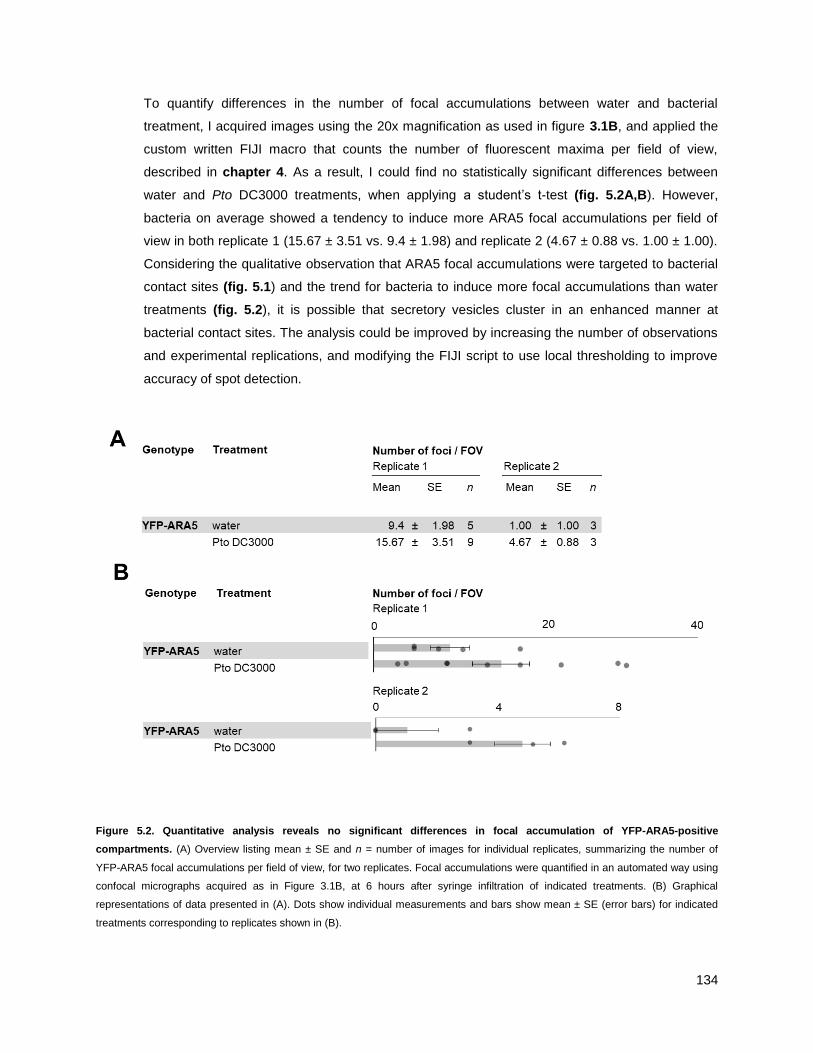
134
To quantify differences in the number of focal accumulations between water and bacterial
treatment, I acquired images using the 20x magnification as used in figure 3.1B, and applied the
custom written FIJI macro that counts the number of fluorescent maxima per field of view,
described in chapter 4. As a result, I could find no statistically significant differences between
water and Pto DC3000 treatments, when applying a student‘s t-test (fig. 5.2A,B). However,
bacteria on average showed a tendency to induce more ARA5 focal accumulations per field of
view in both replicate 1 (15.67 ± 3.51 vs. 9.4 ± 1.98) and replicate 2 (4.67 ± 0.88 vs. 1.00 ± 1.00).
Considering the qualitative observation that ARA5 focal accumulations were targeted to bacterial
contact sites (fig. 5.1) and the trend for bacteria to induce more focal accumulations than water
treatments (fig. 5.2), it is possible that secretory vesicles cluster in an enhanced manner at
bacterial contact sites. The analysis could be improved by increasing the number of observations
and experimental replications, and modifying the FIJI script to use local thresholding to improve
accuracy of spot detection.
Figure 5.2. Quantitative analysis reveals no significant differences in focal accumulation of YFP-ARA5-positive
compartments. (A) Overview listing mean ± SE and n = number of images for individual replicates, summarizing the number of
YFP-ARA5 focal accumulations per field of view, for two replicates. Focal accumulations were quantified in an automated way using
confocal micrographs acquired as in Figure 3.1B, at 6 hours after syringe infiltration of indicated treatments. (B) Graphical
representations of data presented in (A). Dots show individual measurements and bars show mean ± SE (error bars) for indicated
treatments corresponding to replicates shown in (B).
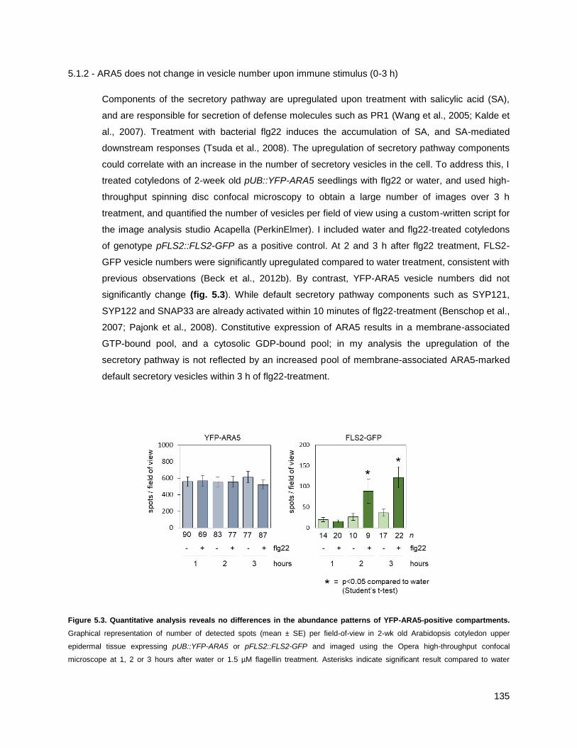
135
5.1.2 - ARA5 does not change in vesicle number upon immune stimulus (0-3 h)
Components of the secretory pathway are upregulated upon treatment with salicylic acid (SA),
and are responsible for secretion of defense molecules such as PR1 (Wang et al., 2005; Kalde et
al., 2007). Treatment with bacterial flg22 induces the accumulation of SA, and SA-mediated
downstream responses (Tsuda et al., 2008). The upregulation of secretory pathway components
could correlate with an increase in the number of secretory vesicles in the cell. To address this, I
treated cotyledons of 2-week old pUB::YFP-ARA5 seedlings with flg22 or water, and used high-
throughput spinning disc confocal microscopy to obtain a large number of images over 3 h
treatment, and quantified the number of vesicles per field of view using a custom-written script for
the image analysis studio Acapella (PerkinElmer). I included water and flg22-treated cotyledons
of genotype pFLS2::FLS2-GFP as a positive control. At 2 and 3 h after flg22 treatment, FLS2-
GFP vesicle numbers were significantly upregulated compared to water treatment, consistent with
previous observations (Beck et al., 2012b). By contrast, YFP-ARA5 vesicle numbers did not
significantly change (fig. 5.3). While default secretory pathway components such as SYP121,
SYP122 and SNAP33 are already activated within 10 minutes of flg22-treatment (Benschop et al.,
2007; Pajonk et al., 2008). Constitutive expression of ARA5 results in a membrane-associated
GTP-bound pool, and a cytosolic GDP-bound pool; in my analysis the upregulation of the
secretory pathway is not reflected by an increased pool of membrane-associated ARA5-marked
default secretory vesicles within 3 h of flg22-treatment.
Figure 5.3. Quantitative analysis reveals no differences in the abundance patterns of YFP-ARA5-positive compartments.
Graphical representation of number of detected spots (mean ± SE) per field-of-view in 2-wk old Arabidopsis cotyledon upper
epidermal tissue expressing pUB::YFP-ARA5 or pFLS2::FLS2-GFP and imaged using the Opera high-throughput confocal
microscope at 1, 2 or 3 hours after water or 1.5 µM flagellin treatment. Asterisks indicate significant result compared to water

136
treatments at p<0.05 (Student‘s t-test). n = number of images analyzed across 24 cotyledons (YFP-ARA5) or 6 cotyledons (FLS2-
GFP). Graphs show data of one experiment, representative of three independent replications.

137
5.2 - THE ARA5 CO-PURIFYING PROTEOME CHANGES UPON IMMUNE STIMULUS
5.2.1 - Purifying ARA5 from seedlings under water and flg22 conditions
Because I showed that ARA5 vesicles can cluster under bacterial contact sites, I was interested
in revealing changes in the cargoes of these vesicles, which could be delivered to the location of
pathogen attack. To investigate this, I applied the same technique as I used for analysis of ARA7
cargoes described in chapter 4. Briefly, I cultivated 8-day old Arabidopsis seedlings of genotype
pUB::YFP-ARA5 in liquid MS, alongside pUB::YFP as a negative control. I then treated the
seedlings of both genotypes by applying the bacterial MAMP flg22 to the growth medium to a final
concentration of 1.5 µM, and subsequently applied a vacuum to infiltrate the seedlings. I used
water treatment as a negative control. Since SA induced the upregulation of defence-related
secretory pathway genes, and SA is produced within hours of flg22 treatment (Tsuda et al.,
2008), after 3 h of flg22 treament, I used immunoprecipitation as described before (Heard et al.,
2015); Chapter 4 in this thesis), and subjected the samples to SDS-PAGE and mass
spectrometry in collaboration with Jan Sklenar, Frank Menke and Paul Derbyshire (Proteomics
Support Team, The Sainsbury Laboratory, Norwich UK).
After filtering the resulting proteomics data using Scaffold, I revealed 228 proteins in the YFP-
ARA5 co-purifying proteome and 1101 proteins in the YFP co-purifying proteome (fig. 5.4A). In
the same way as performed in Chapter 4 of this thesis, to select proteins that interacted
specifically with ARA5, and not with affinity beads or the YFP fluorescent tags, for each protein I
compared the spectral counts (number of peptides detected) across all three replications of YFP-
ARA5 purifications and YFP purifications. I required the combined number of peptides in YFP-
ARA5 purifications to be greater than 4x those in YFP purifications, and obtained a set of 77
proteins that I named the ARA5 co-purifying proteome (fig. 5.4A, table 5.1).
Table 5.1: ARA5 co-purifying proteome. Listed here is the ARA5 co-purifying proteome under water treated conditions. 8-day old
seedlings of genotypes pUB::YFP-ARA5 and pUB::YFP were treated with water or 1.5 µM flg22 for three hours, and subjected to
immunoprecipitation using YFP affinity beads. Mass spectrometry was performed in collaboration with Jan Sklenar, Paul Derbyshire
and Frank Menke (The Sainsbury Laboratory, Norwich, UK). To determine co-purifying proteomes, Scaffold software was used to
retain proteins with a ProteinProphet score of 99% based on a minimum of 2 spectra with a PeptideProphet score of 95%. MS Excel
was then used to retain proteins with at least 5 spectra across all three replicates in the YFP-ARA5 water-treated samples, with an
occurence in more than 1 of these replicates, and an abundance increase of at least 4x compared to matching YFP controls.
(Columns 1-8) Observed in # of replicates, and Spectra in combined replicates: Numbers and associated green cell-shading
intensities indicate in how many experimental replicates the protein was detected, or how many spectra for that protein were
identified in all three replicates combined, separated for water or flagellin treatment. (Columns 9-10) Enrichment factors: For each
protein, the number of total spectra across three replicates of the green-labeled (rightmost) pulldown was divided by the number of
total spectra in red-labeled (left-most) pulldown. Log10 values that correspond to these ratios are displayed in this table. Green or
red bars are graphical representations of magnitude and direction of positive (green) or negative (red) values, centered at 0.
(Columns 11-12) Locus ID and associated names. Source: TAIR10, accessed through www.genevestigator.org. The table is sorted
for total spectral numbers in ―YFP-ARA5 water‖, in descending order.

138
Ta
ble
5.1
: A
RA
5 c
o-p
uri
fyin
g p
rote
om
e
wate
rflg
22
wate
rflg
22
wate
rflg
22
wate
rflg
22
Lo
cu
s ID
Pro
tein
na
me
33
2636
664
52.1
0E
+00
1.8
7E
-02
AT1G
02130
AR
A-5
, A
RA
5,
ATR
AB
1B
, A
TR
AB
D2A
, R
A-5
, R
AB
D2A
, R
AS
5
33
33
519
594
84
37
7.9
1E
-01
5.8
6E
-02
AT1G
59870
PE
N3
33
181
125
8.2
6E
+00
-1.6
1E
-01
AT5G
16280
Tetr
atr
icopeptide r
epeat
(TP
R)-
like s
uperfam
ily p
rote
in
33
1147
104
12.1
7E
+00
-1.5
0E
-01
AT5G
65950
unknow
n p
rote
in,
hom
olo
gy t
o T
RA
PP
I-com
ponents
33
1144
114
12.1
6E
+00
-1.0
1E
-01
AT2G
47960
unknow
n p
rote
in,
hom
olo
gy t
o T
RA
PP
33
126
131
8.1
0E
+00
1.6
9E
-02
AT3G
07160
ATG
SL10,
CA
LS
9,
gsl1
0,
GS
L10,
glu
can s
ynth
ase-lik
e 1
0
33
33
119
105
10
51.0
8E
+00
-5.4
4E
-02
AT2G
29550
TU
B7,
tubulin
beta
-7 c
hain
33
67
49
7.8
3E
+00
-1.3
6E
-01
AT4G
03550
PM
R4
33
166
46
21.5
2E
+00
-1.5
7E
-01
AT4G
39820
Tetr
atr
icopeptide r
epeat
(TP
R)-
like s
uperfam
ily p
rote
in
33
161
74
21.4
8E
+00
8.3
9E
-02
AT2G
36850
ATG
SL08,
ATG
SL8,
CH
OR
, G
SL08,
GS
L8,
glu
can s
ynth
ase-lik
e 8
33
32
61
45
93
8.3
1E
-01
-1.3
2E
-01
AT5G
28540
BIP
1,
heat
shock p
rote
in 7
0 (
Hsp 7
0)
fam
ily p
rote
in
33
55
40
7.7
4E
+00
-1.3
8E
-01
AT1G
30450
ATC
CC
1,
CC
C1,
HA
P5,
cation-c
hlo
ride c
o-t
ransport
er
1
33
11
51
77
11
1.7
1E
+00
1.7
9E
-01
AT3G
11820
AT-S
YR
1,
ATS
YP
121,
ATS
YR
1,
PE
N1,
SY
P121,
SY
R1,
synta
xin
of pla
nts
121
33
49
35
7.6
9E
+00
-1.4
6E
-01
AT1G
77140
ATV
PS
45,
VP
S45,
vacuola
r pro
tein
sort
ing 4
5
33
22
47
37
63
8.9
4E
-01
-1.0
4E
-01
AT2G
14720
BP
80-2
;1,
MTV
4,
VS
R2;1
, V
SR
4,
vacuola
r sort
ing r
ecepto
r 4
33
146
39
17.6
6E
+00
-7.1
7E
-02
AT1G
61250
SC
3,
SC
AM
P3,
secre
tory
carr
ier
3
33
45
33
7.6
5E
+00
-1.3
5E
-01
AT2G
34420
LH
B1B
2,
LH
CB
1.5
, photo
syste
m II lig
ht
harv
esting c
om
ple
x g
ene B
1B
2
33
40
64
7.6
0E
+00
2.0
4E
-01
AT1G
65350
UB
Q13,
ubiq
uitin
13
33
37
23
7.5
7E
+00
-2.0
6E
-01
AT1G
59820
ALA
3,
am
inophospholip
id A
TP
ase 3
33
36
33
7.5
6E
+00
-3.7
8E
-02
AT3G
17900
unknow
n p
rote
in
33
35
29
7.5
4E
+00
-8.1
7E
-02
AT5G
54440
AtT
RS
130,
CLU
B,
CLU
B
33
135
28
11.5
4E
+00
-9.6
9E
-02
AT2G
28520
VH
A-A
1,
vacuola
r pro
ton A
TP
ase A
1
32
34
12
7.5
3E
+00
-4.5
2E
-01
AT2G
34510
Pro
tein
of unknow
n function,
DU
F642
33
11
34
36
22
1.2
3E
+00
2.4
8E
-02
AT4G
18800
ATH
SG
BP
, A
TR
AB
11B
, A
TR
AB
A1D
, R
AB
A1d,
RA
B G
TP
ase h
om
olo
g A
1D
33
22
32
17
65
7.2
7E
-01
-2.7
5E
-01
AT4G
12650
Endom
em
bra
ne p
rote
in 7
0 p
rote
in fam
ily
32
131
14
48.8
9E
-01
-3.4
5E
-01
AT5G
49720
ATG
H9A
1,
DE
C,
GH
9A
1,
IRX2,
KO
R,
KO
R1,
RS
W2,
TS
D1,
gly
cosyl hydro
lase 9
A1
32
29
97.4
6E
+00
-5.0
8E
-01
AT5G
17230
PS
Y,
PH
YTO
EN
E S
YN
TH
AS
E
33
28
11
7.4
5E
+00
-4.0
6E
-01
AT3G
52850
ATE
LP
, A
TE
LP
1,
ATV
SR
1,
BP
-80,
BP
80,
BP
80-1
;1,
BP
80B
, G
FS
1,
VS
R1,
VS
R1;1
33
128
17
21.1
5E
+00
-2.1
7E
-01
AT4G
32410
AtC
ES
A1,
CE
SA
1,
RS
W1,
cellu
lose s
ynth
ase 1
33
27
23
7.4
3E
+00
-6.9
6E
-02
AT3G
58460
ATR
BL15,
RB
L15,
RH
OM
BO
ID-lik
e p
rote
in 1
5
33
27
16
7.4
3E
+00
-2.2
7E
-01
AT3G
05280
Inte
gra
l m
em
bra
ne Y
ip1 fam
ily p
rote
in
33
26
11
7.4
1E
+00
-3.7
4E
-01
AT5G
64740
CE
SA
6,
E112,
IXR
2,
PR
C1,
cellu
lose s
ynth
ase 6
33
126
16
11.4
1E
+00
-2.1
1E
-01
AT5G
05170
ATC
ES
A3,
ATH
-B,
CE
SA
3,
CE
V1,
IXR
1,
Cellu
lose s
ynth
ase fam
ily p
rote
in
33
126
13
11.4
1E
+00
-3.0
1E
-01
AT5G
59840
Ras-r
ela
ted s
mall
GTP
-bin
din
g fam
ily p
rote
in
33
25
17
7.4
0E
+00
-1.6
7E
-01
AT1G
25570
Di-glu
cose b
indin
g p
rote
in w
ith L
eucin
e-r
ich r
epeat
dom
ain
33
22
11
7.3
4E
+00
-3.0
1E
-01
AT5G
58030
TR
S31,
TR
AP
PI-com
ponent
32
122
11
21.0
4E
+00
-3.0
1E
-01
AT1G
17840
AB
CG
11,
ATW
BC
11,
CO
F1,
DS
O,
WB
C11,
white-b
row
n c
om
ple
x h
om
olo
g p
rote
in 1
1
33
19
21
7.2
8E
+00
4.3
5E
-02
AT4G
31490
Coato
mer,
beta
subunit
33
18
24
7.2
6E
+00
1.2
5E
-01
AT1G
53440
Leucin
e-r
ich r
epeat
transm
em
bra
ne p
rote
in k
inase
33
18
15
7.2
6E
+00
-7.9
2E
-02
AT3G
15380
Pla
sm
a-m
em
bra
ne c
holin
e t
ransport
er
fam
ily p
rote
in
33
117
15
37.5
3E
-01
-5.4
4E
-02
AT5G
11040
AtT
RS
120,
TR
S120,
TR
S120
33
16
13
7.2
0E
+00
-9.0
2E
-02
AT3G
05710
ATS
YP
43,
SY
P43,
synta
xin
of pla
nts
43
Y5 w
ate
r
Y5 f
lg22
Ob
serv
ed
in
# o
f re
pli
ca
tes
Sp
ectr
a i
n c
om
bin
ed
re
pli
ca
tes
En
rich
me
nt
facto
r (l
og
10)
Pro
tein
in
form
ati
on
YF
P
YF
P-A
RA
5Y
FP
-AR
A5
YF
PY
FP
-AR
A5
YF
P

139
33
116
15
11.2
0E
+00
-2.8
0E
-02
AT1G
28490
ATS
YP
61,
OS
M1,
SY
P61,
synta
xin
of pla
nts
61
33
15
15
7.1
8E
+00
0.0
0E
+00
AT4G
36440
unknow
n p
rote
in
32
14
11
7.1
5E
+00
-1.0
5E
-01
AT4G
16120
CO
BL7,
SE
B1,
CO
BR
A-lik
e p
rote
in-7
pre
curs
or
32
13
57.1
1E
+00
-4.1
5E
-01
AT1G
24560
PLA
NT-U
NIQ
UE
RA
B5 E
FF
EC
TO
R 2
, P
UF
2
33
13
97.1
1E
+00
-1.6
0E
-01
AT1G
26670
ATV
TI1
2,
VTI1
2,
VTI1
B,
Vesic
le t
ransport
v-S
NA
RE
fam
ily p
rote
in
33
113
22
28.1
3E
-01
2.2
8E
-01
AT5G
06320
NH
L3,
ND
R1/H
IN1-lik
e 3
32
112
11
11.0
8E
+00
-3.7
8E
-02
AT4G
30260
Inte
gra
l m
em
bra
ne Y
ip1 fam
ily p
rote
in
32
112
91
1.0
8E
+00
-1.2
5E
-01
AT1G
56190
Phosphogly
cera
te k
inase fam
ily p
rote
in
33
21
12
13
21
7.7
8E
-01
3.4
8E
-02
AT1G
02080
transcription r
egula
tors
33
11
97.0
4E
+00
-8.7
2E
-02
AT4G
20110
BP
80-3
;1,
VS
R3;1
, V
SR
7,
VA
CU
OLA
R S
OR
TIN
G R
EC
EP
TO
R 7
33
11
97.0
4E
+00
-8.7
2E
-02
AT1G
54320
LE
M3 (
ligand-e
ffect
modula
tor
3)
fam
ily p
rote
in /
CD
C50 fam
ily p
rote
in
31
11
27.0
4E
+00
-7.4
0E
-01
AT5G
09870
CE
SA
5,
cellu
lose s
ynth
ase 5
33
111
24
11.0
4E
+00
3.3
9E
-01
AT4G
23180
CR
K10
32
111
41
1.0
4E
+00
-4.3
9E
-01
AT1G
51500
AB
CG
12,
ATW
BC
12,
CE
R5,
D3,
WB
C12,
AB
C-2
type t
ransport
er
fam
ily p
rote
in
33
110
13
26.9
9E
-01
1.1
4E
-01
AT3G
28710
ATP
ase,
V0/A
0 c
om
ple
x,
subunit C
/D
23
110
72
6.9
9E
-01
-1.5
5E
-01
AT1G
53840
ATP
ME
1,
PM
E1,
pectin m
eth
yle
ste
rase 1
23
915
6.9
5E
+00
2.2
2E
-01
AT5G
54830
DO
MO
N d
om
ain
-conta
inin
g p
rote
in
32
96
6.9
5E
+00
-1.7
6E
-01
AT3G
05200
ATL6,
RIN
G/U
-box s
uperfam
ily p
rote
in
32
96
6.9
5E
+00
-1.7
6E
-01
AT1G
29980
Pro
tein
of unknow
n function,
DU
F642
23
95
6.9
5E
+00
-2.5
5E
-01
AT1G
28340
AtR
LP
4,
RLP
4,
recepto
r lik
e p
rote
in 4
33
94
6.9
5E
+00
-3.5
2E
-01
AT3G
14790
ATR
HM
3,
RH
M3,
rham
nose b
iosynth
esis
3
33
19
91
9.5
4E
-01
0.0
0E
+00
AT5G
57350
AH
A3,
ATA
HA
3,
HA
3,
H(+
)-A
TP
ase 3
32
89
6.9
0E
+00
5.1
2E
-02
AT1G
51630
O-fucosyltra
nsfe
rase fam
ily p
rote
in
33
810
6.9
0E
+00
9.6
9E
-02
AT5G
04870
ATC
PK
1,
CP
K1,
calc
ium
dependent
pro
tein
kin
ase 1
33
822
6.9
0E
+00
4.3
9E
-01
AT5G
03700
D-m
annose b
indin
g lectin p
rote
in w
ith A
pple
-lik
e c
arb
ohydra
te-b
indin
g d
om
ain
23
87
6.9
0E
+00
-5.8
0E
-02
AT5G
60670
Rib
osom
al pro
tein
L11 fam
ily p
rote
in
33
85
6.9
0E
+00
-2.0
4E
-01
AT5G
61060
ATH
DA
5,
HD
A05,
HD
A5,
his
tone d
eacety
lase 5
31
82
6.9
0E
+00
-6.0
2E
-01
AT1G
13900
Purp
le a
cid
phosphata
ses s
uperfam
ily p
rote
in
33
75
6.8
5E
+00
-1.4
6E
-01
AT1G
01960
BIG
3,
ED
A10,
AR
F-G
EF
33
65
6.7
8E
+00
-7.9
2E
-02
AT2G
14740
ATV
SR
3,
BP
80-2
;2,
VS
R2;2
, V
SR
3,
VS
R3,
vaculo
lar
sort
ing r
ecepto
r 3
32
63
6.7
8E
+00
-3.0
1E
-01
AT1G
27930
Pro
tein
of unknow
n function (
DU
F579)
33
611
6.7
8E
+00
2.6
3E
-01
AT2G
45010
PLA
C8 fam
ily p
rote
in
22
63
6.7
8E
+00
-3.0
1E
-01
AT2G
20840
Secre
tory
carr
ier
mem
bra
ne p
rote
in (
SC
AM
P)
fam
ily p
rote
in
21
64
6.7
8E
+00
-1.7
6E
-01
AT5G
58420
Rib
osom
al pro
tein
S4 (
RP
S4A
) fa
mily
pro
tein
33
16
61
7.7
8E
-01
0.0
0E
+00
AT5G
22360
ATV
AM
P714,
VA
MP
714,
VA
MP
714,
vesic
le-a
ssocia
ted m
em
bra
ne p
rote
in 7
14

140
Figure 5.4. Identification of proteins associated with YFP-ARA5 in a flg22-responsive manner. (A) Venn-diagram shows
proteins co-purifying with YFP-ARA5 from Arabidopsis seedlings, compared to proteins co-purifying with a YFP control, as revealed
by immunoprecipitation and mass spectrometry. The YFP-ARA5 co-purifying proteome was defined based on quality control criteria
(present in >1 replicates, peptides = >5) and fold-change ratio of >4 in YFP-ARA5 compared to YFP control. Details available in
Table 5.1. (B) Venn-diagram showing YFP-ARA5 co-purifying proteins as revealed by immunoprecipitation and mass spectrometry,
overlaid with known trafficking regulators (source: Heard et al. 2015). Call-out indicates prominent trafficking regulators found in
YFP-ARA5. (C) Venn-diagram showing YFP-ARA5 co-purifying proteins as above, comparing before and after 3 hours of flagellin
treatment. Call-outs indicate proteins present in both groups (top) or in the flagellin-treated group (bottom). Details available in Table
5.3. (D) Venn-diagram showing YFP-ARA5 co-purifying proteins as above, overlaid with YFP-ARA7 co-purifying proteins, both in the
absence of flg22 treatment. Call-out indicates proteins present in both groups. Details available in table 5.4. (E) Venn-diagram
showing YFP-ARA5 co-purifying proteins after 3 hours flagellin treatment, overlaid with YFP-ARA7 co-purifying proteins after 3
hours flagellin treatment. Call-out indicates proteins present in both groups. Details available in table 5.5.
141
5.2.2 - The ARA5 proteome contains known secretory pathway-associated proteins
In order to further verify the quality of the so-defined ARA5 co-purifying proteome, I compared the
proteome to a comprehensive manually curated list of known Arabidopsis trafficking regulators
(Heard 2015) and listed the results in table 5.2. 19 out of 77 proteins classified as trafficking
regulators in this way (fig. 5.4B, table 5.2). Upon inspection, I observed that a majority of
trafficking regulators found interacting with ARA5 were also identified in the water-treated ARA7
co-purifying proteome as defined in chapter 4. However, several regulators were unique to ARA5
and, based on existing knowledge, corresponded to the known Golgi and TGN/EE localization of
ARA5.
Signature proteins identified in purifications of ARA5 specifically
RABA1d is a member of the Arabidopsis RAB11/RABA group, which consist of predominantly
TGN/EE-to-PM secretory functioning RAB GTPases (Asaoka et al., 2013). RabA1 members were
found to localize at TGN/EE, partially overlapping with TGN/EE-markers SYP43 and VHA-a1
(Asaoka et al., 2013). RABA members have different roles contributing to the cell wall
composition, with single mutants affecting pectin, cellulose and hemicellulose (Lunn et al., 2013).
In immunity, RABA1b, which is a close homolog of RABA1d, governs the secretion of newly
synthesized cell-surface receptor FLS2 (Choi et al., 2013). More distant homologs of RABA1,
such as RABA6a, are thought to have a function in endocytic traffic at the TGN/EE (Choi et al.,
2013). Finding RABA1d in the ARA5 proteome corresponds to the partial TGN/EE localization of
ARA5.
Tubulin 7 (TUB7), is protein found at high abundance interacting specifically with ARA5, and the
only cytoskeletal component found in my ARA5 and ARA7 purifications. Golgi bodies attach to
microtubules, and their motility along the cytoskeleton regulates the dynamics of the delivery and
removal of cell-surface cargoes such as cellulose synthase complexes (CESA, (Crowell et al.,
2009).
Coatomer (subunit AT4G31490) localizes to the Golgi, where multimeric complexes regulate
retrograde traffic (Brandizzi and Barlowe, 2013). The ARA5 partial Golgi-localization leads me to
expect Golgi-components in the purification. While this subunit was specific to ARA5, other
coatomer subunits were identified in ARA7 purifications (chapter 4, tables 4.2, 4.3; AT1G62020,
AT3G15980).
Table 5.2: Trafficking regulators in the ARA5 co-purifying proteome. Listed here is the ARA5 co-purifying proteome under
water treated conditions, copied from Table 5.1, and filtered to retain only endomembrane trafficking regulators as defined in Heard
et al. 2015.

142
Ta
ble
5.2
: T
raff
ic r
eg
ula
tors
in
th
e A
RA
5 c
o-p
uri
fyin
g p
rote
om
e
wate
rflg
22
wate
rflg
22
wate
rflg
22
wate
rflg
22
Lo
cu
s ID
Pro
tein
na
me
33
2636
664
52.1
0E
+00
1.8
7E
-02
AT1G
02130
AR
A-5
, A
RA
5,
ATR
AB
1B
, A
TR
AB
D2A
, R
A-5
, R
AB
D2A
, R
AS
5
33
181
125
8.2
6E
+00
-1.6
1E
-01
AT5G
16280
Tetr
atr
icopeptide r
epeat
(TP
R)-
like s
uperfam
ily p
rote
in
33
33
119
105
10
51.0
8E
+00
-5.4
4E
-02
AT2G
29550
TU
B7,
tubulin
beta
-7 c
hain
33
11
51
77
11
1.7
1E
+00
1.7
9E
-01
AT3G
11820
AT-S
YR
1,
ATS
YP
121,
ATS
YR
1,
PE
N1,
SY
P121,
SY
R1,
synta
xin
of pla
nts
121
33
22
47
37
63
8.9
4E
-01
-1.0
4E
-01
AT2G
14720
BP
80-2
;1,
MTV
4,
VS
R2;1
, V
SR
4,
vacuola
r sort
ing r
ecepto
r 4
33
146
39
17.6
6E
+00
-7.1
7E
-02
AT1G
61250
SC
3,
SC
AM
P3,
secre
tory
carr
ier
3
33
35
29
7.5
4E
+00
-8.1
7E
-02
AT5G
54440
AtT
RS
130,
CLU
B,
CLU
B
33
11
34
36
22
1.2
3E
+00
2.4
8E
-02
AT4G
18800
ATH
SG
BP
, A
TR
AB
11B
, A
TR
AB
A1D
, R
AB
A1d,
RA
B G
TP
ase h
om
olo
g A
1D
33
28
11
7.4
5E
+00
-4.0
6E
-01
AT3G
52850
ATE
LP
, A
TE
LP
1,
ATV
SR
1,
BP
-80,
BP
80,
BP
80-1
;1,
BP
80B
, G
FS
1,
VS
R1,
VS
R1;1
33
27
16
7.4
3E
+00
-2.2
7E
-01
AT3G
05280
Inte
gra
l m
em
bra
ne Y
ip1 fam
ily p
rote
in
33
22
11
7.3
4E
+00
-3.0
1E
-01
AT5G
58030
TR
S31,
TR
AP
PI-com
ponent
33
19
21
7.2
8E
+00
4.3
5E
-02
AT4G
31490
Coato
mer,
beta
subunit
33
117
15
37.5
3E
-01
-5.4
4E
-02
AT5G
11040
AtT
RS
120,
TR
S120,
TR
S120
33
16
13
7.2
0E
+00
-9.0
2E
-02
AT3G
05710
ATS
YP
43,
SY
P43,
synta
xin
of pla
nts
43
33
116
15
11.2
0E
+00
-2.8
0E
-02
AT1G
28490
ATS
YP
61,
OS
M1,
SY
P61,
synta
xin
of pla
nts
61
33
13
97.1
1E
+00
-1.6
0E
-01
AT1G
26670
ATV
TI1
2,
VTI1
2,
VTI1
B,
Vesic
le t
ransport
v-S
NA
RE
fam
ily p
rote
in
32
112
11
11.0
8E
+00
-3.7
8E
-02
AT4G
30260
Inte
gra
l m
em
bra
ne Y
ip1 fam
ily p
rote
in
33
11
97.0
4E
+00
-8.7
2E
-02
AT4G
20110
BP
80-3
;1,
VS
R3;1
, V
SR
7,
VA
CU
OLA
R S
OR
TIN
G R
EC
EP
TO
R 7
33
75
6.8
5E
+00
-1.4
6E
-01
AT1G
01960
BIG
3,
ED
A10,
AR
F-G
EF
33
65
6.7
8E
+00
-7.9
2E
-02
AT2G
14740
ATV
SR
3,
BP
80-2
;2,
VS
R2;2
, V
SR
3,
VS
R3,
vaculo
lar
sort
ing r
ecepto
r 3
33
16
61
7.7
8E
-01
0.0
0E
+00
AT5G
22360
ATV
AM
P714,
VA
MP
714,
VA
MP
714,
vesic
le-a
ssocia
ted m
em
bra
ne p
rote
in 7
14
Ob
serv
ed
in
# o
f re
pli
ca
tes
Sp
ectr
a i
n c
om
bin
ed
re
pli
ca
tes
En
rich
me
nt
facto
r (l
og
10)
Pro
tein
in
form
ati
on
YF
P-A
RA
5Y
FP
YF
P-A
RA
5Y
FP
YF
P
YF
P-A
RA
5Y
5 w
ate
r
Y5 f
lg22
143
The protein with the highest abundance in non-elicited ARA5 purifications is PEN3. Briefly, PEN3
is a PM-localized transporter, thought to function in the export of antimicrobial compounds
produced by PEN2 which is localized on mitochondria (Stein et al., 2006; Fuchs et al., 2015b). In
chapter 4, I discussed the tendency for PEN3 to accumulate higher in ARA7 purifications under
flg22 treatment when compared to water treatment (chapter 4), but in ARA5 purifications, PEN3
accumulates to similar levels as in flg22-elicited conditions (519 vs. 594 spectra, table 5.1). PEN3
shuttles between the PM-TGN/EE, but is also consitutively secreted. Both the secretory pathway
as well as the endocytic pathway were shown to contribute to PEN3-accumulations in chemically-
induced Golgi-TGN/EE aggregates (Mao et al., 2016). In un-elicited states, PEN3 furthermore
localized to the PM, but also to mobile punctae adjacent to SYP32-marked Golgi (Underwood and
Somerville, 2013; Mao et al., 2016). In flg22-elicited conditions, PEN3 accumulated at endosomal
compartments (Underwood et al., 2017), but this is not reflected in a marked increase of spectra
co-purifying with ARA5.
Novel proteins identified in ARA5 purifications
Unexpectedly abundant in ARA5 purifications, BiP1/Heat Shock Protein70 (HSP70) is a major
ER-localized and functioning protein (Srivastava et al., 2013). This could reflect a contamination,
or an interaction partner of ARA5 during its constitutive biosynthesis. However, since dominant
negative studies showed that ARA5 functioned in the early secretory trafficking step between ER
and Golgi, it could also reflect a previously unknown functional interactor in the ARA5 complex at
early secretory steps.
5.2.3 - Proteins that are abundant in ARA5 purifications which also occur in ARA7
The following proteins identified in ARA5 purifications (table 5.1) were found for ARA7 as well
(tables 4.1, 5.4). Here, I describe these proteins in the context of the secretory pathway
localization and functionality of ARA5. In this section, I selected abundant proteins as found in
ARA5 purifications, and in further sections where I describe the common proteome of ARA5 and
ARA7, I prioritize proteins that are of most similar abundance in both.
Trafficking regulators
PEN1/SYP121 has been shown to be PM-localized, and recycle between the PM and TGN/EE
compartment (Nielsen and Thordal-Christensen, 2012; Collins et al., 2003; Uemura et al., 2004).
PEN1 engages the default secretory complex consisting of other SNAREs VAMP721, VAMP722
and SNAP33 in order to confer immunity against powdery mildew (Kwon et al., 2008). However,
besides PEN1, members of this complex were not identified in my ARA5 purifications. PEN1
functions in the formation of the callose-rich extracellular papilla that is formed at sites of
penetration attempts by filamentous pathogens, and contributes to the timely formation of these

144
structures (Collins et al., 2003; Assaad et al., 2004). Separate from its functioning in regulating
membrane traffic, PEN1/SYP121 interacts with the potassium channel KC1 and positively affects
its inward potassium permeability, thus regulating guard cell dynamics in stomatal opening
(Grefen et al., 2010; Honsbein et al., 2009; Eisenach et al., 2012).
Two tetratricopeptide-repeat (TPR)-containing proteins (AT5G16280, AT4G39820) were identified
with relatively high abundance in ARA5 purifications. While these proteins have yet to be
characterized, a different TPR-containing protein HYPERSENSITIVE TO LATRUNCILIN B1
(HLB1) has recently been shown to localize to the TGN/EE, and regulates both secretory and
endocytic traffic passing through this compartment (Sparks et al., 2016). Interestingly, HLB1 was
found to interact and colocalize with and actin filaments, raising the TPR proteins I identified as
interacting with ARA5 as possible candidates linking ARA5 compartments to the cytoskeleton.
Secretory Carrier Membrane Protein 3 (SC3) is part of a bigger family of Secretory Carrier
Membrane Proteins (SCAMPs). In eukaryotes, SCAMPs are transmembrane proteins regulating
post-Golgi traffic, and are associated with endocytic uptake and recycling to the PM, and localize
to PM and endomembrane compartments (Fernández-Chacón and Südhof, 2000). While SC3 in
Arabidopsis is uncharacterized, in rice, OsSCAMP1 localized to the PM and early endocytic
compartments (Lam et al., 2007). Functionally, based on RNAi studies of SCAMPs in poplar
(Populus tremula), they are implicated in the control of secondary cell-wall precursors and
proteins involved in their trafficking (Obudulu et al., 2018).
VAMP714 is a member of the VAMP71 subgroup of Arabidopsis SNAREs, of which members are
predominantly tonoplast-localized (Uemura et al., 2004). VAMP714 localizes to the tonoplast in
localization and proteomic analysis studies, but it was also found on Golgi membranes (Uemura
et al., 2004; Szponarski et al., 2004). The precise function of VAMP714 has yet to be determined,
but its localization suggests a putative role in Golgi-vacuole trafficking (Leshem et al., 2006).
Finally, Vacuolar Sorting Receptor 4/Modified Transport to the Vacuole 4 (VSR4/MTV4) is
involved in vacuolar delivery of vacuolar storage proteins (Zouhar et al., 2009), predominantly
localized to the PM but also to SYP61-positive TGN/EE punctae (Nishimura et al., 2016). It must
be noted that the SYP61 proteome contained ARA5 (Drakakaki et al., 2012), and my ARA5
proteome contained SYP61 (tables 5.1, 5.2).
Non-trafficking regulators
Glucan Synthase-Like (GSL) 8 and 10 are callose synthases that function in normal plant growth,
with knockdown or knockout of the genes causing severe plant defects already at seedling stages
(Töller et al., 2008; Chen et al., 2009). Additionally, GSL10 has been shown to function in male

145
gametogenesis, regulating asymmetric cell division at the microspore stage (Huang et al., 2009).
Importantly, PMR4/GSL5 was found in ARA5 and ARA7 purifications.
PMR4 is a callose synthase involved in callose production in response to wounding and pathogen
detection (Dong et al., 2005; Jacobs et al., 2003; Ellinger et al., 2013). PMR4 contributes to the
formation of the callose-rich papilla upon pathogen attack, and accumulates in encasements
surrounding Golovinomyces orontii haustoria (Meyer et al., 2009). PMR4 is also required for
flg22-induced callose deposition (Clay et al., 2009). While bacterial infection, or treatment with the
bacterial MAMP flagellin, trigger a significant extracellular accumulation of callose, the number of
detected PMR4-associated spectra did not markedly increase or decrease in ARA5 or ARA7
purifications at 3 h after flagellin treatment.
Cation-Chloride Cotransporter 1 (CCC1) is member of a group of co-transporters that are thought
to mediate the transmembrane exchange of potassium and sodium with chloride (Henderson et
al., 2015). CCCs have been consistently found in earlier proteomic studies of the TGN/EE
(Drakakaki et al., 2012; Groen et al., 2014), and localized to the Golgi and TGN/EE (Henderson
et al., 2015), corresponding to the known localization of ARA5.
5.2.4 - The ARA5 co-purifying proteome changes upon 3-h flg22 treatment
Upon flg22 treatment for 3 h, the ARA5 co-purifying proteome changes (fig. 5.4C). 20 proteins
absent from water treated samples were found to interact with ARA5 (table 5.3). Of two proteins
(CRK10 and a lectin protein) identified under water treated conditions, associated spectra
increased more than 2-fold in flg22 treated conditions.
Phosphorus metabolism and multi-organism-processes are overrepresented in the ARA5 co-
purifying proteome upon flg22-stimulus
To obtain an impression of the global pattern of functional changes in the ARA5 co-purifying
proteome, I performed an overrepresentation analysis of GO-terms listing biological functions
(table S4) using the PANTHER Classification System (pantherdb.org). Most notably, processes
including terms such as ―phosphorylation‖ and ―phosphorus metabolic process‖ are
overrepresented. Looking at the details of the ARA5 co-purifying proteome under flg22 conditions
(table 5.3), the list is enriched in protein kinases, receptor kinases and LRR-RLKs. Since these
proteins can phosphorylate interaction partners, an overrepresentation of GO-processes including
phosphorus can be explained. Finally, terms such as ―defense to other organism‖ and ―multi-
organism process‖ appear, which makes sense in the context of studying proteomic changes
upon application of a bacterial MAMP.

146
Proteins co-purifying with ARA5 upon flg22 stimulus that are not found in ARA7
Notable changes in the ARA5 co-purifying proteome upon 3 h flagellin treatment include INDOLE-
GLUCOSINOLATE O-METHYLTRANSFERASE 2 (IGMT2) and MILDEW RESISTANCE LOCUS
12 (MLO12). IGMT2 is an enzyme that plays a catalytic role in the final steps of indole-
glucosinolate derived antimicrobial production (Pfalz et al., 2011). It has been shown to convert
indole-3-methylglucosinolate (I3M) into 4-methoxyindole-3-methylglucosinolate (4MOI3M), which
in turn is converted by the mitochondrial-localized atypical myrosinase PEN2 (Fuchs et al.,
2015b) into derivaties with broad antimicrobial activity (Stahl et al., 2016; Lipka et al., 2005; Oa et
al., 2010). Interestingly, out of both ARA5 and ARA7 purifications, IGMT2 was only identified in
flg22-treated ARA5 samples. In comparison, I identified the less well-characterized homolog
IGMT4 specifically in the ARA7 proteome as strongly positively responsive to flg22 treatment
(table 4.3). Previously, indole glucosinolate-related antimicrobial metabolism was shown to be
required for flg22-induced callose deposition (Clay et al., 2009), which in turn is dependent on the
glucan synthase PMR4 that was detected in ARA5 before flg22 elicitation (table 5.1).
Table 5.3: ARA5 co-purifying proteins that are more abundant upon flagellin treatment. Listed here is the ARA5 co-purifying
proteome under flagellin treated conditions. 8-day old seedlings of genotypes pUB::YFP-ARA5 and pUB::YFP were treated with
water or 1.5 µM flg22 for three hours, and subjected to immunoprecipitation using YFP affinity beads. Mass spectrometry was
performed in collaboration with Jan Sklenar, Paul Derbyshire and Frank Menke (The Sainsbury Laboratory, Norwich, UK). To
determine co-purifying proteomes, Scaffold software was used to retain proteins with a ProteinProphet score of 99% based on a
minimum of 2 spectra with a PeptideProphet score of 95%. MS Excel was then used to retain proteins with at least 5 spectra across
all three replicates in the YFP-ARA5 flg22-treated samples, with an occurence in more than 1 of these replicates, an abundance
increase of at least 4x compared to matching YFP controls, and an abundance increase of at least 2x compared to matching water
treated controls. (Columns 1-8) Observed in # of replicates, and Spectra in combined replicates: Numbers and associated green
cell-shading intensities indicate in how many experimental replicates the protein was detected, or how many spectra for that protein
were identified in all three replicates combined, separated for water or flagellin treatment. (Columns 9-10) Enrichment factors: For
each protein, the number of total spectra across three replicates of the green-labeled (rightmost) pulldown was divided by the
number of total spectra in red-labeled (left-most) pulldown. Log10 values that correspond to these ratios are displayed in this table.
Green or red bars are graphical representations of magnitude and direction of positive (green) or negative (red) values, centered at
0. (Columns 11-12) Locus ID and associated names. Source: TAIR10, accessed through www.genevestigator.org. The table is
sorted for total spectral numbers in ―YFP-ARA5 flg22‖, in descending order.

147
Ta
ble
5.3
: A
RA
5 c
o-p
uri
fyin
g p
rote
ins
tha
t a
re m
ore
ab
un
da
nt
up
on
fla
ge
llin
tre
atm
en
t
wate
rflg
22
wate
rflg
22
wate
rflg
22
wate
rflg
22
Lo
cu
s ID
Pro
tein
na
me
333
0.0
0E
+00
7.5
2E
+00
AT1G
21120
IGM
T2
33
111
24
11.0
4E
+00
3.3
9E
-01
AT4G
23180
CR
K10
33
822
6.9
0E
+00
4.3
9E
-01
AT5G
03700
D-m
annose b
indin
g lectin p
rote
in w
ith A
pple
-lik
e c
arb
ohydra
te-b
indin
g d
om
ain
322
0.0
0E
+00
7.3
4E
+00
AT1G
14540
PE
R4,
PE
RO
XID
AS
E 4
23
220
6.3
0E
+00
1.0
0E
+00
AT5G
04930
ALA
1,
am
inophospholip
id A
TP
ase 1
23
11
216
22
0.0
0E
+00
9.0
3E
-01
AT1G
03220
Eukary
otic a
spart
yl pro
tease fam
ily p
rote
in
33
15
14
50.0
0E
+00
4.4
7E
-01
AT5G
49760
Leucin
e-r
ich r
epeat
pro
tein
kin
ase fam
ily p
rote
in
23
313
6.4
8E
+00
6.3
7E
-01
AT1G
06840
Leucin
e-r
ich r
epeat
pro
tein
kin
ase fam
ily p
rote
in
23
13
13
14.7
7E
-01
6.3
7E
-01
AT1G
51800
IOS
1
33
511
6.7
0E
+00
3.4
2E
-01
AT3G
62370
hem
e b
indin
g p
rote
in
13
11
10
6-7
.78E
-01
1.0
0E
+00
AT4G
08850
LR
R-R
LK
, M
IK2
23
39
6.4
8E
+00
4.7
7E
-01
AT1G
52200
PLA
C8 fam
ily p
rote
in
13
19
6.0
0E
+00
9.5
4E
-01
AT2G
01180
ATLP
P1,
ATP
AP
1,
LP
P1,
PA
P1,
phosphatidic
acid
phosphata
se 1
29
0.0
0E
+00
6.9
5E
+00
AT1G
30700
FA
D-b
indin
g B
erb
erine fam
ily p
rote
in
23
38
6.4
8E
+00
4.2
6E
-01
AT1G
09970
LR
R X
I-23,
RLK
7,
Leucin
e-r
ich r
ecepto
r-lik
e p
rote
in k
inase fam
ily p
rote
in
22
37
6.4
8E
+00
3.6
8E
-01
AT1G
19110
inte
r-alp
ha-t
rypsin
inhib
itor
heavy
chain
-rela
ted
12
32
725
-1.1
0E
+00
5.4
4E
-01
AT2G
32730
26S
pro
teasom
e r
egula
tory
com
ple
x,
non-A
TP
ase s
ubcom
ple
x,
Rpn2/P
sm
d1 s
ubunit
37
0.0
0E
+00
6.8
5E
+00
AT2G
39200
MLO
12
27
0.0
0E
+00
6.8
5E
+00
AT3G
52400
ATS
YP
122,
SY
P122,
synta
xin
of pla
nts
122
13
12
61
3.0
1E
-01
4.7
7E
-01
AT3G
17410
Pro
tein
kin
ase s
uperfam
ily p
rote
in
12
22
618
-9.5
4E
-01
4.7
7E
-01
AT3G
14840
Leucin
e-r
ich r
epeat
transm
em
bra
ne p
rote
in k
inase
22
66
-6.7
8E
+00
6.7
8E
+00
AT4G
20830
OG
OX1,
OLIG
OG
ALA
CTU
RO
NID
E O
XID
AS
E 1
Ob
serv
ed
in
# o
f re
pli
ca
tes
Sp
ectr
a i
n c
om
bin
ed
re
pli
ca
tes
En
rich
me
nt
facto
r (l
og
10)
Pro
tein
in
form
ati
on
Y5 w
ate
r
Y5 f
lg22
YF
P
YF
P-A
RA
5Y
FP
-AR
A5
YF
PY
FP
-AR
A5
YF
P

148
Mildew Resistance Locus O 12 (MLO12) is a member of the MLO family of susceptibility factors,
of which Arabidopsis MLO2 has been best characterized (Consonni et al., 2006; Panstruga,
2005; Kusch and Panstruga, 2017). They contain 7 transmembrane domains, and Arabidopsis
MLO2 has been shown to be PM-localized (Bhat et al., 2005). In barley (Hordeum vulgare),
where MLO was originally identified as a susceptibility locus to powdery mildew infection
(Blumeria graminis f.sp. hordei, Bgh, (Büschges et al., 1997), MLO accumulates at the PM and
enriched under pathogen penetration sites (Bhat et al., 2005). In my proteomic analysis, I identify
MLO12 as positively responsive to flg22, which combined with data on ARA5 vesicles focally
accumulating under bacterial attack sites (fig. 5.1) could indicate MLO12 also focally
accumulates upon pathogen challenge.
Loss of MLO genes increases resistance to powdery mildews, but little is known about functions
in antibacterial resistance. Loss of function mutations in pepper (Capsicum annuum) MLO2, of
which the product localizes to the PM, enhanced resistance to bacterial pathogens, while
introduction of CaMLO2 into Arabidopsis enhanced its susceptibility to Pto DC3000 (Kim and
Hwang, 2012). Mlo2-based resistance requires trafficking processes involving PEN1/SYP121,
and the production of glucosinolate-derived antimicrobials in the PEN2/PEN3 pathway, of which
components were also found in my proteomic studies (Assaad et al., 2004; Consonni et al., 2006;
Dittgen et al., 2006; Bednarek et al., 2009). Little is known about the localization and function of
MLO12, but enhanced mildew resistance in mlo2/6/12 triple mutants did not have the trafficking
and antimicrobial requirements as described for mlo2 (Kuhn et al., 2017).
Aminophospholipid ATPase 1 (ALA1) is a lipid flippase. Flippases are generally involved in
translocating lipids between leaflets of the lipid bilayer, thus achieving lipid asymmetry. For
previously characterized lipid flippases, this process can contribute to the budding of vesicles by
overcoming energetically unfavorable intermediate stages in membrane bending (Pomorski and
Menon, 2006). ALA1 was found to be PM-localized, and together with ALA2 has a function in
RNAi-based antiviral immunity, where it works in the same pathway as RNA-dependent DNA-
polymerases (López-Marqués et al., 2012; Zhu et al., 2017). The precise functions of ALA1 are
unclear, but its known PM-localization suggests it does not function at the TGN/EE. It could be
that upon immune stimulus, ALA1 traffics to the cell surface to support membrane trafficking
events there.
From GO-analysis showing a strong overrepresentation of terms involving phosphorus
metabolism, and from inspection of the proteomic results, it becomes evident that proteins
upregulated in ARA5 purifications upon flg22 treatment contain a number of receptor kinases
(table 5.3, table S4). In fact, four LRR-RLKs were found in ARA5 upon flg22 treatment, which
were not found in ARA7 purifications (AT5G49760, AT1G06840, AT3G14840 and LRR XI-23).
Engagement of default secretion to support the MAMP-induced delivery of defence compounds to

149
the cell surface has been described before, with roles for PEN1/SYP121 in callose deposition and
SYP132 in antimicrobial PR-1 secretion (Assaad et al., 2004; Kalde et al., 2007).
5.3 - THE ARA7 AND ARA5 CO-PURYFING PROTEOMES SHOW DIFFERENTIAL OVERLAP IN
RESTING AND IMMUNE ACTIVATED CONDITIONS
5.3.1 - The shared ARA7 and ARA5 co-purifying proteome contains TGN/EE, cell wall, and membrane
trafficking-associated proteins
In order to gain insight into the shared proteins interacting with both the secretory ARA5 and the
endocytic ARA7, I combined both water-treated co-puryfing proteomes, and filtered the list to only
retain proteins present in both purifications (table 5.4, figure 3.4D). Upon inspection, the list
reveals the presence of typical TGN/EE markers such as VHA-a1, SYP43, SYP61 and VTI12,
which is in accordance with the partial localization of both ARA7 and ARA5 to the TGN/EE
(Gendre et al., 2014). Furthermore, when analyzing the shared proteome for overrepresentation
of GO-terms in the classification of ―biological function‖ (table S5), two themes appear, which
broadly consist of (A) cell wall metabolism: callose and cellulose metabolism, primary cell wall
formation, polysaccharide localization and biosynthesis, cell shape and cytokinesis; and (B)
membrane trafficking: secretion, vacuolar targeting, vesicle tethering, docking and fusion, vesicle
organisation and exocytosis. The appearance of these themes is in line with the expectation
when considering the purifications were performed from rapidly developing 8-day old seedlings,
with immunoprecipitation baits that both partially localize to the TGN/EE sorting hub. When
studying the list of specific proteins present in both proteomes, prominent interactors with
implications in both trafficking and immunity occur.
Table 5.4: ARA7 and ARA5 common co-purifying proteome. Listed here is the common ARA7- and ARA5 co-purifying proteome
under water treated conditions. Proteomes were defined as described in legends of Tables 1 and 3 (water-treated YFP-ARA7 and
YFP-ARA5 purifications), and associated values were listed in this table. (Columns 1-8) Observed in # of replicates, and Spectra in
combined replicates: Numbers and associated green cell-shading intensities indicate in how many experimental replicates the
protein was detected, or how many spectra for that protein were identified in all three replicates combined, separated for water or
flagellin treatment. (Column 9) Enrichment factors: For each protein, the number of total spectra across three replicates of the
green-labeled (rightmost) pulldown was divided by the number of total spectra in red-labeled (left-most) pulldown. Log10 values that
correspond to these ratios are displayed in this table. Green or red bars are graphical representations of magnitude and direction of
positive (green) or negative (red) values, centered at 0. (Columns 10-11) Locus ID and associated names. Source: TAIR10,
accessed through www.genevestigator.org. The table is sorted for absolute differences in protein spectral counts between YFP-
ARA5 and YFP-ARA7 in ascending order.

150
Ta
ble
5.4
: A
RA
7 a
nd
AR
A5 s
ha
red
pro
teo
me
En
rich
me
nt
facto
r (l
og
10)
wate
rflg
22
wate
rflg
22
wate
rflg
22
wate
rflg
22
Lo
cu
s ID
Pro
tein
na
me
33
33
22
17
22
11
0.0
0E
+00
AT5G
58030
TR
S31,
TR
AP
PI-com
ponent
33
33
12
24
13
22
3.4
8E
-02
AT5G
06320
NH
L3,
ND
R1/H
IN1-lik
e 3
, tr
ansm
em
bra
ne p
rote
in
32
33
95
75
-1.0
9E
-01
AT1G
01960
BIG
3,
ED
A10,
AR
F-G
EF
33
32
12
22
14
11
6.6
9E
-02
AT4G
16120
CO
BL7,
SE
B1,
CO
BR
A-lik
e p
rote
in-7
pre
curs
or
22
33
611
99
1.7
6E
-01
AT5G
57350
AH
A3,
ATA
HA
3,
HA
3,
H(+
)-A
TP
ase 3
33
33
17
19
13
9-1
.17E
-01
AT1G
26670
ATV
TI1
2,
VTI1
2,
VTI1
B,
Vesic
le t
ransport
v-S
NA
RE
fam
ily p
rote
in
22
33
23
37
27
16
6.9
6E
-02
AT3G
05280
Inte
gra
l m
em
bra
ne Y
ip1 fam
ily p
rote
in
33
33
23
21
17
15
-1.3
1E
-01
AT5G
11040
AtT
RS
120,
TR
S120,
TR
S120
32
33
22
15
16
15
-1.3
8E
-01
AT1G
28490
ATS
YP
61,
OS
M1,
SY
P61,
synta
xin
of pla
nts
61
22
33
912
16
13
2.5
0E
-01
AT3G
05710
ATS
YP
43,
SY
P43,
synta
xin
of pla
nts
43
33
32
13
17
63
-3.3
6E
-01
AT1G
27930
Pro
tein
of unknow
n function (
DU
F579)
33
33
34
26
26
16
-1.1
7E
-01
AT5G
05170
ATC
ES
A3,
ATH
-B,
CE
SA
3,
CE
V1,
IXR
1,
Cellu
lose s
ynth
ase fam
ily p
rote
in
22
33
41
23
49
35
7.7
4E
-02
AT1G
77140
ATV
PS
45,
VP
S45,
vacuola
r pro
tein
sort
ing 4
5
33
33
41
51
32
17
-1.0
8E
-01
AT4G
12650
Endom
em
bra
ne p
rote
in 7
0 p
rote
in fam
ily
33
32
19
18
29
91.8
4E
-01
AT5G
17230
PS
Y,
PH
YTO
EN
E S
YN
TH
AS
E
33
33
35
37
46
39
1.1
9E
-01
AT1G
61250
SC
3,
SC
AM
P3,
secre
tory
carr
ier
3
33
33
55
41
66
46
7.9
2E
-02
AT4G
39820
Tetr
atr
icopeptide r
epeat
(TP
R)-
like s
uperfam
ily p
rote
in
33
33
40
63
51
77
1.0
6E
-01
AT3G
11820
AT-S
YR
1,
ATS
YP
121,
ATS
YR
1,
PE
N1,
SY
P121,
SY
R1,
synta
xin
of pla
nts
121
32
33
15
14
28
17
2.7
1E
-01
AT4G
32410
AtC
ES
A1,
CE
SA
1,
RS
W1,
cellu
lose s
ynth
ase 1
22
33
24
26
37
23
1.8
8E
-01
AT1G
59820
ALA
3,
am
inophospholip
id A
TP
ase 3
22
33
10
525
17
3.9
8E
-01
AT1G
25570
Di-glu
cose b
indin
g p
rote
in w
ith L
eucin
e-r
ich r
epeat
dom
ain
22
33
10
826
11
4.1
5E
-01
AT5G
64740
CE
SA
6,
E112,
IXR
2,
PR
C1,
cellu
lose s
ynth
ase 6
22
33
99
26
13
4.6
1E
-01
AT5G
59840
Ras-r
ela
ted s
mall
GTP
-bin
din
g fam
ily p
rote
in
33
33
17
25
35
28
3.1
4E
-01
AT2G
28520
VH
A-A
1,
vacuola
r pro
ton A
TP
ase A
1
32
33
24
66
5-6
.02E
-01
AT2G
14740
ATV
SR
3,
BP
80-2
;2,
VS
R2;2
, V
SR
3,
VS
R3,
vaculo
lar
sort
ing r
ecepto
r 3
23
33
15
19
35
29
3.6
8E
-01
AT5G
54440
AtT
RS
130,
CLU
B,
CLU
B
33
33
36
36
15
15
-3.8
0E
-01
AT4G
36440
unknow
n p
rote
in,
G-p
rote
in c
ouple
d r
ecepto
r
32
33
13
18
36
33
4.4
2E
-01
AT3G
17900
unknow
n p
rote
in,
heat-
inducib
le t
ranscription r
epre
ssor
33
33
32
15
55
40
2.3
5E
-01
AT1G
30450
ATC
CC
1,
CC
C1,
HA
P5,
cation-c
hlo
ride c
o-t
ransport
er
1
33
33
30
38
66
-6.9
9E
-01
AT5G
22360
ATV
AM
P714,
VA
MP
714,
VA
MP
714,
vesic
le-a
ssocia
ted m
em
bra
ne p
rote
in 7
14
33
32
57
51
31
14
-2.6
5E
-01
AT5G
49720
ATG
H9A
1,
DE
C,
GH
9A
1,
IRX2,
KO
R,
KO
R1,
RS
W2,
TS
D1,
gly
cosyl hydro
lase 9
A1
33
33
27
51
61
74
3.5
4E
-01
AT2G
36850
ATG
SL08,
ATG
SL8,
CH
OR
, G
SL08,
GS
L8,
glu
can s
ynth
ase-lik
e 8
33
32
48
53
12
11
-6.0
2E
-01
AT4G
30260
Inte
gra
l m
em
bra
ne Y
ip1 fam
ily p
rote
in
22
33
12
19
67
49
7.4
7E
-01
AT4G
03550
ATG
SL05,
ATG
SL5,
GS
L05,
GS
L05,
GS
L5,
PM
R4,
glu
can s
ynth
ase-lik
e 5
33
33
75
48
11
9-8
.34E
-01
AT4G
20110
BP
80-3
;1,
VS
R3;1
, V
SR
7,
VA
CU
OLA
R S
OR
TIN
G R
EC
EP
TO
R 7
33
33
73
62
144
114
2.9
5E
-01
AT2G
47960
unknow
n p
rote
in,
hom
olo
gy t
o T
RA
PP
33
33
70
65
147
104
3.2
2E
-01
AT5G
65950
unknow
n p
rote
in,
hom
olo
gy t
o T
RA
PP
33
33
112
91
27
23
-6.1
8E
-01
AT3G
58460
ATR
BL15,
RB
L15,
RH
OM
BO
ID-lik
e p
rote
in 1
5
33
33
122
79
28
11
-6.3
9E
-01
AT3G
52850
ATE
LP
, A
TE
LP
1,
ATV
SR
1,
BP
-80,
BP
80,
BP
80-1
;1,
BP
80B
, G
FS
1,
VS
R1,
VS
R1;1
33
33
17
28
126
131
8.7
0E
-01
AT3G
07160
ATG
SL10,
CA
LS
9,
gsl1
0,
GS
L10,
glu
can s
ynth
ase-lik
e 1
0
33
33
64
65
181
125
4.5
1E
-01
AT5G
16280
Tetr
atr
icopeptide r
epeat
(TP
R)-
like s
uperfam
ily p
rote
in
33
32
147
132
13
5-1
.05E
+00
AT1G
24560
PLA
NT-U
NIQ
UE
RA
B5 E
FF
EC
TO
R 2
, P
UF
2
33
33
186
141
47
37
-5.9
7E
-01
AT2G
14720
BP
80-2
;1,
MTV
4,
VS
R2;1
, V
SR
4,
vacuola
r sort
ing r
ecepto
r 4
33
33
27
25
636
664
1.3
7E
+00
AT1G
02130
AR
A-5
, A
RA
5,
ATR
AB
1B
, A
TR
AB
D2A
, R
A-5
, R
AB
D2A
, R
AS
5
Ob
serv
ed
in
# o
f re
pli
ca
tes
Sp
ectr
a i
n c
om
bin
ed
re
pli
ca
tes
Pro
tein
in
form
ati
on
YF
P-A
RA
7Y
FP
-AR
A5
YF
P-A
RA
7Y
FP
-AR
A5
AR
A7
AR
A5

151
5.3.2 - Trafficking regulators identified in the shared ARA7 and ARA5 co-purifying proteome
The protein which occurred in both purifications with the most equal number of detected peptides
was Trafficking Protein Particle Complex subunit 31 (TRS31). TRS31 belongs to the Trafficking
Protein Particle complex I (TRAPP-I), which is a multimeric complex of tethering factors that in
yeast mediates vesicle fusion events in the ER-Golgi step of the secretory pathway, and is
thought to fulfil the same role in Arabidopsis (Vukašinović and Žárský, 2016; Cai et al., 2007).
TRS120 and TRS13/CLUB, both TRAPP-II components, were also found in both proteomes.
TRAPP-II functions in concert with the Exocyst tethering complex in secretion (Vukašinović and
Žárský, 2016). TRAPP-II was shown to be functionally linked to RABA-class secretory Rab
GTPases, where it is thought to act as the Guanine Exchange Factor, or activator, of these Rabs
(Qi and Zheng, 2011). Notably, TRAPP-II did not show interaction or functional relationships with
RABD-class RAB GTPases, to which ARA5/RABD2a belongs, with regard to the role for TRAPP-
II in polar secretion of cell-surface localized PIN2 (Qi and Zheng, 2011; Pinheiro et al., 2009).
Previous proteomic studies that looked at TGN/EE marked by SYP61 and VHA-a1, which occur
in my ARA7-ARA5 shared proteome, have identified both TRAPPI and TRAPPII subunits
(Drakakaki et al., 2012; Groen et al., 2014). In yeast, TRAPPI works with coatomer COPII as a
tethering factor in ER-to-Golgi delivery (Cai et al., 2007). While separate coatomer (COPI, COPII)
subunits were identified in both ARA7 and ARA5, none of them were identified in my ARA7/ARA5
shared proteome.
Brefeldin-A-Inhibited Guanine nucleotide-exchange protein 3 (BIG3) is an Adenosine diphosphate
Ribosylation Factor-Guanine Exchange Factor (ARF-GEF), or an activator of ARF proteins which
recruit coat proteins to budding vesicles (Donaldson and Jackson, 2000). The family of BIG
proteins in Arabidopsis consist of five members, which localize to the TGN/EE (Tanaka et al.,
2009; Richter et al., 2014). Unlike the name suggests, BIG3 is Brefeldin-A (BFA)-insensitive and
does not accumulate in the Golgi-TGN/EE clusters formed upon BFA treatment (Tanaka et al.,
2009). BIG1-4, including BIG3, mediate default secretion, and not PM-TGN/EE-recycling, as
tested using the recycling marker PIN1 (Richter et al., 2014). While BIG3 does not have a
described role in immunity, the TGN/EE-localized BIG5/HopM1-interactor 7(MIN7) is required for
full PTI and ETI to the bacterial pathogen Pto DC3000, and is targeted by the Pto DC3000 type-3
secreted effector HopM1 for proteolytic degradation (Nomura et al., 2011; Lozano-Durán et al.,
2014).
Finding Aminophospholipid ATPase 3 (ALA3) in both ARA5 and ARA7 is interesting, as it is an
established, Golgi-localized lipid flippase (Poulsen et al., 2008). ALA3 has been genetically
shown to function in trichome development (Zhang and Oppenheimer, 2009), and in functional
and localizatino studies shown to operate at the TGN/EE in mediating the TGN/EE-exit of the cell-

152
surface defense module PEN3, which then focally accumulates at pathogen attack sites
(Underwood et al., 2017). While ALA3 has not been conclusively shown to localize to the TGN/EE
before, identifying ALA3 in both Golgi-TGN/EE-localizing ARA5 and TGN/EE-LE/MVB-localizing
ARA7 in my purifications supports a population of ALA3 existing at the TGN/EE. PEN3 normally
recycles between the TGN/EE and PM under unelicited conditions, potentially implicating ARA5-
marked TGN/EE in the endocytic recycling of PM cargoes. While ALA3 has an important role in
facilitating the flg22-induced dynamic trafficking of PEN3 (Underwood et al., 2017), its peptide
numbers associated with ARA7 and ARA5 pulldowns in my studies did not dramatically change
upon flg22-treatment (table 5.4).
5.3.3 - Cell wall metabolism and biotic stress related proteins in the shared ARA7 and ARA5 co-purifyng
proteome
COBRA-like protein 7 (CBL7) belongs to the larger group of cell-surface localized COBRA
proteins, which are involved in cellulose biosynthesis, and which bind to cellulose (Sorek et al.,
2014). CBL7 contains a cellulose-binding domain (Roudier, 2002), and COBRA proteins are
thought to function as ―polysaccharide chaperones‖, facilitating the synthesis of cellulose (Sorek
et al., 2014). The founding member of the family, COBRA itself, is GPI-anchored and necessary
for cell expansion (Schindelman et al., 2001). In tomato (Solanum lycopersicum), knockdown of
SlCOBRA-like leads to cell wall integrity problems and degradation in the tomato fruit, and
overexpression resulted in thicker cell walls (Cao et al., 2012).
CELLULOSE SYNTHASE 1 (CESA1), CESA3 and CESA6, all present in both resting ARA7 and
ARA5 co-purifying proteomes, comprise the full primary cell wall synthesis complex that is highly
active at the seedling stage (Endler and Persson, 2011). Cellulose synthase complexes are
delivered through canonical secretion to the PM, where they then deposit extracellular cellulose
fibrils, guided by the orientation of cortical microtubules, and are removed from the PM through
clathrin-mediated endocytosis (Paredez et al., 2006; Bringmann et al., 2012; Bashline et al.,
2013). CESA secretion is Golgi-dependent, and regulated by the dynamics of individual Golgi
stacks tracking microtubules (Crowell et al., 2009) – of which Tubulin 7 was found in my ARA5
purifications (table 5.1). In fact, CESA3 colocalized in fluorescent studies with Golgi and TGN/EE
markers (Crowell et al., 2009), corresponding to my proteomic findings. CESA1,3 and 6 were also
shown to be cargoes of the early clathrin-mediated endocytosis multimeric adaptor TPLATE
complex (Sánchez-Rodríguez et al., 2018), although no clathrin or TPLATE complex components
were identified in any of my purifications.
The protein that was second-to-most equally abundant in both ARA7 and ARA5 purifications is
NON RACE-SPECIFIC DISEASE RESISTANCE 1(NDR1)/HAIRPIN INDUCED GENE 1(HIN1)-
LIKE PROTEIN 3 (NHL3). NHL3 is predominantly PM-localized, and overexpression conferred
153
immunity to Pto DC3000 (Varet et al., 2003). The abundance of its mRNA was shown to increase
upon bacterial infection, but only when ETI was activated by effector detection (Varet et al.,
2003). In my proteomic studies, NHL3 is moderately positively responsive in detected peptides
associating with both ARA7 and ARA5, but does not increase with a fold-ratio of greater than 2 to
be considered flagellin responsive. While the role of NHL3 in immunity is not clarified, the PM-
localized homolog NDR1 shows similarity to integrins, which in mammals are involved in cell-
adhesion, and NDR1 is responsible for carrying out a subset of downstream responses of the
NLRs RPS2 and RPS5 (Century et al., 1997). NDR1 has a role in cell membrane-cell wall
adhesion, and interacts with RIN4, a PM-localized defense regulator, which is guarded by the
NLRs RPS2 and RPM1 (Mackey et al., 2002; Axtell and Staskawicz, 2003). This raises the
question of whether NHL3 could fulfil a similar cell-wall adhesive role.
5.3.3 - The shared ARA7 and ARA5 co-purifying proteome changes upon 3-h flg22 stimulus
In order to gain insight into cargoes that were upregulated in both the secretory and endocytic
pathway upon immunity, I combined the ARA7 and ARA5 co-purifying proteomes under 3 h flg22-
elicitation, and filtered the list to retain only proteins of which the number of peptides were
upregulated with a factor of 2 or greater in both purifications (table 5.5). Out of 22 responding
proteins in ARA5, and 70 in ARA7, 9 were shared between both baits (figure 3.4E, table 5.5). In
line with ARA7 upon flg22 treatment, also the shared flg22-treated ARA7/ARA5 proteome
conspicuously presents cell-wall integrity and oxidative stress themes, and contains receptor
kinases.
Table 5.5: Proteins interacting with ARA7 and ARA5 that are more abundant upon flagellin treatment. Listed here are co-
purifying proteins that are both upregulated upon flagellin treatment in ARA7- and ARA5 immunopurifications. Proteomes were
defined as described in legends of Tables 2 and 4 (flg22-treated YFP-ARA7 and YFP-ARA5 purifications), and associated values
were listed in this table. (Columns 1-8) Observed in # of replicates, and Spectra in combined replicates: Numbers and associated
green cell-shading intensities indicate in how many experimental replicates the protein was detected, or how many spectra for that
protein were identified in all three replicates combined, separated for water or flagellin treatment. (Column 9) Enrichment factors:
For each protein, the number of total spectra across three replicates of the green-labeled (rightmost) pulldown was divided by the
number of total spectra in red-labeled (left-most) pulldown. Log10 values that correspond to these ratios are displayed in this table.
Green or red bars are graphical representations of magnitude and direction of positive (green) or negative (red) values, centered at
0. (Columns 10-11) Locus ID and associated names. Source: TAIR10, accessed through www.genevestigator.org. The table is
sorted for absolute differences in protein spectral counts between YFP-ARA5 and YFP-ARA7 in ascending order.

154
Ta
ble
5.5
: P
rote
ins
inte
racti
ng
wit
h A
RA
7 a
nd
AR
A5 t
ha
t a
re m
ore
ab
un
da
nt
up
on
fla
ge
llin
tre
atm
en
t
En
rich
me
nt
facto
r (l
og
10)
wate
rflg
22
wate
rflg
22
wate
rflg
22
wate
rflg
22
Lo
cu
s ID
Pro
tein
na
me
12
13
310
110
0.0
0E
+00
AT4G
08850
LR
R-R
LK
, M
IK2
12
13
16
26
0.0
0E
+00
AT3G
17410
Pro
tein
kin
ase s
uperfam
ily p
rote
in
12
22
66
0.0
0E
+00
AT4G
20830
OG
OX1,
OLIG
OG
ALA
CTU
RO
NID
E O
XID
AS
E 1
13
23
210
39
-4.5
8E
-02
AT1G
52200
PLA
C8 fam
ily p
rote
in
12
23
110
313
1.1
4E
-01
AT1G
51800
LR
R R
LK
, IO
S1
22
33
418
822
8.7
2E
-02
AT5G
03700
D-m
annose b
indin
g lectin p
rote
in w
ith A
pple
-lik
e c
arb
ohydra
te-b
indin
g d
om
ain
12
23
19
216
2.5
0E
-01
AT1G
03220
Eukary
otic a
spart
yl pro
tease fam
ily p
rote
in
33
11
22
3.0
1E
-01
AT1G
14540
PE
R4,
PE
RO
XID
AS
E 4
23
312
11
24
3.0
1E
-01
AT4G
23180
CR
K10
Ob
serv
ed
in
# o
f re
pli
ca
tes
Sp
ectr
a i
n c
om
bin
ed
re
pli
ca
tes
Pro
tein
in
form
ati
on
YF
P-A
RA
7Y
FP
-AR
A5
YF
P-A
RA
7Y
FP
-AR
A5
AR
A7 f
lg22
AR
A5 f
lg22
155
Some proteins, especially the receptor kinases MIK2 and IOS1, identified in this flg22-responsive
shared proteome are already discussed in the endocytic context of ARA7 in chapter 4. Finding
cell-surface receptors upregulated in the endocytic pathway upon immune stimulus suggests they
are internalized through endocytosis in an increased manner. Coupled to this, it is interesting to
find IOS1 which works together with a range of other cell-surface receptors, also upregulated in
the secretory pathway, while several RLKs upregulated in secretory ARA5 (table 5.3) were not
found upregulated in endocytic ARA7 (table 4.3). Potentially, IOS1 traffics together with its
interacting receptor kinases, a concept which is reminiscent of the ligand-induced co-
internalization of the receptor-like protein (RLP) Cf-4 and its interacting RLK (Postma et al.,
2016).
OLIGALACTURONIDE OXIDASE 1 (OGOX1) is a flavin adenine dinucleotide (FAD)-binding
berberine protein family of oxidoreductases that operates in cell wall metabolism (Daniel et al.,
2015). Specifically, OGOX proteins are thought to control the spurious activation of DAMP-
signaling activated by oligogalacturonides (OGs), which are pectin fragments that normally
originate from plant cell wall damage (Benedetti et al., 2018). Indeed, OGOX1 has a high affinity
for OGs, and oxidizes them through local production of H2O2 (Benedetti et al., 2018).
Interestingly, the FAD-binding berberine proteins to which OGOX1 belongs are largely apoplastic,
but OGOX1 contains a predicted GPI-anchoring site and can be released from membranes using
phospholipase treatment (Elortza et al., 2006). OGOX1 contributes to the resistance to the
necrotrophic pathogen Botrytis cinerea (Benedetti et al., 2018). The increase in spectral counts
for OGOX1 in flg22-treated ARA7 and ARA5 purifications suggests this protein undergoes
increased turnover at the cell surface, perhaps in preparation for the buffering of DAMP signaling
normally triggered by pathogen-induced cell wall damage.
AT1G52200, a Placenta-specific-gene 8 (PLAC8) family protein, is a cysteine-rich, predicted
transmembrane domain-containing protein with a yet uncharacterized function (Song et al.,
2011). Homologous PLAC8 proteins in Arabidopsis have been better studied, with PLANT
CADMIUM RESISTANCE 1 (PCR1) and PCR2 conferring tolerance to heavy metals such as zinc
and cadmium, likely through multimeric complexes that produce a heavy metal transporting effect
(Song et al., 2010, 2011). While not much is known about the role of heavy metals in plant
immunity, they can be toxic themselves, or rate-limiting enzymatic co-factors. In the interaction
between bacterial pathogens and their hosts, it has been shown that iron scavenging is an
important strategy: Arabidopsis IRT-mediated iron uptake was involved in immune-signaling
triggered by the presence of the bacterial siderophore deferroxiamine (Aznar et al., 2014).
Bacteria require siderophores to compete with the host for iron, which is necessary for virulence,
and bacterial overexpression of infection-upregulated Pto DC3000 iron metabolism-related genes

156
conferred enhanced virulence (Aznar et al., 2014; Nobori et al., 2018). Potentially, the cysteine-
rich PLAC8 protein fulfils a metal-competing role in the apoplast.
Finally, the predicted protein kinase AT3G17410, upregulated in both ARA5 and ARA7 upon
flg22, has been identified in Y2H studies as an interactor of the Pseudomonas type-3 effector
AvrB (Weßling et al., 2014; Mukhtar et al., 2011). AvrB has been shown to trigger necrosis
dependent on the host NLR RPM1, which localizes to the PM, and AT3G17410 has previously
been identified in a proteomic analysis of the PM (El Kasmi et al., 2017; Marmagne et al., 2007).
While AvrB triggers RPM1-dependent necrosis through modification of the host guardee RIN4
(Mackey et al., 2002), protein kinases have been shown to be required for these modifications
(Liu et al., 2011; Xu et al., 2017). AT3G17410 could be an additional player in this complex,
increasing in turnover at the PM upon engagement of PTI signaling.
5.4 - ARA7 AND ARA5 LOCALIZE TO DISTINCT VESICLE POPULATIONS IN RESTING AND
IMMUNE ACTIVATED CONDITIONS
5.4.1 - ARA7 and ARA5 do not co-localize at resting state
ARA7 and ARA5 function in endocytic and secretory processes respectively, but both partially
localize to the TGN/EE (Pinheiro et al., 2009; Scheuring et al., 2011), and in my proteomic
analysis of ARA7 and ARA5 purifications, showed partial overlap in their co-purifying proteomes
(table 5.4). While Rab GTPases generally function and localize in distinct populations of vesicles,
they can also overlap, as evidenced by the co-localization of ARA7 and ARA6/RABF2a on
LE/MVBs (Ueda et al., 2004). In order to address whether ARA7 and ARA5 might also partially
localize to the same compartment, I co-expressed fluorescently tagged versions under control of
the Arabidopsis Ubiquitin-10 promoter in a transient manner in N. benthamiana. In addition, I
crossed published Arabidopsis lines stably expressing fluorescently tagged versions of ARA7 and
ARA5 also driven by the Ubiquitin-10 promoter (Geldner et al., 2009; Dettmer et al., 2006), and
analyzed leaf epidermal cells of both systems using confocal microscopy (fig. 5.5). As a result, at
resting state, YFP-ARA5 and mCherry-ARA7 localized to distinct populations of vesicles (fig.
5.5A,B top panels), corresponding to their known localization when expressed individually
(Geldner et al., 2009; Dettmer et al., 2006). Since a subset of vesicles marked by both fluorescent
proteins consists of TGN/EE, I conclude that they mark different subpopulations, or alternatively,
different subdomains of TGN/EE.
5.4.2 - ARA7 and ARA5 do not co-localize upon 3 h flg22-treated conditions
Having established earlier that both ARA7 and ARA5 did not change in total vesicle numbers
upon immune stimulus within 3 h flg22 treatment, in both purifications co-purifying proteins
responsive to flg22 did show overlap (table 5.5). Therefore, I was interested in testing whether

157
this could be partially explained by a change in ARA7/ARA5 co-localization. In order to test this, I
treated both N. benthamiana and Arabidopsis co-expressing fluorescently marked ARA7 and
ARA5 with flg22, and observed leaf epidermal cells at 3 hpi to match timings used in vesicle
quantification as well as proteomic analysis. In both systems, the co-localization pattern of ARA7
and ARA5 did not markedly change, as both still localized to distinct vesicle populations (fig.
5.5A,B bottom panels). While TGN/EE that is not marked by either ARA7 or ARA5 may exist,
the above data taken together provide evidence that proteins co-purifying with both Rab GTPases
are present at physically separate endocytic and secretory trafficking pathways at the same time.
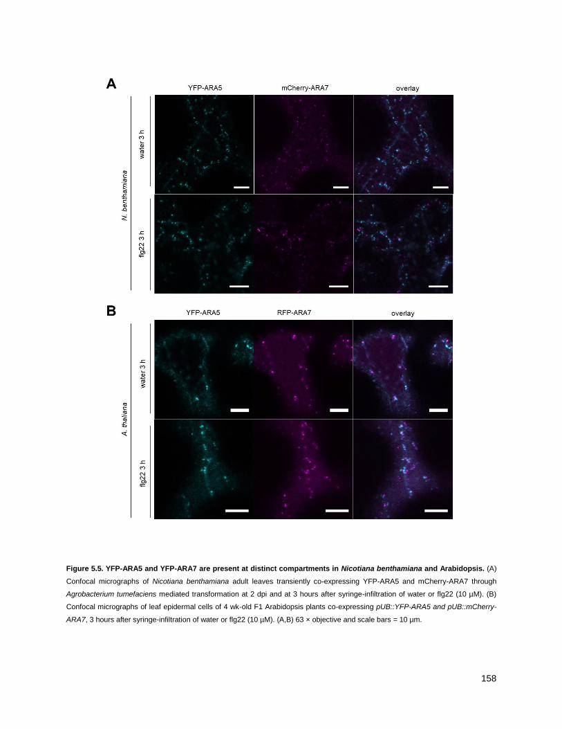
158
Figure 5.5. YFP-ARA5 and YFP-ARA7 are present at distinct compartments in Nicotiana benthamiana and Arabidopsis. (A)
Confocal micrographs of Nicotiana benthamiana adult leaves transiently co-expressing YFP-ARA5 and mCherry-ARA7 through
Agrobacterium tumefaciens mediated transformation at 2 dpi and at 3 hours after syringe-infiltration of water or flg22 (10 µM). (B)
Confocal micrographs of leaf epidermal cells of 4 wk-old F1 Arabidopsis plants co-expressing pUB::YFP-ARA5 and pUB::mCherry-
ARA7, 3 hours after syringe-infiltration of water or flg22 (10 µM). (A,B) 63 × objective and scale bars = 10 µm.

159
DISCUSSION
5.5 - ARA5 vesicles concentrate at the cell surface
ARA5 vesicles were observed to concentrate under bacterial contact sites after ca. 6 h of
infiltration with Pto DC3000 (figs. 5.1-2), although upon quantification, no significant differences
in number of ARA5 concentrations were observed between water treatment and Pto DC3000
infiltrations, which both triggered the response. It could be that Pto DC3000 partially suppresses
the response, so it would be interesting to test flg22-only treatments too. In chapter 4, I could
show that water also triggers ARA7 clustering to some extent, but for both ARA7 and ARA5,
clusters were observed at bacterial infection sites. Concentration of ARA5-marked secretory
pathway compartments that could be Golgi and/or TGN/EE, is in line with existing data that
describe the secretory Rab GTPase RABE1d to focally accumulate upon bacterial challenge.
Furthermore, expression of a constitutive active version fo RABE1d increased resistance against
Pto DC3000 (Speth et al., 2009). While it has not been shown that RabE1d clusters are
positioned under bacterial infection sites, secretory Rab GTPases can be themselves involved in
defining spatially distinct locations at the cell surface, as evidenced by RABA5c, which located at
the geometric cell edges and functioned in cell wall homeostasis there (Kirchhelle et al., 2016).
The factors defining the precise locations of clustering of secretory Rab GTPases are unknown,
but it is conceivable that in both pathogen and developmental context, the spatial cues could be
conferred by physical stimuli.
The number of ARA5 vesicles did not change up to 3 hours after flg22 treatment in my high
throughput confocal experiments. ARA5 vesicle clustering also was not observed before ca. 6 hpi
upon bacterial infection. These findings together indicate that at up to 3 h of immune stimulus,
while proteomic cargoes do change, the ARA5 vesicles do not respond by changing their
dynamics. Genes functioning in the secretory pathway were upregulated upon application of SA
to support PR1-secretion (Wang et al., 2005), and flg22-treatment has been shown to induce SA
accumulation already at early time points (Tateda et al., 2015). However, the described gene
upregulation response was measured at 1 dpi, instead of earlier time points at which I observed
ARA5. This corresponds to the absence of the SA-induced secretory cargo PR1 in my ARA5
purifications, which might be identified upon purification at 1 dpi (Wang et al., 2005). Based on my
data, I cannot exclude that at earlier time points, ARA5 compartment motility and/or turnover
rates are altered, which may underlie an early increased secretory activity.

160
5.6 - The ARA5-specific proteome contains defence-related proteins and changes upon flg22
stimulus
In order to reveal proteomic changes along the default secretory pathway upon immune stimulus,
I purified YFP-ARA5 from flg22-treated cells at 3 hrs, and performed mass spectrometry analysis.
In addition, to gain insight in commonalities and differences between the endocytic and secretory
pathways under these conditions, I compared the ARA5 resting and immune stimulated co-
purifying proteomes to those of ARA7.
Proteins that exclusively fell within the ARA5 co-purifying proteome corresponded to its known
localization and function, such as the secretory TGN/EE-localized Rab GTPase RABA1d (Asaoka
et al., 2013), Tubulin, which guides Golgi dynamic localization (Crowell et al., 2009) and
Coatomer, which functions in intra-Golgi trafficking. While a small pool of YFP-ARA5 may localize
to the Golgi during its constitutive biosynthesis as driven by the Ubiquitin-10 promoter, the
majority of ARA5 is likely to localize to its site of functioning on trans-Golgi and TGN/EE (Pinheiro
et al., 2009; Geldner et al., 2009).
A highly abundant protein identified in ARA5, at similar abundance under water and flg22-treated
conditions, is PEN3, which is known to predominantly localize to the PM (Underwood and
Somerville, 2013). Its non-responsiveness to flg22 treatment is consistent with published data
that de novo synthesis does not contribute to PEN3 focal accumulations at the cell surface
(Underwood and Somerville, 2013), which together with my data suggests that ARA5
compartments and ARA5 TGN/EE does not contribute to this response. Conversely, in ARA7
purifications, PEN3 passed my criteria for co-purifying only upon flg22 treatment, in line with
LE/MVBs underlying the focal accumulation response.
At steady state, PEN3 was found to traffic in a polar way. In roots, PEN3 localized to the outer
lateral membrane, and was found to traffic through VHA-a1/SYP61/VTI12-positive typical
TGN/EE compartments to do this (Mao et al., 2016) – all TGN/EE markers which were found in
both my ARA5 and ARA7 purifications. Both secretory and endocytic traffic contributed to PEN3
localization at the TGN/EE (Mao et al., 2016), but in water treated conditions, I identified PEN3
only in ARA5, implicating ARA5-positive TGN/EE, rather than ARA7, in the steady state polar
trafficking of PEN3. Its absence in ARA7 under my criteria suggests that its route to the cell
surface at resting state does not depend on redirected LE/MVBs as it does under pathogen
stimulated conditions.
Specifically upon flg22 treatment, and only in ARA5 purifications, MLO12 was identified (table
5.3, fig. 5.4). Little is known about the function of MLO12 in immunity, but together with MLO2
and MLO6 it belongs to the clade which is co-orthologous with barley Mlo, in which broad
spectrum mlo-based resistance was originally identified (Panstruga, 2005). Out of this clade,

161
mlo2-based resistance is best studied. MLO2 localized to the PM, and polarly accumulates under
pathogen attack sites (Panstruga, 2005; Consonni et al., 2006; Kusch and Panstruga, 2017; Bhat
et al., 2005). While MLO12 subcellular localization has not been studied, the MLO homolog
NORTIA was found to localize to the Golgi in ovules, functioning in pollen tube reception – a
process that does not occur in the seedlings from which I purified ARA5.
Mlo2 mutants require components of indolic acid metabolism (PEN2, PEN3), and membrane
trafficking (PEN1/SYP121) for full resistance against powdery mildew (Kuhn et al., 2017). Mlo2,
mlo6 and mlo12 cooperatively contribute to mildew resistance, with mlo2 resistance evident in
single mutants, but mlo6 and mlo12 effects only visible as additive effects when in double
mutants with mlo2 or in the mlo2/6/12 triple mutant (Consonni et al., 2006). Interestingly,
mlo2/6/12 triple mutants did not have the same requirements for indole glucosinolate metabolism
or subcellular trafficking as compared to mlo2 single mutants (Kuhn et al., 2017). This could
mean that the function of MLO12 does not involve these components.In line with this, I do not find
flg22-induced upregulation of PEN2, PEN3 or PEN1/SYP121 in ARA5 purifications, but IGMT2, a
major player in the indole glucosinolate defense pathway is strongly upregulated.
While the above data describe the role of MLOs in immunity against fungal pathogens, MLOs
may have different roles in modulating antibacterial immunity. Evidence for MLOs contributing to
susceptibility comes from effector studies, as Pseudomonas HopZ2 targets MLO2 and requires it
for full virulence (Lewis et al., 2012). In addition, loss-of-function mutations in pepper CaMLO2
enhanced resistance to bacterial pathogens, and CaMLO2 expression in Arabidopsis conferred
enhanced susceptibility to Pto DC3000 (Kim and Hwang, 2012). On the other hand, mlo2/6/12
triple mutants, which were more resistant to powdery mildew, were more susceptible to the non-
adapted bacterial pathogen P. syringae pv. maculicola (Kusch and Panstruga, 2017), and mlo2
and mlo2/6/12 were not differentially affected in their resistance to P. syringae pv. maculicola,
while the triple mutant was more susceptible to powdery mildew (Gruner et al., 2018). Taken
together, MLOs can have ambiguous effects on antibacterial immunity, and it is yet unclear if
MLO12 plays a role therein.This provides evidence that flg22-induced changes in the ARA5 co-
puryfing proteome may be nonspecific to the type of MAMP used, but represent a more general
status change independent of trigger. This could be consistent with the finding that mutants in
flg22-upregulated ARA7-interacting proteins are susceptible to powdery mildew, and not bacterial
pathogens.
MLOs have been implicated in modulating ROS signaling (Cui et al., 2018). I identified CRK10,
PER4, and OGOX1 in ARA5 purifications upregulated upon flg22 treatment, all of which are
implicated in processes involving ROS. CRK10 has a potential role in extracellular ROS
perception (Bourdais et al., 2015), and MLO2 was recently found to be a negative regulator of
extracellular ROS sensing, with all MLO proteins hypothesized to exert a similar role (Cui et al.,
162
2018). Their co-occurrence in my flg22-treated ARA5 purifications could point at a functional
relationship, although the regulatory MLO2 role in ROS perception was shown to be partially
uncoupled from its elevated resistance against mildew (Cui et al., 2018).
Barley MLO contains a calmodulin-binding domain, and its interaction with calmodulin is
necessary for its function in downregulating immunity (Kim et al., 2002). MLO12 also contains a
predicted calmodulin-binding domain (UniProt, accessed July 2018). Interestingly, PEN3, present
in the ARA5-associated proteome at high abundance both under water and flg22 treated
conditions, has been shown to interact with calmodulin, which, like PEN3 itself, is required for
non-host resistance (Campe et al., 2016). PEN3 focally accumulates in response to bacteria and
flg22, and is also required for full mlo-based resistance (Consonni et al., 2006; Underwood and
Somerville, 2013; Xin et al., 2013). I find that both ARA7 and ARA5 cluster at the cell surface,
which could mean that they both target the same location and delivering cargoes that otherwise
would not co-accumulate. With MLOs functioning as susceptibility factors, and my data showing
that PEN3 and MLO12 may be co-delivered at the cell surface, it is conceivable that MLO12 may
downregulate calmodulin-involving processes on co-accumulating cell-surface defense
components such as PEN3.
Taken together, while a function for MLO12 in antibacterial resistance has not been described,
my proteomic results show that MLO12 co-purified with secretory ARA5 upon treatment with with
a bacterial MAMP, and provide potential leads to further investigation of the modulation of
immunity by MLOs in general.
5.7 - The shared ARA7 and ARA5 proteome contains TGN/EE proteins
Upon comparison of the resting state co-purifying proteomes of ARA7 and ARA5, their contents
suggest that I purified TGN/EE-associated pools of both. Common TGN/EE markers such as
SYP61, SYP43 and VHA-a1 are found (table 5.4), in addition to cargoes previously identified in
TGN/EE-proteomic studies, such as SCAMP3, CESA1 and CCC1 (Groen et al., 2014; Drakakaki
et al., 2012). In ARA7 purifications, ARA5 was identified, although at low abundance, which could
explain why a majority of ARA5 interacting proteins were also captured by ARA7 purifications
(fig. 5.4D). While upon microscopic analysis I did not find punctate overlap of ARA7 and ARA5 in
Arabidopsis or N. benthamiana (fig. 5.5), upon co-expression in BiFC (split-YFP) experiments in
N. benthamiana, ARA7 and ARA5 together reconstituted a punctate signal (fig. 4.9), indicating
that they exist in close proximity, likely on separate domains of TGN/EE.
In ARA7 purifications I also identified ARA6, which reconstituted a BiFC signal with ARA7, and
which has been described to largely localize to a subpopulation of ARA7 endosomes (Ueda et al.,
2001). Because of their large overlap in published microscopic studies, I expected a high
abundance of ARA6 in ARA7 purifications. However, it was recently shown that these two Rab5

163
GTPases compete for activation by VPS9a through their common effector PUF2 (Ito et al., 2018),
and since Rab GTPase activation status governs their capacity for membrane association, this
could indicate that they do not preferably exist in the same protein complex in a stable manner,
thus leading to low levels of co-purification of ARA6 with ARA7.Additionally, seeing as the
purification method used likely yields greater Rab complexes and not intact membrane
compartments, it could be that despite their localization to the same membrane vesicles, ARA7
and ARA6 do not interact with each other.
Based on co-localization analyses from various publications, broadly two populations of TGN/EE
can be distinguished, either marked by RabA1b, RabA2a and VAMP721 with known secretory
functionality, or marked by SYP61, SYP43 and VHA-a1, with roles in both secretion and also
endocytosis (Gendre et al., 2015). In the shared ARA5 and ARA7 co-purfying proteome, I did not
capture RabA1b, RabA2a and VAMP721, but consistently identified SYP61, SYP43 and VHA-a1,
independent of flg22 treatment (table 5.4). Since ARA5 and ARA7 did not overlap on punctae in
confocal microscopy (fig. 5.5), I conclude that they can further divide this population of TGN/EE
into subpopulations with secretory and endocytic functions respectively.
The shared ARA5 and ARA7 co-purifying proteome was rich in cell wall metabolism proteins, with
cargoes such as CESA1, 3 and 6, comprising the full primary cell wall cellulose synthesis
complex, glucan synthases including PMR4, CBL7 which facilitates cellulose biosynthesis, and
NHL3, which is implicated in PM-cell wall adhesion. The callose synthase PMR4 has a positive
role in defense against some pathogens, such as Bgh (Ellinger et al., 2013), but pmr4 mutants
show constitutively elevated SA levels, defense gene expression coupled with increased
resistance to pathogens such as G. cichoracearum, providing challenges to studies that probe the
role of PMR4-dependent callose deposition in immunity (Nishimura et al., 2003; Jacobs et al.,
2003). PMR4 itself localizes predominantly to the PM, with increased vesicle localization and
focal accumulation under the site of attempted penetration by powdery mildew at ca. 6 h after
spore application (Ellinger et al., 2013).
While the PMR4 focal accumulation response has not been tested under flg22 treatment, this
stimulus triggers cell-surface localized callose depositions fully dependent on PMR4 (Luna et al.,
2011). Contrary to PEN3 found in ARA7 purifications upon flg22 treatment, PMR4 did not
markedly change in abundance in my ARA7 or ARA5 purifications. Therefore, the defence-
associated focal accumulation of PMR4 is unlikely to be underpinned by redirected endosomal
traffic. A role for the secretory pathway is more likely, as PMR4 co-purified with ARA5 at higher
abundance than with ARA7 (table 5.4). Here, PMR4 could be delivered from the secretory
pathway, and undergo turnover and degradation through endocytosis. In line with PMR4
dependency on default secretory processes, overexpression of the secretory RabA4c has been

164
shown to enhance callose deposition and resistance to adapted G. cichoracearum, both
dependent on PMR4 (Ellinger et al., 2014b).
5.8 - Upon flg22 treatment, the shared ARA7 and ARA5 proteome enriches with proteins
functioning at the cell surface
Upon 3 h flg22 treatment, a small group of 9 proteins was upregulated in abundance in both
ARA5 and ARA7 purifications. Due to the common TGN/EE localization of ARA5 and ARA7,
proteins identified in both purifications are also likely to localize to this compartment, but do not
necessarily function there. Shared interactors can function at other locations, as especially
evidenced by the identification of resting state cargoes which have PM localizations and functions
such as NHL3, CBL7, PEN1 and CESAs. Knowing that ARA5 and ARA7 maintain separate
localization upon flg22 treatment, and that the majority of flg22-upregulated shared proteins have
a cell surface localization (table 5.5), I interpret their upregulation in both purifications as a sign of
an increased turnover at the cell surface, and discuss them in that context.
Three cell-surface receptor-like kinases (MIK2, CRK10, IOS1) were identified. While IOS1 has
been shown to be a common interactor of many immune receptors (Yeh et al., 2016), MIK2 is a
cell wall integrity sensor (Van der Does et al., 2017) and CRKs have been functionally implicated
in ROS-perception upon host-microbe interactions, either directly or through spatial organization
of ROS-perception modules (Bourdais et al., 2015; Kimura et al., 2017). The increased turnover
of these receptors 3 h after flg22 treatment could point at ligand-induced endocytosis to achieve
receptor degradation, as is observed for FLS2 (Beck et al., 2012b), coupled to upregulated
secretion to replenish the PM-localized signaling capable pool. While these receptors have not
been previously shown to undergo this process, cell surface receptors are capable of undergoing
ligand-induced endocytosis upon perception of endogenous signals, such as BRI1 upon
brassinolide steroid hormones in growth, (Martins et al., 2015), ERECTA-LIKE 2 upon epf
peptides in stomatal development (Ho et al., 2016) and PEPR1 upon pep1 peptides as DAMP
signals (Ortiz-Morea et al., 2016). ROS themselves can function as extracellular second
messengers, and can induce antibacterial resistance (Sharma et al., 1996; Sewelam et al., 2016).
For the cysteine-rich GPI-anchored enzyme OGOX1, which neutralizes oligogalacturonide
DAMPs in the apoplast, increased turnover could be necessary to maintain an enzymatically
active pool of receptors. For all the above membrane-associated candidates, it would be
interesting to use fluorescence microscopy to study the subcellular dynamics at the 3 h flg22
treated time point, upon chemical disturbance of the cell wall, or external application of ROS, to
confirm their ligand-induced endocytosis.
The above data taken together, I hypothesize that upon perception of flg22 stimulus, the plant is
anticipating defense-related ROS-signaling processes and cell wall modifications. Why anticipate

165
cell wall damage in the interaction with bacterial pathogens? Pto DC3000 inserts type-3-secretion
needles, but is not known to grossly modify the cell wall. However, infection with some bacteria,
such as the opportunistic pathogen Pseudomonas aeruginosa, causes cavities in the cell wall at
bacterial positions as seen in electron microscopy studies (Plotnikova et al., 2000).
Coincidentally, while all flagellin perception in Col-0 is initiated by the singular flagellin receptor
FLS2, the elicitor peptide flg22 corresponds to the immunogenic epitope on flagellin of P.
aeruginosa (Felix et al., 1999). I hypothesize that the upregulation of cell wall integrity and ROS-
sensing capacity upon flg22 elicitation is a generalized response, that prepares for a range of
potential modifications by incoming pathogens, or function in sensing endogenous, potentially
ROS-mediated cell wall modifications and ensure integrity is maintained even upon ―false alarms‖
in the absence of continued infection.
Some LRR-RLKs upregulated in ARA5 upon flg22 were not identified in ARA7. While their ligands
are yet unknown, this could provide evidence for increasing detection capacity at the cell surface
in response to pathogen stimulus. It has been described that treatment with SA potentiates cells
for the detection of flagellin and chitin, which is mediated through cell-surface receptors FLS2 and
CERK1 (Tateda et al., 2015). There, increased potentiation is thought to be achieved through
increased secretion of cell-surface receptors mediated through interaction with the ER-localized
protein ACCELERATED CELL DEATH 6 (ACD6, (Zhang et al., 2014b). SA treatment increased
the PM-resident pool of FLS2 in an ACD6-dependent manner, but when measured at 24 and 48
hpi, which does not match the timing of my flagellin treatments (Zhang et al., 2014b). However,
treatment with flg22 has been shown to induce endogenous SA-signaling, detectable as early as
3 hours (Tsuda et al., 2008).
Determining the cognate ligands for the uncharacterized secreted, but not internalized receptors
would be interesting, as they could be on the lookout for various anticipated MAMPs or DAMPs
that associate with infection in a non-pathogen specific manner, making these receptors
interesting in the context of engineering broad-spectrum resistance. Finally, to gain further insight
in the specificity of upregulated proteins in my ARA5 and ARA7 purifications to various types of
pathogens, it would be interesting to perform the same purifications upon treatment with a panel
of additional MAMPs, such as chitin, lipopolysaccharides or elicitins.
5.9 - Expanding on the method
While the majority of co-purfying proteins in ARA5 and ARA7 purifications were not considered to
be upregulated in response flg22 treatment (figs. 4.6, 5.4), they could still fulfil important
functions in immunity. To address this, it would be relevant to perform my purifications under
inhibition of dephosphorylation, and subject samples to phosphoproteomic analysis using mass
spectrometry. This could reveal activity status changes on proteins such as receptor kinases

166
along the trafficking pathways, thus providing an extra layer of information that is not revealed by
looking at abundance changes only.
Finally, my current data reveal proteomic changes between 3 h water and flg22 treatment, but it is
possible that various suites of secretory and endocytic cargoes change in abundance at different
time points after immune stimulus, perhaps relating in function to earlier occurring responses
such as MAMP-induced endocytosis, apoplastic alkalinization and ROS burst, or later responses
such as callose deposition, stomatal closure and systemic acquired resistance (Khaled et al.,
2015). In combination with more precise methods of peptide quantitation, correlating protein
abundance changes in secretory and endocytic pathways at different time points after immune
stimulus may reveal novel functional relationships between groups of proteins that have not been
implicated to work together before (Thul et al., 2017).

167
CHAPTER 6
GENERAL DISCUSSION
In the first stage of my PhD project, I set myself the following goals:
Reveal the subcellular localization of Cf-4
Reveal the subcellular localization of constitutively interacting Cf-4-SOBIR1 complexes
Probe for ligand-induced subcellular localization dynamics of Cf-4 upon activation by Avr4
Investigate the requirements for SERK members in Cf-dependent receptor outputs and immunity
Using microscopic analysis, I could successfully show that the tomato RLP-type PRR Cf-4 is localized to
the plasma membrane, which confirms its long standing expected localization, in line with its function in
detecting apoplastic Avr4 secreted by C. fulvum upon infection (Joosten et al., 1994; van den Burg et al.,
2006). I could also show that it interacts there with its constitutive interaction partner SOBIR1, an RLK
that is required for Cf-4 function (Liebrand et al., 2013). Upon activation, Cf-4 and SOBIR1 co-internalize,
in a SOBIR1-kinase-activity dependent manner. This supports the notion of Cf-4-SOBIR1 functioning as a
two-component PRR (Liebrand et al., 2014; Gust and Felix, 2014), as this response is shared by the well-
characterized one-component RLK-type PRRs FLS2 (Robatzek et al., 2006; Khaled et al., 2015). These
observations expand our view and reveal that the mechanism that underlies PRR turnover at the PM is
generalisable to diverse classes of receptors.
I confirmed that Cf-4-SOBIR1 recruits the same early activation module BAK1/SERK3 and depend on it
for outputs, which shows that RLP-SOBIR1-type PRR and RLK-type PRR activation converge at early
steps. In the time since this discovery, several other RLP-SOBIR1 systems have been shown to engage
with SERK members upon activation, further supporting the notion that this is a broadly generalisable
PRR activation mechanism (Albert et al., 2015; Domazakis et al., 2018; Du et al., 2015; Wang et al.,
2018).
My finding that Cf-4-SOBIR1 activation upon detection of the race-specific Avr4 effector shares
components with detectors of broadly conserved MAMPs. This, combined with mounting evidence that
Cf-4 detects conserved patterns on Avr4 orthologues from distantly related fungi independent of their
ability to exert their main chitin binding effector function (Kohler et al., 2016; Mesarich et al., 2016),
supports the increasingly blurring classic distinction between effector-triggered immunity and MAMP-
triggered immunity (Thomma et al., 2011; Cook et al., 2015).
My data in combination with related recent discoveries on RLP activation broaden our understanding of
cell-surface PRR dependent immune activation, but leaves the question: which SERKs specify which
outputs? I could show that multiple SERKs were recruited to Cf-SOBIR1 complexes, and it has previously

168
been shown that one SERK can engage with RLKs that function in both defense and development
(Chinchilla et al., 2009). Does the same apply to RLPs?
SOBIR1 affects developmental pathways too. Specifically, it downregulates the outputs of the RLK-type
HAESA (HAE) and HAESA-LIKE 2 (HSL2), which form a cell-surface based developmental peptide
receptor platform (Leslie et al., 2010a; Gubert and Liljegren, 2014). It is thought that SOBIR1 engages
with HAE/HSL2 to target them for degradation, which in the light of my findings, could be through the
endocytic vacuolar-targeted pathway. Indeed, subcellular trafficking components seem to be required for
SOBIR1 effects on HAE/HSL2 (Liljegren et al., 2009).
Some RLPs in development seem to avoid SOBIR1 altogether, but engage with other scaffolding RLKs,
such as the RLP CLAVATA 2 which depends on the RLK CORYNE for accumulation at the PM and
signaling (Somssich et al., 2016). Interestingly, they too engage with poised PM-localized RLKs, as
evidenced by their interaction with the RLK CLAVATA 1 (Muller et al., 2008; Bleckmann et al., 2010). No
evidence for the involvement of SERK members in this system is reported, pointing at potential limits for
the generalisability of the role of SOBIR1 and SERK in RLP-mediated signaling.
In the second stage of my PhD, I wanted to further clarify the role of Rab GTPases in immunity, and set
myself the following goals:
Investigate localization changes of ARA7 and ARA5 upon bacterial infection
Reveal changes in ARA7 and ARA5-associated proteins after flg22-stimulus
Identify flg22-responsive ARA7-interacting proteins which are defence-related
Further characterize a subset of flg22-responsive ARA7 interacting defence-related proteins for
roles in immunity
I revealed that especially the endocytic Rab GTPase ARA7 has the tendency to intensely concentrate
under bacterial contact sites upon infection, which is shared by the secretory Rab GTPase ARA5 to a
lower extent. This matches known endosomal responses in well-established fungal and oomyecete
pathosystems (Inada et al., 2016; Nielsen et al., 2012; Cai et al., 2018), and expands the concept of
LE/MVB spatial reorganisation to antibacterial immunity. Indeed, bacterial infection has been shown to
upregulate extracellular vesicle secretion, a process which is thought to be underpinned by LE/MVB-
dependent secretion (Wang et al., 2014, 2015; Rutter and Innes, 2017b).
I mapped proteomic changes along endocytic and secretory processes by performing mass spectrometry
on purifications of ARA7 and ARA5 after the activation of the immune response to bacterial flagellin, and
reveal that I most likely purified the greater complex associating with these Rab GTPases. Associating in
an increased manner with ARA5 upon flagellin elicitation, I find several uncharacterized RLKs that may
potentiate the cell surface with detection capacity for yet unknown ligands that may not be bacteria-
specific. Additionally, I find a member of the MLO family, which are negative regulators of immunity

169
(Kusch and Panstruga, 2017). Based on its co-upregulation on ARA5 complexes with proteins that work
in processes which have previously been suggested to be the client of MLO activity, I provide avenues for
further study of MLO-based susceptibility (Kim et al., 2002; Campe et al., 2016; Cui et al., 2018).
Comparative proteomics in combination with co-localization studies between ARA7 and ARA5, provided
evidence for these Rab GTPases further dividing a main TGN/EE sorting hub into secretory and
endocytic domains respectively (Gendre et al., 2015; Wattelet-Boyer et al., 2016). In response to immune
stimulus, shared proteins were upregulated, which points at increased secretion and endocytosis, and
thus cell-surface turnover. These include proteins associated with ROS-metabolism and signaling, as well
as cell-wall integrity sensing. These are known processes associated with host-activated modifications to
the extracellular space upon MAMP perception.
Specifically associating with the endocytic Rab GTPase ARA7 upon immune stimulus, I find atypical
resistance proteins TN3 and HR4, which I could show function in resistance to nonadapted powdery
mildew, further supporting a generalized defense programme to be upregulated in the ARA7 complex. In
parallel to TN3, I found the upregulation of a PRA1 Rab GTPase regulatory protein, which is a predicted
target of both fungal and oomycete effectors (Weßling et al., 2014), together pointing at potential host
guarding of the regulation of RabF-dependent processes. Further research should elucidate whether TN3
is the NLR responsible for monitoring endocytic traffic, and potentially unconventional endosomal
secretion in plant immunity.

170
SUPPLEMENTAL MATERIALS
FIGURES
Fig. S.1. GFP-tagged Cf-4 migrates with the predicted molecular weight and is functional. (A) Western blot analysis of Cf-4-
GFP transiently and stably expressed in N. benthamiana, without and with Avr4 (100 µM) treatment, as indicated. For transient
expression, leaf samples were harvested at three days post infiltration (dpi) and Avr4 elicitation was done for 90 min. Blots were
incubated with anti-GFP antibodies for the detection of Cf-4-GFP. CBB, Coomassie Brilliant Blue staining. (B) Images of N.
benthamiana wild type (WT) leaves (top panel) and leaves transiently expressing Cf-4-GFP (bottom panel), treated with either Avr2
or Avr4 (300 µM), as indicated. Images were taken at five dpi and at four days after treatment with the Avrs.

171
Fig. S.2. Subcellular localization of Cf-4, SlSOBIR1-like and AtSOBIR1. Confocal micrographs show N. benthamiana leaf
epidermal cells stably expressing Cf-4-GFP and transiently expressing SlSOBIR1-like-GFP and AtSOBIR1-GFP as indicated (left
panels), and co-expressing plasma membrane-localized ACA8-mCherry (middle panels). Overlay images indicate co-localization of
the proteins fused to GFP and mCherry, as a yellow colour is produced (right panels). Images were taken at three dpi; scale bars =
10 μm.

172

173
Fig. S.3. SlSOBIR1-like-GFP and AtSOBIR1-GFP co-localize with endosomal markers. Confocal micrographs show N.
benthamiana leaf epidermal cells transiently expressing SlSOBIR1-like-GFP or AtSOBIR1-GFP (left panels), and co-expressed
mCherry/RFP-tagged organelle markers (middle, left panels). Overlay images indicate co-localization through generation of a yellow
colour (middle, right panels). Dashed boxes indicated in the middle right panels are depicted as detail pictures on the right (right
panels). Arrowheads point at co-localizing endosomes. Images were taken at three dpi; scale bars = 10 μm. (A) Co-expression of
SlSOBIR1-like-GFP/AtSOBIR1-GFP with Golgi marker MEMB12-mCherry. (B) Co-expression of SlSOBIR1-like-GFP/AtSOBIR1-
GFP with trans-Golgi network marker VHA-a1-RFP. (C) Co-expression of SlSOBIR1-like-GFP/AtSOBIR1-GFP with endosome
marker RFP-ARA7/RabF2b. (D) Co-expression of SlSOBIR1-like-GFP/AtSOBIR1-GFP with late endosome marker ARA6/RabF1-
RFP.

174
Fig. S.4. Line intensity profiles of Cf-4, SlSOBIR1 and membrane markers. Confocal micrographs show transient co-expression
of Cf-4-GFP, SlSOBIR1-GFP or Cf-4-YFPc/SlSOBIR1-YFPn with indicated membrane markers and effector treatments. Left column
of panels correspond to confocal micrographs displayed in (A) Fig. 3.1, (B) Fig. 3.2A, (C) Fig. 3.2b. Dashed squares in these panels
are shown as detailed pictures (magnified in middle column of panels). White lines in detailed pictures indicate the regions of
interest (ROIs) that correspond to intensity profiles in the last panels. Intensity profiles indicate grey-value of pixels across the ROI in
the green and red channels on a scale of 1-300.

175
Fig. S.5. SlSOBIR1 does not interact with FLS2 in BiFC experiments. (A) Confocal micrographs show N. benthamiana leaf
epidermal cells transiently co-expressing FLS2 and SlSOBIR1 C-terminally fused to the C- or N-terminal halves of YFP (YFPc and
YFPn, respectively), as indicated. Left panels show absence or presence of YFP fluorescence, the middle panels show
autofluorescence, and the right panels show the overlay of the fluorescence signals. Images were taken at two dpi, scale bars = 100
μm. (B) Confocal micrographs of N. benthamiana leaf epidermal cells transiently co-expressing FLS2-YFPc and FLS2-YFPn, either
without or with flg22 treatment (10 µM) for 90 min, as indicated. Left panels show absence or presence of YFP fluorescence, the
middle panels show autofluorescence, and the right panels show the overlay of the fluorescence signals. Images were taken at two
dpi, scale bars = 10 μm.

176
Fig. S.6. Cf-4-GFP undergoes Avr4-triggered endocytosis in stable transgenic plants. Confocal micrographs of Cf-4-GFP-
transgenic N. benthamiana plants show leaf epidermal cells expressing Cf-4-GFP, either treated with Avr4 or Avr2 (both at 100 µM),
as indicated. Left panels show GFP fluorescence, middle left panels show autofluorescence, and middle right panels show the
overlay of the fluorescence signals shown in the left and middle left panels. Micrographs in rows 1 and 3 display maximum projected
z-stacks to reveal vesicles, and rows 2 and 4 display their selected single focal plane outtakes to capture plasma membrane signal.
Arrowheads in the detail pictures (right panels) point at mobile vesicles (top right panel). Images were taken at 90 minutes after
elicitation; scale bars = 10 μm.

177
178
Fig. S.7. SlSERK3 is required for Avr4-induced HR. (A) Leaves of Cf-4-GFP-N. benthamiana stable plants were TRV-silenced for
NbSERK3a/b for three weeks and subsequently used to transiently express AtBAK1 and AtBAK1-KD, without treatment of Avr4.
Images are representative of vesicle quantification data presented in Fig. 3.4. (B) qRT-PCR analysis showing NbSERK1,
NbSERK3a/b and NbSOBIR1 expression in N. benthamiana inoculated with the indicated TRV VIGS constructs. Expression of
query genes was normalized to endogenous NbActin expression levels. Data presented have been combined from twelve
(NbSOBIR1 expression) or 24 (NbSERK1 and NbSERK3 expression) individual qRT-PCRs, based on three independent biological
replicates, for each gene of which the expression was studied. Statistical differences (α=0.05) between groups were calculated by a
one-way ANOVA and different groupings are indicated. Error bars represent the standard deviation. These qRT-PCR experiments
were performed by Ruby Bye (C) Immunoblots from total protein extracts of Cf-4-GFP-transgenic N. benthamiana plants that were
TRV-silenced for NbSERK3a/b and subsequently transiently transformed with AtBAK1 and AtBAK1-KD. Cf-4-GFP was revealed
with αGFP. Ponceau staining is shown for equal loading. (D) RT-PCR on cDNA obtained from total RNA extracts of Cf-4-GFP-
transgenic N. benthamiana plants that were TRV-silenced for NbSERK3a/b and subsequently transiently transformed with AtBAK1
and AtBAK1-KD. (E) Images show N. benthamiana leaves from TRV::NbSERK3a/b-inoculated plants and TRV-inoculated controls,
transiently expressing Cf-4-GFP and treated with Avr4 protein (300 µM). Cf-4-GFP was agro-infiltrated at 3 weeks post inoculation
with the recombinant TRV constructs and one day later the Avr4 protein was infiltrated. Images were taken at six days after Avr4
infiltration. HR is observed as brownish cell death.
179
Fig. S.8. Cf-4 endocytosis requires SOBIR1 kinase activity. Leaves of Cf-4-GFP transgenic N. benthamiana plants that had
been TRV-silenced for NbSOBIR1/-like were used for transient expression of AtSOBIR1-Myc and its kinase-inactive variant
AtSOBIR1-KD-Myc. (A) Confocal micrographs show Cf-4-GFP localisation upon treatment with Avr4 (100µM, left panels) and detail
pictures from dashed squares (middle panels). Arrowheads point at Cf-4-GFP-positive vesicles. Images were taken at three weeks
after inoculation with TRV, at three dpi for transient expression, and at 90 min after elicitation; scale bars = 10 μm. (B) Quantification
of Cf-4-GFP-positive vesicles was done with EndoQuant (bars depict means ± 2 SE; n = 6; p < 0.05; statistical significant
differences are indicated by asterisks). Transient co-expression of AtBAK1, but not its kinase-inactive variant AtBAK1-KD, increased
Cf-4-GFP-positive vesicles in NbSOBIR1/-like-silenced leaves. (C) Immunoblots from extracted total proteins of Cf-4-GFP-
transgenic N. benthamiana plants that were TRV-silenced for NbSOBIR1/-like and subsequently transiently transformed with
AtSOBIR1-Myc or AtSOBIR1-KD-Myc. Cf-4-GFP, AtSOBIR1-Myc and AtSOBIR1-KD-Myc were revealed with αGFP and αMyc
antibodies as indicated. Ponceau staining is shown for equal loading.
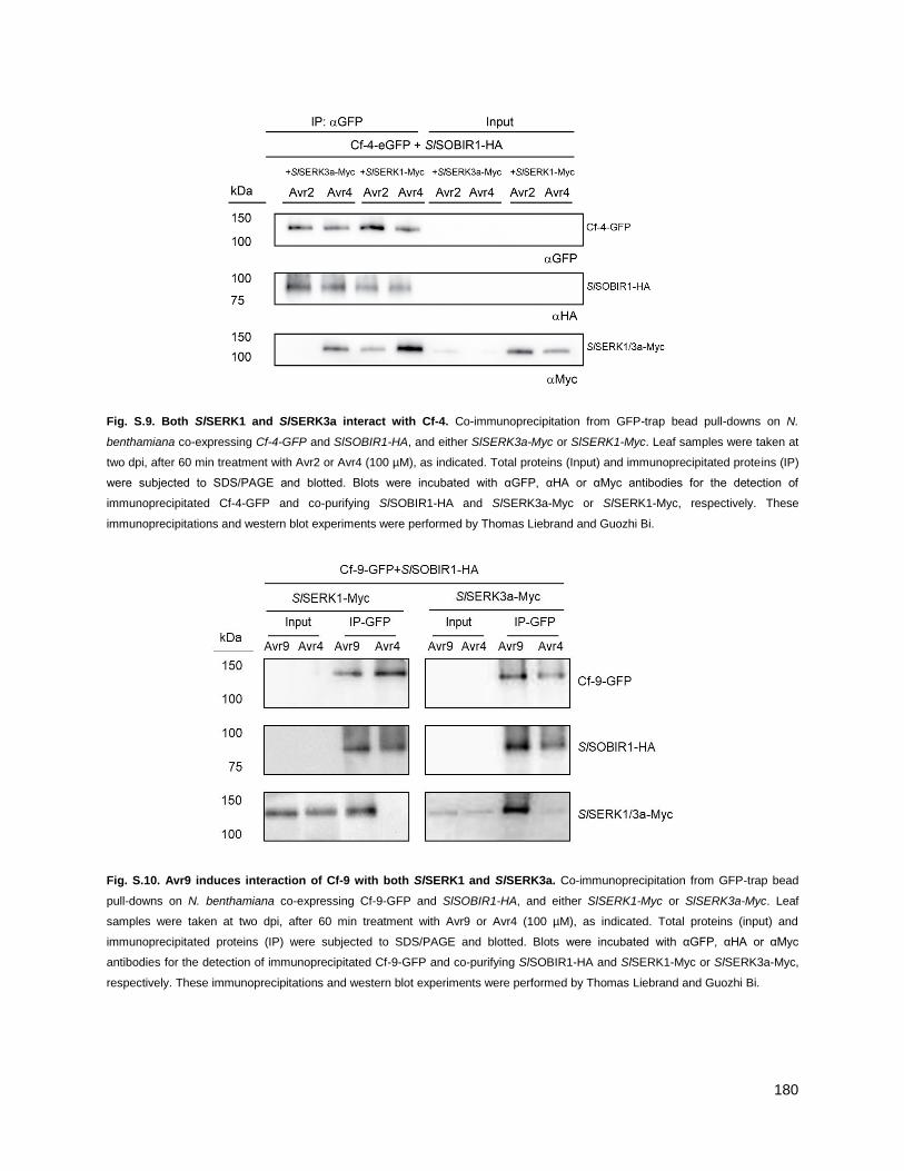
180
Fig. S.9. Both SlSERK1 and SlSERK3a interact with Cf-4. Co-immunoprecipitation from GFP-trap bead pull-downs on N.
benthamiana co-expressing Cf-4-GFP and SlSOBIR1-HA, and either SlSERK3a-Myc or SlSERK1-Myc. Leaf samples were taken at
two dpi, after 60 min treatment with Avr2 or Avr4 (100 µM), as indicated. Total proteins (Input) and immunoprecipitated proteins (IP)
were subjected to SDS/PAGE and blotted. Blots were incubated with αGFP, αHA or αMyc antibodies for the detection of
immunoprecipitated Cf-4-GFP and co-purifying SlSOBIR1-HA and SlSERK3a-Myc or SlSERK1-Myc, respectively. These
immunoprecipitations and western blot experiments were performed by Thomas Liebrand and Guozhi Bi.
Fig. S.10. Avr9 induces interaction of Cf-9 with both SlSERK1 and SlSERK3a. Co-immunoprecipitation from GFP-trap bead
pull-downs on N. benthamiana co-expressing Cf-9-GFP and SlSOBIR1-HA, and either SlSERK1-Myc or SlSERK3a-Myc. Leaf
samples were taken at two dpi, after 60 min treatment with Avr9 or Avr4 (100 µM), as indicated. Total proteins (input) and
immunoprecipitated proteins (IP) were subjected to SDS/PAGE and blotted. Blots were incubated with αGFP, αHA or αMyc
antibodies for the detection of immunoprecipitated Cf-9-GFP and co-purifying SlSOBIR1-HA and SlSERK1-Myc or SlSERK3a-Myc,
respectively. These immunoprecipitations and western blot experiments were performed by Thomas Liebrand and Guozhi Bi.

181
Fig. S.11. Immunoprecipitation and gel-staining of YFP-ARA7 and YFP in preparation for mass spectrometry. (A,B)
Coommassie-stained gel pictures show individual replicates of anti-GFP immunoprecipitations from pUB::YFP-ARA7 or pUB::YFP
seedlings after running in SDS-PAGE on 4-20% agarose gradient gels. Boxes indicate incisions made to obtain separate gel
samples for mass spectrometry analysis. Numbers (left) indicate molecular weight of individual ladder bands (Page Ruler Plus). (C)
Immunoblot shows samples used in (A,B), probed with anti-YFP with anti-GFP-HRP conjugated antibodies. Numbers (right) indicate
molecular weight of ladder bands (not shown).

182
Fig. S.12. Immunoprecipitation and gel-staining of YFP-ARA5 in preparation for mass spectrometry. (A) Coommassie-
stained gel pictures of individual replicates of GFP-antibody immunoprecipitations from pUB::YFP-ARA7 seedlings after running in
SDS-PAGE on 4-20% agarose gradient gels. Boxes indicate incisions made to obtain separate gel samples for mass spectrometry
analysis. Numbers (left) indicate molecular weight of individual ladder bands (Page Ruler Plus). (B) Western blot image of samples
used in (A), probing for YFP with a GFP-HRP conjugated antibody. Numbers (right) indicate molecular weight of ladder bands (not
shown).
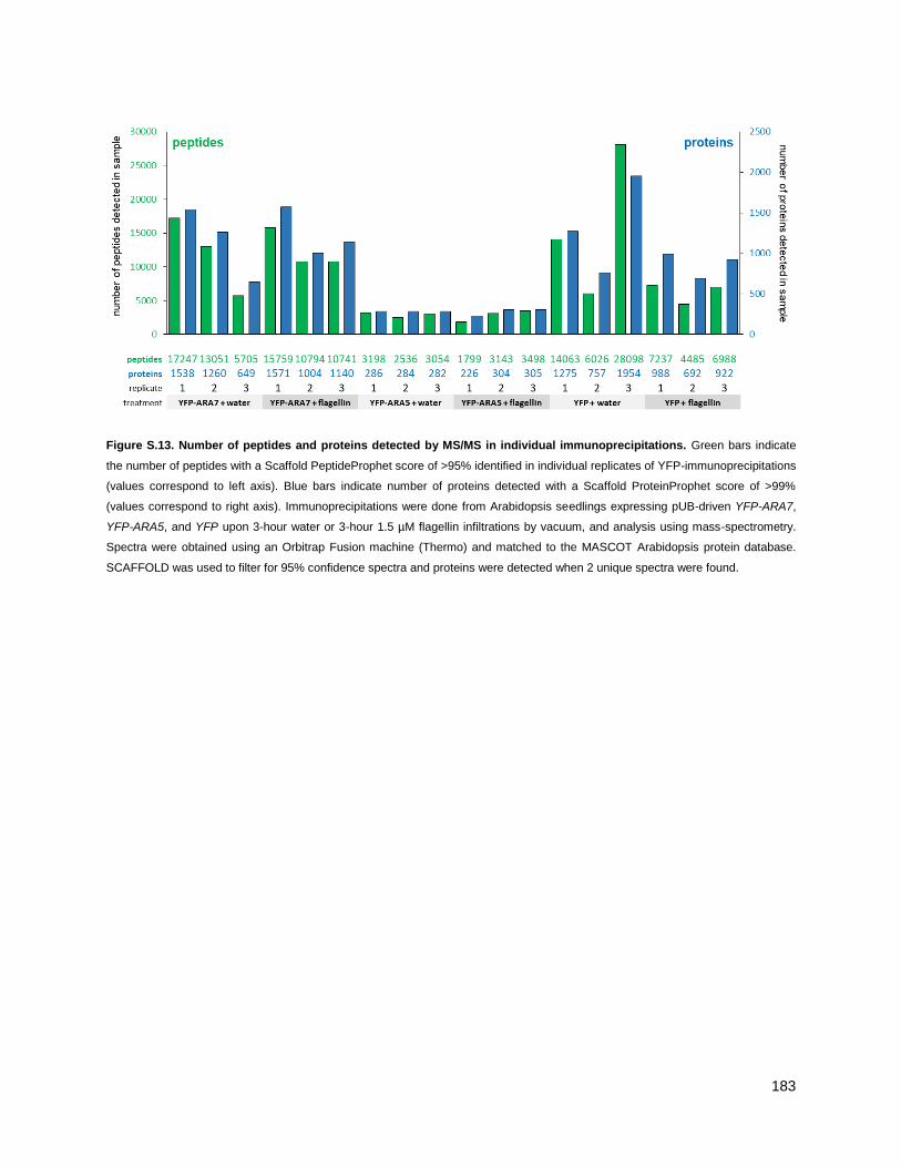
183
Figure S.13. Number of peptides and proteins detected by MS/MS in individual immunoprecipitations. Green bars indicate
the number of peptides with a Scaffold PeptideProphet score of >95% identified in individual replicates of YFP-immunoprecipitations
(values correspond to left axis). Blue bars indicate number of proteins detected with a Scaffold ProteinProphet score of >99%
(values correspond to right axis). Immunoprecipitations were done from Arabidopsis seedlings expressing pUB-driven YFP-ARA7,
YFP-ARA5, and YFP upon 3-hour water or 3-hour 1.5 µM flagellin infiltrations by vacuum, and analysis using mass-spectrometry.
Spectra were obtained using an Orbitrap Fusion machine (Thermo) and matched to the MASCOT Arabidopsis protein database.
SCAFFOLD was used to filter for 95% confidence spectra and proteins were detected when 2 unique spectra were found.
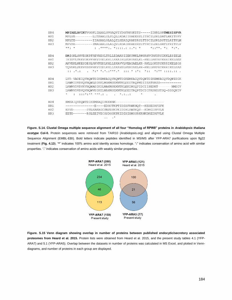
184
Figure. S.14. Clustal Omega multiple sequence alignment of all four “Homolog of RPW8” proteins in Arabidopsis thaliana
ecotype Col-0. Protein sequences were retrieved from TAIR10 (Arabidopsis.org) and aligned using Clustal Omega Multiple
Sequence Alignment (EMBL-EBI). Bold letters indicate peptides identified in MS/MS after YFP-ARA7 purifications upon flg22
treatment (Fig. 4.13). ―*‖ indicates 100% amino acid identity across homologs. ―:‖ indicates conservation of amino acid with similar
properties. ―.‖ indicates conservation of amino acids with weakly similar properties.
Figure. S.15 Venn diagram showing overlap in number of proteins between published endocytic/secretory associated
proteomes from Heard et al. 2015. Protein lists were obtained from Heard et al. 2015, and the present study tables 4.1 (YFP-
ARA7) and 5.1 (YFP-ARA5). Overlap between the datasets in number of proteins was calculated in MS Excel, and plotted in Venn-
diagrams, and number of proteins in each group are displayed.

185
TABLES
Table S1: GO-term enrichment in the ARA7 co-purifying proteome. Listed here are the Gene Ontology (GO) terms (GO-
database of 04-04-2018, ―cellular component‖ group) associated with proteins in the ARA7 co-purifying proteome as defined in
Table 4.1. To capture over- or underrepresentation of a GO-term in the dataset, an enrichment factor is listed, comparing the term‗s
occurrence in the ARA7 co-purifying proteome to its occurrence in the TAIR10 reference genome. For this, the PANTHER
classification system (version 20171205) and Fisher‘s exact test (p < 0.05) were used. Finally, the number of proteins in the ARA7
co-purifying proteome associated with the GO classification is listed. Dots indicate GO terms that correspond to the known
localization and function of ARA7. Bars indicate relative values.
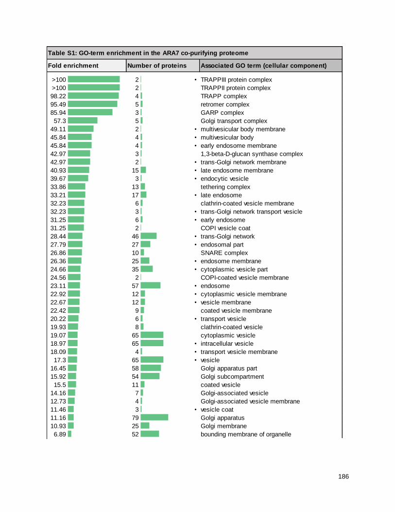
186
Table S1: GO-term enrichment in the ARA7 co-purifying proteome
Fold enrichment Number of proteins Associated GO term (cellular component)
>100 2 • TRAPPIII protein complex
>100 2 TRAPPII protein complex
98.22 4 TRAPP complex
95.49 5 retromer complex
85.94 3 GARP complex
57.3 5 Golgi transport complex
49.11 2 • multivesicular body membrane
45.84 4 • multivesicular body
45.84 4 • early endosome membrane
42.97 3 1,3-beta-D-glucan synthase complex
42.97 2 • trans-Golgi network membrane
40.93 15 • late endosome membrane
39.67 3 • endocytic vesicle
33.86 13 tethering complex
33.21 17 • late endosome
32.23 6 clathrin-coated vesicle membrane
32.23 3 • trans-Golgi network transport vesicle
31.25 6 • early endosome
31.25 2 COPI vesicle coat
28.44 46 • trans-Golgi network
27.79 27 • endosomal part
26.86 10 SNARE complex
26.36 25 • endosome membrane
24.66 35 • cytoplasmic vesicle part
24.56 2 COPI-coated vesicle membrane
23.11 57 • endosome
22.92 12 • cytoplasmic vesicle membrane
22.67 12 • vesicle membrane
22.42 9 coated vesicle membrane
20.22 6 • transport vesicle
19.93 8 clathrin-coated vesicle
19.07 65 cytoplasmic vesicle
18.97 65 • intracellular vesicle
18.09 4 • transport vesicle membrane
17.3 65 • vesicle
16.45 58 Golgi apparatus part
15.92 54 Golgi subcompartment
15.5 11 coated vesicle
14.16 7 Golgi-associated vesicle
12.73 4 Golgi-associated vesicle membrane
11.46 3 • vesicle coat
11.16 79 Golgi apparatus
10.93 25 Golgi membrane
6.89 52 bounding membrane of organelle

187
6.89 52 bounding membrane of organelle
6.69 89 • endomembrane system
6.64 58 organelle subcompartment
6.52 37 • whole membrane
6.19 24 • membrane protein complex
5.06 55 organelle membrane
4.59 7 integral component of plasma membrane
3.5 12 plasma membrane part
3.49 9 intrinsic component of plasma membrane
3.35 20 endoplasmic reticulum
3.19 20 plasmodesma
3.19 20 symplast
3.18 20 cell-cell junction
3.18 20 cell junction
2.96 92 • intracellular organelle part
2.96 52 • protein-containing complex
2.95 92 • organelle part
2.8 18 vacuole
2.52 33 • cytosol
2.22 74 • membrane part
2.19 108 • membrane
2.11 132 • cytoplasmic part
2.11 48 plasma membrane
2.07 60 • integral component of membrane
2 61 • intrinsic component of membrane
1.92 51 cell periphery
1.8 139 • cytoplasm
1.29 135 organelle
1.29 135 intracellular organelle
1.27 131 membrane-bounded organelle
1.25 145 • intracellular part
1.24 145 • intracellular
1.24 127 intracellular membrane-bounded organelle
1.15 148 • cell
1.15 148 • cell part
0.58 33 nucleus
0.4 8 mitochondrion
0.29 5 extracellular region

188
Table S2: GO-term enrichment in the ARA7 co-purifying proteome upon flagellin treatment. Listed here are the Gene
Ontology (GO) terms (GO-database of 04-04-2018, ―biological function‖ group) associated with proteins in the ARA7 co-purifying
proteome as defined in Table 4.3. To capture over- or underrepresentation of a GO-term in the dataset, an enrichment factor is
listed, comparing the term‗s occurrence in the ARA7 co-purifying proteome to its occurrence in the TAIR10 reference genome. For
this, the PANTHER classification system (version 20171205) and Fisher‘s exact test (p < 0.05) were used. Finally, the number of
proteins in the ARA7 co-purifying proteome associated with the GO classification is listed. Dots indicate GO terms that correspond
to the known localization and function of ARA7. Bars indicate relative values.
Table S3: GO-term enrichment in the ARA5 co-purifying proteome. Listed here are the Gene Ontology (GO) terms (GO-
database of 04-04-2018, ―cellular component‖ group) associated with proteins in the ARA5 co-purifying proteome as defined in
Table 5.1. To capture over- or underrepresentation of a GO-term in the dataset, an enrichment factor is listed, comparing the term‗s
occurrence in the ARA5 co-purifying proteome to its occurrence in the TAIR10 reference genome. For this, the PANTHER
classification system (version 20171205) and Fisher‘s exact test (p < 0.05) were used. Finally, the number of proteins in the ARA5
co-purifying proteome associated with the GO classification is listed. Dots indicate GO terms that correspond to the known
localization and function of ARA5. Bars indicate relative values.
Table S2: GO-term enrichment in ARA7 co-purifying proteins upregulated by flagellin treatment
Fold enrichment Number of proteins Associated GO term (biological process)
12.09 5 Golgi vesicle transport
7.72 10 vesicle-mediated transport
7.19 8 cellular amino acid metabolic process
6.39 8 monocarboxylic acid metabolic process
6.15 16 carboxylic acid metabolic process
5.93 9 carboxylic acid biosynthetic process
5.93 9 organic acid biosynthetic process
5.76 17 oxoacid metabolic process
5.74 17 organic acid metabolic process
5.56 11 small molecule biosynthetic process
4.84 9 drug metabolic process
4.02 18 small molecule metabolic process
3.84 11 macromolecule localization
2.52 20 response to chemical
1.97 33 response to stimulus

189
Table S3: GO-term enrichment in the ARA5 co-purifying proteome
Fold enrichment Number of proteins Associated GO term (cellular component)
>100 2 • TRAPPIII protein complex
>100 2 • TRAPPII protein complex
>100 4 • trans-Golgi network membrane
>100 3 TRAPP complex
94.02 4 • Golgi transport complex
88.15 3 1,3-beta-D-glucan synthase complex
78.35 2 vacuolar proton-transporting V-type ATPase complex
54.24 2 proton-transporting V-type ATPase, V0 domain
47.01 2 • early endosome membrane
44.07 4 • clathrin-coated vesicle membrane
44.07 2 • trans-Golgi network transport vesicle
42.74 4 • early endosome
40.59 32 • trans-Golgi network
39.18 7 late endosome membrane
37.4 7 tethering complex
37.11 4 • transport vesicle membrane
34.57 5 • transport vesicle
33.58 2 proton-transporting V-type ATPase complex
31.34 8 • cytoplasmic vesicle membrane
31 8 • vesicle membrane
30.66 6 • coated vesicle membrane
28.05 7 late endosome
27.55 5 SNARE complex
25.78 31 • endosome
25.55 5 • clathrin-coated vesicle
22.11 38 • Golgi apparatus part
21.17 35 • Golgi subcompartment
21.11 10 • endosomal part
20.46 34 • cytoplasmic vesicle
20.35 34 • intracellular vesicle
20.23 14 • cytoplasmic vesicle part
20.23 7 • coated vesicle
19.59 3 • Golgi-associated vesicle membrane
18.56 34 • vesicle
17.31 8 • endosome membrane
16.59 4 • Golgi-associated vesicle
14.2 49 • Golgi apparatus
13.46 15 • Golgi membrane
8.46 36 • organelle subcompartment
8.25 4 plant-type vacuole
7.71 50 • endomembrane system
7.34 27 • bounding membrane of organelle
7.23 20 whole membrane
6.73 5 integral component of plasma membrane

190
6.57 11 plasma membrane part
6.35 12 • membrane protein complex
5.88 18 plasmodesma
5.88 18 symplast
5.87 18 cell-cell junction
5.87 18 cell junction
5.57 7 intrinsic component of plasma membrane
5.28 28 • organelle membrane
4.92 9 vacuolar membrane
4.89 9 vacuolar part
4.46 14 vacuole
3.88 43 plasma membrane
3.4 44 cell periphery
3.3 50 intracellular organelle part
3.29 50 • organelle part
2.92 25 • protein-containing complex
2.89 43 • intrinsic component of membrane
2.83 46 • membrane part
2.82 40 • integral component of membrane
2.58 62 • membrane
2 61 • cytoplasmic part
1.7 64 • cytoplasm
1.28 65 • intracellular organelle
1.27 65 • organelle
1.25 63 • membrane-bounded organelle
1.18 74 • cell part
1.18 74 • cell
0.43 12 nucleus

191
Table S4: GO-term enrichment in the ARA5 co-purifying proteome upon flagellin treatment. Listed here are the Gene
Ontology (GO) terms (GO-database of 04-04-2018, ―biological function‖ group) associated with proteins in the ARA5 co-purifying
proteome as defined in Table 5.3. To capture over- or underrepresentation of a GO-term in the dataset, an enrichment factor is
listed, comparing the term‗s occurrence in the ARA7 co-purifying proteome to its occurrence in the TAIR10 reference genome. For
this, the PANTHER classification system (version 20171205) and Fisher‘s exact test (p < 0.05) were used. Finally, the number of
proteins in the ARA7 co-purifying proteome associated with the GO classification is listed. Dots indicate GO terms that correspond
to the known localization and function of ARA7. Bars indicate relative values.
Table S5: GO-term enrichment in the shared ARA7 and ARA5 co-puryfing proteome. Listed here are the Gene Ontology (GO)
terms (GO-database of 04-04-2018, ―biological function‖ group) associated with proteins in the shared ARA7 and ARA5 co-purifying
proteome as defined in Table 5.4. To capture over- or underrepresentation of a GO-term in the dataset, an enrichment factor is
listed, comparing the term‗s occurrence in the shared ARA7 and ARA5 co-purifying proteome to its occurrence in the TAIR10
reference genome. For this, the PANTHER classification system (version 20171205) and Fisher‘s exact test (p < 0.05) were used.
Finally, the number of proteins in the shared ARA7 and ARA5 co-purifying proteome associated with the GO classification is listed.
Bars indicate relative values.
Table S4: GO-term enrichment in ARA5 co-purifying proteins upregulated by flagellin treatment
Fold enrichment Number of proteins Associated GO term (biological process)
20.56 5 protein autophosphorylation
10.05 8 protein phosphorylation
8.31 6 defense response to other organism
7.53 8 phosphorylation
6.04 9 phosphate-containing compound metabolic process
5.82 8 multi-organism process
5.81 9 phosphorus metabolic process
3.86 11 response to stress

192
Table S4: GO-term enrichment in the shared ARA7 and ARA5 co-purifying proteome
Fold enrichment Number of proteins Associated GO term (biological process)
> 100 3 (1->3)-beta-D-glucan biosynthetic process
> 100 3 (1->3)-beta-D-glucan metabolic process
> 100 5 Golgi to vacuole transport
89.29 5 post-Golgi vesicle-mediated transport
83.34 2 cell plate assembly
78.13 5 protein targeting to vacuole
78.13 6 protein localization to vacuole
75.44 7 beta-glucan biosynthetic process
72.12 3 plant-type primary cell wall biogenesis
66.49 5 establishment of protein localization to vacuole
64.66 3 regulation of cell shape
62.5 3 protein secretion
62.5 3 peptide secretion
58.97 5 vesicle fusion
54.83 5 organelle membrane fusion
54.35 4 cellulose biosynthetic process
52.09 3 polysaccharide localization
50.88 7 beta-glucan metabolic process
44.65 5 vesicle docking
44.02 5 organelle localization by membrane tethering
44.02 5 membrane docking
42.81 5 membrane fusion
42.62 6 vacuolar transport
39.07 7 glucan biosynthetic process
38.66 6 vesicle organization
36.77 11 Golgi vesicle transport
34.34 5 organelle fusion
34.25 4 exocytosis
33.79 4 cellulose metabolic process
33.25 5 mitotic cytokinesis
32.22 5 cytoskeleton-dependent cytokinesis
32.22 5 cytokinesis
31.89 5 secretion by cell
29.94 8 cellular polysaccharide biosynthetic process
29.21 5 secretion
25.38 8 polysaccharide biosynthetic process
24.04 4 ER to Golgi vesicle-mediated transport
23.59 8 cellular carbohydrate biosynthetic process
23.15 3 monovalent inorganic cation homeostasis
21.51 17 vesicle-mediated transport
21.14 7 cellular glucan metabolic process
20.97 5 plant-type cell wall biogenesis
20.83 3 regulation of cell morphogenesis
20.35 7 glucan metabolic process

193
19.63 6 mitotic cell cycle process
19.41 5 organelle localization
18.75 6 membrane organization
18.66 8 cellular polysaccharide metabolic process
17.86 6 protein targeting
15.11 8 carbohydrate biosynthetic process
14.6 5 cell wall biogenesis
14.42 6 mitotic cell cycle
13.83 15 cellular macromolecule localization
12.86 5 establishment of protein localization to organelle
12.84 6 protein localization to organelle
12.61 14 intracellular transport
12.58 13 cellular protein localization
12.56 8 cellular carbohydrate metabolic process
12.39 11 intracellular protein transport
11.95 7 cell cycle process
11.91 14 establishment of localization in cell
11.89 16 protein localization
11.75 5 plant-type cell wall organization or biogenesis
11.7 17 cellular localization
11.54 14 protein transport
11.42 14 establishment of protein localization
11.09 20 macromolecule localization
10.71 14 peptide transport
10.55 8 polysaccharide metabolic process
10.52 14 amide transport
9.33 5 cell division
9.33 8 cell wall organization
8.74 8 external encapsulating structure organization
8.42 14 nitrogen compound transport
8.15 9 cell wall organization or biogenesis
7.81 5 growth
7.8 7 cell cycle
7.4 16 organic substance transport
6.04 25 localization
5.71 22 transport
5.61 22 establishment of localization
4.74 8 carbohydrate metabolic process
4.44 9 cellular component biogenesis
4.32 19 cellular component organization
4.26 8 regulation of biological quality
4.03 20 cellular component organization or biogenesis
3.87 10 organelle organization
0.08 1 nitrogen compound metabolic process
< 0.01 0 cellular nitrogen compound metabolic process

194
REFERENCES
Albert, I., Böhm, H., Albert, M., Feiler, C.E., Imkampe, J., Wallmeroth, N., Brancato, C., Raaymakers, T.M., Oome, S., Zhang, H., Krol, E., Grefen, C., Gust, A.A., Chai, J., Hedrich, R., Van Den Ackerveken, G., and Nürnberger, T. (2015). An RLP23-SOBIR1-BAK1 complex mediates NLP-triggered immunity. Nat. Plants 1: 1–30.
Alvim Kamei, C.L., Boruc, J., Vandepoele, K., Van den Daele, H., Maes, S., Russinova, E., Inze, D., and De Veylder, L. (2008). The PRA1 Gene Family in Arabidopsis. Plant Physiol. 147: 1735–1749.
An, Q., Hückelhoven, R., Kogel, K.H., and van Bel, A.J.E. (2006). Multivesicular bodies participate in a cell wall-associated defence response in barley leaves attacked by the pathogenic powdery mildew fungus. Cell. Microbiol. 8: 1009–1019.
Asaoka, R., Uemura, T., Ito, J., Fujimoto, M., Ito, E., Ueda, T., and Nakano, A. (2013). Arabidopsis RABA1 GTPases are involved in transport between the trans-Golgi network and the plasma membrane, and are required for salinity stress tolerance. Plant J. 73: 240–249.
Assaad, F.F., Qiu, J., Youngs, H., Ehrhardt, D., Zimmerli, L., Kalde, M., Wanner, G., Peck, S.C., Edwards, H., Ramonell, K., Somerville, C.R., and Thordal-christensen, H. (2004). The PEN1 syntaxin defines a novel cellular compartment assembly of papillae. Am. Soc. Cell Biol. 15: 5118–
5129.
Austin, A.T. and Ballaré, C.L. (2014). Plant interactions with other organisms: molecules, ecology and evolution. New Phytol. 204: 257-260.
Axtell, M.J. and Staskawicz, B.J. (2003). Initiation of RPS2-specified disease resistance in Arabidopsis is coupled to the AvrRpt2-directed elimination of RIN4. Cell 112: 369–377.
Aznar, A., Chen, N.W.G., Rigault, M., Riache, N., Joseph, D., Desmaele, D., Mouille, G., Boutet, S., Soubigou-Taconnat, L., Renou, J.-P., Thomine, S., Expert, D., and Dellagi, A. (2014). Scavenging iron: a novel mechanism of plant immunity activation by microbial siderophores. Plant Physiol. 164: 2167–2183.
Bar, M. and Avni, A. (2009a). EHD2 inhibits ligand-induced endocytosis and signaling of the leucine-rich repeat receptor-like protein LeEix2. Plant J. 59: 600–611.
Bar, M. and Avni, A. (2009b). EHD2 inhibits signaling of leucine rich repeat receptor-like proteins. Plant Signal. Behav. 4: 682–684.
Bashline, L., Li, S., Anderson, C.T., Lei, L., and Gu, Y. (2013). The endocytosis of cellulose synthase in Arabidopsis is dependent on µ2, a clathrin-mediated endocytosis adaptin. Plant Physiol. 163: 150–
160.
Beck, M., Heard, W., Mbengue, M., and Robatzek, S. (2012a). The INs and OUTs of pattern recognition receptors at the cell surface. Curr. Opin. Plant Biol. 15: 367–374.
Beck, M., Zhou, J., Faulkner, C., MacLean, D., and Robatzek, S. (2012b). Spatio-temporal cellular dynamics of the Arabidopsis flagellin receptor reveal activation status-dependent endosomal sorting. Plant Cell 24: 4205–4219.
Bednarek, P., Kwon, C., and Schulze-Lefert, P. (2010). Not a peripheral issue: Secretion in plant-microbe interactions. Curr. Opin. Plant Biol. 13: 378–387.
Bednarek, P., Piślewska-Bednarek, M., Svatoš, A., Schneider, B., Doubský, J., Mansurova, M., Humphry, M., Consonni, C., Panstruga, R., Sanchez-Vallet, A., Molina, A., and Schulze-Lefert, P. (2009). A glucosinolate metabolism pathway in living plant cells mediates broad-spectrum

195
antifungal defense. Science 323: 101–106.
Bendahmane, A., Farnham, G., Moffett, P., and Baulcombe, D.C. (2002). Constitutive gain-of-function mutants in a nucleotide binding site-leucine rich repeat protein encoded at the Rx locus of potato. Plant J. 32: 195–204.
Benedetti, M., Verrascina, I., Pontiggia, D., Locci, F., Mattei, B., De Lorenzo, G., and Cervone, F. (2018). Four Arabidopsis berberine bridge enzyme-like proteins are specific oxidases that inactivate the elicitor-active oligogalacturonides. Plant J. 94: 260-273.
Benschop, J.J., Mohammed, S., O’Flaherty, M., Heck, A.J.R., Slijper, M., and Menke, F.L.H. (2007). quantitative phosphoproteomics of early elicitor signaling in Arabidopsis. Mol. Cell. Proteomics 6: 1198–1214.
Bent, A. (2006). Arabidopsis thaliana floral dip transformation method. Methods Mol. Biol. 343: 87–103.
Berkey, R., Zhang, Y., Ma, X., King, H., Zhang, Q., Wang, W., and Xiao, S. (2017). Homologues of the RPW8 resistance protein are localized to the extrahaustorial membrane that is likely synthesized de novo. Plant Physiol. 173: 600–613.
Bhat, R.A., Miklis, M., Schmelzer, E., Schulze-Lefert, P., and Panstruga, R. (2005). Recruitment and interaction dynamics of plant penetration resistance components in a plasma membrane microdomain. Proc. Natl. Acad. Sci. 102: 3135–3140.
Birkenbihl, R.P., Kracher, B., and Somssich, I.E. (2017). Induced genome-wide binding of three Arabidopsis WRKY transcription factors during early MAMP-triggered immunity. Plant Cell 29: 20–
38.
Bleckmann, A., Weidtkamp-Peters, S., Seidel, C.A.M., and Simon, R. (2010). Stem cell signaling in Arabidopsis requires CRN to localize CLV2 to the plasma membrane. Plant Physiol. 152: 166-76.
Boch, J., Joardar, V., Gao, L., Robertson, T.L., Lim, M., and Kunkel, B.N. (2002). Identification of Pseudomonas syringae pv. tomato genes induced during infection of Arabidopsis thaliana. Mol. Microbiol. 44: 73-88.
Boevink, P.C. (2017). Exchanging missives and missiles: the roles of extracellular vesicles in plant–pathogen interactions. J. Exp. Bot. 68: 5411–5414.
Boller, T. and Felix, G. (2009). A renaissance of elicitors: perception of microbe-associated molecular patterns and danger signals by pattern-recognition receptors. Annu. Rev. Plant Biol. 60: 379-406.
Bonardi, V., Tang, S., Stallmann, A., Roberts, M., Cherkis, K., and Dangl, J.L. (2011). Expanded functions for a family of plant intracellular immune receptors beyond specific recognition of pathogen effectors. Proc. Natl. Acad. Sci. 108: 16463–16468.
Bonifacino, J.S. and Glick, B.S. (2004). The mechanisms of vesicle budding and fusion. Cell 116: 153–
166.
Borhan, M.H., Gunn, N., Cooper, A., Gulden, S., Tör, M., Rimmer, S.R., and Holub, E.B. (2008). WRR4 Encodes a TIR-NB-LRR protein that confers broad-spectrum white rust resistance in Arabidopsis thaliana to four physiological races of Albugo candida. 21: 757–768.
Borner, G.H.H., Sherrier, D.J., Weimar, T., Michaelson, L. V, Hawkins, N.D., Macaskill, A., Napier, J.A., Beale, M.H., Lilley, K.S., and Dupree, P. (2005). Analysis of detergent-resistant membranes in Arabidopsis. Evidence for plasma membrane lipid rafts. Plant Physiol. 137: 104–116.
Bos, J.I.B., Armstrong, M.R., Gilroy, E.M., Boevink, P.C., Hein, I., Taylor, R.M., Zhendong, T., Engelhardt, S., Vetukuri, R.R., Harrower, B., Dixelius, C., Bryan, G., Sadanandom, A.,

196
Whisson, S.C., Kamoun, S., and Birch, P.R.J. (2010). Phytophthora infestans effector AVR3a is essential for virulence and manipulates plant immunity by stabilizing host E3 ligase CMPG1. Proc. Natl. Acad. Sci. 107: 9909–9914.
Bourdais, G., Burdiak, P., Gauthier, A., Nitsch, L., Salojärvi, J., Rayapuram, C., Idänheimo, N., Hunter, K., Kimura, S., Merilo, E., Vaattovaara, A., Oracz, K., Kaufholdt, D., Pallon, A., Anggoro, D.T., Glów, D., Lowe, J., Zhou, J., Mohammadi, O., Puukko, T., Albert, A., Lang, H., Ernst, D., Kollist, H., Brosché, M., Durner, J., Borst, JW., Collinge, D.B. Karpiński, S. Lyngkjær, M.F. Robatzek, S. Wrzaczek, M. Kangasjärvi, J., CRK Consortium. (2015). Large-scale phenomics identifies primary and fine-tuning roles for CRKs in responses related to oxidative stress. PLoS Genet. 11: e1005373
Boutrot, F. and Zipfel, C. (2017). Function, discovery, and exploitation of plant pattern recognition receptors for broad-spectrum disease resistance. Annu. Rev. Phytopathol. 55: 257–286.
Bozkurt, T.O., Belhaj, K., Dagdas, Y.F., Chaparro-Garcia, A., Wu, C.H., Cano, L.M., and Kamoun, S. (2015). Rerouting of plant late endocytic trafficking toward a pathogen interface. Traffic 16: 204–
226.
Bozkurt, T.O., Schornack, S., Win, J., Shindo, T., Ilyas, M., Oliva, R., Cano, L.M., Jones, A.M.E., Huitema, E., van der Hoorn, R.A.L., and Kamoun, S. (2011). Phytophthora infestans effector AVRblb2 prevents secretion of a plant immune protease at the haustorial interface. Proc. Natl. Acad. Sci. 108: 20832–20837.
Brading, P.A., Hammond-Kosack, K.E., Parr, A., and Jones, J.D.G. (2000). Salicylic acid is not required for Cf-2- and Cf-9-dependent resistance of tomato to Cladosporium fulvum. Plant J. 23:
305-18.
Brandizzi, F. and Barlowe, C. (2013). Organization of the ER-Golgi interface for membrane traffic control. Nat. Rev. Mol. Cell Biol. 14: 382–392.
Bringmann, M., Landrein, B., Schudoma, C., Hamant, O., Hauser, M.T., and Persson, S. (2012). Cracking the elusive alignment hypothesis: The microtubule-cellulose synthase nexus unraveled. Trends Plant Sci. 17: 666–674.
Brumell, J.H. and Scidmore, M.A. (2007). Manipulation of Rab GTPase function by intracellular bacterial pathogens. Microbiol. Mol. Biol. Rev. 71: 636–652.
Buono, R.A., Leier, A., Paez-Valencia, J., Pennington, J., Goodman, K., Miller, N., Ahlquist, P., Marquez-Lago, T.T., and Otegui, M.S. (2017). ESCRT-mediated vesicle concatenation in plant endosomes. J. Cell Biol. 216: 2167–2177.
Van den Burg, H.A., Harrison, S.J., Joosten, M.H.A.J., Vervoort, J., and de Wit, P.J.G.M. (2006). Cladosporium fulvum Avr4 protects fungal cell walls against hydrolysis by plant chitinases accumulating during infection. Mol. Plant-Microbe Interact. 19: 1420–1430.
Van Den Burg, H.A., Spronk, C.A.E.M., Boeren, S., Kennedy, M.A., Vissers, J.P.C., Vuister, G.W., De Wit, P.J.G.M., and Vervoort, J. (2004). Binding of the AVR4 elicitor of Cladosporium fulvum to chitotriose units is facilitated by positive allosteric protein-protein interactions: The chitin-binding site of AVR4 represents a novel binding site on the folding scaffold shared between the invertebrate and the plant chitin-binding domain. J. Biol. Chem. 279: 16786–16796.
van den Burg, H.A., Tsitsigiannis, D.I., Rowland, O., Lo, J., Rallapalli, G., MacLean, D., Takken, F.L.W., and Jones, J.D.G. (2008). The F-Box protein ACRE189/ACIF1 regulates cell death and defense responses activated during pathogen recognition in tobacco and tomato. Plant Cell 20: 697–719.
Van den Burg, H.A., Westerink, N., Francoijs, K.J., Roth, R., Woestenenk, E., Boeren, S., De Wit,

197
P.J.G.M., Joosten, M.H.A.J., and Vervoort, J. (2003). Natural disulfide bond-disrupted mutants of AVR4 of the tomato pathogen Cladosporium fulvum are sensitive to proteolysis, circumvent Cf-4-mediated resistance, but retain their chitin binding ability. J. Biol. Chem. 278: 27340–27346.
Burow, M. and Halkier, B.A. (2017). How does a plant orchestrate defense in time and space? Using glucosinolates in Arabidopsis as case study. Curr. Opin. Plant Biol. 38: 142–147.
Burr, C.A., Leslie, M.E., Orlowski, S.K., Chen, I., Wright, C.E., Daniels, M.J., and Liljegren, S.J. (2011). CASTAWAY, a membrane-associated receptor-like kinase, inhibits organ abscission in Arabidopsis. Plant Physiol. 156: 1837–1850.
Büschges, R., Hollricher, K., Panstruga, R., Simons, G., Wolter, M., Frijters, A., van Daelen, R., van der Lee, T., Diergaarde, P., Groenendijk, J., Töpsch, S., Vos, P., Salamini, F., and Schulze-Lefert, P. (1997). The barley Mlo gene: a novel control element of plant pathogen resistance. Cell 88: 695–705.
Cai, H., Yu, S., Menon, S., Cai, Y., Lazarova, D., Fu, C., Reinisch, K., Hay, J.C., and Ferro-Novick, S. (2007). TRAPPI tethers COPII vesicles by binding the coat subunit Sec23. Nature 445: 941–944.
Cai, Q., Cai, Q., Qiao, L., Wang, M., He, B., Lin, F., Palmquist, J., Huang, H., and Jin, H. (2018). Plants send small RNAs in extracellular vesicles to fungal pathogen to silence virulence genes. Science 360: 1126-1129.
Campe, R., Langenbach, C., Leissing, F., Popescu, G. V., Popescu, S.C., Goellner, K., Beckers, G.J.M., and Conrath, U. (2016). ABC transporter PEN3/PDR8/ABCG36 interacts with calmodulin that, like PEN3, is required for Arabidopsis nonhost resistance. New Phytol. 209: 294–306.
Cao, Y., Liang, Y., Tanaka, K., Nguyen, C.T., Jedrzejczak, R.P., Joachimiak, A., and Stacey, G. (2014). The kinase LYK5 is a major chitin receptor in Arabidopsis and forms a chitin-induced complex with related kinase CERK1. Elife. 03766
Cao, Y., Tang, X., Giovannoni, J., Xiao, F., and Liu, Y. (2012). Functional characterization of a tomato COBRA-like gene functioning in fruit development and ripening. BMC Plant Biol. 12: 211
Carter, C. (2004). The vegetative vacuole proteome of Arabidopsis thaliana reveals predicted and unexpected proteins. Plant Cell 16: 3285–3303.
Century, K.S., Shapiro, A.D., Repetti, P.P., Dahlbeck, D., Holub, E., and Staskawicz, B.J. (1997). NDR1, a pathogen-induced component required for Arabidopsis disease resistance. Science. 278:
1963-1965.
Cesari, S., Bernoux, M., Moncuquet, P., Kroj, T., and Dodds, P.N. (2014). A novel conserved mechanism for plant NLR protein pairs: the "integrated decoy" hypothesis. Front. Plant Sci. 5: 1–11.
Chaparro-Garcia, A., Schwizer, S., Sklenar, J., Yoshida, K., Petre, B., Bos, J.I.B., Schornack, S., Jones, A.M.E., Bozkurt, T.O., and Kamoun, S. (2015). Phytophthora infestans RXLR-WY effector AVR3a associates with dynamin-related protein 2 required for endocytosis of the plant pattern recognition receptor FLS2. PLoS One 10: 1–27.
Chatre, L., Brandizzi, F., Hocquellet, A., Hawes, C., and Moreau, P. (2005). Sec22 and Memb11 are v-SNAREs of the anterograde endoplasmic reticulum-Golgi pathway in tobacco leaf epidermal cells. Plant Physiol. 139: 1244–1254.
Chen, N., Hsiang, T., and Goodwin, P.H. (2003). Use of green fluorescent protein to quantify the growth of Colletotrichum during infection of tobacco. J. Microbiol. Methods 53: 113–122.
Chen, X.-Y., Liu, L., Lee, E., Han, X., Rim, Y., Chu, H., Kim, S.-W., Sack, F., and Kim, J.-Y. (2009). The Arabidopsis callose synthase gene GSL8 is required for cytokinesis and cell patterning. Plant

198
Physiol. 150: 105–113.
Chinchilla, D., Shan, L., He, P., de Vries, S., and Kemmerling, B. (2009). One for all: the receptor-associated kinase BAK1. Trends Plant Sci. 14: 535-41.
Chinchilla, D., Zipfel, C., Robatzek, S., Kemmerling, B., Nürnberger, T., Jones, J.D.G., Felix, G., and Boller, T. (2007). A flagellin-induced complex of the receptor FLS2 and BAK1 initiates plant defence. Nature. 448: 467-500.
Choi, H., Larsen, B., Lin, Z., Breitkreutz, A., Fermin, D., Qin, Z.S., and Tyers, M. (2011). SAINT: probabilistic scoring of affinity purification-mass spectrometry data. Nature Methods. 8: 70–73.
Choi, H.W. and Klessig, D.F. (2016). DAMPs, MAMPs, and NAMPs in plant innate immunity. BMC Plant Biol. 1: 232
Choi, S.W., Tamaki, T., Ebine, K., Uemura, T., Ueda, T., and Nakano, A. (2013). RABA members act in distinct steps of subcellular trafficking of the FLAGELLIN SENSING2 receptor. Plant Cell 25: 1174–
1187.
Clay, N.K., Adio, A.M., Denoux, C., Jander, G., and Ausubel, F.M. (2009). Glucosinolate metabolites required for an Arabidopsis innate immune response. Science 323: 95–101.
Collier, S.M., Hamel, L.-P., and Moffett, P. (2011). Cell death mediated by the N-terminal domains of a unique and highly conserved class of NB-LRR protein. Mol. Plant-Microbe Interact. 24: 918–931.
Collings, D.A., Gebbie, L.K., Howles, P.A., Hurley, U.A., Birch, R.J., Cork, A.H., Hocart, C.H., Arioli, T., and Williamson, R.E. (2008). Arabidopsis dynamin-like protein DRP1A: A null mutant with widespread defects in endocytosis, cellulose synthesis, cytokinesis, and cell expansion. J. Exp. Bot. 2: 361-76.
Collins, N.C., Thordal-Christensen, H., Lipka, V., Bau, S., Kombrink, E., Qiu, J.L., Hückelhoven, R., Steins, M., Freialdenhoven, A., Somerville, S.C., and Schulze-Lefert, P. (2003). SNARE-protein-mediated disease resistance at the plant cell wall. Nature 425: 973–977.
Consonni, C., Humphry, M.E., Hartmann, H.A., Livaja, M., Durner, J., Westphal, L., Vogel, J., Lipka, V., Kemmerling, B., Schulze-Lefert, P., Somerville, S.C., and Panstruga, R. (2006). Conserved requirement for a plant host cell protein in powdery mildew pathogenesis. Nat. Genet. 38: 716–720.
Cook, D.E., Mesarich, C.H., and Thomma, B.P.H.J. (2015). Understanding plant immunity as a surveillance system to detect invasion. Annu. Rev. Phytopathol. 53: 541-63.
Crowell, E.F., Bischoff, V., Desprez, T., Rolland, A., Stierhof, Y.-D., Schumacher, K., Gonneau, M., Hofte, H., and Vernhettes, S. (2009). Pausing of Golgi bodies on microtubules regulates secretion of cellulose synthase complexes in Arabidopsis. Plant Cell 21: 1141–1154.
Cui, F., Wu, H., Safronov, O., Zhang, P., Kumar, R., Kollist, H., Salojärvi, J., Panstruga, R., and Overmyer, K. (2018). Arabidopsis MLO2 is a negative regulator of sensitivity to extracellular reactive oxygen species. Plant Cell Environ. 41: 782–796.
Cui, X., Lu, F., Li, Y., Xue, Y., Kang, Y., Zhang, S., Qiu, Q., Cui, X., Zheng, S., Liu, B., Xu, X., and Cao, X. (2013). Ubiquitin-specific proteases UBP12 and UBP13 act in circadian clock and photoperiodic flowering regulation in Arabidopsis. Plant Physiol. 162: 897–906.
Cui, Y., Zhao, Q., Gao, C., Ding, Y., Zeng, Y., Ueda, T., Nakano, A., and Jiang, L. (2014a). Activation of the Rab7 GTPase by the MON1-CCZ1 complex is essential for PVC-to-vacuole trafficking and plant growth in Arabidopsis. Plant Cell 26: 2080–2097.
Daniel, B., Pavkov-Keller, T., Steiner, B., Dordic, A., Gutmann, A., Nidetzky, B., Sensen, C.W., Van

199
Der Graaff, E., Wallner, S., Gruber, K., and Macheroux, P. (2015). Oxidation of monolignols by members of the berberine bridge enzyme family suggests a role in plant cell wall metabolism. J. Biol. Chem. 290: 18770–18781.
Daskalov, A., Habenstein, B., Sabaté, R., Berbon, M., Martinez, D., Chaignepain, S., Coulary-Salin, B., Hofmann, K., Loquet, A., and Saupe, S.J. (2016). Identification of a novel cell death-inducing domain reveals that fungal amyloid-controlled programmed cell death is related to necroptosis. Proc. Natl. Acad. Sci. 113: 2720–2725.
Dean, R., Van Kan, J.A.L., Pretorius, Z.A., Hammond-Kosack, K.E., Di Pietro, A., Spanu, P.D., Rudd, J.J., Dickman, M., Kahmann, R., Ellis, J., and Foster, G.D. (2012). The Top 10 fungal pathogens in molecular plant pathology. Mol. Plant Pathol. 4: 414-30
Denoux, C., Galletti, R., Mammarella, N., Gopalan, S., Werck, D., De Lorenzo, G., Ferrari, S., Ausubel, F.M., and Dewdney, J. (2008). Activation of defense response pathways by OGs and Flg22 elicitors in Arabidopsis seedlings. Mol. Plant. 3: 423-45
Dettmer, J., Hong-Hermesdorf, A., Stierhof, Y.-D., and Schumacher, K. (2006). Vacuolar H+-ATPase activity is required for endocytic and secretory trafficking in Arabidopsis. Plant Cell 18: 715–730.
Dittgen, J., Sa, C., Hou, B., Molina, A., Schulze-lefert, P., Lipka, V., Somerville, S., Stein, M., Dittgen, J., Sánchez-Rodríguez, C., Hou, B., Molina, A., Schulze-lefert, P., Lipka, V., and Somerville, S. (2006). Arabidopsis PEN3/PDR8, an ATP binding cassette transporter, contributes to nonhost resistance to inappropriate pathogens that enter by direct penetration. Plant Cell 18: 731–746.
Dixon, M.S., Jones, D.A., Keddie, J.S., Thomas, C.M., Harrison, K., and Jones, J.D.G. (1996). The tomato Cf-2 disease resistance locus comprises two functional genes encoding leucine-rich repeat proteins. Cell 84: 451–459.
Van der Does, D., Boutrot, F., Engelsdorf, T., Rhodes, J., McKenna, J., Vernhettes, S., Koevoets, I., Tintor, N., Veerabagu, M., Miedes, E., Segonzac, C., Roux, M., Breda, A., Hardtke, C., Molina, A., Rep, M., Testering, C., Mouille, G., Höfte, H., Hamann, T., Zipfel, C. (2017). The Arabidopsis leucine-rich repeat receptor kinase MIK2/LRR-KISS connects cell wall integrity sensing, root growth and response to abiotic and biotic stresses. PLoS Genet. 13: 1–27.
Domazakis, E., Wouters, D., Visser, R.G.F., Kamoun, S., Joosten, M.H.A.J., and Vleeshouwers, V.G.A.A. (2018). The ELR-SOBIR1 complex functions as a two-component receptor-like kinase to mount defense against Phytophthora infestans. Mol. Plant-Microbe Interact. 8: 795–802.
Domínguez-Ferreras, A., Kiss-Papp, M., Jehle, A.K., Felix, G., and Chinchilla, D. (2015). An overdose of the arabidopsis coreceptor BRASSINOSTEROID INSENSITIVE1-ASSOCIATED RECEPTOR KINASE1 or its ectodomain causes autoimmunity in a SUPPRESSOR OF BIR1-1-dependent manner. Plant Physiol. 168: 1106–1121.
Donaldson, J.G. and Jackson, C.L. (2000). Regulators and effectors of the ARF GTPases. Curr. Opin. Cell Biol. 12: 475–482.
Dong, O.X., Tong, M., Bonardi, V., El Kasmi, F., Woloshen, V., Wünsch, L.K., Dangl, J.L., and Li, X. (2016). TNL-mediated immunity in Arabidopsis requires complex regulation of the redundant ADR1 gene family. New Phytol. 210: 960–973.
Dong, X., Hong, Z., Sivaramakrishnan, M., Mahfouz, M., and Verma, D.P.S. (2005). Callose synthase (CalS5) is required for exine formation during microgametogenesis and for pollen viability in Arabidopsis. Plant J. 42: 315–328.
Downie, J.A. (2014). Legume nodulation. Curr. Biol. 24: 184–190.
200
Drakakaki, G., Van De Ven, W., Pan, S., Miao, Y., Wang, J., Keinath, N.F., Weatherly, B., Jiang, L., Schumacher, K., Hicks, G., and Raikhel, N. (2012). Isolation and proteomic analysis of the SYP61 compartment reveal its role in exocytic trafficking in Arabidopsis. Cell Res. 22: 413–424.
Du, J., Verzaux, E., Chaparro-Garcia, A., Bijsterbosch, G., Keizer, L.C.P., Zhou, J., Liebrand, T.W.H., Xie, C., Govers, F., Robatzek, S., Van Der Vossen, E.A.G., Jacobsen, E., Visser, R.G.F., Kamoun, S., and Vleeshouwers, V.G.A.A. (2015). Elicitin recognition confers enhanced resistance to Phytophthora infestans in potato. Nat. Plants 1: 1–29.
Du, Y., Tejos, R., Beck, M., Himschoot, E., Li, H., Robatzek, S., Vanneste, S., and Friml, J. (2013). Salicylic acid interferes with clathrin-mediated endocytic protein trafficking. Proc. Natl. Acad. Sci. 110: 7946-51.
Dubiella, U., Seybold, H., Durian, G., Komander, E., Lassig, R., Witte, C.-P., Schulze, W.X., and Romeis, T. (2013). Calcium-dependent protein kinase/NADPH oxidase activation circuit is required for rapid defense signal propagation. Proc. Natl. Acad. Sci. 110: 8744–8749.
Ebine, K., Fujimoto, M., Okatani, Y., Nishiyama, T., Goh, T., Ito, E., Dainobu, T., Nishitani, A., Uemura, T., Sato, M.H., Thordal-Christensen, H., Tsutsumi, N., Nakano, A., and Ueda, T. (2011). A membrane trafficking pathway regulated by the plant-specific RAB GTPase ARA6. Nat. Cell Biol. 13: 853–859.
Ebine, K., Miyakawa, N., Fujimoto, M., Uemura, T., Nakano, A., and Ueda, T. (2012). Endosomal trafficking pathway regulated by ARA6, a RAB5 GTPase unique to plants. Small GTPases. 3: 23-7.
Ebine, K., Okatani, Y., Uemura, T., Goh, T., Shoda, K., Niihama, M., Morita, M.T., Spitzer, C., Otegui, M.S., Nakano, A., and Ueda, T. (2008). A SNARE complex unique to seed plants is required for protein storage vacuole biogenesis and seed development of Arabidopsis thaliana. Plant Cell 20:
3006–3021.
Ederli, L., Madeo, L., Calderini, O., Gehring, C., Moretti, C., Buonaurio, R., Paolocci, F., and Pasqualini, S. (2011). The Arabidopsis thaliana cysteine-rich receptor-like kinase CRK20 modulates host responses to Pseudomonas syringae pv . tomato DC3000 infection. J. Plant Physiol. 168: 1784–1794.
Eisenach, C., Chen, Z., Grefen, C., and Blatt, M.R. (2012). The trafficking protein SYP121 of Arabidopsis connects programmed stomatal closure and K
+ channel activity with vegetative growth.
Plant J. 69: 241–251.
Ellinger, D., Glöckner, A., Koch, J., Naumann, M., Stürtz, V., Schütt, K., Manisseri, C., Somerville, S.C., and Voigt, C. a. (2014). Interaction of the Arabidopsis GTPase RabA4c with its effector PMR4 results in complete penetration resistance to powdery mildew. Plant Cell 26: 3185–200.
Ellinger, D., Naumann, M., Falter, C., Zwikowics, C., Jamrow, T., Manisseri, C., Somerville, S.C., and Voigt, C.A. (2013). Elevated early callose deposition results in complete penetration resistance to powdery mildew in Arabidopsis. Plant Physiol. 161: 1433–1444.
Ellinger, D. and Voigt, C.A. (2014). Callose biosynthesis in arabidopsis with a focus on pathogen response: What we have learned within the last decade. Ann. Bot. 114: 1349–1358.
Ellis, J.G. (2016). Integrated decoys and effector traps: How to catch a plant pathogen. BMC Biol. 14:
14–16.
Elortza, F., Mohammed, S., Bunkenborg, J., Foster, L.J., Nühse, T.S., Brodbeck, U., Peck, S.C., and Jensen, O.N. (2006). Modification-specific proteomics of plasma membrane proteins: Identification and characterization of glycosylphosphatidylinositol-anchored proteins released upon phospholipase D treatment. J. Proteome Res. 5: 935–943.

201
Endler, A. and Persson, S. (2011). Cellulose synthases and synthesis in Arabidopsis. Mol. Plant 4: 199–
211.
Engelhardt, S., Boevink, P.C., Armstrong, M.R., Ramos, M.B., Hein, I., and Birch, P.R.J. (2012). Relocalization of late blight resistance protein R3a to endosomal compartments is associated with effector recognition and required for the immune response. Plant Cell 24: 5142–5158.
van Esse, H.P., Bolton, M.D., Stergiopoulos, I., de Wit, P.J.G.M., Thomma, B.P.H.J., Esse, H.P. Van, Bolton, M.D., Stergiopoulos, I., Wit, P.J.G.M. De, Thomma, B.P.H.J., van Esse, H.P., Bolton, M.D., Stergiopoulos, I., de Wit, P.J.G.M., and Thomma, B.P.H.J. (2007). The chitin-binding Cladosporium fulvum effector protein Avr4 is a virulence factor. Mol. Plant. Microbe. Interact. 20:
1092–101.
Ewan, R., Pangestuti, R., Thornber, S., Craig, A., Carr, C., O’Donnell, L., Zhang, C., and Sadanandom, A. (2011). Deubiquitinating enzymes AtUBP12 and AtUBP13 and their tobacco homologue NtUBP12 are negative regulators of plant immunity. New Phytol. 191: 92–106.
Farid, A., Malinovsky, F.G., Veit, C., Schoberer, J., Zipfel, C., and Strasser, R. (2013). Specialized Roles of the conserved subunit OST3/6 of the oligosaccharyltransferase complex in innate immunity and tolerance to abiotic stresses. Plant Physiol. 162: 24–38.
Felix, G., Duran, J.D., Volko, S., and Boller, T. (1999). Plants have a sensitive perception system for the most conserved domain of bacterial flagellin. Plant J. 18: 265–276.
Feraru, E., Feraru, M.I., Asaoka, R., Paciorek, T., De Rycke, R., Tanaka, H., Nakano, A., and Friml, J. (2012). BEX5/RabA1b Regulates trans-Golgi network-to-plasma membrane protein trafficking in Arabidopsis. Plant Cell 24: 3074–3086.
Fernández-Chacón, R. and Südhof, T.C. (2000). Novel SCAMPs lacking NPF repeats: ubiquitous and synaptic vesicle-specific forms implicate SCAMPs in multiple membrane-trafficking functions. J. Neurosci. 20: 7941–7950.
Foresti, O. and Denecke, J. (2008). Intermediate organelles of the plant secretory pathway: Identity and function. Traffic. 9: 1599-612.
Fradin, E.F., Abd-El-Haliem, A., Masini, L., van den Berg, G.C.M., Joosten, M.H.A.J., and Thomma, B.P.H.J. (2011). Interfamily transfer of tomato Ve1 Mediates Verticillium resistance in Arabidopsis. PLANT Physiol. 156: 2255–2265.
Frei dit Frey, N., Mbengue, M., Kwaaitaal, M., Nitsch, L., Altenbach, D., Haweker, H., Lozano-Duran, R., Njo, M.F., Beeckman, T., Huettel, B., Borst, J.W., Panstruga, R., and Robatzek, S. (2012). plasma membrane calcium ATPases are important components of receptor-mediated signaling in plant immune responses and development. Plant Physiol. 159: 798–809.
Frick, M., Bright, N.A., and Riento, K. (2007). Coassembly of Flotillins induces formation of membrane microdomains, membrane curvature, and vesicle budding. Curr. Biol. 17: 1151–1156.
Fuchs, R., Kopischke, M., Klapprodt, C., Hause, G., Meyer, A.J., Schwarzländer, M., Fricker, M.D., and Lipka, V. (2015). Immobilized subpopulations of leaf epidermal mitochondria mediate PEN2-dependent pathogen entry control in Arabidopsis. Plant Cell 28: 130-45.
Furman-Matarasso, N., Cohen, E., Du, Q., Chejanovsky, N., Hanania, U., and Avni, A. (1999). A point mutation in the ethylene-inducing xylanase elicitor inhibits the beta-1-4-endoxylanase activity but not the elicitation activity. Plant Physiol. 121: 345-51.
Gamir, J., Darwiche, R., van’t Hof, P., Choudhary, V., Stumpe, M., Schneiter, R., and Mauch, F. (2017). The sterol-binding activity of PATHOGENESIS-RELATED PROTEIN 1 reveals the mode of action of an antimicrobial protein. Plant J. 89: 502-509.

202
Gao, C., Zhuang, X., Shen, J., and Jiang, L. (2017). Plant ESCRT complexes: moving beyond endosomal sorting. Trends Plant Sci. 22: 986-998.
Gao, M., Wang, X., Wang, D., Xu, F., Ding, X., Zhang, Z., Bi, D., Cheng, Y.T., Chen, S., Li, X., and Zhang, Y. (2009). Regulation of cell death and innate immunity by two receptor-like kinases in Arabidopsis. Cell Host Microbe 6: 34–44.
Garcia-Guzman, G. and Morales, E. (2007). Life-history strategies of plant pathogens: Distribution patterns and phylogenetic analysis. Ecology 88: 589-96.
Gasson, M.J. (1980). Indicator technique for antimetabolic toxin production by phytopathogenic species of Pseudomonas. Appl. Environ. Microbiol. 39: 25-9.
Geldner, N., Dénervaud-Tendon, V., Hyman, D.L., Mayer, U., Stierhof, Y.D., and Chory, J. (2009). Rapid, combinatorial analysis of membrane compartments in intact plants with a multicolor marker set. Plant J. 59: 169–178.
Gendre, D., Jonsson, K., Boutté, Y., and Bhalerao, R.P. (2015). Journey to the cell surface—the central role of the trans-Golgi network in plants. Protoplasma 252: 385–398.
Gilroy, E.M., Taylor, R.M., Hein, I., Boevink, P., Sadanandom, A., and Birch, P.R.J. (2011). CMPG1-dependent cell death follows perception of diverse pathogen elicitors at the host plasma membrane and is suppressed by Phytophthora infestans RXLR effector AVR3a. New Phytol. 190: 653–666.
Glebov, O.O., Bright, N.A., and Nichols, B.J. (2006). Flotillin-1 defines a clathrin-independent endocytic pathway in mammalian cells. Nat. Cell. Biol. 8: 46-54.
Goh, T., Uchida, W., Arakawa, S., Ito, E., Dainobu, T., Ebine, K., Takeuchi, M., Sato, K., Ueda, T., and Nakano, A. (2007). VPS9a, the common activator for two distinct types of Rab5 GTPases, Is essential for the development of Arabidopsis thaliana. Plant Cell 19: 3504–3515.
Göhre, V., Spallek, T., Häweker, H., Mersmann, S., Mentzel, T., Boller, T., de Torres, M., Mansfield, J.W., and Robatzek, S. (2008). Plant pattern-recognition receptor FLS2 is directed for degradation by the bacterial ubiquitin ligase AvrPtoB. Curr. Biol. 18: 1824–1832.
Greenberg, J.T., Silverman, F.P., and Liang, H. (2000). Uncoupling salicylic acid-dependent cell death and defense-related responses from disease resistance in the Arabidopsis mutant acd5. Genetics 156: 341-50.
Grefen, C., Chen, Z., Honsbein, A., Donald, N., Hills, A., and Blatt, M.R. (2010). A novel motif essential for SNARE interaction with the K
+ channel KC1 and channel gating in Arabidopsis. Plant
Cell 22: 3076–3092.
Griffis, A.H.N., Groves, N.R., Zhou, X., and Meier, I. (2014). Nuclei in motion: movement and positioning of plant nuclei in development, signaling, symbiosis, and disease. Front. Plant Sci. 5:
129.
Groen, A.J., Sancho-Andrés, G., Breckels, L.M., Gatto, L., Aniento, F., and Lilley, K.S. (2014). Identification of trans-Golgi network proteins in Arabidopsis thaliana root tissue. J. Proteome Res. 13: 763–776.
Grosshans, B.L., Ortiz, D., and Novick, P. (2006). Rabs and their effectors: Achieving specificity in membrane traffic. Proc. Natl. Acad. Sci. 103: 11821-7.
Gruner, K., Zeier, T., Aretz, C., and Zeier, J. (2018). A critical role for Arabidopsis MILDEW RESISTANCE LOCUS O2 in systemic acquired resistance. Plant J. 94: 1064-1082.
Gu, Y. and Innes, R.W. (2012). The KEEP ON GOING protein of Arabidopsis regulates intracellular

203
protein trafficking and is degraded during fungal infection. Plant Cell. 24: 4717-30.
Gubert, C.M. and Liljegren, S.J. (2014). HAESA and HAESA-LIKE2 activate organ abscission downstream of NEVERSHED and EVERSHED in Arabidopsis flowers. Plant Signal. Behav. 9:
e29115.
Gust, A.A. and Felix, G. (2014). Receptor like proteins associate with SOBIR1-type of adaptors to form bimolecular receptor kinases. Curr. Opin. Plant Biol. 21: 104–111.
Hammond-Kosack, K.E. and Kanyuka, K. (2007). Resistance genes (R genes) in plants. Encycl. Life
Sci.
Haney, C.H., Riely, B.K., Tricoli, D.M., Cook, D.R., Ehrhardt, D.W., and Long, S.R. (2011). Symbiotic rhizobia bacteria trigger a change in localization and dynamics of the Medicago truncatula receptor kinase LYK3. Plant Cell 23: 2774–2787.
Häweker, H., Rips, S., Koiwa, H., Salomon, S., Saijo, Y., Chinchilla, D., Robatzek, S., and Von Schaewen, A. (2010). Pattern recognition receptors require N-glycosylation to mediate plant immunity. J. Biol. Chem. 285: 4629–4636.
He, Q., McLellan, H., Boevink, P.C., Sadanandom, A., Xie, C., Birch, P.R.J., and Tian, Z. (2015). U-box E3 ubiquitin ligase PUB17 acts in the nucleus to promote specific immune pathways triggered by Phytophthora infestans. J. Exp. Bot. 66: 3189–3199.
Heard, W., Sklenář, J., Tomé, D.F.A., Robatzek, S., and Jones, A.M.E. (2015). Identification of regulatory and cargo proteins of endosomal and secretory pathways in Arabidopsis thaliana by proteomic dissection. Mol. Cell. Proteomics 14: 1796–813.
Heese, A., Hann, D.R., Gimenez-ibanez, S., Jones, A.M.E., He, K., Li, J., Schroeder, J.I., Peck, S.C., Rathjen, J.P., and Kinase, R. (2007). The receptor-like kinase SERK3/BAK1 is a central regulator of innate immunity in plants. Proc. Natl. Acad. Sci. 104: 12217–12222.
Hegenauer, V., Fürst, U., Kaiser, B., Smoker, M., Zipfel, C., Felix, G., Stahl, M., and Albert, M. (2016). Detection of the plant parasite Cuscuta reflexa by a tomato cell surface receptor. Science 353: 478–481.
Henderson, S.W., Wege, S., Qiu, J., Blackmore, D.H., Walker, A.R., Tyerman, S., Walker, R.R., and Gilliham, M. (2015). Grapevine and Arabidopsis cation-chloride cotransporters localise to the Golgi and trans-Golgi network and indirectly influence long-distance ion homeostasis and plant salt tolerance. Plant Physiol. 169: 2215-29.
Heucken, N. and Ivanov, R. (2017). The retromer, sorting nexins and the plant endomembrane protein trafficking. J. Cell Sci. 131.
Ho, C.M.K., Paciorek, T., Abrash, E., and Bergmann, D.C. (2016). Modulators of stomatal lineage signal transduction alter membrane contact sites and reveal specialization among ERECTA kinases. Dev. Cell 38: 345–357.
Holsters, M., Silva, B., Van Vliet, F., Genetello, C., De Block, M., Dhaese, P., Depicker, A., Inzé, D., Engler, G., Villarroel, R., Van Montagu, M., and Schell, J. (1980). The functional organization of the nopaline A. tumefaciens plasmid pTiC58. Plasmid 3: 212–230.
Honsbein, A., Sokolovski, S., Grefen, C., Campanoni, P., Paneque, M., Chen, Z., Johansson, I., and Blatt, M.R. (2009). A tripartite SNARE-K
+ channel complex mediates in channel-dependent K
+
nutrition in Arabidopsis. 21: 2859–2877.
Van der Hoorn, R.A.L. (2001). Identification of distinct specificity determinants in resistance protein Cf-4 allows construction of a Cf-9 mutant that confers recognition of avirulence protein AVR4. Plant Cell
204
13: 273–285.
Huang, L., Chen, X.Y., Rim, Y., Han, X., Cho, W.K., Kim, S.W., and Kim, J.Y. (2009). Arabidopsis glucan synthase-like 10 functions in male gametogenesis. J. Plant Physiol. 166: 344–352.
Husebye, H., Halaas, Ø., Stenmark, H., Tunheim, G., Sandanger, Ø., Bogen, B., Brech, A., Latz, E., and Espevik, T. (2006). Endocytic pathways regulate Toll-like receptor 4 signaling and link innate and adaptive immunity. EMBO J. 25: 683–692.
Imkampe, J., Halter, T., Huang, S., Schulze, S., Mazzotta, S., Schmidt, N., Manstretta, R., Postel, S., Wierzba, M., Yang, Y., van Dongen W.M.A.M., Stahl, M., Zipfel, C., Goshe, M.B., Clouse, S., de Vries, S.C., Tax, F., Wang, X., Kemmerling, B. (2017). The Arabidopsis Leucine-rich repeat receptor kinase BIR3 negatively regulates BAK1 receptor complex formation and stabilizes BAK1. Plant Cell 29: 2285-2303.
Inada, N., Betsuyaku, S., Shimada, T.L., Ebine, K., Ito, E., Kutsuna, N., Hasezawa, S., Takano, Y., Fukuda, H., Nakano, A., and Ueda, T. (2016). Modulation of plant RAB GTPase-mediated membrane trafficking pathway at the interface between plants and obligate biotrophic pathogens. Plant Cell Physiol. 57: 1854–1864.
Ishiga, Y., Takeuchi, K., Taguchi, F., Inagaki, Y., Toyoda, K., Shiraishi, T., and Ichinose, Y. (2005). Defense responses of Arabidopsis thaliana inoculated with Pseudomonas syringae pv. tabaci wild type and defective mutants for flagellin (ΔfliC) and flagellin-glycosylation (Δorf1). J. Gen. Plant Pathol. 71: 302–307.
Ito, E., Ebine, K., Choi, S., and Ichinose, S. (2018). Integration of two RAB5 groups during endosomal
transport in plants. Elife e34064.
Ito, Y., Toyooka, K., Fujimoto, M., Ueda, T., Uemura, T., and Nakano, A. (2017). The trans-Golgi network and the Golgi stacks behave independently during regeneration after Brefeldin-A treatment in tobacco BY-2 cells. Plant Cell Physiol. 58: 811–821.
Jacobs, A.K., Lipka, V., Burton, R.A., Panstruga, R., Strizhov, N., Schulze-Lefert, P., and Fincher, G.B. (2003). An Arabidopsis callose synthase, GSL5, is required for wound and papillary callose formation. Plant Cell 15: 2503–2513.
Jarsch, I.K., Konrad, S.S.A., Stratil, T.F., Urbanus, S.L., Szymanski, W., Braun, P., Braun, K.-H., and Ott, T. (2014). Plasma membranes are subcompartmentalized into a plethora of coexisting and diverse microdomains in Arabidopsis and Nicotiana benthamiana. Plant Cell 26: 1698–1711.
Jarsch, I.K. and Ott, T. (2011). Perspectives on remorin proteins, membrane rafts, and their role during plant–microbe interactions. Mol. Plant-Microbe Interact. 24: 7–12.
Jia, T., Gao, C., Cui, Y., Wang, J., Ding, Y., Cai, Y., Ueda, T., and Nakano, A. (2013). ARA7 (Q69L) expression in transgenic Arabidopsis cells induces the formation of enlarged multivesicular bodies. J. Exp. Bot. 7: 1–13.
Jiménez-Góngora, T., Kim, S.-K., Lozano-Durán, R., and Zipfel, C. (2015). Flg22-triggered immunity negatively regulates key BR biosynthetic genes. Front. Plant Sci. 6: 981.
Jonge, R. De, Esse, H.P. Van, Maruthachalam, K., Bolton, M.D., and Santhanam, P. (2012). Tomato immune receptor Ve1 recognizes effector of multiple fungal pathogens uncovered by genome and RNA sequencing. Proc. Natl. Acad. Sci. 109: 5110-5.
Joosten, M. and de Wit, P. (1999). The tomato-Cladosporium fulvum interaction: A versatile experimental system to study plant-pathogen interactions. Annu. Rev. Phytopathol. 37: 335-367.
Joosten, M.H.A.J., Cozijnsen, T.J., and De Wit, P.J.G.M. (1994). Host resistance to a fungal tomato
205
pathogen lost by a single base-pair change in an avirulence gene. Nature. 367: 384-6.
Jung, C.J., Hui Lee, M., Ki Min, M., and Hwang, I. (2011). Localization and trafficking of an isoform of the AtPRA1 family to the Golgi apparatus depend on both N- and C-terminal sequence motifs. Traffic 12: 185–200.
Kadota, Y., Sklenar, J., Derbyshire, P., Stransfeld, L., Asai, S., Ntoukakis, V., Jones, J.D., Shirasu, K., Menke, F., Jones, A., and Zipfel, C. (2014). Direct regulation of the NADPH oxidase RBOHD by the PRR-associated kinase BIK1 during plant immunity. Mol. Cell 54: 43–55.
Kalde, M., Nuhse, T.S., Findlay, K., and Peck, S.C. (2007). The syntaxin SYP132 contributes to plant resistance against bacteria and secretion of pathogenesis-related protein 1. Proc. Natl. Acad. Sci. 104: 11850–11855.
Kamoun, S., Furzer, O., Jones, J.D., Judelson, H.S, Ali, G.S., Dalio, R.J., Roy, S.G., Schena, L., Zambounis, A., Panabières, F., Cahill, D., Ruocco, M., Figueiredo, A., Chen, X.R., Hulvey, J., Stam, R., Lamour, K., Gijzen, M., Tyler, B.M., Grünwald, N.J., Mukhtar, M.S., Tomé, D.F., Tör, M., Van Den Ackerveken, G., McDowell, J., Daayf, F., Fry, W.E., Lindqvist-Kreuze, H., Meijer, H.J., Petre, B., Ristaino, J., Yoshida, K., Birch, P.R., Govers, F. (2015). The Top 10 oomycete pathogens in molecular plant pathology. Mol. Plant Pathol. 16: 413-34.
El Kasmi, F., Chung, E.-H., Anderson, R.G., Li, J., Wan, L., Eitas, T.K., Gao, Z., and Dangl, J.L. (2017). Signaling from the plasma-membrane localized plant immune receptor RPM1 requires self-association of the full-length protein. Proc. Natl. Acad. Sci. 114: 7385-7394.
El Kasmi, F. and Nishimura, M.T. (2016). Structural insights into plant NLR immune receptor function. Proc. Natl. Acad. Sci. 113: 12619–12621.
Khaled, S. Ben, Postma, J., and Robatzek, S. (2015). A moving view: subcellular trafficking processes in pattern recognition receptor–triggered plant immunity. Annu. Rev. Phytopathol. 53: 379–402.
Kim, D.S. and Hwang, B.K. (2012). The pepper MLO gene, CaMLO2, is involved in the susceptibility cell-death response and bacterial and oomycete proliferation. Plant J. 72: 843–855.
Kim, H., Connell, R.O., Maekawa-yoshikawa, M., Uemura, T., Neumann, U., and Schulze-lefert, P. (2014). The powdery mildew resistance protein RPW8.2 is carried on VAMP721/722 vesicles to the extrahaustorial membrane of haustorial complexes. Plant J. 79: 835–847.
Kim, M.C., Panstruga, R., Elliott, C., Möller, J., Devoto, A., Yoon, H.W., Park, H.C., Cho, M.J., and Schulze-Lefert, P. (2002). Calmodulin interacts with MLO protein to regulate defence against mildew in barley. Nature 416: 447-51.
Kimura, S., Waszczak, C., Hunter, K., and Wrzaczek, M. (2017). Bound by fate: the role of reactive oxygen species in receptor-like kinase signaling. Plant Cell 29: 638–654.
Kirchhelle, C., Chow, C.M., Foucart, C., Neto, H., Stierhof, Y.D., Kalde, M., Walton, C., Fricker, M., Smith, R.S., Jérusalem, A., Irani, N., and Moore, I. (2016). The specification of geometric edges by a plant Rab GTPase is an essential cell-patterning principle during organogenesis in Arabidopsis. Dev. Cell 36: 386–400.
Kohler, A.C., Chen, L.-H., Hurlburt, N., Salvucci, A., Schwessinger, B., Fisher, A.J., and Stergiopoulos, I. (2016). Structural Analysis of an Avr4 effector ortholog offers insight into chitin binding and recognition by the Cf-4 receptor. Plant Cell 28: 1945–1965.
Kohorn, B.D. and Kohorn, S.L. (2012). The cell wall-associated kinases, WAKs, as pectin receptors. Front. Plant Sci. 3: 88.
Kuhn, H., Lorek, J., Kwaaitaal, M., Consonni, C., Becker, K., Micali, C., Ver Loren van Themaat, E.,

206
Bednarek, P., Raaymakers, T.M., Appiano, M., Bai, Y., Meldau, D., Baum, S., Conrath, U., Feussner, I., and Panstruga, R. (2017). Key components of different plant defense pathways are dispensable for powdery mildew resistance of the Arabidopsis mlo2 mlo6 mlo12 triple mutant. Front. Plant Sci. 8: 1–19.
Kumar, M. and Turner, S. (2015). Plant cellulose synthesis: CESA proteins crossing kingdoms. Phytochemistry 112: 91-9.
Künzl, F., Früholz, S., Fäßler, F., Li, B., and Pimpl, P. (2016). Receptor-mediated sorting of soluble vacuolar proteins ends at the trans-Golgi network/early endosome. Nat. Plants 2: 1–10.
Kusch, S. and Panstruga, R. (2017). Mlo-based resistance: an apparently universal "weapon" to defeat powdery mildew disease. Mol. Plant-Microbe Interact. 30: 179–189.
Kwon, C., Neu, C., Pajonk, S., Yun, H.S., Lipka, U., Humphry, M., Bau, S., Straus, M., Kwaaitaal, M., Rampelt, H., Kasmi, F. El, Jürgens, G., Parker, J., Panstruga, R., Lipka, V., and Schulze-Lefert, P. (2008). Co-option of a default secretory pathway for plant immune responses. Nature 451: 835–
840.
Lacombe, S., Rougon-cardoso, A., Sherwood, E., Peeters, N., Dahlbeck, D., Esse, H.P. Van, Smoker, M., Rallapalli, G., Thomma, B.P.H.J., Staskawicz, B., Jones, J.D.G., and Zipfel, C. (2010). Interfamily transfer of a plant pattern-recognition receptor confers broad-spectrum bacterial resistance. Nat. Biotechnol. 28: 365–370.
Lacomme, C. and Santa Cruz, S. (1999). Bax-induced cell death in tobacco is similar to the hypersensitive response. Proc. Natl. Acad. Sci. 96: 7956–7961.
Lam, S.K., Siu, C.L., Hillmer, S., Jang, S., An, G., Robinson, D.G., and Jiang, L. (2007). Rice SCAMP1 defines clathrin-coated, trans-Golgi-located tubular-vesicular structures as an early endosome in tobacco BY-2 cells. Plant Cell 19: 296–319.
LaMontagne, E.D. and Heese, A. (2017). Trans-Golgi network/early endosome: a central sorting station for cargo proteins in plant immunity. Curr. Opin. Plant Biol. 40: 114–121.
Langhorst, M.F., Solis, G.P., Hannbeck, S., Plattner, H., and Stuermer, C.A.O. (2007). Linking membrane microdomains to the cytoskeleton: Regulation of the lateral mobility of reggie-1/flotillin-2 by interaction with actin. FEBS Lett. 581: 4697–4703.
Lee, H.Y., Bowen, C.H., Popescu, G. V., Kang, H.-G., Kato, N., Ma, S., Dinesh-Kumar, S., Snyder, M., and Popescu, S.C. (2011). Arabidopsis RTNLB1 and RTNLB2 reticulon-like proteins regulate intracellular trafficking and activity of the FLS2 immune receptor. Plant Cell 23: 3374-91.
Lemmon, M.A. and Schlessinger, J. (2010). Cell signaling by receptor tyrosine kinases. Cell 141: 1117–
1134.
Leshem, Y., Melamed-Book, N., Cagnac, O., Ronen, G., Nishri, Y., Solomon, M., Cohen, G., and Levine, A. (2006). Suppression of Arabidopsis vesicle-SNARE expression inhibited fusion of H2O2-containing vesicles with tonoplast and increased salt tolerance. Proc. Natl. Acad. Sci. 103: 18008–
18013.
Leslie, M.E., Lewis, M.W., Youn, J.-Y., Daniels, M.J., and Liljegren, S.J. (2010a). The EVERSHED receptor-like kinase modulates floral organ shedding in Arabidopsis. Development 137: 467–476.
Lewis, J.D., Wan, J., Ford, R., Gong, Y., Fung, P., Nahal, H., Wang, P.W., Desveaux, D., and Guttman, D.S. (2012). Quantitative interactor screening with next-generation sequencing (QIS-seq) identifies Arabidopsis thaliana MLO2 as a target of the Pseudomonas syringae type III effector HopZ2. BMC Genomics 13: 8.

207
Li, J., Zhao-Hui, C., Batoux, M., Nekrasov, V., Roux, M., Chinchilla, D., Zipfel, C., and Jones, J.D.G. (2009). Specific ER quality control components required for biogenesis of the plant innate immune receptor EFR. Proc. Natl. Acad. Sci. 106: 15973–15978.
Li, R., Liu, P., Wan, Y., Chen, T., Wang, Q., Mettbach, U., Baluska, F., Samaj, J., Fang, X., Lucas, W.J., and Lin, J. (2012). A membrane microdomain-associated protein, Arabidopsis Flot1, is involved in a clathrin-independent endocytic pathway and is required for seedling development. Plant Cell 24: 2105–2122.
Li, X., Kapos, P., and Zhang, Y. (2015). NLRs in plants. Curr. Opin. Immunol. 32: 114–121.
Li, X. and Pan, S.Q. (2017). Agrobacterium delivers VirE2 protein into host cells via clathrin-mediated endocytosis. Sci. Adv. 3: e1601528.
Li, X., Yang, Q., Tu, H., Lim, Z., and Pan, S.Q. (2014). Direct visualization of Agrobacterium-delivered VirE2 in recipient cells. Plant J. 77: 487–495.
Li, Y.B., Rogers, S.W., Tse, Y.C., Lo, S.W., Sun, S.S.M., Jauh, G.Y., and Jiang, L. (2002). BP-80 and homologs are concentrated on post-Golgi, probable lytic prevacuolar compartments. Plant Cell Physiol. 43: 726–742.
Liang, X., Ding, P., Lian, K., Wang, J., Ma, M., Li, L., Li, L., Li, M., Zhang, X., Chen, S., Zhang, Y., and Zhou, J.M. (2016). Arabidopsis heterotrimeric G proteins regulate immunity by directly coupling to the FLS2 receptor. Elife 5: e13568.
Liebrand, T.W.H., van den Berg, G.C.M., Zhang, Z., Smit, P., Cordewener, J.H.G., America, A.H.P., Sklenar, J., Jones, A.M.E., Tameling, W.I.L., Robatzek, S., Thomma, B.P.H.J., and Joosten, M.H.A.J. (2013). Receptor-like kinase SOBIR1/EVR interacts with receptor-like proteins in plant immunity against fungal infection. Proc. Natl. Acad. Sci. 110: 10010–10015.
Liebrand, T.W.H., van den Burg, H.A., and Joosten, M.H.A.J. (2014). Two for all: Receptor-associated kinases SOBIR1 and BAK1. Trends Plant Sci. 19: 123–132.
Liebrand, T.W.H., Smit, P., Abd-el-haliem, A., Jonge, R. De, Cordewener, J.H.G., America, A.H.P., Sklenar, J., Jones, A.M.E., Robatzek, S., Thomma, B.P.H.J., Tameling, W.I.L., and Joosten, M.H.A.J. (2012). Endoplasmic reticulum-quality control chaperones facilitate the biogenesis of Cf receptor-like proteins involved in pathogen resistance of tomato. Plant Phys. 159: 1819–1833.
Liljegren, S.J., Leslie, M.E., Darnielle, L., Lewis, M.W., Taylor, S.M., Luo, R., Geldner, N., Chory, J., Randazzo, P.A., Yanofsky, M.F., and Ecker, J.R. (2009). Regulation of membrane trafficking and organ separation by the NEVERSHED ARF-GAP protein. Development 136: 1909–1918.
Lipka, V., Dittgen, J., Bednarek, P., Bhat, R., Wiermer, M., Stein, M., Landtag, J., Brandt, W., Rosahl, S., Scheel, D., Llorente, F., Molina, A., Parker, J., Somerville, S., and Schulze-Lefert, P. (2005). Pre- and postinvasion defenses both contribute to nonhost resistance in Arabidopsis. Science 310: 1180–1183.
Lipka, V., Kwon, C., and Panstruga, R. (2007). SNARE-ware: The role of SNARE-domain proteins in plant biology. Annu. Rev. Cell Dev. Biol. 23: 147–174.
Liu, J., Elmore, J.M., Lin, Z.J.D., and Coaker, G. (2011). A receptor-like cytoplasmic kinase phosphorylates the host target RIN4, leading to the activation of a plant innate immune receptor. Cell Host Microbe 9: 137–146.
Liu, N., Hake, K., Wang, W., Zhao, T., Romeis, T., and Tang, D. (2017). CALCIUM-DEPENDENT PROTEIN KINASE5 associates with the truncated NLR protein TIR-NBS2 to contribute to exo70B1-mediated immunity. Plant Cell 29: 746-759.

208
Liu, Y., Huang, X., Li, M., He, P., and Zhang, Y. (2016). Loss-of-function of Arabidopsis receptor-like kinase BIR1 activates cell death and defense responses mediated by BAK1 and SOBIR1. New Phytol. 212: 637–645.
van Loon, L.C. (1975). Polyacrylamide disc electrophoresis of the soluble leaf proteins from Nicotiana tabacum var. ―Samsun‖ and ―Samsun NN‖. IV. Similarity of qualitative changes of specific proteins after infection with different viruses and their relationship to acquired resistance. Virology. 67: 566-
75.
Van Loon, L.C. and Van Strien, E.A. (1999). The families of pathogenesis-related proteins, their activities, and comparative analysis of PR-1 type proteins. Physiol. Mol. Plant Pathol. 55: 85-97.
López-Marqués, R.L., Poulsen, L.R., and Palmgren, M.G. (2012). A putative plant aminophospholipid flippase, the Arabidopsis p4 ATPase ALA1, localizes to the plasma membrane following association with a β-subunit. PLoS One 7: 1–10.
Lozano-Durán, R., Bourdais, G., He, S.Y., and Robatzek, S. (2014). The bacterial effector HopM1 suppresses PAMP-triggered oxidative burst and stomatal immunity. New Phytol. 202: 259–269.
Lu, D., Lin, W., Gao, X., Wu, S., Cheng, C., Avila, J., Heese, A., Devarenne, T.P., He, P., and Shan, L. (2011). Direct ubiquitination of pattern recognition receptor FLS2 attenuates plant innate immunity. Science 332: 1439–1442.
Lu, X., Dittgen, J., Pi, M., Molina, A., and Schneider, B. (2015). Mutant allele-specific uncoupling of PENETRATION3 functions reveals engagement of the ATP-binding cassette transporter in distinct tryptophan metabolic pathways. Plant Phys. 168: 814–827.
Lu, X., Tintor, N., Mentzel, T., Kombrink, E., Boller, T., Robatzek, S., Schulze-Lefert, P., and Saijo, Y. (2009). Uncoupling of sustained MAMP receptor signaling from early outputs in an Arabidopsis endoplasmic reticulum glucosidase II allele. Proc. Natl. Acad. Sci. 106: 22522–22527.
Lu, Y.J., Schornack, S., Spallek, T., Geldner, N., Chory, J., Schellmann, S., Schumacher, K., Kamoun, S., and Robatzek, S. (2012). Patterns of plant subcellular responses to successful oomycete infections reveal differences in host cell reprogramming and endocytic trafficking. Cell. Microbiol. 14: 682–697.
Luna, E., Pastor, V., Robert, J., Flors, V., Mauch-Mani, B., and Ton, J. (2011). Callose deposition: a multifaceted plant defense response. Mol. Plant-Microbe Interact. 24: 183–193.
Lunn, D., Gaddipati, S.R., Tucker, G.A., and Lycett, G.W. (2013). Null mutants of individual RABA genes impact the proportion of different cell wall components in stem tissue of Arabidopsis thaliana. PLoS One 8: 1–7.
Mackey, D., Holt, B.F., Wiig, A., and Dangl, J.L. (2002). RIN4 interacts with Pseudomonas syringae type III effector molecules and is required for RPM1-mediated resistance in Arabidopsis. Cell 108:
743–754.
Malinovsky, F.G., Brodersen, P., Fiil, B.K., McKinney, L.V., Thorgrimsen, S., Beck, M., Nielsen, H.B., Pietra, S., Zipfel, C., Robatzek, S., Petersen, M., Hofius, D., and Mundy, J. (2010). Lazarus1, a DUF300 protein, contributes to programmed cell death associated with Arabidopsis acd11 and the hypersensitive response. PLoS One. 5: e12586.
Mansfield, J., Genin, S., Magori, S., Citovsky, V., Sriariyanum, M., Ronald, P., Dow, M., Verdier, V., Beer, S. V., Machado, M.A., Toth, I., Salmond, G., and Foster, G.D. (2012). Top 10 plant pathogenic bacteria in molecular plant pathology. Mol. Plant Pathol. 13: 614-29.
Mao, H., Nakamura, M., Viotti, C., and Grebe, M. (2016). A framework for lateral membrane trafficking and polar tethering of the PEN3 ATP-binding cassette transporter. Plant Physiol. 172: 2245–2260.

209
Marmagne, A., Ferro, M., Meinnel, T., Bruley, C., Kuhn, L., Garin, J., Barbier-Brygoo, H., and Ephritikhine, G. (2007). A high content in lipid-modified peripheral proteins and integral receptor kinases features in the Arabidopsis plasma membrane proteome. Mol. Cell. Proteomics. 6: 1980-96.
Martins, S., Dohmann, E.M.N., Cayrel, A., Johnson, A., Fischer, W., Pojer, F., Satiat-Jeunemaître, B., Jaillais, Y., Chory, J., Geldner, N., and Vert, G. (2015). Internalization and vacuolar targeting of the brassinosteroid hormone receptor BRI1 are regulated by ubiquitination. Nat. Commun. 6:
6151.
Mayers, J.R., Hu, T., Wang, C., Càrdenas, J.J., Tan, Y., Pan, J., and Bednarek, S. (2017). SCD1 and SCD2 form a complex that functions with the Exocyst and RabE1 in exocytosis and cytokinesis. Plant Cell 29: 2610-2625.
Mbengue, M., Bourdais, G., Gervasi, F., Beck, M., Zhou, J., and Spallek, T. (2016). Clathrin-dependent endocytosis is required for immunity mediated by pattern recognition receptor kinases. Proc. Natl. Acad. Sci. 27: 11034-9.
Meister, M. and Tikkanen, R. (2014). Endocytic trafficking of membrane-bound cargo: A flotillin point of view. Membranes (Basel). 4: 356–371.
Melotto, M. and Kunkel, B.N. (2013). Virulence strategies of plant pathogenic bacteria. The Prokaryotes.
Springer, Berlin, Heidelberg..
Melotto, M., Underwood, W., Koczan, J., Nomura, K., and He, S.Y. (2006). Plant stomata function in innate immunity against bacterial invasion. Cell. 126: 969-80.
Mesarich, C.H., Stergiopoulos, I., Beenen, H.G., Cordovez, V., Guo, Y., Karimi Jashni, M., Bradshaw, R.E., and de Wit, P.J.G.M. (2016). A conserved proline residue in Dothideomycete Avr4 effector proteins is required to trigger a Cf-4-dependent hypersensitive response. Mol. Plant Pathol. 17: 84–95.
Meyer, D., Pajonk, S., Micali, C., Connell, R.O., and Schulze-lefert, P. (2009). Extracellular transport and integration of plant secretory proteins into pathogen-induced cell wall compartments. Plant J. 57: 986–999.
Micali, C.O., Neumann, U., Grunewald, D., Panstruga, R., and Connell, R.O. (2011). Biogenesis of a specialized plant – fungal interface during host cell internalization of Golovinomyces orontii haustoria. Cell. Microbiol. 13: 210–226.
Monaghan, J. and Zipfel, C. (2012). Plant pattern recognition receptor complexes at the plasma membrane. Curr. Opin. Plant Biol. 15: 349–357.
Mondragon-Palomino, M. and Gaut, B.S. (2005). Gene conversion and the evolution of three leucine-rich repeat gene families in Arabidopsis thaliana. Mol. Biol. Evol. 22: 2444–2456.
Mudgett, M.B. and Staskawicz, B.J. (1999). Characterization of the Pseudomonas syringae pv. tomato AvrRpt2 protein: Demonstration of secretion and processing during bacterial pathogenesis. Mol. Microbiol. 32: 927-41.
Mukhtar, M.S., Carvunis, A.R., Dreze, M., Epple, P., Steinbrenner, J., Moore, J., Tasan, M., Galli, M., Hao, T., Nishimura, M.T., Pevzner, S.J., Donovan, S.E., Ghamsari, L., Santhanam, B., Romero, V., Poulin, M.M., Gebreab, F., Gutierrez, B.J., Tam, S., Monachello, D., Boxem, M., Harbort, C.J., McDonald, N., Gai, L., Chen, H., He, Y.; European Union Effectoromics Consortium, Vandenhaute, J., Roth, F.P., Hill, D.E., Ecker, J.R., Vidal, M., Beynon, J., Braun, P., Dangl, J.L. (2011). Independently evolved virulence effectors converge onto hubs in a plant immune system network. Science 333: 596–601.
Muller, R., Bleckmann, A., and Simon, R. (2008). The receptor kinase CORYNE of Arabidopsis

210
transmits the stem cell-limiting signal CLAVATA3 independently of CLAVATA1. Plant Cell 20: 934-
46..
Nandety, R.S., Caplan, J.L., Cavanaugh, K., Perroud, B., Wroblewski, T., Michelmore, R.W., and Meyers, B.C. (2013). The role of TIR-NBS and TIR-X proteins in plant basal defense responses. Plant Physiol. 162: 1459–1472.
Navarro, L., Zipfel, C., Rowland, O., Keller, I., Robatzek, S., Boller, T., and Jones, J.D.G. (2004). The transcriptional innate immune response to flg22. Interplay and Overlap with Avr gene-dependent defense responses and bacterial pathogenesis Plant Phys. 135: 1113–1128.
Nekrasov, V., Li, J., Batoux, M., Roux, M., Chu, Z.H., Lacombe, S., Rougon, A., Bittel, P., Kiss-Papp, M., Chinchilla, D., van Esse, H.P,. Jorda, L., Schwessinger, B., Nicaise, V., Thomma, B.P., Molina, A., Jones, J.D., Zipfel, C. (2009). Control of the pattern-recognition receptor EFR by an ER protein complex in plant immunity. EMBO J. 28: 3428–3438.
Nelson, B.K., Cai, X., and Nebenfu, A. (2007). A multicolored set of in vivo organelle markers for co-localization studies in Arabidopsis and other plants. Plant J. 51: 1126–1136.
Nielsen, E., Cheung, A.Y., and Ueda, T. (2008). The regulatory RAB and ARF GTPases for vesicular trafficking. Plant Physiol. 147: 1516–1526.
Nielsen, M.E., Feechan, A., Bohlenius, H., Ueda, T., and Thordal-Christensen, H. (2012). Arabidopsis ARF-GTP exchange factor, GNOM, mediates transport required for innate immunity and focal accumulation of syntaxin PEN1. Proc. Natl. Acad. Sci. 109: 11443–11448.
Nielsen, M.E., Jürgens, G., and Thordal-Christensen, H. (2017). VPS9a activates the Rab5 GTPase ARA7 to confer distinct pre- and post-invasive plant innate immunity. Plant Cell 29: 1927-1937.
Nielsen, M.E. and Thordal-Christensen, H. (2012). Recycling of Arabidopsis plasma membrane PEN1 syntaxin. Plant Signal. Behav. 7: 1541–1543.
Nielsen, M.E. and Thordal-Christensen, H. (2013). Transcytosis shuts the door for an unwanted guest. Trends Plant Sci. 18: 611–616.
Nishimura, K., Matsunami, E., Yoshida, S., Kohata, S., Yamauchi, J., Jisaka, M., Nagaya, T., Yokota, K., and Nakagawa, T. (2016). The tyrosine-sorting motif of the vacuolar sorting receptor VSR4 from Arabidopsis thaliana, which is involved in the interaction between VSR4 and AP1M2, μ1-adaptin type 2 of clathrin adaptor complex 1 subunits, participates in the post-Golgi sorting of VS. Biosci. Biotechnol. Biochem. 80: 694–705.
Nishimura, M.T., Stein, M., Hou, B.H., Vogel, J.P., Edwards, H., and Somerville, S.C. (2003). Loss of a callose synthase results in salicylic acid-dependent disease resistance. Science 15: 969-72.
Nobori, T., Velásquez, A.C., Wu, J., Kvitko, B.H., Kremer, J.M., Wang, Y., He, S.Y., and Tsuda, K. (2018). Transcriptome landscape of a bacterial pathogen under plant immunity. Proc. Natl. Acad. Sci. 115: 3055-3064.
Nomura, K., DebRoy, S., Lee, Y.H., Pumplin, N., Jones, J., and He, S.Y. (2006). A bacterial virulence protein suppresses host innate immunity to cause plant disease. Science 313: 220–223.
Nomura, K., Mecey, C., Lee, Y.-N., Imboden, L.A., Chang, J.H., and He, S.Y. (2011). Effector-triggered immunity blocks pathogen degradation of an immunity-associated vesicle traffic regulator in Arabidopsis. Proc. Natl. Acad. Sci. 108: 10774–10779.
Oa, M.C.W., Consonni, C., Bednarek, P., Humphry, M., Francocci, F., Ferrari, S., Harzen, A., Ver, E., Themaat, L. Van, and Panstruga, R. (2010). Tryptophan-derived metabolites are required for antifungal defense in the Arabidopsis mlo2 mutant. Plant Phys. 152: 1544–1561.
211
Obudulu, O., Mähler, N., Skotare, T., Bygdell, J., Abreu, I.N., Ahnlund, M., Gandla, M.L., Petterle, A., Moritz, T., Hvidsten, T.R., Jönsson, L.J., Wingsle, G., Trygg, J., and Tuominen, H. (2018). A multi-omics approach reveals function of Secretory Carrier-Associated Membrane Proteins in wood formation of Populus trees. BMC Genomics 19: 1–18.
Ortiz-Morea, F.A., Savatin, D. V., Dejonghe, W., Kumar, R., Luo, Y., Adamowski, M., Van den Begin, J., Dressano, K., Pereira de Oliveira, G., Zhao, X., Lu, Q., Madder, A., Friml, J., Scherer de Moura, D., and Russinova, E. (2016). Danger-associated peptide signaling in Arabidopsis requires clathrin. Proc. Natl. Acad. Sci. 113: 11028–11033.
Otto, G.P. and Nichols, B.J. (2011). The roles of flotillin microdomains - endocytosis and beyond. J. Cell Sci. 124: 3933–3940.
Paez Valencia, J., Goodman, K., and Otegui, M.S. (2016). Endocytosis and endosomal trafficking in Plants. Annu. Rev. Plant Biol. 67: 309–335.
Pajonk, S., Kwon, C., Panstruga, R., Pajonk, S., Kwon, C., Clemens, N., Panstruga, R., and Schulze-lefert, P. (2008). Activity Determinants and Functional Specialization of Arabidopsis PEN1 syntaxin in innate immunity J. Biol. Chem. 283: 26974-84.
Panstruga, R. (2005). Serpentine plant MLO proteins as entry portals for powdery mildew fungi. Biochem. Soc. Trans. 33: 389–392.
Paredez, A.R., Somerville, C.R., and Ehrhardt, D.W. (2006). Visualization of cellulose synthase demonstrates functional association with microtubules. Science 312: 1491–1495.
Park, E., Lee, H.-Y., Woo, J., Choi, D., and Dinesh-Kumar, S.P. (2017). Spatiotemporal monitoring of Pseudomonas syringae effectors via type III secretion using split fluorescent protein fragments. Plant Cell 29: 1571-1584.
Peart, J.R., Lu, R., Sadanandom, A., Malcuit, I., Moffett, P., Brice, D.C., Schauser, L., Jaggard, D.A.W., Xiao, S., Coleman, M.J., Dow, M., Jones, J.D.G., Shirasu, K., and Baulcombe, D.C. (2002). Ubiquitin ligase-associated protein SGT1 is required for host and nonhost disease resistance in plants. Proc. Natl. Acad. Sci. 99: 10865–10869.
Peart, J.R., Mestre, P., Lu, R., Malcuit, I., and Baulcombe, D.C. (2005). NRG1, a CC-NB-LRR protein, together with N, a TIR-NB-LRR protein, mediates resistance against tobacco mosaic virus. Curr. Biol. 15: 968–973.
Pečenková, T., Pleskot, R., and Ţárský, V. (2017). Subcellular localization of Arabidopsis pathogenesis-related 1 (PR1) protein. Int. J. Mol. Sci. 18: 1–13.
Pfalz, M., Mikkelsen, M.D., Bednarek, P., Olsen, C.E., Halkier, B.A., and Kroymann, J. (2011). Metabolic engineering in Nicotiana benthamiana reveals key enzyme functions in Arabidopsis indole glucosinolate modification. Plant Cell 23: 716–29.
Pfalz, M., Mukhaimar, M., Perreau, F., Kirk, J., Hansen, C.I.C., Olsen, C.E., Agerbirk, N., and Kroymann, J. (2016). Methyl transfer in glucosinolate biosynthesis mediated by indole glucosinolate O-methyltransferase 5. Plant Physiol. 172: 2190–2203.
Pinheiro, H., Samalova, M., Geldner, N., Chory, J., Martinez, A., and Moore, I. (2009). Genetic evidence that the higher plant Rab-D1 and Rab-D2 GTPases exhibit distinct but overlapping interactions in the early secretory pathway. J. Cell Sci. 122: 3749–58.
Plotnikova, J.M., Rahme, L.G., and Ausubel, F.M. (2000). Pathogenesis of the human opportunistic pathogen Pseudomonas aeruginosa PA14 in Arabidopsis. Plant Physiol. 124: 1766–74.
Pomorski, T. and Menon, A.K. (2006). Lipid flippases and their biological functions. Cell. Mol. Life Sci.
212
63: 2908–2921.
Postma, J., Liebrand, T.W.H., Bi, G., Evrard, A., Bye, R.R., Mbengue, M., Kuhn, H., Joosten, M.H.A.J., and Robatzek, S. (2016). Avr4 promotes Cf-4 receptor-like protein association with the BAK1/SERK3 receptor-like kinase to initiate receptor endocytosis and plant immunity. New Phytol. 210: 627-42.
Poulsen, L.R., Lopez-Marques, R.L., McDowell, S.C., Okkeri, J., Licht, D., Schulz, A., Pomorski, T., Harper, J.F., and Palmgren, M.G. (2008). The Arabidopsis P4-ATPase ALA3 localizes to the Golgi and requires a beta-subunit to function in lipid translocation and secretory vesicle formation. Plant Cell 20: 658–676.
Prince, D.C., Rallapalli, G., Xu, D., Schoonbeek, H., Çevik, V., Asai, S., Kemen, E., Cruz-mireles, N., Kemen, A., Belhaj, K., Schornack, S., Kamoun, S., Holub, E.B., Halkier, B.A., and Jones, J.D.G. (2017). Albugo-imposed changes to tryptophan-derived antimicrobial metabolite biosynthesis may contribute to suppression of non-host resistance to Phytophthora infestans in Arabidopsis
thaliana. BMC Biol. 15: 1–22.
Qi, X. and Zheng, H. (2011). Arabidopsis TRAPPII is functionally linked to Rab-A, but not Rab-D in polar protein trafficking in trans-Golgi network. Plant Signal. Behav. 6: 1679–1683.
Ranf, S., Gisch, N., Schäffer, M., Illig, T., Westphal, L., Knirel, Y.A., Sánchez-Carballo, P.M., Zähringer, U., Hückelhoven, R., Lee, J., and Scheel, D. (2015). A lectin S-domain receptor kinase mediates lipopolysaccharide sensing in Arabidopsis thaliana. Nat. Immunol. 16: 246-33.
Rasmussen, M.W., Roux, M., Petersen, M., and Mundy, J. (2012). MAP kinase cascades in Arabidopsis innate immunity. Front. Plant Sci. 3: 169.
Richter, S., Kientz, M., Brumm, S., Nielsen, M.E., Park, M., Gavidia, R., Krause, C., Voss, U., Beckmann, H., Mayer, U., Stierhof, Y.D., and Jürgens, G. (2014). Delivery of endocytosed proteins to the cell-division plane requires change of pathway from recycling to secretion. Elife: e02131.
Rivas, S. and Thomas, C.M. (2005). Molecular interactions between tomato and the leaf mold pathogen Cladosporium fulvum. Annu. Rev. Phytopathol. 43: 395–436.
Robatzek, S., Chinchilla, D., and Boller, T. (2006). Ligand-induced endocytosis of the pattern recognition receptor FLS2 in Arabidopsis. Genes Dev. 20: 537–542.
Robinson, D.G. and Neuhaus, J.M. (2016). Receptor-mediated sorting of soluble vacuolar proteins: Myths, facts, and a new model. J. Exp. Bot. 67: 4435–4449.
Rojo, E. and Denecke, J. (2008). What is moving in the secretory pathway of plants? Plant Phys. 147:
1493-503.
Romeis, T., Ludwig, A.A., Martin, R., and Jones, J.D. (2001). Calcium-dependent protein kinases play an essential role in a plant defence response. EMBO J. 20: 5556–5567.
Ron, M. (2004). The receptor for the fungal elicitor ethylene-inducing xylanase is a member of a resistance-like gene family in tomato. Plant Cell 16: 1604-15.
Rosado, B.H.P., Almeida, L.C., Alves, L.F., Lambais, M.R., and Oliveira, R.S. (2018). The importance of phyllosphere on plant functional ecology: A phyllo trait manifesto. New Phytol. 219: 1145-1149.
Roth, C., Lüdke, D., Klenke, M., Quathamer, A., Valerius, O., Braus, G.H., and Wiermer, M. (2017). The truncated NLR protein TIR-NBS13 is a MOS6/IMPORTIN-α3 interaction partner required for plant immunity. Plant J. 92: 808-821.
213
Roudier, F. (2002). The COBRA family of putative GPI-anchored proteins in Arabidopsis. a new fellowship in expansion. Plant Physiol. 130: 538–548.
Roux, M., Schwessinger, B., Albrecht, C., Chinchilla, D., Jones, A., Holton, N., Malinovsky, F.G., Tör, M., de Vries, S., and Zipfel, C. (2011). The Arabidopsis leucine-rich repeat receptor–like kinases BAK1/SERK3 and BKK1/SERK4 are required for innate immunity to hemibiotrophic and biotrophic pathogens. Plant Cell 23: 2440–2455.
Russinova, E. (2004). Heterodimerization and endocytosis of arabidopsis brassinosteroid receptors BRI1 and AtSERK3 (BAK1). Plant Cell 16: 3216–3229.
Rutter, B.D. and Innes, R.W. (2017). Extracellular vesicles isolated from the leaf apoplast carry stress-response proteins. Plant Physiol. 173: 728–741.
Saint-Jean, B., Seveno-Carpentier, E., Alcon, C., Neuhaus, J.-M., and Paris, N. (2010). The cytosolic tail dipeptide Ile-Met of the pea receptor BP80 is required for recycling from the prevacuole and for endocytosis. Plant Cell 22: 2825–2837.
Saito, C. and Ueda, T. (2009). Functions of RAB and SNARE proteins in plant life. Int. Rev. Cell Mol. Biol. 274: 183-233.
Sakurai, H.T., Inoue, T., Nakano, A., and Ueda, T. (2016). ENDOSOMAL RAB EFFECTOR WITH PX-DOMAIN, an Interacting Partner of RAB5 GTPases, regulates membrane trafficking to protein storage vacuoles in Arabidopsis. Plant Cell 28: 1490–1503.
Sánchez-Rodríguez, C., Shi, Y., Kesten, C., Zhang, D., Sancho-Andrés, G., Ivakov, A., Lampugnani, E.R., Sklodowski, K., Fujimoto, M., Nakano, A., Bacic, A., Wallace, I.S., Ueda, T., Van Damme, D., Zhou, Y., and Persson, S. (2018). The cellulose synthases are cargo of the TPLATE adaptor complex. Mol. Plant 11: 346–349.
Sappl, P.G., Carroll, A.J., Clifton, R., Lister, R., Whelan, J., Harvey Millar, A., and Singh, K.B. (2009). The Arabidopsis glutathione transferase gene family displays complex stress regulation and co-silencing multiple genes results in altered metabolic sensitivity to oxidative stress. Plant J. 58: 53–
68.
Sarris, P.F., Cevik, V., Dagdas, G., Jones, J.D.G., and Krasileva, K. V. (2016). Comparative analysis of plant immune receptor architectures uncovers host proteins likely targeted by pathogens. BMC Biol. 14: 8.
Saur, I.M.L., Kadota, Y., Sklenar, J., Holton, N.J., Smakowska, E., Belkhadir, Y., Zipfel, C., and Rathjen, J.P. (2016). NbCSPR underlies age-dependent immune responses to bacterial cold shock protein in Nicotiana benthamiana. Proc. Natl. Acad. Sci. 113: 3389–3394.
Scheuring, D., Viotti, C., Kruger, F., Kunzl, F., Sturm, S., Bubeck, J., Hillmer, S., Frigerio, L., Robinson, D.G., Pimpl, P., and Schumacher, K. (2011). Multivesicular bodies mature from the trans-Golgi network/early endosome in Arabidopsis. Plant Cell Online 23: 3463–3481.
Schindelman, G., Morikami, A., Jung, J., Baskin, T.I., Carpita, N.C., Derbyshire, P., McCann, M.C., and Benfey, P.N. (2001). COBRA encodes a putative GPI-anchored protein, which is polarly localized and necessary for oriented cell expansion in Arabidopsis. Genes Dev. 15: 1115–1127.
Schwessinger, B., Roux, M., Kadota, Y., Ntoukakis, V., Sklenar, J., Jones, A., and Zipfel, C. (2011). Phosphorylation-dependent differential regulation of plant growth, cell death, and innate immunity by the regulatory receptor-like kinase BAK1. PLoS Genet. 7: 1–32.
Selin, C., Kievit, T.R. De, Belmonte, M.F., Fernando, W.G.D., and Monaghan, J. (2016). Elucidating the role of effectors in plant-fungal interactions: progress and challenges. Front. Microbiol. 7: 1–21.

214
Serrano, M., Coluccia, F., Torres, M., L'Haridon, F., and Métraux, J.-P. (2014). The cuticle and plant defense to pathogens. Front. Plant Sci. 5: 274.
Sewelam, N., Kazan, K., and Schenk, P.M. (2016). Global plant stress signaling: reactive oxygen species at the cross-road. Front. Plant Sci. 7: 1–21.
Sharfman, M., Bar, M., Ehrlich, M., Schuster, S., Melech-Bonfil, S., Ezer, R., Sessa, G., and Avni, A. (2011). Endosomal signaling of the tomato leucine-rich repeat receptor-like protein LeEix2. Plant J. 68: 413–423.
Sharma, Y.K., Leon, J., Raskin, I., and Davis, K.R. (1996). Ozone-induced responses in Arabidopsis thaliana: The role of salicylic acid in the accumulation of defense-related transcripts and induced resistance. Proc. Natl. Acad. Sci. 93: 5099–5104.
Singh, M.K., Krüger, F., Beckmann, H., Brumm, S., Vermeer, J.E.M., Munnik, T., Mayer, U., Stierhof, Y.D., Grefen, C., Schumacher, K., and Jürgens, G. (2014). Protein delivery to vacuole requires SAND protein-dependent Rab GTPase conversion for MVB-vacuole fusion. Curr. Biol. 24: 1383–
1389.
Smith, J.M., Salamango, D.J., Leslie, M.E., Collins, C.A., and Heese, A. (2014). Sensitivity to flg22 is modulated by ligand-induced degradation and de novo synthesis of the endogenous flagellin-receptor FLAGELLIN-SENSING2. Plant Physiol. 164: 440–454.
Somssich, M., Bleckmann, A., and Simon, R. (2016). Shared and distinct functions of the pseudokinase CORYNE (CRN) in shoot and root stem cell maintenance of Arabidopsis. J. Exp. Bot. 67: 4901–4915.
Song, W.-Y., Choi, K.S., Kim, D.Y., Geisler, M., Park, J., Vincenzetti, V., Schellenberg, M., Kim, S.H., Lim, Y.P., Noh, E.W., Lee, Y., and Martinoia, E. (2010). Arabidopsis PCR2 is a zinc exporter involved in both zinc extrusion and long-distance zinc transport. Plant Cell 22: 2237–2252.
Song, W.Y., Hörtensteiner, S., Tomioka, R., Lee, Y., and Martinoia, E. (2011). Common functions or only phylogenetically related? The large family of PLAC8 motif-containing/PCR genes. Mol. Cells 31: 1–7.
Sorek, N., Sorek, H., Kijac, A., Szemenyei, H.J., Bauer, S., Hématy, K., Wemmer, D.E., and Somerville, C.R. (2014). The Arabidopsis COBRA protein facilitates cellulose crystallization at the plasma membrane. J. Biol. Chem. 289: 34911–34920.
Spallek, T., Beck, M., Ben Khaled, S., Salomon, S., Bourdais, G., Schellmann, S., and Robatzek, S. (2013). ESCRT-I mediates FLS2 endosomal sorting and plant immunity. PLoS Genet. 9: e1004035.
Spanò, S. and Galán, J.E. (2017). Taking control: Hijacking of Rab GTPases by intracellular bacterial pathogens. Small GTPases 1248: 1–10.
Sparks, J.A., Kwon, T., Renna, L., Liao, F., Brandizzi, F., and Blancaflor, E.B. (2016). HLB1 is a tetratricopeptide repeat domain-containing protein that operates at the intersection of the exocytic and endocytic pathways at the TGN/EE in Arabidopsis. Plant Cell 28: 746–769.
Speth, E.B., Imboden, L., Hauck, P., and He, S.Y. (2009). Subcellular localization and functional analysis of the Arabidopsis GTPase RabE. Plant Physiol. 149: 1824–1837.
Srivastava, R., Deng, Y., Shah, S., Rao, A.G., and Howell, S.H. (2013). BINDING PROTEIN is a master regulator of the endoplasmic reticulum stress sensor/transducer bZIP28 in Arabidopsis. Plant Cell 25: 1416–1429.
Stahl, E., Bellwon, P., Huber, S., Schlaeppi, K., and Bernsdorff, F. (2016). Regulatory and functional aspects of indolic metabolism in plant systemic acquired resistance. Mol. Plant 9: 662-681.

215
Stefano, G., Renna, L., Lai, Y., Slabaugh, E., Mannino, N., Buono, R.A., Otegui, M.S., and Brandizzi, F. (2015). ER network homeostasis is critical for plant endosome streaming and endocytosis. Cell Discov. 17: 15033.
Stefano, G., Renna, L., Wormsbaecher, C., Gamble, J., Zienkiewicz, K., and Brandizzi, F. (2018). plant endocytosis requires the ER membrane-anchored proteins VAP27-1 and VAP27-3. Cell Rep. 23: 2299–2307.
Stein, M., Dittgen, J., Sánchez-Rodríguez, C., Hou, B.-H., Molina, A., Schulze-Lefert, P., Lipka, V., and Somerville, S. (2006). Arabidopsis PEN3/PDR8, an ATP binding cassette transporter, contributes to nonhost resistance to inappropriate pathogens that enter by direct penetration. Plant Cell 18: 731–746.
Stein, M.P., Müller, M.P., and Wandinger-Ness, A. (2012). Bacterial pathogens commandeer Rab GTPases to establish intracellular niches. Traffic. 13: 1565-88.
Stergiopoulos, I., van den Burg, H.A., Okmen, B., Beenen, H.G., van Liere, S., Kema, G.H.J., and de Wit, P.J.G.M. (2010). Tomato Cf resistance proteins mediate recognition of cognate homologous effectors from fungi pathogenic on dicots and monocots. Proc. Natl. Acad. Sci. 107: 7610–7615.
Stergiopoulos, I. and de Wit, P.J.G.M. (2009). Fungal Effector Proteins. Annu. Rev. Phytopathol. 47:
233–263.
Stotz, H.U., Mitrousia, G.K., de Wit, P.J.G.M., and Fitt, B.D.L. (2014). Effector-triggered defence against apoplastic fungal pathogens. Trends Plant Sci. 19: 491–500.
Stulemeijer, I.J.E., Stratmann, J.W., and Joosten, M.H.A.J. (2007). Tomato mitogen-activated protein kinases LeMPK1, LeMPK2, and LeMPK3 are activated during the Cf-4/Avr4-induced hypersensitive response and have distinct phosphorylation specificities. Plant Physiol. 144: 1481–1494.
Szabo, L.J. and Bushnell, W.R. (2001). Hidden robbers: the role of fungal haustoria in parasitism of plants. Proc. Natl. Acad. Sci. 98: 7654–5.
Szponarski, W., Sommerer, N., Boyer, J.C., Rossignol, M., and Gibrat, R. (2004). Large-scale characterization of integral proteins from Arabidopsis vacuolar membrane by two-dimensional liquid chromatography. Proteomics 4: 397–406.
Takken, F.L.W., Thomas, C.M., Joosten, M.H.A.J., Golstein, C., Westerink, N., Hille, J., Nijkamp, H.J.J., De Wit, P.J.G.M., and Jones, J.D.G. (1999). A second gene at the tomato Cf-4 locus confers resistance to Cladosporium fulvum through recognition of a novel avirulence determinant. Plant J. 20: 279–288.
Tameling, W.I.L. and Baulcombe, D.C. (2007). Physical association of the NB-LRR resistance protein Rx with a Ran GTPase-activating protein is required for extreme resistance to Potato virus X. Plant Cell 19: 1682–1694.
Tanaka, H., Kitakura, S., De Rycke, R., De Groodt, R., and Friml, J. (2009). Fluorescence imaging-based screen identifies ARF GEF component of early endosomal trafficking. Curr. Biol. 19: 391–
397.
Tang, D. and Zhou, J.-M. (2016). PEPRs spice up plant immunity. EMBO J. 35: 4-5.
Tateda, C., Zhang, Z., Greenberg, J.T., Tateda, C., Zhang, Z., and Greenberg, J.T. (2015). Linking pattern recognition and salicylic acid responses in Arabidopsis through ACCELERATED CELL DEATH6 and receptors.Plant Signal. Behav. 10: e1010912.
Tenhaken, R. (2015). Cell wall remodeling under abiotic stress. Front. Plant Sci. 5: 1–9.

216
Thomas, C.M., Jones, D. a, Parniske, M., Harrison, K., Balint-Kurti, P.J., Hatzixanthis, K., and Jones, J.D. (1997). Characterization of the tomato Cf-4 gene for resistance to Cladosporium fulvum identifies sequences that determine recognitional specificity in Cf-4 and Cf-9. Plant Cell 9: 2209–2224.
Thomas, C.M., Tang, S., Hammond-Kosack, K., and Jones, J.D.G. (2000). Comparison of the hypersensitive response induced by the tomato Cf-4 and Cf-9 genes in Nicotiana spp. Mol. Plant-Microbe Interact. 13: 465–469.
Thomma, B.P.H.J., Nürnberger, T., and Joosten, M.H.A.J. (2011). Of PAMPs and effectors: the blurred PTI-ETI dichotomy. Plant Cell 23: 4–15.
Thul, P.J., Åkesson, L., Wiking, M., Mahdessian, D., Geladaki, A., Ait Blal, H., Alm, T., Asplund, A., Björk, L., Breckels, L.M., Bäckström, A., Danielsson, F., Fagerberg, L., Fall, J., Gatto, L., Gnann, C., Hober, S., Hjelmare, M., Johansson, F., Lee, S., Lindskog, C., Mulder, J., Mulvey, C.M., Nilsson, P., Oksvold, P., Rockberg, J., Schutten, R., Schwenk, J.M., Sivertsson, Å., Sjöstedt, E., Skogs, M., Stadler, C., Sullivan, D.P., Tegel, H., Winsnes, C., Zhang, C., Zwahlen, M., Mardinoglu, A., Pontén, F., von Feilitzen, K., Lilley, K.S., Uhlén, M., Lundberg, E. (2017). A subcellular map of the human proteome. Science 356: 6340.
Töller, A., Brownfield, L., Neu, C., Twell, D., and Schulze-Lefert, P. (2008). Dual function of Arabidopsis glucan synthase-like genes GSL8 and GSL10 in male gametophyte development and plant growth. Plant J. 54: 911–923.
Tsuda, K., Glazebrook, J., and Katagiri, F. (2008). The interplay between MAMP and SA signaling. Plant Signal. Behav. 3: 359–361.
Ueda, T., Uemura, T., Sato, M.H., and Nakano, A. (2004). Functional differentiation of endosomes in Arabidopsis cells. Plant J. 40: 783–789.
Ueda, T., Yamaguchi, M., Uchimiya, H., and Nakano, A. (2001). Ara6, a plant-unique novel type Rab GTPase, functions in the endocytic pathway of Arabidopsis thaliana. EMBO J. 20: 4730–4741.
Uemura, T., Kim, H., Saito, C., Ebine, K., Ueda, T., Schulze-Lefert, P., and Nakano, A. (2012). Qa-SNAREs localized to the trans-Golgi network regulate multiple transport pathways and extracellular disease resistance in plants. Proc. Natl. Acad. Sci. 109: 1784–1789.
Uemura, T., Suda, Y., Ueda, T., and Nakano, A. (2014). Dynamic behavior of the trans-golgi network in root tissues of Arabidopsis revealed by super-resolution live imaging. Plant Cell Physiol. 55: 694–
703.
Uemura, T., Ueda, T., Ohniwa, R.L., Nakano, A., Takeyasu, K., and Sato, M.H. (2004). Systematic analysis of SNARE molecules in Arabidopsis: dissection of the post-Golgi network in plant cells. Cell Struct. Funct. 29: 49–65.
Underwood, W. (2012). The plant cell wall: a dynamic barrier against pathogen invasion. Front. Plant Sci. 3: 1–6.
Underwood, W., Ryan, A., and Somerville, S.C. (2017). An Arabidopsis lipid flippase is required for timely recruitment of defenses to the host–pathogen interface at the plant cell surface. Mol. Plant 10: 805–820.
Underwood, W. and Somerville, S.C. (2008). Focal accumulation of defences at sites of fungal pathogen attack. J. Exp. Bot. 59: 3501–3508.
Underwood, W. and Somerville, S.C. (2013). Perception of conserved pathogen elicitors at the plasma membrane leads to relocalization of the Arabidopsis PEN3 transporter. Proc. Natl. Acad. Sci. U. S. A. 110: 12492–12497.

217
Varet, A., Hause, B., Hause, G., Scheel, D., and Lee, J. (2003). The Arabidopsis NHL3 gene encodes a plasma membrane protein and its overexpression correlates with increased resistance to Pseudomonas syringae pv. tomato DC3000. Plant Physiol. 132: 2023–2033.
Voigt, C.A. (2014). Callose-mediated resistance to pathogenic intruders in plant defense-related papillae. Front. Plant Sci. 5: 1–7.
Vukašinović, N. and Ţárský, V. (2016). Tethering Complexes in the Arabidopsis endomembrane system. Front. Cell Dev. Biol. 4: 1–13.
Wang, D., Weaver, N.D., Kesarwani, M., and Dong, X. (2005). Induction of protein secretory pathway is required for systemic acquired resistance. Science 308: 1036–1040.
Wang, F., Shang, Y., Fan, B., Yu, J.Q., and Chen, Z. (2014). Arabidopsis LIP5, a positive regulator of multivesicular body biogenesis, is a critical target of pathogen-responsive MAPK cascade in plant basal defense. PLoS Pathog. 10: e1004243.
Wang, F., Yang, Y., Wang, Z., Zhou, J., Fan, B., and Chen, Z. (2015). A critical role of Lyst-interacting protein5, a positive regulator of multivesicular body biogenesis, in plant responses to heat and salt stresses. Plant Physiol. 169: 497–511.
Wang, W., Devoto, A., Turner, J.G., and Xiao, S. (2007). Expression of the membrane-associated resistance protein RPW8 enhances basal defense against biotrophic pathogens. Mol. Plant-Microbe Interact. 20: 966–976.
Wang, W., Zhang, Y., Wen, Y., Berkey, R., Ma, X., Pan, Z., Bendigeri, D., King, H., Zhang, Q., and Xiao, S. (2013). A comprehensive mutational analysis of the Arabidopsis resistance protein RPW8.2 reveals key amino acids for defense activation and protein targeting. Plant Cell 25: 4242–61.
Wang, Y., Xu, Y., Sun, Y., Wang, H., Qi, J., Wan, B., Ye, W., Lin, Y., Shao, Y., Dong, S., Tyler, B.M., and Wang, Y. (2018). Leucine-rich repeat receptor-like gene screen reveals that Nicotiana RXEG1 regulates glycoside hydrolase 12 MAMP detection. Nat. Commun. 9: 594.
Watanabe, S., Shimada, T.L., Hiruma, K., and Takano, Y. (2013). Pathogen infection trial increases the secretion of proteins localized in the endoplasmic reticulum body of arabidopsis. Plant Physiol. 163: 659–664.
Wattelet-Boyer, V., Brocard, L., Jonsson, K., Esnay, N., Joubès, J., Domergue, F., Mongrand, S., Raikhel, N., Bhalerao, R.P., Moreau, P., and Boutté, Y. (2016). Enrichment of hydroxylated C24-and C26-acyl-chain sphingolipids mediates PIN2 apical sorting at trans-Golgi network subdomains. Nat. Commun. 7: 12788.
Wei, C., Hsu, S., Deng, W., Wen, Y., and Huang, H. (2012). Plant innate immunity induced by flagellin suppresses the hypersensitive response in non-host plants elicited by Pseudomonas syringae pv . averrhoi. PLoS One 7: e41056.
Weßling, R., Epple, P., Altmann, S., He, Y., Yang, L., Henz, S.R., McDonald, N., Wiley, K., Bader, K.C., Gläßer, C., Mukhtar, M.S., Haigis, S., Ghamsari, L., Stephens, A.E., Ecker, J.R., Vidal, M., Jones, J.D., Mayer, K.F., Ver Loren van Themaat, E., Weigel, D., Schulze-Lefert, P., Dangl, J.L., Panstruga, R., Braun, P. (2014). Convergent targeting of a common host protein-network by pathogen effectors from three kingdoms of life. Cell Host Microbe 16: 364–375.
Whalen, M.C. (1991). Identification of Pseudomonas syringae pathogens of Arabidopsis and a bacterial locus determining avirulence on both Arabidopsis and soybean. Plant Cell 3: 49-59.
Whisson, S.C., Boevink, P.C., Wang, S., and Birch, P.R. (2016). The cell biology of late blight disease. Curr. Opin. Microbiol. 34: 127-135.

218
Williams, S.J., Sohn, K.H., Wan, L., Bernoux, M., Sarris, P.F., Segonzac, C., Ve, T., Ma, Y., Saucet, S.B., Ericsson, D.J., Casey, L.W., Lonhienne, T., Winzor, D.J., Zhang, X., Coerdt, A., Parker, J.E., Dodds, P.N., Kobe, B., Jones, J.D.G. (2014). Structural basis for assembly and function of a heterodimeric plant immune receptor. Science 344: 299–303.
de Wit, P.J., van der Burgt, A., Ökmen, B., Stergiopoulos, I., Abd-Elsalam, K.A., Aerts, A.L., Bahkali, A.H., Beenen, H.G., Chettri, P., Cox, M.P., Datema, E., de Vries, R.P., Dhillon, B., Ganley, A.R., Griffiths, S.A., Guo, Y., Hamelin, R.C., Henrissat, B., Kabir, M.S., Jashni, M.K., Kema, G., Klaubauf, S., Lapidus, A., Levasseur, A., Lindquist, E., Mehrabi, R., Ohm, R.A., Owen, T.J., Salamov, A., Schwelm, A., Schijlen, E., Sun, H., van den Burg, H.A., van Ham, R.C., Zhang, S., Goodwin, S.B., Grigoriev, I.V., Collemare, J., Bradshaw, R.E. (2012). The genomes of the fungal plant pathogens Cladosporium fulvum and Dothistroma septosporum reveal adaptation to different hosts and lifestyles but also signatures of common ancestry. PLoS Genet. 8:
e100308.
Woollard, A.A. and Moore, I. (2008). The functions of Rab GTPases in plant membrane traffic. Curr. Opin. Plant Biol. 11:610-9.
Wu, C.-H., Abd-El-Haliem, A., Bozkurt, T.O., Belhaj, K., Terauchi, R., Vossen, J.H., and Kamoun, S. (2017). NLR network mediates immunity to diverse plant pathogens. Proc. Natl. Acad. Sci.: 114:
811.-8118.
Wu, C.-H., Derevnina, L., and Kamoun, S. (2018). Receptor networks underpin plant immunity. Science 360: 1300–1301.
Wu, Y. and Zhou, J.M. (2013). Receptor-like kinases in plant innate immunity. J. Integr. Plant Biol. 55:
1271–1286.
Wulff, B.B.H., Chakrabarti, A., and Jones, D.A. (2009). Recognitional specificity and evolution in the tomato–Cladosporium fulvum pathosystem. Mol. Plant-Microbe Interact. 22: 1191–1202.
Wulff, B.B.H., Thomas, C.M., Parniske, M., and Jones, J.D.G. (2004). Genetic variation at the tomato Cf-4/Cf-9 locus induced by EMS mutagenesis and intralocus recombination. Genetics 167: 459–
470.
Xiao, S., Calis, O., Patrick, E., Zhang, G., Charoenwattana, P., Muskett, P., Parker, J.E., and Turner, J.G. (2005). The atypical resistance gene, RPW8, recruits components of basal defence for powdery mildew resistance in Arabidopsis. Plant J. 42: 95–110.
Xiao, S., Ellwood, S., Calis, O., Patrick, E., Li, T., Coleman, M., Turner, J.G., Xiao, S., Ellwood, S., Callis, O., Patrick, E., Li, T., Coleman, M., and Turner, J.G. (2001). Broad-spectrum mildew resistance in Arabidopsis mediated by RPW8. Science 291: 118–120.
Xin, X.-F., Nomura, K., Underwood, W., and He, S.Y. (2013). Induction and suppression of PEN3 focal accumulation during Pseudomonas syringae pv. tomato DC3000 infection of Arabidopsis. Mol. plant-microbe Interact. 26: 861–7.
Xu, J., Meng, J., Meng, X., Zhao, Y., Liu, J., Sun, T., Liu, Y., Wang, Q., and Zhang, S. (2016). Pathogen-responsive MPK3 and MPK6 reprogram the biosynthesis of indole glucosinolates and their derivatives in Arabidopsis immunity. Plant Cell 28: 1144-62.
Xu, N., Luo, X., Li, W., Wang, Z., and Liu, J. (2017). The bacterial effector AvrB-induced RIN4 hyperphosphorylation is mediated by a receptor-like cytoplasmic kinase complex in Arabidopsis. Mol. Plant-Microbe Interact. 30: 502–512.
Yamada, K., Yamashita-Yamada, M., Hirase, T., Fujiwara, T., Tsuda, K., Hiruma, K., and Saijo, Y. (2016). Danger peptide receptor signaling in plants ensures basal immunity upon pathogen-induced depletion of BAK1. Eur. Mol. Biol. Organ. J. 35: 46–61.

219
Yang, C.W., Gonzalez-Lamothe, R., Ewan, R.A., Rowland, O., Yoshioka, H., Shenton, M., Ye, H., O’Donnell, E., Jones, J.D., and Sadanandom, A. (2006). The E3 ubiquitin ligase activity of arabidopsis PLANT U-BOX17 and its functional tobacco homolog ACRE276 are required for cell death and defense. Plant Cell 18: 1084–1098.
Yeh, Y.-H., Panzeri, D., Kadota, Y., Huang, Y.-C., Huang, P.-Y., Tao, C.-N., Roux, M., Chien, H.-C., Chin, T.-C., Chu, P.-W., Zipfel, C., and Zimmerli, L. (2016). The Arabidopsis malectin-like/LRR-RLK IOS1 is critical for BAK1-dependent and BAK1-independent pattern-triggered immunity. Plant Cell 28: 1701–21.
Yu, M., Liu, H., Dong, Z., Xiao, J., Su, B., Fan, L., Komis, G., Šamaj, J., Lin, J., and Li, R. (2017). The dynamics and endocytosis of Flot1 protein in response to flg22 in Arabidopsis. J. Plant Physiol. 215:
73–84.
Zbierzak, A.M., Porfirova, S., Griebel, T., Melzer, M., Parker, J.E., and Dörmann, P. (2013). A TIR-NBS protein encoded by Arabidopsis Chilling Sensitive 1 (CHS1) limits chloroplast damage and cell death at low temperature. Plant J. 75: 539–552.
Zelazny, E., Santambrogio, M., Pourcher, M., Chambrier, P., Berne-Dedieu, A., Fobis-Loisy, I., Miège, C., Jaillais, Y., and Gaude, T. (2013). Mechanisms governing the endosomal membrane recruitment of the core retromer in Arabidopsis. J. Biol. Chem. 288: 8815–8825.
Zhang, L., Kars, I., Essenstam, B., Liebrand, T.W.H., Wagemakers, L., Elberse, J., Tagkalaki, P., Tjoitang, D., van den Ackerveken, G., and van Kan, J.A.L. (2014a). Fungal endopolygalacturonases are recognized as microbe-associated molecular patterns by the Arabidopsis receptor-like protein RESPONSIVENESS TO BOTRYTIS POLYGALACTURONASES1. Plant Physiol. 164: 352–364.
Zhang, L., Zhang, H., Liu, P., Hao, H., Jin, J.B., and Lin, J. (2011a). Arabidopsis R-SNARE proteins VAMP721 and VAMP722 are required for cell plate formation. PLoS One 6.
Zhang, X. and Oppenheimer, D.G. (2009). Irregular Trichome Branch 2 (ITB2) encodes a putative aminophospholipid translocase that regulates trichome branch elongation in Arabidopsis. Plant J. 60: 195–206.
Zhang, X., Zhao, H., Gao, S., Wang, W.C., Katiyar-Agarwal, S., Huang, H. Da, Raikhel, N., and Jin, H. (2011b). Arabidopsis Argonaute 2 regulates innate immunity via miRNA393*-mediated silencing of a Golgi-localized SNARE gene, MEMB12. Mol. Cell 42: 356–366.
Zhang, Y., Nikolovski, N., Sorieul, M., Vellosillo, T., McFarlane, H.E., Dupree, R., Kesten, C., Schneider, R., Driemeier, C., Lathe, R., Lampugnani, E., Yu, X., Ivakov, A., Doblin, M.S, Mortimer, J.C., Brown, S.P., Persson, S., Dupree, P. (2016). Golgi-localized STELLO proteins regulate the assembly and trafficking of cellulose synthase complexes in Arabidopsis. Nat. Commun. 7: 11656.
Zhang, Y., Wang, Y., Liu, J., Ding, Y., Wang, S., Zhang, X., Liu, Y., and Yang, S. (2017). Temperature-dependent autoimmunity mediated by chs1 requires its neighboring TNL gene SOC3. New Phytol. 213: 1330–1345.
Zhang, Z., Lenk, A., Andersson, M.X., Gjetting, T., Pedersen, C., Nielsen, M.E., Newman, M.A., Hou, B.H., Somerville, S.C., and Thordal-Christensen, H. (2008). A lesion-mimic syntaxin double mutant in Arabidopsis reveals novel complexity of pathogen defense signaling. Mol. Plant. 1: 510-27.
Zhang, Z., Shrestha, J., Tateda, C., and Greenberg, J.T. (2014b). Salicylic acid signaling controls the maturation and localization of the arabidopsis defense protein ACCELERATED CELL DEATH6. Mol. Plant 7: 1365–1383.

220
Zhao, T., Rui, L., Li, J., Nishimura, M.T., Vogel, J.P., Liu, N., Liu, S., Zhao, Y., Dangl, J.L., and Tang, D. (2015). A truncated NLR Protein, TIR-NBS2, is required for activated defense responses in the exo70B1 mutant. PLoS Genet. 11. e1004945.
Zhong, Y. and Cheng, Z.M. (2016). A unique RPW8-encoding class of genes that originated in early land plants and evolved through domain fission, fusion, and duplication. Sci. Rep. 6: 1–13.
Zhu, B., Gao, H., Xu, G., Wu, D., Song, S., Jiang, H., Zhu, S., Qi, T., and Xie, D. (2017). Arabidopsis ALA1 and ALA2 mediate RNAi-based antiviral immunity. Front. Plant Sci. 8: 1–9.
Zipfel, C., Robatzek, S., Navarro, L., Oakeley, E.J., Jones, J.D.G., Felix, G., and Boller, T. (2004). Bacterial disease resistance in Arabidopsis through flagellin perception. Nature 428: 764–767.
Zouhar, J., Rojo, E., and Bassham, D.C. (2009). AtVPS45 is a positive regulator of the SYP41/SYP61/VTI12 SNARE complex involved in trafficking of vacuolar cargo. Plant Physiol. 149: 1668–1678.

221