universidade de lisboa -...
TRANSCRIPT
Universidade de Lisboa
Faculdade de Ciências
Departamento de Química e Bioquímica
The role of mRNA translation on nonsense-mediated decay inhibition of transcripts carrying a short open
reading frame
Isabel Cristina Ramos Peixeiro
Tese orientada por: Doutora Luísa Romão Loison (Instituto Nacional de Saúde Dr. Ricardo Jorge) Professora Doutora Margarida D. Amaral (Faculdade de Ciências da Universidade de Lisboa)
Doutoramento em Bioquímica (Genética Molecular)
2011


iii
As opiniões expressas nesta publicação são da exclusiva responsabilidade da sua autora.


v
“I like nonsense, it wakes up the brain cells.”
Theodor Seuss Geiseil
(American writer, 1904-1991)

vii
Prefácio
Prefácio
O trabalho de investigação descrito na presente tese de Doutoramento foi realizado na
Unidade de Investigação e Desenvolvimento do Departamento de Genética do Instituto
Nacional de Saúde Dr. Ricardo Jorge, sob a orientação da Doutora Luísa Romão Loison e
co-orientação da Professora Doutora Margarida Amaral, membro da Faculdade de
Ciências da Universidade de Lisboa.
Este estudo teve como objectivo principal identificar o mecanismo molecular subjacente
à inibição do mecanismo de nonsense-mediated mRNA decay (NMD) previamente
descrita para transcritos portadores de um codão de terminação prematuro localizado
na proximidade do codão de iniciação. Este trabalho contribuiu para a identificação de
novos determinantes implicados na indução do mecanismo de NMD e na tradução.
Em conformidade com o disposto no nº 5 do artigo 41º do Regulamento dos Estudos
Pós-Graduados da Universidade de Lisboa, deliberação nº 93/2006, publicado em Diário
da República, 2ª série – Nº 209 – 30 de Outubro de 2006, esta tese apresenta-se em
língua inglesa e inclui um resumo em português com mais de 1200 palavras (ver
Resumo).
De acordo com o nº 1 do mesmo artigo do referido Regulamento, na elaboração desta
tese foi aproveitado o resultado do trabalho de colaboração já publicado numa revista
de circulação internacional, estando a minha contribuição pessoal devidamente
indicada:
Peixeiro I, Inácio A, Barbosa C, Silva AL, Liebhaber SA and Romão L. (2011) Interaction of
PABPC1 with the translation initiation complex is critical to the NMD resistance of AUG-
proximal nonsense mutations. Nucleic Acids Research doi:10.1093/nar/gkr820.
No âmbito do trabalho realizado para a obtenção desta dissertação foi ainda publicado
um artigo de revisão numa revista de circulação internacional:
Peixeiro I, Silva AL, Romão L. (2011) Control of human beta-globin mRNA stability and its
impact on beta-thalassemia phenotype. Haematologica 96(6): 905-913

viii
Prefácio
Este trabalho foi financiado pela Fundação para a Ciência e Tecnologia (FCT) na forma de
uma Bolsa de Doutoramento com a Referência SFRH/BD/35962/2007 e através do
Programa de Financiamento Plurianual do Center for Biodiversity, Functional and
Integrative Genomics (BioFIG) e do Centro de Investigação em Genética Molecular
Humana (CIGMH), e pela Fundação Luso-Americana para o Desenvolvimento (FLAD).
O desenvolvimento deste trabalho não teria sido possível sem a colaboração de várias
pessoas, às quais quero expressar os meus agradecimentos.
Gostaria de começar por agradecer à minha orientadora, Dra. Luísa Romão Loison, pela
oportunidade de trabalhar no seu grupo. Foram a sua paciência, entusiasmo e
orientação que constituíram os pilares desta tese. Durante o desenvolvimento deste
trabalho, partilhou comigo as suas ideias brilhantes e palavras de encorajamento.
Agradeço-lhe ainda pelo seu apoio, pelos seus conselhos e amizade.
Gostaria de agradecer à Prof. Margarida Amaral pela co-orientação deste trabalho de
Doutoramento, pelo seu apoio e pelo interesse demonstrado no desenvolvimento do
mesmo.
Gostaria de expressar os meus agradecimentos ao Prof. Steve Liebhaber pelas
discussões produtivas e ideias que tanto contribuíram para o desenvolvimento do
trabalho aqui apresentado.
Gostaria ainda de expressar o meu reconhecimento ao Dr. João Lavinha por me acolher
no Departamento de Genética do Instituto Nacional de Saúde Dr. Ricardo Jorge e pelas
suas sugestões e comentários nos manuscritos produzidos durante o desenvolvimento
desta tese.
Agradeço também ao Dr. José Manuel Furtado pelo seu apoio na cultura de células e
pelas suas palavras encorajadoras.
Aos meus colegas no instituto gostaria de agradecer por partilharem comigo os altos e
baixos que o trabalho científico acarreta. Agradeço também por ter tido a oportunidade
de conhecer excelentes pessoas, como os meus colegas de laboratório, Cristina Barbosa,
Ana Luísa Silva, Ana Morgado, Francisco Pereira, Rute Martins, Alexandre Teixeira, Ana
Ramos, Rafaela Lacerda, David Cruz e Cláudia Onofre; com quem pude partilhar as
experiências e dificuldades não só laboratoriais, mas também de vida. Agradeço
principalmente à Cristina, que é agora uma das minhas melhores e mais queridas

ix
Prefácio
amigas, pela sua compreensão, apoio e incentivo durante todo o processo, e acima de
tudo, pela sua amizade e constante presença nos melhores e nos piores momentos.
Não poderia deixar de agradecer à Cristina e à Rute pela paciência e ajuda preciosa nas
correcções desta dissertação.
Agradeço a todos os meus amigos, alguns dos quais já mencionados. Agradeço à Inês, à
Sylvie, à Tânia, ao Hugo, ao Rui, à Sónia, à Andreia e ao resto do grupo da Faculdade de
Ciências pelo seu apoio e amizade, e por conseguirem estar sempre presentes, mesmo
quando uma grande distância nos separa. Agradeço ainda à Cláudia, a minha amiga de
sempre, pelo seu constante apoio; à Bisca pela sua alegria e boa disposição; à Maria, à
Daniela e a tantos outros por todo o apoio, pelo carinho e amizade.
Gostaria de agradecer também à minha família pelo apoio e amor com que, sempre, me
envolveram. Agradeço aos meus pais, irmão e irmã por sempre me incentivarem a
seguir o meu coração e os meus sonhos. Foram os sábios conselhos e o apoio incessante
dos meus pais que me forneceram as ferramentas necessárias para enfrentar a vida, e
para tal, um simples obrigada não é suficiente.
Acima de tudo, agradeço ao meu marido André pela sua paciência e apoio constante.
Sem o seu encorajamento e compreensão teria sido impossível concluir este trabalho.
Obrigada pelo teu amor total e incondicional que me faz ultrapassar tudo. Por fim,
gostaria de agradecer ao meu filho. O Tiago ilumina a minha vida e, por isso, lhe dedico
esta tese.

xi
Acknowledgements
Acknowledgements
This work would have not been completed without the support of all the kind people
around me. Though it will not be enough to express my gratitude in words, I would still
like to thank to those who gave me the possibility to complete this thesis.
First of all, I would like to give my sincere thanks to my supervisor Dr. Luísa Romão
Loison, for accepting me in her research group. Her patience, enthusiasm and guidance
have provided a good basis for the present thesis. Throughout my PhD, she provided
encouragement, excellent teaching and lots of good ideas. For her support, good advice
and friendship I am extremely grateful.
I would like to thank Prof. Margarida Amaral for the co-supervision of my PhD, for her
support and interest in the development of this work.
I am most grateful to Prof. Steve Liebhaber for the productive discussions and valuable
ideas related to my work, which have been very helpful.
I also acknowledge Dr. João Lavinha for allowing me to carry out this study at
Departamento de Genética from Instituto Nacional de Saúde Dr. Ricardo Jorge and for
his helpful suggestions.
I am also grateful to Dr. José M. Furtado for his support in cell culture, for his generosity
and encouragement.
To my colleagues in the institute I would like to thank for sharing the glory and sadness
of day-to-day research. It was a pleasure to share doctoral studies and life with
wonderful people like Cristina Barbosa, Ana Luísa Silva, Ana Morgado, Francisco Pereira,
Rute Martins, Alexandre Teixeira, Ana Ramos, Rafaela Lacerda, David Cruz and Cláudia
Onofre. I am especially grateful to Cristina, who had become a very close friend, for her
understanding, support and encouragement throughout, but above all, for her
friendship and help in difficult times. I also thank Cristina and Rute for their precious
help in proof reading this document.
To all of my friends, some of whom have already been mentioned, I express my
gratitude. I thank Inês, Sylvie, Tânia, Hugo, Rui, Sónia, Andreia and the rest of the group
from Faculdade de Ciências for their support and devoted friendship, and for always
being around. I also thank Cláudia, Maria, Bisca, Daniela and so many others for their
support and caring.

xii
Acknowledgements
I would also like to thank my family for the support and love they provided me through
my entire life. I thank my parents, brother and sister who had always encouraged me to
follow my heart. My parents provided me a good foundation with which to meet life, for
which my mere expression of thanks likewise does not suffice.
Above all I would like to thank my husband André for his great patience and support,
without any complain or regret. Without his encouragement and understanding, it
would have been impossible for me to finish my PhD. His complete and unconditional
love carries me through always. In addition, these acknowledgements would not be
complete if I did not mention my son, Tiago. He has been a bright light in my life. To him
I dedicate this thesis.

xiii
Resumo
Resumo O mecanismo de decaimento do mRNA mediado por mutações nonsense, ou nonsense-
mediated mRNA decay (NMD), é um processo de controlo de qualidade da expressão
génica que permite a eliminação de transcritos portadores de codões de terminação da
tradução prematura (CTPs). A eliminação dos transcritos anómalos permite reduzir a
produção de proteínas truncadas, protegendo a célula dos efeitos dominantes negativos
resultantes da acumulação de péptidos potencialmente tóxicos (Behm-Ansmant &
Izaurralde, 2006; Chang et al, 2007). Este mecanismo desempenha não só uma função
como modulador de doenças genéticas, mas também uma importante função na
regulação de transcritos fisiológicos envolvidos em vários processos celulares (McGlincy
et al, 2010; Mendell et al, 2004; Rehwinkel et al, 2006).
Nos mamíferos, durante o processo de splicing, é depositado um complexo proteico,
cerca de 20-24 nucleótidos (nt) a montante de cada junção exão-exão, designado
complexo junção exão-exão (exon junction complex – EJC) (Le Hir et al, 2001). Estes
complexos permanecem associados ao mRNA durante o seu transporte para o
citoplasma e são removidos pelo ribossoma durante a primeira ronda de tradução
(Ishigaki et al, 2001; Lejeune et al, 2002; Maquat, 2004). Assim, admite-se que, no caso
de existir um CTP situado a 50-55 nt a montante da última junção exão-exão, o
transcrito permanecerá associado a pelo menos um EJC, que poderá induzir a activação
do mecanismo de NMD. A relação entre os EJCs e o NMD é mediada por três factores
UPF1, UPF2 e UPF3, considerados os principais efetores do processo de NMD. A
interacção das proteínas UPF2 e UPF3, componentes do EJC, com a proteína UPF1, que é
recrutada para o complexo de terminação pelos factores de terminação da tradução
eRF1 e eRF3, resulta na activação do mecanismo de NMD (Kashima et al, 2006; Le Hir et
al, 2001). Por outro lado, quando o codão de terminação se localiza depois da última
junção exão-exão, o ribossoma atinge o codão de terminação depois de remover todos
os EJCs, que não poderão, assim, desencadear a degradação do transcrito (Maquat,
2004).
No decorrer do últimos anos, vários estudos têm vindo a evidenciar o envolvimento da
proteína citoplasmática 1 de ligação à cauda poli(A), PABPC1, no processo de
discriminação entre codões de terminação normais e prematuros. A proteína PABPC1

xiv
Resumo
compete com a UPF1 pela interacção com o complexo de terminação e estimula a
eficiência da terminação da tradução. De facto, existem evidências experimentais de que
a proximidade da PABPC1 ao codão de terminação é suficiente para inibir o mecanismo
de NMD, mesmo na presença de um EJC a jusante (Eberle et al, 2008; Ivanov et al, 2008;
Silva et al, 2008; Singh et al, 2008). Desta forma, quanto maior a distância entre o codão
de terminação prematuro e a cauda poli(A), maior será a probabilidade de desencadear
o NMD, e a presença de um EJC a jusante poderá funcionar como um potenciador da
degradação do transcrito (Stalder & Muhlemann, 2008).
De acordo com este modelo, a localização de um codão de terminação prematuro na
proximidade do codão de iniciação (AUG) será suficiente para activar o NMD. No
entanto, verificou-se que os transcritos da β-globina humana portadores de CTPs no
início do primeiro exão constituem uma excepção a esta regra, uma vez que apresentam
uma estabilidade semelhante à do transcrito normal (Romao et al, 2000). A análise
funcional destes transcritos permitiu concluir que a resistência ao mecanismo de NMD
não é uma consequência da ocorrência de re-iniciação da tradução e é independente
quer da identidade do transcrito quer do contexto da sequência (Inacio et al, 2004; Silva
et al, 2006). De facto, estudos anteriores indicam que a proximidade do codão de
terminação ao AUG é o principal determinante na inibição do NMD (Silva et al, 2006;
Silva et al, 2008). Com o objectivo de compreender o mecanismo molecular subjacente
ao efeito da proximidade do CTP ao AUG foi proposto um modelo que postula que a
PABPC1, por se encontrar na vizinhança do AUG iniciador durante o processo de
tradução de uma grelha de leitura curta, poderá estar implicada na resistência ao NMD
dos transcritos portadores de um CTP próximo do codão de iniciação (Silva et al, 2008;
Silva & Romao, 2009). Este modelo baseia-se na capacidade da PABPC1 interagir com a
cauda poli(A), na extremidade 3’ do mRNA, e em simultâneo com o factor de iniciação
eIF4G, associado à extremidade 5’ do transcrito. Esta interacção entre as duas
extremidades dos mRNAs durante a tradução resulta na aquisição de uma conformação
circular (Wells et al, 1998). As evidências experimentais que indicam que alguns dos
factores de iniciação da tradução envolvidos no processo de scanning permanecem
associados ao ribossoma durante os primeiros passos do alongamento da tradução
(Poyry et al, 2004) levaram-nos a sugerir que a PABPC1 poderia ser trazida para a
vizinhança do AUG em associação com o complexo de iniciação. Assim, no caso de um

xv
Resumo
evento de terminação demasiadamente prematuro, a PABPC1 manter-se-ia numa
posição favorável para interagir com o complexo de terminação e estimular a eficiência
da terminação da tradução, com a consequente supressão do NMD (Silva & Romao,
2009).
O trabalho apresentado nesta dissertação focou-se na identificação dos determinantes
da resistência ao NMD de transcritos portadores de um CTP na proximidade do AUG,
contribuindo, assim, para a compreensão do mecanismo de discriminação de um codão
prematuro e do mecanismo geral da tradução.
Com o objectivo de confirmar o papel da PABPC1 na inibição do NMD de transcritos
portadores de um CTP localizado na proximidade do codão de iniciação, analisámos o
efeito da ausência da interacção PABPC1-eRF3 na estabilidade destes transcritos.
Mediante a inibição específica da expressão da proteína PABPC1 endógena por
interferência de RNA e expressão de uma proteína mutante PABPC1 com uma deleção
na região C-terminal (domínio responsável pela interacção com eRF3), observámos uma
desestabilização dos mRNAs portadores de CTP na proximidade do AUG. Este resultado
foi corroborado pela análise da estabilidade destes transcritos em condições de inibição
da expressão da eRF3 endógena e expressão de uma proteína mutante eRF3 sem a
região de interacção com PABPC1 (domínio N-terminal). Estes resultados sugerem que, à
semelhança do que acontece nos casos de codões de terminação na parte 3’ do último
exão, quando o CTP se encontra posicionado na vizinhança do AUG, a PABPC1 interage
com o complexo de terminação, impedindo a interacção deste complexo com a UPF1,
com a consequente inibição do NMD.
Adicionalmente, demonstrámos que a resistência ao NMD dos transcritos portadores de
um CTP próximo ao AUG pode ser reduzida pela inibição da interacção entre a PABPC1 e
o factor do complexo de iniciação eIF4G resultante da sobreexpressão da proteína
PAIP2. De facto, foi possível reverter o efeito destabilizador da sobreexpressão desta
proteína mediante a inibição da expressão da UPF1, o principal mediador do mecanismo
de NMD.
O factor de iniciação da tradução eIF3 é um complexo multiproteico que interage com o
ribossoma e a proteína eIF4G, funcionando como um elo de ligação entre os dois
durante o processo de scanning (Hinnebusch, 2006; LeFebvre et al, 2006). O facto deste
complexo ter a capacidade de se manter associado ao ribossoma durante os primeiros
xvi
Resumo
passos de elongação (Pestova, 2007) levou-nos a questionar se algumas subunidades do
eIF3 poderiam estar envolvidas no posicionamento da PABPC1 na vizinhança do CTP
próximo do AUG. Os nossos resultados demonstraram não só que as subunidades eIF3F
e eIF3h são essências para a interacção do complexo eIF3 com o factor eIF4G e o
ribossoma, respetivamente, mas também para a supressão do NMD de transcritos
portadores de um CTP na proximidade do codão de iniciação.
Por outro lado, uma vez que existem evidências experimentais em plantas que
demonstram que a subunidade eIF3h é essencial para a re-iniciação da tradução num
codão AUG a jusante do codão de terminação de pequenas grelhas de leitura a jusante
(upstream open reading frame – uORF) da ORF principal; a destabilização dos transcritos
em estudo em células com uma expressão reduzida da eIF3h poderia ser um resultado
da incapacidade do ribossoma re-iniciar a tradução num AUG a jusante do CTP. No
entanto, o estudo do efeito da eIF3h na re-iniciação da tradução após a tradução da
uORF da eritropoietina humana demonstrou que a eficiência da re-iniciação da tradução
não é afectada em condições de inibição da expressão da eIF3h.
Em resumo, o trabalho desenvolvido nesta dissertação contribuiu para a elucidação da
base molecular subjacente ao efeito da proximidade do AUG na inibição do NMD de
transcritos portadores de um CTP próximo ao codão de iniciação. Os resultados obtidos
demonstram que esta resistência é dependente da interacção da PABPC1 com o
complexo de iniciação da tradução, através do eIF4G e das subunidades eIF3f e eIF3h,
que, no caso de um CTP localizado na proximidade do AUG iniciador, posiciona a PABPC1
numa situação favorável para interagir com o complexo de terminação, através do factor
de terminação eRF3, com a consequente supressão do NMD. Deste modo, o trabalho
aqui apresentado, que inclui resultados já publicados num artigo científico e outros que
serão submetidos em breve para publicação, contribui para a caracterização dos
determinantes da discriminação de um codão prematuro e activação/inibição do
mecanismo de NMD. A elucidação da função de factores de iniciação da tradução no
efeito supressor de NMD resultante da proximidade do AUG ao CTP contribui também
para a compreensão de conceitos da tradução, nomeadamente para o caso de pequenas
grelhas de leitura.

xvii
Resumo
Palavras-chave mRNA; β-globina humana; codão de terminação prematuro; decaimento do mRNA
mediado por codões nonsense; controlo de qualidade do mRNA; tradução

xix
Abstract
Abstract
Nonsense-mediated mRNA decay (NMD) is a surveillance pathway that recognizes and
rapidly degrades mRNAs containing premature termination codons (PTC). The unified
model for NMD, proposes that the decision of NMD triggering is the outcome of the
competition between the cytoplasmatic poly(A)-binding protein 1 (PABPC1) and the
NMD effector UPF1 for the termination complex. Consequently, PTCs located far, in a
linear sense, from the poly(A) tail and associated PABPC1 in mRNAs containing residual
downstream exon junction complexes (EJCs) are expected to elicit NMD. Nevertheless, it
has been reported that mRNAs containing PTCs in close proximity to the translation
initiation codon (AUG-proximal PTCs) can substantially evade NMD. The work described
in this thesis was focused on the mechanistic basis for this NMD resistance, exemplified
by human β-globin transcripts carrying an AUG-proximal PTC. We demonstrated that
translation termination at an AUG-proximal PTC lacks the ribosome stalling that is
evident in an NMD-sensitive PTC. In fact, we have shown that the establishment of an
efficient translation termination reaction at the AUG-proximal PTC is dependent on
PABPC1 interaction with the initiation factor eIF4G and with the release factor eRF3 at
the terminating ribosome. These interactions underlie critical 3’-5’ linkage of translation
initiation with efficient termination at the AUG-proximal PTC and contribute to an NMD-
resistant PTC definition at an early phase of translation elongation. Furthermore, we
provide strong evidence that the eukaryotic initiation factor 3 is involved in delivering
eIF4G-associated PABPC1 into the vicinity of the AUG-proximal PTC through eIF3h and
eIF3f subunits. These data support the “AUG-proximity effect” as a major inhibitor of the
NMD pathway.
The work presented here corroborates a role for PABPC1 on NMD evasion of AUG-
proximal transcripts and provides further insights into the mechanistic details of PTC
definition and translation initiation.
Keywords mRNA; human β-globin; premature termination codon; nonsense-mediated mRNA
decay; mRNA surveillance; translation


xxi
Abbreviations
Abbreviations
4E-BP1 eukaryotic translation initiation factor 4E-binding protein 1 A adenosine Aa aminoacid Ab antibody ADP adenosine diphosphate A-site aminoacyl-site ATP adenosine triphosphate bp base pairs BRCA1 breast cancer protein 1 BTZ Barentz C cytidine CBC cap binding complex CBP cap binding protein cDNA mRNA-complementary DNA CH cysteine- and histidine-rich domains CHX cyclohexamide C-terminal carboxyl-terminal DCP decapping-protein DEAD Asp-Glu-Ala-Asp motif DECID decay-inducing complex DHX34 DEAH box polypeptide 34 DMD Duchenne muscular dystrophy DMEM Dulbecco’s modified Eagle medium DMSO dimethyl sulfoxide DNA deoxyrybonucleic acid DNase deoxyribonuclease dNTP deoxynucleoside triphosphate DSE downstream sequence element DTT dithiothreitol EDTA ethylenediaminotetraacetic acid eEF eukaryotic translation elongation factor eIF eukaryotic translation initiation factor EJC exon junction complex EPO erythropoietin eRF eukaryotic translation release factor E-site exit-site G guanosine GAPDH glyceraldehyde-3-phosphate dehydrogenase GCN2 general control non-derepressible-2 protein GDP guanosine diphosphate GFP green-fluorescent protein GTP guanosine triphosphate HDAC histone deacetylase 1 Hrp1p yeast heterogeneous ribonucleoprotein 1

xxii
Abbreviations
Ig immunoglobulin IP immunoprecipitation IRES internal ribosome entry site LUC luciferase m7G 7-methylguanosine MAGOH mago-nashi homolog Met methionine Met-tRNAi methionine-loaded initiator tRNA MIF4G middle of eIF4G-like domain mRNA messenger ribonucleic acid mRNP messenger ribonucleoprotein particle mTOR mammalian target of rapamycin NLS nuclear localization signals NMD nonsense-mediated mRNA decay NP40 nonidet-P40 nt nucleotide N-terminal amino-terminus ORF open reading frame p53 tumour protein 53 PABP poly(A)-binding protein PABPC1 cytoplasmic poly(A)-binding protein 1 PABPN nuclear poly(A)-binding protein PAGE polyacrilamide gel electrophoresis PAIP2 poly(A)-binding protein-interacting protein 2 PAN poly(A) nuclease PARN poly(A) ribonuclease P-bodies processing bodies PBS phosphate buffer saline PCR polymerase chain reaction PERK PKR-like endoplasmic reticulum kinase Pi inorganic phosphate PIC pre-initiation complex PIKK phosphatidylinositol 3-kinase-related protein PIN PilT N terminus PKR protein kinase activated by double-stranded RNAs Poly(A) poly-adenilate PP2A protein phosphatase 2a Pre-mRNA messenger ribonucleic acid precursor P-site peptidyl-site PTC premature translation termination codon Puror puromycin resistance PVDF polyvinylidene difluoride REF RNA and export factor binding protein RNA ribonucleic acid RNAi RNA interference RNase ribonuclease RNPS1 RNA binding protein S1
xxiii
Abbreviations
RPA ribonuclease protection assay rpS6 ribosomal protein small subunit 6 RRL rabbit reticulocyte lysate RRM RNA recognition motif RT reverse-transcription RT-qPCR reverse transcription quantitative polymerase chain reaction SDS sodium dodecyl sulphate Ser serine siRNA short interfering RNA SKI superkiller SMG suppressor of morphological defects on genitalia sORF short open reading frame Srm160 SR-related nuclear matrix protein of 160 kDa STAU1 staufen-1 SURF SMG1-UPF1-eRFs complex T thimidine TCR T‐cell receptor TERRA telomeric repeat-containing RNA Tet tetracycline TPI triosephosphate isomerase Tris tris(hydroxymethyl)aminomethane tRNA transfer ribonucleic acid U uridine uORF upstream open reading frame UPF up-frameshift protein UTR untranslated region WT wild type WT1 Wilms tumour protein 1 XRN1 5’-3’ exoribonuclease 1 β15 human β‐globin transcript with PTC at codon 15 β39 human β‐globin transcript with PTC at codon 39 βN normal human β‐globin transcript (also termed βWT)


xxv
Table of Contents
Table of Contents
Prefácio vii
Acknowledgements xi
Resumo xiii
Palavras-chave xvii
Abstract xix
Keywords xix
Abbreviations xxi
Table of Contents xxv
Chapter I – General Introduction 29
I.1. mRNA metabolism and quality control 31
I.1.1. mRNA translation 32
I.1.2. Translation regulation 35
I.2. Nonsense-mediated mRNA decay as a surveillance mechanism 37
I.2.1. Aberrant and natural NMD targets 37
I.2.2. Nonsense-mediated mRNA decay implications in disease 39
I.2.3. Molecular basis of the NMD pathway 42
I.2.3.1. UPFs: the core of the NMD machinery 44
I.2.3.2. SMGs – determining the phosphorylation status of UPF1 46
I.2.3.3. Splicing and the EJC 47
I.2.4. PTC definition 49
I.2.4.1. Pioneer round of translation 50
I.2.4.2. Proper versus aberrant termination 52
I.2.4.3. Alternative branches 53
I.2.4.4. An evolutionary model with different second signals 54
I.2.4.5. AUG-proximal PTCs unexpectedly evade NMD 57
I.2.5. Degradation of NMD targets 59
I.2.6. Feedback regulatory mechanisms 61
I.2.7. NMD effectors: other functions & new players 63
I.2.7.1. NMD factors and genome stability 63
I.2.7.2. Functions in other mRNA turnover pathways 64
I.2.7.3. NMD factors and translation 64
I.2.8. Human β-globin as a model system for studying NMD 65
I.3. Aims 67
xxvi
Table of Contents
Chapter II – Interaction of PABPC1 with the translation initiation complex is critical to the NMD-resistance of AUG-proximal nonsense mutations
69
Author’s note 71
II.1. Abstract 73
II.2. Introduction 73
II.3. Materials and methods 75
II.3.1. Plasmid constructs 75
II.3.2. Cell culture, plasmid and siRNA transfection 77
II.3.3. RNA isolation 77
II.3.4. Reverse transcription-coupled quantitative PCR 77
II.3.5. Semi-quantitative RT-PCR 78
II.3.6. SDS-PAGE and Western blotting 78
II.3.7. In vitro transcription and capping 79
II.3.8. Toeprinting assay 79
II.4. Results 80
II.4.1. Translation termination at an AUG-proximal PTC obviates ribosome stalling characteristic of NMD-sensitive PTCs.
80
II.4.2. PABPC1 plays an essential role in establishing NMD-resistance of an AUG-proximal nonsense-mutated mRNA
83
II.4.3. Interaction of PABPC1 with the ribosome release factor, eRF3, is critical to NMD-evasion by an AUG-proximal nonsense-mutated mRNA
86
II.4.4. Inhibition of PABPC1 function by PAIP2 sensitizes an AUG-proximal nonsense-mutated mRNA to NMD
87
II.4.5. The eIF3 subunits that tether the 40S ribosomal subunit to eIF4G are required for NMD-resistance of an AUG-proximal nonsense-mutated mRNA
89
II.5. Discussion 92
II.6. Funding 96
II.6. Acknowledgements 96
Chapter III – Further insights into the role of eIF3f and eIF3h non-conserved subunits on translational regulation
97
Author’s note 99
III.1. Abstract 101
III.2. Introduction 101
III.3. Materials and methods 104
III.3.1. Plasmid constructs 104
III.3.2. Cell culture, plasmid and siRNA transfection 105
III.3.3. RNA isolation 106

xxvii
Table of Contents
III.3.4. Semi-quantitative RT-PCR 106
III.3.5. Dual luciferase assay 107
III.3.6. In vitro transcription and capping 107
III.3.7. Immunoprecipitation 107
III.3.8. SDS-PAGE and Western blotting 108
III.3.9. GST-Pull down assays 108
III.4. Results 109
III.4.1. mRNA acquires a closed-loop conformation during in vitro translation in rabbit reticulocyte lysate
109
III.4.2. eIF3f silencing affects eIF3h expression 110
III.4.3. eIF3f depletion decreases eIF4G-eIF3 interaction and increases CBP80-mRNA association
112
III.4.4. eIF3h is involved in eIF3 interaction with the 40S ribosomal subunit 113
III.4.5. Efficiency of translation reinitiation downstream the erythropoietin uORF is not affected by the knockdown of eIF3 non-conserved subunits
114
III.5. Discussion 115
III.6. Funding 118
III.6. Acknowledgements 119
Chapter IV – General Discussion 121
Conclusions and future directions 127
Chapter V – References 129
Figures
I.1. The canonical pathway of eukaryotic translation 33
I.2. Nonsense-mediated mRNA decay modulates the disease phenotype 40
I.3. Nonsense-mediated mRNA decay occurs during the pioneer round of translation
51
I.4. The unified model for nonsense-mediated mRNA decay 56
I.5. Model for the AUG-proximity effect on NMD-evasion of transcripts harboring an AUG-proximal PTC
59
I.6. Decay pathways of human NMD 61
II.1. Translation termination at the AUG-proximal β15 PTC occurs in the absence of the ribosome stalling evident at the more distal β39 PTC
81
II.2. PABPC1 plays an essential role in NMD-resistance of an AUG-proximal PTC 84
II.3. The N-terminal domain of eRF3 is critical to NMD-evasion by an AUG-proximal nonsense-mutated mRNA
87

xxviii
Table of Contents
II.4. Over-expression of PAIP2 induces NMD sensitivity in the β15 nonsense-mutated mRNA
88
II.5. The translation initiation factors eIF3h and eIF3f are required for the NMD-resistant phenotype of the β15 nonsense-mutated mRNA
90
II.6. A model for NMD-resistance of AUG-proximal nonsense-mutated mRNAs 92
III.1. 5’-3’ mRNA ends interaction during translation in rabbit reticulocyte lysates 110
III.2. Knocking down eIF3f subunit destabilizes eIF3h expression 111
III.3. eIF3f depletion decreases eIF4G-eIF3 interaction and increases CBP80-mRNA association
113
III.4. eIF3h depletion affects eIF3 interaction with the 40S ribosomal subunit 114
III.5. Efficiency of translation reinitiation downstream the erythropoietin uORF is not affected by eIF3h, eIF3f or eIF3e depletion
116
Tables
II.1. DNA oligonucleotides used in the current work 76
II.2. Sequences of the siRNAs used in the current work 78
III.1. DNA oligonucleotides used in the current work 105
III.2. Sequences of the siRNAs used in the current work 106

CChhaapptteerr II
General Introduction

31
General Introduction
I.1. mRNA metabolism and quality control
Eukaryotic gene expression involves a sequence of biochemical processes that initiates
with transcription of the genetic material into messenger RNAs (mRNAs) followed by
protein synthesis and that, ultimately, culminates in mRNA and protein degradation. The
key intermediate, mRNA precursor, is produced by DNA transcription in the nucleus. The
pre-mRNA is then subjected to removal of introns (splicing) and addition of the cap
structure and the poly(A) tail, and this mature mRNA is transported into the cytoplasm
where translation occurs. Throughout this process, mRNA molecules are in dynamic
association with RNA-binding proteins constituting the messenger ribonucleoprotein
particle (mRNP) (Dreyfuss et al, 2002; Isken & Maquat, 2007; Moore, 2005). The mRNP
content influences the mRNA fate by mediating its cellular localization, translation and
decay, responding to cellular signal networks (Moore, 2005). This post-transcriptional
regulatory mechanism allows the cells to rapidly adapt to changes in their environment
by altering the patterns of gene expression (Silva & Romao, 2009). A tight control of such
a mechanism is therefore essential to guarantee cell survival. Thus, to ensure the quality
and fidelity of RNA metabolism and function, cells have evolved quality control
mechanisms that preferentially degrade aberrant transcripts or nonfunctional RNAs
(Doma & Parker, 2007). These quality control mechanisms, also called mRNA
surveillance pathways, operate in the nucleus, where improperly processed mRNAs are
degraded before export; and in the cytoplasm, where the quality control mechanisms
assess the translatability of the mRNA and degrade those that lack translation
termination codons or that carry premature translation termination codons (Behm-
Ansmant et al, 2007b). In fact, the quality-control mechanisms do not need to recognize
specific defective features of an RNA or RNP but, instead, can function on a broad range
of defects affecting the rate of a given step in metabolism or function (Doma & Parker,
2007). Moreover, the function of these mRNA surveillance pathways extends to the
post-transcriptional regulation of gene expression, since they act not only in aberrant
mRNAs, but also regulate the expression of naturally occurring transcripts that present
features recognized by the surveillance machinery (Behm-Ansmant et al, 2007b).

32
Chapter I
I.1.1. mRNA Translation
Translation regulation of existing mRNAs allows for a spatial and temporal fine-tuning of
levels of the encoded proteins, allowing the maintenance of cellular homeostasis
(Gebauer & Hentze, 2004; Sonenberg & Hinnebusch, 2009). The translation process can
be divided into four stages - initiation, elongation, termination and ribosome recycling –
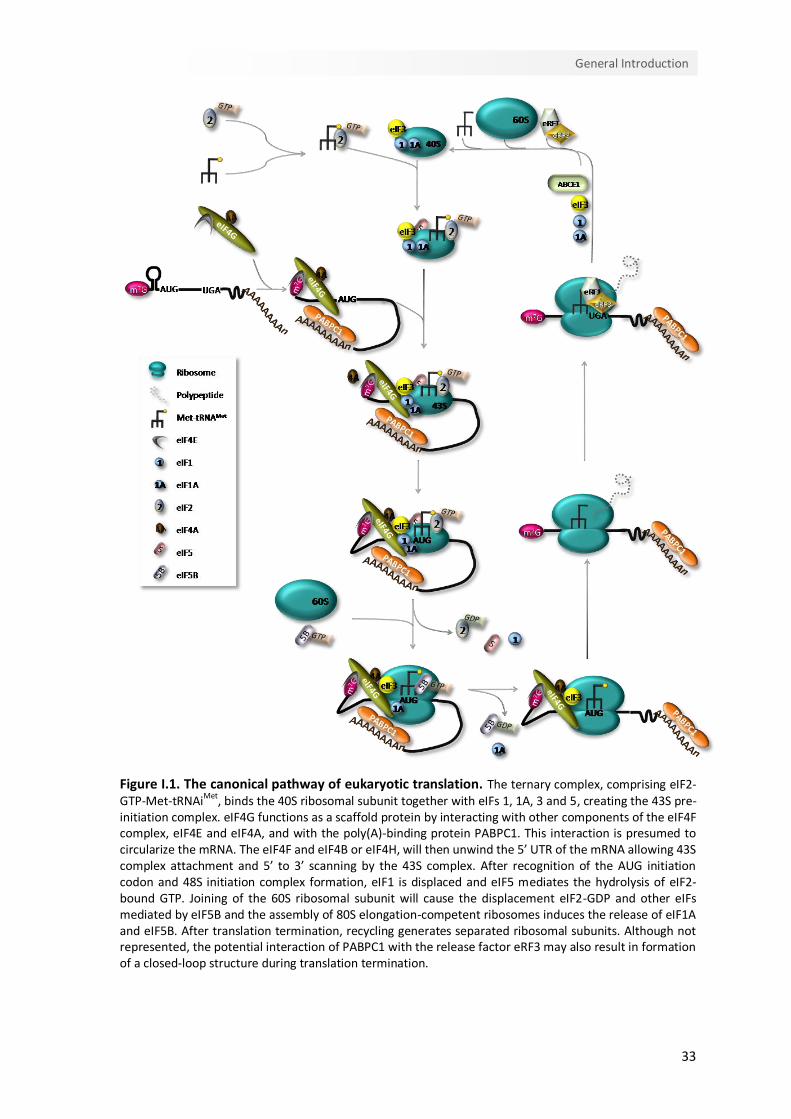
each of which requires a particular set of conditions and factors (Figure I.1) (Sonenberg
& Hinnebusch, 2009). Translation initiation is the rate-limiting step and requires the
function of several eukaryotic translation initiation factors (eIFs) (Livingstone et al,
2010). This complex event involves the assembly of elongation-competent 80S
ribosomes at the initiation codon (AUG). On most mRNAs, the start codon is identified
by a scanning mechanism, where the 43S pre-initiation complex binds to the mRNA near
the 5’ end and scans the 5’ untranslated region (5’UTR) for an AUG codon. The 43S pre-
initiation complex comprises the small (40S) ribosomal subunit, the eIFs 3, 1, 1A and 5,
and a ternary complex, which consists of the methionine-loaded initiator tRNA (Met-
tRNAiMet) and eIF2 coupled to GTP (Gebauer & Hentze, 2004). Binding of this complex to
the mRNA requires the cooperative action of eIF4F and eIF4B or eIF4H, which unwind
the 5’UTR of the mRNA allowing ribosomal attachment. eIF4F is composed by eIF4E
protein, that binds the m7G cap structure, the DEAD-box RNA helicase eIF4A and by
eIF4G, which functions as a scaffold protein by interacting with eIF4E, eIF4A, eIF3 and
with the cytoplasmic poly(A)-binding protein 1 (PABPC1) (Jackson et al, 2010). The
simultaneous interaction of eIF4E and PABP with eIF4G is believed to circularize the
mRNA, bringing the 3’UTR in close proximity to the 5’end of the mRNA (Wells et al,
1998). The functional connection between the mRNA ends during translation is
highlighted by the fact that most known regulatory sequences are found within the
3’UTR (Gebauer & Hentze, 2004).
After loading to the mRNA, 43S complex scans downstream to the initiation codon. The
scanning process is assisted by eIF4A, eIF4G, eIF4B and possibly by eIF3 (Jackson et al,
2010; Pestova & Kolupaeva, 2002). eIF3 plays a key role in recruiting the 43S pre-
initiation complex to the mRNA and, at least in mammals, also forms a bridge to the
mRNA by interacting with eIF4G (Hinnebusch, 2006; Pestova, 2007).
33
General Introduction
Figure I.1. The canonical pathway of eukaryotic translation. The ternary complex, comprising eIF2-GTP-Met-tRNAiMet, binds the 40S ribosomal subunit together with eIFs 1, 1A, 3 and 5, creating the 43S pre-initiation complex. eIF4G functions as a scaffold protein by interacting with other components of the eIF4F complex, eIF4E and eIF4A, and with the poly(A)-binding protein PABPC1. This interaction is presumed to circularize the mRNA. The eIF4F and eIF4B or eIF4H, will then unwind the 5’ UTR of the mRNA allowing 43S complex attachment and 5’ to 3’ scanning by the 43S complex. After recognition of the AUG initiation codon and 48S initiation complex formation, eIF1 is displaced and eIF5 mediates the hydrolysis of eIF2-bound GTP. Joining of the 60S ribosomal subunit will cause the displacement eIF2-GDP and other eIFs mediated by eIF5B and the assembly of 80S elongation-competent ribosomes induces the release of eIF1A and eIF5B. After translation termination, recycling generates separated ribosomal subunits. Although not represented, the potential interaction of PABPC1 with the release factor eRF3 may also result in formation of a closed-loop structure during translation termination.
34
Chapter I
eIF1 has a crucial role in the selection of initiation codons, allowing scanning 43S
complexes to discriminate against codon-anticodon mismatches and preventing
premature eIF5-induced hydrolysis of eIF2-GTP and Pi release (Holcik & Pestova, 2007).
eIF1A also regulates start codon selection promoting continued scanning at non-AUG
codons or by arresting scanning and promoting eIF1 release at AUG codons (Sonenberg
& Hinnebusch, 2009). The Met-tRNAiMet is anchored to the 43S complex by the GTP-
bound form of eIF2, and a perfect match with an AUG start codon [usually the first AUG
triplet in an optimum context – GCC(A/G)CCAUGG, with a purine at the -3 and a G at the
+4 positions (relative to the A of the AUG codon, which is designated +1) (Kozak, 1991)]
triggers the arrest of scanning and irreversible hydrolysis of GTP in the eIF2-GTP-Met-
tRNAi ternary complex. This step, during which the ribosome becomes committed to
initiation at that codon, is catalyzed by eIF5, an eIF2-specific GTPase-activating protein
(Gebauer & Hentze, 2004; Jackson et al, 2010). With the release of eIF2-GDP and other
eIFs, mediated by eIF5B, the large (60S) subunit joins to form an 80S initiation complex
ready to accept the appropriate aminoacyl-tRNA into the A- (aminoacyl) site and
synthesize the first peptide bond (Pestova, 2007). Because eIF3 binds mainly to the
solvent side of the 40S subunit, the dissociation of this factor is not essential for subunit
joining and may thus be delayed (Szamecz et al, 2008; Valasek et al, 2002).
During the elongation stage, amino acids are added sequentially to the growing
polypeptide chain (Abbott & Proud, 2004). The ribosome has three tRNA-binding sites:
the A-site, which accepts the incoming aminoacyl-tRNA, the P- (peptidyl) site, which
holds the tRNA with the nascent peptide chain and the E- (exit) site that holds the
deacylated tRNA before it leaves the ribosome (Proud, 1994). The key factor in this
process is the elongation factor (eEF) 1A which delivers cognate tRNA to the A-site, upon
hydrolysis of GTP. The nascent peptide chain is then transferred to the amino acid of the
A-site aminoacyl-tRNA. The translocation of peptidyl-tRNA from A- to P- and of
deacylated tRNA from P- to E- sites is then promoted by eEF2; this process is also
dependent on GTP hydrolysis (Abbott & Proud, 2004; Pisarev et al, 2007).
Termination of translation is triggered by peptide release factors eRF1 and eRF3. When a
stop codon enters the A-site, eRF1 induces the hydrolysis of the ester bond of the P-site
peptidyl-tRNA (Pestova, 2007). The function of eRF3 is to ensure the rapid and efficient
hydrolysis of the peptidyl-tRNA by eRF1 (Alkalaeva et al, 2006). It is suggested that eRF1,

35
General Introduction
eRF3 and GTP bind the pre-termination complexes as a ternary complex. However, the
mechanism of post-termination dissociation of the release factors is unknown (Pisarev
et al, 2007).
After translation termination, the mRNA and P-site deacylated tRNA remain associated
with ribosomes in post-termination complexes. eIFs 3, 1, 1A and 3j cooperatively
dissociate such complexes into free 60S subunits and mRNA- and tRNA-bound 40S
subunits (Pisarev et al, 2007). According to the model proposed by Pisarev et al. (2007),
eIF1 promotes dissociation of P-site deacylated and eIF3j mediates release of mRNA.
Ribosomal recycling might influence post-termination events such as reinitiation and
possibly mRNA decay pathways.
Reinitiation may occur after translation of an upstream open reading frame (uORF), if
the 40S ribosomal subunit remains bound to the mRNA molecule upon translation
termination. Following translation-termination and the consequent release of the 60S
ribosomal subunit, the 40S subunit may resume scanning downstream from the uORF,
exhibiting a conditional loss of reinitiation competence. The de novo recruitment of the
eIF2-GTP-Met-tRNAiMet ternary complex, that will allow recognizing the AUG of the main
ORF, determines the acquirement of full reinitiation competence (Kozak, 1987; Kozak,
2001). The reinitiation efficiency depends on i) cis-acting mRNA features; ii) the time
required for the uORF translation, which is determined by the relative length of the
uORF and the translation elongation rate; and iii) the translation initiation factors
involved in the translation initiation event (Kozak, 2001; Poyry et al, 2004). It has been
hypothesized that the eIFs involved in resuming scanning remain at least transiently
associated with the elongating ribosome, and increasing the uORF length or the
ribosome transit time increases the odds of these factors dissociate (Kozak, 2001).
Corroborating this hypothesis, in yeast, eIF3 remains 80S-bound for several rounds of
elongation and enhances the reinitiation competence of post-termination 40S
ribosomes (Szamecz et al, 2008).
I.1.2. Translation regulation
Global control of protein synthesis is mostly exerted at the stage of translation initiation.
The mechanisms regulating initiation may impact on the eIFs or ribosomes, affecting
36
Chapter I
scanning-dependent mechanisms; or may impact directly on the mRNA, through
sequence-specific RNA-binding proteins or microRNAs.
The best well-known examples of regulation mechanisms impacting on the eIFs are the
control of the availability of active eIF2 and eIF4F by reversible protein phosphorylation.
Phosphorylation of the α subunit of eIF2 at Ser51 blocks the GTP-exchange reaction by
reducing the dissociation rate of eIF2 from eIF2B (Gebauer & Hentze, 2004). There are
four mammalian protein kinases that phosphorylate eIF2α at Ser51 decreasing global
translation: haem-regulated kinase, which is stimulated by haem depletion; PKR (protein
kinase activated by double-stranded RNAs), which is activated by viral infection; GCN2
(general control non-derepressible-2), which is activated by amino-acid starvation; and
PERK (PKR-like endoplasmic reticulum kinase), which is activated under circumstances of
endoplasmic reticulum stress (Jackson et al, 2010). The availability of the cap-binding
protein eIF4E is also used to regulate general translation rates. When
hypophosphorylated, a eIF4E-binding protein (4E-BP) binds eIF4E competitively
displacing eIF4G; but phosphorylation of the 4E-BP at multiple sites (mainly by mTOR)
releases eIF4E for assimilation into eIF4F, thus promoting translation initiation (Gebauer
& Hentze, 2004). The phosphorylation of several other eIFs (eIF1, eIF2β, several eIF3
subunits, eIF4G, eIF4B, eIF4H, eIF5 and eIF5B) and of ribosomal protein 6 (RPS6)
increasing under conditions in which translation is activated has also been described.
However, there is no concrete evidence that these phosphorylation events are the cause
of such activation (Jackson et al, 2010).
Regulation of translation initiation through the mRNA itself, by a sequence-specific RNA-
binding protein, is exclusive of the mRNAs that contain the relevant RNA sequence motif
and is generally inhibitory, with the activation of such mRNAs requiring the inactivation
or the degradation of the inhibitory protein. The regulation by specific 5’UTR-protein
interactions is surprisingly rare, being the ferritin mRNAs the only well-studied example.
In contrast, there are several cases, most of them important in development, of control
by 3’UTR-protein interactions (Jackson et al, 2010).
While the mentioned protein-RNA interactions usually result in decreased translation,
the binding of PABPC1 to the poly(A) tail stimulates translation (Borman et al, 2000).
This stimulatory effect appears to result mainly from the ability of PABPC1 to interact
with eIF4G, mediating mRNA circularization by linking the cap and the poly(A) tail
37
General Introduction
(Imataka et al, 1998; Wells et al, 1998). By interacting with eIF4G, PABPC1 enhances
eIF4F binding to the cap of the mRNA (Kahvejian et al, 2005) and, therefore, stimulates
mRNA binding do the 43S pre-initiation complex (Pestova, 2007). In fact, an in vitro
study showed that the formation of the closed-loop occurs before or during the 48S
complex assembly, independently of a prior termination event (Amrani et al, 2008). The
formation of a second closed-loop mRNP, comprising PABPC1, eIF4G, eIF4E, eIF3 and the
termination factors eRF1 and eRF3, was also described during the subunit joining to
form the 80S ribosome (Amrani et al, 2008). The formation of such a circular
conformation could facilitate reinitiation by post-termination ribosomes. Moreover,
PABP also stimulates 60S subunit joining (Kahvejian et al, 2005) and is required to block
the inhibitory effect of RNA helicases on subunit joining factor eIF5B (Searfoss et al,
2001).
I.2. Nonsense-mediated mRNA decay as a surveillance mechanism
The best characterized mRNA quality control system in eukaryotes is nonsense-
mediated mRNA decay (NMD). This surveillance mechanism detects and ensures the
rapid degradation of faulty mRNAs harboring premature translation termination codons
(PTCs) that would otherwise result in the synthesis of C-terminally truncated proteins.
Hence, the quality control function of NMD relies in protecting the cell from the
deleterious dominant-negative or gain-of-function effects of these truncated proteins
(Behm-Ansmant & Izaurralde, 2006; Chang et al, 2007).
I.2.1. Aberrant and natural NMD targets
Cells produce a large number of faulty mRNAs harboring PTCs that are recognized and
rapidly eliminated by NMD. PTC-harboring transcripts can arise at the DNA level as the
result of germline or somatic alterations (direct nonsense-mutations; frame-shifting
deletions and insertions that can lead to PTCs downstream of the shift; mutations within
either an intron or an exon that results in inaccurate intron removal from the pre-mRNA
and create an intron-derived PTC or a frameshift) or at the RNA level due to errors in
transcription or pre-mRNA processing (Kuzmiak & Maquat, 2006; Stalder & Muhlemann,

38
Chapter I
2008). Other types of faulty mRNAs are transcripts from non-functional pseudogenes,
endogenous retroviral and transposon RNAs or mRNA-like non-protein coding RNAs
from intergenic regions (Mendell et al, 2004; Nicholson et al, 2010; Rehwinkel et al,
2006). By targeting these transcripts for decay, NMD exerts a role in reducing genomic
noise.
In addition, PTCs can also be generated by non-faulty regulated processes of the mRNA
metabolism, such as somatic rearrangements in the DNA, alternative splicing or
utilization of alternative AUG initiation sites that lead to a shift in the main ORF
(Holbrook et al, 2004; Kuzmiak & Maquat, 2006). In fact, unproductive alternative
splicing is believed to constitute the major source of PTC-containing mRNAs in
mammals, since bioinformatic approaches studies suggested that about one-third of the
alternatively spliced human mRNAs contain a PTC that is targeted for NMD (Lewis et al,
2003); and, at least for same cases, it constitutes a form of autoregulation of the
expression of the canonical protein-encoding isoforms, as the protein products of these
genes are responsible for NMD triggering (Neu-Yilik & Kulozik, 2008). Thus, besides
ensuring the degradation of faulty transcripts, NMD also has a crucial role in regulating
gene expression. Indeed, genome-wide screens in NMD-deficient budding yeast, fruitfly
and human cells have revealed that NMD controls the abundance of 3% - 10% of the
transcriptome (He et al, 2003; Mendell et al, 2004; Rehwinkel et al, 2005; Rehwinkel et
al, 2006). Examples of natural NMD targets include transcripts containing a regulatory
ORF that resides upstream of the primary downstream ORF (uORFs); introns in the 3’
UTR; programmed frameshifts; and long 3’ UTRs (Kuzmiak & Maquat, 2006; Nicholson et
al, 2010). Notably, mRNAs containing UGA triplets that direct selenocysteine
incorporation can also elicit NMD, since the UGA codon can be interpreted as a PTC
when the selenium concentration in the cell is low [reviwed in (Bhuvanagiri et al, 2010;
Nicholson et al, 2010)]. The process of maturation of the immune system is other
example of the physiological role of NMD. This programmed somatic rearrangements
and hypermutations generate PTCs at a high frequency (Kuzmiak & Maquat, 2006;
Nicholson et al, 2010).
Remarkably, many of the identified natural NMD targets encode proteins that could be
grouped in clusters acting in related pathways (Neu-Yilik et al, 2004; Weischenfeldt et al,
2005). Such mRNAs are involved in a variety of cellular processes such as stress
39
General Introduction
responses, hematopoietic stem cell development, regulation of alternative splice forms,
genomic stability, cell-cycle, telomere length maintenance and embryonic development
(Bhuvanagiri et al, 2010; Stalder & Muhlemann, 2008).
I.2.2. Nonsense-mediated mRNA decay implications in disease
The biological and medical significance of the NMD pathway is evidenced by the fact
that approximately 30% of all inherited genetic disorders are due to PTCs, and in many
of these cases, NMD influences the severity of the clinical phenotype (Holbrook et al,
2004; Stalder & Muhlemann, 2008). In most cases, NMD has a beneficial effect by
eliminating transcripts harboring PTCs that would otherwise give rise to truncated
proteins either with a complete loss of function or with a dominant-negative function
leading to toxicity (Bhuvanagiri et al, 2010). The benefits that can be achieved by the
elimination of mRNAs encoding these faulty proteins are clearly illustrated in β-
thalassemia (Frischmeyer & Dietz, 1999). β-Thalassemia is characterized by the absence
or reduction in the synthesis of β-globin polypeptide chains, one of the hemoglobin
subunits (Weatherall, 2000). This disorder exemplifies the phenotypic impact of the
polar effect of PTCs at different positions between the same β-globin gene (Holbrook et
al, 2004). If a PTC is located at a position that activates NMD, the disease results in a
recessive mode of inheritance with the heterozygous being asymptomatic (Figure I.2). In
this case the defective β-globin transcript is degraded by NMD resulting in a limited
synthesis of the truncated product. The excess of free α-globin, as well as the limited
amount of truncated β-globin protein (if translated) are proteolytically degraded.
Indeed, it has been shown that these defects result in very low amounts of β-globin
chain production from the affected allele causing a reduction about 50% of total β-
globin chain synthesis in the heterozygote. This produces the clinically asymptomatic
phenotype of β-thalassemia trait (also known as thalassemia minor), as heterozygotes
usually synthesize enough β-globin from the remaining normal allele to support near-
normal haemoglobin levels (Holbrook et al, 2004; Peixeiro et al, 2011b). If the PTC is
located at a position that does not induce NMD, at less than 55 nucleotides (nts)
upstream the last exon-exon junction or at the 5’ part of exon 3, the amount of
truncated protein that is synthesized might be small enough to be efficiently degraded

40
Chapter I
by the proteolytic system of the red blood cell, together with the excess of free α-globin
chains. In this case, the heterozygotes are also asymptomatic and the disease still results
in a recessive mode of inheritance (Figure I.2). In contrast, if the PTC is located further
downstream at exon 3 of the β-globin mRNA, NMD is not triggered; instead, substantial
amounts of mutant β-globin mRNA are present in the patients, encoding for translation
products that are long enough to overburden the cellular proteolytic system (Figure I.2).
Under these conditions, accumulation of the truncated products, as well as free α-globin
chains in excess, act in a dominant negative manner, leading to deleterious effects on
the cell, as the nonfunctional globin chains cause toxic precipitation of insoluble globins
(Holbrook et al, 2004; Neu-Yilik & Kulozik, 2008; Peixeiro et al, 2011b). This condition is
related to a symptomatic form of the disease in heterozygotes named “thalassemia
intermedia” with a dominant mode of inheritance (Hall & Thein, 1994).
Figure I.2. Nonsense-mediated mRNA decay modulates the disease phenotype. The example here presented shows the human β-globin mRNA. AUG-proximal PTCs do not trigger NMD but heterozygotes are asymptomatic, as the translated short β-globin peptides, along with the β-globin chains in excess, are effectively degraded. If the PTC is located downstream of codon 23 and more than 55 nucleotides (nt) upstream of the last exon-exon junction, the corresponding transcript is targeted for NMD and heterozygotes are also protected from thalassemia. In contrast, transcripts bearing PTCs located less than 55 nt upstream of the last exon-exon junction, or in the 5’-part of exon 3, escape NMD, resulting in the production of truncated proteins that are small enough to be efficiently degraded, along with the α-globin chains in excess, and heterozygotes are also asymptomatic for β-thalassemia. However, if the PTC is located further downstream, the encoded truncated nonfunctional β-globin proteins overwhelm the cellular proteolytic system and cause toxic precipitation of insoluble globin chains. Heterozygotes with these mutations are affected with dominantly inherited β-thalassemia intermedia.
Other examples of the protective impact of NMD and of its disease-modulator function
are retinal degeneration, von Willebrand disease, myotonia congenital and factor X
41
General Introduction
deficiency (Behm-Ansmant et al, 2007b; Khajavi et al, 2006). A potential influence of
NMD in cancer has also been suggested. In fact, transcripts of several mutant forms of
the tumor suppressor proteins breast cancer 1 (BRCA1), p53 and Wilms tumor (WT1)
have been shown to be eliminated by NMD. The targeting for degradation of these
nonsense-transcripts that would convert the tumor suppressors into dominant-negative
oncoproteins, protects the heterozygous carriers from developing cancer (Behm-
Ansmant et al, 2007b; Bhuvanagiri et al, 2010).
However, it is important to note that NMD can also aggravate the disease phenotype by
eliminating mRNAs that would otherwise support the synthesis of partially functional
proteins, leading to haploinsufficiency (Khajavi et al, 2006). Examples of the detrimental
effect of NMD are cystic fibrosis, Hurler syndrome and X-linked nephrogenic diabetes
insipidus (Neu-Yilik & Kulozik, 2008). The clinical picture of protein deficiency induced by
NMD is also clearly illustrated in Duchenne muscular dystrophy (DMD), where the rare
truncating mutations that occur near the 3’ end of dystrophin gene result in variable
mild phenotypes while the 5’ PTCs are associated with a severe form of DMD and fail to
rescue the phenotype as a consequence of NMD triggering (Khajavi et al, 2006). In these
cases, where NMD has such a detrimental effect, therapeutics that specifically
suppresses premature translation termination by promoting translational readthrough
would be clinically useful (Holbrook et al, 2004; Kuzmiak & Maquat, 2006; Neu-Yilik &
Kulozik, 2008). The use of aminoglycosides as a therapeutic approach has been tested in
the treatment of the above mentioned genetic disorders. The aminoglycoside antibiotics
bind to the decoding center of the ribosome and reduce the accuracy requirements for
the codon-anticodon pairing, promoting the incorporation of an amino-acid at the stop
codon (Eustice & Wilhelm, 1984), generating functionally active, although missense-
mutated, proteins. This therapeutic strategy was tested by using gentamicin in clinical
trials performed in patients with DMD, Becker muscular dystrophy (Wagner et al, 2001)
and cystic fibrosis (Clancy et al, 2001), showing that these antibiotics can promote in
vivo readthrough of nonsense mutations and lead to the expression of full-length
proteins. However, the variability in the response to the aminoglycoside treatment, the
side-effects (such as kidney damage), and the fact that these suppressing agents may
lead to a general loss in the fidelity of translation termination by promoting nonspecific
42
Chapter I
readthrough of normal termination codons, limited the use of its application
(Bhuvanagiri et al, 2010; Frischmeyer & Dietz, 1999).
Recently, PTC124, a new drug similar to gentamicin, has been shown to effectively
promote the selective readthrough of nonsense codons without interfering with
translation termination at the normal stop codon. Unlike aminoglycosides, PTC124 is
efficient at very low dosages and its side effects appear to be rare and mild (Welch et al,
2007), which makes this new drug a promising clinical application for the treatment of
PTC-associated genetic disorders.
The elimination of the PTC-carrying portion of a nonsense-mutated transcript
constitutes another viable approach for the treatment of NMD-associated diseases. One
example of these repairing strategies is the use of antisense oligoribonucleotides to
redirect splicing avoiding the creation of PTCs and it was first implemented to correct
aberrant splicing of the β-globin gene (Dominski & Kole, 1993). The major drawback of
this strategy relates to the lack a proper delivery system, issues of transfection efficiency
and undesired side effects (Bhuvanagiri et al, 2010).
Biological evidence suggests that individuals with similar genetic mutations may exhibit
different phenotypic severities as a result of variability in NMD efficiency (Kerr et al,
2001). This points out the modulation of NMD itself as another potential therapeutic
strategy, by stimulation or inhibition of this decay pathway, rather than modulating
recognition of PTCs. Even though this could benefit cases where the truncated proteins
retain a certain level of function, the variety of functions of NMD and its cofactors in
RNA surveillance and gene expression makes very challenging to identify a strategy to
attack NMD systemically without undesired side-effects (Bhuvanagiri et al, 2010;
Holbrook et al, 2004). Therefore, a balance between the potential side-effects and
efficacy of these drugs will have to be found for therapeutic strategies targeting the
physiological NMD function.
I.2.3. Molecular basis of the NMD pathway
NMD was first described in 1979 through the observation that in Saccharomyces
cerevisiae (S. cerevisiae) nonsense mutations in the Ura3 resulted in reduced amount of
the affected transcript (Losson & Lacroute, 1979) and that the absence of human β-
43
General Introduction
globin expression in β0-thalassemic patients was due to the presence of PTCs within the
coding sequence of the transcripts (Chang & Kan, 1979). Subsequent studies revealed
NMD as a well phylogenetic conserved surveillance mechanism by documenting that the
fast degradation of nonsense-mutated organisms is common to other organisms such as
Caenorhabditis elegans (C. elegans) (Pulak & Anderson, 1993), Drosophila melanogaster
(D. melanogaster) (Brogna, 1999) and plants (Jofuku et al, 1989; Voelker et al, 1990).
Three key steps constitute the NMD pathway: i) the identification of the PTC; ii) the
assembly of the surveillance complex on the mRNA and iii) the degradation of the NMD
target. The mechanism by which premature stop codons are recognized and
discriminated from natural stop codons is translation-dependent and leads to the
recruitment of NMD trans-acting factors. Once assembled, the surveillance complex
interacts with eukaryotic translation termination factors, eRF1 and eRF3, and triggers
the fast degradation of the aberrant mRNA (Behm-Ansmant et al, 2007b; Conti &
Izaurralde, 2005).
The NMD machinery comprises three trans-acting factors designated up-frameshift
(UPF) proteins and seven proteins designated SMGs (for suppressor of morphological
defects on genitalia). These effectors were first identified through genetic screens in S.
cerevisiae and C. elegans and were later identified in other organisms by homology
searches (Amrani et al, 2006b; Cali et al, 1999; Conti & Izaurralde, 2005; Hodgkin et al,
1989; Leeds et al, 1991; Leeds et al, 1992; Pulak & Anderson, 1993). The UPF1, UPF2 and
UPF3 yeast factors, homologous of C. elegans SMG2, SMG3 and SMG4, respectively, are
essential for NMD and orthologues are present in all eukaryotes. The SMG1, SMG5,
SMG6 and SMG7 are conserved in almost all high eukaryotes, with the exception of D.
melanogaster, which contains no clear orthologue for SMG7. Homologues of SMG1,
SMG5 and SMG6 as regulators of NMD are not present in S. cerevisiae (Behm-Ansmant
et al, 2007b; Muhlemann, 2008; Neu-Yilik & Kulozik, 2008; Nicholson et al, 2010). In
human cells, the NMD pathway comprises the factors UPF1, UPF2 and UPF3A and UPF3B
and the factors SMG1, SMG5, SMG6 and SMG7 (Culbertson & Leeds, 2003). However,
additional NMD effectors are likely to exist. Indeed, recently, new NMD factors have
been described: NAG and DHX34, the human homologous of C. elegans SMGL-1 and
SMGL-2 (Longman et al, 2007); SMG8 and SMG9, which were shown to regulate the

44
Chapter I
SMG1 kinase activity (Yamashita et al, 2009) and RUVBL1 and RUVBL2, which also
regulate SMG1 (Izumi et al, 2010).
I.2.3.1. UPFs: the core of the NMD machinery
UPF1 is the most conserved NMD factor and appears to be a key factor of this
surveillance pathway in all organisms (Culbertson & Leeds, 2003). UPF1 is an RNA
helicase and nucleic-acid-dependent ATPase. The conserved central region of this
complex phosphoprotein harbors two cysteine-rich zinc-fingers and seven group I
helicase motifs, two of which are responsible for the ATPase activity. The ATPase activity
is essential for NMD in yeast and humans and is linked to the ATP-dependent 5’ to 3’
helicase activity (Bhattacharya et al, 2000; Chang et al, 2007). In spite of being primarily
a cytoplasmic protein, UPF1 has been shown to shuttle between the nucleus and the
cytoplasm (Mendell et al, 2002). UPF1 interacts with UPF2 through its N-terminal
cysteine- and histidine-rich domains (CH), which forms three zinc-binding motifs
arranged in two tandem modules (Kadlec et al, 2006). UPF1 also interacts with the
termination factors eRF1 and eRF3 (Czaplinski et al, 1998; Ivanov et al, 2008) and
immunoprecipitation studies revealed an association of UPF1 with SMG1, eRF1 and eRF3
that constitutes the SURF complex (Kashima et al, 2006). In higher eukaryotes, UPF1
activity is regulated by phosphorylation/dephosphorylation cycles (Ohnishi et al, 2003).
Phosphorylation of its serine-glutamine C-terminal motifs is catalyzed by SMG1 and
requires UPF2 and UPF3 (Grimson et al, 2004; Kashima et al, 2006; Page et al, 1999;
Yamashita et al, 2001). Nevertheless, recent studies have provided evidence for the
existence of UPF2-independent and UPF3-independent NMD pathways (Chan et al,
2007; Gehring et al, 2005; Ivanov et al, 2008). SMG5, SMG6 and SMG7 mediate the
dephosphorylation of phospho-UPF1 by the protein phosphatase 2A (PP2A) (Anders et
al, 2003; Chiu et al, 2003; Ohnishi et al, 2003). In coimmunoprecipitation studies, UFP1
has been found to interact with a variety of other NMD factors, such as exon-junction
complex (EJC) components, but also with other proteins with no obvious role in this
decay mechanism, such as the initiation factor eIF3, through eIF3e subunit (Isken et al,
2008; Morris et al, 2007). Moreover, a recent study showed that the cap binding protein
CBP80 at the 5’ mRNA end chaperones SMG1 and UPF1 to an EJC, thus promoting NMD
45
General Introduction
by augmenting UPF1-UPF2 (presumably EJC-bound UPF2) interaction (Hwang et al,
2011).
UPF2 is also a phosphoprotein and functions as bridge between UPF1 and UPF3 (Chiu et
al, 2003; Lykke-Andersen et al, 2000; Mendell et al, 2000). The interaction of UPF2 with
UPF3 involves the highly conserved residues in the last three middle of eIF4G-like
(MIF4G) domains of UPF2 and the β-sheet surface of the RNA-binding domain of UPF3B
(Kadlec et al, 2004). Recently, a structural study determined the precise interaction of
the C-terminal region of UPF2 with the CH domain of UPF1 (Clerici et al, 2009). UPF2
interacts with UPF1 mainly through its C-terminal region (amino acids 1084-1272) but
the amino acids 94-133 from the N-terminal domain also contribute to this interaction
(Serin et al, 2001). It is not yet clear if the UPF2- and eRF3-binding sites in UPF1 overlap,
but the bipartite nature of UPF2-binding may allow to UPF2 and eRF3 simultaneously
bind UPF1 in an initial interaction (Clerici et al, 2009). Albeit the presence of nuclear
localization signals in the N-terminal domain of UPF2, this protein localizes
predominantly in the cytoplasm. Besides the function in bridging UPF1 to UPF3, UPF2
also plays a role in UPF1 phosphorylation (Wittmann et al, 2006). Indeed,
immunoprecipitation assays showed that UPF2 directly binds SMG1 and is essential for
the association of SMG1 with the mRNP (Kashima et al, 2006).
The human genome encodes two UPF3 genes, designated UPF3A (on chromosome 13)
and UPF3B (also known as UPF3X, as it is localized on the X chromosome) (Lykke-
Andersen et al, 2000; Serin et al, 2001). Splice variants generated by both genes have
been described. UPF3AL and UPF3AS retain or skip exon 4, respectively, and UPF3B
isoforms are generated by alternatively splicing exon 8 (Lykke-Andersen et al, 2000;
Serin et al, 2001). All UPF3 isoforms, components of the EJC (Le Hir et al, 2001), shuttle
between the nucleus and the cytoplasm, although their predominantly location resides
in the nucleus (Lykke-Andersen et al, 2000). The N-terminal RNA-binding domain of
these proteins is responsible for the interaction with UPF2 and is absent in the UPF3AS
isoform (Kadlec et al, 2004). The C-terminal domains, involved in the assemble of the
complex containing the EJC proteins Y14 and MAGOH (Gehring et al, 2003), from UPF3A
and UPF3B are considerably divergent. This divergence explains the weaker ability of
UPF3A, compared to UPF3B, to trigger NMD (Kunz et al, 2006). Evidence that UPF3A is
an NMD factor regulated by UPF3B and UPF2 has emerged recently (Chan et al, 2009).
46
Chapter I
Chan and colleagues (2009) showed that, as a consequence of competitive binding of
UPF3A and UPF3B for UPF2, when UPF3B levels are low, UPF3A is able to bind UPF2.
Conversely, when UPF3B levels are high, less UPF3A molecules can bind UPF2, which
results in decreased UPF3A levels, since UPF3A alone is intrinsically unstable. The UPF3A
proteins appear to specify two UPF1-containing complexes, one containing phospho-
UPF1, UPF2, UPF3B and UPF3AL, and the other containing phospho-UPF1, UPF3B, SMG5,
SMG7 and UPF3AS (Ohnishi et al, 2003). The exchange between the two UPF3A isoforms
might represent the switch between a phosphorylation and a pre-dephosphorylation
UPF1-complex, based on the hypothesis that phospho-UPF1 recruits the SMG5-SMG7
complex that together with PP2A and UPF3AS induce dephosphorylation of UPF1
(Anders et al, 2003; Ohnishi et al, 2003). Moreover, a role for these isoforms in
translation termination at the PTC, instead of in the degradation phase of NMD, is likely
since there is evidence that UPF3A proteins interact with a UPF1-eRF1-ERF3 complex
even when the binding of UPF2, UPF3B and EJC-components to UPF1 is abrogated
(Kashima et al, 2006).
I.2.3.2. SMGs - determining the phosphorylation status of UPF1
As above mentioned, SMG1 and SMG5-7 control the phosphorylation status of UPF1
(Wilkinson, 2003). SMG1 is a protein kinase that phosphorylates specifically serine and
threonine residues and belongs to the phosphatidylinositol 3-kinase-related protein
(PIKK) family (Yamashita et al, 2001). A recent report showed that the novel identified
NMD factors SMG8 and SMG9 assemble a complex with SMG1 and suppress SMG1
kinase activity in this isolated complex. SMG8 protein is further required to the
recruitment of SMG1 to form the SURF complex (Yamashita et al, 2009) and SMG9
indirectly regulates SMG1 through the recruitment of SMG8 to the N-terminal region of
SMG1 (Arias-Palomo et al, 2011). In addition to the SURF complex, SMG1 presence on
other multiprotein complexes was revealed by immunoprecipitations assays. SMG1
interacts with the EJC components eIF4A, Y14 and MAGOH and with the NMD factors
UPF1, UPF2, UPF3B and SMG7 (Kashima et al, 2006).
C. elegans mutants for SMG5, SMG6 or SMG7 accumulate hyperphosphorylated SMG2
suggesting that these proteins are responsible for UPF1 dephosphorylation. SMG5,
SMG6 and SMG7 share a common 14-3-3-like domain, which is usually associated with
47
General Introduction
protein-protein interactions. This domain in SMG7 is thought to promote the
dephosphorylation of UPF1 by binding phospho-UPF1 (Fukuhara et al, 2005) and also
contributes to an interaction with the 14-3-3-like domain of SMG5 (Anders et al, 2003;
Unterholzner & Izaurralde, 2004). Tethering of SMG7 to a reporter mRNA is sufficient to
trigger decay even in the absence of a PTC and the NMD factors UPF1, SMG5 and SMG6,
indicating that SMG7 functions at a late step in NMD (Unterholzner & Izaurralde, 2004).
SMG5 also interacts with phospho-UPF1 and PP2A, which might give PP2A specificity for
UPF1 (Anders et al, 2003). SMG5 and SMG6 contain a C-terminal PIN (for PilT N
terminus) domain which characteristically exhibits nuclease activity. However, only the
SMG6 has the triad of acidic residues for the RNase H activity (Glavan et al, 2006).
Actually, studies in D. melanogaster, zebrafish and human cells showed that the
endonuclease activity of SMG6 can initiate cleavage of NMD targets (Eberle et al, 2009;
Gatfield & Izaurralde, 2004; Huntzinger et al, 2008). The requirement of the 14-3-3-like
domain of SMG6 in NMD was recently revealed by a study suggesting that the N-
terminal domain of this protein is required for association with other NMD factors and
also mediates binding to the EJC (Kashima et al, 2010). Accordingly, SMG6 was shown to
co-purify with PP2A, SMG1, UPF1, UPF2 and UPF3B (Chiu et al, 2003). As the N-terminal
domain of SMG6 contains conserved motifs related to a C-terminal motif present in
UPF3B that is responsible for binding to the EJC (Buchwald et al, 2010; Gehring et al,
2003), UPF3B may block SMG6 recruitment to mRNAs lacking PTCs. However, the
assembly of the surveillance complex with phospho-UPF1 on mRNAs harboring PTCs
could result in the replacement of UPF3B for the shorter isoform UPF3AS with low
affinity to the EJC. Such a rearrangement would confer specificity in SMG6 recruitment
to the EJC of PTC-containing transcripts (Kashima et al, 2010).
I.2.3.3. Splicing and the EJC
Even earlier than the discovery of NMD effectors, studies in mammalian cells revealed
that PTCs located more than 50-54 nucleotides (nt) upstream the last exon-exon
junction were able to target the mRNA for rapid decay (Nagy & Maquat, 1998). This
evidence implicates splicing in mammalian NMD by supporting the view that PTC
recognition is dependent on the definition of exon-exon junctions.
48
Chapter I
In metazoans, during pre-mRNA splicing, the exon junction complex (EJC) assembles 20-
24 nt upstream of each exon-exon junction (Le Hir et al, 2000a). The EJC is formed by the
assembly of four conserved core proteins - Y14, MAGOH, eIF4AIII and Barentz (BTZ) –
that associate with the mRNA in the nucleus and travels with the mRNP to the cytoplasm
(Bono et al, 2006). The EJC is a dynamic structure, and therefore, additional EJC factors
(RNPS1, Pinin, SRm160, UAP56, TAP, the mRNA export-related proteins Aly/REF, the
RNA-binding protein PYM) associate with the mRNA more transiently, either by
assembling in the nucleus but dissociating before mRNA export, or by only binding the
EJC during the subsequent processes of mRNA metabolism in the cytoplasm (Chang et al,
2007; Le Hir et al, 2001; Le Hir et al, 2000a). It was recently shown that EJC deposition is
a sequential process coordinated by splicing, involving a pre-EJC as an intermediate. The
pre-EJC, which consists of eIF4AIII and Y14-MAGOH heterodimer, is assembled before
exon ligation and provides a binding platform for peripheral EJC components (Gehring et
al, 2009).
The EJC is involved in translation and mRNA transport and turnover (Nott et al, 2004;
Tange et al, 2004); and the identification of UPF2 and UPF3 as EJC components
suggested a role for this complex also in NMD (Le Hir et al, 2000a). UPF3b associates
with the EJC in the nucleus by interacting with the complex eIF4AIII:Y14-MAGOH
(Buchwald et al, 2010). The NMD-competent core EJC is accomplished by the association
of BTZ, which most likely occurs after nuclear export (Gehring et al, 2009). Supporting a
function for EJC in this surveillance mechanism, the artificially tethering of RNPS1, Y14,
MAGOH and PYM at more than 50 nt downstream the native stop codon of β-globin
transcripts resulted in NMD triggering (Bono et al, 2004; Gehring et al, 2003; Lykke-
Andersen et al, 2001). Moreover, the knockdown of Y14, eIF4AIII or BTZ by RNA
interference was shown to impair NMD activation of PTC-harboring mRNAs (Gehring et
al, 2003; Palacios et al, 2004). In contrast, studies with β-hexosaminidase, Igµ and hybrid
mouse/human β-globin transcripts revealed that NMD could be triggered without the
requirement of an EJC downstream the PTC (Buhler et al, 2006; Rajavel & Neufeld, 2001;
Zhang et al, 1998b). Additionally, wild-type Igµ minigene, β-globin or TPI transcripts with
3’UTRs extended in length were targeted by the NMD pathway (Buhler et al, 2006;
Eberle et al, 2008). Noteworthy, the EJC is dispensable for NMD in C. elegans and D.
melanogaster (Gatfield et al, 2003; Longman et al, 2007). Therefore, the EJC may have
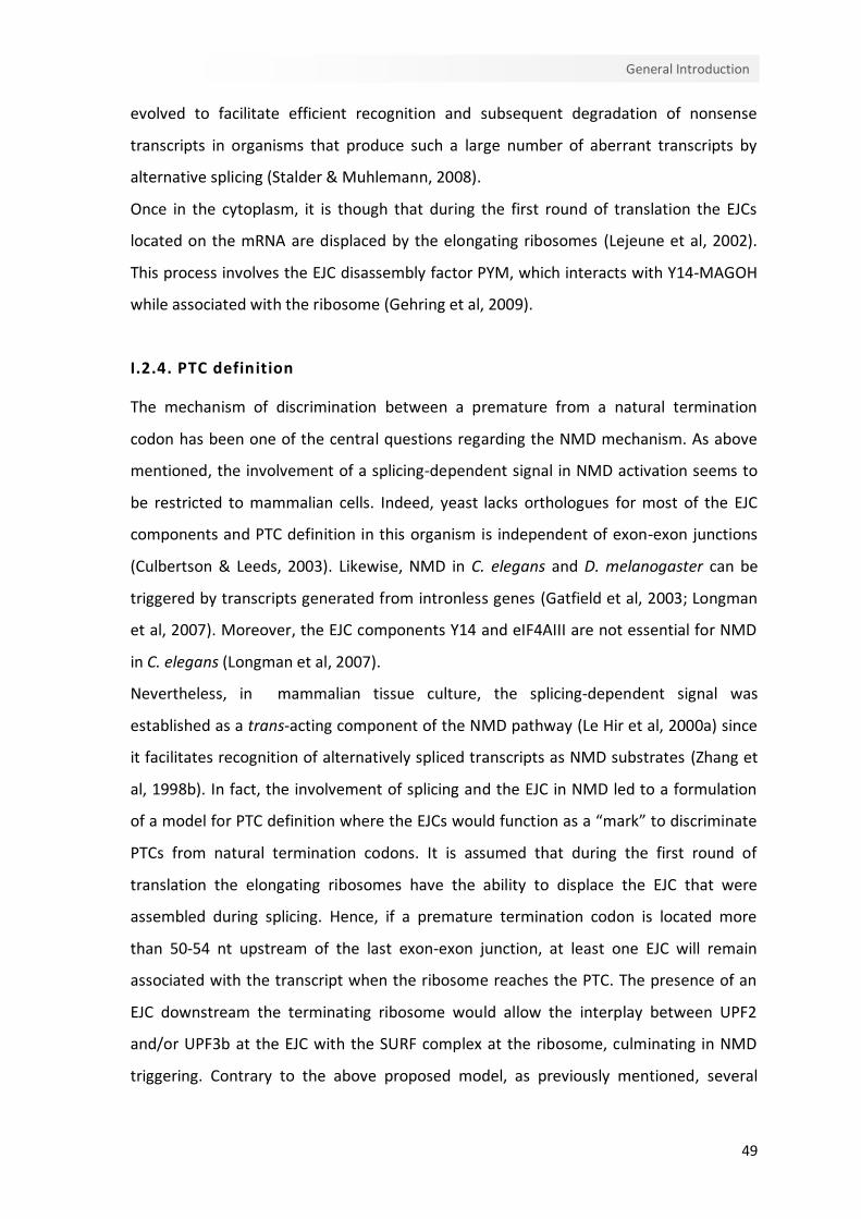
49
General Introduction
evolved to facilitate efficient recognition and subsequent degradation of nonsense
transcripts in organisms that produce such a large number of aberrant transcripts by
alternative splicing (Stalder & Muhlemann, 2008).
Once in the cytoplasm, it is though that during the first round of translation the EJCs
located on the mRNA are displaced by the elongating ribosomes (Lejeune et al, 2002).
This process involves the EJC disassembly factor PYM, which interacts with Y14-MAGOH
while associated with the ribosome (Gehring et al, 2009).
I.2.4. PTC definition
The mechanism of discrimination between a premature from a natural termination
codon has been one of the central questions regarding the NMD mechanism. As above
mentioned, the involvement of a splicing-dependent signal in NMD activation seems to
be restricted to mammalian cells. Indeed, yeast lacks orthologues for most of the EJC
components and PTC definition in this organism is independent of exon-exon junctions
(Culbertson & Leeds, 2003). Likewise, NMD in C. elegans and D. melanogaster can be
triggered by transcripts generated from intronless genes (Gatfield et al, 2003; Longman
et al, 2007). Moreover, the EJC components Y14 and eIF4AIII are not essential for NMD
in C. elegans (Longman et al, 2007).
Nevertheless, in mammalian tissue culture, the splicing-dependent signal was
established as a trans-acting component of the NMD pathway (Le Hir et al, 2000a) since
it facilitates recognition of alternatively spliced transcripts as NMD substrates (Zhang et
al, 1998b). In fact, the involvement of splicing and the EJC in NMD led to a formulation
of a model for PTC definition where the EJCs would function as a “mark” to discriminate
PTCs from natural termination codons. It is assumed that during the first round of
translation the elongating ribosomes have the ability to displace the EJC that were
assembled during splicing. Hence, if a premature termination codon is located more
than 50-54 nt upstream of the last exon-exon junction, at least one EJC will remain
associated with the transcript when the ribosome reaches the PTC. The presence of an
EJC downstream the terminating ribosome would allow the interplay between UPF2
and/or UPF3b at the EJC with the SURF complex at the ribosome, culminating in NMD
triggering. Contrary to the above proposed model, as previously mentioned, several

50
Chapter I
studies demonstrated that PTCs can activate NMD in the absence of downstream EJCs
(Buhler et al, 2006; Matsuda et al, 2007). However, a lower extent of mRNA decay is
observed for transcripts without EJCs when compared to the decay of NMD targets
harboring an EJC in the 3’UTR. The presence of the EJC downstream of the termination
codon may, thus, function as a potent NMD enhancer (Stalder & Muhlemann, 2008).
I.2.4.1. Pioneer round of translation
The 5’end of mRNAs acquires a modified guanine residue during transcription that binds
to the nuclear cap-binding protein (CBP) complex CBP80-CBP20 (CBC) that supports the
pioneer round of translation (Ishigaki et al, 2001). The cap is then replaced by eIF4E that
directs subsequent rounds of translation. Spliced CBC-bound mRNAs differ from eIF4E-
bound mRNAs in being associated with one or more EJCs. This feature suggests a role for
CBC in nonsense-mediated mRNA decay. Indeed, it has been proposed that NMD occurs
exclusively during the pioneer round of translation and that transcripts bound to eIF4E
are NMD-insensitive (Chiu et al, 2004; Ishigaki et al, 2001; Lejeune et al, 2002).
Additionally, CBC-bound complexes contain the nuclear poly(A) binding protein 1
(PABPN1) (Chiu et al, 2004; Kashima et al, 2006). The distinguished feature between the
pioneer round of translation complexes and the steady-state translation initiation
complexes contribute to the specialized function of each complex. Nonetheless, there
are likely to be more similarities than differences between the two mRNPs: both include
PABPC1, eIF2α, eIF3, eIF4G, eRF1 and eRF3 (Chiu et al, 2004; Hosoda et al, 2006; Isken et
al, 2008; Kashima et al, 2006; Lejeune et al, 2002) and both assemble into polysomes
(Lejeune et al, 2002; Sato & Maquat, 2009). Although translation contributes to
remodeling the pioneer round of translation complexes by augmenting the displacement
of EJC components, the replacement of CBC by eIF4E is promoted by the binding of the
nuclear transport receptor importin β (Sato & Maquat, 2009).
In support for a role of CBC in NMD, the recently shown interaction of CBP80 with UPF1
promotes NMD in two sequential steps (Hwang et al, 2011). First CBP80 chaperones
SMG1-UPF1 to eRF1-eRF3 at the PTC, promoting SURF complex assembly; and
subsequently CBP80 physically joins the distal-EJC while still chaperoning SMG1-UPF1,
which results in SMG1-mediated UPF1 phosphorylation (Hwang et al, 2011) (Figure I.3).

51
General Introduction
AAAAAnm7G
Splicing
Pioneer round of translation
SURF complex assembly
mRNA decay
m7G
UPF3
UPF2
UPF3
UPF2
STOP PTC
CBP20
CBP80
PABPC1AUG
DECID
PABPC1
EJCm7G
STOP
CBP80
AUG
PTC
UPF3
UPF2SMG1
UPF1P
m7G
UPF2
STOP
CBP20
CBP80
PABPC1UPF3
UPF2
AUG PTC
PABPC1
EJCm7G
UPF3
UPF2
STOP
CBP80
P
AUG
PTC eRF1
eRF3
Ribosome
Polypeptide
EJC
SMG8
SMG9
Figure I.3. Nonsense-mediated mRNA decay occurs during the pioneer round of translation. During splicing, exon junction complexes (EJCs) assemble 20-24 nucleotides (nt) upstream of exon-exon junctions. The translating ribosome displaces the EJCs during the pioneer round of translation. If a PTC is located more than 50-54 nt upstream from the last EJC, the terminating complex is able to interact with NMD factors EJC-associated. The CBP80 protein at the mRNA 5’end can bridge UPF1-SMG-1 to eRF1-eRF3 at the PTC, leading to the formation of the SURF complex. The interaction of UPF1 with EJC-bound UPF2 induces the SMG1-mediated phosphorylation of UPF1 and dissociation of eRF1-eRF3. Phosphorylated UPF1 recruits SMG-7 and SMG-5 proteins and EJC recruits SMG-6, triggering the decay of the PTC-containing transcript.
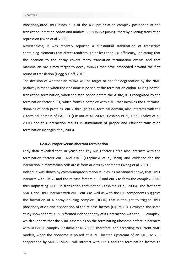
52
Chapter I
Phosphorylated-UPF1 binds eIF3 of the 43S preinitiation complex positioned at the
translation initiation codon and inhibits 60S subunit joining, thereby eliciting translation
repression (Isken et al, 2008).
Nevertheless, it was recently reported a substantial stabilization of transcripts
containing elements that direct readthrough at less than 1% efficiency, indicating that
the decision to the decay covers many translation termination events and that
mammalian NMD may target to decay mRNAs that have proceeded beyond the first
round of translation (Hogg & Goff, 2010).
The decision of whether an mRNA will be target or not for degradation by the NMD
pathway is made when the ribosome is poised at the termination codon. During normal
translation termination, when the stop codon enters the A-site, it is recognized by the
termination factor eRF1, which forms a complex with eRF3 that involves the C-terminal
domains of both proteins. eRF3, through its N-terminal domain, also interacts with the
C-terminal domain of PABPC1 (Cosson et al, 2002a; Hoshino et al, 1999; Kozlov et al,
2001) and this interaction results in stimulation of proper and efficient translation
termination (Mangus et al, 2003).
I.2.4.2. Proper versus aberrant termination
Early data revealed that, in yeast, the key NMD factor Upf1p also interacts with the
termination factors eRF1 and eRF3 (Czaplinski et al, 1998) and evidence for this
interaction in mammalian cells arose from in vitro experiments (Wang et al, 2001).
Indeed, it was shown by coimmunoprecipitation studies, as mentioned above, that UPF1
interacts with SMG1 and the release factors eRF1 and eRF3 to form the complex SURF,
thus implicating UPF1 in translation termination (Kashima et al, 2006). The fact that
SMG1 and UPF1 interact with eRF1-eRF3 as well as with the EJC components suggests
the formation of a decay-inducing complex (DECID) that is thought to trigger UPF1
phosphorylation and dissociation of the release factors (Figure I.3). However, the same
study showed that SURF is formed independently of its interaction with the EJC complex,
which supports that the SURF assembles on the terminating ribosome before it interacts
with UPF2/EJC complex (Kashima et al, 2006). Therefore, and according to current NMD
models, when the ribosome is poised at a PTC located upstream of an EJC, SMG1 -
chaperoned by SMG8-SMG9 - will interact with UPF1 and the termination factors to
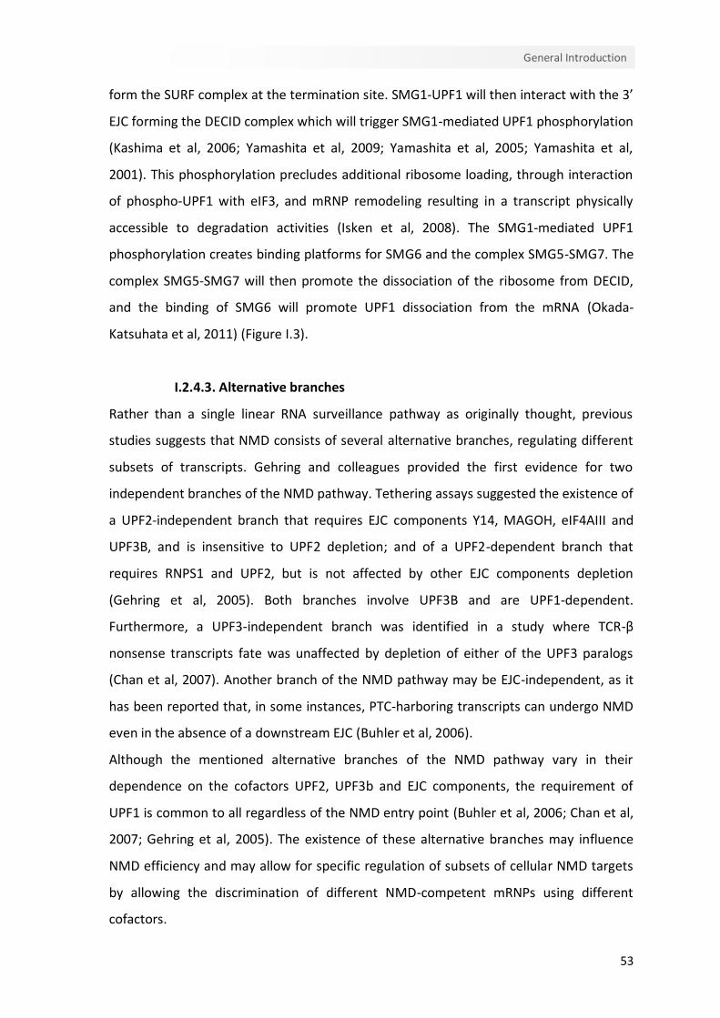
53
General Introduction
form the SURF complex at the termination site. SMG1-UPF1 will then interact with the 3’
EJC forming the DECID complex which will trigger SMG1-mediated UPF1 phosphorylation
(Kashima et al, 2006; Yamashita et al, 2009; Yamashita et al, 2005; Yamashita et al,
2001). This phosphorylation precludes additional ribosome loading, through interaction
of phospho-UPF1 with eIF3, and mRNP remodeling resulting in a transcript physically
accessible to degradation activities (Isken et al, 2008). The SMG1-mediated UPF1
phosphorylation creates binding platforms for SMG6 and the complex SMG5-SMG7. The
complex SMG5-SMG7 will then promote the dissociation of the ribosome from DECID,
and the binding of SMG6 will promote UPF1 dissociation from the mRNA (Okada-
Katsuhata et al, 2011) (Figure I.3).
I.2.4.3. Alternative branches
Rather than a single linear RNA surveillance pathway as originally thought, previous
studies suggests that NMD consists of several alternative branches, regulating different
subsets of transcripts. Gehring and colleagues provided the first evidence for two
independent branches of the NMD pathway. Tethering assays suggested the existence of
a UPF2-independent branch that requires EJC components Y14, MAGOH, eIF4AIII and
UPF3B, and is insensitive to UPF2 depletion; and of a UPF2-dependent branch that
requires RNPS1 and UPF2, but is not affected by other EJC components depletion
(Gehring et al, 2005). Both branches involve UPF3B and are UPF1-dependent.
Furthermore, a UPF3-independent branch was identified in a study where TCR-β
nonsense transcripts fate was unaffected by depletion of either of the UPF3 paralogs
(Chan et al, 2007). Another branch of the NMD pathway may be EJC-independent, as it
has been reported that, in some instances, PTC-harboring transcripts can undergo NMD
even in the absence of a downstream EJC (Buhler et al, 2006).
Although the mentioned alternative branches of the NMD pathway vary in their
dependence on the cofactors UPF2, UPF3b and EJC components, the requirement of
UPF1 is common to all regardless of the NMD entry point (Buhler et al, 2006; Chan et al,
2007; Gehring et al, 2005). The existence of these alternative branches may influence
NMD efficiency and may allow for specific regulation of subsets of cellular NMD targets
by allowing the discrimination of different NMD-competent mRNPs using different
cofactors.
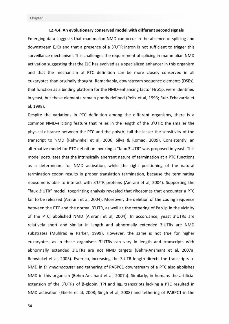
54
Chapter I
I.2.4.4. An evolutionary conserved model with different second signals
Emerging data suggests that mammalian NMD can occur in the absence of splicing and
downstream EJCs and that a presence of a 3’UTR intron is not sufficient to trigger this
surveillance mechanism. This challenges the requirement of splicing in mammalian NMD
activation suggesting that the EJC has evolved as a specialized enhancer in this organism
and that the mechanism of PTC definition can be more closely conserved in all
eukaryotes than originally thought. Remarkably, downstream sequence elements (DSEs),
that function as a binding platform for the NMD-enhancing factor Hrp1p, were identified
in yeast, but these elements remain poorly defined (Peltz et al, 1993; Ruiz-Echevarria et
al, 1998).
Despite the variations in PTC definition among the different organisms, there is a
common NMD-eliciting feature that relies in the length of the 3’UTR: the smaller the
physical distance between the PTC and the poly(A) tail the lesser the sensitivity of the
transcript to NMD (Rehwinkel et al, 2006; Silva & Romao, 2009). Consistently, an
alternative model for PTC definition invoking a “faux 3’UTR” was proposed in yeast. This
model postulates that the intrinsically aberrant nature of termination at a PTC functions
as a determinant for NMD activation, while the right positioning of the natural
termination codon results in proper translation termination, because the terminating
ribosome is able to interact with 3’UTR proteins (Amrani et al, 2004). Supporting the
“faux 3’UTR” model, toeprinting analysis revealed that ribosomes that encounter a PTC
fail to be released (Amrani et al, 2004). Moreover, the deletion of the coding sequence
between the PTC and the normal 3’UTR, as well as the tethering of Pab1p in the vicinity
of the PTC, abolished NMD (Amrani et al, 2004). In accordance, yeast 3’UTRs are
relatively short and similar in length and abnormally extended 3’UTRs are NMD
substrates (Muhlrad & Parker, 1999). However, the same is not true for higher
eukaryotes, as in these organisms 3’UTRs can vary in length and transcripts with
abnormally extended 3’UTRs are not NMD targets (Behm-Ansmant et al, 2007a;
Rehwinkel et al, 2005). Even so, increasing the 3’UTR length directs the transcripts to
NMD in D. melanogaster and tethering of PABPC1 downstream of a PTC also abolishes
NMD in this organism (Behm-Ansmant et al, 2007a). Similarly, in humans the artificial
extension of the 3’UTRs of β-globin, TPI and Igµ transcripts lacking a PTC resulted in
NMD activation (Eberle et al, 2008; Singh et al, 2008) and tethering of PABPC1 in the
55
General Introduction
vicinity of a PTC suppresses NMD, even in the presence of a downstream EJC (Eberle et
al, 2008; Kashima et al, 2006; Silva et al, 2008; Singh et al, 2008). In addition, spatial
rearrangements in the 3’UTR increased the stabilization of nonsense transcripts,
suggesting that the physical distance rather than the number of nucleotides between
the termination codon and the poly(A) tail is a crucial determinant for PTC definition
(Eberle et al, 2008). Therefore, the inhibitory effect of PABPC1 on NMD as a result of
stimulation of proper translation termination, as the “faux 3’UTR” model proposes,
might be extended to all species. In fact, depletion of PABPC1 increases the level of
readthrough at all three stop codons (Ivanov et al, 2008). Surprisingly, the knockdown of
UPF1 results in a decrease of readthrough level, indicating that UPF1 and PABPC1 have
antagonistic effects on translation termination efficiency that can be reflected in their
respective stimulatory and inhibitory effects on NMD (Ivanov & Anderson, 2010).
Furthermore, it was shown in vitro that PABPC1 competes with UPF1 for interaction with
eRF3, which is consistent with NMD being impaired when the termination codon is
located in proximity of PABPC1 (Singh et al, 2008). Thus, when the terminating ribosome
stalls at a termination codon distant from PABPC1, UPF1 is able to bind eRF3; on the
other hand, the proximity of the termination codon to PABPC1 allows this protein to
interact with eRF3 in detriment of UPF1, enhancing efficient translation termination.
While there is considerable support to the “faux 3’UTR” model, it was observed that, in
yeast, NMD occurs in deadenylated transcripts or in mutant strains lacking PABP (Meaux
et al, 2008). Additionally, a variety of studies shows that cis-acting elements, such as
long 3’UTRs or EJCs located downstream of the termination codon, are sufficient to
trigger NMD, suggesting that PTC definition is not likely to rely exclusively on PABPC
relative position to the PTC. Instead, additional molecular signals might influence the
nature of the termination event. The unified 3’UTR model integrates elements from
both the “downstream marker” model and the “faux 3’UTR” model and proposes that
the discrimination between a normal and a premature termination event is the outcome
of the combination of antagonistic signals (Muhlemann, 2008; Shyu et al, 2008; Silva &
Romao, 2009; Singh et al, 2008). At a translation termination event, the decision of NMD
triggering will be the result from the competition between PABPC1 and UPF1 for the
termination complex (Figure I.4). If PAPBC1 is favorably located to interact with the
terminating ribosome - through eRF3 - a normal termination event will occur, impairing

56
Chapter I
Aberrant translationtermination
STOP
m7G
PABPC1STOP PTCP eRF1
eRF3
CBP80
UPF3
UPF2
m7G
PABPC1STOP PTCP eRF1
eRF3
CBP80
m7G
PABPC1STOP PTCP eRF1
eRF3
CBP80
PP
m7G
PABPC1STOP PTCP eRF1
eRF3
CBP80
UPF3
UPF2PP
m7GCBP80
PABPC1STOP PTCP eRF1
eRF3
mRNAdecay
SURF formation
Licensing step
Ribosome
Polypeptide
EJC
SMG8
SMG9
Normal translation termination
STOP
CBP80
STOP eRF1
eRF3
Figure I.4. The unified model for nonsense-mediated mRNA decay. The model proposes that normal translation termination involves PABPC1 interaction with the termination complex, via eRF3, inhibiting UPF1 binding to eRF3. PABPC1 interaction with eRF3 promotes efficient translation termination. However, when the premature stop codon (PTC) is located far from the poly(A) tail, PABPC1 fails to interact with the termination complex, and the ribosome stalls for a prolonged period of time, allowing UPF1 to interact with eRF3. After SURF formation, UPF2 and/or UPF3 stimulate SMG1-mediated UPF1 phosphorylation. At this point, the presence of a downstream EJC will function as a strong NMD enhancer, since the proximity of UPF2 and/or UPF3 at the EJC will facilitate the interaction with UPF1, and the recruitment of the UPF proteins is likely to take longer in the absence of an EJC. Phosphorylation of UPF1 will irreversibly commit the mRNA to decay.
57
General Introduction
the association of UPF1, thus repressing NMD triggering, even in the presence of
downstream EJCs. Conversely, the failure of PABPC1 (and/or other associated 3’UTR
elements) to interact with the terminating complex will favor the association of
dephosphorylated UPF1, together with SMG1, to the terminating complex, forming the
SURF complex. Then, a second signal, such as UPF1 interaction with UPF2 and/or UPF3b
that induces SMG1-mediated UPF1 phosphorylation, will trigger NMD, with the presence
of downstream EJCs functioning, in some cases, as a secondary NMD enhancer (Figure
I.4).
An alternative model called the “ribosome-release” model was recently proposed, but
remains to be experimentally tested (Brogna & Wen, 2009). This model hypothesizes
that NMD would be a consequence of the release of post-termination ribosomes from a
region of the mRNA that normally would be translated. Therefore, in the case of a
premature termination event, a long region of the mRNA would not be traversed by
ribosomes subunits which would result in mRNA destabilization. UPF1 would have an
active role, by interacting with eIF3 to stimulate ribosome release.
So far, it is not possible to unambiguously predict the importance of each mentioned
mRNP feature in NMD-triggering. It seems that neither a long 3’UTR or a downstream
EJC are absolutely required for NMD, instead a combination of several features is likely
to be involved in the mechanism of PTC definition, with the only definite requirement
for NMD being the translation-dependent recruitment of UPF1 to the mRNA (Nicholson
et al, 2010).
I.2.4.5. AUG-proximal PTCs unexpectedly evade NMD
Even though a nonsense codon at position +1 with respect to the initiation AUG codon is
sufficient to trigger NMD (Lew et al, 1998), there are several reports of PTCs close to the
5’ end of the first exon that fail to elicit NMD (Asselta et al, 2001; Boyer et al, 2005;
Buisson et al, 2006; Buzina & Shulman, 1999; Danckwardt et al, 2002; Denecke et al,
2004; Harries et al, 2004; McLean et al, 2003; Paulsen et al, 2006; Perrin-Vidoz et al,
2002; Rolfini & Cabrini, 1993; Romao et al, 2000; Tournier et al, 2004; Zhang & Maquat,
1997). In fact, our lab reported that human β-globin transcripts harboring a naturally
occurring nonsense mutation at codon 15 accumulate to levels comparable to those of
wild-type (Romao et al, 2000). Similarly, PTCs generated at codons 5 and 17 were also
58
Chapter I
shown to fail to trigger NMD on these transcripts (Romao et al, 2000). For some AUG-
proximal PTCs, the NMD-resistance is explained by translation reinitiation, since the
ribosome that initiates at an in-frame AUG downstream of the early PTC removes the
remaining EJCs from the mRNA and terminates translation at the normal termination
codon, culminating in the production of an N-terminal truncated protein (Perrin-Vidoz et
al, 2002; Zhang & Maquat, 1997). However, translation reinitiation does not fully explain
the NMD-resistance observed for some AUG-proximal PTCs. For instance, in Igµ
transcripts bearing PTCs located at codons 3 or 32, expressed in hybridoma cells,
translation reinitiation occurs at Met100 with an efficiency of 4% of the normal rate
(Buzina & Shulman, 1999); still, no detectable protein products corresponding to
translation reinitiation at Met100 were found in HeLa cells expressing Igµ transcripts
with a nonsense codon in position 32 (Buhler et al, 2004). Likewise, the NMD-resistance
of β-globin transcripts bearing a PTC at position 15 is not attributable to translation
reinitiation since blocking potential reinitiation sites does not result in NMD-typical
mRNA levels (Inacio et al, 2004; Silva et al, 2006). Moreover, abnormal splicing and
impaired translation have also been shown not to be the determinants of this resistance
to NMD (Inacio et al, 2004; Silva et al, 2006). Indeed, our lab has shown that, in these
transcripts, NMD inhibition reflects the proximity of the PTC to the translation initiation
codon (Inacio et al, 2004; Silva et al, 2006). This “AUG-proximity effect”, which is
independent of sequence context and transcript identity and appears to be a general
attribute of mammalian NMD (Silva et al, 2006), may reflect the nature of the early
termination event. In the case of such a premature termination event, the translation
round might be brief enough to maintain PABPC1 in close proximity to the AUG codon,
as a consequence of mRNA circularization. This favorable location might allow PABPC1
to interact with the translation termination complex by the time the ribosome reaches
the stop codon, superimposing its inhibitory effect on NMD (Silva et al, 2008; Silva &
Romao, 2009) (Figure I.5).
Nonetheless, a recent study (Neu-Yilik et al, 2011) reported that translation reinitiation
is the determinant of the NMD-resistance of β-globin AUG-proximal nonsense-mutated
transcripts. This inconsistency with our lab previous findings might be explained by
differences in the experimental settings, since in the study of Neu-Yilik et al. (2011), the
natural β-globin exon 1 was transformed into a functional exon 2 by the introduction of
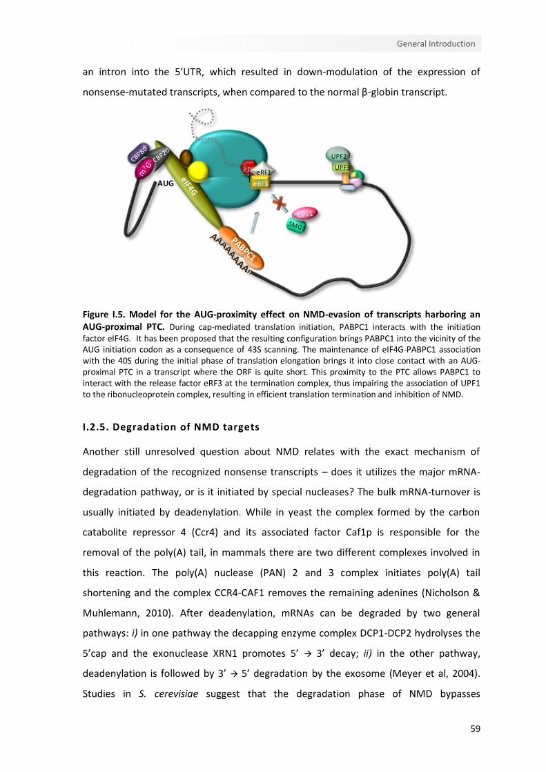
59
General Introduction
AUG
UPF3
UPF2
PTC eRF1
eRF3
an intron into the 5’UTR, which resulted in down-modulation of the expression of
nonsense-mutated transcripts, when compared to the normal β-globin transcript.
Figure I.5. Model for the AUG-proximity effect on NMD-evasion of transcripts harboring an AUG-proximal PTC. During cap-mediated translation initiation, PABPC1 interacts with the initiation factor eIF4G. It has been proposed that the resulting configuration brings PABPC1 into the vicinity of the AUG initiation codon as a consequence of 43S scanning. The maintenance of eIF4G-PABPC1 association with the 40S during the initial phase of translation elongation brings it into close contact with an AUG-proximal PTC in a transcript where the ORF is quite short. This proximity to the PTC allows PABPC1 to interact with the release factor eRF3 at the termination complex, thus impairing the association of UPF1 to the ribonucleoprotein complex, resulting in efficient translation termination and inhibition of NMD.
I.2.5. Degradation of NMD targets
Another still unresolved question about NMD relates with the exact mechanism of
degradation of the recognized nonsense transcripts – does it utilizes the major mRNA-
degradation pathway, or is it initiated by special nucleases? The bulk mRNA-turnover is
usually initiated by deadenylation. While in yeast the complex formed by the carbon
catabolite repressor 4 (Ccr4) and its associated factor Caf1p is responsible for the
removal of the poly(A) tail, in mammals there are two different complexes involved in
this reaction. The poly(A) nuclease (PAN) 2 and 3 complex initiates poly(A) tail
shortening and the complex CCR4-CAF1 removes the remaining adenines (Nicholson &
Muhlemann, 2010). After deadenylation, mRNAs can be degraded by two general
pathways: i) in one pathway the decapping enzyme complex DCP1-DCP2 hydrolyses the
5’cap and the exonuclease XRN1 promotes 5’ 3’ decay; ii) in the other pathway,
deadenylation is followed by 3’ 5’ degradation by the exosome (Meyer et al, 2004).
Studies in S. cerevisiae suggest that the degradation phase of NMD bypasses

60
Chapter I
deadenylation before decapping (Muhlrad & Parker, 1994). However, PTC-dependent
accelerated deadenylation has also been reported and the initiation of NMD in this
organism by decapping or deadenylation seems to be dependent on the relative position
of the PTC to the cap and poly(A) tail (Cao & Parker, 2003; Mitchell & Tollervey, 2003). In
contrast, in D. melanogaster, where there is no SMG7 ortholog, nonsense mRNAs are
degraded by endonucleolytic cleavage within the vicinity of the PTC. The resulting 5’ and
3’ intermediates are then rapidly degraded by XRN1 and the exosome, respectively
(Gatfield et al, 2003).
In mammals, available data indicates that nonsense transcripts degradation initiates
with deadenylation followed by decapping and XRN1-mediated decay (Chen & Shyu,
2003; Couttet & Grange, 2004; Lejeune et al, 2003). Consistently, UPF proteins are able
to form a complex with XRN1, XRN2 and some exosome proteins (Lejeune et al, 2003).
Recently, the proline-rich nuclear receptor co-regulatory protein2 (PNRC2) was shown to
be involved in promoting degradation of NMD targets. As this protein interacts with
phospho-UPF1 and DCP1, it might provide a link between UPF1-bound mRNAs and the
decapping enzymes (Cho et al, 2009).
Nonetheless, emerging data has revealed that degradation of human nonsense
transcripts can be initiated by endonucleolytic cleavage in the vicinity of the PTC (Eberle
et al, 2009). As above mentioned, the PIN domain of SMG6 exhibits the endonucleolytic
activity responsible for initiating NMD in human cells as in D. melanogaster (Eberle et al,
2009). Therefore, mammalian NMD seems to consist of two independent routes for RNA
decay: an endonucleotic SMG6-dependent route, and other decapping-dependent
exonucleolytic route. In either case, the resulting RNA fragments are subjected to
exonucleolytic decay (Muhlemann & Lykke-Andersen, 2010b; Nicholson & Muhlemann,
2010) (Figure I.6). The determinants of which pathway to be activated remain unclear. It
has been shown that SMG7, when tethered to a reporter mRNA, activated mRNA decay
in a DCP2- and XRN1-dependent manner, but independently of SMG6. Since SMG5 and
SMG7 co-localize in processing bodies (P-bodies) while SMG6 forms separate foci
(Unterholzner & Izaurralde, 2004); it is possible that the binding of SMG5/7 or SMG6 to
phospho-UPF1 represents a branching point. Even so, the relative contribution of each
pathway remains to be clarified.

61
General Introduction
PABPC1
UPF3
UPF2
STOP
P
PTC eRF1
eRF3 m7G
P
PABPC1STOP PTC
UPF3
UPF2SMG1
UPF1
m7G
A
A
AA
A
STOP PTC
PABPC1STOP PTC
UPF3
UPF2SMG1
m7G
m7G
PABPC1STOP
SMG1-mediated UPF1 phosphorylation
SMG6-mediated endonucleolysis
Deadenylationand decapping
Exonucleolytic decayfrom internal sites
Exonucleolyticdecay from termini
Ribosome
Polypeptide
SMG9
Exosome
EJC
SMG8
DCP1-DCP2 complex
XRN1
Figure I.6. Decay pathways of human NMD. SMG1-mediated UPF1 phosphorylation induces a conformational change in UPF1 that results in an increased affinity for the mRNA and irreversibly marks the mRNA for degradation. Phosphorylated UPF1 can either interact with SMG6 or with the heterodimer SMG5-SMG7. Both routes will lead to PP2A recruitment and UPF1 dephosphorylation. SMG6 is an endonuclease and, upon UPF1 binding, will cleave the mRNA in the vicinity of the premature termination codon. The resulting decay intermediates will be rapidly degraded 5’ 3’ by XRN1 and 3’ 5’ by the exosome. On the other hand, UPF1 binding to SMG5-SMG7 results in the recruitment of deadenylases. Deadenylation is followed by the removal of the cap structure by the DCP1/DCP2 decapping complex and the transcripts will be degraded 5’ 3’ by XRN1 and 3’ 5’ by the exosome.
I.2.6. Feedback regulatory mechanisms
Given that NMD plays a role in gene regulation and mRNA surveillance, the perturbation
of this surveillance pathway could have catastrophic consequences due to the variation
in transcripts profiles and to the production of potentially toxic truncated proteins
62
Chapter I
(Muhlemann et al, 2008). A buffering system that confers robustness to the NMD
mechanism would be essential to protect against this deleterious consequences. Indeed,
evidence for such homeostatic pathway acting on NMD has emerged from reports
showing that UPF3a is stabilized in response to loss of its paralog UPF3b (Chan et al,
2009) and that UPF1 depletion stabilizes SMG5 encoding mRNA (Mendell et al, 2004;
Singh et al, 2008).
Consistently, Huang and colleagues recently identified a large number of NMD factors
that are upregulated upon NMD perturbation. This NMD regulation consists of a series
of negative feedback regulatory loops. Analysis of the UPF and SMG mRNA half-lifes in
UPF1- and SMG1-depleted cells suggests that mRNAs encoding UPF1 and a subset of
other NMD factors are direct NMD targets (Huang et al, 2011). Moreover, the same
study showed that overexpression of SMG1, SMG5 and SMG6 augments NMD,
suggesting that these three factors are rate limiting for NMD. Another recent study of
transcriptome profiling in human UPF1-, SMG6- or SMG7-depleted cells revealed that
most mRNAs coding for NMD factors are NMD-sensitive, all possess long 3’UTRs and
some of them harbor uORFs (Yepiskoposyan et al, 2011). In addition, the long 3’UTRs of
UPF1, SMG5 and SMG7 mRNAs were shown to constitute the main NMD-inducing
features of these mRNAs (Yepiskoposyan et al, 2011).
The NMD feedback regulation is a highly directed response that is mediated by different
branches of the NMD pathway, probably because, in some cases, NMD may selectively
increase the levels of NMD factors of the same branch of the perturbed factor,
maximizing NMD buffering, or because the upregulation of partially redundant factors
will bolster NMD. As cell types differ in their NMD factor profile and sensitivity, there are
cell-type specific differences in rate-limiting and redundant NMD factors. Therefore, this
buffering system provides versatile and subtle feedback control to confer robustness to
NMD, by fine-tuning the level of NMD factors in order to achieve optimal NMD activity
(Huang et al, 2011). This mechanism may be conserved in eukaryotes, as recent studies
show that SMG5 and SMG6 in D. melanogaster (Rehwinkel et al, 2005) and SMG7 and
UPF3 in Arabidopsis thaliana (Riehs et al, 2008; Saul et al, 2009) are upregulated in
response to NMD depletion.
63
General Introduction
I.2.7. NMD effectors: other functions & new players
NMD factors either alone or in groups participate in additional cellular pathways and
functions that range from other RNA surveillance pathways over functions in genome
stability, telomere maintenance and translation. So far, it is not known if these other
functions are always independent from NMD or if they converge in functional networks
(Neu-Yilik & Kulozik, 2008).
I.2.7.1. NMD factors and genome stability
The first link between NMD factors and telomere maintenance arose from studies in
yeast showing that Upf1p, Upf2p and Upf3p mutations led to telomere shortening (Lew
et al, 1998). In addition, SMG5, SMG6 and SMG7 were found to be homologous of
hEST1B, hEST1A and hEST1C, respectively. These proteins are orthologues of yeast Est1p
protein, which is involved in telomerase regulation and promotes telomere elongation.
Human SMG6 overexpression leads to progressive telomere shortening and SMG5 and
SMG6 were found to associate with telomerase activity in human cell extracts
(Reichenbach et al, 2003). Surprisingly, UPF1, UPF2, SMG1 and SMG6 were found to be
enriched at telomeric chromatin fractions, and they negatively regulate the association
of the recently discovered set of telomeric repeat-containing RNAs (TERRAs) with
chromatin (Azzalin et al, 2007). This data suggests that the NMD factors link TERRA
regulation with telomere maintenance, and thus, with genome stability.
SMG1 is a member of the PIK family and, like ATM and ATR kinases, it functions in
genome surveillance. Both SMG1 and ATM phosphorylate the cell-cycle checkpoint
protein p53, and SMG1 depletion leads to spontaneous DNA damage and increased
sensitivity to ionizing radiation (Brumbaugh et al, 2004). Moreover, UPF1 also has a role
in DNA replication: UPF1 depletion results in early S-phase arrest and induces an ATR-
dependent DNA-damage response (Azzalin & Lingner, 2006). The identification of UPF1
as the DNA polymerase -associated helicase corroborates its function in DNA repair
(Carastro et al, 2002) that is separable and likely independent of its role in NMD, since
ATR depletion impairs chromatin loading of UPF1 without interfering with the NMD
pathway (Brumbaugh et al, 2004).
64
Chapter I
I.2.7.2. Functions in other mRNA turnover pathways
In addition to its role in guiding NMD targets for decay, UPF1 also serves in other mRNA
turnover pathways. Yeast two-hybrid analysis and GST-pull down assays showed that
Staufen-1 (STAU1), which plays a role in mRNA decay, interacts with UPF1. Tethering of
STAU1 to a reporter transcript results in UPF1-recruitment and induces translation-
dependent STAU1-mediated mRNA decay (SMD) without involving other NMD effectors
(Kim et al, 2005). UPF1 has also been reported to play a role in replication-dependent
histone-mRNA degradation (Kaygun & Marzluff, 2005), however its function in histone
mRNA metabolism remains unclear (Nicholson et al, 2010).
I.2.7.3. NMD factors and translation
There are several studies indicating a role for the UPF proteins in translation. Analysis of
premature translation in yeast cell-free extracts demonstrated that a significant fraction
of ribosomes terminating translation at a PTC reinitiated at proximal AUG codons in a
UPF1-dependent fashion (Amrani et al, 2004). Furthermore, the NMD effectors UPF1,
UPF2, UPF3A, UPF3B, MAGOH, Y14 and RNPS1 promote polysome association and
increase translation when tethered within the ORF of a reporter mRNA (Kunz et al, 2006;
Nott et al, 2004). Fascinatingly, the NMD and the translation functions of UPF3A and
UPF3B require different protein domains and interacting factors. Indeed, the UPF3
proteins require Y14/MAGOH/eIF4IIIA/BTZ to trigger NMD while their activity in
enhancing translation is independent of these EJC components (Kunz et al, 2006), even
though Y14 and MAGOH also stimulate translation (Nott et al, 2004).
Whilst increasing the rate of translation, some NMD factors also appear to stimulate the
efficiency of translation termination as, in yeast, codon readthrough is enhanced when
any of the three upframeshift proteins is inactive or absent (Wilkinson, 2005).
Nevertheless, a recent study demonstrated that the apparent loss of termination fidelity
seems to be a indirect consequence of the NMD-sensitive ALR1 mRNA that results in
increased intracellular Mg2+ levels (Johansson & Jacobson, 2010).
Several data have implicated possible roles for the UPF proteins in the regulation of
translation termination, repression and initiation, both in yeast and in mammals, which
seem to be independent of their activity on mRNA decay (Czaplinski et al, 1998; Isken et
al, 2008; Ivanov et al, 2008; Muhlrad & Parker, 1999; Wang et al, 2001). In fact, an

65
General Introduction
hyperphosphorylated mutant of UPF1 that is impaired for NMD, repressed translation of
a reporter mRNA in rabbit retyculocyte lysates (Isken et al, 2008). Consistently, data
from a ribosome reutilization assay showing that the elimination of any of the UPF
proteins led to reduced availability of ribosomes for translation initiation, suggest that
UPF1 regulates initiation efficiency of ribosomes entering a new cycle of translation
(Ghosh et al, 2010). Furthermore, toeprinting assays revealed that extracts lacking UPF1
had fewer 80S ribosomes at the AUG, while exhibiting similar 40S recruitment to the
wild-type extracts, indicating that UPF1 loss affects subunit joining (Ghosh et al, 2010).
At the same time, as new functions for NMD factors are revealed, it becomes more
apparent that factors involved in translation play multiple roles that may link translation
initiation with termination and/or NMD (Amrani et al, 2008). eIF3, together with eIF1,
eIF1A and the eIF3j subunit, has been implicated in recycling by stimulating the in vitro
dissociation of post-termination 80S complexes, and remains bound to 40S subunits
after this process (Pisarev et al, 2007). Moreover, eIF3e depletion impairs NMD in a
manner similar to UPF1 depletion (Morris et al, 2007) and interacts with phospho-UPF1,
triggering translation repression (Isken et al, 2008). It has also been shown that, at
elevated temperatures, yeast strains harboring the temperature-sensitive prt1-1
mutation (in eIF3) lose the ability to initiate translation and promote NMD (Welch &
Jacobson, 1999). The role of eIF3 in recycling may, thus, be linked to its role in NMD, for
example, in the resolution of the SURF/post-termination complex.
I.2.8. Human β-globin as a model system for studying NMD
The human β-globin gene has two introns and three exons that are flanked by 5’ and 3’
UTRs (Huisman, 1993). Thus, after the two introns are spliced out, two EJCs might be
assembled on the transcript (Le Hir et al, 2000a; Le Hir et al, 2000b). A β-globin NMD-
behaviour corroborating the “50-55 nt boundary rule” has been broadly described,
suggesting that the EJC functions as an enhancer in β-globin NMD. Indeed, the first
indications regarding whether a β-globin PTC elicits NMD, concerning its position
relatively to the downstream intron, were obtained by Kulozik and Maquat labs
(Thermann et al, 1998; Zhang et al, 1998b). These authors showed that β-globin
nonsense mutations located in the 3’ region of exon 1 (at codon 26) and within the 5’
66
Chapter I
two-thirds of exon 2 (at codons 36, 60-61, 75, and 82) elicit NMD. In contrast, mRNAs
bearing PTCs towards the 3’ end of exon 2 (at codons 88, 91, 95, 98, 101 and 103) and
those with PTCs in exon 3 (at codons 106, 107, 114, 121 and 141) are all NMD-resistant.
This illustrates a clear boundary between 48 and 66 nt upstream of the last exon-exon
junction. Although the onset of NMD requires a minimal distance between the nonsense
mutation and the final exon-exon junction, on β-globin transcripts, the requirement for a
maximal distance appears to be considerably less crucial. Indeed, a β-globin mRNA with
a nonsense mutation at codon 39 (Baserga & Benz, 1988) remains NMD competent,
even when the distance between the PTC and the 3’ exon-exon junction is increased
from the normal 180 nt up to 654 nt (Neu-Yilik et al, 2001). This is a much longer
distance than the one required for NMD in the triosephosphate isomerase (TPI)
transcripts (Ruiz-Echevarria et al, 1998; Sun & Maquat, 2000; Zhang et al, 1998a). On the
other hand, an intronless human β-globin gene, carrying a nonsense mutation at codon
39, generates an mRNA that lacks the EJCs and, consequently, is immune to NMD (Neu-
Yilik et al, 2001). This is supported by the fact that naturally intronless mRNAs containing
PTCs are not usually degraded by NMD (Maquat & Carmichael, 2001). Furthermore, the
insertion of an intron more than 50 nt downstream of the native stop codon, redefines
this codon as premature and triggers this mRNA to NMD (Thermann et al, 1998).
Nevertheless, exceptions to the “50-55 nt boundary rule” have also been reported for
the human β-globin mRNAs. As above mentioned, our lab showed that β-globin
transcripts containing naturally occurring nonsense mutations in the 5’ region of exon 1
accumulate to levels similar to those of normal β-globin transcripts, being, unexpectedly
according to their position, NMD-resistant (Romao et al, 2000). Furthermore, NMD was
found to be triggered in hybrid mouse-human β-globin transcripts (carrying nonsense
mutations at codons 21-22, 39 or 60-61) in the absence of the last intron (Zhang et al,
1998b) and therefore, in the absence of downstream EJCs. Even so, Neu-Yilik and
colleagues have shown that splicing is an indispensable component of the β-globin NMD
pathway (Neu-Yilik et al, 2001). Additionally, a study using the IVS1 +5 G→A thalassemic
β-globin gene as a model system, showed that the splicing of these pre-mRNAs at a
cryptic site, generates a mature transcript carrying a PTC at codon 30 (located more
than 55 nt 5’ of the final exon-exon junction) that is immune to NMD (Danckwardt et al,
2002). Although the mechanism underlying this NMD-behavior was not clarified, it was

67
General Introduction
demonstrated that neither abnormalities of splicing nor translation reinitiation at
downstream AUG codons is the cause for the NMD resistance of this transcript
(Danckwardt et al, 2002).
I.3. Aims
In summary, there are several examples of PTC-containing transcripts, expected to
contain residual EJCs, that escape NMD even when the PTC is located, in a linear sense,
quite far from the poly(A) tail and PABP. Previous studies of AUG-proximal PTC mRNAs
revealed that its observed NMD resistance does not reflect downstream translation
reinitiation or extension of ribosome elongation 3’ of the PTC and is instead a direct
effect of the termination event being located in close proximity to the AUG. This effect
constitutes a general attribute of mammalian NMD that is independent of sequence
context and independent of the 5’UTR length. The aim of the work presented in this
thesis was to clarify the mechanistic basis for the NMD resistance of mRNAs carrying
AUG-proximal PTCs and expand the current models for NMD and translation. Therefore,
the following objectives were established:
To confirm the inhibitory effect of PABPC1 on the NMD commitment of AUG-
proximal nonsense-mutated transcripts.
To evaluate the biochemical mechanism by which PABPC1 controls ribosomal
termination and how it may alter the recruitment of UPF1 to a terminating
ribosome at an AUG-proximal PTC.
To investigate the molecular basis by which translation initiation factors are
involved in the NMD inhibition of mRNAs harboring an AUG-proximal PTC.

CChhaapptteerr IIII
Interaction of PABPC1 with the translation initiation
complex is critical to the NMD-resistance of AUG-
proximal nonsense mutations
Isabel Peixeiro, Ângela Inácio, Cristina Barbosa, Ana Luísa Silva, Stephen A.
Liebhaber and Luísa Romão


71
Translation initiation function In NMD inhibition
Author’s note
This chapter was published, with minor modifications, in Nucleic Acids Research (2011)
doi: 10.1093/nar/gkr820. Some of the presented data in this chapter were obtained by
the other indicated authors. Ângela Inácio was responsible for the toeprinting assays
and Cristina Barbosa contributed to the test of eIF3 subunits depletion in general
translation.


73
Translation initiation function in NMD inhibition
II.1. Abstract
Nonsense-mediated mRNA decay (NMD) is a surveillance pathway that recognizes and
rapidly degrades mRNAs containing premature termination codons (PTC). The strength
of the NMD response appears to reflect multiple determinants on a target mRNA. We
have previously reported that mRNAs containing PTCs in close proximity to the
translation initiation codon (AUG-proximal PTCs) can substantially evade NMD. Here, we
explore the mechanistic basis for this NMD resistance. We demonstrate that translation
termination at an AUG-proximal PTC lacks the ribosome stalling that is evident in an
NMD-sensitive PTC. This difference is associated with demonstrated interactions of the
cytoplasmic poly(A)-binding protein 1, PABPC1, with the cap-binding complex subunit,
eIF4G, and the 40S recruitment factor eIF3 as well as the ribosome release factor, eRF3.
These interactions, in combination, underlie critical 3’-5’ linkage of translation initiation
with efficient termination at the AUG-proximal PTC and contribute to an NMD-resistant
PTC definition at an early phase of translation elongation.
II.2. Introduction
Nonsense-mediated decay (NMD) targets mRNAs harboring premature translation-
termination codons (PTCs) for rapid decay. This surveillance pathway limits the synthesis
of potentially deleterious C-terminally truncated proteins encoded by mutant mRNAs
(Muhlemann & Lykke-Andersen, 2010a; Nicholson et al, 2010; Rebbapragada & Lykke-
Andersen, 2009; Silva & Romao, 2009). As such, NMD serves as an important modifier of
many genetic disorders (Behm-Ansmant & Izaurralde, 2006; Holbrook et al, 2004;
Peixeiro et al, 2011b). The NMD pathway also functions as an important determinant of
wild-type gene expression with approximately 10% of the mammalian transcriptome
impacted by components of the NMD apparatus (McGlincy et al, 2010; Mendell et al,
2004; Rehwinkel et al, 2006). A comprehensive description of the determinants and
mechanisms of NMD is therefore central to the understanding of both normal and
mutant gene expression.
A substantial body of evidence supports a role for the supramolecular exon junction
complexes (EJC) in triggering NMD. The EJC is deposited 20-24 nucleotides (nts)
74
Chapter II
upstream of each exon-exon junction during transcript splicing in the nucleus (Le Hir et
al, 2001). These complexes are subsequently displaced from the mature mRNA in the
cytoplasm by the elongating ribosome (Maquat, 2004) during the first round of
translation *“pioneer round of translation”; (Ishigaki et al, 2001; Lejeune et al, 2002)]. If
a PTC is located more than 50-54 nts 5’ to the last exon-exon junction, one or more EJCs
will remain beyond the reach of the elongating ribosome and will be retained on the
mRNA. The retained EJC(s) can interact with the translation termination complex via
bridging interactions between the release complex associated proteins, UPF1 and SMG1
(Kashima et al, 2006) and the EJC-associated factors, UPF2-UPF3 (Le Hir et al, 2001). This
bridging interaction has been proposed to trigger accelerated decay (i.e., NMD) of the
PTC-containing mRNA.
While multiple reports support a role for the EJC-release complex interaction in NMD, an
accumulating body of data indicates that additional determinants may play a significant
role in this surveillance pathway. Converging lines of evidence from studies in D.
melanogaster, C. elegans, and S. cerevisiae reveal that NMD can be triggered
independently of transcript splicing and EJC deposition (Conti & Izaurralde, 2005;
Rehwinkel et al, 2006). These studies further reveal that 3’ untranslated region (UTR)
length and the proximity of the PTC to the cytoplasmic poly(A)-binding protein 1
(PABPC1) may constitute critical determinants of NMD. Studies in yeast support these
findings by demonstrating that correct positioning of the termination codon relative to
PABPC1 are necessary for efficient termination and NMD resistance (Amrani et al, 2004).
Recent studies in mammalian cell culture support the conclusion that the strength of the
NMD response is inversely related to the distance between the PTC and PABPC1 (Eberle
et al, 2008; Singh et al, 2008); shortening this distance by tethering PABPC1 in close
proximity to an otherwise NMD-sensitive PTC suppresses NMD, even in the presence of
a downstream EJC (Eberle et al, 2008; Ivanov et al, 2008; Silva et al, 2008; Singh et al,
2008). In further support of this model, it has been demonstrated in vitro that PABPC1
can competitively block the association of UPF1 with eRF3 (Singh et al, 2008) with a
corresponding blunting of UPF1 actions and the NMD response (Singh et al, 2008). Thus
the impact of PABPC1 on NMD appears to reflect its ability to interact with, and alter the
impact of, supramolecular interactions at the translation termination complex.

75
Translation initiation function in NMD inhibition
We have previously reported that mRNAs containing PTCs in close proximity to the
translation initiation AUG codon (AUG-proximal PTCs) escape NMD. This was initially
surprising as these mRNAs would be expected to contain residual EJCs and in addition
would situate the PTC quite far, in a linear sense, from the poly(A) tail and PABP (Romao
et al, 2000). Detailed analyses of AUG-proximal PTC mRNAs revealed that its observed
NMD resistance did not reflect downstream translation reinitiation or extension of
ribosome elongation 3’ of the PTC and was instead a direct effect of the termination
event being located in close proximity to the AUG (Inacio et al, 2004; Silva et al, 2006).
Based on these studies, and on the observation by others that PABPC1 is able to bind
simultaneously to the cap-binding complex subunit eIF4G and to the poly(A) tail,
*“closed-loop” mRNP configuration (Wells et al, 1998)], we have proposed a model in
which the short open reading frame on an AUG-proximal nonsense-mutated mRNA,
situates PABPC1 and its associated cap-binding complex, in close proximity to the PTC.
This proximity would allow PABPC1 to alter the structure and/or function of the
translation termination complex with a consequent inhibitory effect on NMD (Silva et al,
2008; Silva & Romao, 2009).
In the present report we further focus on the mechanism of NMD-resistance of AUG-
proximal PTCs. These studies support the model in which PABPC1 is brought into close
proximity with an AUG-proximal PTC via interactions with the translation initiation
complexes. This proximity of PABP to the AUG-proximal PTC allows PABP to interact with
eRF3 with a consequent enhancement of the release reaction and repression of the
NMD response.
II.3. Materials and Methods
II.3.1. Plasmid constructs
The wild type β-globin gene (βN), as well as the human β-globin variants β15 and β39
were cloned into the pTRE2pur vector (BD Biosciences) as previously described (Silva et
al, 2006). The pDEST26-PABPC1delC construct was obtained by deleting the DNA
fragment encoding the C-terminal portion of PABPC1 from pDEST26-PABPC1 plasmid
(IOH13850-pDEST26; RZPD). To accomplish this deletion, the coding sequence of
PABPC1, excluding the C-terminal domain (NM 002568, nucleotides 2140-2394,
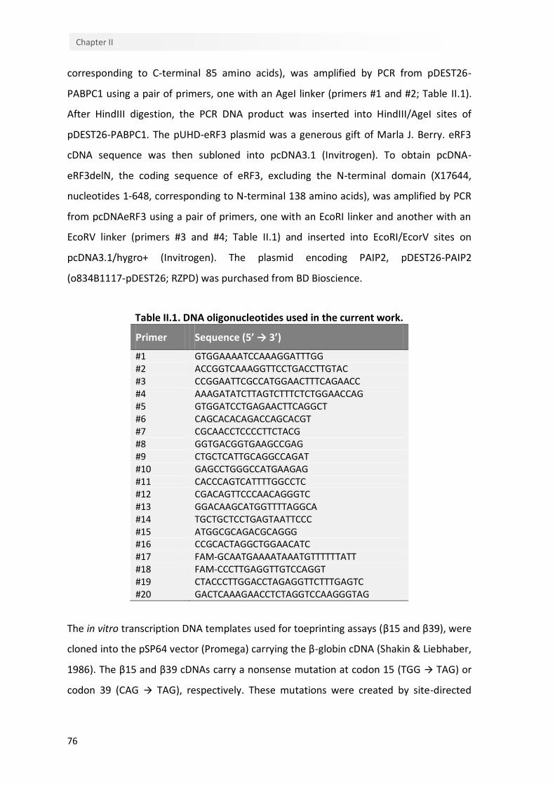
76
Chapter II
corresponding to C-terminal 85 amino acids), was amplified by PCR from pDEST26-
PABPC1 using a pair of primers, one with an AgeI linker (primers #1 and #2; Table II.1).
After HindIII digestion, the PCR DNA product was inserted into HindIII/AgeI sites of
pDEST26-PABPC1. The pUHD-eRF3 plasmid was a generous gift of Marla J. Berry. eRF3
cDNA sequence was then subloned into pcDNA3.1 (Invitrogen). To obtain pcDNA-
eRF3delN, the coding sequence of eRF3, excluding the N-terminal domain (X17644,
nucleotides 1-648, corresponding to N-terminal 138 amino acids), was amplified by PCR
from pcDNAeRF3 using a pair of primers, one with an EcoRI linker and another with an
EcoRV linker (primers #3 and #4; Table II.1) and inserted into EcoRI/EcorV sites on
pcDNA3.1/hygro+ (Invitrogen). The plasmid encoding PAIP2, pDEST26-PAIP2
(o834B1117-pDEST26; RZPD) was purchased from BD Bioscience.
Table II.1. DNA oligonucleotides used in the current work.
Primer Sequence (5’ → 3’)
#1 GTGGAAAATCCAAAGGATTTGG #2 ACCGGTCAAAGGTTCCTGACCTTGTAC #3 CCGGAATTCGCCATGGAACTTTCAGAACC #4 AAAGATATCTTAGTCTTTCTCTGGAACCAG #5 GTGGATCCTGAGAACTTCAGGCT #6 CAGCACACAGACCAGCACGT #7 CGCAACCTCCCCTTCTACG #8 GGTGACGGTGAAGCCGAG #9 CTGCTCATTGCAGGCCAGAT #10 GAGCCTGGGCCATGAAGAG #11 CACCCAGTCATTTTGGCCTC #12 CGACAGTTCCCAACAGGGTC #13 GGACAAGCATGGTTTTAGGCA #14 TGCTGCTCCTGAGTAATTCCC #15 ATGGCGCAGACGCAGGG #16 CCGCACTAGGCTGGAACATC #17 FAM‐GCAATGAAAATAAATGTTTTTTATT #18 FAM‐CCCTTGAGGTTGTCCAGGT #19 CTACCCTTGGACCTAGAGGTTCTTTGAGTC #20 GACTCAAAGAACCTCTAGGTCCAAGGGTAG
The in vitro transcription DNA templates used for toeprinting assays (β15 and β39), were
cloned into the pSP64 vector (Promega) carrying the β-globin cDNA (Shakin & Liebhaber,
1986). The β15 and β39 cDNAs carry a nonsense mutation at codon 15 (TGG TAG) or
codon 39 (CAG TAG), respectively. These mutations were created by site-directed

77
Translation initiation function in NMD inhibition
mutagenesis using the QuickChangeTMSite-directed Mutagenesis Kit (Stratagene) and
mutagenic primers #19 and #20 and the plasmid with the βN cDNA as template.
II.3.2. Cell culture, plasmid and siRNA transfection
HeLa cells, stably expressing the tet transactivator (HeLa/tTA) (Kong et al, 2003), were
grown in Dulbecco’s modified Eagle’s medium supplemented with 10% fetal bovine
serum. siRNA duplexes (Table II.2) were designed as 19-mers with 3’-dTdT overhangs
and purchased from Thermo. Transfections of cells with siRNAs were carried out using
Lipofectamine 2000 Transfection Reagent (Invitrogen), following the manufacturer’s
instructions, in 35-mm plates using 200 pmol of siRNA oligonucleotides and 4 µl of
transfection reagent. Twenty four hours later, cells were transfected with an additional
50 pmol of siRNAs along with 300 ng of the test construct DNA and 1700 ng of pEGFP
vector (BD Biosciences) to control for transfection efficiency. Where specified, 1700 ng
of pDEST26-PABPC1, pDEST26-PABPC1delC, pcDNA-eRF3delN or 1000 ng of pDEST26-
PAIP2 [since in the study of Khalegpour et al. (Khaleghpour et al, 2001) 5000ng of PAIP2
DNA resulted in a decrease of about 30% of the reporter translation in HeLa cells] were
co-transfected with 300 ng of the test β-globin construct DNA.
II.3.3. RNA isolation
Total RNA from transfected cells was prepared using the Nucleospin RNA extraction II
(Marcherey-Nagel) following the manufacturer’s instructions.
II.3.4. Reverse transcription-coupled quantitative PCR (RT-qPCR)
Synthesis of cDNA was carried out using 2 µg of total RNA and Superscript II Reverse
Transcriptase (Invitrogen), according to the manufacturer’s instructions. Real-Time PCR
was performed in ABI Prism 7000 Sequence Detection System, using SybrGreen Master
Mix (Applied Biosystems). Primers specific for β-globin (primers #5 and #6, Table 1) and
for puromycin (primers #7 and #8, Table 1) were designed using the ABI Primer Express
software. Quantification was performed using the relative standard curve method (ΔΔCt,
Applied Biosystems). The following cycling parameters were used: 10 min at 95°C and 40

78
Chapter II
cycles of 15 sec at 95°C and 1 min at 60°C. Technical triplicates from 3-4 independent
experiments were assessed in all cases.
Table II.2. Sequences of the siRNAs used in the current work.
siRNA Sequence (5’ → 3’)
PABPC11 UCACUGGCAUGUUGUUGGA PAB3UTR UUGAUCAGGGACCAUGAAA eRF3a1 GAGGAACAGUCAUUGUGUG eRF3a2 UCCUCUCAAGAGGAACAGU UPF1a AAGAUGCAGUUCCGCUCCAUU UPF1b GCAGCCACAUUGUAAAUCAUU eIF3h1 ACUGCCCAAGGAUCUCUCU eIF3h2 GAUCGGCUUGAAAUUACCA eIF3f1 GUGAAGGAGAAAUGGGUUU eIF3f2 AUACGCGUACUACGACACU eIF3e1 CCAGGGAUGGUAGGAUGCU eIF3e2 UGCAGAAUUGGGAUGCAGC Luc CGUACGCGGAAUACUUCGA
II.3.5. Semi-quantitative RT-PCR
1500 ng of total mRNA were reverse-transcribed (RT) with Superscript II Reverse
Transcriptase (Invitrogen) according to the manufacturer’s standard protocol and using
250 ng of Random Primers (Invitrogen) in a final volume of 20 µl. The PCR reactions for
eIF3h, eIF3f or eIF3e and histone deacetylase 1 (HDAC1) cDNAs were performed in
parallel at similar conditions: 3 µl of the RT product was amplified in a 50 µl reaction
volume using 0.2 mM dNTPs, 1.5 mM MgCl2, 15 pmol of each primer (primers #9 and
#10 for eIF3h, primers #11 and #12 for eIF3f, primers #13 and #14 for eIF3e and primers
#15 and #16 for HDAC1; Table II.1), 0.75 U of Amplitaq (Promega), and 1X PCR buffer
(Promega). Thermocycler conditions were 95°C for 4 min followed by 26 cycles of 95°C
for 45 sec, 56°C for 45 sec, and 72°C for 45 sec followed by a final extension of 72°C for
10 min. Ten-microliter aliquots from each RT-PCR sample were analyzed by
electrophoresis on 1.8% agarose gels.
II.3.6. SDS-PAGE and Western blotting
Protein lysates were resolved, according to standard protocols, in 10% SDS-PAGE and
transferred to PVDF membranes (Bio-Rad). Membranes were probed using mouse

79
Translation initiation function in NMD inhibition
monoclonal anti-α-tubulin (Sigma) at 1:10,000 dilution, goat polyclonal anti-hUPF1
(Bethyl Labs) at 1:500 dilution, rabbit polyclonal anti-PABPC1 N-terminal domain (Cell
Signaling) at 1:500 dilution, rabbit polyclonal anti-eRF3 (Abcam) at 1:500 dilution, goat
polyclonal anti-PAIP2 (Santa Cruz) at 1:250 dilution or anti-GFP (Abcam) at 1:5000
dilution. Detection was carried out using secondary peroxidase-conjugated anti-mouse
IgG (Bio-Rad), anti-rabbit IgG (Bio-Rad) or anti-goat IgG (Sigma) antibodies followed by
chemiluminescence.
II.3.7. In vitro transcription and capping
The pSP64 plasmids carrying the βN, β15 or β39 cDNA containing a (T)30-tail were
linearized with HindIII and purified. Capped and polyadenylated RNA was synthesized
with SP6 RNA polymerase using the DNA linearized templates and the mMessage
mMachine kit (Ambion), according to the supplier’s protocol. The RNA samples were
treated with RNase-free DNase I (Ambion), purified by phenol:chloroform extraction and
the yield of RNA quantified.
II.3.8. Toeprinting assay
The #17 or #18 primers (Table II.1) were synthesized and end labeled with 5’‐FAM
(Invitrogen). Twenty picomoles of primer and 200 ng of capped and polyadenylated
mRNA were combined in 50 mM Tris‐HCl, pH 7.5, heated to 68oC for 2 min and cooled to
37oC for 8 min and immediately added to the translation mixture, comprising 60% rabbit
reticulocyte lysate (Promega), 28 mM AcK, 180 μM MgCl2, 102 μg/ml creatine
phosphokinase (Sigma), 6 mM creatine phosphate (Sigma), 24 mM KCl, 15 mM Hepes,
27 μM amino‐acid mixture (Promega) and 0.6 U/μl RNase inhibitor (Invitrogene). Where
appropriated, 500 μg/mL cycloheximide (Sigma) was added before the reaction. The
reactions were incubated at 30oC for 20 min. For the primer extension stage, 4 μl of the
translation reaction were supplemented with 50 mM Tris‐HCl, pH 7.5, 40 mM KCl, 3.5
mM MgCl2, 5mM DTT, 0.8 mM each dNTP, 500 μg/mL cycloheximide, 1.5 U/μl
ribonuclease inhibitor, and 5 U/μl Superscript II reverse transcriptase (Invitrogen) in a
final volume of 20 μl and placed at 37oC for 10 min. Primer extension products were
extracted with phenol, and the ethanol‐precipitated pellets were resuspended in 3 μl of

80
Chapter II
Hi‐Di Formamide (Applied Biosystems) and brought to a total volume of 12 μl aliquot
with Hi‐Di Formamide, which included 0.5 μl ROX 500 or 2500 size standard (Applied
Biosystems). The products were separated by electrophoresis using standard GeneScan
conditions and analyzed using the GeneMapper Software – version 3.7 (Applied
Biosystems).
II.4. Results
II.4.1. Translation termination at an AUG-proximal PTC obviates ribosome
stalling characteristic of NMD-sensitive PTCs.
Given the demonstrated linkage between NMD and defects in translation termination
(Amrani et al, 2006a; Amrani et al, 2004; Singh et al, 2008), we hypothesized that
translation termination at an NMD-resistant AUG-proximal PTC might be more efficient
than that at a more distal NMD-sensitive PTC. This possibility was explored by an in vitro
“toeprinting” analysis. We compared the residency of terminating ribosomes at the
termination codon of the wild-type β-globin mRNA (βN), at a NMD resistant “AUG-
proximal” stop codon (β15), and at a more distal NMD sensitive PTC (β39). Capped,
polyadenylated βN, β15, and β39 mRNAs were each incubated in translationally-active
rabbit reticulocyte lysate (RRL) followed by primer extension using a fluorescently
labeled primer (#17 primer; Figure II.1A and Table II.1) and analysis of reverse
transcription products by capillary electrophoresis (Gould et al, 2005; Sachs et al, 2002)
(Figure II.1B). Parallel controls included incubation of the mRNAs in RRL containing
cycloheximide (CHX) and incubation in buffers in the absence of RRL. CHX at the levels
used, blocks peptidyl transferase and should therefore ablate toeprinting signals
corresponding to the terminating ribosome and accentuate signals corresponding to
ribosomes blocked at the initiation AUG codon (“AUG toeprint”). mRNA incubated in the
absence of RRL was used to distinguish genuine ribosome-generated toeprints (TP) from
primer extension “short-stops” due to mRNA secondary structures.
The toeprinting analyses of βN, β15 and β39 mRNAs under each of the three incubation
conditions are summarized in figure II.1B. The predominant peak at 626 nts observed on
each of the three mRNAs represents the full-length transcripts. A second peak
corresponding to the initiation AUG toeprint (at 555 nts, AUG TP) is seen for all three

81
Translation initiation function in NMD inhibition
Figure II.1. Translation termination at the AUG-proximal β15 PTC occurs in the absence of the ribosome stalling evident at the more distal β39 PTC. (A) Diagram of the human β‐globin mRNA showing positions of initiation and termination codons [native (N) or premature (at position 15 or 39)]. The arrow indicates the position and orientation of the #17 primer (Table 1) used in the primer‐extension (toeprinting) assays. The lines below the mRNA diagram represent the length (in nucleotides; nts) of the
synthesized cDNA from the #17 primer to the 5’‐end of the mRNA, the initiation AUG codon, or to the N,
15, and 39 translation termination codons. (B) Electropherograms of βN, β15 and β39 toeprint assays using the fluorescently labeled #17 primer. The toeprint reactions were performed in the presence or absence of cycloheximide (+CHX or ‐CHX, respectively). A control reaction was carried out in the absence of added rabbit reticulocyte lysate (‐RRL). Primer extension products were suspended in a mix containing

82
Chapter II
ROX 2500 molecular weight marker (Applied Biosystems) and subjected to capillary electrophoresis. Size standard peaks are shown below and sequence position is indicated in nts. The toeprint peaks at 626 nts represent the full length transcript; the peaks at 620 nts correspond to the cap‐binding complex‐bound full length transcript; the peaks at 555 nts map 18 nts downstream the AUG codon (AUG TP). The peak at 440 nts maps to a position 16 nts downstream the β39 stop codon (PTC TP). (C) The arrow below the mRNA diagram represents position and orientation of #18 primer (Table 1) used in the second, high resolution toeprinting assays. The lines below the mRNA diagram represent the length in nts of the primer-extended cDNA to the 5’‐end of the mRNA, to the initiation AUG, as well as to the translation termination codons at positions 15 or 39. (D) Electropherograms of β15 and β39 toeprint assays using the fluorescently labeled #18 primer. A parallel sequencing reaction on the βN cDNA, performed with the same primer is shown on the top. Underlined sequences indicate codon position: 0 (AUG), 15, and 39. The toeprint reactions were performed in the presence or absence of cycloheximide (+CHX or ‐CHX, respectively). A control reaction lacking rabbit reticulocyte lysate (‐RRL) is shown. Primer extension and sequencing products were resuspended in a mix containing ROX 500 (Applied Biosystems) molecular weight standard and separated by capillary electrophoresis. Sequence position is indicated in nts. Peaks at 230 nts correspond to the initiation AUG toeprint that maps 18 nts downstream the AUG codon. The peak at 115 nts, present uniquely in the β39 analysis, maps 16 nts downstream of the PTC.
mRNAs (βN, β15 and β39 mRNAs) and in each case it is appropriately enhanced by CHX.
A toeprint at 440 nts is specifically observed on the β39 mRNA incubated in the RRL. This
band, mapping to a position 16 nts 3’ to codon 39, is ablated in the presence of CHX and
is not generated in the -RRL incubation. The position of this toeprint is consistent with
the presence of a stalled ribosome at the β39 termination codon (PTC TP; Figure II.1B).
The parallel analysis of the βN and β15 mRNAs failed to reveal and extension product of
116 or 512 nts that could be ascribed to ribosome stalling at the normal stop codon or at
codon 15, respectively.
To confirm and extend the initial toeprinting study, a re-analysis of the β15 and β39
transcripts was performed with a primer positioned closer to the PTCs to achieve higher
resolution (#18 primer; Figure II.1C and Table II.1). The β15 and β39 mRNAs both
generate a peak at 230 nts (Figure II.1D) that is accentuated in the presence of CHX
(AUG TP; Figure II.1D). This product maps to a position 18 nts 3’ the AUG codon, as
confirmed by direct sequencing with the same primer (Figure II.1D). Additional CHX-
independent peaks, observed for both transcripts in the presence but not in the absence
of RRL most likely represent mRNA regions where mRNA binding proteins or structures
impede cDNA synthesis. The analysis of the β39 mRNA generated an additional peak at
115 nts that was not seen in the presence of CHX; this signal corresponds to a toeprint at
16 nts downstream the PTC at codon 39 (PTC TP; Figure II.1D). No corresponding band
was observed immediately 3’ to codon 15 of the β15 mRNA. It is unlikely that the
absence of a toeprint corresponding to the β15 PTC is due to inefficient translation

83
Translation initiation function in NMD inhibition
because these transcripts show a normal initiator AUG toeprint that responds positively
to CHX (Figure II.1B and D). Together, these in vitro studies reveal aberrant ribosome
termination/release at the β39 PTC that is not seen at the β15 PTC.
II.4.2. PABPC1 plays an essential role in establish ing NMD-resistance of an
AUG-proximal nonsense-mutated mRNA
Prior studies have demonstrated that PABPC1 can associate simultaneously with the
cap-associated eIF4G and the poly(A) tail (Wells et al, 1998). This dual binding has the
potential to bridge the 3’ and 5’ termini of an mRNA resulting in a circularized or “closed
loop” conformation (Wells et al, 1998). Such a conformation would bring PABPC1 into
close proximity with 5’-terminal translation complexes. In related studies, PABPC1 has
been shown to inhibit NMD when located close to the PTC (Eberle et al, 2008; Ivanov et
al, 2008; Silva et al, 2008; Singh et al, 2008) and this effect is dependent on its C-terminal
domain (Silva et al, 2008). With these observations in mind, we hypothesized that
PABPC1 might be brought into the vicinity of the AUG-proximal PTC during cap-
dependent translation by binding to and tracking with the initiation complex. This would
allow it to interact with eRF3 and repress NMD. To test this model, HeLa cells were
transfected with a siRNA targeting PABPC1. Twenty four hours later, these cells were
transiently co-transfected with a plasmid encoding PABPC1 lacking the 85 residues of
the C-terminal segment critical to eRF3 interactions (Hoshino et al, 1999), (PABPdelC,
see Materials and Methods) and with plasmid encoding βN, β15, or β39 mRNAs (Figure
II.2A). Levels of endogenous PABPC1 and exogenous PABPdelC proteins were monitored
by Western blot (Figure II.2B; lanes 4-6) and the impact of altering the levels and
structure of the PABPC1 on the expression of the various β-globin mRNAs was
monitored by quantitative RT-PCR (RT-qPCR) (Figure II.2C). The studies were controlled
by a parallel study in cells treated with a control (Luciferase; LUC) siRNA (Figure II.2B;
lanes 1-3). The data revealed that expression of the C-terminally-deleted PABPC1 in
PABPC1-depleted cells (Figure II.2B; Lanes 4-6) results in a significant decrease on β15
mRNA (from 103% to 70% of βN). In contrast, the same alteration in PABP constitution
fails to alter β39 mRNA expression levels (Figure II.2C). These observations are
consistent with the model that NMD-evasion of β15 transcripts is dependent on
PABPC1, and specifically on a function mediated by its C-terminal domain. Efforts at

84
Chapter II
testing the single effect of endogenous PABPC1 depletion on the NMD-resistance of β15
transcripts failed because HeLa cells became detached from plates after the second
transfection. The apparent toxicity of the siRNA-mediated PABPC1 depletion suggests
that the knock down of this critical factor was effective (unpublished data).
Figure II.2. PABPC1 plays an essential role in NMD-resistance of an AUG-proximal PTC. (A) Diagram representing βN, the NMD-resistant β-globin mRNA with an AUG-proximal nonsense mutation at
codon 15 (UGA; β15) and the NMD-sensitive 39 mRNA. The positions of initiation (AUG) and termination (native and premature) codons are indicated. (B) Representative Western blot analysis of HeLa cells extracts transfected with human PABPC1 siRNA (lanes 4-6) or with a control Luciferase siRNA target (LUC
siRNA; lanes 1-3). Twenty-four hours after siRNA treatment, cells were transfected with the -globin reporter constructs (βN, β15 or β39) with or without a plasmid expressing PABPdelC mutant protein (pDESTPABPC1delC plasmid); lanes 4-6 or 1-3, respectively. Twenty-four hours post transfection, protein and RNA were isolated from the cells for analysis. Immunoblotting to confirm PABPC1 knockdown was
carried out with anti-PABPC1 (specific for the N-terminal domain) and with anti--tubulin antibodies as a

85
Translation initiation function in NMD inhibition
loading control (lanes 4-6 versus lanes 1-3). Identification of each band is on the right. (C) Depletion of endogenous PABPC1 in conjunction with expression of exogenous PABPdelC protein represses β15 mRNA levels. Relative β-globin mRNA levels under control conditions (LUC siRNA-treated HeLa cells; dark bars) and at conditions of PABPdelC expression in PABPC1-depleted HeLa cells (PABPC1 siRNA + PABPdelC overexpression; light bars), normalized to the levels of puromycin resistance mRNA (Puro
r; plasmids
carrying the reporter β-globin gene also contain the Puror gene), were determined by quantitative RT-
qPCR and compared to the corresponding βN mRNA levels (defined as 100%). Average and standard deviation (SD) of three independent experiments corresponding to three independent transfections are shown in the histogram. (D) Representative Western blot analysis of HeLa cells extracts transfected with control Luciferase siRNA (LUC siRNA; lanes 1-3) or with siRNA targeting the human PABPC1 3’UTR (PABPC1 3’UTR siRNA; lanes 4-9). After siRNA treatment, cells were transfected with the plasmids expressing βN, β15 or β39 mRNAs (lanes 1-3) or co-transfected with these plasmids in combination with a plasmid expressing PABPdelC mutant protein [pDESTPABPC1delC plasmid as above in (B); lanes 4-6], or with a plasmid expressing wild-type PABPC1 (encoded by an mRNA with an heterologous 3’UTR resistant to the PABPC1 siRNA; lanes 7-9). Protein levels present in the cell extracts were analyzed by Western blot for PABPC1 and α-tubulin (loading control) as in (B), to monitor endogenous PABPC1 knockdown (lanes 4-9 versus lanes 1-3) and exogenous expression of mutant PABPdelC protein (lanes 4-6) or PABPC1 expression rescue (lanes 7-9). Identification of each band is indicated to the right of the gel image. The histogram below the immunoblot shows the average and standard deviation values of three independent experiments for quantification of relative PABPC1 protein levels. (E) Expression of wild type PABPC, but not PABPdelC, restores β15 expression in PABPC1 depleted cells. Relative β-globin mRNA levels for the control conditions (LUC siRNA-treated cells; dark bars), at conditions of PABPdelC expression in PABPC1-depleted HeLa cells (PABPC1 siRNA + PABPdelC overexpression; light bars), and at conditions of PABPC1 rescue (PABPC1 siRNA-treated cells plus expression of exogenous PABPC1; light bars) were determined by RT-qPCR and compared to the corresponding βN mRNA levels as in (C). Average values and SD of three independent experiments corresponding to three independent transfections are shown in the histogram. All values are represented as a percentage (%) of the corresponding βN mRNA (defined as 100%).
The preceding experiment was repeated by directly comparing the impact of the native
PABPC1 versus the PABPdelC mutant in the cells depleted of endogenous PABPC1
(Figure II.2D and F). HeLa cells were transfected with a siRNA targeting the 3’UTR region
of PABPC1 encoding mRNA (PAB3UTR; Table II.2).
HeLa cells were transfected with a siRNA targeting the 3’UTR region of PABPC1 encoding
mRNA (PAB3UTR; Table II.2). Twenty four hours later, the PABPC1-depleted HeLa cells
were co-transfected with plasmids encoding the β-globin reporters and with a plasmid
encoding PABPdelC protein (Figure II.2D; lanes 4-6) or encoding exogenous PABPC1
(with a 3’UTR resistant to PABPC1-directed siRNA, Figure II.2D; lanes 7-9). Levels of β-
globin mRNAs were measured by RT-qPCR (Figure II.2E). As before, we observed that
depletion of PABPC1, followed by expression of PABPdelC (Figure II.2D; lanes 4-6 versus
lanes 1-3), decreased β15 mRNA levels while the corresponding levels of the β39 mRNA
levels remained unaltered (Figure II.2F). In contrast, repletion of the cells with wild type
PABPC1 from about 40-50% to approximately 75-95% of the normal protein levels
(Figure II.2D; lanes 7-9 versus lanes 4-6) fully restored β15 mRNA levels so that they
were comparable to βN mRNA levels, while β39 mRNA remained at low levels (Figure
86
Chapter II
II.2E). Together, these results demonstrate that the NMD-resistance of the β15 RNA is
dependent on the action of PABPC1 and in particular on the function of its C-terminal
domain.
II.4.3. Interaction of PABPC1 with the ribosome release factor, eRF3, is
critical to NMD-evasion by an AUG-proximal nonsense-mutated mRNA
Prior studies have revealed that PABPC1 can bind to eRF3 (Cosson et al, 2002a;
Czaplinski et al, 1998; Hoshino et al, 1999; Kashima et al, 2006; Kozlov et al, 2001;
Mangus et al, 2003; Uchida et al, 2002). This binding blocks UPF1 binding to the release
complex. PABPC1 is thus able to antagonize the UPF1-eRF3 interaction involved in NMD
(Singh et al, 2008). PABPC1 interacts with the N-terminal domain of eRF3 (Cosson et al,
2002a; Cosson et al, 2002b), while UPF1 interacts with the non-overlapping eRF3 GTPase
domain (Ivanov et al, 2008). To further test the model that NMD-resistance of the AUG-
proximal nonsense-mutated transcripts relies on the PABPC1-eRF3 interaction, we
depleted HeLa cells of eRF3 by siRNA treatment and subsequently co-transfected these
cells with plasmids bearing the β-globin reporters genes (βN, β15 or β39) and with a
plasmid encoding an eRF3 mutant protein lacking its N-terminal domain [residues 1 to
183 (eRF3delN)]. Importantly, this deletion does not impair eRF3-eRF1 or eRF3-UPF1
interactions (Ivanov et al, 2008; Uchida et al, 2002). eRF3 levels as monitored by
Western blot were depleted to about 25% of wild-type levels (Figure II.3A, lanes 4-6
versus lanes 1-3). Under these eRF3-depleted conditions, the expression eRF3delN
resulted in a significant repression of β15 mRNAs to 55% of βN mRNA levels, while levels
of β39 transcript were not appreciably altered (Figure II.3B). These results, along with
those summarized in figure II.2, lead us to conclude that an AUG-proximal nonsense-
mutated mRNA can evade NMD through a mechanism that is dependent on the
interaction of PABPC1 with eRF3.
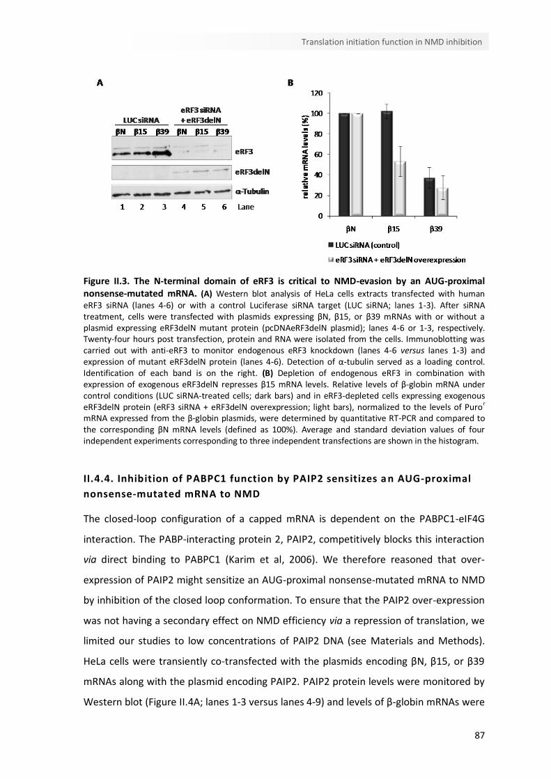
87
Translation initiation function in NMD inhibition
Figure II.3. The N-terminal domain of eRF3 is critical to NMD-evasion by an AUG-proximal nonsense-mutated mRNA. (A) Western blot analysis of HeLa cells extracts transfected with human eRF3 siRNA (lanes 4-6) or with a control Luciferase siRNA target (LUC siRNA; lanes 1-3). After siRNA treatment, cells were transfected with plasmids expressing βN, β15, or β39 mRNAs with or without a plasmid expressing eRF3delN mutant protein (pcDNAeRF3delN plasmid); lanes 4-6 or 1-3, respectively. Twenty-four hours post transfection, protein and RNA were isolated from the cells. Immunoblotting was carried out with anti-eRF3 to monitor endogenous eRF3 knockdown (lanes 4-6 versus lanes 1-3) and expression of mutant eRF3delN protein (lanes 4-6). Detection of α-tubulin served as a loading control. Identification of each band is on the right. (B) Depletion of endogenous eRF3 in combination with expression of exogenous eRF3delN represses β15 mRNA levels. Relative levels of β-globin mRNA under control conditions (LUC siRNA-treated cells; dark bars) and in eRF3-depleted cells expressing exogenous eRF3delN protein (eRF3 siRNA + eRF3delN overexpression; light bars), normalized to the levels of Puror mRNA expressed from the β-globin plasmids, were determined by quantitative RT-PCR and compared to the corresponding βN mRNA levels (defined as 100%). Average and standard deviation values of four independent experiments corresponding to three independent transfections are shown in the histogram.
II.4.4. Inhibition of PABPC1 function by PAIP2 sensitizes a n AUG-proximal
nonsense-mutated mRNA to NMD
The closed-loop configuration of a capped mRNA is dependent on the PABPC1-eIF4G
interaction. The PABP-interacting protein 2, PAIP2, competitively blocks this interaction
via direct binding to PABPC1 (Karim et al, 2006). We therefore reasoned that over-
expression of PAIP2 might sensitize an AUG-proximal nonsense-mutated mRNA to NMD
by inhibition of the closed loop conformation. To ensure that the PAIP2 over-expression
was not having a secondary effect on NMD efficiency via a repression of translation, we
limited our studies to low concentrations of PAIP2 DNA (see Materials and Methods).
HeLa cells were transiently co-transfected with the plasmids encoding βN, β15, or β39
mRNAs along with the plasmid encoding PAIP2. PAIP2 protein levels were monitored by
Western blot (Figure II.4A; lanes 1-3 versus lanes 4-9) and levels of β-globin mRNAs were
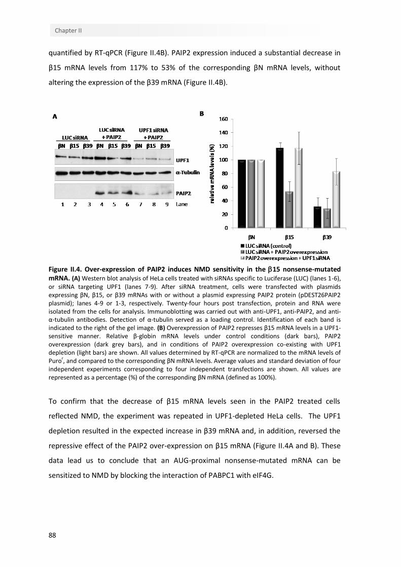
88
Chapter II
quantified by RT-qPCR (Figure II.4B). PAIP2 expression induced a substantial decrease in
β15 mRNA levels from 117% to 53% of the corresponding βN mRNA levels, without
altering the expression of the β39 mRNA (Figure II.4B).
Figure II.4. Over-expression of PAIP2 induces NMD sensitivity in the 15 nonsense-mutated mRNA. (A) Western blot analysis of HeLa cells treated with siRNAs specific to Luciferase (LUC) (lanes 1-6), or siRNA targeting UPF1 (lanes 7-9). After siRNA treatment, cells were transfected with plasmids expressing βN, β15, or β39 mRNAs with or without a plasmid expressing PAIP2 protein (pDEST26PAIP2 plasmid); lanes 4-9 or 1-3, respectively. Twenty-four hours post transfection, protein and RNA were isolated from the cells for analysis. Immunoblotting was carried out with anti-UPF1, anti-PAIP2, and anti-α-tubulin antibodies. Detection of α-tubulin served as a loading control. Identification of each band is indicated to the right of the gel image. (B) Overexpression of PAIP2 represses β15 mRNA levels in a UPF1-sensitive manner. Relative β-globin mRNA levels under control conditions (dark bars), PAIP2 overexpression (dark grey bars), and in conditions of PAIP2 overexpression co-existing with UPF1 depletion (light bars) are shown. All values determined by RT-qPCR are normalized to the mRNA levels of Puro
r, and compared to the corresponding βN mRNA levels. Average values and standard deviation of four
independent experiments corresponding to four independent transfections are shown. All values are represented as a percentage (%) of the corresponding βN mRNA (defined as 100%).
To confirm that the decrease of β15 mRNA levels seen in the PAIP2 treated cells
reflected NMD, the experiment was repeated in UPF1-depleted HeLa cells. The UPF1
depletion resulted in the expected increase in β39 mRNA and, in addition, reversed the
repressive effect of the PAIP2 over-expression on β15 mRNA (Figure II.4A and B). These
data lead us to conclude that an AUG-proximal nonsense-mutated mRNA can be
sensitized to NMD by blocking the interaction of PABPC1 with eIF4G.
89
Translation initiation function in NMD inhibition
II.4.5. The eIF3 subunits that tether the 40S ribosomal subunit to eIF4G are
required for NMD-resistance of an AUG-proximal nonsense-mutated mRNA
Interactions between mRNA 5’ end and the poly(A) tail are thought to be established
during the initial round of translation. These interactions involve interactions of PABPC1
with the initiation factor eIF4G. eIF4G can simultaneously interact with PABPC1 and with
the 40S recruitment factor, eIF3 (Amrani et al, 2008). Mammalian eIF3 is composed of
13 subunits designated eIF3a to eIF3m (Masutani et al, 2007). The structure and
organization of this multisubunit has not been complete elucidated, although
crystallography, mass spectrometry and immunoprecipitation studies allows the
prediction of an interaction map of the free eIF3 as it is represented in Figure II.5A
[adapted from references (Masutani et al, 2007; Wei et al, 2004; Zhou et al, 2008)]. The
eIF3-eIF4G interaction is established via eIF3e and eIF3f subunits (LeFebvre et al, 2006;
Masutani et al, 2007). This interaction of eIF4G with eIF3 has the potential to retain the
eIF4G-bound PABPC1 on the scanning 40S ribosome and on the 80S ribosome during the
early phase of elongation. Besides, in plants, the eIF3h subunit helps to prevent the
permanent loss of reinitiation competence (Roy et al, 2010), suggesting that this subunit
might also be involved in tethering 40S to eIF4G. Such an activity could blunt NMD of an
AUG-proximal nonsense-mutated mRNA by bringing the PABPC1 into close proximity
with the PTC. To test this hypothesis, we assessed the impact of depleting eIF3 subunits
on the expression of an AUG-proximal PTC mRNA. Efficient knock-down was confirmed
at the mRNA level for each respective eIF3 subunit mRNA, by normalization to mRNA
levels of the histone deacetylase 1 (HDAC1) (Figure II.5B, C and D). HDAC1 mRNA was
chosen as an internal control for these analyses since it is constitutively expressed (de
Ruijter et al, 2003). We further demonstrated that under the experimental conditions
used in the study, these depletions did not alter cellular translation as assessed by GFP
reporter expression (Figure II.5E; lanes 3-5 versus lane 2 and 1, respectively).
We next evaluated the effect of individually depleting each of the three eIF3 subunits on
NMD. Plasmids encoding the β15, β39, and βN mRNAs were each transiently expressed
in eIF3h-, eIF3f- or eIF3e-depleted HeLa cells. β-globin mRNA levels, quantified by RT-
qPCR, revealed that eIF3h depletion (Figure II.5F; lanes 4-6 versus lanes 1-3) results in a
decrease of β15 mRNA level from 113% to 79% of the corresponding normal control βN,
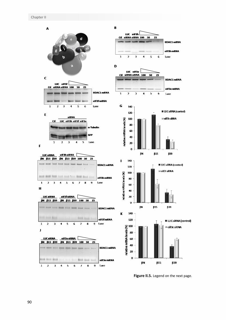
90
Chapter II
Figure II.5. Legend on the next page.
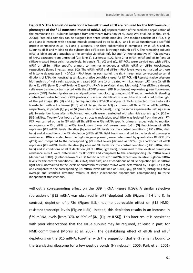
91
Translation initiation function in NMD inhibition
Figure II.5. The translation initiation factors eIF3h and eIF3f are required for the NMD-resistant
phenotype of the 15 nonsense-mutated mRNA. (A) Representation of the predicted organization of the mammalian eIF3 subunits [adapted from references (Masutani et al, 2007; Wei et al, 2004; Zhou et al, 2008)]. Free eIF3 complex can be assigned into three stable modules. One module consists of eIF3a, b, g and i, and it interacts with a second module composed by eIF3c, d, e, l and k. eIF3b functions as a scaffold protein connecting eIF3a, c, i and g subunits. The third subcomplex is composed by eIF3f, h and m. Subunits eIF3f and m bind to the subcomplex eIF3 c:d:e:l:k through subunit eIF3h. The remaining subunit eIF3j, a labile subunit, attaches to the complex via eIF3b. (B), (C) and (D) Representative RT-PCR analyses of RNAs extracted from untreated (Ctl; lane 1), Luciferase (LUC; lane 2) or eIF3h, eIF3f and eIF3e (lane 3) siRNAs-treated HeLa cells, respectively, in panels (B), (C) and (D). RT-PCRs were carried out with eIF3h, eIF3f or eIF3e mRNA specific primers to monitor endogenous eIF3h, eIF3f or eIF3e knockdown, respectively (lanes 3 versus lanes 1-2). The eIF3h, eIF3f and eIF3e mRNA levels were normalized to those of histone deacetylase 1 (HDAC1) mRNA level. In each panel, the right three lanes correspond to serial dilutions of RNA, demonstrating semiquantitative conditions used for RT-PCR. (E) Representative Western blot analysis of HeLa cells extracts, untreated (Ctl, lane 1) or treated with Luciferase (LUC; lane 2), eIF3h (lane 3), eIF3f (lane 4) or eIF3e (lane 5) specific siRNAs (see Material and Methods). After siRNA treatment, cells were transiently transfected with the pEGFP plasmid (BD Biosciences) expressing green fluorescent protein (GFP). Protein lysates were analyzed by immunoblotting using anti-GFP and anti-α-tubulin (loading control) antibodies to monitor GFP protein expression. Identification of each band is indicated to the right of the gel image. (F), (H) and (J) Semiquantitative RT-PCR analyses of RNAs extracted from HeLa cells transfected with a Luciferase (LUC) siRNA target (lanes 1-3) or human eIF3h, eIF3f or eIF3e siRNAs, respectively, at panels (F), (H) or (J) (lanes 4-6 of each panel), using the same experimental settings as in
(B). Twenty-four hours after siRNA-treatment, cells were transfected with plasmids expressing N, 15 or
39 mRNAs. Twenty-four hours after constructs transfection, total RNA was isolated from the cells. RT-PCR was carried out as in (B) with eIF3h, eIF3f or eIF3e mRNA specific primers, respectively, to monitor endogenous eIF3h, eIF3f or eIF3e knockdown (lanes 4-6 versus lanes 1-3). (G) Knockdown of eIF3h represses β15 mRNA levels. Relative β-globin mRNA levels for the control conditions (LUC siRNA; dark bars) and at conditions of eIF3h depletion (eIF3h siRNA; light bars), normalized to the levels of puromycin resistance mRNA encoded from the β-globin gene plasmid, were determined by quantitative RT-PCR (RT-qPCR) and compared to the corresponding βN mRNA levels (defined as 100%). (I) Knockdown of eIF3f represses β15 mRNA levels. Relative β-globin mRNA levels for the control conditions (LUC siRNA; dark bars) and at conditions of eIF3f depletion (eIF3f siRNA; light bars), normalized to the levels of puromycin resistance mRNA were determined by RT-qPCR and compared to the corresponding βN mRNA levels (defined as 100%). (K) Knockdown of eIF3e fails to repress β15 mRNA expression. Relative β-globin mRNA levels for the control conditions (LUC siRNA; dark bars) and at conditions of eIF3e depletion (eIF3e siRNA; light bars), normalized to the levels of puromycin resistance mRNA were determined by RT-qPCR as in (G) and compared to the corresponding βN mRNA levels (defined as 100%). (G), (I) and (K) histograms show average and standard deviation values of three independent experiments corresponding to three independent transfections.
without a corresponding effect on the β39 mRNA (Figure II.5G). A similar selective
repression of β15 mRNA was observed in eIF3f-depleted cells (Figure II.5H and I). In
contrast, depletion of eIF3e (Figure II.5J) had no appreciable effect on β15 NMD-
resistant transcript levels (Figure II.5K). Instead, this depletion results in an increase in
β39 mRNA levels [from 37% to 59% of βN; (Figure II.5K)]. This later result is consistent
with prior observations that the eIF3e subunit may be required, at least in part, for
NMD-commitment (Morris et al, 2007). The destabilizing effect of eIF3h and eIF3f
depletions on the β15 mRNA, together with the suggestion that eIF3 remains bound to
the translating ribosome for a few peptide bonds (Hinnebusch, 2006; Park et al, 2001)

92
Chapter II
are consistent with the model where eIF3h and eIF3f have a role in bringing PABPC1 in
association with eIF4G into the vicinity of the PTC of an mRNA with a short open reading
frame. A schematic of this model is shown in Figure II.6.
Figure II.6. A model for NMD-resistance of AUG-proximal nonsense-mutated mRNAs. The current and prior data supports the model shown in this figure. During cap-mediated translation initiation, PABPC1 interacts with the initiation factor eIF4G. This interaction indirectly tethers PABPC1 to the 40S ribosomal subunit via the interaction of eIF4G with eIF3 subunits. The resulting configuration brings PABPC1 into in the vicinity of the AUG initiation codon as a consequence of 43S scanning and the maintenance of eIF4G-PABPC1 association with the 40S during the initial phase of translation elongation brings it into close contact with an AUG-proximal PTC in a transcript where the ORF is quite short. This proximity to the PTC allows PABPC1 to interact with the release factor eRF3 at the termination complex, thus impairing the association of UPF1 to the ribonucleoprotein complex, resulting in efficient translation termination and inhibition of NMD.
II.5. Discussion
We have previously reported that mammalian mRNAs bearing AUG-proximal PTCs evade
NMD (Romao et al, 2000; Silva et al, 2006). This immunity to NMD is independent of
sequence context of the open reading frame and PTC, independent of transcript
identity, and appears to be a general attribute of the mammalian NMD pathway (Silva et
al, 2006). Extensive mapping studies, using the human β-globin gene as a primary model,
have revealed that this effect reflects a close proximity of a PTC to the translation
initiation codon (Inacio et al, 2004; Silva et al, 2006). In the case of the β-globin mRNA,
PTCs that occur up to 23 codons into the open reading frame effectively evade NMD
while mRNAs with PTCs at a greater distance from the AUG are targeted for decay. We
93
Translation initiation function in NMD inhibition
have further demonstrated that NMD evasion by AUG-proximal PTCs does not, as a rule,
reflect defects in transcript splicing, impaired translation, or reinitiation of translation 3’
to the PTC (Inacio et al, 2004; Silva et al, 2006). Thus, the mechanism underlying the
NMD resistance of the AUG-proximal nonsense-mutated mRNA remains to be more
clearly delineated.
The potential role of translation reinitiation in NMD resistance of AUG-proximal PTCs
has been the subject of a recent report. In that study (Neu-Yilik et al, 2011) the NMD-
resistance of β-globin AUG-proximal nonsense-mutated mRNAs was attributed to
translation reinitiation 3’ to the PTC. It may be of importance, however, to note that the
interpretation of these studies is complicated by the use of a β-globin gene in which
exon 1 was transformed into a functional exon 2 by the introduction of an exogenous
intron into the 5’UTR. The expression of this recombinant β-globin gene, even lacking a
PTC, was repressed when compared to the normal β-globin transcript. In contrast, we
have found using globin genes with native structures, that even in cases in which we do
detect some level of reinitiation 3’ to the AUG-proximal PTC, site-specific elimination of
the reinitiation sites fails to fully restore NMD-sensitivity (Silva et al, 2006). Thus, while a
contribution of translation reinitiation in NMD-evasion cannot be completely ruled out
in any particular circumstance, our detailed analyses of the human β-globin gene lead us
to conclude that AUG-proximity is a major inhibitor of the NMD pathway and that other
mechanisms must be considered.
The present data points to a major role for PABPC1 in protecting mRNAs harboring AUG-
proximal PTCs from the NMD surveillance pathway (Silva et al, 2008; Silva & Romao,
2009). A critical contribution of interactions between PABPC1 and components of
translation initiation complexes is highlighted by the dependence of the NMD evasion on
interactions of PABPC1 with the eIF4G and most probably of eIF4G with eIF3 subunits.
The data further reveal that these interactions are linked to the establishment of an
efficient translation termination reaction at the AUG-proximal PTC. Toeprinting analysis
of capped and polyadenylated human β-globin transcripts reveals substantial ribosome
stalling at the NMD sensitive β39 stop codon that is absent at the termination codon of
the β15 mRNA, as well as at the stop codon of the normal transcript (Figure II.1). The
results of these studies lead us to conclude the interactions of PABPC1 with the
preinitiation of the mRNA specifically blunt the NMD response on an AUG-proximal

94
Chapter II
nonsense-mutated mRNA? Our current observations lead us to propose that this
repositioned PABPC1 can enhance the efficiency of the translation termination at the
AUG-proximal PTC and in so doing represses NMD. It has been demonstrated by others
that PABPC1 can compete with UPF1 for binding to eRF3 with a consequent antagonistic
effect on NMD response (Ivanov et al, 2008; Singh et al, 2008). Recruitment of PABPC1
to the 5’ end of the mRNA, with retention of ribosome association during the early
phase of elongation, would position PABPC1 in close proximity to an early PTC. An
impact on the termination reaction is supported by our prior demonstration that AUG-
proximal nonsense-mutated transcripts fail to bind UPF1 as efficiently as NMD-sensitive
transcripts (Silva et al, 2008). The interactions that support the repositioning of PABPC1
close to an AUG-proximal PTC are further defined by the in vitro observation that
interactions of PABPC1 with eIF4G, eIF4E and eIF3, and the termination factors eRF1 and
eRF3 can form a closed-loop structure during translation of short mRNAs (Amrani et al,
2008). The proposed role for PABPC1 in blunting NMD on AUG-proximal nonsense-
mutated mRNAs is also consistent with the observation by ourselves and others of an
inverse relationship between the distance among the PTC and an artificially tethered
PABPC1 and the strength of NMD (Rebbapragada & Lykke-Andersen, 2009; Silva &
Romao, 2009). It is worth to note that the destabilization of transcripts bearing AUG-
proximal PTCs obtained by the impairment of PABPC1-eRF3 interaction corroborates the
importance of the AUG-proximity effect in NMD-evasion of such transcripts.
In the case of the AUG-proximal nonsense-mutated mRNA, our data supports the
conclusion that the PABPC1-eIF4G interaction enables PABPC1 to travel with the
eIF4F/43S complex, as it scans from the cap to the AUG. The dependence of the AUG-
proximity effect on the function of eIF3 (Figure II.5), which bridges 43S complex to eIF4G
(Hinnebusch, 2006), further supports this model. Thus, we provide evidence that eIF4G
and eIF3 initiation factors are both implicated in PABPC1 inhibitory effect on NMD. This
model is further supported by the observation that the interruption of the PABPC1-
eIF4G interaction by over-expression of PAIP2 leads to NMD of transcripts bearing an
AUG-proximal PTC (Figure II.4). The observations that eIF3 can remain bound to the
translating ribosome during the initial phase of elongation (Hinnebusch, 2006; Park et al,
2001) and that it can function as the link between the eIF4F-bound mRNA (through
eIF4G) and the 40S ribosomal subunit (LeFebvre et al, 2006), supports the notion that
95
Translation initiation function in NMD inhibition
eIF3 subunits might be involved in the delivery of eIF4G-associated PABPC1 to the
vicinity of the AUG-proximal PTC. The strength of the NMD effect decay would then be
determined by the physical distance between PABPC1 at the AUG vicinity and the PTC;
the closer this distance, the more effectively PABPC1 could interact with eRF3,
diminishing the binding of UPF1, with the consequent enhancement of translation
termination and blunting of the NMD response (Figure II.6).
Considering that mRNA circularization (Kahvejian et al, 2001; Wells et al, 1998) and
PABPC1 function as NMD inhibitor when located in the PTC proximity (Amrani et al,
2004; Eberle et al, 2008; Ivanov et al, 2008; Silva et al, 2008; Singh et al, 2008) are
common features in all eukaryotes, the model for the AUG-proximity effect that we
propose here might be also a general attribute for these organisms. Here, we show that
the human eIF3h and eIF3f subunits, which are not conserved in Saccharomyces
cerevisiae, have a role in bringing PABPC1 in association with eIF4G into the vicinity of an
AUG-proximal PTC. While the individual functions of the different mammalian subunits
are not yet well established, many of the eIF3 subunits are thought to be conserved
(Abbott & Proud, 2004; Hinnebusch, 2006; Masutani et al, 2007; Wei et al, 2004; Zhou et
al, 2008). Indeed, the six subunits that constitute the yeast eIF3 - eIF3a, b, c, g, i and j -
are conserved in mammalian (Asano et al, 1998; Hinnebusch, 2000; Hinnebusch, 2006).
However, mammalian eIF3 has evolved to include additional subunits, which are likely to
function as specific regulatory factors and some extra structural motifs provide the
capacity to mediate extra protein-protein or protein-RNA interactions. Accordingly,
eIF3e possesses a PCI (proteosome-COP9-initiation factor) domain, and eIF3f and eIF3h
have a MPN (Mpr1-Pad1-N-terminal) domain. PCI or MPN domains are found in
components of large protein complexes and have been implicated in protein-protein
interactions (Hinnebusch, 2000). In fact, several results have indicated that the non-core
subunits of the mammalian eIF3 regulate specific mRNAs (Choudhuri et al, 2010; Guo et
al, 2000; Ray et al, 2008). Also, one attractive hypothesis is that the presence of a
particular non-core subunit, e.g., eIF3h in the eIF3 protein complex, may in turn govern
the efficiency with which the 43S preinitiation complex is able to scan through the 5’-
UTR of a particular set of mRNAs (Kim et al, 2007). In addition, the non-core subunits
may serve to stabilize the eIF3 complex (Masutani et al, 2007) and/or may have
redundant functions (Choudhuri et al, 2010). It has been shown that mammalian
96
Chapter II
subunits eIF3f:h:m constitute a stable module that is located on the periphery of the
complex and is not involved in interactions between other subunits (Zhou et al, 2008).
Also, it is known that subunit eIF3h is essential for binding the trimer f:h:m to the core
eIF3c subunit (Zhou et al, 2008). Based on these data, the possibility exists that in yeast,
the model for the AUG-proximity effect may be dependent on the eIF3c, which may not
evolved in order to become as specialized as its mammalian ortholog and thus it
performs different biological functions, among which, those attributable to the
mammalian eIF3f and h subunits. The relevance of the mammalian non-conserved eIF3f
and h subunits in bridging eIF4G in association with PABPC1 to the ribosome during the
process of translation initiation, which seem to be maintained during the first steps of
elongation, might be linked to the need of a tighter regulation of translation in higher
eukaryotes.
In conclusion, the present data supports a role for PABPC1 and associated translation
initiation factors in NMD evasion of AUG-proximal nonsense-mutated transcripts. Future
efforts addressing the biochemical mechanism by which PABPC1 is involved in cap-
dependent translation initiation and how it participates in the eIF4F/43S scanning will
contribute to our understanding of mRNA translation.
II.6. Funding
This work was supported by Fundação Luso-Americana para o Desenvolvimento and
Fundação para a Ciência e Tecnologia [(SFRH/BD/35962/2007 to I.P.,
SFRH/BD/63581/2009 to C.B., SFRH/BPD/34583/2007 to A.L.S., SFRH/BPD/20644/2004
to A.I., and Center for Biodiversity, Functional and Integrative Genomics (BioFIG)].
II.7. Acknowledgments
We thank Marla J. Berry (University of Hawaii, Manoa, HI) for providing the pUHD-eRF3
plasmid. We also thank João Lavinha (Instituto Nacional de Saúde Dr. Ricardo Jorge,
Lisboa, Portugal) for comments on the manuscript.
CChhaapptteerr IIIIII
Further insights into the role of eIF3f and eIF3h non-
conserved subunits on translational regulation
Isabel Peixeiro, Cristina Barbosa and Luísa Romão


99
eIF3f and eIF3h function in translation regulation
Author’s note
Some of the presented data in this chapter were obtained by the other indicated author.
Cristina Barbosa was responsible for cloning the luciferase reporter plasmid constructs
and contributed for the luciferase assays.


101
eIF3f and eIF3h function in translation regulation
III.1. Abstract
The mammalian eIF3 is the most complex initiation factor and it comprises 13 non-
identical subunits. eIF3h and eIF3f subunits are non-conserved and seem to function in
translational regulation. Although in plants eIF3h plays a role in translation reinitiation,
and eIF3f is able to interact with the mammalian mTOR pathway, the function of these
subunits in the eIF3 complex is still unclear. A role for these subunits in bridging eIF4G
and PABPC1 to the ribosome during translation has been recently shown in HeLa cells.
Here we explore the role of eIF3f and eIF3h subunits in translational regulation.
Supporting the previously published data, we demonstrate that eIF3h and eIF3f
depletion in HeLa cells affect the association of the eIF3 complex with the ribosome and
eIF4G, respectively. Consistently, eIF3f silencing by RNA interference augments CBP80
association with eIF3-mRNA complexes. Surprisingly, eIF3h does not seem to be involved
in translation reinitiation downstream of a short uORF in human cells. From these
observations, we propose that eIF3f and eIF3h are involved in the interaction of eIF4G
with the ribosome and that, at least the eIF3f subunit, is required for the transition from
the first round of translation to subsequent rounds of translation. These data further
support a function of these non-conserved subunits in translational regulation in higher
eukaryotes.
III.2. Introduction
The efficiency and accuracy of the initiation phase of translation is ensured by numerous
proteins designated eukaryotic initiation factors (eIFs). The first step comprises the
recruitment of the eIF2-GTP-Met-tRNAiMet ternary complex to the small ribosomal
subunit (40S), producing the 43S pre-initiation complex (PIC). This process is mediated
by the multiprotein complex eIF3 together with eIFs 1, 1A and 5. The mRNA 5’ capped-
end binds the eukaryotic initiation factor 4F (eIF4F) complex, composed of the cap-
binding protein eIF4E, as well as eIF4A and eIF4G, which then recruits the 43S pre-
initiation scanning complex (Holcik & Pestova, 2007; Jackson et al, 2010; Sonenberg &
Hinnebusch, 2009). The 3’ end of the mRNA also participates in this process. In fact,

102
Chapter III
PABPC1 is able to bind simultaneously to the cap-associated eIF4G and to the poly(A) tail
(Wells et al, 1998) and it has been described a formation of a closed-loop mRNP during
translation in yeast extracts, that involves PABP, the initiation factors eIF4G, eIF4E and
eIF3 (Amrani et al, 2008). The formed 48S PIC scans the mRNA searching usually for the
first AUG codon. When an AUG codon is recognized by the anticodon of Met-tRNAiMet, in
a process involving the concerted action of eIFs 1, 1A, 2 and 5, eIF5 induces irreversible
hydrolysis of GTP in the eIF2-GTP-Met-tRNAi ternary complex. The Met-tRNAiMet is then
released to the P-site and the large (60S) subunit joins to form an 80S initiation complex
ready to accept the appropriate aminoacyl-tRNA into the A-site to synthesize the first
peptide bond (Pestova, 2007). Subunit joining is catalyzed by eIF5B (Pestova & Hellen,
2000) and is thought to mediate the release of eIF2-GDP and other eIFs. Because eIF3
binds mainly to the solvent side of the 40S subunit, the dissociation of this factor is not
essential for subunit joining and may thus be delayed (Jackson et al, 2010).
eIF3 is the most complex initiation factor and it comprises 13 non-identical subunits
designated from eIF3a to eIF3m in mammalian cells (Unbehaun et al, 2004). In
Saccharomyces cerevisiae, eIF3 only comprises the orthologues of the four core essential
subunits eIF3a, b, c, and g and one noncore subunit eiF3j (Hinnebusch, 2006). The real
composition of the mammalian eIF3 core and the interactions of the different eIF3
subunits are not yet well established. Nevertheless, experiments to reconstitute the
human eIF3 in vitro suggested that the functional core comprises subunits eIF3a, b, c, e,
f and h (Masutani et al, 2007), while a recent study based on tandem mass spectrometry
and solution disruption assays identified the formation of three stable modules:
eIF3(a:b:i:g) resembling the yeast eIF3 core, eIF3(c:d:e:l:k) and eIF3(f:h:m) (Zhou et al,
2008). This multiprotein complex clearly plays a central role in translation initiation
including promoting the binding of the ternary complex and other eIFs to the 40S
subunit, subsequent mRNA recruitment and scanning for AUG recognition. In fact, eIF3
functions as a bridge of the 40S ribosomal subunit to the mRNA by interacting with
eIF4G (Hinnebusch, 2006).
In addition to its functions in general translation, eIF3 was also implicated in translation
reinitiation. Translation reinitiation is a mechanism for up- or downregulation of the
expression of regulatory proteins such as transcription factors and proto-oncogenes in
response to environmental stimuli (Kozak, 2005). Reinitiation may occur after translation
103
eIF3f and eIF3h function in translation regulation
of an upstream open reading frame (uORF), if the 40S ribosomal subunit remains bound
to the mRNA molecule upon translation termination. Following translation termination
and the consequent release of the 60S ribosomal subunit, the 40S subunit may resume
scanning downstream from the uORF. The de novo recruitment of the eIF2-GTP-Met-
tRNAi ternary complex, that will allow recognizing the AUG of the main ORF, determines
the acquirement of full reinitiation competence (Kozak, 1987; Kozak, 2001). The
reinitiation efficiency depends on i) cis-acting mRNA features; ii) the time required for
the uORF translation, which is determined by the relative length of the uORF and the
translation elongation rate; and iii) the translation initiation factors involved in the
translation initiation event (Kozak, 2001; Poyry et al, 2004). It is thought that the eIFs
involved in resuming scanning remain at least transiently associated with the elongating
ribosome, and increasing the uORF length or the ribosome transit time increases the
likelihood of these factors to dissociate (Kozak, 2001). Supporting this idea, in yeast, eIF3
remains 80S-bound for several rounds of elongation and enhances the reinitiation
competence of post-termination 40S ribosomes (Szamecz et al, 2008). Moreover, studies
in yeast have implicated eIF3a and g subunits in this process (Cuchalova et al, 2010;
Szamecz et al, 2008; Valasek et al, 2002), while experiments in plants point for a role of
eIF3h subunit in promoting reinitiation competence during translation of mRNAs
harboring uORFs (Roy et al, 2010).
The role of eIF3 subunits in translation reinitiation further supports the hypothesis that
some subunits of this multiprotein complex are involved in translation regulation rather
than directly participating in translation initiation. Consistently, mammalian eIF3e
subunit, which directly binds eIF4G (LeFebvre et al, 2006), is required for nonsense-
mediated mRNA decay (NMD) (Morris et al, 2007; Peixeiro et al, 2011a). Nonsense-
mediated mRNA decay is a translation-dependent mRNA surveillance mechanism that
degrades transcripts bearing premature translation termination codons (PTCs). The
current model for mammalian NMD proposes that the discrimination between a normal
and a premature termination event is the outcome of the combination of antagonistic
signals at the translation termination event: the decision of NMD triggering will be the
result from the competition between the poly(A)-binding protein 1 (PABPC1) and the
NMD factor UPF1 for the termination complex (Nicholson et al, 2010).

104
Chapter III
We have previously reported that eIF4G and eIF3 initiation factors are both implicated in
PABPC1 inhibitory effect on NMD. Our data demonstrate that human eIF3h and eIF3f
subunits are involved in the NMD-resistance of mRNAs containing PTCs in close
proximity to the translation initiation AUG codon (AUG-proximal PTCs), suggesting that
these non-conserved subunits have a role in bringing PABPC1 in association with eIF4G
into the vicinity of an AUG-proximal PTC (Peixeiro et al, 2011a). Here we aim to provide
further insights into eIF3h and eIF3f subunits regulatory functions. Our data indicates
that eIF3h and eIF3f depletion affect the association of the eIF3 complex with the
ribosome and eIF4G. Surprisingly, eIF3h is not involved in translation reinitiation after a
short uORF. These data support a role for eIF3h and eIF3f subunits in bridging eIF4G-
associated PABPC1 to the ribosome, by interacting both with the 40S ribosomal subunit
and eIF4G.
III.3. Materials and Methods
III.3.1. Plasmid constructs
The pGEMβN-MS2 vector, that carries the binding site for the MS2 phage coat protein,
was obtained by reverse-transcription (RT) of βN-MS2 mRNA, after RNA extraction from
HeLa cells expressing the βN-MS2 plasmid previously described (Silva et al, 2008), with
Superscript II (Invitrogen) according to the manufacturer’s standard protocol and using 2
pmol of reverse primer #1 and 0.5 mM espermidin. The RT product was amplified by
polymerase chain reaction (PCR) using primers #2 and #3 (Table III.1), and then
subjected to a second PCR amplification with primers with HindIII and XbaI linkers (#4
and #5, respectively, Table III.1) and inserted into XbaI/HindIII sites of pGEM plasmid
(Promega). To obtain pGEMβNins5’UTR-MS2 construct, the βWTins5’UTR plasmid (Silva
et al, 2006) was amplified by PCR using one primer with a SmaI linker (#6) and primer #7
(Table III.1) containing a NcoI site. The obtained fragment was cloned into NcoI/SmaI
sites of pGEMβN-MS2.
The plasmid encoding firefly luciferase (pGL2hCMV) was obtained by insertion of hCMV
promoter sequence from pcDNA3.1/hygro+ BglII/HindIII digested, into BglII/HindIII sites
of pGL2-enhancer (Promega). The puORFEPOLuc construct was obtained by inserting the
erythropoietin (EPO) uORF sequence upstream of the luciferase ORF in the pGL2hCMV

105
eIF3f and eIF3h function in translation regulation
plasmid. This was by overlap-extension PCR with flanking primers #8 (with a HindIII
linker) and #9 and overlapping primers #10 and #11 (Table III.1), and the EPO gene and
the luciferase ORF in pGL2hCMV plasmid as templates. The obtained fragment was
cloned into HindIII/XbaI sites of pGL2hCMV. The pGL2uORF(TTG)EPO variant, carrying a
mutation at the uORF AUG codon (ATG TTG), was obtained by overlap-extension PCR
with flanking primers #8 and #9 and overlapping primers #12 and #13, and the
pGL2uORFEPO as template and the resultant primer was cloned into HindIII/XbaI. The
pRL-TK plasmid encoding Renilla luciferase was a kind gift of Margarida Gama-Carvalho.
Table III.1. DNA oligonucleotides used in the current work.
Primer Sequence (5’ → 3’)
#1 AAGCAGTGGTATCAACGCAGAGTAC(T)30 #2 AAGCAGTGGTATCAACGCAGA #3 ACATTTGCTTCTGACACAAC #4 GCCAAGCTTGTATCAACGCAGAGTACTTT #5 TGCTCTAGAACATTTGCTTCTGACACAAC #6 TCCCCCGGGACATTTGCTTCTGACACAACTG #7 TGCACCATGGTGTCTGTTTG #8 CCCAAGCTTCCCGGAGCCGGACCGG #9 CGTACGTGATGTTCACCTC #10 GCCAGGCGCGGAGATGGAAGACGCCAAAAACATAAAG #11 CTTTATGTTTTTGGCGTCTTCCATCTCCGCGCCTGGC #12 CTTCCCGGGTTGAGGGC #13 GCCCTGAACCCGGGAAG #14 CTGCTCATTGCAGGCCAGAT #15 GAGCCTGGGCCATGAAGAG #16 CACCCAGTCATTTTGGCCTC #17 CGACAGTTCCCAACAGGGTC #18 GGACAAGCATGGTTTTAGGCA #19 TGCTGCTCCTGAGTAATTCCC #20 ATGGCGCAGACGCAGGG #21 CCGCACTAGGCTGGAACATC #22 AAACTCTCAAGGATCTTTCC #23 TTGGACTTAGGGAACAAAGG #24 ACATTTGCTTCTGACACAAC #25 TGCCACACTGAGTGAGCTG
III.3.2. Cell culture, plasmid and siRNA transfection
HeLa cells, stably expressing the tet transactivator (HeLa/tTA) (Kong et al, 2003), were
grown in Dulbecco’s modified Eagle’s medium supplemented with 10% fetal bovine
serum. The short RNA interference (siRNA) duplexes (Table III.2) were designed as 19-

106
Chapter III
mers with 3’-dTdT overhangs and purchased from Thermo. For the Luciferase assay
transfections of cells with siRNAs were carried out using Lipofectamine 2000
Transfection Reagent (Invitrogen), following the manufacturer’s instructions, in 35-mm
plates using 200 pmol of siRNA oligonucleotides and 4µl of transfection reagent. Twenty
four hours later, 750 ng of pGL2hCMV, pGL2uORFEPO or pGL2uORF(TTG)EPO were co-
transfected with 750ng of the pRL-TK plasmid. For immunoprecipitations, HeLa cells
were seeded in 60-mm dishes and transfected 20 hours later using 400 pmol of siRNA
oligonucleotides and 8 µl of Lipofectamine 2000 reagent, according to the supplier’s
protocol.
Table III.2. Sequences of the siRNAs used in the current work.
siRNA Sequence (5’ → 3’)
eIF3h1 ACUGCCCAAGGAUCUCUCU eIF3h2 GAUCGGCUUGAAAUUACCA eIF3f1 GUGAAGGAGAAAUGGGUUU eIF3f2 AUACGCGUACUACGACACU eIF3e1 CCAGGGAUGGUAGGAUGCU eIF3e2 UGCAGAAUUGGGAUGCAGC GFP GGCUACGUCCAGGAGCGCAC
III.3.3. RNA isolation
Total RNA from transfected cells was prepared using the Nucleospin RNA extraction II
(Marcherey-Nagel) following the manufacturer’s instructions.
III.3.4. Semi-quantitative RT-PCR
1500 ng of total mRNA were reverse-transcribed with Superscript II Reverse
Transcriptase (Invitrogen) according to the manufacturer’s standard protocol and using
250 ng of Random Primers (Invitrogen) in a final volume of 20 µl. The PCR reactions for
eIF3h, eIF3f or eIF3e and histone deacetylase 1 (HDAC1) cDNAs were performed in
parallel at similar conditions: 3 µl of the RT product was amplified in a 50-µl reaction
volume using 0.2 mM dNTPs, 1.5 mM MgCl2, 15 pmol of each primer (primers #14 and
#15 for eIF3h, primers #16 and #17 for eIF3f, primers #18 and #19 for eIF3e and primers
#20 and #21 for HDAC1; Table III.1), 0.75 U of Amplitaq (Promega), and 1X PCR buffer
(Promega). Thermocycler conditions were 95°C for 4 min followed by 26 cycles of 95°C
107
eIF3f and eIF3h function in translation regulation
for 45 sec, 56°C for 45 sec, and 72°C for 45 sec followed by a final extension of 72°C for
10 min. Ten-microliter aliquots from each RT-PCR sample were analyzed by
electrophoresis on 1.8% agarose gels.
III.3.5. Dual luciferase assay
Co-transfected HeLa cells were lysed with Passive lysis buffer (Promega) and
luminescence was measured in Lucy 2 Luminometer (Anthos Labtec) with the Dual
Luciferase Assay System (Promega) according to the manufacturer’s indications.
III.3.6. In vitro transcription and capping
The pGEMβN-MS2 and pGEMβNins5’UTR-MS2 plasmids carrying the βN-MS2 cDNA
containing a (T)30-tail were linearized with HindIII and purified. Polyadenylated RNA was
synthesized with T7 RNA polymerase using the DNA linearized templates and the
Ribomax large scale kit (Promega), according to the supplier’s protocol, with or without
cap addition. The RNA samples were treated with RNase-free DNase I (Ambion), purified
by phenol:chloroform extraction and the yield of RNA quantified.
III.3.7. Immunoprecipitation
HeLa cells cultured in 60-mm dishes and treated with siRNAs were collected 72h after
transfection. Cells were lysed in 300 µl of NP40 buffer [500mM Tris-HCl, pH 7.4; 100 mM
NaCl; 10mM MgCl2; 0.1% (v/v) Nonidet P-40; 100 U of RNase inhibitor per ml
(Invitrogen); protease inhibitor mixture (Sigma)]. Total lysates were cleared by
centrifugation at 2,500g for 5 min. A 40 µl aliquot of total lysate (Pre-IP) was saved for
RNA and protein analysis (a 20 µl sample was used for RNA extraction as described
above, prior to semi-quantitative RT-PCR analysis; 4 µl of 5x SDS loading buffer was
added to the remaining 20 µl sample for Western-blot analysis). The lysates were
incubated for 2 hours at 4oC with rabbit polyclonal anti-eIF3F (Abcam) or rabbit
polyclonal anti-eIF3H (Cell Signaling) at a 1:100 dilution. An 80 µl volume of protein G-
agarose beads (Roche) were then added and the samples were incubated for 1 hour at
4oC, washed 4 times with excess NP40 buffer and ressuspended in 30 µl of 2x SDS
sample buffer.

108
Chapter III
III.3.8. SDS-PAGE and Western blotting
Protein lysates were resolved, according to standard protocols, in 10% SDS-PAGE and
transferred to PVDF membranes (Bio-Rad). Membranes were probed using mouse
polyclonal anti-PCNA (Abcam) at 1:10,000 dilution (as a loading control), goat polyclonal
anti-hUPF1 (Bethyl Labs) at 1:500 dilution; rabbit polyclonal anti-PABPC1 (Cell Signaling)
at 1:500 dilution, rabbit monoclonal anti-eIF3H (Cell Signaling), rabbit polyclonal anti-
CPB80 (generous gift from E. Izaurralde), rabbit polyclonal anti-eIF4G (Cell Signaling) or
mouse monoclonal anti-RPS6 (Cell Signaling) at 1:250 dilution. Detection was carried out
using secondary peroxidase-conjugated anti-mouse IgG (Bio-Rad), anti-rabbit IgG (Bio-
Rad) or anti-goat IgG (Sigma) antibodies followed by chemiluminescence.
III.3.9. GST-Pull down assays
Recombinant GST-MS2 was expressed by transforming DH5α with pGEX-MS2 plasmid.
250 ml of LB media supplemented with 50 µg/ml ampicillin were inoculated with 5 ml of
overnight culture. At OD600 ≈ 0.9, expression was induced with 0.1 mM IPTG for 3h and
cells were pellet. Cell pellet was resuspended in 2 ml of lysis buffer [50 mM TrisHCl pH
7.5, 50 mM NaCl, 5 mM MgCl2, 1 mM DTT and protease inhibitor mixture (Sigma)] ,
sonicated for 4x 30 sec bursts, and centrifugated at 6,000rpm for 30min at 4oC.
Supernatant was bound to 1ml of glutathione-agarose beads (Sigma) overnight at 4oC,
washed 4 times with excess PBS-T and resuspendend in PBS (1:1).
Transcripts lacking the poly(A) tail were obtained by hybridization of uncapped
GEMβNins5’UTR mRNA with 500ng of oligo(dT)18 (Promega), followed by RNase H
(Promega) digestion (4.0 U per 100 µl of total volume) in RNase H buffer (20 mM HEPES,
50 mM KCl, 10 mM MgCl2, 1 mM DTT). The undeadenylated RNAs were purified by
phenol:chloroform extraction.
300 ng of each substrate for in vitro pull-down assays were first heated to 65oC for 5 min
and cooled to 37oC for 5 min and immediately added to the translation mixture,
comprising 25 µl of supplemented untreated rabbit reticulocyte lysate [URRL (Promega)
containing 0.6 M AcK, 5 mM MgCl2, 1 mg/ml creatine phosphokinase (Sigma), 100 mM
creatine phosphate (Sigma), 0.2 mM amino‐acid mixture (Promega), 2.5 mM DTT, 0.25
109
eIF3f and eIF3h function in translation regulation
mg/ml E. coli tRNA, 0.02 mM hemin, 0.6 U/μl RNase inhibitor (Invitrogene) and 0.5 g/L
cyclohexamide (CHX)]. The reactions were incubated at 30oC for 10 min. For RNase H
digestion, the translation mixture was supplemented with 500 mg of ssDNA probe
(5’CCCACGTTCTTCGGG3’), 10 µl of RNase H Buffer 10x and 4.0 U of RNase H (Promega)
in a final volume of 100 µl and incubated for 2h at 37oC. After the addition of 200 µl RSB-
T (10 mM TrisHCl pH 7.5, 100 mM NaCl, 2.5 mM MgCl2, 0.5 v/v Triton X-100) and 25 µl of
GST-MS2 agarose beads and nutation for 3h at 4oC, the beads were washed 6 times with
excess RSB-T and resuspended in elution buffer (100 mM TrisHCl pH 7.0, 150 mM NaCl,
12.5 mM EDTA, 1% SDS) and heated at 95oC for 3min. RNA samples were then purified
by phenol:chloroform extraction and reversed-transcribed with Superscript II
(Invitrogen) according to the manufacturer’s standard protocol and using 2 pmol of
primer #22 for the 5’UTR fragment or #23 for the 3’UTR fragment. The RT products
were amplified by PCR using primers #22 and #24 for the 5’UTR fragment and primers
#23 and # 25 for the 3’UTR fragment.
III.4. Results
III.4.1. mRNA acquires a closed-loop conformation during in vitro
translation in rabbit reticulocyte lysate
Mammalian PABPC1 directly binds eIF4G (Imataka et al, 1998). We thus hypothesized
that, as in yeast (Amrani et al, 2008), this interaction may mediate the formation of a
closed-loop mRNP in mammalian extracts. To test this hypothesis, we employed an in
vitro pull-down assay. We synthesized transcripts that included three binding sites for
the bacteriophage MS2 coat protein at the 3’ end of the βN capped and with a 30nt
poly(A) tail or βN uncapped and deadenylated (Figure III.1A). These mRNAs were
subjected to in vitro translation in supplemented URRL containing CHX to block peptidyl
transferase, thus enriching the sample with stalled ribosomes at the AUG initiation
codon. These mRNAs were cleaved by RNase H upstream of the AUG codon (Figure
III.1A, ssDNA probe), the 3’ fragments were selected by binding to recombinant GST
fused to MS2 coat protein (GST-MS2) and the pellet was assayed for the presence of the
5’ fragment (Figure III.1B). Efficient co-selection of the 5’ fragment was dependent of an
intact cap structure and poly(A) tail. This result suggest that the mRNA acquires a closed-

110
Chapter III
5’ insm7G
AUG
MS2
5’ fragment 3’ fragmentssDNAprobe
βNins5’-MS2 capped and polyadenylated
5’ ins
AUG
MS2
5’ fragment 3’ fragmentssDNAprobe
βNins5’-MS2 uncapped and deadenylated
5’ fragment
3’ fragment
1 2 3 Lane
ssDNAprobe
+ +- RT-PCR:
A B
loop structure during in vitro translation due to 5’ – 3’ ends interactions that is
maintained after ribosome scanning and is dependent of the presence of an intact
poly(A) tail.
Figure III.1. 5’-3’ mRNA ends interaction during translation in rabbit reticulocyte lysates. (A) Schematic representation of the human β-globin mRNAs assayed in the GST-pull down showing positions of the MS2-binding site, of the 5’ spacer insertion, and complementarity to the oligonucleotide used for RNase H cleavage. The arrows indicate the position and orientation of the primers used for RT-PCR. (B) Pannels show the pellet of the 5’ and 3’ fragment as indicated. Samples in lanes 2 and 3 were treated with the oligonucleotide and subjected to RNase H cleavage prior to the GST-pull down.
III.4.2. eIF3f silencing affects eIF3h expression
Since eIF3h and eIF3f subunits interact with each other and assemble a subcomplex of
eIF3 by also interacting with the subunit eIF3m (Zhou et al, 2008), we investigated the
effect of individually depleting eIF3h and eIF3f subunits on eIF3h proteins levels. HeLa
cells were eIF3h- or eIF3f-depleted by RNAi and, two days later, protein lysates were
analyzed by Western blot. Both eIF3h and eIF3f knockdown resulted in decreased eIF3h
protein levels (Figure III.2A and B). The incubation with the anti-eIF3h antibody revealed
two distinct bands, that may correspond to eIF3h isoforms 3 (UniProt O15372) and eIF3h
isoform 8 (UniProt E5RJT0), according to the molecular weight of ≈ 40 kDa and ≈ 28 kDa,
respectively. To confirm that eIF3h destabilization observed in eIF3f-depleted cells was
specific of eIF3f depletion and not an outcome of eIF3 complex destabilization, protein
lysates from eIF3e-depleted HeLa cells were also analyzed by Western blot. The
incubation with anti-eIF3h antibody revealed that eIF3e depletion does not significantly
affect the amount of eIF3h subunit (Figure III.2C). Efficient knockdown was confirmed at
the mRNA level for eIF3h and eIF3f subunits mRNA, by normalization to mRNA levels of
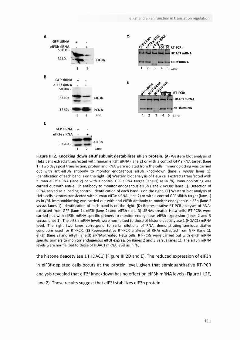
111
eIF3f and eIF3h function in translation regulation
DA
E
100
eIF3h mRNA
HDAC1 mRNA
50
2 3 4 5 Lane1
RT-PCR:
B
HDAC1 mRNA
RT-PCR:
eIF3f mRNA
2 3 4 5 Lane1
100 50
eIF3h
GFP siRNA
37 kDa -
eIF3h siRNA50 kDa -
+ -- +
21
Lane
+ -
37 kDa -
50 kDa -
GFP siRNA
eIF3f siRNA - +
PCNA37 kDa -
21
eIF3h
Lane
+ -
37 kDa -
GFP siRNA
eIF3e siRNA - +
21
eIF3h
C
Figure III.2. Knocking down eIF3f subunit destabilizes eIF3h protein. (A) Western blot analysis of HeLa cells extracts transfected with human eIF3h siRNA (lane 2) or with a control GFP siRNA target (lane 1). Two days post transfection, protein and RNA were isolated from the cells. Immunoblotting was carried out with anti-eIF3h antibody to monitor endogenous eIF3h knockdown (lane 2 versus lanes 1). Identification of each band is on the right. (B) Western blot analysis of HeLa cells extracts transfected with human eIF3f siRNA (lane 2) or with a control GFP siRNA target (lane 1) as in (B). Immunoblotting was carried out with anti-eIF3h antibody to monitor endogenous eIF3h (lane 2 versus lanes 1). Detection of PCNA served as a loading control. Identification of each band is on the right. (C) Western blot analysis of HeLa cells extracts transfected with human eIF3e siRNA (lane 2) or with a control GFP siRNA target (lane 1) as in (B). Immunoblotting was carried out with anti-eIF3h antibody to monitor endogenous eIF3h (lane 2 versus lanes 1). Identification of each band is on the right. (D) Representative RT-PCR analyses of RNAs extracted from GFP (lane 1), eIF3f (lane 2) and eIF3h (lane 3) siRNAs-treated HeLa cells. RT-PCRs were carried out with eIF3h mRNA specific primers to monitor endogenous eIF3h expression (lanes 2 and 3 versus lanes 1). The eIF3h mRNA levels were normalized to those of histone deacetylase 1 (HDAC1) mRNA level. The right two lanes correspond to serial dilutions of RNA, demonstrating semiquantitative conditions used for RT-PCR. (E) Representative RT-PCR analyses of RNAs extracted from GFP (lane 1), eIF3h (lane 2) and eIF3f (lane 3) siRNAs-treated HeLa cells. RT-PCRs were carried out with eIF3f mRNA specific primers to monitor endogenous eIF3f expression (lanes 2 and 3 versus lanes 1). The eIF3h mRNA levels were normalized to those of HDAC1 mRNA level as in (D).
the histone deacetylase 1 (HDAC1) (Figure III.2D and E). The reduced expression of eIF3h
in eIF3f-depleted cells occurs at the protein level, given that semiquantitative RT-PCR
analysis revealed that eIF3f knockdown has no effect on eIF3h mRNA levels (Figure III.2E,
lane 2). These results suggest that eIF3f stabilizes eIF3h protein.
112
Chapter III
III.4.3. eIF3f depletion decreases eIF4G -eIF3 interaction and increases
CBP80-mRNA association
It has been suggested that eIF3f subunit plays a role in translation regulation by directly
binding mTOR, which controls the association of eIF4G with eIF3 (Harris et al, 2006). Our
previous findings showing that eIF3f subunit is required for the NMD-resistance of AUG-
proximal nonsense-mutated mRNAs (Peixeiro et al, 2011a) also support a possible
function of eIF3f in mediating eIF4G (and associated PABPC1) interaction with eIF3-
bound ribosome.
To address eIF3f role in eIF4G-eIF3 interaction, HeLa cells were treated with control or
eIF3f-specific siRNAs and total lysates were generated two days later. Relative to the
Pre-IP, 6.5-fold more amount of each lysate (GFP-depleted or eIF3f-depleted cells
lysates) was separately immunoprecipitated using anti-eIF3h antibody and protein G-
agarose beads or protein G-agarose beads alone to control for nonspecific
immunoprecipitation (IP). Western blot analysis using eIF4G1 antibody revealed that
eIF3h immunoprecipitated eIF4G (Figure III.3, top panel) in a RNA-independent manner.
Even though we were unable to detect full-length eIF4G in eIF3f-depleted cells, eIF3h
specifically immunoprecipitated two proteins (of ≈ 130 kDa and ≈ 110 kDa) in eIF3f
siRNA-treated cells detected with the anti-eIF4G antibody, even after RNase treatment.
These bands might correspond to eIF4G cleavage fragments, as the ones that result from
caspase-mediated proteolytic cleavage in apoptotic cells (Marissen & Lloyd, 1998).
Most capped mRNAs are subjected to NMD almost immediately after transcription,
when they are in transit to the cytoplasm and still bound by the cap binding complex
(CBC) CBP80-CBP20 (Chiu et al, 2004). The CBC supports the pioneer round of
translation, and the cap is then replaced by eIF4E that directs subsequent rounds of
translation (Ishigaki et al, 2001). Since eIF3f depletion targets AUG-proximal nonsense-
mutated mRNAs for decay, we investigated whether eIF3f silencing could increase
CBP80 association with complexes containing eIF3. Immunoblotting using anti-CBP80
antibody shows an increased RNA-dependent association of CBP80 with eIF3h
containing complexes in eIF3f-depleted cells (Figure III.3, lane 5 and 7 vs lane 4 and 6).
Together these results suggest that eIF3f subunit is involved in eIF3-eIF4G interaction
and that it might be required for the transition from the pioneer round of translation to
subsequent rounds of translation.
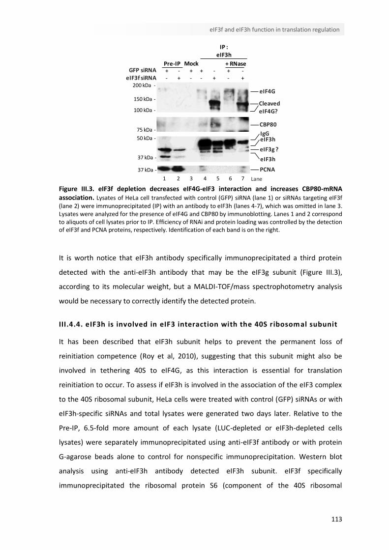
113
eIF3f and eIF3h function in translation regulation
37 kDa -
IP :eIF3h
200 kDa -
150 kDa -
100 kDa -
75 kDa -
37 kDa -
50 kDa -
eIF4G
CleavedeIF4G?
CBP80
eIF3h
eIF3h
eIF3g ?
IgG
-GFP siRNAPre-IP Mock
eIF3f siRNA+ - + + -- + - - +
+- +
+ RNase
PCNA
2 3 4 Lane1 5 6 7
Figure III.3. eIF3f depletion decreases eIF4G-eIF3 interaction and increases CBP80-mRNA association. Lysates of HeLa cell transfected with control (GFP) siRNA (lane 1) or siRNAs targeting eIF3f (lane 2) were immunoprecipitated (IP) with an antibody to eIF3h (lanes 4-7), which was omitted in lane 3. Lysates were analyzed for the presence of eIF4G and CBP80 by immunoblotting. Lanes 1 and 2 correspond to aliquots of cell lysates prior to IP. Efficiency of RNAi and protein loading was controlled by the detection of eIF3f and PCNA proteins, respectively. Identification of each band is on the right.
It is worth notice that eIF3h antibody specifically immunoprecipitated a third protein
detected with the anti-eIF3h antibody that may be the eIF3g subunit (Figure III.3),
according to its molecular weight, but a MALDI-TOF/mass spectrophotometry analysis
would be necessary to correctly identify the detected protein.
III.4.4. eIF3h is involved in eIF3 interaction with the 40S ribosomal subunit
It has been described that eIF3h subunit helps to prevent the permanent loss of
reinitiation competence (Roy et al, 2010), suggesting that this subunit might also be
involved in tethering 40S to eIF4G, as this interaction is essential for translation
reinitiation to occur. To assess if eIF3h is involved in the association of the eIF3 complex
to the 40S ribosomal subunit, HeLa cells were treated with control (GFP) siRNAs or with
eIF3h-specific siRNAs and total lysates were generated two days later. Relative to the
Pre-IP, 6.5-fold more amount of each lysate (LUC-depleted or eIF3h-depleted cells
lysates) were separately immunoprecipitated using anti-eIF3f antibody or with protein
G-agarose beads alone to control for nonspecific immunoprecipitation. Western blot
analysis using anti-eIF3h antibody detected eIF3h subunit. eIF3f specifically
immunoprecipitated the ribosomal protein S6 (component of the 40S ribosomal
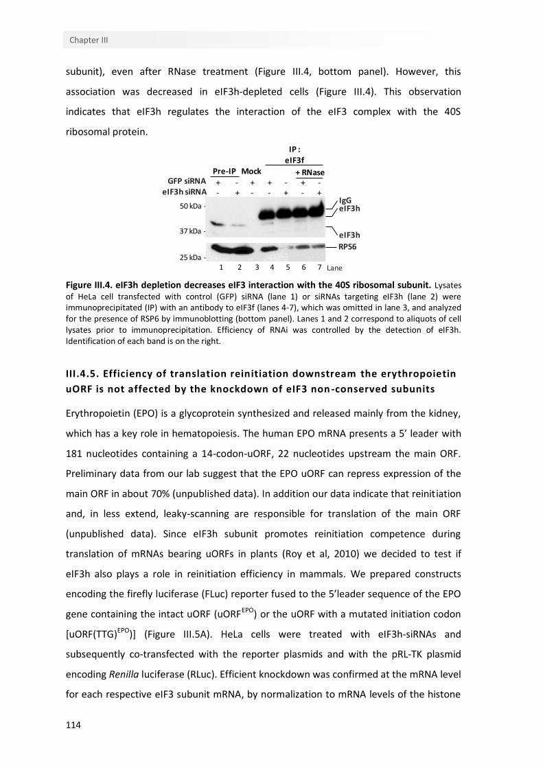
114
Chapter III
-
eIF3h
GFP siRNAPre-IP Mock
IP :eIF3f
37 kDa -
eIF3h siRNA
50 kDa -
eIF3h
IgG
+ - + + -- + - - +
+- +
+ RNase
25 kDa -
RPS6
2 3 4 Lane1 5 6 7
subunit), even after RNase treatment (Figure III.4, bottom panel). However, this
association was decreased in eIF3h-depleted cells (Figure III.4). This observation
indicates that eIF3h regulates the interaction of the eIF3 complex with the 40S
ribosomal protein.
Figure III.4. eIF3h depletion decreases eIF3 interaction with the 40S ribosomal subunit. Lysates of HeLa cell transfected with control (GFP) siRNA (lane 1) or siRNAs targeting eIF3h (lane 2) were immunoprecipitated (IP) with an antibody to eIF3f (lanes 4-7), which was omitted in lane 3, and analyzed for the presence of RSP6 by immunoblotting (bottom panel). Lanes 1 and 2 correspond to aliquots of cell lysates prior to immunoprecipitation. Efficiency of RNAi was controlled by the detection of eIF3h. Identification of each band is on the right.
III.4.5. Efficiency of translation reinitiation downstream the erythropoietin
uORF is not affected by the knockdown of eIF3 non -conserved subunits
Erythropoietin (EPO) is a glycoprotein synthesized and released mainly from the kidney,
which has a key role in hematopoiesis. The human EPO mRNA presents a 5’ leader with
181 nucleotides containing a 14-codon-uORF, 22 nucleotides upstream the main ORF.
Preliminary data from our lab suggest that the EPO uORF can repress expression of the
main ORF in about 70% (unpublished data). In addition our data indicate that reinitiation
and, in less extend, leaky-scanning are responsible for translation of the main ORF
(unpublished data). Since eIF3h subunit promotes reinitiation competence during
translation of mRNAs bearing uORFs in plants (Roy et al, 2010) we decided to test if
eIF3h also plays a role in reinitiation efficiency in mammals. We prepared constructs
encoding the firefly luciferase (FLuc) reporter fused to the 5’leader sequence of the EPO
gene containing the intact uORF (uORFEPO) or the uORF with a mutated initiation codon
[uORF(TTG)EPO)] (Figure III.5A). HeLa cells were treated with eIF3h-siRNAs and
subsequently co-transfected with the reporter plasmids and with the pRL-TK plasmid
encoding Renilla luciferase (RLuc). Efficient knockdown was confirmed at the mRNA level
for each respective eIF3 subunit mRNA, by normalization to mRNA levels of the histone
115
eIF3f and eIF3h function in translation regulation
deacetylase 1 (HDAC1) (Figure III.5B). In transient expression the EPO uORF repressed
luciferase activity by about 3-fold in control conditions (GFP siRNA) and by about 3.5-
fold in eIF3h-depleted cells (Figure III.5C). These results failed to reveal a significant role
for eIF3h in regulation of translation reinitiation efficiency. The same experiment was
done for eIF3f- and eIF3e-depleted cells (Figure III.5D and III.5E, respectively). Similar
results were obtained in control cells and in eIF3f- or eIF3e-depleted cells (Figure III.5F)
suggesting that these subunits are not involved in translation reinitiation efficiency
downstream of the EPO uORF.
III.5. Discussion
Our findings suggest that eIF3h and eIF3f subunits are involved in the association of the
eIF3 complex to the ribosome and eIF4G. Even though it has been attributed a role for
these subunits in translation regulation, the functional contribution of these subunits in
the eIF3 complex has not been defined. Orthologues for eIF3h and eIF3f subunits are not
found in S.cerevisae; however, in S. pombe, eIF3f is essential for cell viability and general
translation (Zhou et al, 2005) and eIF3h physically associates with the 40S ribosomal
particles but is not indispensable for global protein synthesis (Ray et al, 2008). eIF3f and
eIF3h both contain a MPN (Mpr1-Pad1-N-terminal) domain. MPN domains are found in
components of large protein complexes and have been implicated in protein-protein
interactions (Hinnebusch, 2000) supporting a possible interaction of these proteins with
other initiation factor complexes. In addition, the non-core subunits may serve to
stabilize the eIF3 complex (Masutani et al, 2007). In accordance, our findings suggest
that eIF3f stabilizes eIF3h subunit, since the knockdown of eIF3f resulted in reduced
eIF3h protein levels (Figure III.2).
eIF3f subunit was reported to bind mTOR, which controls the association of eIF3 and
eIF4G (Harris et al, 2006). Besides eIF4G, one of the best characterized targets of mTOR
is the eIF4E binding protein (4EBP1). Hypophosphorylated 4EBP1 inhibits cap-dependent
translation by blocking eIF4E binding to eIF4G (Gingras et al, 1999a). Interestingly, eIF3f
depletion reduced the amount of intact eIF4G associated with the eIF3 complex by
apparently leading to an increase in the amount of eIF4G cleavage fragments (Figure
III.3). An attractive explanation for this observation is that when mTOR-eIF3f interaction
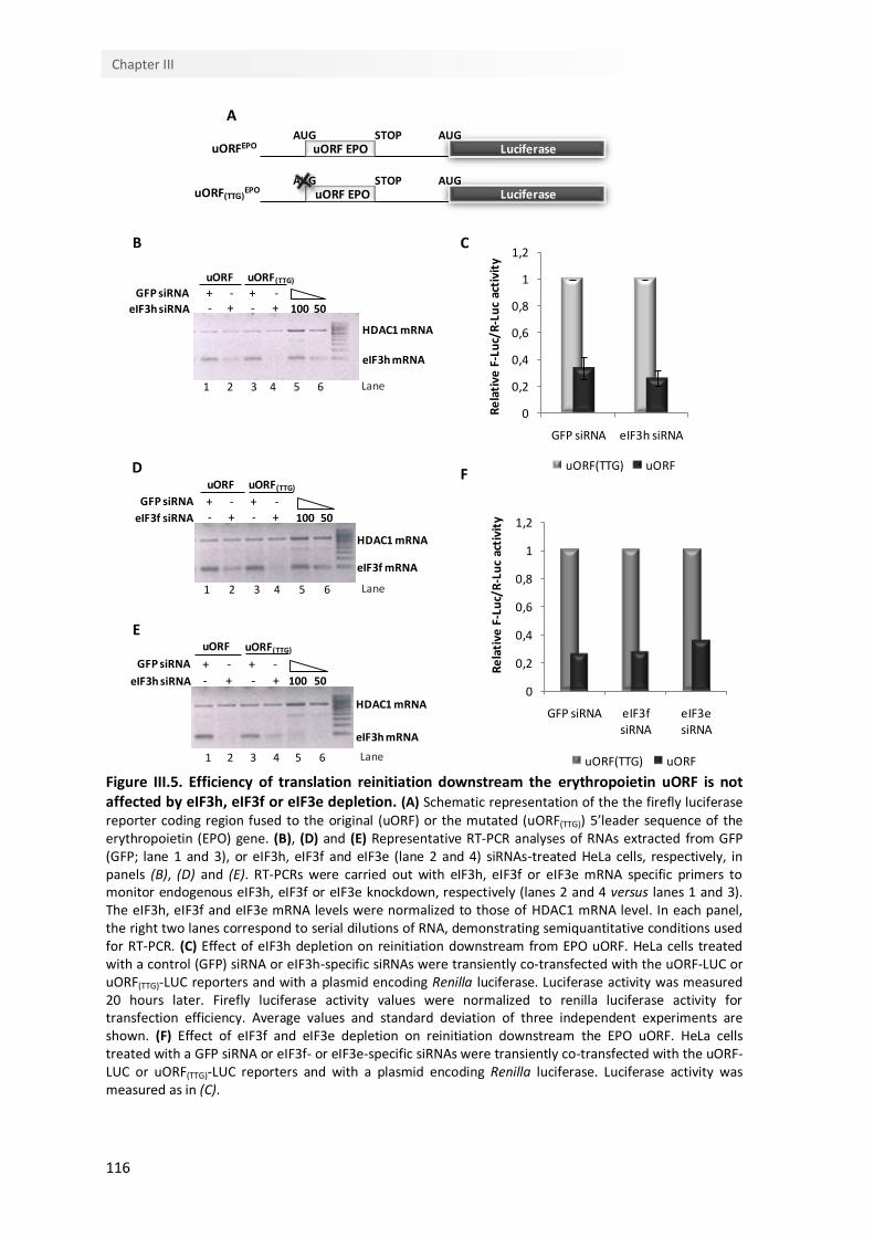
116
Chapter III
eIF3h mRNA
HDAC1 mRNA
GFP siRNA
eIF3h siRNA 100 50
1 2 3 4 5 6 Lane
uORF uORF(TTG)
+ -- +
+ -- +
eIF3f mRNA
HDAC1 mRNA
GFP siRNA
eIF3f siRNA 100 50
1 2 3 4 5 6 Lane
uORF uORF(TTG)
+ -- +
+ -- +
eIF3h mRNA
HDAC1 mRNA
GFP siRNA
eIF3h siRNA
1 2 3 4 Lane5 6
uORF uORF(TTG)
+ -- +
+ -- + 100 50
LuciferaseuORF EPOAUG AUGSTOP
LuciferaseuORF EPOAUG AUGSTOP
A
B C
D F
E
uORFEPO
uORF(TTG)EPO
0
0,2
0,4
0,6
0,8
1
1,2
GFP siRNA eIF3h siRNA
Re
lati
ve F
-Lu
c/R
-Lu
c ac
tivi
ty
uORF(TTG) uORF
0
0,2
0,4
0,6
0,8
1
1,2
GFP siRNA eIF3f siRNA
eIF3e siRNA
Re
lati
ve F
-Lu
c/R
-Lu
c ac
tivi
ty
uORF(TTG) uORF
Figure III.5. Efficiency of translation reinitiation downstream the erythropoietin uORF is not affected by eIF3h, eIF3f or eIF3e depletion. (A) Schematic representation of the the firefly luciferase reporter coding region fused to the original (uORF) or the mutated (uORF(TTG)) 5’leader sequence of the erythropoietin (EPO) gene. (B), (D) and (E) Representative RT-PCR analyses of RNAs extracted from GFP (GFP; lane 1 and 3), or eIF3h, eIF3f and eIF3e (lane 2 and 4) siRNAs-treated HeLa cells, respectively, in panels (B), (D) and (E). RT-PCRs were carried out with eIF3h, eIF3f or eIF3e mRNA specific primers to monitor endogenous eIF3h, eIF3f or eIF3e knockdown, respectively (lanes 2 and 4 versus lanes 1 and 3). The eIF3h, eIF3f and eIF3e mRNA levels were normalized to those of HDAC1 mRNA level. In each panel, the right two lanes correspond to serial dilutions of RNA, demonstrating semiquantitative conditions used for RT-PCR. (C) Effect of eIF3h depletion on reinitiation downstream from EPO uORF. HeLa cells treated with a control (GFP) siRNA or eIF3h-specific siRNAs were transiently co-transfected with the uORF-LUC or uORF(TTG)-LUC reporters and with a plasmid encoding Renilla luciferase. Luciferase activity was measured 20 hours later. Firefly luciferase activity values were normalized to renilla luciferase activity for transfection efficiency. Average values and standard deviation of three independent experiments are shown. (F) Effect of eIF3f and eIF3e depletion on reinitiation downstream the EPO uORF. HeLa cells treated with a GFP siRNA or eIF3f- or eIF3e-specific siRNAs were transiently co-transfected with the uORF-LUC or uORF(TTG)-LUC reporters and with a plasmid encoding Renilla luciferase. Luciferase activity was measured as in (C).
117
eIF3f and eIF3h function in translation regulation
is impaired, hypophosphorylated 4EBP1 can tightly associate with eIF4E, inhibiting eIF4E
binding to eIF4G, thus decreasing eIF4F bound to the 5’ cap region of the mRNA (Gingras
et al, 1999b). Moreover, the accumulation of eIF4G cleavage fragments could disrupt the
closed-loop mRNA conformation (Amrani et al, 2008; Wells et al, 1998) acquired during
translation (Figure III.1). The possible disruption of 5’-3’ mRNA ends interaction in the
absence of eIF3f would support a role for eIF3f subunit in bridging eIF4G and associated
PABPC1 to the ribosome as it was previously suggested (Peixeiro et al, 2011a). Our
results showing that eIF3f silencing augments CBP80 association with the mRNA-bound
eIF3 complex also suggest a function for eIF3f subunit in promoting the transition from
the pioneer round of translation to subsequent rounds of steady-state translation
(Figure III.3). Indeed, it has been suggested that the composition of eIF3 is likely to be
dynamic, and that a specific subcomplex (including eIF3e subunit) would be implicated
in the pioneer round of translation, while other subcomplex would be specific for
steady-state translation (Morris et al, 2007). Additionally, the absence of eIF3f-mTOR
interaction that can result in impaired eIF4E binding to the capped mRNA could also
explain the arrest in the pioneer round of translation. Further research will be needed to
determine if the observed eIF4G cleavage and the increased CBP80-eIF3-mRNA
association upon eIF3f depletion are outcomes of inhibiting the mTOR signaling
pathway.
Several studies have implicated the non-core subunits of the mammalian eIF3 in
regulation of specific mRNAs (Choudhuri et al, 2010; Guo et al, 2000; Ray et al, 2008). It
has been proposed that the presence of a particular non-core subunit, e.g., eIF3h in the
eIF3 protein complex, may in turn govern the efficiency with which the 43S preinitiation
complex is able to scan through the 5’-UTR of a particular set of mRNAs in plants (Kim et
al, 2007). Consistently, we confirmed that human eIF3h subunit is involved in the eIF3
binding to the 40S ribosomal subunit (Figure III.4), suggesting a potential role for eIF3h
in scanning processivity by the 40S, resumption of scanning or reinitiation downstream
of a uORF. With the expectation that, as in plants (Roy et al, 2010), mammalian eIF3h
could be part of a larger functional module responsible for efficient translation
reinitiation after uORF translation, we sought to investigate the effect of eIF3h depletion
on translation reinitiation after a short uORF. Surprisingly, eIF3h silencing had no
significant effect on repression of translation of the main ORF after EPO uORF
118
Chapter III
translation (Figure III.5). These data did not reproduce the eIF3h effect observed in A.
thaliana, where eIF3h assists the ribosome to retain competence for reinitiation during
translation of an uORF by enhancing efficient resume of scanning (Roy et al, 2010).
Nevertheless, computational modeling of mutant eIF3h plants indicated that in the wild
type, 50% loss of reinitiation competence was estimated at 58 nucleotides (nt) of
translated uORF, while in the mutant 50% loss of competence was estimated at 22 nt
(Roy et al, 2010). Moreover, reinitiation after the short uORF of the AtbZip11 leader of
18 codons was predicted to be only moderately eIF3h-dependent (Roy et al, 2010). In
the light of the length of the EPO uORF (14 codons), translation reinitiation at the main
ORF is not likely to be eIF3h-dependent because the uORF is not long enough to make
reinitiation highly inefficient in eIF3h-depleted cells. Another possible explanation for
the failure to detect a significant eIF3h-dependence of translation reinitiation during
translation of the EPO uORF, might rely in the length of the intercistronic spacer
sequence (of only 22 nt in the EPO gene) since translation reinitiation is less efficient
when the distance between the uORF termination codon and the main ORF initiation
codon is too short (Kozak, 1987). Additional research is required to determine if eIF3h
facilitates post-initiation retention of eIF3 on the ribosome, thereby facilitating resumed
scanning and translation reinitiation at a downstream AUG.
In conclusion, our findings support a function for the non-conserved eIF3f and eIF3h
subunits in mediating the interaction of the eIF3 complex with eIF4G and with the
ribosome, thereby contributing for translation regulation.
III.6. Funding
This work was supported by Fundação Luso-Americana para o Desenvolvimento and
Fundação para a Ciência e Tecnologia [(SFRH/BD/35962/2007 to I.P.,
SFRH/BD/63581/2009 to C.B. and Center for Biodiversity, Functional and Integrative
Genomics (BioFIG)]

119
eIF3f and eIF3h function in translation regulation
III.7. Acknowledgments
We thank M. Gama-Carvalho (Faculty of Sciences, University of Lisbon, Portugal) for
providing the pRL-TK plasmid and E. Izaurralde (Max Planck Institute for Developmental
Biology, Tubingen, Germany) for providing the anti-CBP80 antibody. We also thank
Stephen A. Liebhaber (University of Pennsylvania, USA) for numerous discussions
regarding the RNase H cleavage and the GST-pull down assay.


CChhaapptteerr IIVV
General Discussion

123
General Discussion
IV.1. General Discussion
NMD is a translation-dependent mRNA quality control mechanism that, in several cases,
also exhibits a dependence on splicing. The “unified model” for NMD proposes that the
decision of NMD triggering is the outcome of the competition between PABPC1 and
UPF1 for the termination complex (Muhlemann, 2008; Shyu et al, 2008; Silva & Romao,
2009; Singh et al, 2008). Consequently, PTCs located far, in a linear sense, from the
poly(A) tail and associated PABPC1, in mRNAs containing residual downstream EJCs, are
expected to elicit NMD. However, our lab has reported that mammalian mRNAs bearing
AUG-proximal PTCs evade NMD (Romao et al, 2000; Silva et al, 2006). This immunity to
NMD was proven to be independent of sequence context of the open reading frame and
independent of transcript identity (Silva et al, 2006). Previous studies, using the human
β-globin gene as a primary model, have revealed that this effect appears to be a general
attribute of the mammalian NMD pathway and reflects a close proximity of a PTC to the
translation initiation codon (Inacio et al, 2004; Silva et al, 2006). Additional studies
demonstrated that this “AUG-proximity effect” does not, as a rule, reflect defects in
transcript splicing, impaired translation, or translation reinitiation downstream the PTC
(Inacio et al, 2004; Silva et al, 2006).
Based on the emerging inhibitory role of PABPC1 on NMD, we have proposed a model in
which the short open reading frame on an AUG-proximal nonsense-mutated mRNA
situates PABPC1 into the vicinity of the PTC during cap-dependent translation. This
proximity would allow PABPC1 to alter the structure and/or function of the translation
termination complex with a consequent inhibitory effect on NMD (Silva et al, 2008; Silva
& Romao, 2009).
The aim of the work presented in this doctoral thesis was to clarify the mechanistic basis
of the “AUG-proximity effect”. Results point to a major role for PABPC1 in protecting
mRNAs harboring AUG-proximal PTCs from the NMD surveillance pathway (Silva et al,
2008; Silva & Romao, 2009). In fact, NMD-resistance of AUG-proximal nonsense-
mutated transcripts is dependent on PABPC1-eIF4G interaction that allows the
establishment of an efficient translation termination reaction at the AUG-proximal PTC
by enhancing PABPC1 interaction with the termination complex. The present data
further supports the notion that eIF3f and eIF3h subunits might be involved in delivering
124
Chapter IV
eIF4G-associated PABPC1 to the vicinity of the AUG-proximal PTC. Indeed, our results
indicate that eIF3f and eIF3h subunits are involved in the eIF3 complex association to the
ribosome and to eIF4G, respectively.
While, in the light of the “unified NMD model”, it is straightforward to conceptualize the
NMD resistance of mRNAs harboring PTCs in the 3’ part of the terminal exon; the NMD
evasion of AUG-proximal nonsense-mutated mRNAs is not so clear. For some AUG-
proximal PTCs translation reinitiation has been pointed out as the cause for the
observed NMD-resistance (Buisson et al, 2006; Buzina & Shulman, 1999; Howard et al,
2004; Paulsen et al, 2006; Perrin-Vidoz et al, 2002; Zhang & Maquat, 1997). Even
though, for most of these cases it was not addressed whether or not preventing
reinitiation was sufficient to fully restore NMD triggering (Buisson et al, 2006; Howard et
al, 2004; Paulsen et al, 2006).
For the case of β-globin, the elimination of potential downstream reinitiation codons
revealed that precluding translation reinitiation has no effect on NMD inhibition (Inacio
et al, 2004). However, our findings are in conflict with a recent report (Neu-Yilik et al,
2011) suggesting that the NMD-resistance of β-globin AUG-proximal nonsense-mutated
mRNAs was attributed to translation reinitiation 3’ to the PTC. It may be of importance,
though, to note that this study uses a -globin gene in which exon 1 was transformed
into a functional exon 2 by the introduction of an exogenous intron into the 5’UTR,
leading to the downregulation of this recombinant -globin gene, even lacking a PTC. In
contrast, our studies based on globin genes with native structures show that, even in
cases in which we detect some level of reinitiation downstream the PTC, the elimination
of the reinitiation sites fails to fully restore NMD-sensitivity (Silva et al, 2006). Therefore,
while a contribution of translation reinitiation in NMD-evasion cannot be completely
ruled out, our previous data demonstrate that the presence of a short ORF (sORF) is, by
itself, sufficient to circumvent NMD, being the AUG-proximity a major inhibitor of the
NMD pathway.
Considering that, in mammalian systems, when the 5’ proximal AUG initiation codon is
followed by a short ORF (sORF), a fraction of the ribosomes have the potential to resume
scanning after translation termination at the sORF stop codon and are likely to reinitiate
at a downstream AUG (Kozak, 1987; Kozak, 2001); it is tempting to assume that
translation reinitiation and translation of a sORF are related mechanisms. Nonetheless,
125
General Discussion
the process of translation reinitiation does not seem to be sufficient to explain the
overall NMD-resistance (Buzina & Shulman, 1999; Silva et al, 2006; Zhang & Maquat,
1997). Thus, one might conjecture that translation reinitiation may be, as the observed
NMD-resistance, a consequence of the process of translation of a sORF. What is the
mechanism basis for a sORF translation that can lead to NMD-inhibition and/or
translation reinitiation?
As translation reinitiation efficiency is dependent on initiation factors and translation
time, it has been proposed that some initiation factors remain transiently bound to the
elongating ribosome, and increasing the ORF length would increase the likelihood of the
eIFs to dissociate (see Chapter I.1.1. mRNA translation). In fact, while ribosome subunit
joining causes the dissociation of some initiation factors, the dissociation of the eIFs that
bind mainly to the solvent part of the 40S subunit, like eIF3 and eIF4G, may be delayed
(Szamecz et al, 2008; Valasek et al, 2002).
Additionally, it has been described in yeast that PABPC1 is able to bind simultaneously to
the mRNA poly(A) tail and to eIF4F complex component eIF4G (Amrani et al, 2008; Wells
et al, 1998). Our findings further support the formation of a closed-loop mRNA structure
dependent on an intact poly(A) tail in mammalian extracts (Figure III.1). This closed-loop
conformation is also likely to be formed during the pioneer round of translation, as
CBP80-associated mRNPs were shown to contain both eIF4G and PABPC1 (Chiu et al,
2004). Thus, the recruitment of PABPC1 to the 5’ end of the mRNA, with retention of
ribosome association during the early phase of elongation, would position PABPC1 in
close proximity to an early PTC.
Strong evidence that the positioning of the PABPC1 at the 5’ end of the mRNA
specifically blunts the NMD response on an AUG-proximal nonsense-mutated mRNA
arose from PABPC1 role in stimulating efficient translation termination (Ivanov et al,
2008) and on inhibiting triggering of NMD mechanism (Amrani et al, 2006a; Behm-
Ansmant et al, 2007a; Ivanov et al, 2008; Silva et al, 2008; Singh et al, 2008).
Consistently, toeprinting analysis revealed that the translation termination at an AUG-
proximal PTC obviates ribosome stalling characteristic of NMD-sensitive PTCs (Figure
II.1) leading us to propose that this repositioned PABPC1 can enhance the efficiency of
the translation termination at the AUG-proximal PTC and, in so doing, represses NMD.
The destabilization of transcripts bearing AUG-proximal PTCs obtained by the
126
Chapter IV
impairment of PABPC1-eRF3 interaction (Figure II.2 and II.3) further supports the
importance of the AUG-proximity effect in NMD-resistance of such transcripts.
Importantly, eIF4G function in PABPC1 inhibitory effect on NMD was corroborated by
the NMD eliciting of AUG-proximal nonsense-mutated mRNAs due to abrogation of
PABPC1-eIF4G interaction (Figure II.4).
The present data also shed new light on the question of how PABPC1-eIF4G interaction
enables PABPC1 to travel with the eIF4F/43S complex, as it scans from the cap to the
AUG. The function of eIF3 as the link between eIF4G and the ribosome (Hinnebusch,
2006; LeFebvre et al, 2006) as well as its ability to remain bound to the elongating
ribosome by binding to the solvent portion of the 40S subunit (Szamecz et al, 2008)
supports the hypothesis that eIF3 subunits might be involved in bringing eIF4G-
associated PABPC1 to the vicinity of the AUG-proximal PTC. Indeed, our results suggest
that eIF3h and eIF3f subunits are required not only for the interaction of eIF3 complex
with the ribosome and with eIF4G, respectively (Figures III.3 and III.4), but also for the
NMD-resistance of transcripts harboring an AUG-proximal PTC (Figure II.5). Furthermore,
eIF3f silencing increases CBP80 association with the mRNA-bound eIF3 complex also
suggesting a possible arrest in the pioneer round of translation (Figure III.3).
As it has been demonstrated that the eIF3h subunit promotes reinitiation competence
during translation of uORFs in plants (Roy et al, 2010), one could argue that the
observed destabilization of AUG-proximal nonsense-mutated transcripts upon eIF3h
depletion could reflect a loss of reinitiation competence. However, this is unlikely
because eIF3h (as well as eIF3f and eIF3e) depletion failed to significantly affect
translation reinitiation efficiency after translation of a 14 nt ORF (Figure III.5). In
accordance, in plants the reinitiation competence after translation of an 18 nt ORF was
predicted to be only moderately eIF3h-dependent (Roy et al, 2010).
Considering that the mRNA closed-loop (Kahvejian et al, 2001; Wells et al, 1998) and
PABPC1 function as an NMD inhibitor are common features to all eukaryotes (Amrani et
al, 2004; Eberle et al, 2008; Ivanov et al, 2008; Silva et al, 2008; Singh et al, 2008), we
can envision that the proposed AUG-proximity effect might be a general attribute for
these organisms. Although S. cerevisiae lacks orthologues for eIF3h and eIF3f subunits,
the model for the AUG-proximity effect may be dependent on a different eIF3 subunit
while in higher eukaryotes, new non-core eIF3 subunits may have evolved to function as
127
General Discussion
specialized translation regulators. Indeed, the mammalian eIF3e subunit, that also lacks
an ortholog in budding yeast, is required for NMD [Figure II.5 and (Morris et al, 2007)].
Thus, the relevance of the mammalian non-conserved eIF3f and h subunits in bridging
eIF4G in association with PABPC1 to the ribosome during the process of translation
initiation might be related to the requirement of a tighter regulation of translation in
higher eukaryotes. With the ability of PABPC1 to travel with the elongating ribosome,
the physical distance between the initiation codon and the PTC would then determine
the strength of PABPC1 inhibitory effect: the closer this distance, the more likely PABPC1
would remain associated with eIF3-bound eIF4G and more effectively could interact with
eRF3, diminishing the binding of UPF1, with the consequent enhancement of translation
termination and blunting of the NMD response.
IV.2. Conclusions and future directions
Even though the NMD mechanism has been subject of intensive investigation for several
years, the molecular basis underlying this surveillance pathway is not yet fully
understood. According to the unified NMD model, the physical distance between the
termination codon and the poly(A) tail is a crucial determinant for PTC definition, as
PABPC1 and UPF1 compete for eRF3-binding.
The work presented in this doctoral thesis corroborates PABPC1 function in PTC
definition. Indeed, our results support the AUG-proximity effect as a major determinant
of NMD inhibition, providing a role for PABPC1 also in the NMD-resistance of 5’ proximal
PTCs, due to 5’-3’ mRNA ends interaction during translation initiation. Furthermore, the
data indicate that eIF3 subunits might be involved in the delivery of eIF4G-associated
PABPC1 to the vicinity of the AUG-proximal PTC. The strength of the NMD effect decay
would then be determined by the physical distance between PABPC1 at the AUG vicinity
and the PTC; the closer this distance, the more effectively PABPC1 could interact with
eRF3, diminishing the binding of UPF1 and circumventing the full activity of the NMD
pathway. Finally, our findings support the model that the NMD-resistance of AUG-
proximal nonsense-mutated transcripts is related to translation of a sORF. The
intrinsically nature of a sORF may allow the maintenance of the elongating ribosome

128
Chapter IV
interaction with initiation factors, namely eIF4G and eIF3, while increasing the ORF
length increases the likelihood of disengaging these transient associations.
The results here presented can provide the basis for new lines of mechanistic research,
which may expand the current models of NMD and translation in eukaryotes. One
important aspect of future investigation will be to address the particular features of a
sORF translation and what is the critical size that allows PABPC1 to interact with the
terminating complex by participating in the eIF4F/43S scanning. As the maintenance of
eIFs-ribosome interaction is also a requirement for translation reinitiation downstream a
uORF, determining the parameters of sORFs PTC definition may be critical for the
elucidation of the mechanistic details of translation regulation by uORFs.

CChhaapptteerr VV
References

131
References
Abbott CM, Proud CG (2004) Translation factors: in sickness and in health. Trends Biochem Sci 29(1): 25-31
Alkalaeva EZ, Pisarev AV, Frolova LY, Kisselev LL, Pestova TV (2006) In vitro reconstitution of eukaryotic translation reveals cooperativity between release factors eRF1 and eRF3. Cell 125(6): 1125-1136
Amrani N, Dong S, He F, Ganesan R, Ghosh S, Kervestin S, Li C, Mangus DA, Spatrick P, Jacobson A (2006a) Aberrant termination triggers nonsense-mediated mRNA decay. Biochem Soc Trans 34(Pt 1): 39-42
Amrani N, Ganesan R, Kervestin S, Mangus DA, Ghosh S, Jacobson A (2004) A faux 3'-UTR promotes aberrant termination and triggers nonsense-mediated mRNA decay. Nature 432(7013): 112-118
Amrani N, Ghosh S, Mangus DA, Jacobson A (2008) Translation factors promote the formation of two states of the closed-loop mRNP. Nature 453(7199): 1276-1280
Amrani N, Sachs MS, Jacobson A (2006b) Early nonsense: mRNA decay solves a translational problem. Nat Rev Mol Cell Biol 7(6): 415-425
Anders KR, Grimson A, Anderson P (2003) SMG-5, required for C.elegans nonsense-mediated mRNA decay, associates with SMG-2 and protein phosphatase 2A. EMBO J 22(3): 641-650
Arias-Palomo E, Yamashita A, Fernandez IS, Nunez-Ramirez R, Bamba Y, Izumi N, Ohno S, Llorca O (2011) The nonsense-mediated mRNA decay SMG-1 kinase is regulated by large-scale conformational changes controlled by SMG-8. Genes Dev 25(2): 153-164
Asano K, Phan L, Anderson J, Hinnebusch AG (1998) Complex formation by all five homologues of mammalian translation initiation factor 3 subunits from yeast Saccharomyces cerevisiae. J Biol Chem 273(29): 18573-18585
Asselta R, Duga S, Spena S, Santagostino E, Peyvandi F, Piseddu G, Targhetta R, Malcovati M, Mannucci PM, Tenchini ML (2001) Congenital afibrinogenemia: mutations leading to premature termination codons in fibrinogen A alpha-chain gene are not associated with the decay of the mutant mRNAs. Blood 98(13): 3685-3692
Azzalin CM, Lingner J (2006) The human RNA surveillance factor UPF1 is required for S phase progression and genome stability. Curr Biol 16(4): 433-439
Azzalin CM, Reichenbach P, Khoriauli L, Giulotto E, Lingner J (2007) Telomeric repeat containing RNA and RNA surveillance factors at mammalian chromosome ends. Science 318(5851): 798-801
Baserga SJ, Benz EJ, Jr. (1988) Nonsense mutations in the human beta-globin gene affect mRNA metabolism. Proc Natl Acad Sci U S A 85(7): 2056-2060
Behm-Ansmant I, Gatfield D, Rehwinkel J, Hilgers V, Izaurralde E (2007a) A conserved role for cytoplasmic poly(A)-binding protein 1 (PABPC1) in nonsense-mediated mRNA decay. EMBO J 26(6): 1591-1601
Behm-Ansmant I, Izaurralde E (2006) Quality control of gene expression: a stepwise assembly pathway for the surveillance complex that triggers nonsense-mediated mRNA decay. Genes Dev 20(4): 391-398
Behm-Ansmant I, Kashima I, Rehwinkel J, Sauliere J, Wittkopp N, Izaurralde E (2007b) mRNA quality control: an ancient machinery recognizes and degrades mRNAs with nonsense codons. FEBS Lett 581(15): 2845-2853
Bhattacharya A, Czaplinski K, Trifillis P, He F, Jacobson A, Peltz SW (2000) Characterization of the biochemical properties of the human Upf1 gene product that is involved in nonsense-mediated mRNA decay. RNA 6(9): 1226-1235
Bhuvanagiri M, Schlitter AM, Hentze MW, Kulozik AE (2010) NMD: RNA biology meets human genetic medicine. Biochem J 430(3): 365-377
Bono F, Ebert J, Lorentzen E, Conti E (2006) The crystal structure of the exon junction complex reveals how it maintains a stable grip on mRNA. Cell 126(4): 713-725
Bono F, Ebert J, Unterholzner L, Guttler T, Izaurralde E, Conti E (2004) Molecular insights into the interaction of PYM with the Mago-Y14 core of the exon junction complex. EMBO Rep 5(3): 304-310
Borman AM, Michel YM, Kean KM (2000) Biochemical characterisation of cap-poly(A) synergy in rabbit reticulocyte lysates: the eIF4G-PABP interaction increases the functional affinity of eIF4E for the capped mRNA 5'-end. Nucleic Acids Res 28(21): 4068-4075
Boyer J, Crosnier C, Driancourt C, Raynaud N, Gonzales M, Hadchouel M, Meunier-Rotival M (2005) Expression of mutant JAGGED1 alleles in patients with Alagille syndrome. Hum Genet 116(6): 445-453
132
Chapter V
Brogna S (1999) Nonsense mutations in the alcohol dehydrogenase gene of Drosophila melanogaster correlate with an abnormal 3' end processing of the corresponding pre-mRNA. RNA 5(4): 562-573
Brogna S, Wen J (2009) Nonsense-mediated mRNA decay (NMD) mechanisms. Nat Struct Mol Biol 16(2): 107-113
Brumbaugh KM, Otterness DM, Geisen C, Oliveira V, Brognard J, Li X, Lejeune F, Tibbetts RS, Maquat LE, Abraham RT (2004) The mRNA surveillance protein hSMG-1 functions in genotoxic stress response pathways in mammalian cells. Mol Cell 14(5): 585-598
Buchwald G, Ebert J, Basquin C, Sauliere J, Jayachandran U, Bono F, Le Hir H, Conti E (2010) Insights into the recruitment of the NMD machinery from the crystal structure of a core EJC-UPF3b complex. Proc Natl Acad Sci U S A 107(22): 10050-10055
Buhler M, Paillusson A, Muhlemann O (2004) Efficient downregulation of immunoglobulin mu mRNA with premature translation-termination codons requires the 5'-half of the VDJ exon. Nucleic Acids Res 32(11): 3304-3315
Buhler M, Steiner S, Mohn F, Paillusson A, Muhlemann O (2006) EJC-independent degradation of nonsense immunoglobulin-mu mRNA depends on 3' UTR length. Nat Struct Mol Biol 13(5): 462-464
Buisson M, Anczukow O, Zetoune AB, Ware MD, Mazoyer S (2006) The 185delAG mutation (c.68_69delAG) in the BRCA1 gene triggers translation reinitiation at a downstream AUG codon. Hum Mutat 27(10): 1024-1029
Buzina A, Shulman MJ (1999) Infrequent translation of a nonsense codon is sufficient to decrease mRNA level. Mol Biol Cell 10(3): 515-524
Cali BM, Kuchma SL, Latham J, Anderson P (1999) smg-7 is required for mRNA surveillance in Caenorhabditis elegans. Genetics 151(2): 605-616
Cao D, Parker R (2003) Computational modeling and experimental analysis of nonsense-mediated decay in yeast. Cell 113(4): 533-545
Carastro LM, Tan CK, Selg M, Jack HM, So AG, Downey KM (2002) Identification of delta helicase as the bovine homolog of HUPF1: demonstration of an interaction with the third subunit of DNA polymerase delta. Nucleic Acids Res 30(10): 2232-2243
Chan WK, Bhalla AD, Le Hir H, Nguyen LS, Huang L, Gecz J, Wilkinson MF (2009) A UPF3-mediated regulatory switch that maintains RNA surveillance. Nat Struct Mol Biol 16(7): 747-753
Chan WK, Huang L, Gudikote JP, Chang YF, Imam JS, MacLean JA, 2nd, Wilkinson MF (2007) An alternative branch of the nonsense-mediated decay pathway. EMBO J 26(7): 1820-1830
Chang JC, Kan YW (1979) beta 0 thalassemia, a nonsense mutation in man. Proc Natl Acad Sci U S A 76(6): 2886-2889
Chang YF, Imam JS, Wilkinson MF (2007) The nonsense-mediated decay RNA surveillance pathway. Annu Rev Biochem 76: 51-74
Chen CY, Shyu AB (2003) Rapid deadenylation triggered by a nonsense codon precedes decay of the RNA body in a mammalian cytoplasmic nonsense-mediated decay pathway. Mol Cell Biol 23(14): 4805-4813
Chiu SY, Lejeune F, Ranganathan AC, Maquat LE (2004) The pioneer translation initiation complex is functionally disctint from but structurally overlaps with the steady-state translation initiation complex. Genes Dev 18: 745-754
Chiu SY, Serin G, Ohara O, Maquat LE (2003) Characterization of human Smg5/7a: a protein with similarities to Caenorhabditis elegans SMG5 and SMG7 that functions in the dephosphorylation of Upf1. RNA 9(1): 77-87
Cho H, Kim KM, Kim YK (2009) Human proline-rich nuclear receptor coregulatory protein 2 mediates an interaction between mRNA surveillance machinery and decapping complex. Mol Cell 33(1): 75-86
Choudhuri A, Evans T, Maitra U (2010) Non-core subunit eIF3h of translation initiation factor eIF3 regulates zebrafish embryonic development. Dev Dyn 239(6): 1632-1644
Clancy JP, Bebok Z, Ruiz F, King C, Jones J, Walker L, Greer H, Hong J, Wing L, Macaluso M, Lyrene R, Sorscher EJ, Bedwell DM (2001) Evidence that systemic gentamicin suppresses premature stop mutations in patients with cystic fibrosis. Am J Respir Crit Care Med 163(7): 1683-1692
133
References
Clerici M, Mourao A, Gutsche I, Gehring NH, Hentze MW, Kulozik A, Kadlec J, Sattler M, Cusack S (2009) Unusual bipartite mode of interaction between the nonsense-mediated decay factors, UPF1 and UPF2. EMBO J 28(15): 2293-2306
Conti E, Izaurralde E (2005) Nonsense-mediated mRNA decay: molecular insights and mechanistic variations across species. Curr Opin Cell Biol 17(3): 316-325
Cosson B, Berkova N, Couturier A, Chabelskaya S, Philippe M, Zhouravleva G (2002a) Poly(A)-binding protein and eRF3 are associated in vivo in human and Xenopus cells. Biol Cell 94(4-5): 205-216
Cosson B, Couturier A, Chabelskaya S, Kiktev D, Inge-Vechtomov S, Philippe M, Zhouravleva G (2002b) Poly(A)-binding protein acts in translation termination via eukaryotic release factor 3 interaction and does not influence [PSI(+)] propagation. Mol Cell Biol 22(10): 3301-3315
Couttet P, Grange T (2004) Premature termination codons enhance mRNA decapping in human cells. Nucleic Acids Res 32(2): 488-494
Cuchalova L, Kouba T, Herrmannova A, Danyi I, Chiu WL, Valasek L (2010) The RNA recognition motif of eukaryotic translation initiation factor 3g (eIF3g) is required for resumption of scanning of posttermination ribosomes for reinitiation on GCN4 and together with eIF3i stimulates linear scanning. Mol Cell Biol 30(19): 4671-4686
Culbertson MR, Leeds PF (2003) Looking at mRNA decay pathways through the window of molecular evolution. Curr Opin Genet Dev 13(2): 207-214
Czaplinski K, Ruiz-Echevarria MJ, Paushkin SV, Han X, Weng Y, Perlick HA, Dietz HC, Ter-Avanesyan MD, Peltz SW (1998) The surveillance complex interacts with the translation release factors to enhance termination and degrade aberrant mRNAs. Genes Dev 12(11): 1665-1677
Danckwardt S, Neu-Yilik G, Thermann R, Frede U, Hentze MW, Kulozik AE (2002) Abnormally spliced beta-globin mRNAs: a single point mutation generates transcripts sensitive and insensitive to nonsense-mediated mRNA decay. Blood 99(5): 1811-1816
de Ruijter AJ, van Gennip AH, Caron HN, Kemp S, van Kuilenburg AB (2003) Histone deacetylases (HDACs): characterization of the classical HDAC family. Biochem J 370(Pt 3): 737-749
Denecke J, Kranz C, Kemming D, Koch HG, Marquardt T (2004) An activated 5' cryptic splice site in the human ALG3 gene generates a premature termination codon insensitive to nonsense-mediated mRNA decay in a new case of congenital disorder of glycosylation type Id (CDG-Id). Hum Mutat 23(5): 477-486
Doma MK, Parker R (2007) RNA quality control in eukaryotes. Cell 131(4): 660-668
Dominski Z, Kole R (1993) Restoration of correct splicing in thalassemic pre-mRNA by antisense oligonucleotides. Proc Natl Acad Sci U S A 90(18): 8673-8677
Dreyfuss G, Kim VN, Kataoka N (2002) Messenger-RNA-binding proteins and the messages they carry. Nat Rev Mol Cell Biol 3(3): 195-205
Eberle AB, Lykke-Andersen S, Muhlemann O, Jensen TH (2009) SMG6 promotes endonucleolytic cleavage of nonsense mRNA in human cells. Nat Struct Mol Biol 16(1): 49-55
Eberle AB, Stalder L, Mathys H, Orozco RZ, Muhlemann O (2008) Posttranscriptional gene regulation by spatial rearrangement of the 3' untranslated region. PLoS Biol 6(4): e92
Eustice DC, Wilhelm JM (1984) Fidelity of the eukaryotic codon-anticodon interaction: interference by aminoglycoside antibiotics. Biochemistry 23(7): 1462-1467
Frischmeyer PA, Dietz HC (1999) Nonsense-mediated mRNA decay in health and disease. Hum Mol Genet 8(10): 1893-1900
Fukuhara N, Ebert J, Unterholzner L, Lindner D, Izaurralde E, Conti E (2005) SMG7 is a 14-3-3-like adaptor in the nonsense-mediated mRNA decay pathway. Mol Cell 17(4): 537-547
Gatfield D, Izaurralde E (2004) Nonsense-mediated messenger RNA decay is initiated by endonucleolytic cleavage in Drosophila. Nature 429(6991): 575-578
Gatfield D, Unterholzner L, Ciccarelli FD, Bork P, Izaurralde E (2003) Nonsense-mediated mRNA decay in Drosophila: at the intersection of the yeast and mammalian pathways. EMBO J 22(15): 3960-3970
Gebauer F, Hentze MW (2004) Molecular mechanisms of translational control. Nat Rev Mol Cell Biol 5(10): 827-835

134
Chapter V
Gehring NH, Kunz JB, Neu-Yilik G, Breit S, Viegas MH, Hentze MW, Kulozik AE (2005) Exon-junction complex components specify distinct routes of nonsense-mediated mRNA decay with differential cofactor requirements. Mol Cell 20(1): 65-75
Gehring NH, Lamprinaki S, Hentze MW, Kulozik AE (2009) The hierarchy of exon-junction complex assembly by the spliceosome explains key features of mammalian nonsense-mediated mRNA decay. PLoS Biol 7(5): e1000120
Gehring NH, Neu-Yilik G, Schell T, Hentze MW, Kulozik AE (2003) Y14 and hUpf3b form an NMD-activating complex. Mol Cell 11(4): 939-949
Ghosh S, Ganesan R, Amrani N, Jacobson A (2010) Translational competence of ribosomes released from a premature termination codon is modulated by NMD factors. RNA 16(9): 1832-1847
Gingras AC, Gygi SP, Raught B, Polakiewicz RD, Abraham RT, Hoekstra MF, Aebersold R, Sonenberg N (1999a) Regulation of 4E-BP1 phosphorylation: a novel two-step mechanism. Genes Dev 13(11): 1422-1437
Gingras AC, Raught B, Sonenberg N (1999b) eIF4 initiation factors: effectors of mRNA recruitment to ribosomes and regulators of translation. Annu Rev Biochem 68: 913-963
Glavan F, Behm-Ansmant I, Izaurralde E, Conti E (2006) Structures of the PIN domains of SMG6 and SMG5 reveal a nuclease within the mRNA surveillance complex. EMBO J 25(21): 5117-5125
Gould PS, Bird H, Easton AJ (2005) Translation toeprinting assays using fluorescently labeled primers and capillary electrophoresis. Biotechniques 38(3): 397-400
Grimson A, O'Connor S, Newman CL, Anderson P (2004) SMG-1 is a phosphatidylinositol kinase-related protein kinase required for nonsense-mediated mRNA Decay in Caenorhabditis elegans. Mol Cell Biol 24(17): 7483-7490
Guo J, Hui DJ, Merrick WC, Sen GC (2000) A new pathway of translational regulation mediated by eukaryotic initiation factor 3. EMBO J 19(24): 6891-6899
Hall GW, Thein S (1994) Nonsense codon mutations in the terminal exon of the beta-globin gene are not associated with a reduction in beta-mRNA accumulation: a mechanism for the phenotype of dominant beta-thalassemia. Blood 83(8): 2031-2037
Harries LW, Ellard S, Jones RW, Hattersley AT, Bingham C (2004) Abnormal splicing of hepatocyte nuclear factor-1 beta in the renal cysts and diabetes syndrome. Diabetologia 47(5): 937-942
Harris TE, Chi A, Shabanowitz J, Hunt DF, Rhoads RE, Lawrence JC, Jr. (2006) mTOR-dependent stimulation of the association of eIF4G and eIF3 by insulin. EMBO J 25(8): 1659-1668
He F, Li X, Spatrick P, Casillo R, Dong S, Jacobson A (2003) Genome-wide analysis of mRNAs regulated by the nonsense-mediated and 5' to 3' mRNA decay pathways in yeast. Mol Cell 12(6): 1439-1452
Hinnebusch AG (2000) in Translational control of gene expression (Sonenberg, N.; Hershey, J.W.B. and Mathews, M.B., eds.), Cold Spring Harbor, NY: Cold Spring Harbor Laboratory Press.
Hinnebusch AG (2006) eIF3: a versatile scaffold for translation initiation complexes. Trends Biochem Sci 31(10): 553-562
Hodgkin J, Papp A, Pulak R, Ambros V, Anderson P (1989) A new kind of informational suppression in the nematode Caenorhabditis elegans. Genetics 123(2): 301-313
Hogg JR, Goff SP (2010) Upf1 senses 3'UTR length to potentiate mRNA decay. Cell 143(3): 379-389
Holbrook JA, Neu-Yilik G, Hentze MW, Kulozik AE (2004) Nonsense-mediated decay approaches the clinic. Nat Genet 36(8): 801-808
Holcik M, Pestova TV (2007) Translation mechanism and regulation: old players, new concepts. Meeting on translational control and non-coding RNA. EMBO Rep 8(7): 639-643
Hoshino S, Imai M, Kobayashi T, Uchida N, Katada T (1999) The eukaryotic polypeptide chain releasing factor (eRF3/GSPT) carrying the translation termination signal to the 3'-Poly(A) tail of mRNA. Direct association of erf3/GSPT with polyadenylate-binding protein. J Biol Chem 274(24): 16677-16680
Hosoda N, Lejeune F, Maquat LE (2006) Evidence that poly(A) binding protein C1 binds nuclear pre-mRNA poly(A) tails. Mol Cell Biol 26(8): 3085-3097
Howard MT, Anderson CB, Fass U, Khatri S, Gesteland RF, Atkins JF, Flanigan KM (2004) Readthrough of dystrophin stop codon mutations induced by aminoglycosides. Ann Neurol 55(3): 422-426
135
References
Huang L, Lou CH, Chan W, Shum EY, Shao A, Stone E, Karam R, Song HW, Wilkinson MF (2011) RNA homeostasis governed by cell type-specific and branched feedback loops acting on NMD. Mol Cell 43(6): 950-961
Huisman TH (1993) The structure and function of normal and abnormal haemoglobins. Baillieres Clin Haematol 6(1): 1-30
Huntzinger E, Kashima I, Fauser M, Sauliere J, Izaurralde E (2008) SMG6 is the catalytic endonuclease that cleaves mRNAs containing nonsense codons in metazoan. RNA 14(12): 2609-2617
Hwang J, Sato H, Tang Y, Matsuda D, Maquat LE (2011) UPF1 association with the cap-binding protein, CBP80, promotes nonsense-mediated mRNA decay at two distinct steps. Mol Cell 39(3): 396-409
Imataka H, Gradi A, Sonenberg N (1998) A newly identified N-terminal amino acid sequence of human eIF4G binds poly(A)-binding protein and functions in poly(A)-dependent translation. Embo J 17(24): 7480-7489
Inacio A, Silva AL, Pinto J, Ji X, Morgado A, Almeida F, Faustino P, Lavinha J, Liebhaber SA, Romao L (2004) Nonsense mutations in close proximity to the initiation codon fail to trigger full nonsense-mediated mRNA decay. J Biol Chem 279(31): 32170-32180
Ishigaki Y, Li X, Serin G, Maquat LE (2001) Evidence for a pioneer round of mRNA translation: mRNAs subject to nonsense-mediated decay in mammalian cells are bound by CBP80 and CBP20. Cell 106(5): 607-617
Isken O, Kim YK, Hosoda N, Mayeur GL, Hershey JW, Maquat LE (2008) Upf1 phosphorylation triggers translational repression during nonsense-mediated mRNA decay. Cell 133(2): 314-327
Isken O, Maquat LE (2007) Quality control of eukaryotic mRNA: safeguarding cells from abnormal mRNA function. Genes Dev 21(15): 1833-1856
Ivanov P, Anderson P (2010) CBP80 choreographs the NMD two-step. Mol Cell 39(3): 317-318
Ivanov PV, Gehring NH, Kunz JB, Hentze MW, Kulozik AE (2008) Interactions between UPF1, eRFs, PABP and the exon junction complex suggest an integrated model for mammalian NMD pathways. EMBO J 27(5): 736-747
Izumi N, Yamashita A, Iwamatsu A, Kurata R, Nakamura H, Saari B, Hirano H, Anderson P, Ohno S (2010) AAA+ proteins RUVBL1 and RUVBL2 coordinate PIKK activity and function in nonsense-mediated mRNA decay. Sci Signal 3(116): ra27
Jackson RJ, Hellen CU, Pestova TV (2010) The mechanism of eukaryotic translation initiation and principles of its regulation. Nat Rev Mol Cell Biol 11(2): 113-127
Jofuku KD, Schipper RD, Goldberg RB (1989) A frameshift mutation prevents Kunitz trypsin inhibitor mRNA accumulation in soybean embryos. Plant Cell 1(5): 567
Johansson MJ, Jacobson A (2010) Nonsense-mediated mRNA decay maintains translational fidelity by limiting magnesium uptake. Genes Dev 24(14): 1491-1495
Kadlec J, Guilligay D, Ravelli RB, Cusack S (2006) Crystal structure of the UPF2-interacting domain of nonsense-mediated mRNA decay factor UPF1. RNA 12(10): 1817-1824
Kadlec J, Izaurralde E, Cusack S (2004) The structural basis for the interaction between nonsense-mediated mRNA decay factors UPF2 and UPF3. Nat Struct Mol Biol 11(4): 330-337
Kahvejian A, Roy G, Sonenberg N (2001) The mRNA closed-loop model: the function of PABP and PABP-interacting proteins in mRNA translation. Cold Spring Harb Symp Quant Biol 66: 293-300
Kahvejian A, Svitkin YV, Sukarieh R, M'Boutchou MN, Sonenberg N (2005) Mammalian poly(A)-binding protein is a eukaryotic translation initiation factor, which acts via multiple mechanisms. Genes Dev 19(1): 104-113
Karim MM, Svitkin YV, Kahvejian A, De Crescenzo G, Costa-Mattioli M, Sonenberg N (2006) A mechanism of translational repression by competition of Paip2 with eIF4G for poly(A) binding protein (PABP) binding. Proc Natl Acad Sci U S A 103(25): 9494-9499
Kashima I, Jonas S, Jayachandran U, Buchwald G, Conti E, Lupas AN, Izaurralde E (2010) SMG6 interacts with the exon junction complex via two conserved EJC-binding motifs (EBMs) required for nonsense-mediated mRNA decay. Genes Dev 24(21): 2440-2450
136
Chapter V
Kashima I, Yamashita A, Izumi N, Kataoka N, Morishita R, Hoshino S, Ohno M, Dreyfuss G, Ohno S (2006) Binding of a novel SMG-1-Upf1-eRF1-eRF3 complex (SURF) to the exon junction complex triggers Upf1 phosphorylation and nonsense-mediated mRNA decay. Genes Dev 20(3): 355-367
Kaygun H, Marzluff WF (2005) Regulated degradation of replication-dependent histone mRNAs requires both ATR and Upf1. Nat Struct Mol Biol 12(9): 794-800
Kerr TP, Sewry CA, Robb SA, Roberts RG (2001) Long mutant dystrophins and variable phenotypes: evasion of nonsense-mediated decay? Hum Genet 109(4): 402-407
Khajavi M, Inoue K, Lupski JR (2006) Nonsense-mediated mRNA decay modulates clinical outcome of genetic disease. Eur J Hum Genet 14(10): 1074-1081
Khaleghpour K, Kahvejian A, De Crescenzo G, Roy G, Svitkin YV, Imataka H, O'Connor-McCourt M, Sonenberg N (2001) Dual interactions of the translational repressor Paip2 with poly(A) binding protein. Mol Cell Biol 21(15): 5200-5213
Kim BH, Cai X, Vaughn JN, von Arnim AG (2007) On the functions of the h subunit of eukaryotic initiation factor 3 in late stages of translation initiation. Genome Biol 8(4): R60
Kim YK, Furic L, Desgroseillers L, Maquat LE (2005) Mammalian Staufen1 recruits Upf1 to specific mRNA 3'UTRs so as to elicit mRNA decay. Cell 120(2): 195-208
Kong J, Ji X, Liebhaber SA (2003) The KH-domain protein alpha CP has a direct role in mRNA stabilization independent of its cognate binding site. Mol Cell Biol 23(4): 1125-1134
Kozak M (1987) Effects of intercistronic length on the efficiency of reinitiation by eucaryotic ribosomes. Mol Cell Biol 7(10): 3438-3445
Kozak M (1991) Structural features in eukaryotic mRNAs that modulate the initiation of translation. J Biol Chem 266(30): 19867-19870
Kozak M (2001) Constraints on reinitiation of translation in mammals. Nucleic Acids Res 29(24): 5226-5232
Kozak M (2005) Regulation of translation via mRNA structure in prokaryotes and eukaryotes. Gene 361: 13-37
Kozlov G, Trempe JF, Khaleghpour K, Kahvejian A, Ekiel I, Gehring K (2001) Structure and function of the C-terminal PABC domain of human poly(A)-binding protein. Proc Natl Acad Sci U S A 98(8): 4409-4413
Kunz JB, Neu-Yilik G, Hentze MW, Kulozik AE, Gehring NH (2006) Functions of hUpf3a and hUpf3b in nonsense-mediated mRNA decay and translation. RNA 12(6): 1015-1022
Kuzmiak HA, Maquat LE (2006) Applying nonsense-mediated mRNA decay research to the clinic: progress and challenges. Trends Mol Med 12(7): 306-316
Le Hir H, Gatfield D, Izaurralde E, Moore MJ (2001) The exon-exon junction complex provides a binding platform for factors involved in mRNA export and nonsense-mediated mRNA decay. Embo J 20(17): 4987-4997
Le Hir H, Izaurralde E, Maquat LE, Moore MJ (2000a) The spliceosome deposits multiple proteins 20-24 nucleotides upstream of mRNA exon-exon junctions. EMBO J 19(24): 6860-6869
Le Hir H, Moore MJ, Maquat LE (2000b) Pre-mRNA splicing alters mRNP composition: evidence for stable association of proteins at exon-exon junctions. Genes Dev 14(9): 1098-1108
Leeds P, Peltz SW, Jacobson A, Culbertson MR (1991) The product of the yeast UPF1 gene is required for rapid turnover of mRNAs containing a premature translational termination codon. Genes Dev 5(12A): 2303-2314
Leeds P, Wood JM, Lee BS, Culbertson MR (1992) Gene products that promote mRNA turnover in Saccharomyces cerevisiae. Mol Cell Biol 12(5): 2165-2177
LeFebvre AK, Korneeva NL, Trutschl M, Cvek U, Duzan RD, Bradley CA, Hershey JW, Rhoads RE (2006) Translation initiation factor eIF4G-1 binds to eIF3 through the eIF3e subunit. J Biol Chem 281(32): 22917-22932
Lejeune F, Ishigaki Y, Li X, Maquat LE (2002) The exon junction complex is detected on CBP80-bound but not eIF4E-bound mRNA in mammalian cells: dynamics of mRNP remodeling. Embo J 21(13): 3536-3545
Lejeune F, Li X, Maquat LE (2003) Nonsense-mediated mRNA decay in mammalian cells involves decapping, deadenylating, and exonucleolytic activities. Mol Cell 12(3): 675-687

137
References
Lew JE, Enomoto S, Berman J (1998) Telomere length regulation and telomeric chromatin require the nonsense-mediated mRNA decay pathway. Mol Cell Biol 18(10): 6121-6130
Lewis BP, Green RE, Brenner SE (2003) Evidence for the widespread coupling of alternative splicing and nonsense-mediated mRNA decay in humans. Proc Natl Acad Sci U S A 100(1): 189-192
Livingstone M, Atas E, Meller A, Sonenberg N (2010) Mechanisms governing the control of mRNA translation. Phys Biol 7(2): 021001
Longman D, Plasterk RH, Johnstone IL, Caceres JF (2007) Mechanistic insights and identification of two novel factors in the C. elegans NMD pathway. Genes Dev 21(9): 1075-1085
Losson R, Lacroute F (1979) Interference of nonsense mutations with eukaryotic messenger RNA stability. Proc Natl Acad Sci U S A 76(10): 5134-5137
Lykke-Andersen J, Shu MD, Steitz JA (2000) Human Upf proteins target an mRNA for nonsense-mediated decay when bound downstream of a termination codon. Cell 103(7): 1121-1131
Lykke-Andersen J, Shu MD, Steitz JA (2001) Communication of the position of exon-exon junctions to the mRNA surveillance machinery by the protein RNPS1. Science 293(5536): 1836-1839
Mangus DA, Evans MC, Jacobson A (2003) Poly(A)-binding proteins: multifunctional scaffolds for the post-transcriptional control of gene expression. Genome Biol 4(7): 223
Maquat LE (2004) Nonsense-mediated mRNA decay: splicing, translation and mRNP dynamics. Nat Rev Mol Cell Biol 5(2): 89-99
Maquat LE, Carmichael GG (2001) Quality control of mRNA function. Cell 104(2): 173-176
Marissen WE, Lloyd RE (1998) Eukaryotic translation initiation factor 4G is targeted for proteolytic cleavage by caspase 3 during inhibition of translation in apoptotic cells. Mol Cell Biol 18(12): 7565-7574
Masutani M, Sonenberg N, Yokoyama S, Imataka H (2007) Reconstitution reveals the functional core of mammalian eIF3. EMBO J 26(14): 3373-3383
Matsuda D, Hosoda N, Kim YK, Maquat LE (2007) Failsafe nonsense-mediated mRNA decay does not detectably target eIF4E-bound mRNA. Nat Struct Mol Biol 14(10): 974-979
McGlincy NJ, Tan LY, Paul N, Zavolan M, Lilley KS, Smith CW (2010) Expression proteomics of UPF1 knockdown in HeLa cells reveals autoregulation of hnRNP A2/B1 mediated by alternative splicing resulting in nonsense-mediated mRNA decay. BMC Genomics 11: 565
McLean WH, Irvine AD, Hamill KJ, Whittock NV, Coleman-Campbell CM, Mellerio JE, Ashton GS, Dopping-Hepenstal PJ, Eady RA, Jamil T, Phillips RJ, Shabbir SG, Haroon TS, Khurshid K, Moore JE, Page B, Darling J, Atherton DJ, Van Steensel MA, Munro CS, Smith FJ, McGrath JA (2003) An unusual N-terminal deletion of the laminin alpha3a isoform leads to the chronic granulation tissue disorder laryngo-onycho-cutaneous syndrome. Hum Mol Genet 12(18): 2395-2409
Meaux S, van Hoof A, Baker KE (2008) Nonsense-mediated mRNA decay in yeast does not require PAB1 or a poly(A) tail. Mol Cell 29(1): 134-140
Mendell JT, ap Rhys CM, Dietz HC (2002) Separable roles for rent1/hUpf1 in altered splicing and decay of nonsense transcripts. Science 298(5592): 419-422
Mendell JT, Medghalchi SM, Lake RG, Noensie EN, Dietz HC (2000) Novel Upf2p orthologues suggest a functional link between translation initiation and nonsense surveillance complexes. Mol Cell Biol 20(23): 8944-8957
Mendell JT, Sharifi NA, Meyers JL, Martinez-Murillo F, Dietz HC (2004) Nonsense surveillance regulates expression of diverse classes of mammalian transcripts and mutes genomic noise. Nat Genet 36(10): 1073-1078
Meyer S, Temme C, Wahle E (2004) Messenger RNA turnover in eukaryotes: pathways and enzymes. Crit Rev Biochem Mol Biol 39(4): 197-216
Mitchell P, Tollervey D (2003) An NMD pathway in yeast involving accelerated deadenylation and exosome-mediated 3'-->5' degradation. Mol Cell 11(5): 1405-1413
Moore MJ (2005) From birth to death: the complex lives of eukaryotic mRNAs. Science 309(5740): 1514-1518
138
Chapter V
Morris C, Wittmann J, Jack HM, Jalinot P (2007) Human INT6/eIF3e is required for nonsense-mediated mRNA decay. EMBO Rep 8(6): 596-602
Muhlemann O (2008) Recognition of nonsense mRNA: towards a unified model. Biochem Soc Trans 36(Pt 3): 497-501
Muhlemann O, Eberle AB, Stalder L, Zamudio Orozco R (2008) Recognition and elimination of nonsense mRNA. Biochim Biophys Acta 1779(9): 538-549
Muhlemann O, Lykke-Andersen J (2010a) How and where are nonsense mRNAs degraded in mammalian cells. RNA biol 7(1): 28-32
Muhlemann O, Lykke-Andersen J (2010b) How and where are nonsense mRNAs degraded in mammalian cells? RNA Biol 7(1): 28-32
Muhlrad D, Parker R (1994) Premature translational termination triggers mRNA decapping. Nature 370(6490): 578-581
Muhlrad D, Parker R (1999) Aberrant mRNAs with extended 3' UTRs are substrates for rapid degradation by mRNA surveillance. RNA 5(10): 1299-1307
Nagy E, Maquat LE (1998) A rule for termination-codon position within intron-containing genes: when nonsense affects RNA abundance. Trends Biochem Sci 23(6): 198-199
Neu-Yilik G, Amthor B, Gehring NH, Bahri S, Paidassi H, Hentze MW, Kulozik AE (2011) Mechanism of escape from nonsense-mediated mRNA decay of human {beta}-globin transcripts with nonsense mutations in the first exon. RNA
Neu-Yilik G, Gehring NH, Hentze MW, Kulozik AE (2004) Nonsense-mediated mRNA decay: from vacuum cleaner to Swiss army knife. Genome Biol 5(4): 218
Neu-Yilik G, Gehring NH, Thermann R, Frede U, Hentze MW, Kulozik AE (2001) Splicing and 3' end formation in the definition of nonsense-mediated decay-competent human beta-globin mRNPs. EMBO J 20(3): 532-540
Neu-Yilik G, Kulozik AE (2008) NMD: multitasking between mRNA surveillance and modulation of gene expression. Adv Genet 62: 185-243
Nicholson P, Muhlemann O (2010) Cutting the nonsense: the degradation of PTC-containing mRNAs. Biochem Soc Trans 38(6): 1615-1620
Nicholson P, Yepiskoposyan H, Metze S, Zamudio Orozco R, Kleinschmidt N, Muhlemann O (2010) Nonsense-mediated mRNA decay in human cells: mechanistic insights, functions beyond quality control and the double-life of NMD factors. Cell Mol Life Sci 67(5): 677-700
Nott A, Le Hir H, Moore MJ (2004) Splicing enhances translation in mammalian cells: an additional function of the exon junction complex. Genes Dev 18(2): 210-222
Ohnishi T, Yamashita A, Kashima I, Schell T, Anders KR, Grimson A, Hachiya T, Hentze MW, Anderson P, Ohno S (2003) Phosphorylation of hUPF1 induces formation of mRNA surveillance complexes containing hSMG-5 and hSMG-7. Mol Cell 12(5): 1187-1200
Okada-Katsuhata Y, Yamashita A, Kutsuzawa K, Izumi N, Hirahara F, Ohno S (2011) N- and C-terminal Upf1 phosphorylations create binding platforms for SMG-6 and SMG-5:SMG-7 during NMD. Nucleic Acids Res
Page MF, Carr B, Anders KR, Grimson A, Anderson P (1999) SMG-2 is a phosphorylated protein required for mRNA surveillance in Caenorhabditis elegans and related to Upf1p of yeast. Mol Cell Biol 19(9): 5943-5951
Palacios IM, Gatfield D, St Johnston D, Izaurralde E (2004) An eIF4AIII-containing complex required for mRNA localization and nonsense-mediated mRNA decay. Nature 427(6976): 753-757
Park HS, Himmelbach A, Browning KS, Hohn T, Ryabova LA (2001) A plant viral "reinitiation" factor interacts with the host translational machinery. Cell 106(6): 723-733
Paulsen M, Lund C, Akram Z, Winther JR, Horn N, Moller LB (2006) Evidence that translation reinitiation leads to a partially functional Menkes protein containing two copper-binding sites. Am J Hum Genet 79(2): 214-229
Peixeiro I, Inacio A, Barbosa C, Silva AL, Liebhaber SA, Romao L (2011a) Interaction of PABPC1 with the translation initiation complex is critical to the NMD resistance of AUG-proximal nonsense mutations. Nucleic Acids Res

139
References
Peixeiro I, Silva AL, Romao L (2011b) Control of human beta-globin mRNA stability and its impact on beta-thalassemia phenotype. Haematologica 96(6): 905-913
Peltz SW, Brown AH, Jacobson A (1993) mRNA destabilization triggered by premature translational termination depends on at least three cis-acting sequence elements and one trans-acting factor. Genes Dev 7(9): 1737-1754
Perrin-Vidoz L, Sinilnikova OM, Stoppa-Lyonnet D, Lenoir GM, Mazoyer S (2002) The nonsense-mediated mRNA decay pathway triggers degradation of most BRCA1 mRNAs bearing premature termination codons. Hum Mol Genet 11(23): 2805-2814
Pestova TV, Hellen CU (2000) The structure and function of initiation factors in eukaryotic protein synthesis. Cell Mol Life Sci 57(4): 651-674
Pestova TV, Kolupaeva VG (2002) The roles of individual eukaryotic translation initiation factors in ribosomal scanning and initiation codon selection. Genes Dev 16(22): 2906-2922
Pestova TV, Lorch, J.R., and Hellen, C.U.T. (2007) The mechanism of translation initiation in eukaryotes. In Translational control in biology and medicine. Michael B. Mathews, Nahum Sonenberg, John W.B. Hershey, eds., Cold Spring Harbor, New York: Cold Spirng Harbor Laboratory Press.
Pisarev AV, Hellen CU, Pestova TV (2007) Recycling of eukaryotic posttermination ribosomal complexes. Cell 131(2): 286-299
Poyry TA, Kaminski A, Jackson RJ (2004) What determines whether mammalian ribosomes resume scanning after translation of a short upstream open reading frame? Genes Dev 18(1): 62-75
Proud CG (1994) Peptide-chain elongation in eukaryotes. Mol Biol Rep 19(3): 161-170
Pulak R, Anderson P (1993) mRNA surveillance by the Caenorhabditis elegans smg genes. Genes Dev 7(10): 1885-1897
Rajavel KS, Neufeld EF (2001) Nonsense-mediated decay of human HEXA mRNA. Mol Cell Biol 21(16): 5512-5519
Ray A, Bandyopadhyay A, Matsumoto T, Deng H, Maitra U (2008) Fission yeast translation initiation factor 3 subunit eIF3h is not essential for global translation initiation, but deletion of eif3h+ affects spore formation. Yeast 25(11): 809-823
Rebbapragada I, Lykke-Andersen J (2009) Execution of nonsense-mediated mRNA decay: what defines a substrate? Curr Opin Cell Biol 21(3): 394-402
Rehwinkel J, Letunic I, Raes J, Bork P, Izaurralde E (2005) Nonsense-mediated mRNA decay factors act in concert to regulate common mRNA targets. RNA 11(10): 1530-1544
Rehwinkel J, Raes J, Izaurralde E (2006) Nonsense-mediated mRNA decay: Target genes and functional diversification of effectors. Trends Biochem Sci 31(11): 639-646
Reichenbach P, Hoss M, Azzalin CM, Nabholz M, Bucher P, Lingner J (2003) A human homolog of yeast Est1 associates with telomerase and uncaps chromosome ends when overexpressed. Curr Biol 13(7): 568-574
Riehs N, Akimcheva S, Puizina J, Bulankova P, Idol RA, Siroky J, Schleiffer A, Schweizer D, Shippen DE, Riha K (2008) Arabidopsis SMG7 protein is required for exit from meiosis. J Cell Sci 121(Pt 13): 2208-2216
Rolfini R, Cabrini G (1993) Nonsense mutation R1162X of the cystic fibrosis transmembrane conductance regulator gene does not reduce messenger RNA expression in nasal epithelial tissue. J Clin Invest 92(6): 2683-2687
Romao L, Inacio A, Santos S, Avila M, Faustino P, Pacheco P, Lavinha J (2000) Nonsense mutations in the human beta-globin gene lead to unexpected levels of cytoplasmic mRNA accumulation. Blood 96(8): 2895-2901
Roy B, Vaughn JN, Kim BH, Zhou F, Gilchrist MA, Von Arnim AG (2010) The h subunit of eIF3 promotes reinitiation competence during translation of mRNAs harboring upstream open reading frames. RNA 16(4): 748-761
Ruiz-Echevarria MJ, Gonzalez CI, Peltz SW (1998) Identifying the right stop: determining how the surveillance complex recognizes and degrades an aberrant mRNA. EMBO J 17(2): 575-589
140
Chapter V
Sachs MS, Wang Z, Gaba A, Fang P, Belk J, Ganesan R, Amrani N, Jacobson A (2002) Toeprint analysis of the positioning of translation apparatus components at initiation and termination codons of fungal mRNAs. Methods 26(2): 105-114
Sato H, Maquat LE (2009) Remodeling of the pioneer translation initiation complex involves translation and the karyopherin importin beta. Genes Dev 23(21): 2537-2550
Saul H, Elharrar E, Gaash R, Eliaz D, Valenci M, Akua T, Avramov M, Frankel N, Berezin I, Gottlieb D, Elazar M, David-Assael O, Tcherkas V, Mizrachi K, Shaul O (2009) The upstream open reading frame of the Arabidopsis AtMHX gene has a strong impact on transcript accumulation through the nonsense-mediated mRNA decay pathway. Plant J 60(6): 1031-1042
Searfoss A, Dever TE, Wickner R (2001) Linking the 3' poly(A) tail to the subunit joining step of translation initiation: relations of Pab1p, eukaryotic translation initiation factor 5b (Fun12p), and Ski2p-Slh1p. Mol Cell Biol 21(15): 4900-4908
Serin G, Gersappe A, Black JD, Aronoff R, Maquat LE (2001) Identification and characterization of human orthologues to Saccharomyces cerevisiae Upf2 protein and Upf3 protein (Caenorhabditis elegans SMG-4). Mol Cell Biol 21(1): 209-223
Shakin SH, Liebhaber SA (1986) Destabilization of messenger RNA/complementary DNA duplexes by the elongating 80 S ribosome. J Biol Chem 261(34): 16018-16025
Shyu AB, Wilkinson MF, van Hoof A (2008) Messenger RNA regulation: to translate or to degrade. Embo J 27: 471-481
Silva AL, Pereira FJ, Morgado A, Kong J, Martins R, Faustino P, Liebhaber SA, Romao L (2006) The canonical UPF1-dependent nonsense-mediated mRNA decay is inhibited in transcripts carrying a short open reading frame independent of sequence context. Rna 12(12): 2160-2170
Silva AL, Ribeiro P, Inacio A, Liebhaber SA, Romao L (2008) Proximity of the poly(A)-binding protein to a premature termination codon inhibits mammalian nonsense-mediated mRNA decay. Rna 14(3): 563-576
Silva AL, Romao L (2009) The mammalian nonsense-mediated mRNA decay pathway: to decay or not to decay! Which players make the decision? FEBS Lett 583(3): 499-505
Singh G, Rebbapragada I, Lykke-Andersen J (2008) A competition between stimulators and antagonists of Upf complex recruitment governs human nonsense-mediated mRNA decay. PLoS Biol 6(4): e111
Sonenberg N, Hinnebusch AG (2009) Regulation of translation initiation in eukaryotes: mechanisms and biological targets. Cell 136(4): 731-745
Stalder L, Muhlemann O (2008) The meaning of nonsense. Trends Cell Biol 18(7): 315-321
Sun X, Maquat LE (2000) mRNA surveillance in mammalian cells: the relationship between introns and translation termination. RNA 6(1): 1-8
Szamecz B, Rutkai E, Cuchalova L, Munzarova V, Herrmannova A, Nielsen KH, Burela L, Hinnebusch AG, Valasek L (2008) eIF3a cooperates with sequences 5' of uORF1 to promote resumption of scanning by post-termination ribosomes for reinitiation on GCN4 mRNA. Genes Dev 22(17): 2414-2425
Tange TO, Nott A, Moore MJ (2004) The ever-increasing complexities of the exon junction complex. Curr Opin Cell Biol 16(3): 279-284
Thermann R, Neu-Yilik G, Deters A, Frede U, Wehr K, Hagemeier C, Hentze MW, Kulozik AE (1998) Binary specification of nonsense codons by splicing and cytoplasmic translation. EMBO J 17(12): 3484-3494
Tournier I, Raux G, Di Fiore F, Marechal I, Leclerc C, Martin C, Wang Q, Buisine MP, Stoppa-Lyonnet D, Olschwang S, Frebourg T, Tosi M (2004) Analysis of the allele-specific expression of the mismatch repair gene MLH1 using a simple DHPLC-Based Method. Hum Mutat 23(4): 379-384
Uchida N, Hoshino S, Imataka H, Sonenberg N, Katada T (2002) A novel role of the mammalian GSPT/eRF3 associating with poly(A)-binding protein in Cap/Poly(A)-dependent translation. J Biol Chem 277(52): 50286-50292
Unbehaun A, Borukhov SI, Hellen CU, Pestova TV (2004) Release of initiation factors from 48S complexes during ribosomal subunit joining and the link between establishment of codon-anticodon base-pairing and hydrolysis of eIF2-bound GTP. Genes Dev 18(24): 3078-3093

141
References
Unterholzner L, Izaurralde E (2004) SMG7 acts as a molecular link between mRNA surveillance and mRNA decay. Mol Cell 16: 587-596
Valasek L, Nielsen KH, Hinnebusch AG (2002) Direct eIF2-eIF3 contact in the multifactor complex is important for translation initiation in vivo. EMBO J 21(21): 5886-5898
Voelker TA, Moreno J, Chrispeels MJ (1990) Expression analysis of a pseudogene in transgenic tobacco: a frameshift mutation prevents mRNA accumulation. Plant Cell 2(3): 255-261
Wagner KR, Hamed S, Hadley DW, Gropman AL, Burstein AH, Escolar DM, Hoffman EP, Fischbeck KH (2001) Gentamicin treatment of Duchenne and Becker muscular dystrophy due to nonsense mutations. Ann Neurol 49(6): 706-711
Wang W, Czaplinski K, Rao Y, Peltz SW (2001) The role of Upf proteins in modulating the translation read-through of nonsense-containing transcripts. EMBO J 20(4): 880-890
Weatherall DJ (2000) Single gene disorders or complex traits: lessons from the thalassaemias and other monogenic diseases. BMJ 321(7269): 1117-1120
Wei Z, Zhang P, Zhou Z, Cheng Z, Wan M, Gong W (2004) Crystal structure of human eIF3k, the first structure of eIF3 subunits. J Biol Chem 279(33): 34983-34990
Weischenfeldt J, Lykke-Andersen J, Porse B (2005) Messenger RNA surveillance: neutralizing natural nonsense. Curr Biol 15(14): R559-562
Welch EM, Barton ER, Zhuo J, Tomizawa Y, Friesen WJ, Trifillis P, Paushkin S, Patel M, Trotta CR, Hwang S, Wilde RG, Karp G, Takasugi J, Chen G, Jones S, Ren H, Moon YC, Corson D, Turpoff AA, Campbell JA, Conn MM, Khan A, Almstead NG, Hedrick J, Mollin A, Risher N, Weetall M, Yeh S, Branstrom AA, Colacino JM, Babiak J, Ju WD, Hirawat S, Northcutt VJ, Miller LL, Spatrick P, He F, Kawana M, Feng H, Jacobson A, Peltz SW, Sweeney HL (2007) PTC124 targets genetic disorders caused by nonsense mutations. Nature 447(7140): 87-91
Welch EM, Jacobson A (1999) An internal open reading frame triggers nonsense-mediated decay of the yeast SPT10 mRNA. EMBO J 18(21): 6134-6145
Wells SE, Hillner PE, Vale RD, Sachs AB (1998) Circularization of mRNA by eukaryotic translation initiation factors. Mol Cell 2(1): 135-140
Wilkinson MF (2003) The cycle of nonsense. Mol Cell 12(5): 1059-1061
Wilkinson MF (2005) A new function for nonsense-mediated mRNA-decay factors. Trends Genet 21(3): 143-148
Wittmann J, Hol EM, Jack HM (2006) hUPF2 silencing identifies physiologic substrates of mammalian nonsense-mediated mRNA decay. Mol Cell Biol 26(4): 1272-1287
Yamashita A, Izumi N, Kashima I, Ohnishi T, Saari B, Katsuhata Y, Muramatsu R, Morita T, Iwamatsu A, Hachiya T, Kurata R, Hirano H, Anderson P, Ohno S (2009) SMG-8 and SMG-9, two novel subunits of the SMG-1 complex, regulate remodeling of the mRNA surveillance complex during nonsense-mediated mRNA decay. Genes Dev 23(9): 1091-1105
Yamashita A, Kashima I, Ohno S (2005) The role of SMG-1 in nonsense-mediated mRNA decay. Biochim Biophys Acta 1754(1-2): 305-315
Yamashita A, Ohnishi T, Kashima I, Taya Y, Ohno S (2001) Human SMG-1, a novel phosphatidylinositol 3-kinase-related protein kinase, associates with components of the mRNA surveillance complex and is involved in the regulation of nonsense-mediated mRNA decay. Genes Dev 15(17): 2215-2228
Yepiskoposyan H, Aeschimann F, Nilsson D, Okoniewski M, Muhlemann O (2011) Autoregulation of the nonsense-mediated mRNA decay pathway in human cells. RNA
Zhang J, Maquat LE (1997) Evidence that translation reinitiation abrogates nonsense-mediated mRNA decay in mammalian cells. EMBO J 16(4): 826-833
Zhang J, Sun X, Qian Y, LaDuca JP, Maquat LE (1998a) At least one intron is required for the nonsense-mediated decay of triosephosphate isomerase mRNA: a possible link between nuclear splicing and cytoplasmic translation. Mol Cell Biol 18(9): 5272-5283
Zhang J, Sun X, Qian Y, Maquat LE (1998b) Intron function in the nonsense-mediated decay of beta-globin mRNA: indications that pre-mRNA splicing in the nucleus can influence mRNA translation in the cytoplasm. RNA 4(7): 801-815

142
Chapter V
Zhou C, Arslan F, Wee S, Krishnan S, Ivanov AR, Oliva A, Leatherwood J, Wolf DA (2005) PCI proteins eIF3e and eIF3m define distinct translation initiation factor 3 complexes. BMC Biol 3: 14
Zhou M, Sandercock AM, Fraser CS, Ridlova G, Stephens E, Schenauer MR, Yokoi-Fong T, Barsky D, Leary JA, Hershey JW, Doudna JA, Robinson CV (2008) Mass spectrometry reveals modularity and a complete subunit interaction map of the eukaryotic translation factor eIF3. Proc Natl Acad Sci U S A 105(47): 18139-18144