urinary stones
TRANSCRIPT
M. N. Jalalian
Faculty of Medicine
Tehran University of Medical Sciences

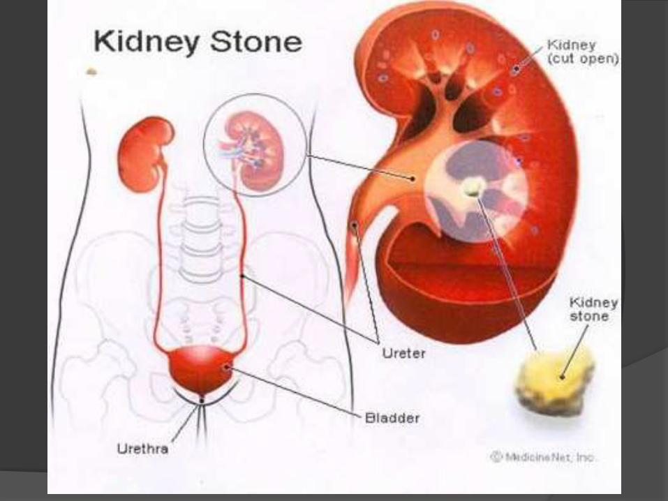
Definition
Presence of calculi (stones) in the kidney or collecting system
Usually small (2-12 mm), solid, crystalline concretions
Calcium salts, uric acid, cystine, or struvite
Stones < 0.5 cm without symptoms
Larger calculi cause pain and obstruction
Staghorn calculi (struvite, cystine, and uric acid) can grow as large as renal pelvis
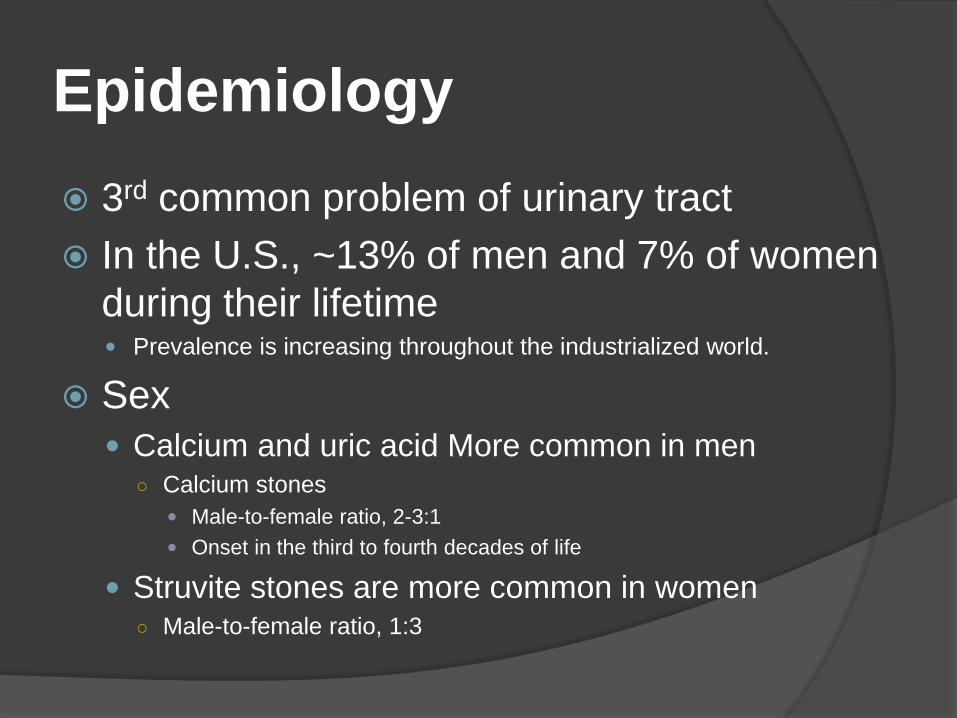
Epidemiology
3rd common problem of urinary tract
In the U.S., ~13% of men and 7% of women
during their lifetime Prevalence is increasing throughout the industrialized world.
Sex
Calcium and uric acid More common in men○ Calcium stones
Male-to-female ratio, 2-3:1
Onset in the third to fourth decades of life
Struvite stones are more common in women○ Male-to-female ratio, 1:3
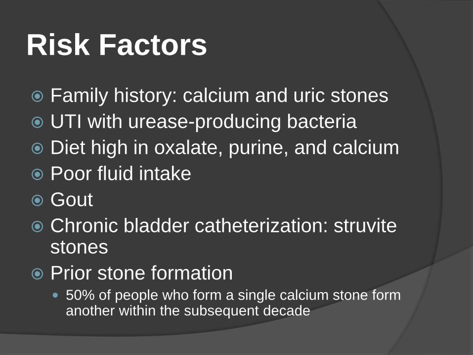
Risk Factors
Family history: calcium and uric stones
UTI with urease-producing bacteria
Diet high in oxalate, purine, and calcium
Poor fluid intake
Gout
Chronic bladder catheterization: struvite stones
Prior stone formation 50% of people who form a single calcium stone form
another within the subsequent decade
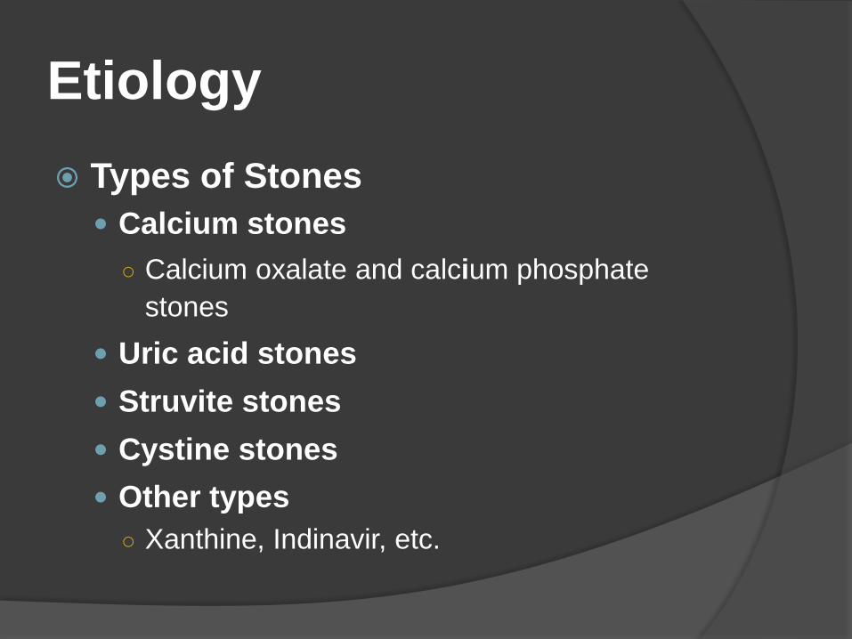
Etiology
Types of Stones
Calcium stones
○ Calcium oxalate and calcium phosphate
stones
Uric acid stones
Struvite stones
Cystine stones
Other types
○ Xanthine, Indinavir, etc.
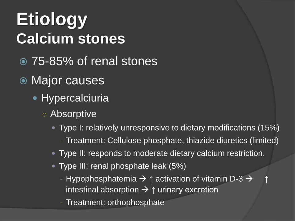
EtiologyCalcium stones
75-85% of renal stones
Major causes
Hypercalciuria
○ Absorptive
Type I: relatively unresponsive to dietary modifications (15%)
- Treatment: Cellulose phosphate, thiazide diuretics (limited)
Type II: responds to moderate dietary calcium restriction.
Type III: renal phosphate leak (5%)
- Hypophosphatemia ↑ activation of vitamin D-3 ↑
intestinal absorption ↑ urinary excretion
- Treatment: orthophosphate
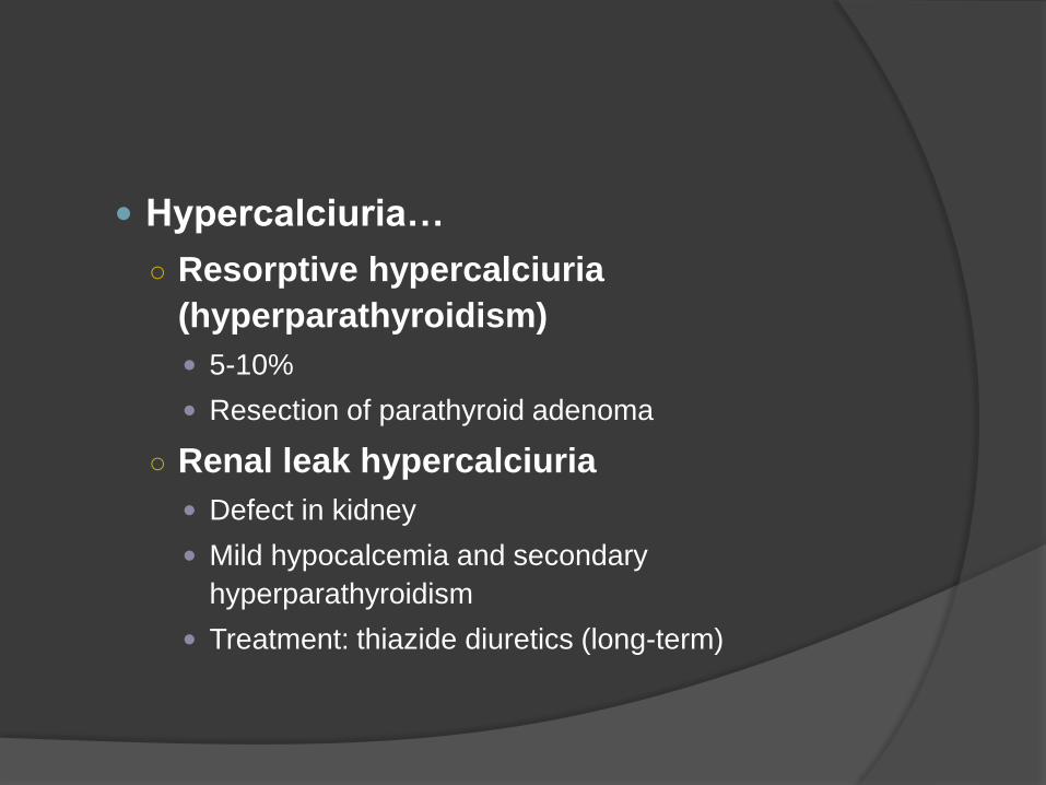
Hypercalciuria…
○ Resorptive hypercalciuria
(hyperparathyroidism)
5-10%
Resection of parathyroid adenoma
○ Renal leak hypercalciuria
Defect in kidney
Mild hypocalcemia and secondary
hyperparathyroidism
Treatment: thiazide diuretics (long-term)
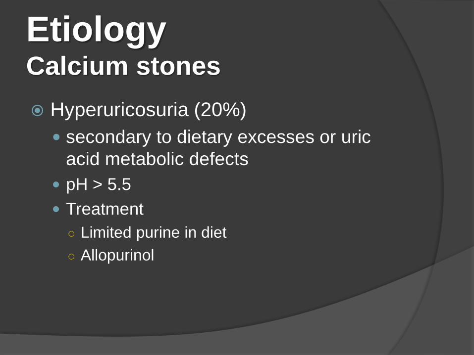
EtiologyCalcium stones
Hyperuricosuria (20%)
secondary to dietary excesses or uric
acid metabolic defects
pH > 5.5
Treatment
○ Limited purine in diet
○ Allopurinol
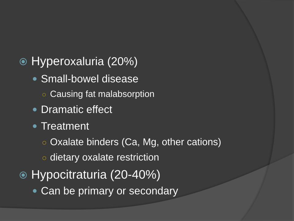
Hyperoxaluria (20%)
Small-bowel disease
○ Causing fat malabsorption
Dramatic effect
Treatment
○ Oxalate binders (Ca, Mg, other cations)
○ dietary oxalate restriction
Hypocitraturia (20-40%)
Can be primary or secondary
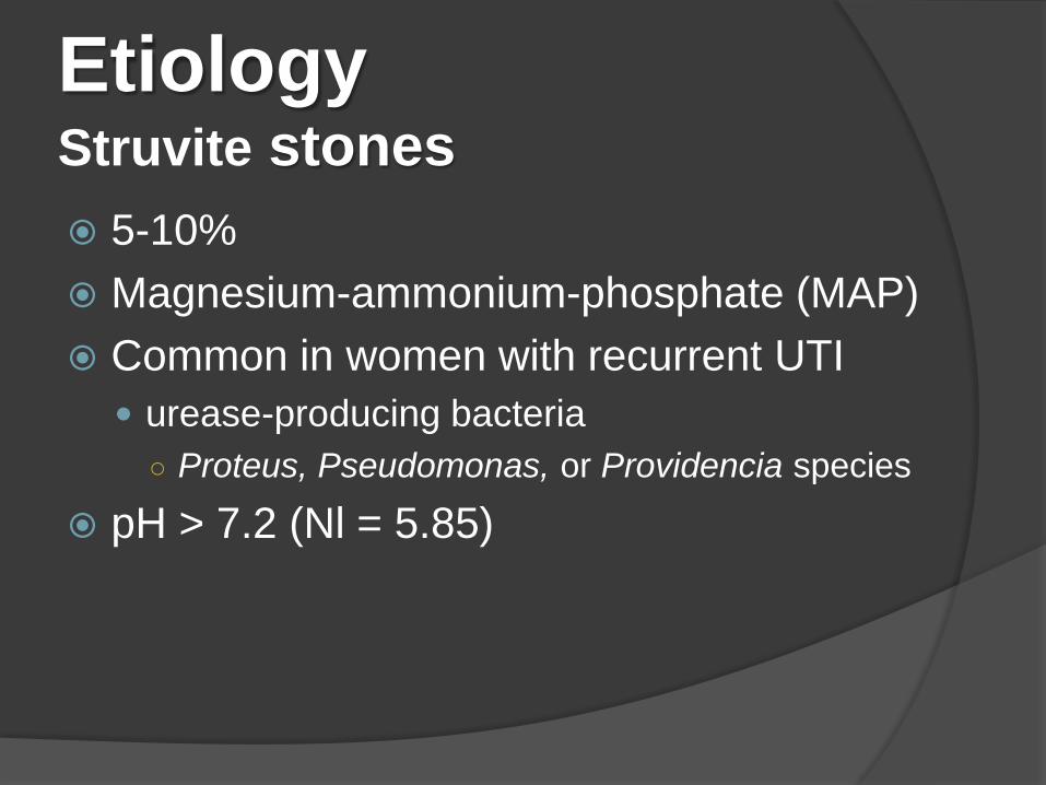
EtiologyStruvite stones
5-10%
Magnesium-ammonium-phosphate (MAP)
Common in women with recurrent UTI
urease-producing bacteria
○ Proteus, Pseudomonas, or Providencia species
pH > 7.2 (Nl = 5.85)
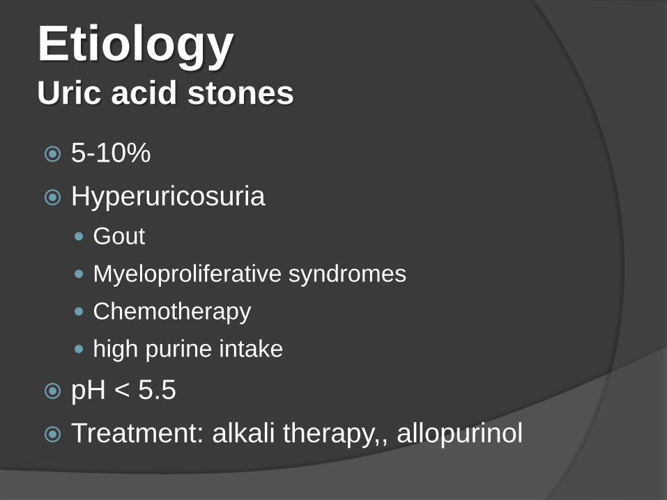
EtiologyUric acid stones
5-10%
Hyperuricosuria
Gout
Myeloproliferative syndromes
Chemotherapy
high purine intake
pH < 5.5
Treatment: alkali therapy,, allopurinol

Etiology
Cystine stones
1-3%
Cystinuria
○ autosomal recessive disorder
○ defective proximal tubular and jejunal transport of
cystine, lysine, arginine, and ornithine
○ Clinical disease due to insolubility of cystine
Drug-induced stone disease
Indinavir
tazanavir; triamterene; silicate
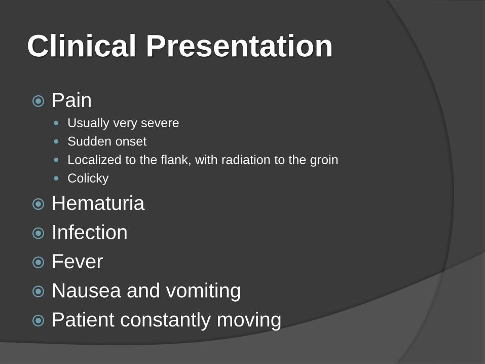
Clinical Presentation
Pain Usually very severe
Sudden onset
Localized to the flank, with radiation to the groin
Colicky
Hematuria
Infection
Fever
Nausea and vomiting
Patient constantly moving
Differential Diagnosis
Pyelonephritis
Acute abdomen
Gynecologic problems
Diverticulitis
Abdominal aortic aneurysm
Aortic dissection
Appendicitis
Biliary colic
Perforating duodenal ulcer
Viral gastroenteritis
Acute pancreatitis
Urinary tract infection
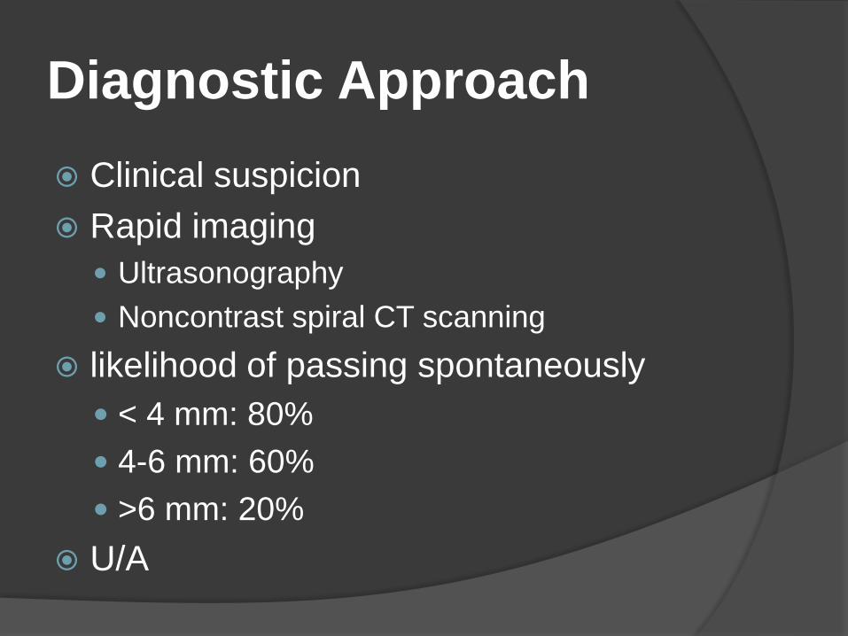
Diagnostic Approach
Clinical suspicion
Rapid imaging
Ultrasonography
Noncontrast spiral CT scanning
likelihood of passing spontaneously
< 4 mm: 80%
4-6 mm: 60%
>6 mm: 20%
U/A
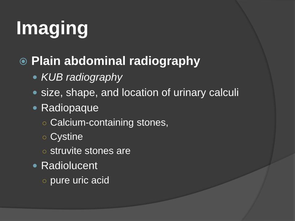
Imaging
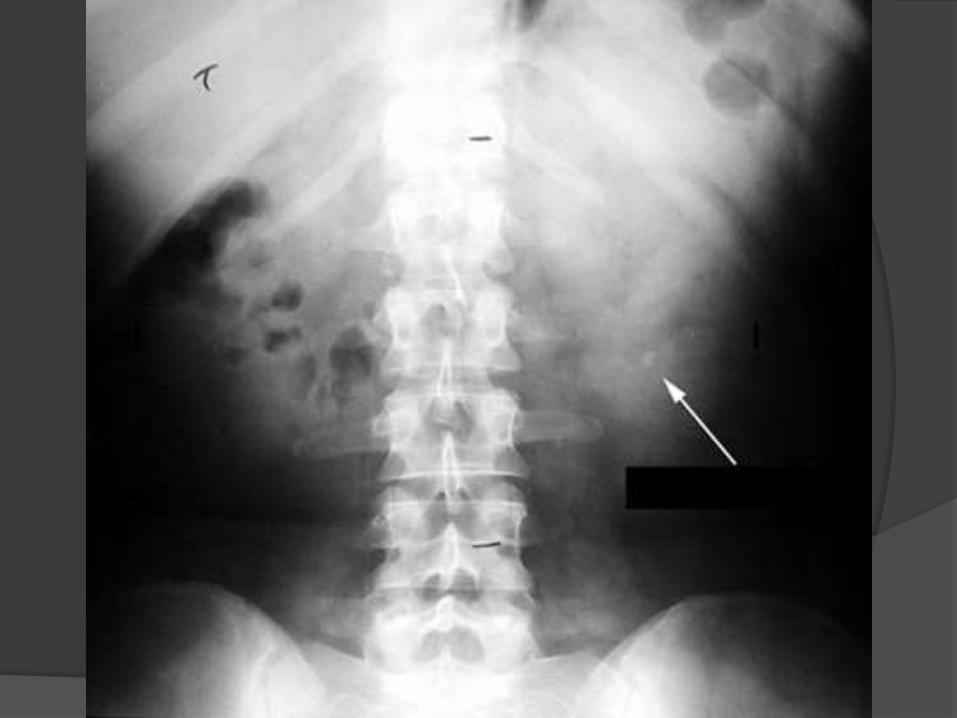
Plain abdominal radiography
KUB radiography
size, shape, and location of urinary calculi
Radiopaque
○ Calcium-containing stones,
○ Cystine
○ struvite stones are
Radiolucent
○ pure uric acid
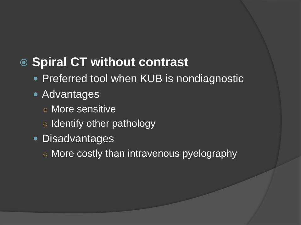
Spiral CT without contrast
Preferred tool when KUB is nondiagnostic
Advantages
○ More sensitive
○ Identify other pathology
Disadvantages
○ More costly than intravenous pyelography
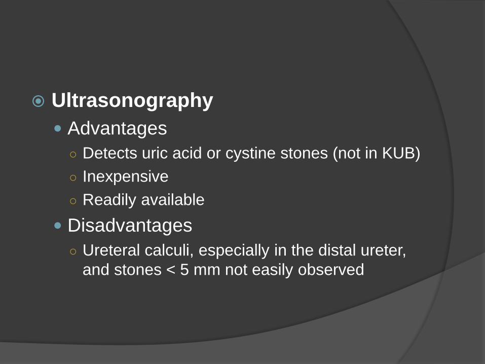
Ultrasonography
Advantages
○ Detects uric acid or cystine stones (not in KUB)
○ Inexpensive
○ Readily available
Disadvantages
○ Ureteral calculi, especially in the distal ureter,
and stones < 5 mm not easily observed
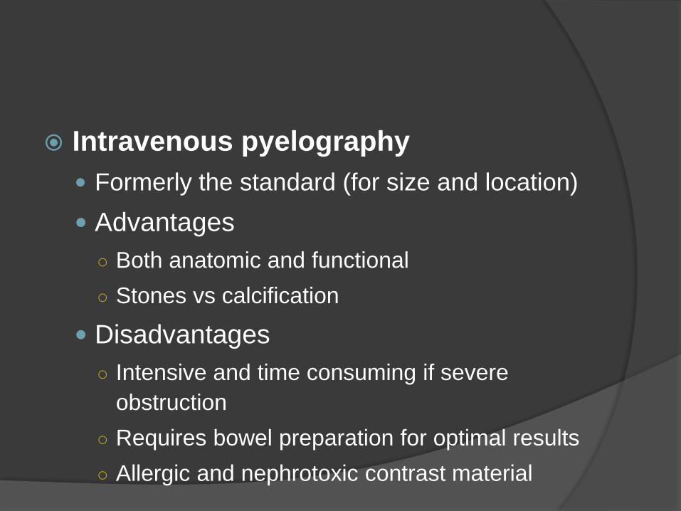
Intravenous pyelography
Formerly the standard (for size and location)
Advantages
○ Both anatomic and functional
○ Stones vs calcification
Disadvantages
○ Intensive and time consuming if severe
obstruction
○ Requires bowel preparation for optimal results
○ Allergic and nephrotoxic contrast material
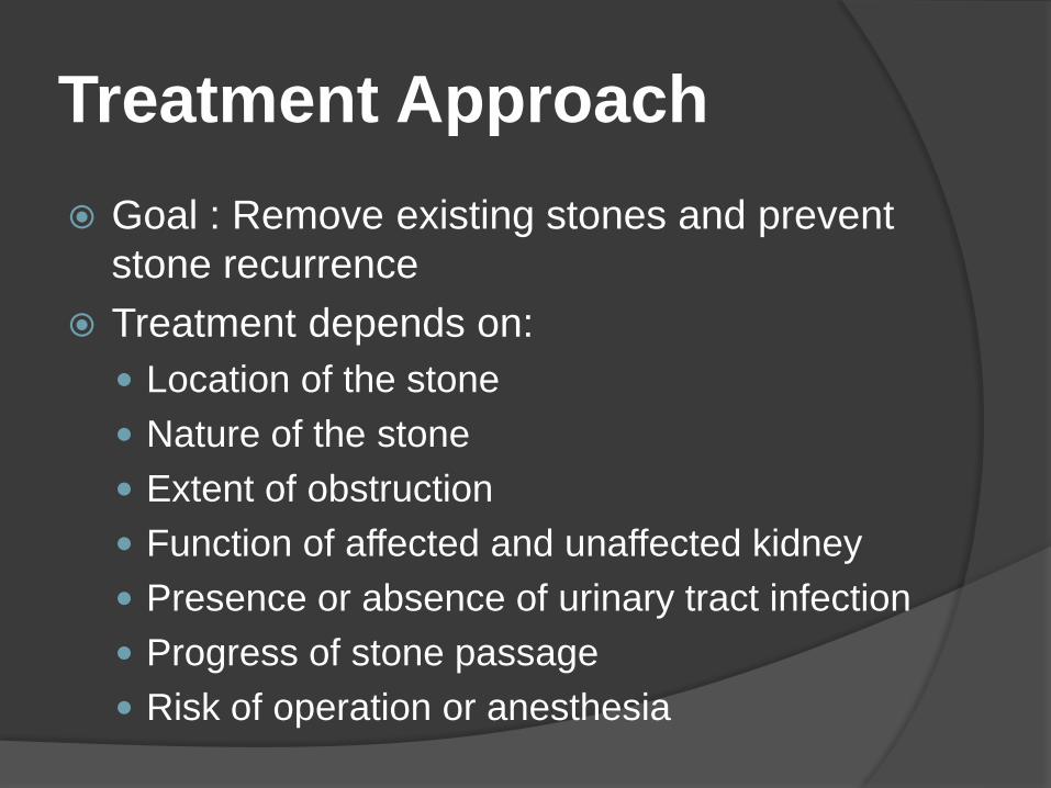
Treatment Approach
Goal : Remove existing stones and prevent
stone recurrence
Treatment depends on:
Location of the stone
Nature of the stone
Extent of obstruction
Function of affected and unaffected kidney
Presence or absence of urinary tract infection
Progress of stone passage
Risk of operation or anesthesia
Stones already present
Combined medical and surgical approach
Oral α1-adrenergic blockers
○ Relax ureteral muscle
○ Reduce time to stone passage
○ Reduce need for surgical removal of small
stones
Indications for stone removal
A stone, usually >5mm, that does not
pass spontaneously
Severe obstruction
Infection
Intractable pain
Serious bleeding
Management of renal colic
Hydration
Pain control Parenteral: morphine sulfate and/or
intravenous NSAID (e.g.,ketorolac)
Oral: narcotic (codeine, oxycodone, hydrocodone) plus acetaminophen together with an NSAID, such as ibuprofen
Antiemetic agents (e.g.,metoclopramide orprochlorperazine)
Strain urine
Antibiotics, if infection is suspected
Agents to relax the ureters
α1-blockers (e.g.,tamsulosin 0.4 mg PO daily
30 minutes after a meal)○ Faster and fewer hospitalization
Calcium-channel blockers
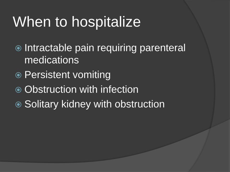
When to hospitalize
Intractable pain requiring parenteral
medications
Persistent vomiting
Obstruction with infection
Solitary kidney with obstruction
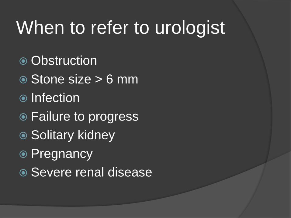
When to refer to urologist
Obstruction
Stone size > 6 mm
Infection
Failure to progress
Solitary kidney
Pregnancy
Severe renal disease

Thank you for your
attention