use of lipoprotein(a) in clinical practice: a ... - lipid.org · original research q1 use of...
TRANSCRIPT
Q1
Q2 Q3
Q24
Q4
Q5
Journal of Clinical Lipidology (2019) -, -–-
123456789
101112131415161718192021222324252627282930313233343536373839404142434445464748495051
52535455
Original Research 5657585960616263646566Use of lipoprotein(a) in clinical practice: Abiomarker whose time has come—A scientificstatement from the National Lipid Association.Don P. Wilson, MD, on behalf of the Writinggroup
6768697071
Don P. Wilson, MD*, Terry A. Jacobson, MD, Peter H. Jones, MD,Marlys L. Koschinsky, PhD, Catherine J. McNeal, MD, PhD,Børge G. Nordestgaard, MD, DMSc, Carl E. Orringer, MD
727374757677787980
Pediatric Endocrinology and Diabetes, Cook Children’s Medical Center, Fort Worth, TX, USA (Dr Wilson); Lipid Clinicand Cardiovascular Risk Reduction Program, Emory University, Atlanta, GA, USA (Dr Jacobson); Baylor College ofMedicine, Houston, TX, USA (Dr Jones); Robarts Research Institute, Schulich School of Medicine and Dentistry, TheUniversity of Western Ontario, London, Ontario, Canada (Dr Koschinsky); Division of Cardiology, Department of InternalMedicine, Baylor Scott & White Health, Temple, TX, USA (Dr McNeal); Department of Clinical Biochemistry, Herlev andGentofte Hospital, Copenhagen University Hospital, Herlev, Denmark (Dr Nordestgaard); and Division of Cardiology,Department of Medicine, University of Miami Miller School of Medicine, Miami, FLUSA (Dr Orringer)
81828384
KEYWORDS:---
* Corresponding author. Lipoprotein
tee, National Lipid Association, Pedia
Cook Children’s Medical Center, 1500
Worth, TX, 76104, USA.
E-mail address: don.wilson@cookch
Submitted April 24, 2019. Accepted
1933-2874/� 2019 National Lipid Ass
https://doi.org/10.1016/j.jacl.2019.04.0
8586878889
Abstract: Lipoprotein(a) [Lp(a)] is a well-recognized, independent risk factor for atherosclerotic car-diovascular disease, with elevated levels estimated to be prevalent in 20% of the population. Observa-tional and genetic evidence strongly support a causal relationship between high plasma concentrationsof Lp(a) and increased risk of atherosclerotic cardiovascular disease–related events, such as myocardialinfarction and stroke, and valvular aortic stenosis. In this scientific statement, we review an array ofevidence-based considerations for testing of Lp(a) in clinical practice and the utilization of Lp(a) levelsto inform treatment strategies in primary and secondary prevention.� 2019 National Lipid Association. All rights reserved.
9091
92 Introduction 939495a. Question: What are the proposed pathophysiologicmechanisms supporting a causal link between increased
(a) Scientific Statement Commit-
tric Endocrinology and Diabetes,
Cooper Street, 2nd Floor, Fort
ildrens.org
for publication April 26, 2019.
ociation. All rights reserved.
10
FLA 5.6.0 DTD � JACL1449_proo
96979899
100101102
circulating concentrations of Lp(a) and (1) atheroscle-rotic cardiovascular disease (ASCVD) and (2) valvularaortic stenosis (VAS)?
Observational and genetic evidence strongly support acausal relationship between high plasma concentrations oflipoprotein(a) [Lp(a)] and increased risk of ASCVD andVAS.1–4 Although the precise pathophysiologic mecha-nism behind these relationships is not completely clear,the mechanism likely involves either or both componentsof Lp(a), that is, the low-density lipoprotein (LDL)-like
f � 13 May 2019 � 9:23 pm

web4C=FPO
Key points
� Apo(a), attached to the apoB segment of an LDL-likeparticle, is a unique protein contained within Lp(a).
� Apo(a) has homology with plasminogen and may inhibitfibrinolysis, thus increasing thrombosis.
� Through inhibition of fibrinolysis at sites of plaquerupture, apo(a) has the potential to cause MI and ischemic
2 Journal of Clinical Lipidology, Vol -, No -, - 2019
103104105106107108109110111112113114115116117118119120121122123124125126127128129130131132133134135136137138139140141142143144145146147148149150151152153154155156157158
159160161162163164165166167168169170171172173174175176177178179180181182183184
particle and the apolipoprotein(a) [apo(a)] attached toapolipoprotein B (apoB) via a disulfide bridge (Fig. 1).The apo(a) protein has homology with plasminogen andin vitro, as well as in some animal models, and inhibitsfibrinolysis.2,5,6 Historically, it has been suggested thathigh concentrations of circulating Lp(a) could have pro-vided a survival benefit by facilitating wound healing,7,8
reduce bleeding, and aiding hemostasis duringchildbirth.4,6
Both ASCVD and VAS share elements of stenosis aswell as cholesterol deposition in the arterial intima andaortic valve leaflets, respectively. In susceptible individ-uals, Lp(a) mediated promotion of thrombosis in vulner-able plaques of coronary arteries or at sites of stenosismay increase risk of myocardial infarction (MI), andthrombotic emboli may increase risk of ischemic stroke(Fig. 1).4
The cholesterol content of the LDL portion of Lp(a) maypromote cholesterol deposition in the arterial intima and ataortic valve leaflets, leading, respectively, to symptomaticatherosclerosis resulting in MI and ischemic stroke, andVAS (Fig. 1). However, even at very high Lp(a) concentra-tions such as 100 mg/dL, the LDL cholesterol (LDL-C)portion of Lp(a) would only amount to 33 mg/dL,9 whichis unlikely to cause substantial deposition of cholesterolin tissues.
Figure 1 Proposed pathophysiologic mechanisms supportinga causal link between elevated circulating concentrations ofLp(a) and (1) atherosclerotic cardiovascular disease and (2)aortic stenosis. LDL, low-density lipoprotein; PL, phospho-lipids; TG, triglycerides; FC, free cholesterol; CE, cholesterylester; ApoB100, apolipoprotein B 100; KIV, kringle IV; KV,kringle V; P, protease; apo(a), apolipoprotein(a); OxPL,oxidized phospholipids.
FLA 5.6.0 DTD � JACL1449_proo
Although ASCVD and VAS are distinct clinical entities,they have several risk factors in common and similarpathological processes. Evidence suggests that oxidizedphospholipids, which modify Lp(a) primarily by covalentbinding to its unique apo(a) component, might hold the keyto Lp(a) pathogenicity and provide a mechanistic linkbetween ASCVD and VAS. Oxidized phospholipids coloc-alize with apo(a)-Lp(a) in arterial and aortic valve lesionsand directly participate in the pathogenesis of thesedisorders by promoting endothelial dysfunction, lipiddeposition, inflammation, and osteogenic differentiation,leading to calcification. Genetic evidence for a contributionof oxidized phospholipids has been presented,10 and associ-ations between elevated oxidized phospholipids on Lp(a)and risk for coronary heart disease (CHD) and valvularaortic stenosis have been detected.10,11
stroke.� Thrombosis at sites of turbulent flow may promoteatherosclerotic and valvular aortic stenosis.
� Apo(a) possesses unique properties that promote initiationand progression of atherosclerosis and calcific valvularaortic stenosis through endothelial dysfunction andproinflammatory responses, and calcification.
� Many of these effects are likely attributable to theoxidized phospholipids, of which Lp(a) is the preferentialcarrier, and which are covalently attached to apo(a).
185186187188189190191192193194195196197198199200201202203204205206207208209210211212213214
b. Question: Do available, high-quality data from meta-analyses, large prospective, population-based studies,large Mendelian randomization studies, and genome-wide association (GWA) studies support a relationshipbetween increased circulating Lp(a) concentrations and(1) ASCVD; (2) VAS; and (3) mortality?
Meta-analyses of prospective, population-based studiesof adults show increased risk of CHD and MI at Lp(a)concentrations above 30 mg/dL (62 nmol/L) and increasedrisk of ischemic stroke at concentrations above 50 mg/dL(100 nmol/L) (Table 1). However, effect sizes were modest,likely due to inclusion of all available studies (1) irrespec-tive of size, study quality, and quality of the Lp(a) assaysused and (2) whether the plasma samples used were freshor had been frozen for prolonged periods of time beforemeasurement of Lp(a).12–15
Another meta-analysis found that individuals withsmaller apo(a) isoforms [and high Lp(a) concentrations]had an approximately 2-fold higher risk of CHD andischemic stroke than those with larger apo(a) isoforms
f � 13 May 2019 � 9:23 pm
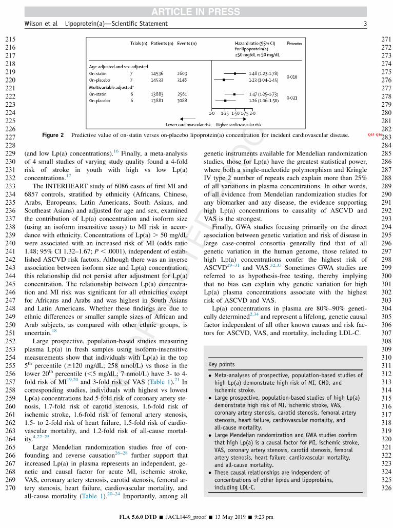
Figure 2 Predictive value of on-statin verses on-placebo lipoprotein(a) concentration for incident cardiovascular disease. Q15 Q16
Key points
� Meta-analyses of prospective, population-based studies ofhigh Lp(a) demonstrate high risk of MI, CHD, andischemic stroke.
� Large prospective, population-based studies of high Lp(a)demonstrate high risk of MI, ischemic stroke, VAS,coronary artery stenosis, carotid stenosis, femoral arterystenosis, heart failure, cardiovascular mortality, andall-cause mortality.
� Large Mendelian randomization and GWA studies confirmthat high Lp(a) is a causal factor for MI, ischemic stroke,VAS, coronary artery stenosis, carotid stenosis, femoralartery stenosis, heart failure, cardiovascular mortality,and all-cause mortality.
� These causal relationships are independent ofconcentrations of other lipids and lipoproteins,including LDL-C.
Wilson et al Lipoprotein(a)—Scientific Statement 3
215216217218219220221222223224225226227228229230231232233234235236237238239240241242243244245246247248249250251252253254255256257258259260261262263264265266267268269270
271272273274275276277278279280281282283284285286287288289290291292293294295296297298299300301302303304305306307308309310311312313314315316317318319320321322323324325326
(and low Lp(a) concentrations).16 Finally, a meta-analysisof 4 small studies of varying study quality found a 4-foldrisk of stroke in youth with high vs low Lp(a)concentrations.17
The INTERHEART study of 6086 cases of first MI and6857 controls, stratified by ethnicity (Africans, Chinese,Arabs, Europeans, Latin Americans, South Asians, andSoutheast Asians) and adjusted for age and sex, examinedthe contribution of Lp(a) concentration and isoform size(using an isoform insensitive assay) to MI risk in accor-dance with ethnicity. Concentrations of Lp(a) . 50 mg/dLwere associated with an increased risk of MI (odds ratio1.48; 95% CI 1.32–1.67; P , .0001), independent of estab-lished ASCVD risk factors. Although there was an inverseassociation between isoform size and Lp(a) concentration,this relationship did not persist after adjustment for Lp(a)concentration. The relationship between Lp(a) concentra-tion and MI risk was significant for all ethnicities exceptfor Africans and Arabs and was highest in South Asiansand Latin Americans. Whether these findings are due toethnic differences or smaller sample sizes of African andArab subjects, as compared with other ethnic groups, isuncertain.18
Large prospective, population-based studies measuringplasma Lp(a) in fresh samples using isoform-insensitivemeasurements show that individuals with Lp(a) in the top5th percentile ($120 mg/dL; 258 nmol/L) vs those in thelower 20th percentile (,5 mg/dL; 7 nmol/L) have 3- to 4-fold risk of MI19,20 and 3-fold risk of VAS (Table 1).21 Incorresponding studies, individuals with highest vs lowestLp(a) concentrations had 5-fold risk of coronary artery ste-nosis, 1.7-fold risk of carotid stenosis, 1.6-fold risk ofischemic stroke, 1.6-fold risk of femoral artery stenosis,1.5- to 2-fold risk of heart failure, 1.5-fold risk of cardio-vascular mortality, and 1.2-fold risk of all-cause mortal-ity.4,22–25
Large Mendelian randomization studies free of con-founding and reverse causation26–28 further support thatincreased Lp(a) in plasma represents an independent, ge-netic and causal factor for acute MI, ischemic stroke,VAS, coronary artery stenosis, carotid stenosis, femoral ar-tery stenosis, heart failure, cardiovascular mortality, andall-cause mortality (Table 1).20–24 Importantly, among all
FLA 5.6.0 DTD � JACL1449_proo
genetic instruments available for Mendelian randomizationstudies, those for Lp(a) have the greatest statistical power,where both a single-nucleotide polymorphism and KringleIV type 2 number of repeats each explain more than 25%of all variations in plasma concentrations. In other words,of all evidence from Mendelian randomization studies forany biomarker and any disease, the evidence supportinghigh Lp(a) concentrations to causality of ASCVD andVAS is the strongest.
Finally, GWA studies focusing primarily on the directassociation between genetic variation and risk of disease inlarge case-control consortia generally find that of allgenetic variation in the human genome, those related tohigh Lp(a) concentrations confer the highest risk ofASCVD29–31 and VAS.32,33 Sometimes GWA studies arereferred to as hypothesis-free testing, thereby implyingthat no bias can explain why genetic variation for highLp(a) plasma concentrations associate with the highestrisk of ASCVD and VAS.
Lp(a) concentrations in plasma are 80%–90% geneti-cally determined2,34 and represent a lifelong, genetic causalfactor independent of all other known causes and risk fac-tors for ASCVD, VAS, and mortality, including LDL-C.
f � 13 May 2019 � 9:23 pm

Q6
Table 1 Do available, high-quality data from meta-analyses, large observational studies, Mendelian randomization studies, andgenome-wide association studies support a relationship between increased circulating Lp(a) concentrations and (1) atheroscleroticcardiovascular disease, (2) valvular aortic stenosis, and (3) mortality?
High-quality data source:
Atherosclerotic cardiovascular disease
Aortic valvestenosis
Cardiovascularmortality
All-causemortality
MyocardialInfarction
Ischemicstroke
Atheroscleroticstenosis*
Meta-analyses of observational studies Yes Yes No No No NoLarge observational studies.†** Yes Yes Yes Yes Yes YesLarge Mendelian randomization studies Yes Yes Yes Yes Yes YesLarge genome-wide association studies Yes No Yes Yes No No
*Clinical symptoms in the form of stable angina pectoris or intermittent claudication or documented atherosclerotic stenosis in coronary, femoral, or
carotid arteries.
†Using isoform insensitive Lp(a) measurements.
4 Journal of Clinical Lipidology, Vol -, No -, - 2019
327328329330331332333334335336337338339340341342343344345346347348349350351352353354355356357358359360361362363364365366367368369370371372373374375376377378379380381382
383384385386387388389390391392393394395396397398
Laboratory measurement of lipoprotein(a)Table 2 Distribution of Lp(a) levels by ethnic group*
N
Lp(a) Level by percentile (nmol/L) Q17
10th 50th 75th 80th 98th 95th
Caucasian Americans2929 1 20 73 100 154 209African Americans 189916 75 130 148 199 234Japanese American 1379 3 19 40 49 75 103
*Data from Marcovina, 2016.
399400401402403404405406407408409410411412413414415416417418419420421422423424425426427428429430431432433434435436437438
a. Question: What are the key laboratory measurement is-sues which impact a clinician’s interpretation of reportedLp(a) values?
Lp(a) has a highly heterogeneous structure owing to thepresence of many different isoform sizes within the pop-ulation. The distribution of plasma Lp(a) levels is highlyskewed and differs considerably among different ethnicgroups. From a clinical perspective, these factors haveimportant implications for Lp(a) measurement.35 Key is-sues include (1) the prevalence of assays reporting Lp(a)values as mass concentrations (units of mg/dL) vs particleconcentrations (nmol/L); (2) the lack of standardizationof Lp(a) assays; and (3) the absence of evidence-basedLp(a) cut points for different risk groups, ethnic popula-tions, and comorbidities.
b. Question: What are the limitations of currently availableassays and how does the performance characteristics ofthe test (ie, accuracy [bias] and precision) affect clini-cian interpretation of the results?
Currently available assays have not been subjected to aglobal standardization regime.36 Although some commer-cially available assays use calibrators that are traceable,such as the WHO/IFCCLM secondary reference materialPRM-2B,37 this is not the case for all, notably those thatreport results in mg/dL. Moreover, harmonization of valuesobtained from different assays, even those reporting innmol/L, has yet to be undertaken.36 The potential exists,therefore, for bias in Lp(a) immunoassays because of thepresence of variable numbers of repeated units in differ-ently sized apo(a) isoforms.35,38,39 Typically, this bias man-ifests as an underestimation of the levels of small Lp(a)isoforms and an overestimation of large Lp(a) isoforms.35
This bias could result in misclassification of patients withLp(a) levels close to a predefined cut point.38 Somecommercially available assays minimize isoform-dependent bias by using a 5-point calibrator, consisting ofa range of Lp(a) isoforms.35
It has been recommended that use of mg/dL units bediscontinued.36 As the PRM-2B is in nmol/L, and Lp(a)
FLA 5.6.0 DTD � JACL1449_proo
isoforms have different molecular weights, unlike otherlipids and lipoproteins, direct conversion between mg/dLand nmol/L is not possible. Universal use of nmol/L would(1) create an opportunity to standardize and harmonizeLp(a) assays, as the output is independent of the molecularweight of the Lp(a) species used as the calibrator and (2)facilitate future clinical studies of Lp(a) and the establish-ment of evidence-based guidelines. Therefore, in theabsence of Lp(a) assay standardization, clinicians shoulduse, where possible, assays that report results in nmol/L, us-ing a 5-point or similar calibrator, and which are calibratedagainst the WHO/IFCCLM secondary reference material.
c. Question: What should be the population Lp(a) cutpoints for defining high risk, based on age, sex, andethnicity?
The evidence base for specific cut points for high riskbased on age, sex, and ethnicity is generally incomplete.This also applies to individuals with comorbid conditionssuch as familial hypercholesterolemia (FH), diabetes mel-litus, or renal disease. There has been debate about whethercut points based on Lp(a) concentrations or population-specific percentiles are most appropriate. This is becausethe distribution of Lp(a) levels differs among ethnic groups(Table 2)35 and is affected by certain disease conditions.40
For example, the Multi-Ethnic Study of Atherosclerosisfound that while a cut point of $50 mg/dL best predictedCHD in Caucasians, Chinese-Americans, and Hispanics,the corresponding value for blacks was $30 mg/dL.41 Onthe other hand, the Atherosclerosis Risk in Communitiesstudy found no difference in risk between Caucasian and
f � 13 May 2019 � 9:23 pm

Q7
Key points
� Measurement of Lp(a) is currently not standardized orharmonized.
� Available assays report Lp(a) in either mg/dL or nmol/Land may exhibit Lp(a) isoform-dependent bias.
� Evidence is incomplete regarding the utility of usingdifferent risk cut points of Lp(a) based on age, gender,ethnicity, or the presence of comorbid conditions.
� Elevated Lp(a) appears to confer elevated risk for ASCVDover a wide range of LDL-C concentrations.
� An Lp(a) level .50 mg/dL (.100 nmol/L) may beconsidered as a risk-enhancing factor favoring theinitiation of statin therapy. This level corresponds tothe 80th population percentile in populations whichare predominantly Caucasian.
� The corresponding 80th population percentile in AfricanAmericans is approximately 150 nmol/L, but it is unclearwhether a different risk threshold or cut point should beapplied. Clinicians should be aware that African Americanshave an approximately 3-fold higher median Lp(a) thanCaucasian populations (75 nmol/L vs 20 nmol/L)
Wilson et al Lipoprotein(a)—Scientific Statement 5
439440441442443444445446447448449450451452453454455456457458459460461462463464465466467468469470471472473474475476477478479480481482483484485486487488489490491492493494
495496497498499500501502503504505506507508509510511512513514515516517518519520521522523524525526527528529530531
black subjects, irrespective of the cut point used.42 More-over, individual studies in different populations (eg, pri-mary vs secondary prevention) have arrived at differentcut points ($30 mg/dL and $50 mg/dL, respectively).36
It is unlikely that these observations reflect differences inthe underlying pathobiology of Lp(a). Although differentgroups likely have varying risk factor profiles, which influ-ence the contribution of Lp(a), it is also possible that thedifferent observed cut points reflect selection bias, differentstatistical power in individual studies, and other confound-ing effects. Therefore, we recommend a tentative, universalcut point of $100 nmol/L (approximately $50 mg/dL),which is supported by the largest meta-analyses in a rangeof populations.16,43
d. Question: Because the cholesterol content of Lp(a) isincluded in the measurement of LDL-C, is there a levelof LDL-C where the measurement of Lp(a) should beconsidered independent of clinical history?
Some studies have shown that lowering LDL-Cattenuates or eliminates risk attributable to elevatedLp(a).44,45 On the other hand, other studies have shownthat Lp(a) clearly contributes to residual risk in statin-treated subjects.43,46,47 In a 2018 meta-analysis, elevatedLp(a) was a stronger risk factor than LDL-C for incidentCVD in statin-treated than in placebo-treated subjects.43
Therefore, it may be reasonable to speculate thatmeasuring Lp(a) in subjects with elevated LDL-C iden-tifies subjects who could benefit from more intensiveLDL-C–lowering therapy, including use of PCSK9 inhib-itors, which have been shown to lower Lp(a) by w20%–30%.48,49 However, this proposition has yet to be directlytested in clinical studies. Notably, current risk predictionalgorithms, such as the Framingham Risk Score or thePooled Cohort Equations, do not include Lp(a), whereasrecommendations from several organizations and soci-eties suggest measuring Lp(a) in subjects with an
Table of Recommendation
I. Laboratory measurement of lipoprotein(a)
1. For the measurement of Lp(a), it is recommendedthat an immunochemical assay that is calibratedagainst theWHO/IFCCM secondary reference materialshould be used and reported in nmol/L.
2. When using values of Lp(a) for clinical risk assessmentand treatment decisions, the use of a factor to convertLp(a) values from mg/dL to nmol/L is not recommended.
3. When Lp(a) values are used for ASCVD risk assessment inCaucasian patients, it is reasonable to use measuredvalues $ 50 mg/dL or $100 nmol/L as levels suggestingincreased risk.
FLA 5.6.0 DTD � JACL1449_proo
intermediate risk score.50,51 Therefore, at present, werecommend that measurement of Lp(a) should be consid-ered when clinically indicated and not necessarily relatedto a high baseline level of LDL-C alone. Because statinsand PCSK9 inhibitors lower LDL-C less effectively inthe setting of a high Lp(a) concentration, the finding ofless-than-anticipated LDL-C lowering in response totreatment with these agents should suggest the possibil-ity of a markedly elevated Lp(a). Some patients withmarkedly elevated LDL-C values, with levels suggestingFH, have been found to have this clinical presentationprimarily because of Lp(a) elevation.52
Q8
Class of Rec(strength)
Levels ofEvidence References/notes
I B-NR Marcovina, 2016; Tsimikas,2018;Marcovina, 2000; Marcovina,2003
III (nobenefit)
E-O Marcovina, 2000, Marcovina,2016;Tsimikas, 2018 JCL
IIa B-R Nordestgaard, 2010;Willeit, 2018, Langsted, 2019
f � 13 May 2019 � 9:23 pm
532533534535536537538539540541542543544545546547548549550
Q9
Q10
11
Key points
Lp(a) testing is reasonable to refine risk assessment forASCVD events in adults with:
� First-degree relatives with premature ASCVD (aged,55 y in men or ,65 y in women).
� A personal history of premature ASCVD.� Primary severe hypercholesterolemia (LDL-C $190mg/dL) or suspected FH.
Lp(a) testing may be reasonable in adults:� To aid in the clinician-patient discussion about whetherto prescribe a statin in those aged 40-75 y withborderline (5%–7.4%) 10-y ASCVD risk.
� To identify a possible cause for a less-than-anticipatedLDL-C lowering to evidence-based LDL-C–loweringtherapy.
� To use in cascade screening of family members withsevere hypercholesterolemia.
� To identify those at risk for progressive VAS.
6 Journal of Clinical Lipidology, Vol -, No -, - 2019
551552553554555556557558559560561562563564565566567568569570571572573574575576577578579580581582583584585586587588589590591592593594595596597598599600601602603604605606
607608609610611612613614615616617618619620621622623624625626627628629630631632633634635636637638639640641642643644645646647648649650651652653654655656657658659660
Lipoprotein(a) testing in clinical practice
a. The importance of shared decision-making
A decision to measure Lp(a) should be made after athoughtful benefit-risk discussion between the patientand his/her health care provider. Shared decision-makingshould reflect an individual’s preferences and values.Decisions should also be based on family history, thepresence of comorbid conditions, race/ethnicity, and/orconcern of future risk. In the absence of an acute illness,the level of Lp(a) is stable throughout an individual’slifetime and unaffected by lifestyle. Therefore, a casecould be made to measure Lp(a) in all individuals, atleast once in a lifetime, based on strong support for theassociation between elevated Lp(a) levels and increasedrisk, together with genetic findings that indicate elevatedLp(a) is causally related to premature ASCVD and VAS.However, there is no current evidence to substantiate thebenefit of such an approach, and there is currently notargeted treatment(s) to lower Lp(a) levels that have beenproven to affect ASCVD outcomes or progression ofVAS. Therefore, although some panel members sup-ported it, a recommendation for universal testing ofLp(a) was not made at this time. The Scientific StatementCommittee acknowledges that there is likely little harmfrom a universal screening approach and that the cost ofthe test is relatively inexpensive compared with othercardiovascular disease screening tests. As more databecome available in the future, the potential role ofuniversal testing should be re-evaluated.
b Question: What clinical factors result in considerationof Lp(a) testing in primary prevention?
A large percentage of the world’s population (20%)has an Lp(a) . 50 mg/dl.53 A prospective population-based study showed that measurement of Lp(a) predictednot only 15-year CVD outcomes but improved CVD riskprediction.54 Several national and international (ESC/EAS) guidelines4,50,51,55 recommend Lp(a) testing if anindividual has documented ASCVD (especially withrecurrent events on optimal lipid-lowering therapy), se-vere hypercholesterolemia or genetic FH, prematureASCVD, or a first-degree family member with prematureASCVD, particularly in the absence of traditional riskfactors. Based on the results of cascade screening of797 patients from a Spanish registry of molecularlydefined heterozygous FH patients, testing for Lp(a) dur-ing cascade screening was found to be an effective meansto identify relatives of the proband with increased risk ofclinical ASCVD, especially when FH and elevated Lp(a)coexist.85
The 2018 ACC-AHA Multi-Organization Guidelineon the Management of Blood Cholesterol does not pro-vide a recommendation on routine measurement of
FLA 5.6.0 DTD � JACL1449_proo
Lp(a).56 However, the 2018 guideline further statesthat if the results of Lp(a) testing are available to theclinician, an elevated concentration of $50 mg/dL or$125 nmol/L may be considered to be a risk-enhancing factor favoring moderate-intensity statin ther-apy in patients at intermediate risk (7.5%–19.9% 10-year risk) (class IIa B-NR) who are aged 40–75 yearsand have an LDL-C of 70–189 mg/dL. In addition, anelevated Lp(a) may aid risk discussion in patients aged40–75 years with borderline risk (5%–7.4%) and anLDL-C 70–189 mg/dL, when initiation of statin therapyis being considered (class IIb B-NR).
A potential caveat to consider in this recommendationemanates from a study examining Lp(a) levels in bloodsamples from female subjects as part of 2 large randomizedclinical trials and one observational study, suggesting thatLp(a) concentrations of .50 mg/dL predicted increasedcardiovascular risk only in those with total cholesterol.220 mg/dL.57 However, other larger studies do not sup-port this perspective.14,19,58
Two ICD-10 codes have been Qadded to justify Lp(a)testing [E78.41 5 elevated Lp(a) and Z83.430 5 FamilyHistory of elevated Lp(a)]. The relative stability of Lp(a)levels over a lifetime supports the perspective that repeatmeasurement is generally unnecessary, provided that theinitial blood sample was not obtained during an acuteillness.59
c. Question: What is the effect of currently available ther-apies on lowering Lp(a) levels and is there evidence thatreducing Lp(a) will reduce the incidence of ASCVD,VAS, or cerebrovascular disease?
f � 13 May 2019 � 9:23 pm
661662

Wilson et al Lipoprotein(a)—Scientific Statement 7
663664665666667668669670671672673674675676677678679680681682683684685686687688689690691692693694695696697698699700701702703704705706707708709710711712713714715716717718
719720721722723724725726727728729730731732733734735736737738739740741742743744745746747748749750751752753754755756757758759760761762763764765766767768769770771772773
Although in general beneficial, lifestyle changes,including low fat diets and moderate-to-vigorous dailyphysical exercise, have no significant effect on Lp(a)levels.57,60,61
Hormone replacement therapy (HRT) in womenlowers Lp(a) levels, and in the Women’s Health Study,HRT was observed to modify CVD risk across Lp(a)quintiles.60 However, in the Heart and Estrogen/progestinReplacement Study (secondary prevention) and theWomen’s Health Initiative (primary prevention) random-ized trials, HRT-related adverse events (breast cancer,stroke, thrombosis) outweighed any benefit on CVD.Therefore, HRT cannot be recommended as the sole pur-pose of lowering Lp(a).62,63
Niacin therapy is associated with a significant reductionin Lp(a) of approximately 23%.64 However, its addition tostatin therapy in high-risk ASCVD patients with LDL-Clevels near or at goal (,75 mg/dl) has not been shown toimprove ASCVD outcomes in AIM HIGH and HPS2THRIVE and has been associated with increased harms(new onset diabetes, bleeding, myopathy, and infec-tions).47,65,66 One potential explanation for this finding isniacin’s limited ability to reduce the concentration ofLp(a) in those with the highest baseline Lp(a) levels andsmall isoform size.67
Statin therapy has demonstrated a clinical benefit inpatients with elevated Lp(a),68 despite evidence thatlevels of Lp(a) may increase after initiation of therapy.3
A 2018 meta-analysis of patients with elevated Lp(a)and history of CV events concluded that those withLp(a) levels .50 mg/dL on statin therapy are at a signif-icantly higher risk of CVD as compared with those withlevels ,30 mg/dL, independent of other conventionalCVD risk factors.43
There is uncertainty about the clinical value ofPCSK9 inhibitor–associated Lp(a) reduction. An anal-ysis of the FOURIER trial demonstrated that evolocu-mab reduced Lp(a) by 27% and that the reduction inMACE was 23% (hazard ratio [HR] 0.77, 95% CI 0.67–0.88) in those patients with Lp(a) . median (37 nmol/L)and by 7% (HR 0.93, 0.80–1.08) in those # median.69
Patients with higher baseline Lp(a) levels had greater ab-solute reductions in Lp(a) and tended to derive greaterbenefit from PCSK9 inhibition. In ODYSSEY OUT-COMES, there was also a greater absolute benefit onMACE with alirocumab in patients with higher baselinelevels of Lp(a).70 In addition, baseline Lp(a) valuespredicted risk of MACE. Although the reduction ofLDL-C was the dominant factor contributing to theevent reduction with alirocumab, an independentcontribution of lowering Lp(a) on MACE and total CVevents was also demonstrated.71 Additional analysisof the PCSK9 inhibitor outcomes trials will be neededto support their use in patients with elevated Lp(a)levels.
FLA 5.6.0 DTD � JACL1449_proo
A modest reduction in Lp(a) of 20%–25% hasbeen reported in homozygous FH patients treatedwith lomitapide, a microsomal triglyceride transferprotein inhibitor. However, there are no studiesshowing the incremental benefit in this unique popula-tion. In the absence of data, lomitapide is notindicated for Lp(a) lowering or for ASCVD riskreduction.
Lipoprotein apheresis (LA), which acutely lowersLDL-C by .60% and reduces plasma levels of oxidizedphospholipid, known mediators of vascular inflammationand predictors of atherosclerosis progression foundpredominantly on Lp(a)-containing fractions,72 may beoffered to individuals with drug resistant, uncontrolledLDL-C levels (.160 mg/dL with and .300 mg/dLwithout CVD). In 2010, the German health caresystem approved LA therapy for ASCVD patients withan elevated Lp(a) (.60 mg/dL; .120 nmol/L) andrecurrent ASCVD events, irrespective of LDL-Clevels.73 Currently, more than 1400 Germansreceive weekly LA therapy for an elevated Lp(a) andCVD prophylaxis.74 Since the initiation of LAtherapy for Lp(a) reduction in Germany, three prospec-tive/retrospective trials involving over 400 individualshave demonstrated a 70% reduction of MACE comparedwith preapheresis events.75–77 In addition, Khan et al78
conducted a single-blind, placebo-controlled, crossovertrial, initiating weekly LA therapy for patientswith refractory angina and elevated Lp(a) levels(.50 mg/dL). Myocardial perfusion reserve, the study’sprimary outcome, increased after LA comparedwith sham treatment, yielding a net treatment increaseof 0.63 (95% CI 0.27–0.89; P , .001 between thegroups). In the United States, LA is performed primarilyto reduce LDL-C in patients with severe FHand ASCVD. Some specialized lipid centers have alsoused LA for both LDL-C and Lp(a) reduction in veryselected very-high-risk patients, such as those withrecurrent ASCVD events despite optimal lipid-lowering drugs.
Presently, no data exist on the lowering of Lp(a) forthe treatment of VAS and the benefits of available lipid-lowering drug therapy, and LA on VAS outcomes isunknown. The use of statins in patients with calcificVAS may modestly raise Lp(a) and oxidized phospho-lipids, effects that theoretically could promoteprogression.79
Phase 2 clinical trials of apo(a) antisense oligonucle-otide (AKCEA apo(a)-LRx) have been completed inpatients with elevated Lp(a) and ASCVD. Thesestudies demonstrated Lp(a) reductions of 35%–80%,depending on the dosage used; however, more trialsare needed to show safety, and improved ASCVDoutcomes, before the drug can be considered for clinicaluse.
f � 13 May 2019 � 9:23 pm
774
Key points
�The measurement of Lp(a) is reasonable in adults with:� Premature ASCVD (men aged ,55 y, women aged,65 y).
� Recurrent or progressive ASCVD, despite optimal lipidlowering.
� Lp(a) is associated with an increased risk of calcific VASproportional to the Lp(a) level, and measuring Lp(a)may be reasonable in patients with this disorder.
� Patients with high Lp(a) levels may have less-than-expected LDL-C lowering on statin therapy.
� There is a lack of current evidence demonstrating thatlowering Lp(a), independently of LDL-C, reduces ASCVDevents in individuals with established ASCVD. It appearsthat large absolute reductions in Lp(a) may be needed todemonstrate a significant clinical benefit.
Key points
� Lifestyle therapy, including diet and physical exercise,has no significant effect on Lp(a) levels.
� Statin therapy does not decrease Lp(a) levels.� Patients with a history of ASCVD who are taking statinsand have an Lp(a) $ 50 mg/dL are at increased risk forASCVD events, independent of other risk factors.
� Niacin lowers Lp(a), has no demonstrated ASCVD riskreduction benefit in patients taking statins, and maycause harms.
� Lomitapide, which is indicated to lower LDL-C in patientswith homozygous FH, also lowers Lp(a) but is notrecommended for ASCVD risk reduction.
� PCSK9 inhibitors lower Lp(a), but the contribution ofLp(a) reduction to their ASCVD risk reduction benefitremains undetermined.
� LDL apheresis lowers Lp(a) and is sometimes used forthose with elevated Lp(a) and recurrent ASCVD events.
8 Journal of Clinical Lipidology, Vol -, No -, - 2019
775776777778779780781782783784785786787788789790791792793794795796797798799800801802803804805806807808809810811812813814815816817818819820821822823824825826827828829830
831832833834835836837838839840841842843844845846847848849850851852853854855856857858859860861862863864865866867868869870871872873874875876877878879880881882883884885886
d. Question: What clinical factors would result in consider-ation of Lp(a) testing in secondary prevention?
Recommendations for Lp(a) screening in patients withestablished ASCVD (stroke, CHD, peripheral arterial dis-ease, and VAS) continue to evolve. The most consistentbarrier to screening is based on a lack of evidencedemonstrating that lowering Lp(a) independently of LDL-C reduces adverse CVD-related events. Although a casecould be made by experienced lipidologists for screeningLp(a) in all secondary prevention patients, the followingdiscussion provides the best available evidence to guide theclinical utility of measuring Lp(a).
Clinical situations in which Lp(a) screening may bereasonable in secondary prevention include adults (1) withpremature ASCVD-related events,80 (2) with recurrentASCVD events, including individuals with target vesselrestenosis after percutaneous intervention and bypass graftfailure, despite adequate risk factor control,69,81 and (3)with ischemic stroke who are aged,55 years.15 Individualsaged ,45 years with premature ASCVD-related eventshave been shown to be more likely to have a Lp(a) level.50 mg/dL, tripling the chance of an acute coronary syn-drome compared with individuals aged .60 years.82
Lp(a) has been shown to be a strong predictor of riskwhen the risk attributable to LDL-C is reduced by statintherapy. A large meta-analysis of 29,069 patients enrolledin 7 primary and secondary prevention placebo-controlledstatin trials43 found that on-statin treatment patients withLp(a) levels .50 mg/dL (15% of the population) had aMACE HR of 1.48 (1.23–1.78), compared with subjectswith Lp(a) , 50 mg/dL in the placebo arm who had anHR of 1.23 (1.04–1.45).
Approximately 1 in 3 individuals with FH also have aLp(a) level .50 mg/dL, which is a significant accelerant ofASCVD and is also an indication for cascade screening of
FLA 5.6.0 DTD � JACL1449_proo
Lp(a) in FH families.83–85 These findings suggest that it isreasonable to measure Lp(a) in FH patients with ASCVD.The relationship of Lp(a) levels and stroke generally sug-gests that Lp(a) is a risk factor for cerebral vascular dis-ease.86–88 A meta-analysis of case-control prospectivecohort studies, which included 5029 stroke events, foundLp(a) to be an independent risk factor for ischemic stroke,especially in adults aged ,55 years.15 Because the prepon-derance of evidence supports Lp(a) as an independent riskfactor, it may be reasonable to measure Lp(a) in adults aged,55 years with ischemic stroke.
It may also be reasonable to measure Lp(a) in in-dividuals with calcific VAS.89 Two single-nucleotide poly-morphisms (rs10455872 and rs3798220), which determineplasma levels of Lp(a) are associated with an increasedrisk of calcific VAS proportional to the Lp(a) level. Onestudy reported HRs for calcific VAS ranging from 1.2 fora Lp(a) , 20 mg/dL to 2.9 for levels .90 mg/dL.21
Another study reported an odds ratio of 1.61 for VAS perlog-unit increase in plasma Lp(a) levels.32
The calculated LDL-C includes the cholesterol con-tained in Lp(a). Because the Lp(a) cholesterol is notreduced by statins, individuals with elevated Lp(a) mayhave a less-than-expected response in LDL-C reduction tostatin therapy. Data from GWA studies have reported thatseveral genetic variants, including rs10455872, within theLPA gene account for as much as a 4% attenuation in LDL-C lowering with statin treatment.90,91
A Mendelian randomization analysis concluded thatlarge absolute reductions of Lp(a) may be needed todemonstrate a meaningful reduction in ASCVD risk.92
The magnitude of this effect is significant, ranging from aproportional risk reduction of 1.3% when the change inLp(a) is 5 mg/dL to a risk reduction of 27.7% if the changeis 120 mg/dL.
f � 13 May 2019 � 9:23 pm
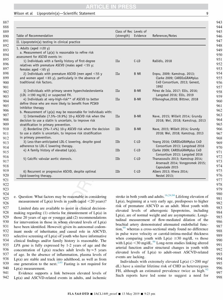
Table of RecommendationClass of Rec(strength)
Levels ofEvidence References/Notes
II. Lipoprotein(a) testing in clinical practice
1. Adults (aged $20 y)a. Measurement of Lp(a) is reasonable to refine risk
assessment for ASCVD events in:1) Individuals with a family history of first-degree
relatives with premature ASCVD (males aged ,55 y;females aged ,65 y)
IIa C-LD Rallidis, 2018
2) Individuals with premature ASCVD (men aged ,55 yand women aged ,65 y), particularly in the absence oftraditional risk factors.
IIa B NR Erqou, 2009; Kamstrup, 2013;Clarke 2009; CARDIoGRAMplusC4D Consortium, 2013; Genest,1992
3) Individuals with primary severe hypercholesterolemia(LDL $190 mg/dL) or suspected FH.
IIa B-NR P�erez de Isla, 2017; Ellis, 2016;Langsted 2016; Ellis, 2019
4) Individuals at very-high-risk** of ASCVD to betterdefine those who are more likely to benefit from PCSK9inhibitor therapy
IIa B-NR O’Donoghue,2018; Bittner, 2018
b. Measurement of Lp(a) may be reasonable for individuals with:1) Intermediate (7.5%–19.9%) 10-y ASCVD risk when the
decision to use a statin is uncertain, to improve riskstratification in primary prevention.
IIa B-NR Nave, 2015; Willeit 2014; Grundy2018; Wei, 2018; Kamstrup, 2013
2) Borderline (5%–7.4%) 10-y ASCVD risk when the decisionto use a statin is uncertain, to improve risk stratificationin primary prevention.
IIb B-NR Nave, 2015; Willeit 2014; Grundy2018; Wei, 2018; Kamstrup, 2013
3) Less-than-anticipated LDL-C lowering, despite goodadherence to LDL-C lowering therapy.
IIb C-LD Yeang 2016; CARDIoGRAMplus C4DConsortium 2013; Langstead 2016
4) A family history of elevated Lp(a). IIb C-LD Clarke 2009; CARDIoGRAMplus C4DConsortium 2013; Langsted 2016
5) Calcific valvular aortic stenosis. IIb C-LD Thanassoulis 2013; Kamstrup 2014;Arsenault 2014; Vongpromek 2015;Capoulade 2015
6) Recurrent or progressive ASCVD, despite optimallipid-lowering therapy.
IIb C-LD Albers 2013; Khera 2014;Nestel 2013;
Wilson et al Lipoprotein(a)—Scientific Statement 9
887888889890891892893894895896897898899900901902903904905906907908909910911912913914915916917918919920921922923924925926927928929930931932933934935936937938939940941942
943944945946947948949950951952953954955956957958959960961962963964965966967968969970971972973974975976977978979980981982983984985986987988989990991992993994995996997998
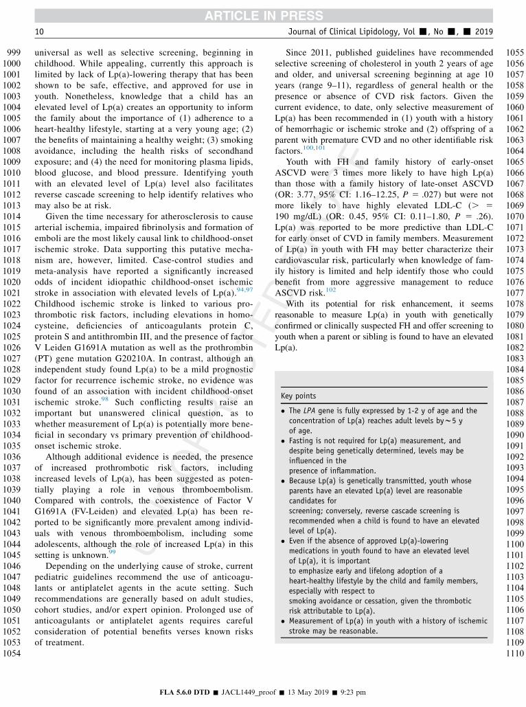
e. Question: What factors may be reasonable in consideringmeasurement of Lp(a) levels in youth (aged ,20 years)?
Limited data are available to assist in clinical decision-making regarding (1) criteria for measurement of Lp(a) inthose 20 years of age or younger and (2) recommendationsfor intervention in those in whom elevated levels of Lp(a)have been identified. However, given its autosomal codom-inant mode of inheritance and causal role in ASCVD,selective screening of Lp(a) of youth who have informativeclinical findings and/or family history is reasonable. TheLPA gene is fully expressed by 1-2 years of age and theconcentration of Lp(a) reaches adult levels byw5 yearsof age. In the absence of inflammation, plasma levels ofLp(a) are stable and track into adulthood, as well as fromone generation to the next.2,93 Fasting is not required forLp(a) measurement.
Evidence supports a link between elevated levels ofLp(a) and ASCVD-related events in adults, and ischemic
FLA 5.6.0 DTD � JACL1449_proo
stroke in both youth and adults.16,24,94 Lifelong elevation ofLp(a), beginning at a very early age, predisposes to higherrisk of premature ASCVD as an adult. Most youth withelevated levels of atherogenic lipoproteins, includingLp(a), are of normal weight and are asymptomatic. Longi-tudinal measurement of flow-mediated dilation of thebrachial artery demonstrated attenuated endothelial func-tion,95 whereas a cross-sectional study found no differencein pulse wave velocity or carotid-intima-medial thicknesswhen comparing youth with Lp(a) $30 mg/dL vs thosewith Lp(a) ,30 mg/dL.96 Long-term studies linking alteredarterial function and/or structural changes in youth withelevated levels of Lp(a) to adult-onset ASCVD-relatedevents are lacking.
Individuals with extremely elevated Lp(a) (.200 mg/dL) have a similar lifetime risk of CHD as heterozygousFH, although an estimated prevalence twice as high.92
Such reports have led some to suggest a need for
f � 13 May 2019 � 9:23 pm
Key points
� The LPA gene is fully expressed by 1-2 y of age and theconcentration of Lp(a) reaches adult levels byw5 yof age.
� Fasting is not required for Lp(a) measurement, anddespite being genetically determined, levels may beinfluenced in thepresence of inflammation.
� Because Lp(a) is genetically transmitted, youth whoseparents have an elevated Lp(a) level are reasonablecandidates forscreening; conversely, reverse cascade screening isrecommended when a child is found to have an elevatedlevel of Lp(a).
� Even if the absence of approved Lp(a)-loweringmedications in youth found to have an elevated levelof Lp(a), it is importantto emphasize early and lifelong adoption of aheart-healthy lifestyle by the child and family members,especially with respect tosmoking avoidance or cessation, given the thromboticrisk attributable to Lp(a).
� Measurement of Lp(a) in youth with a history of ischemicstroke may be reasonable.
10 Journal of Clinical Lipidology, Vol -, No -, - 2019
9991000100110021003100410051006100710081009101010111012101310141015101610171018101910201021102210231024102510261027102810291030103110321033103410351036103710381039104010411042104310441045104610471048104910501051105210531054
1055105610571058105910601061106210631064106510661067106810691070107110721073107410751076107710781079108010811082108310841085108610871088108910901091109210931094109510961097109810991100110111021103110411051106110711081109
universal as well as selective screening, beginning inchildhood. While appealing, currently this approach islimited by lack of Lp(a)-lowering therapy that has beenshown to be safe, effective, and approved for use inyouth. Nonetheless, knowledge that a child has anelevated level of Lp(a) creates an opportunity to informthe family about the importance of (1) adherence to aheart-healthy lifestyle, starting at a very young age; (2)the benefits of maintaining a healthy weight; (3) smokingavoidance, including the health risks of secondhandexposure; and (4) the need for monitoring plasma lipids,blood glucose, and blood pressure. Identifying youthwith an elevated level of Lp(a) level also facilitatesreverse cascade screening to help identify relatives whomay also be at risk.
Given the time necessary for atherosclerosis to causearterial ischemia, impaired fibrinolysis and formation ofemboli are the most likely causal link to childhood-onsetischemic stroke. Data supporting this putative mecha-nism are, however, limited. Case-control studies andmeta-analysis have reported a significantly increasedodds of incident idiopathic childhood-onset ischemicstroke in association with elevated levels of Lp(a).94,97
Childhood ischemic stroke is linked to various pro-thrombotic risk factors, including elevations in homo-cysteine, deficiencies of anticoagulants protein C,protein S and antithrombin III, and the presence of factorV Leiden G1691A mutation as well as the prothrombin(PT) gene mutation G20210A. In contrast, although anindependent study found Lp(a) to be a mild prognosticfactor for recurrence ischemic stroke, no evidence wasfound of an association with incident childhood-onsetischemic stroke.98 Such conflicting results raise animportant but unanswered clinical question, as towhether measurement of Lp(a) is potentially more bene-ficial in secondary vs primary prevention of childhood-onset ischemic stroke.
Although additional evidence is needed, the presenceof increased prothrombotic risk factors, includingincreased levels of Lp(a), has been suggested as poten-tially playing a role in venous thromboembolism.Compared with controls, the coexistence of Factor VG1691A (FV-Leiden) and elevated Lp(a) has been re-ported to be significantly more prevalent among individ-uals with venous thromboembolism, including someadolescents, although the role of increased Lp(a) in thissetting is unknown.99
Depending on the underlying cause of stroke, currentpediatric guidelines recommend the use of anticoagu-lants or antiplatelet agents in the acute setting. Suchrecommendations are generally based on adult studies,cohort studies, and/or expert opinion. Prolonged use ofanticoagulants or antiplatelet agents requires carefulconsideration of potential benefits verses known risksof treatment.
FLA 5.6.0 DTD � JACL1449_proo
Since 2011, published guidelines have recommendedselective screening of cholesterol in youth 2 years of ageand older, and universal screening beginning at age 10years (range 9–11), regardless of general health or thepresence or absence of CVD risk factors. Given thecurrent evidence, to date, only selective measurement ofLp(a) has been recommended in (1) youth with a historyof hemorrhagic or ischemic stroke and (2) offspring of aparent with premature CVD and no other identifiable riskfactors.100,101
Youth with FH and family history of early-onsetASCVD were 3 times more likely to have high Lp(a)than those with a family history of late-onset ASCVD(OR: 3.77, 95% CI: 1.16–12.25, P 5 .027) but were notmore likely to have highly elevated LDL-C (. 5190 mg/dL) (OR: 0.45, 95% CI: 0.11–1.80, P 5 .26).Lp(a) was reported to be more predictive than LDL-Cfor early onset of CVD in family members. Measurementof Lp(a) in youth with FH may better characterize theircardiovascular risk, particularly when knowledge of fam-ily history is limited and help identify those who couldbenefit from more aggressive management to reduceASCVD risk.102
With its potential for risk enhancement, it seemsreasonable to measure Lp(a) in youth with geneticallyconfirmed or clinically suspected FH and offer screening toyouth when a parent or sibling is found to have an elevatedLp(a).
1110
f � 13 May 2019 � 9:23 pm

Table of RecommendationClass of Rec(strength)
Levels ofEvidence References/notes
2. Youth (aged ,20 y)
a. Measurement of Lp(a) may be reasonable with:1) Clinically suspected or genetically confirmed FH. IIb C-LD Burgess, 2018; Sultan, 20182) A family history of first-degree relatives with prematureASCVD (males aged ,55 y, females aged ,65 y).
IIb C-LD Sultan 2018; Expert Panel 2011
3) An unknown cause of ischemic stroke. IIb C-LD Erqou,2009; Kenet, 2010; Goldenberg,2013; Expert Panel 2011
4) A parent or sibling found to have an elevated Lp(a). IIb C-LD Zawacki, 2018
Wilson et al Lipoprotein(a)—Scientific Statement 11
11111112111311141115111611171118111911201121112211231124112511261127112811291130113111321133113411351136113711381139114011411142114311441145114611471148114911501151115211531154115511561157115811591160116111621163116411651166
116711681169117011711172117311741175117611771178117911801181118211831184118511861187118811891190119111921193119411951196119711981199120012011202120312041205120612071208
Treatment
a Question: If Lp(a) is markedly increased, what are theimplications with regard to further LDL-C–loweringtherapy? Is there evidence that supports improved out-comes with greater LDL-C reductions in the presenceof an increased Lp(a)?
In patients receiving LDL-C–lowering therapy,increased baseline and on-statin treatment Lp(a) concen-trations remain a risk factor for ASCVD events.43,46,47 Inanalyses of 29,000 patients from seven randomized statintrials, an Lp(a) $50 mg/dL (105 nmol/L) vs ,15 mg/dL(29 nmol/L) conferred a 1.3-fold ASCVD risk for baselineand a 1.4-fold for on-statin Lp(a) concentrations.43 Statintreatment did not affect Lp(a) concentrations, and highLp(a) was a stronger ASCVD risk predictor in patientson statins vs placebo. Because patients on statins withmarkedly elevated Lp(a) concentrations have a higher ab-solute risk than those without Lp(a) elevation, such pa-tients are likely to exhibit the greatest benefit from moreaggressive LDL-C–lowering therapy. Therefore, as recom-mended in the 2018 ACC/AHA Cholesterol Guidelines,the following recommendations can be made. First, in pri-mary prevention for adults aged 40–75 years with a 10-year ASCVD risk of 7.5%–19.9%, a Lp(a) $50 mg/dLor $100 nmol/L is reasonable to use as a risk-enhancingfactor to favor initiation of a moderate- or high-intensity
Key points
� In statin-treated patients, a high Lp(a) is an independent ASCVD r� In primary prevention for adults aged 40–75 y with a 10-y ASCVDreasonable to be used as a risk-enhancing factor to favor initiatio
� In high-risk* or very-high-risk** patients with LDL-C $70 mg/dL ($100 nmol/L on maximally tolerated statin intensity, it is reasonaand/or PCSK9 inhibitors) to lower LDL-C (and non–HDL-C) to achie
� The presence of an elevated Lp(a) in patients with very-high-risk*$100 mg/dL despite maximally tolerated statin 6 ezetimibe may
� Although niacin and hormone replacement therapy can reduce Lp(ademonstrated ASCVD benefit and the possibility of harm.
FLA 5.6.0 DTD � JACL1449_proo
statin. Second, in high or very-high-risk patients withLDL-C $70 mg/dL (non–HDL-C $100 mg/dL) and aLp(a) $50 mg/dl or $100 nmol/L on maximally toleratedstatin intensity, it is reasonable to consider more intensivetherapies (such as ezetimibe and PCSK9 inhibitors) tolower LDL-C (and non–HDL-C) to achieve greaterASCVD risk reduction.
In the FOURIER trial, the addition of evolocumab to thetreatment regimen of high-risk patients already receivingintensive therapy with high- or moderate-intensity statin(69% vs 30%) 1/2 ezetimibe showed that the greatesttreatment benefit was obtained in those with baseline Lp(a)at or above a clinical threshold of 120 nmol/L (50 mg/dL)as compared with those below the threshold.69 Evolocumabreduced Lp(a) by 27%. However, it is not clear that thisreduction contributed independently to the treatmentbenefit.103 In the ODYSSEY OUTCOMES study, alirocu-mab use in high-risk/very-high-risk patients confers thegreater absolute risk reduction in patients within the highestLp(a) tertile (.60 mg/dL).70 In addition, recent analysisfrom ODYSSEY OUTCOMES suggests that the Lp(a)reduction with alirocumab, independent of LDL-C, contrib-utes to risk reduction.71
As noted in section Laboratory Measurement oflipoprotein(a) b, niacin and hormone replacement treatmentcan reduce Lp(a). However, because there is no evidence ofASCVD benefit, while there is a suggestion of harm, use ofthese therapies are not recommended.
isk factor.risk of 7.5%–19.9%, an Lp(a) $50 mg/dL or $100 nmol/L isn of a moderate or high-intensity statin.non–HDL-C $100 mg/dL) and a Lp(a) $50 mg/dl orble to consider more intensive therapies (such as ezetimibeve greater ASCVD risk reduction.* ASCVD and baseline LDL-C $70 mg/dL or non–HDL-Cbe used as a factor favoring addition of a PCSK9 inhibitor.) levels, these drugs are not recommended because of no
f � 13 May 2019 � 9:23 pm
12091210121112121213121412151216121712181219122012211222
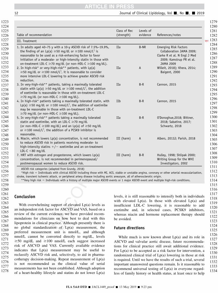
Table of recommendationClass of Rec(strength)
Levels ofevidence References/notes
III. Treatment Q12
1. In adults aged 40–75 y with a 10-y ASCVD risk of 7.5%–19.9%,the finding of an Lp(a) $50 mg/dL or $100 nmol/Lx isreasonable to be used as a risk-enhancing factor to favorinitiation of a moderate- or high-intensity statin in those withon-treatment LDL-C $70 mg/dL (or non–HDL-C $100 mg/dL).
IIa B-NR Emerging Risk FactorsCollaboration JAMA 2009;
Clarke R et al. N Engl J Med2009; Kamstrup PR et al.JAMA 2009
2. In high-risk* or very-high-risk** patients, with Lp(a)$50 mg/dL or $100 nmol/Lx, it is reasonable to considermore intensive LDL-C lowering to achieve greater ASCVD riskreduction.
IIa A Willeit, 2018); Khera, 2014;Baigent, 2000
3. In very-high-risk** patients, taking a maximally toleratedstatin with Lp(a) $50 mg/dL or $100 nmol/Lx, the additionof ezetimibe is reasonable in those with on-treatment LDL-C$70 mg/dL (or non–HDL-C $100 mg/dL).
IIa B-R Cannon, 2015
4. In high-risk* patients taking a maximally tolerated statin, withLp(a) $50 mg/dL or $100 nmol/Lx, the addition of ezetimibemay be reasonable in those with on-treatment LDL-C$70 mg/dL (or non–HDL-C $100 mg/dL).
IIb B-R Cannon, 2015
5. In very-high-risk** patients taking a maximally toleratedstatin and ezetimibe, with an LDL-C $70 mg/dL(or non–HDL-C $100 mg/dL) and an Lp(a) of $50 mg/dLor $100 nmol/Lx, the addition of a PCSK9 inhibitor isreasonable.
IIa B-R O’Donoghue,2018; Bittner,2018; Sabatine, 2017;Schwartz, 2018
6. Niacin, which lowers Lp(a) concentration, is not recommendedto reduce ASCVD risk in patients receiving moderate- tohigh-intensity statins 1/2 ezetimibe and an on-treatmentLDL-C ,80 mg/dL
III (harm) A Albers, 2013J; Parish, 2018
7. HRT with estrogen and progesterone, which lowers Lp(a)concentration, is not recommended in perimenopausal/postmenopausal women to reduce ASCVD risk.
III (harm) B-R Hulley, 1998; Shlipak 2000;Writing Group for the WHIInvestigators, 2002
ASCVD risk categories (adapted from Grundy, 2018)
*High risk 5 Individuals with clinical ASCVD including those with MI, ACS, stable or unstable angina, coronary or other arterial revascularization,
stroke, transient ischemic attack, or peripheral artery disease including aortic aneurysm, all of atherosclerotic origin.
**Very high risk 5 Individuals with a history of multiple major ASCVD events or 1 major ASCVD event and multiple high-risk conditions.
12 Journal of Clinical Lipidology, Vol -, No -, - 2019
12231224122512261227122812291230123112321233123412351236123712381239124012411242124312441245124612471248124912501251125212531254125512561257125812591260126112621263126412651266126712681269127012711272127312741275127612771278
12791280128112821283128412851286128712881289129012911292129312941295129612971298129913001301130213031304130513061307130813091310131113121313131413151316131713181319132013211322132313241325132613271328132913301331133213331334
Conclusion
With overwhelming support of elevated Lp(a) levels asan independent risk factor for ASCVD and VAS, based on areview of the current evidence, we have provided recom-mendations for clinicians on how best to deal with thislipoprotein in clinical practice. Although presently there isno global standardization of Lp(a) measurement, thepreferred measurement unit is nmol/L, and althoughnmol/L cannot be converted directly to mg/dL, levels$50 mg/dL and $100 nmol/L each suggest increasedrisk of ASCVD and VAS. Currently available evidenceindicates that Lp(a) measurement may be useful toreclassify ASCVD risk and, selectively, to aid in pharma-cotherapy decision-making. Repeat measurement of Lp(a)is not recommended as the clinical value of serialmeasurements has not been established. Although adoptionof a heart-healthy lifestyle and statins do not lower Lp(a)
FLA 5.6.0 DTD � JACL1449_proo
levels, it is still reasonable to intensify both in individualswith elevated Lp(a). In those with elevated Lp(a) andinsufficient LDL-C lowering, it is reasonable to addezetimibe and, in selected cases, PCSK9 inhibitors,whereas niacin and hormone replacement therapy shouldbe avoided.
Future directions
While much is now known about Lp(a) and its role inASCVD and valvular aortic disease, future recommenda-tions for clinical practice still await additional evidence.For Lp(a) to be accepted as a risk factor for intervention, arandomized clinical trial of Lp(a) lowering in those at riskis required. Until we have the results of such a trial, severalimportant unanswered questions remain. Is it reasonable torecommend universal testing of Lp(a) in everyone regard-less of family history or health status, at least once to help
f � 13 May 2019 � 9:23 pm
13
Wilson et al Lipoprotein(a)—Scientific Statement 13
13351336133713381339134013411342134313441345134613471348134913501351135213531354135513561357135813591360136113621363136413651366136713681369137013711372137313741375137613771378137913801381138213831384138513861387138813891390
139113921393139413951396139713981399140014011402140314041405140614071408
encourage healthy habits and inform clinical decision-making? Will earlier testing and effective interventionshelp to improve outcomes? What will be the benefit ofmedical interventions that target Lp(a) lowering and howwill such therapies change the outcome of those at-risk andthose currently affected by ASCVD? Will Lp(a)-loweringtherapy be effective in those with low LDL-C, given thedevelopment of new promising LDL-C–lowering therapiesbeyond statins, ezetimibe, and PCSK9 inhibitors?
To answer these and a myriad of other questions, it isencouraging that a randomized, placebo-controlled, double-blind trial of Lp(a) reduction using antisense oligonucleo-tides to block the production of Lp(a) via LPA genesilencing is anticipated to start in 2020. Other pharmaceu-tical companies are developing other promising Lp(a)-lowering therapies such as small interfering RNA inhibitortechnology. Thus, if these early studies continue to showboth safety and efficacy, it is likely that more randomized
Table of recommendation
I. Laboratory measurement of lipoprotein(a)
1. For the measurement of Lp(a), it is recommended that animmunochemical assay that is calibrated against theWHO/IFCCM secondary reference material should be used andreported in nmol/L.
2. When using values of Lp(a) for clinical risk assessment andtreatment decisions, the use of a factor to convert Lp(a) valuesfrom mg/dL to nmol/L is not recommended.
3. When Lp(a) values are used for ASCVD risk assessment inCaucasian patients, it is reasonable to use measured values$ 50 mg/dL or $100 nmol/L as levels suggesting increased risk.
II. Lipoprotein(a) testing in clinical practice
1. Adults (aged $ 20 y)a. Measurement of Lp(a) is reasonable to refine risk assessment for1) Individuals with a family history of first-degree relatives
with premature ASCVD (males aged ,55 y; females aged ,65 y)2) Individuals with premature ASCVD (males aged ,55 y and
females aged ,65 y), particularly in the absence of traditionalrisk factors.
3) Individuals with primary severe hypercholesterolemia (LDL$190 mg/dL) or suspected FH.4) Individuals at very high** risk of ASCVD to better define
those who are more likely to benefit from PCSK9 inhibitor therapyb. Measurement of Lp(a) may be reasonable with:1) Intermediate (7.5%–19.9%) 10-y ASCVD risk when the decisio
to use a statin is uncertain, to improve risk stratification in primarprevention.2) Borderline (5%–7.4%) 10-y ASCVD risk when the decision
to use a statin is uncertain, to improve risk stratification inprimary prevention.3) Less-than-anticipated LDL-C lowering, despite good
adherence to therapy.
FLA 5.6.0 DTD � JACL1449_proo
trials will also be conducted with the aim of reducingASCVD and possibly AVS progression through novel tar-geted Lp(a) reduction.
As discussed in this scientific statement, there is anurgent need for better standardization of Lp(a) measure-ment and an improved understanding of Lp(a) metabolism,physiology, and the pathologic mechanisms by which Lp(a)and oxidized phospholipids on Lp(a) leads to ASCVD andAVS. Finally, we need to address the knowledge gaps thatcurrently exist for unique populations, including therelationship of high Lp(a) with stroke in children and tobetter define the unmet medical needs for Lp(a) reductionin individuals of all ethnicities. Additional data are urgentlyneeded in blacks, South Asians, and those of Hispanicdescent. We hope that this NLA scientific statement Qwillhelp stimulate a thoughtful worldwide discussion that willresult in improved health and outcomes of those entrustedto our care.
Class of Rec(strength)
Levels ofevidence References/notes
I B-NR Marcovina, 2016; Tsimikas,2018; Marcovina, 2000;Marcovina, 2003
III (nobenefit)
E-O Marcovina, 2000, Marcovina,2016; Tsimikas, 2018 JCL
IIa B-R Nordestgaard, 2010;Willeit, 2018, Langsted, 2019
ASCVD events in:IIa C-LD Rallidis, 2018
IIa B NR Erqou, 2009; Kamstrup, 2013;Clarke 2009; CARDIoGRAMplusC4D Consortium, 2013; Genest,
1992IIa B-NR P�erez de Isla, 2017; Ellis, 2016;
Langsted 2016; Ellis, 2019IIa B-NR O’Donoghue,2018; Bittner, 2018
ny
IIa B-NR Nave, 2015; Willeit 2014; Grundy2018; Wei, 2018; Kamstrup,2013
IIb B-NR Nave, 2015; Willeit 2014; Grundy2018; Wei, 2018; Kamstrup,2013
IIb C-LD Yeang 2016; CARDIoGRAMplusC4DConsortium 2013; Langstead2016
(continued on next page)
f � 13 May 2019 � 9:23 pm
14091410141114121413141414151416141714181419142014211422142314241425142614271428142914301431143214331434143514361437143814391440144114421443144414451446

(continued )
Table of recommendationClass of Rec(strength)
Levels ofevidence References/notes
I. Laboratory measurement of lipoprotein(a)
4) A family history of elevated Lp(a). IIb C-LD Clarke 2009; CARDIoGRAMplusC4DConsortium 2013; Langsted2016
5) Calcific valvular aortic stenosis. IIb C-LD Thanassoulis 2013; Kamstrup2014;Arsenault 2014; Vongpromek2015;Capoulade 2015
6) Recurrent or progressive ASCVD, despite optimallipid-lowering therapy.
IIb C-LD Albers 2013; Khera 2014; Nestel2013;
2. Youth (aged , 20 y)a. Measurement of Lp(a) may be reasonable with:1) Clinically suspected or genetically confirmed FH. IIb C-LD Burgess, 2018; Sultan, 20182) A family history of first-degree relatives with premature
ASCVD (males with ,55 y, females aged ,65 y).IIb C-LD Sultan 2018; Expert Panel 2011
3) An unknown cause of ischemic stroke. IIb C-LD Erqou,2009; Kenet, 2010;Goldenberg, 2013;Expert Panel 2011
4) A parent or sibling found to have an elevated Lp(a). IIb C-LD Zawacki,2018
III. Treatment
1. In adults aged 40-75 y with a 10-y ASCVD risk of 7.5%–19.9%,the finding of an Lp(a) $50 mg/dL or $100 nmol/Lx is reasonableto be used as a risk-enhancing factor to favor initiation of amoderate- or high-intensity statin in those with on-treatmentLDL-C $70 mg/dL (or non–HDL-C $100 mg/dL).
IIa B-NR Emerging Risk FactorsCollaborationJAMA 2009;
Clarke R et al. N Engl J Med 2009;Kamstrup PR et al. JAMA 2009.
2. In high-risk* or very-high-risk** patients, with Lp(a) $50 mg/dLor $100 nmol/Lx, it is reasonable to consider more intensive LDL-Clowering to achieve greater ASCVD risk reduction.
IIa A Willeit, 2018; Khera, 2014;Baigent, 2000
3. In very-high-risk** patients, taking a maximally tolerated statinwith Lp(a) $50 mg/dL or $100 nmol/Lx, the addition of ezetimibeis reasonable in those with on-treatment LDL-C $70 mg/dL(or non–HDL-C $100 mg/dL).
IIa B-R Cannon, 2015
4. In high-risk* patients taking a maximally tolerated statin, withLp(a) $50 mg/dL or $100 nmol/Lx, the addition of ezetimibemay be reasonable in those with on-treatment LDL-C $70 mg/dL(or non–HDL-C $100 mg/dL).
IIb B-R Cannon, 2015
5. In very-high-risk** patients taking a maximally tolerated statin andezetimibe, with an LDL-C $70 mg/dL (or non–HDL-C $100 mg/dL)and an Lp(a) of $50 mg/dL or $100 nmol/Lx, the addition of aPCSK9 inhibitor is reasonable.
IIa B-R O’Donoghue,2018; Bittner, 2018;Sabatine, 2017; Schwartz,2018
6. Niacin, which lowers Lp(a) concentration, is not recommended toreduce ASCVD risk in patients receiving moderate- to high-intensitystatins 1/2 ezetimibe and an on-treatment LDL-C ,80 mg/dL
III (harm) A Albers, 2013J; Parish, 2018
7. HRT with estrogen and progesterone, which lowers Lp(a)concentration, is not recommended in perimenopausal/postmenopausal women to reduce ASCVD risk.
III (harm) B-R Hulley, 1998; Shlipak 2000;Writing Group for the WHIInvestigators, 2002
ASCVD risk categories (adapted from Grundy, 2018)
*High risk 5 Individuals with clinical ASCVD including those with MI, ACS, stable or unstable angina, coronary or other arterial revascularization,
stroke, transient ischemic attack, or peripheral artery disease including aortic aneurysm, all of atherosclerotic origin.
**Very high risk 5 Individuals with a history of multiple major ASCVD events or 1 major ASCVD event and multiple high-risk conditions.
14 Journal of Clinical Lipidology, Vol -, No -, - 2019
FLA 5.6.0 DTD � JACL1449_proof � 13 May 2019 � 9:23 pm
14471448144914501451145214531454145514561457145814591460146114621463146414651466146714681469147014711472147314741475147614771478147914801481148214831484148514861487148814891490149114921493149414951496149714981499150015011502
15031504150515061507150815091510151115121513151415151516151715181519152015211522152315241525152615271528152915301531153215331534153515361537153815391540154115421543154415451546154715481549155015511552155315541555155615571558
Q14
Wilson et al Lipoprotein(a)—Scientific Statement 15
15591560156115621563156415651566156715681569157015711572157315741575157615771578157915801581158215831584158515861587158815891590159115921593159415951596159715981599160016011602160316041605160616071608160916101611161216131614
161516161617161816191620162116221623162416251626
Acknowledgments
The authors would like to acknowledge Vivian Grifan-tini, Luke Hamilton and Dena Hanson for their assistancein preparing and editing this manuscript. A special thanksto Dr. Patrick Moriarty, who provided insightful commentsand thoughtful suggestions during manuscript development.There was no funding for the study.
Authors’ contribution: All authors contributed to thisscientific statement, drafting and revising it critically forimportant intellectual content, and have approved the finalversion.
16271628162916301631163216331634163516361637163816391640164116421643164416451646164716481649
Financial disclosure
D.P.W. discloses that in the past 12 months, he hasreceived speaking honorarium from Osler Institute, hasreceived research grants from Merck Sharp & Dohme andNovo Nordisk, and has participated on the advisory boardfor Alexion Pharmaceuticals. T.A.J. discloses that in thepast 12 months, he has received consulting fees fromAmarin, Amgen, AstraZeneca, Esperion, Sanofi Regeneron,and Novartis. P.H.J. discloses that in the past 12 months, hehas received advisory board honorarium from Amgen,Sanofi Regeneron, and AstraZeneca. M.L.K. disclosesthat in the past 12 months, she has received speaker andconsulting honorarium from Eli Lilly, speaker honorariumfrom Pfizer, consulting honorarium from Amgen, andindependent contractor fees from Pfizer, Eli Lilly, Cardio-Vax, and Ionis. C.J.M. discloses that in the past 12 months,she has nothing to disclose. B.G.N. discloses that in the past12 months, he has received consulting honorarium fromAkcea, Amgen, Regeneron, Sanofi, and Kowa. C.E.O.discloses that in the past 12 months, he has nothing todisclose.
16501651165216531654
Conflict of interest
The authors have no conflicts of interest to disclose.
16551656165716581659Uncited figure
Figure 2.
18
16601661166216631664166516661667166816691670
References1. Nordestgaard BG, Chapman MJ, Ray K, et al. Lipoprotein(a) as a
cardiovascular risk factor: Current status. Eur Heart J. 2010;
31(23):2844–2853.
2. Kronenberg F, Utermann G. Lipoprotein(a): Resurrected by genetics.
J Intern Med. 2013;273(1):6–30.
3. Tsimikas S. A test in context: Lipoprotein(a): Diagnosis, prognosis,
controversies, and emerging therapies. J Am Coll Cardiol. 2017;
69(6):692–711.
4. Nordestgaard BG, Langsted A. Lipoprotein (a) as a cause of cardio-
vascular disease: Insights from epidemiology, genetics, and biology.
J Lipid Res. 2016;57(11):1953–1975.
FLA 5.6.0 DTD � JACL1449_proo
5. Boffa MB, Koschinsky ML. Lipoprotein (a): truly a direct prothrom-
botic factor in cardiovascular disease? J Lipid Res. 2016 May;57(5):
745–757.
6. Brown MS, Goldstein JL. Plasma lipoproteins: teaching old dogmas
new tricks. Nat 330. 1987;6144:113–114.
7. Ishikawa S, Kotani K, Kario K, et al. Inverse association between
serum lipoprotein(a) and cerebral hemorrhage in the Japanese popu-
lation. Thromb.Res. 2013;131(2):e54–e58.
8. Langsted A, Kamstrup PR, Nordestgaard BG. High Lipoprotein(a)
and Low Risk of Major Bleeding in Brain and Airways in the Gen-
eral Population: a Mendelian Randomization Study. Clin.Chem.
2017;63(11):1714–1723.
9. Kinpara K, Okada H, Yoneyama A, Okubo M, Murase T. Lipopro-
tein(a)-cholesterol: A significant component of serum cholesterol.
Clin Chim Acta. 2011;412(19-20):1783–1787.
10. Kamstrup PR, Hung MY, Witztum JL, Tsimikas S, Nordestgaard BG.
Oxidized Phospholipids and Risk of Calcific Aortic Valve Disease:
The Copenhagen General Population Study. Arterioscler.Thromb.-
Vasc.Biol. 2017;37(8):1570–1578.
11. Yeang C, Wilkinson MJ, Tsimikas S. Lipoprotein(a) and oxidized
phospholipids in calcific aortic valve stenosis. Curr Opin Cardiol.
2016 Jul;31(4):440–450.
12. Craig WY, Neveux LM, Palomaki GE, Cleveland MM,
Haddow JE. Lipoprotein(a) as a risk factor for ischemic heart dis-
ease: Metaanalysis of prospective studies. Clin Chem. 1998;
44(11):2301–2306.
13. Danesh J, Collins R, Peto R. Lipoprotein(a) and coronary heart dis-
ease. Meta-analysis of prospective studies. Circulation. 2000;
102(10):1082–1085.
14. Erqou S, Kaptoge S, Perry PL, et al. Lipoprotein(a) concentration
and the risk of coronary heart disease, stroke, and nonvascular mor-
tality. JAMA. 2009;302:412–423.
15. Nave AH, Lange KS, Leonards CO, et al. Lipoprotein (a) as a risk
factor for ischemic stroke: A meta-analysis. Atherosclerosis. 2015;
242(2):496–503.
16. Erqou S, Thompson A, Di Angelantonio E, et al. Apolipoprotein(a)
isoforms and the risk of vascular disease: Systematic review of 40
studies involving 58,000 participants. J Am Coll Cardiol. 2010;
55(19):2160–2167.
17. Sultan SM, Schupf N, Dowling MM, Deveber GA, Kirton A,
Elkind MS. Review of lipid and lipoprotein(a) abnormalities
in childhood arterial ischemic stroke. Int J Stroke. 2014;9(1):
79–87.
18. Pare G, Caku A, McQueen M, et al. Lipoprotein(a) levels and the risk
of myocardial infarction among 7 ethnic groups. Circulation. 2019;
139(12):1472–1482.
19. Kamstrup PR, Benn M, Tybjaerg-Hansen A, Nordestgaard BG.
Extreme lipoprotein(a) levels and risk of myocardial infarction in
the general population: The Copenhagen City Heart Study. Circula-
tion. 2008;117(2):176–184.
20. Kamstrup PR, Tybjaerg-Hansen A, Steffensen R, Nordestgaard BG.
Genetically elevated lipoprotein(a) and increased risk of myocardial
infarction. JAMA. 2009;301(22):2331–2339.
21. Kamstrup PR, Tybjaerg-Hansen A, Nordestgaard BG. Elevated lipo-
protein(a) and risk of aortic valve stenosis in the general population.
J Am Coll Cardiol. 2014;63(5):470–477.
22. Kamstrup PR, Tybjaerg-Hansen A, Nordestgaard BG. Genetic evi-
dence that lipoprotein(a) associates with atherosclerotic stenosis
rather than venous thrombosis. Arterioscler Thromb Vasc Biol.
2012;32(7):1732–1741.
23. Kamstrup PR, Nordestgaard BG. Elevated lipoprotein(a) levels, LPA
risk genotypes, and increased risk of heart failure in the general pop-
ulation. JACC Heart Fail. 2016;4(1):78–87.
24. Langsted A, Kamstrup PR, Nordestgaard BG. High lipoprotein(a)
and high risk of mortality. Eur Heart J. 2019. Q
25. Langsted A, Nordestgaard BG, Kamstrup PR. High lipoprotein(a)
and increased risk of ischemic stroke in a large contemporary general
population study. J Am Coll Cardiol. 2019(in press).
f � 13 May 2019 � 9:23 pm

19
16 Journal of Clinical Lipidology, Vol -, No -, - 2019
16711672167316741675167616771678167916801681168216831684168516861687168816891690169116921693169416951696169716981699170017011702170317041705170617071708170917101711171217131714171517161717171817191720172117221723172417251726
17271728172917301731173217331734173517361737173817391740174117421743174417451746174717481749175017511752175317541755175617571758175917601761176217631764176517661767176817691770177117721773177417751776177717781779178017811782
26. Smith GD, Ebrahim S. ’Mendelian randomization’: Can genetic
epidemiology contribute to understanding environmental determi-
nants of disease? Int J Epidemiol. 2003;32(1):1–22.
27. Smith GD, Ebrahim S, Lewis S, Hansell AL, Palmer LJ, Burton PR.
Genetic epidemiology and public health: Hope, hype, and future
prospects. Lancet. 2005;366(9495):1484–1498.
28. Benn M, Nordestgaard BG. From genome-wide association studies to
mendelian randomization: Novel opportunities for understanding car-
diovascular disease causality, pathogenesis, prevention, and treat-
ment. Cardiovasc Res. 2018;114(9):1192–1208.
29. Clarke R, Peden JF, Hopewell JC, et al. Genetic variants associated
with Lp(a) lipoprotein level and coronary disease. N Engl J Med.
2009;361(26):2518–2528.
30. Tregouet DA, Konig IR, Erdmann J, et al. Genome-wide haplotype as-
sociation study identifies the SLC22A3-LPAL2-LPA gene cluster as a
risk locus for coronary artery disease. Nat Genet. 2009;41(3):283–285.
31. Schunkert H, Konig IR, Kathiresan S, et al. Large-scale association
analysis identifies 13 new susceptibility loci for coronary artery dis-
ease. Nat Genet. 2011;43(4):333–338.
32. Thanassoulis G, Campbell CY, Owens DS, et al. Genetic associations
with valvular calcification and aortic stenosis. N Engl J Med. 2013;
368(6):503–512.
33. Helgadottir A, Thorleifsson G, Gretarsdottir S, et al. Genome-wide
analysis yields new loci associating with aortic valve stenosis. Nat
Commun. 2018;9(1):987.
34. Utermann G. Lipoprotein(a). In: Scriver CR, Beaudet AL, Sly WS,
Valle D, editors. The metabolic & molecular bases of inherited dis-
ease. 9th ed. New York: McGraw-Hill, 2001. p. 2753–2787.
35. Marcovina SM, Albers JJ. Lipoprotein (a) measurements for clinical
application. J Lipid Res. 2016;57(4):526–537.
36. Tsimikas S, Fazio S, Ferdinand KC, et al. NHLBI working group rec-
ommendations to reduce lipoprotein(a)-mediated risk of cardiovascu-
lar disease and aortic stenosis. J Am Coll Cardiol. 2018;71(2):
177–192.
37. Marcovina SM, Albers JJ, Scanu AM, et al. Use of a reference ma-
terial proposed by the International Federation of Clinical Chemistry
and Laboratory Medicine to evaluate analytical methods for the
determination of plasma lipoprotein(a). Clin Chem. 2000;46(12):
1956–1967.
38. Marcovina SM, Koschinsky ML, Albers JJ, Skarlatos S. Report of the
National Heart, Lung, and Blood Institute Workshop on Lipopro-
tein(a) and Cardiovascular Disease: Recent advances and future di-
rections. Clin Chem. 2003;49(11):1785–1796.
39. Marcovina SM, Albers JJ, Gabel B, Koschinsky ML, Gaur VP. Effect
of the number of apolipoprotein(a) kringle 4 domains on immuno-
chemical measurements of lipoprotein(a). Clin Chem. 1995;41(2):
246–255.
40. Enkhmaa B, Anuurad E, Berglund L. Lipoprotein (a): Impact by
ethnicity and environmental and medical conditions. J Lipid Res.
2016;57(7):1111–1125.
41. Guan W, Cao J, Steffen BT, et al. Race is a key variable in assigning
lipoprotein(a) cutoff values for coronary heart disease risk assess-
ment: The Multi-Ethnic Study of Atherosclerosis. Arterioscler
Thromb Vasc Biol. 2015;35(4):996–1001.
42. Virani SS, Brautbar A, Davis BC, et al. Associations between lipo-
protein(a) levels and cardiovascular outcomes in black and white
subjects: The atherosclerosis risk in communities (ARIC) study. Cir-
culation. 2012;125(2):241–249.
43. Willeit P, Ridker PM, Nestel PJ, et al. Baseline and on-statin treat-
ment lipoprotein(a) levels for prediction of cardiovascular events: In-
dividual patient-data meta-analysis of statin outcome trials. Lancet.
2018;392(10155):1311–1320.
44. O’Donoghue ML, Morrow DA, Tsimikas S, et al. Lipoprotein(a) for
risk assessment in patients with established coronary artery disease. J
Am Coll Cardiol. 2014;63(6):520–527.
45. Cantin B, Gagnon F, Moorjani S, et al. Is lipoprotein(a) an indepen-
dent risk factor for ischemic heart disease in men? The Quebec Car-
diovascular Study. J Am Coll Cardiol. 1998;31(3):519–525.
FLA 5.6.0 DTD � JACL1449_proo
46. Khera AV, Everett BM, Caulfield MP, et al. Lipoprotein(a) concentra-
tions, rosuvastatin therapy, and residual vascular risk: An analysis
from the JUPITER Trial (Justification for the Use of Statins in Pre-
vention: An Intervention Trial Evaluating Rosuvastatin). Circulation.
2014;129(6):635–642.
47. Albers JJ, Slee A, O’Brien KD, et al. Relationship of apolipopro-
teins A-1 and B, and lipoprotein(a) to cardiovascular outcomes:
The AIM-HIGH trial (Atherothrombosis Intervention in Meta-
bolic Syndrome with Low HDL/High Triglyceride and Impact
on Global Health Outcomes). J Am Coll Cardiol. 2013;62(17):
1575–1579.
48. Gaudet D, Watts GF, Robinson JG, et al. Effect of alirocumab on lip-
oprotein(a) over ./51.5 years (from the phase 3 ODYSSEY pro-
gram). Am J Cardiol. 2017;119(1):40–46.
49. Raal FJ, Giugliano RP, Sabatine MS, et al. Reduction in lipopro-
tein(a) with PCSK9 monoclonal antibody evolocumab (AMG 145):
A pooled analysis of more than 1,300 patients in 4 phase II trials.
J Am Coll Cardiol. 2014;63(13):1278–1288.
50. Catapano AL, Graham I, De Backer G, et al. 2016 ESC/EAS Guide-
lines for the Management of Dyslipidaemias. Eur Heart J. 2016;
37(39):2999–3058.
51. Anderson TJ, Gregoire J, Pearson GJ, et al. Canadian Cardiovascular
Society Guidelines for the Management of Dyslipidemia for the Pre-
vention of Cardiovascular Disease in the Adult. Can J Cardiol 2016.
2016;32(11):1263–1282.
52. Langsted A, Kamstrup PR, Benn M, Tybjaerg-Hansen A,
Nordestgaard BG. High lipoprotein(a) as a possible cause of clinical
familial hypercholesterolaemia: A prospective cohort study. Lancet
Diabetes Endocrinol. 2016;4(7):577–587.
53. Utermann G, Hoppichler F, Dieplinger H, Seed M, Thompson G,
Boerwinkle E. Defects in the low density lipoprotein receptor gene
affect lipoprotein (a) levels: Multiplicative interaction of two gene
loci associated with premature atherosclerosis. Proc Natl Acad Sci
U S A. 1989;86(11):4171–4174.
54. Willeit P, Kiechl S, Kronenberg F, et al. Discrimination and net re-
classification of cardiovascular risk with lipoprotein(a): Prospective
15-year outcomes in the bruneck study. J Am Coll Cardiol. 2014;
64(9):851–860.
55. Jacobson TA, Ito MK, Maki KC, et al. National Lipid Association
recommendations for patient-centered management of dyslipidemia:
Part 1–full report. J Clin Lipidol. 2015;9(2):129–169.
56. Grundy SM, Stone NJ, Bailey AL, et al. 2018 AHA/ACC/AACV-
PR/AAPA/ABC/ACPM/ADA/AGS/APhA/ASPC/NLA/PCNA Guide-
line on the Management of Blood Cholesterol. Circulation. 2018.
57. Cook NR, Mora S, Ridker PM. Lipoprotein(a) and cardiovascular
risk prediction among women. J Am Coll Cardiol. 2018;72(3):
287–296.
58. Verbeek R, Hoogeveen RM, Langsted A, et al. Cardiovascular dis-
ease risk associated with elevated lipoprotein(a) attenuates at low
LDL-C levels in a primary prevention setting. Eur Heart J. 2018;
39:2589–2596.
59. Min WK, Lee JO, Huh JW. Relation between lipoprotein(a) concen-
trations in patients with acute-phase response and risk analysis for
coronary heart disease. Clin Chem. 1997;43(10):1891–1895.
60. Danik JS, Rifai N, Buring JE, Ridker PM. Lipoprotein(a), hormone
replacement therapy, and risk of future cardiovascular events. J Am
Coll Cardiol. 2008;52(2):124–131.
61. Chasman DI, Shiffman D, Zee RY, et al. Polymorphism in the apoli-
poprotein(a) gene, plasma lipoprotein(a), cardiovascular disease, and
low-dose aspirin therapy. Atherosclerosis. 2009;203(2):371–376.
62. The WHI Writing Group JAMA. 2002. Q
63. Hulley S, Grady D, Bush T, et al. Randomized trial of estrogen plus
progestin for secondary prevention of coronary disease in postmeno-
pausal women. JAMA. 1998;280:605–613.
64. Sahebkar A, Reiner Z, Simental-Mendia LE, Ferretti G, Cicero AF.
Effect of extended-release niacin on plasma lipoprotein(a) levels: A
systematic review and meta-analysis of randomized placebo-
controlled trials. Metabolism. 2016;65(11):1664–1678.
f � 13 May 2019 � 9:23 pm
Q20
Q21 Q22
Q23
Wilson et al Lipoprotein(a)—Scientific Statement 17
17831784178517861787178817891790179117921793179417951796179717981799180018011802180318041805180618071808180918101811181218131814181518161817181818191820182118221823182418251826182718281829183018311832183318341835183618371838
18391840184118421843184418451846184718481849185018511852185318541855185618571858185918601861186218631864186518661867186818691870187118721873187418751876187718781879188018811882188318841885188618871888188918901891
65. HPS2-THRIVE Collaborative Group, Landray MJ, Haynes R,
Hopewell JC, et al. Effects of extended-release niacin with laropi-
prant in high-risk patients. N Engl J Med. 2014;371(3):203–212.
66. Anderson TJ, Boden WE, Desvigne-Nickens P, et al. Safety profile of
extended-release niacin in the AIM-HIGH Trial. N Engl J Med. 2014;
371(3):288–290.
67. Parish S, Hopewell JC, Hill MR, et al. Impact of Apolipoprotein(a)
Isoform Size on Lipoprotein(a) Lowering in the HPS2-THRIVE
Study. Circ Genome Precis Med. 2018;11:e001696.
68. Ridker PM, Danielson E, Fonseca FA, et al. Rosuvastatin to prevent
vascular events in men and women with elevated C-reactive protein.
N Engl J Med. 2008;359(21):2195–2207.
69. O’Donoghue M, Giugliano R, Keech A, et al. Lipoprotein(a), PCSK9
inhibition and cardiovascular risk: Insights from the fourier trial.
Atherosclerosis. 2018;275:e9–e10.
70. Bittner V, Szarek M, Aylward PE, et al. Lp(A) and cardiovascular
outcomes: An analysis from the odyssey outcomes trial. Atheroscler
Suppl. 2018;32:24–25.
71. Bittner V, Szarek M, Aylward P. Lipoprotein (a) lowering by alirocu-
mab contributes to event reduction independent of low-density lipopro-
tein cholesterol in the ODYSSEY OUTCOMES trial. Paper presented
at: 68th Annual Scientific Session & Exposition of the American Col-
lege of Cardiology; March 16-18, 2019; New Orleans, LA.
72. Arai K, Orsoni A, Mallat Z, et al. Acute impact of apheresis on
oxidized phospholipids in patients with familial hypercholesterole-
mia. J Lipid Res. 2012;53(8):1670–1678.
73. Moriarty PM, Hemphill L. Lipoprotein apheresis. Cardiol Clin.
2015;33(2):197–208.
74. Schettler VJJ, Neumann CL, Peter C, et al. The german lipoprotein
apheresis registry (GLAR) - almost 5 years on. Clin Res Cardiol
Suppl. 2017;12(Suppl 1):44–49.
75. Jaeger BR, Richter Y, Nagel D, et al. Longitudinal cohort study on
the effectiveness of lipid apheresis treatment to reduce high lipopro-
tein(a) levels and prevent major adverse coronary events. Nat Clin
Pract Cardiovasc Med. 2009;6(3):229–239.
76. Rosada A, et al. Artif Organs. 2013;128:2567–2576.
77. Roeseler E, Julius U, Heigl F, et al. Lipoprotein apheresis for
lipoprotein(a)-associated cardiovascular disease: Prospective 5 years
of follow-up and apolipoprotein(a) characterization. Arterioscler
Thromb Vasc Biol. 2016;36(9):2019–2027.
78. Khan TZ, Hsu L-Y, Arai AE, et al. Apheresis as novel treatment for
refractory angina with raised lipoprotein(a): A randomized controlled
crossover trial. E Heart J. 2017;38:1561–1569.
79. Capoulade R, Chan KL, Yeang C, et al. Oxidized Phospholipids, Lip-
oprotein(a), and Progression of Calcific Aortic Valve Stenosis. J Am
Coll Cardiol. 2015;66(11):1236–1246.
80. Rallidis LS, Pavlakis G, Foscolou A, et al. High levels of lipoprotein
(a) and premature acute coronary syndrome. Atherosclerosis. 2018;
269:29–34.
81. Wei WQ, Li X, Feng Q, et al. LPA variants are associated with resid-
ual cardiovascular risk in patients receiving statins. Circulation.
2018;138(17):1839–1849.
82. Rallidis LS, Pavlakis G, Foscolou A, et al. High levels of lipoprotein
(a) and premature acute coronary syndrome. Atherosclerosis. 2018;
269:29–34.
83. Vuorio A, Watts GF, Kovanen PT. Depicting new pharmacological
strategies for familial hypercholesterolaemia involving lipoprotein
(a). Eur Heart J. 2017;38(48):3555–3559.
84. Miller PE, Haberlen SA, Metkus T, et al. HIV and coronary arterial
remodeling from the Multicenter AIDS Cohort Study (MACS).
Atherosclerosis. 2015;241(2):716–722.
85. Ellis KL, Perez de Isla L, Alonso R, Fuentes F, Watts GF, Mata P.
Value of measuring lipoprotein(a) during cascade testing for familial
hypercholesterolemia. J Am Coll Cardiol. 2019;73(9):1029–1039.
FLA 5.6.0 DTD � JACL1449_proo
86. Smolders B, Lemmens R, Thijs V. Lipoprotein (a) and stroke: A
meta-analysis of observational studies. Stroke. 2007;38(6):
1959–1966.
87. Kotani K, Serban MC, Penson P, Lippi G, Banach M. Evidence-based
assessment of lipoprotein(a) as a risk biomarker for cardiovascular
diseases - some answers and still many questions. Crit Rev Clin
Lab Sci. 2016;53(6):370–378.
88. Langsted, Kamstrup PR, Nordestgaard BG. High lipoprotein(a) and
high risk of mortality. Eur Heart J. 2019.
89. Chen HY, Dufresne L, Burr H, et al. Association of LPA variants with
aortic stenosis: A large-scale study using diagnostic and procedural
codes from electronic health records. JAMA Cardiol. 2018;3(1):
18–23.
90. Postmus I, Trumpet S, Deshmukh HA, et al. Pharmacogenetic meta-
analysis of genome-wide association studies of LDL cholesterol
response to statins. Nat Commun. 2014;5:506–508.
91. Deshmukh HA, Colhoun HM, Johnson T, et al. Genome-wide asso-
ciation study of genetic determinants of LDL-c response to atorvas-
tatin therapy: Importance of Lp(a). J Lipid Res. 2012;53(5):
1000–1011.
92. Burgess S, Ference BA, Staley JR, et al. Association of LPA variants
with risk of coronary disease and the implications for lipoprotein(a)-
lowering therapies: A Mendelian randomization analysis. JAMA Car-
diol. 2018;3(7):619–627.
93. Marcovina SM, Moriarty PM, Koschinsky ML, Guyton JR. JCL
roundtable—Lipoprotein(a): The emerging risk factor. J Clin Lipidol.
2018;12(6):1335–1345.
94. Kenet G, Lutkhoff LK, Albisetti M, et al. Impact of thrombophilia
on risk of arterial ischemic stroke or cerebral sinovenous throm-
bosis in neonates and children: A systematic review and meta-
analysis of observational studies. Circulation. 2010;121(16):
1838–1847.
95. Lapinleimu J, Raitakari OT, Lapinleimu H, et al. High lipoprotein(a)
concentrations are associated with impaired endothelial function in
children. J Pediatr. 2015;166(4):947–952e1-2.
96. Qayum Omar, Alshami Noor, Alshami Ibezim, et al. Lipoprotein (a):
Examination of Cardiovascular Risk in a Pediatric Referral Popula-
tion. Pediatr Cardiol. 2018;39:1540–1546.
97. Nowak-Gottl U, Strater R, Heinecke A, et al. Lipoprotein (a) and
genetic polymorphisms of clotting factor V, prothrombin, and
methylenetetrahydrofolate reductase are risk factors of sponta-
neous ischemic stroke in childhood. Blood. 1999;94(11):
3678–3682.
98. Goldenberg NA, Bernard TJ, Hillhouse J, et al. Elevated lipoprotein
(a), small apolipoprotein (a), and the risk of arterial ischemic stroke
in North American children. Haematologica. 2013;98(5):802–807.
99. von Depka M, Nowak-Gottl U, Eisert R, et al. Increased lipoprotein
(a) levels as an independent risk factor for venous thromboembolism.
Blood. 2000;96(10):3364–3368.
100. Sultan S, Dowling M, Kirton A, et al. Dyslipidemia in children with
arterial ischemic stroke: Prevalence and risk factors. Pediatr Neurol.
2018;78:46–54.
101. Expert Panel on Integrated Guidelines for Cardiovascular Health and
Risk Reduction in Children and Adolescents, National Heart, Lung,
and Blood Institute. Expert panel on integrated guidelines for cardio-
vascular health and risk reduction in children and adolescents: Sum-
mary report. Pediatrics. 2011;128(Suppl 5):S213–S256.
102. Zawacki AW, Dodge A, Woo KM, Ralphe JC, Peterson AL. In pedi-
atric familial hypercholesterolemia, lipoprotein(a) is more predictive
than LDL-C for early onset of cardiovascular disease in family mem-
bers. J Clin Lipidol. 2018;12(6):1445–1451.
103. Sabatine MS, Giugliano RP, Keech AC, et al. Evolocumab and clin-
ical outcomes in patients with cardiovascular disease. N Engl J Med.
2017;376(18):1713–1722.
f � 13 May 2019 � 9:23 pm
189218931894